Embed Size (px)
Citation preview

Distal Radius Fractures
John T. Capo, MD
John T. Capo, MD; Version 4, November 2015 John T. Capo, MD; Version 3, November 2009
John T. Capo, MD; Revised January 2006 Original Author: Thomas F. Varecka, MD; March 2004

The Problem of Distal Radius Fractures
Common injury: >450,000/yr. in USA High potential for functional impairment and
frequent complications
Introduction Distal radius fractures occur through the distal
metaphysis of the radius May involve articular surface
frequently involving the ulnar styloid Most often result from a fall on the outstretched
hand. – forced extension of the carpus,
– impact loading of the distal radius. Associated injuries may accompany distal radius
fractures.

Introduction
Classified by: – presence or absence of intra-
articular involvement, – degree of comminution,
– dorsal vs. volar displacement, – involvement of the distal
radioulnar joint.
Diagnosis: History and Physical Findings
History of mechanism of injury A visible deformity of the wrist is usually noted,
with the hand most commonly displaced in the dorsal direction.
Movement of the hand and wrist are painful. Adequate and accurate assessment of the
neurovascular status of the hand is imperative
Diagnosis: Diagnostic Tests and Examination
Evaluation of the injured joint, and a joint above and below
Radiographs of the injured wrist Radiographs of other areas, if symptoms
warrant. CT scan of the distal radius in selected
instances.
Treatment Goals
Preserve hand and wrist function
Realign normal osseous anatomy – Articular surface
– Alignment of radial platform in space
Promote bony healing
Allow early finger and elbow ROM

Osseous Anatomy
Distal radius – 80% of axial load – Scaphoid fossa – Lunate fossa
Distal ulna – 20% axial load
Sigmoid notch – DRUJ
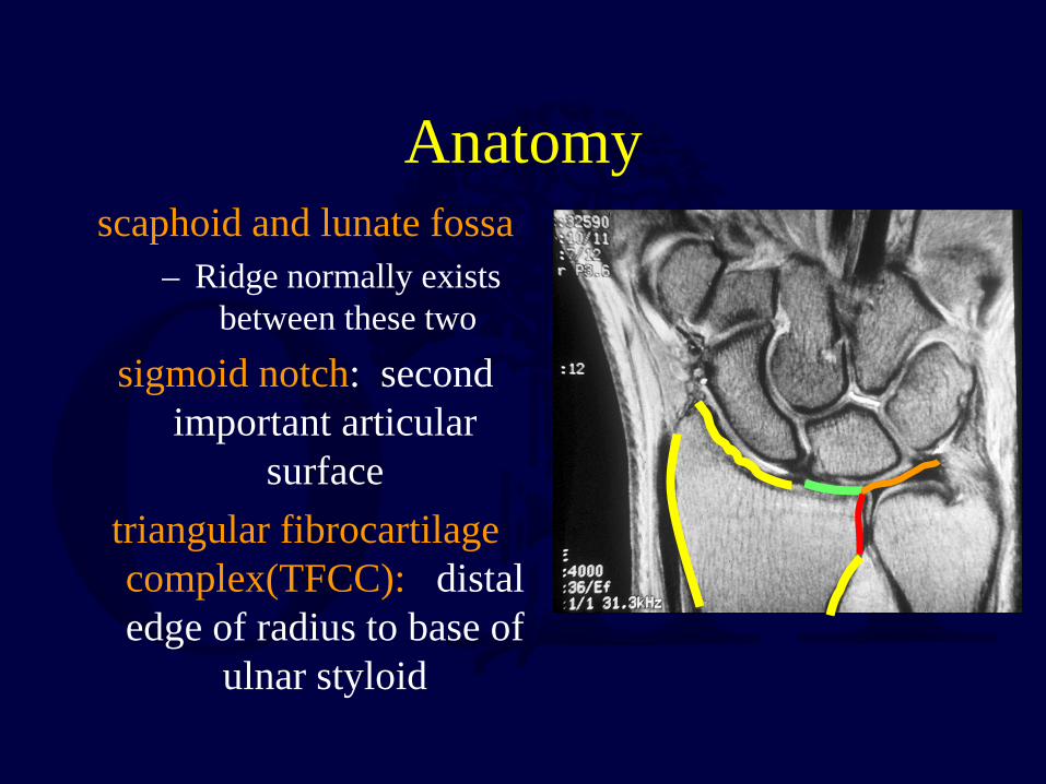
Anatomy scaphoid and lunate fossa
– Ridge normally exists between these two
sigmoid notch: second important articular
surface triangular fibrocartilage complex(TFCC): distal edge of radius to base of
ulnar styloid
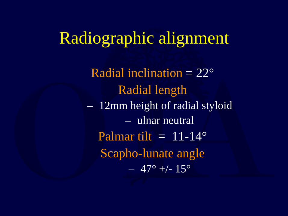
Radiographic alignment
Radial inclination = 22° Radial length
– 12mm height of radial styloid – ulnar neutral
Palmar tilt = 11-14° Scapho-lunate angle
– 47° +/- 15°

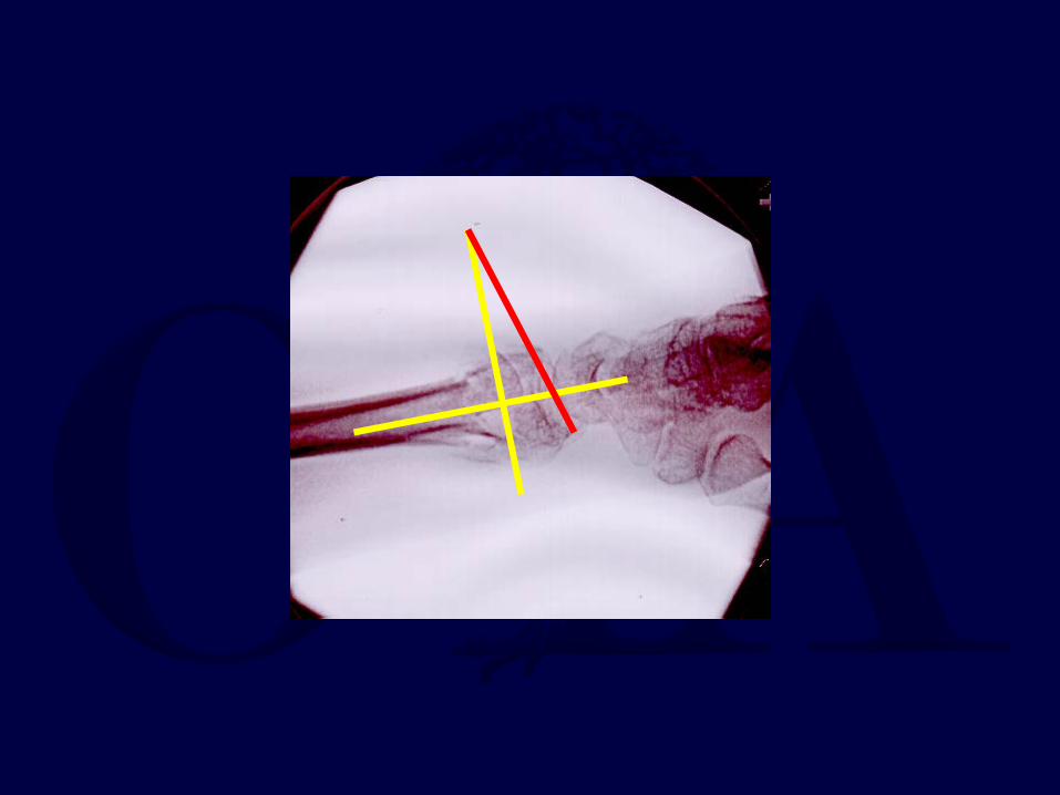
Measurement of Radial Length and Inclination
Inclination = 23 degrees

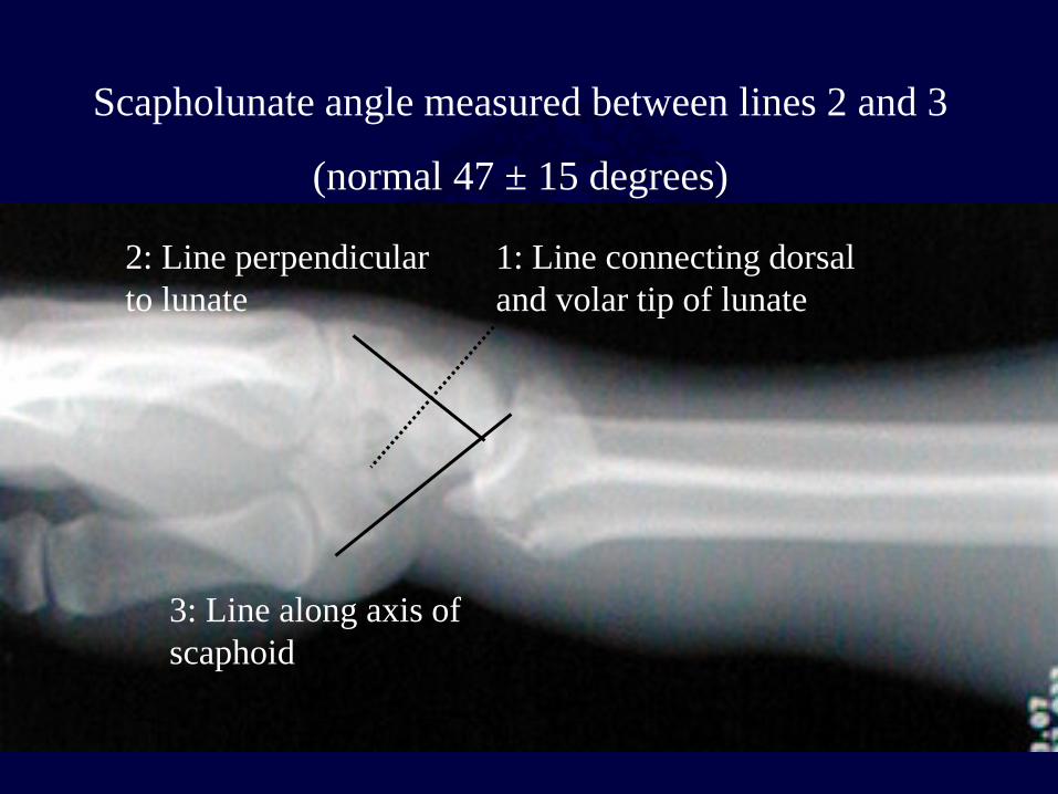
1: Line connecting dorsal and volar tip of lunate
2: Line perpendicular to lunate
3: Line along axis of scaphoid
Scapholunate angle measured between lines 2 and 3
(normal 47 ± 15 degrees)

Computed Tomography Indications:
Intra-articular fxs with multiple fragments centrally impacted fragments
DRUJ incongruity
19 consecutive fx, CT had better sensitivity for intraarticular fragments
management change in 5 pts
Cole et al: J Hand Surg, 1997

Classification of Distal Radius Fractures
Ideal system should describe: – Type of injury
– Severity – Evaluation – Treatment – Prognosis

Common Classifications
Frykman Weber (AO/ASIF)
Melone Column theory
Fernandez (mechanism)
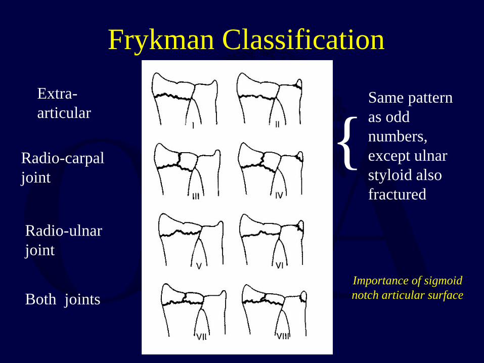
Frykman Classification
Extra-articular
Radio-carpal joint
Radio-ulnar joint
Both joints
{ Same pattern as odd numbers, except ulnar styloid also fractured
Importance of sigmoid notch articular surface

AO/ OTA Classification
Group A: Extra-articular
Group B: Partial Intra-articular
Group C: Complete Intra-articular
Volar and dorsal
Barton fxs

Column Theory
Rikli & Regazzoni, 1996
3 Columns: lateral, intermediate, medial

Classification – Fernandez (1997)
I. Bending-metaphysis fails under tensile
stress (Colles, Smith)
II. Shearing-fractures of joint surface (Barton,
radial styloid)
importance of mechanism and energy level of injury

Classification – Fernandez (1997) III. Compression - intraarticular fracture
with impaction of subchondral and
metaphyseal bone (die-punch)
IV. Avulsion- fractures of ligament attachments (ulna, radial styloid)
V. Combined complex -
high velocity injuries
Assessment of X-rays
Assess involvement of dorsal or volar rim – is comminution mainly volar or dorsal?
– is one of four cortices intact?
Look for “die-punch” lesions of the scaphoid or lunate fossa.
Assess amount of shortening Look for DRUJ involvement
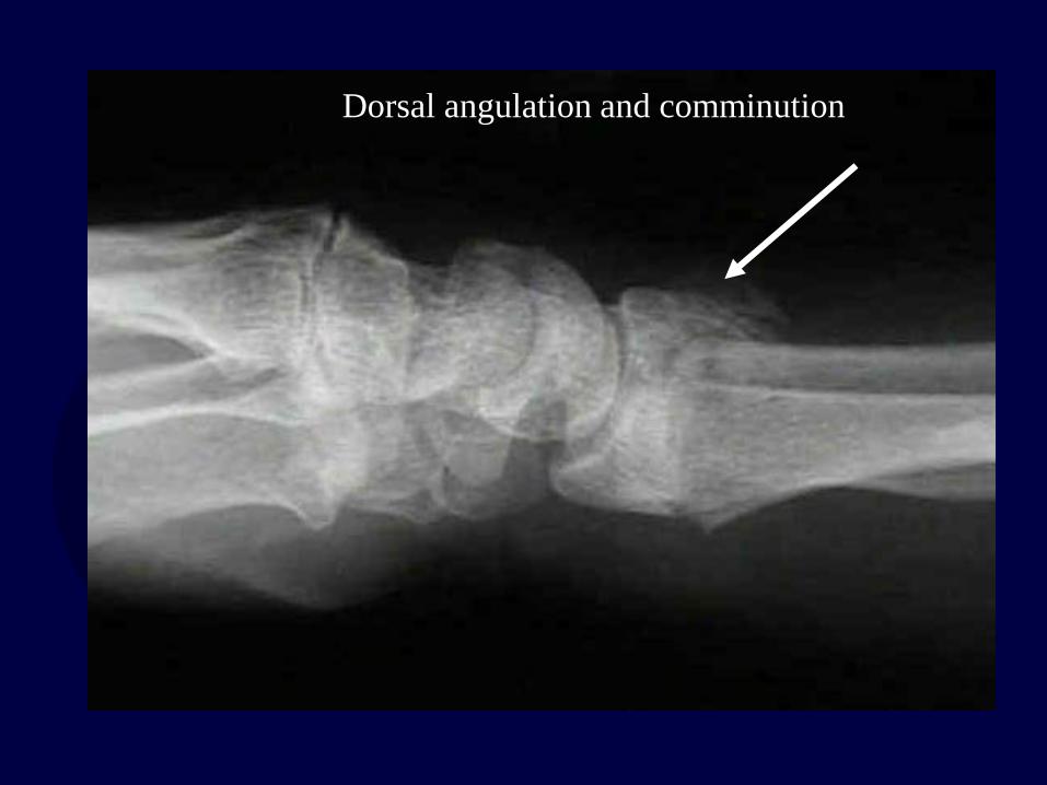
Dorsal angulation and comminution

Volar subluxation of carpus with fracture fragment
Options for Treatment Casting
– Long arm vs. short arm – Sugar-tong splint
External Fixation – Joint-spanning – Non bridging
Percutaneous pinning Internal Fixation
– Dorsal plating – Volar plating
– Combined dorsal/volar plating – focal (fracture specific) plating
Indications for Closed Treatment
Low-energy fracture Low-demand patient
Medical co-morbidities Minimal displacement- acceptable
alignment Match treatment to demands of the
patient

Closed Treatment of Distal Radial Fractures
Obtaining and then maintaining an acceptable reduction
Immobilization:
– long arm (cast or sugar-tong for high demand) – short arm adequate for elderly patients
Frequent follow-up necessary in order to diagnose
re-displacement.

Technique of Closed Reduction Anesthesia – Hematoma block
– Intravenous sedation – Bier block
Traction: finger traps and weights Reduction Maneuver (dorsally angulated fracture):
– hyperextension of the distal fragment, – Maintain weighted traction and reduce the distal to the proximal
fragment with pressure applied to the distal radius. Apply well-molded “sugar-tong” splint or cast, with wrist in
neutral to slight flexion. Avoid Extreme Positions!

Acceptable Reduction Criteria
Dorsal angulation < 10° > 15 ° of inclination
Articular step-off < 2mm < 5 mm shortening DRUJ congruent
After-treatment Watch for median nerve symptoms
– parasthesias common but should diminish over few hours
– If persist release pressure on cast, take wrist out of flexion
– Acute carpal tunnel: symptoms progress; CTR required Follow-up x-rays needed in 1-2 weeks to evaluate
reduction. Change to short-arm cast after 2-3 weeks, continue
until fracture healing.

Management of Redisplacement
Repeat reduction and casting – high rate of failure
Repeat reduction and percutaneous pinning External Fixation
ORIF

Treatment Choice
Depends on assessment of fracture stability Indicators of instability are:
– Patient age – Metaphyseal Comminution – Shortening: ulnar variance
Mackenney, McQueen, Elbton, JBJS 2006 Sep;88(9):1944-51., Prediction of instability in distal radial fractures
Indications for Surgical Treatment
High-energy injury with instability Open injury
Radial inclination < 15° Articular step-off, or gap > 2mm
Dorsal tilt > 10 ° DRUJ incongruity

Operative Management of Distal Radius Fractures

External fixation:
The treatment of choice for distal radius fractures in the 1980’s
Has fallen out of favor


Spanning
A spanning fixator is one which fixes distal radius fractures by spanning the carpus; I.e., fixation into radius and metacarpals

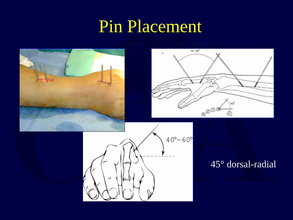
Pin Placement
45° dorsal-radial
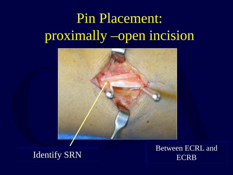
Pin Placement: proximally –open incision
Identify SRN Between ECRL and
ECRB

45 deg dorsal-radial position of fixator
Wrist in neutral
Allows retropulsion of thumb

Ex fix and supplementary Pins
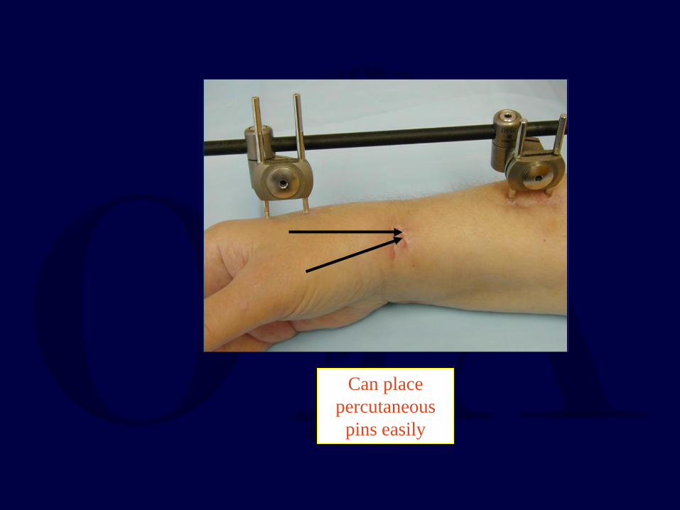
Can place percutaneous
pins easily

Can Remove Ex Fix or Pins Sooner if Needed
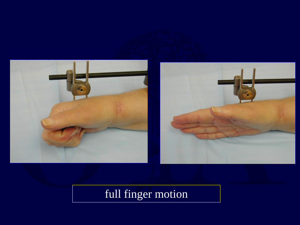
full finger motion
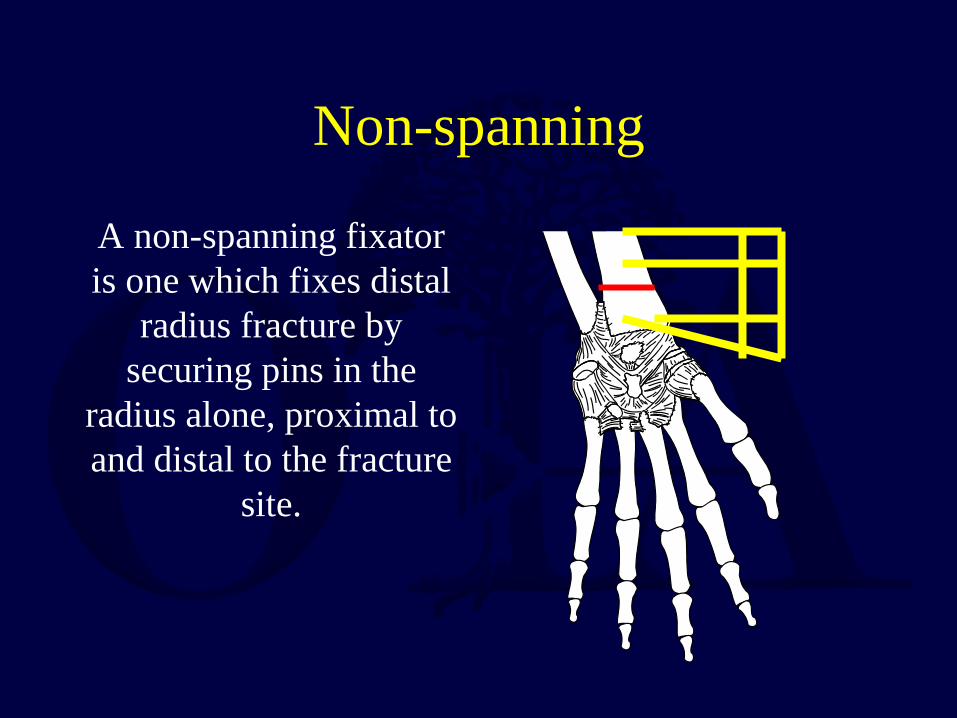
Non-spanning
A non-spanning fixator is one which fixes distal
radius fracture by securing pins in the
radius alone, proximal to and distal to the fracture
site.

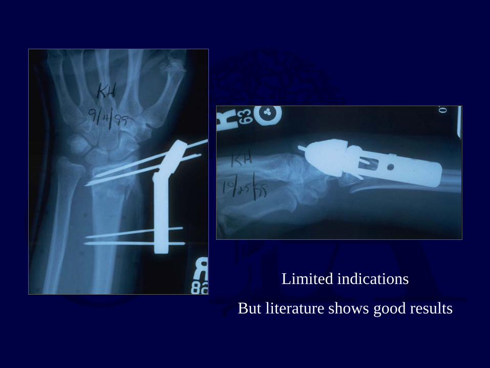
Limited indications
But literature shows good results

Courtesy of Hill Hastings,MD
Early ROM permitted
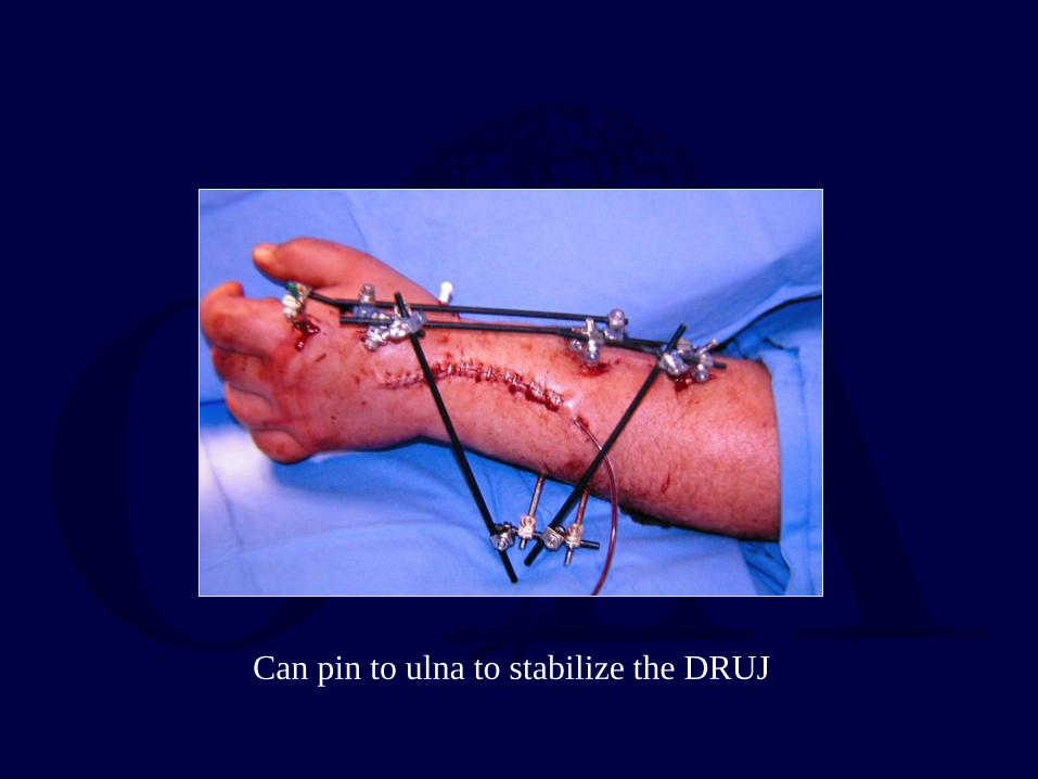
Can pin to ulna to stabilize the DRUJ
Factors Affecting Functional Outcome
McQueen (1996): carpal alignment after distal radius fractures is the main influence on
final outcome – malalignment has significant negative effect on
function – failure to restore volar tilt predisposes to carpal
collapse and carpal malalignment

Reduction Tactics
DePalma (1952) introduced traction / distraction as means of reducing distal
radius fractures Spanning fixator relies on distraction as
principle method of reducing fracture fragments
Distraction (Ligamentotaxis) excellent for restoring length

Ligamentotaxis
Bartosh, J Hand Surg 15A, 1990 19 cadaver hands with distal radius osteotomy
Ligamentotaxis with 10# and 20# of traction @ 100, 200 and 300 of flexion
volar tilt of distal radius could not be re-established
Ligamentous Anatomy
Volar ligaments – Straight fibers
– Stout – Tighten readily
Dorsal ligaments – Zigzag – Elastic
– Tighten slowly
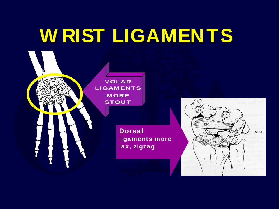
VOLAR LIGAMENTS
MORE STOUT
W RIST LIGAMENTSW RIST LIGAMENTS
Dorsal ligaments more lax, zigzag

Non-Spanning vs. Spanning Fixator
McQueen, JBJS-B, 1998 Prospectively studied 30 spanning vs. 30 non-
spanning fixator patients Non-spanning better preserved volar tilt, prevented
carpal malalignment, gave better grip strength and hand function (all with p<.001) Complication rate 50% lower
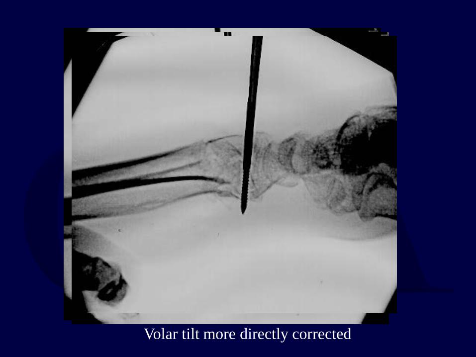
Volar tilt more directly corrected
Complications
Complication rates high in almost all reported series
– Pin track infection – RSD Finger stiffness
– Loss of reduction; early vs. late – Tendon rupture
But do not throw away the external fixator

Indirect reduction and percutaneous fixation versus open reduction and internal fixation for displaced intra-articular fractures of the distal radius: a
randomized, controlled trial. Kreder, Hanel et al., JBJS Br, 2005 Jun;87(6):829-36.
179 adult patients with displaced intra-articular fractures of
the distal radius was randomized – indirect percutaneous reduction and external fixation (n = 88)
– open reduction and internal fixation (n = 91)
During the first year functional scores , pinch strength and grip strength improved significantly in all patients.
No difference in the radiological restoration of anatomical features or the ROM
At 2 years – Ex Fix patients had a more rapid return of function and a better
functional outcome than ORIF group – provided that the intra-articular step and gap deformity were minimized.

Spanning Plate “Internal Ex Fix”

Indications
High energy comminuted fractures
ICU patients where perc pins are undesirable
Pts that can’t tolerate an external fixator
Courtesy Doug Hanel, MD

Courtesy Doug Hanel, MD

Pins Out 6wks Plate Out 16Weeks
Courtesy Doug Hanel, MD

Courtesy Doug Hanel, MD
Percutaneous Pinning-Methods variety described
most common radial styloid pinning + dorsal-ulnar corner of radius pinning
supplemental immobilization with cast, splint in conjunction with external fixation
(Augmented external fixation)

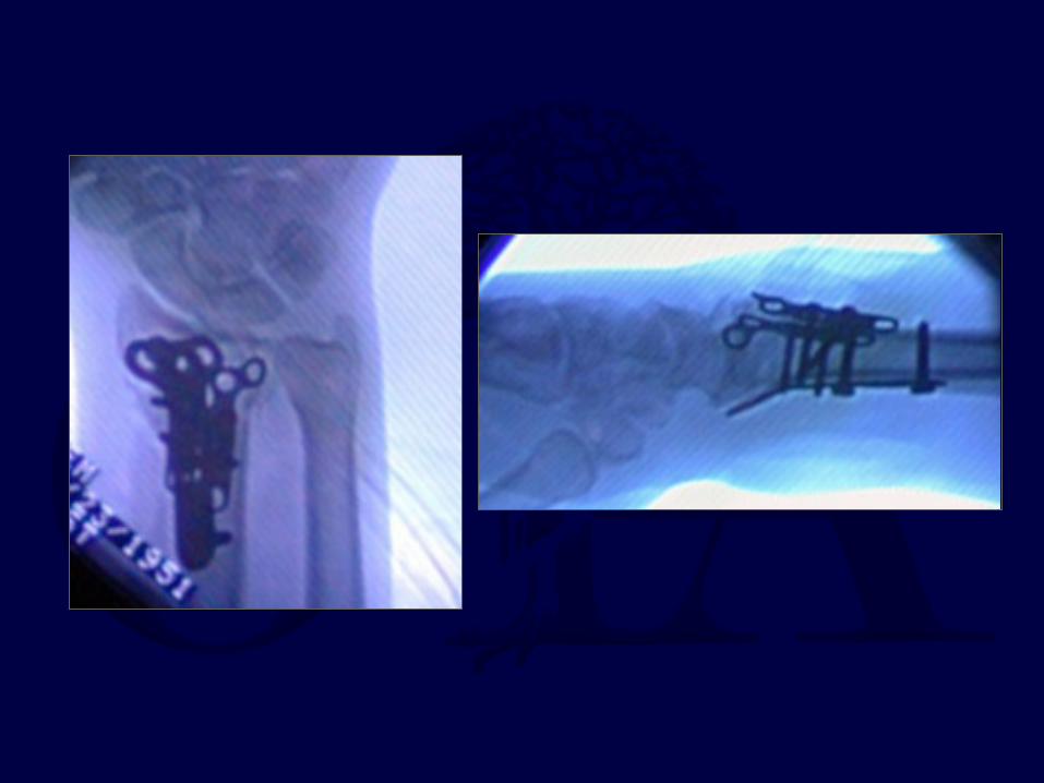
Percutaneous Pins
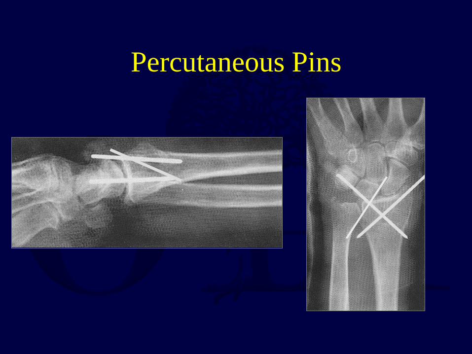
Percutaneous Pins

Percutaneous Pinning
2 radial styloid pins, (Mah and Atkinson, J Hand Surg 1992)
– excellent anatomic 82% – good-excellent functional results 100% Crossed Pins - (Clancey JBJS 1984)
– prospective study – 30 pts excellent anatomic results in 90%
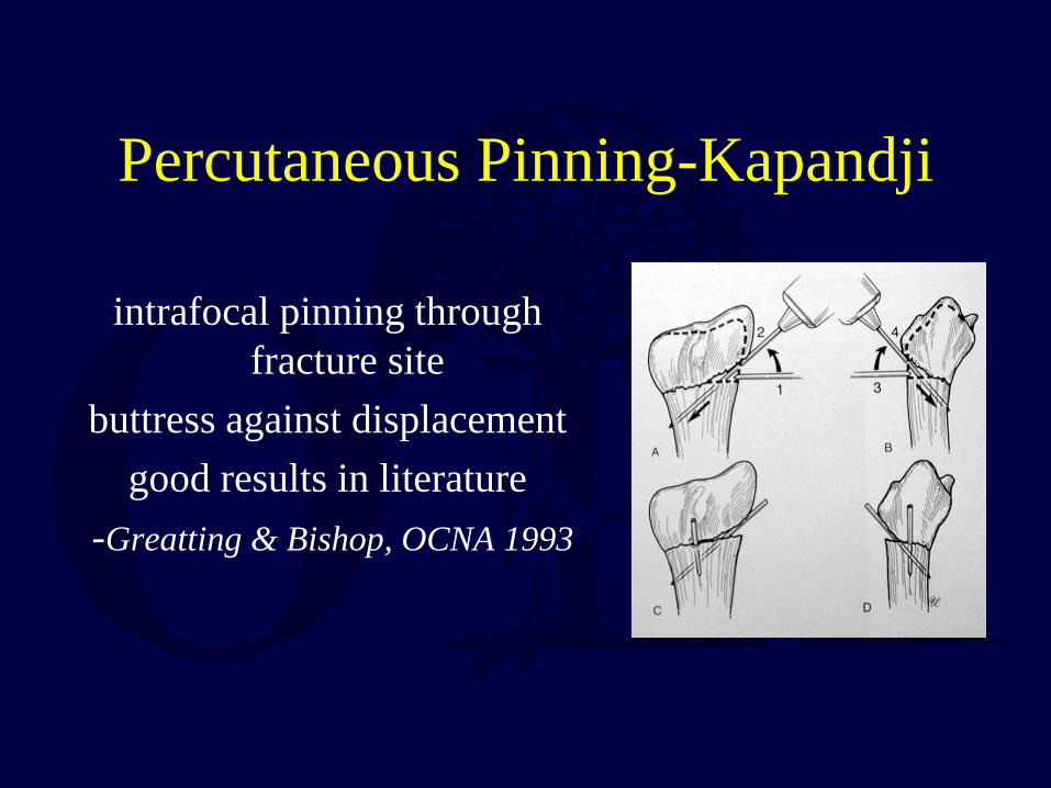
Percutaneous Pinning-Kapandji
intrafocal pinning through fracture site
buttress against displacement good results in literature
-Greatting & Bishop, OCNA 1993

Internal Fixation of Distal Radius Fractures
elevation of depressed articular fragments required if articular fragments can not be
adequately reduced with percutaneous methods
Volar approaches most common *Significant increase in use over last 5 years

Selection of Approach
Based on location of fracture and displacement Volar approach for volar rim fractures and
comminuted fractures that can be reduced Radial styloid approach for buttressing of styloid
Dorsal approach – Occasionally for dorsally displaced fractures that can’
be reduced from volar approach Combined approaches needed for high-energy
fractures with significant axial impaction.

-
WHICH APPROACH?
DORSAL 3rd DC –EPL
(extensile) 1-2nd DC

Classical Henry approach Extended carpal tunnel approach
VOLAR
Useful for volar ulnar corner fragment or Fxs associated with
CTS

Distal Radius- “volar Barton” 64 y.o. M, MVA, contralateral tibial shaft Fx

Carpal subluxation


Volar –Henry Approach
Courtesy Diego Fernandez, MD
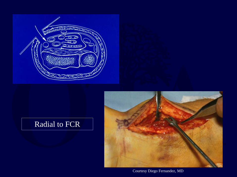
Radial to FCR
Courtesy Diego Fernandez, MD

Elevate Pronator Quadratus
Courtesy Diego Fernandez, MD
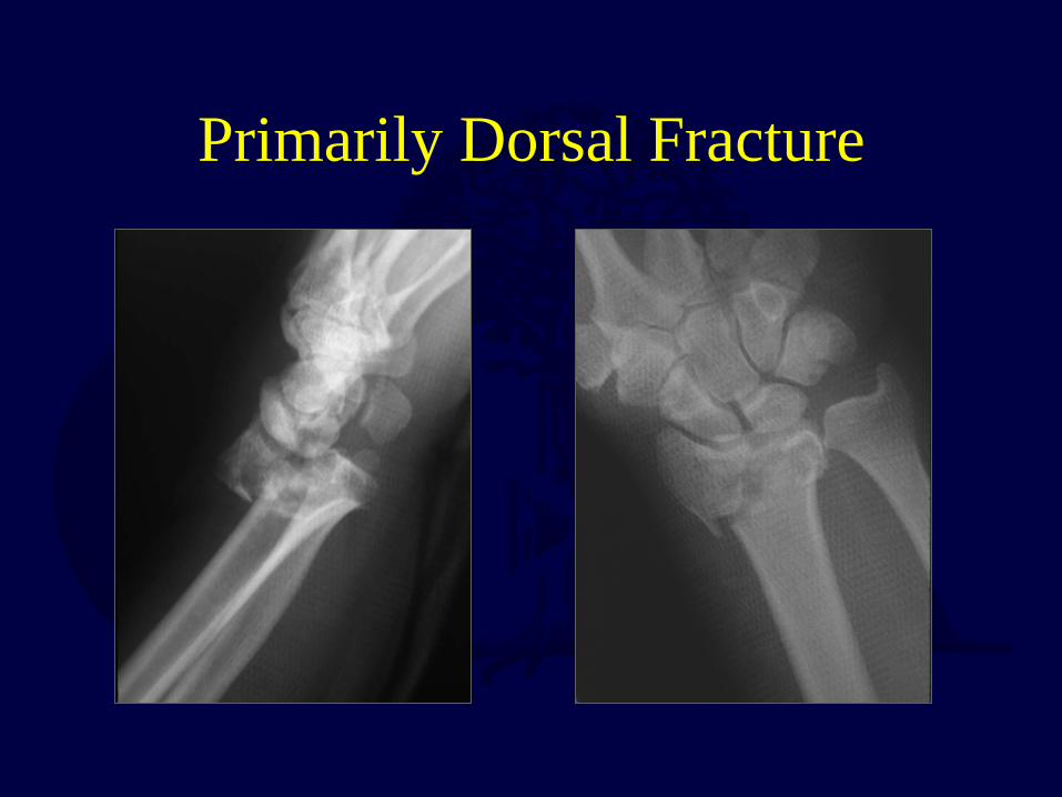
Primarily Dorsal Fracture
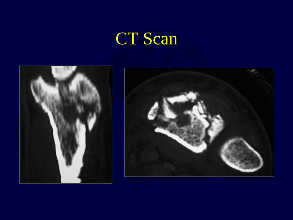
CT Scan
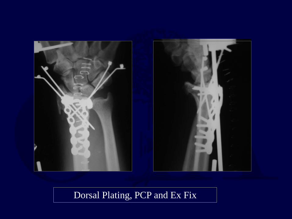
Dorsal Plating, PCP and Ex Fix

Joints aligned plates removed

-less tendon irritation than dorsal plating
- indirect reduction
-better tolerated than Ex fix
Volar Plating for Dorsal Fractures

Fixed angle locked screws

Courtesy Jorge Orbay, MD
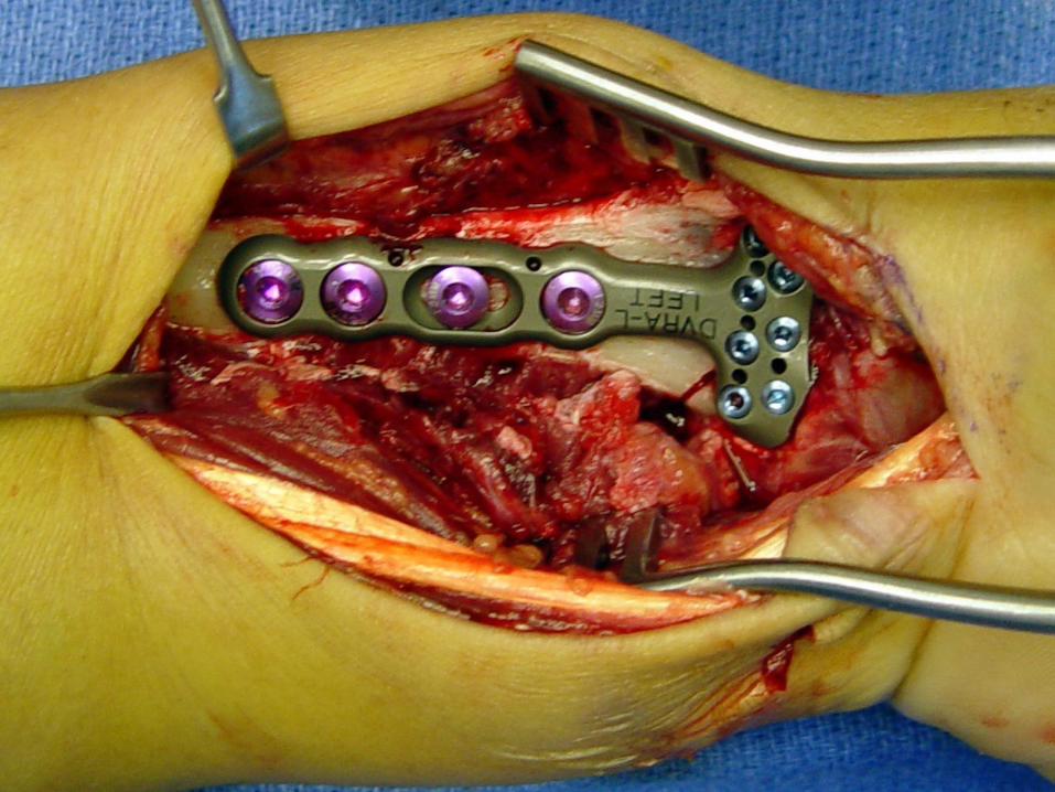
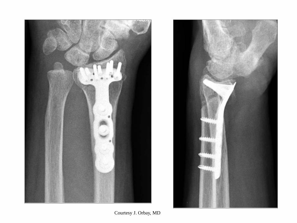
Courtesy J. Orbay, MD

Three Column Theory
Radial Column Lateral side of
radius Intermediate Column Ulnar side of
radius Ulnar Column
distal ulna
Radial column
Intermediate column
Ulnar column
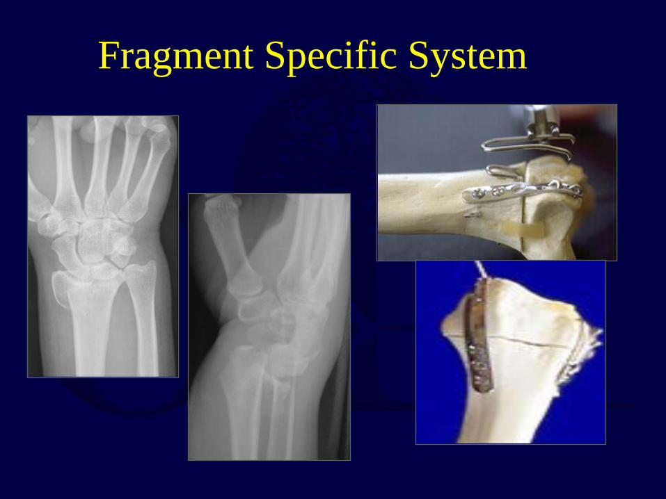
Fragment Specific System

Radial and Ulnar Columns
-Pin plates -90-90 plating
technique

Focal Plating
Radial Styloid Fragment Dorsal ulnar fragment
70 – 90 degrees apart

Dorsal Fracture
Radial Styloid and dorsal-ulnar corner

Dorsal Case: focal plating
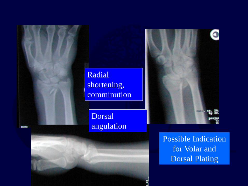
Radial shortening, comminution
Dorsal angulation
Possible Indication for Volar and
Dorsal Plating
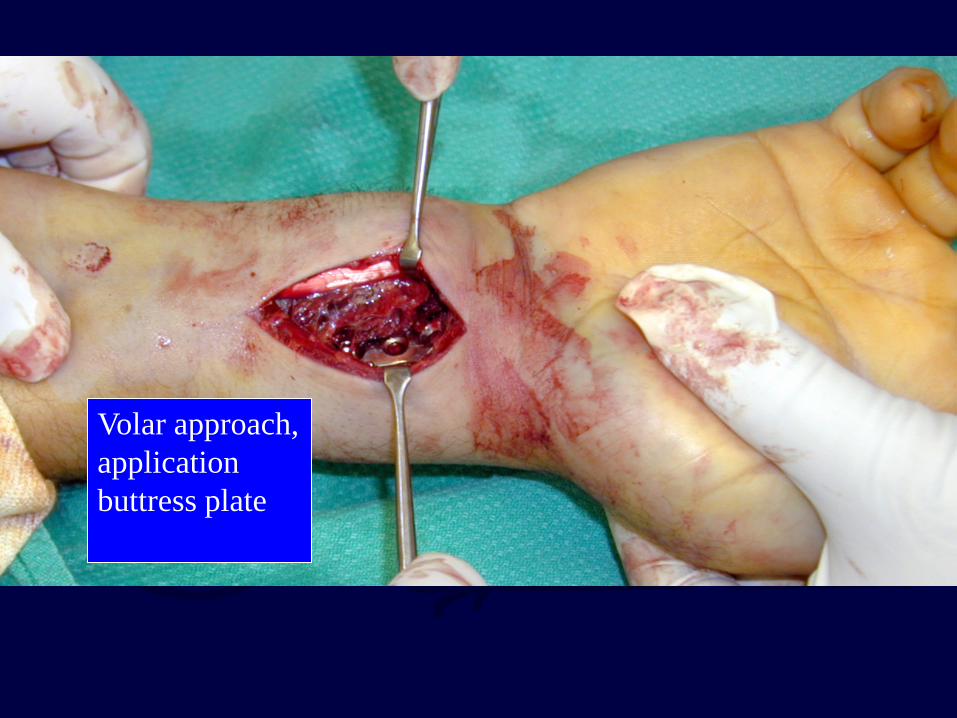
Volar approach, application buttress plate
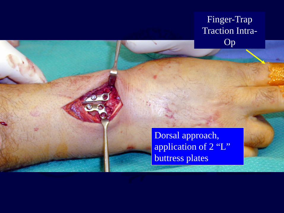
Dorsal approach, application of 2 “L” buttress plates
Finger-Trap Traction Intra-
Op
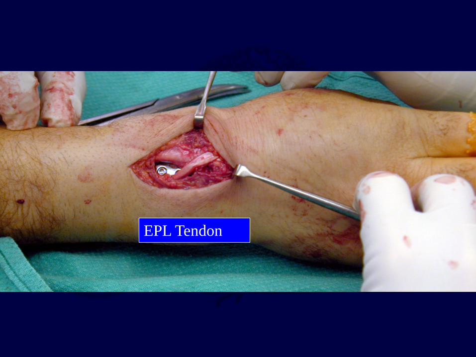
EPL Tendon

Extensor retinaculum repaired beneath EPL to prevent irritation from plate- EPL left transposed

Advanced Techniques Arthroscopic-Assisted
reduce articular incongruities also diagnose associated soft tissue lesions
minimally invasive

Arthroscopic-Assisted
Culp and Osterman, OCNA 26(4) 1995

Malunion of Distal Radius Fractures
Changes load-bearing patterns on the distal radius and load sharing between the radius
and ulna. Can lead to arthrosis.
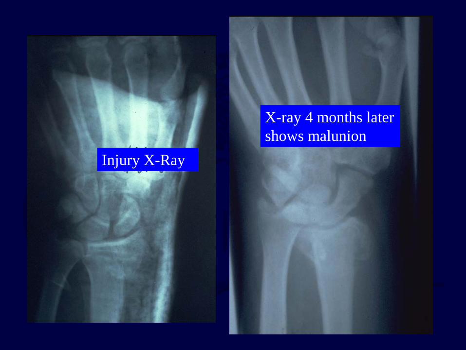
Injury X-Ray
X-ray 4 months later shows malunion

Normal loading patterns
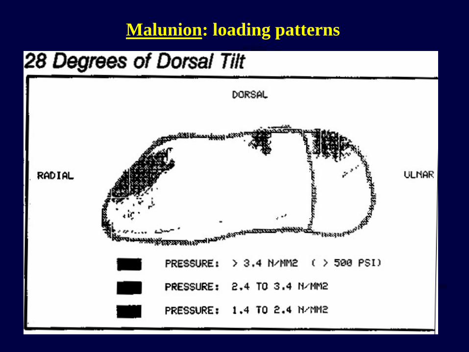
Malunion: loading patterns
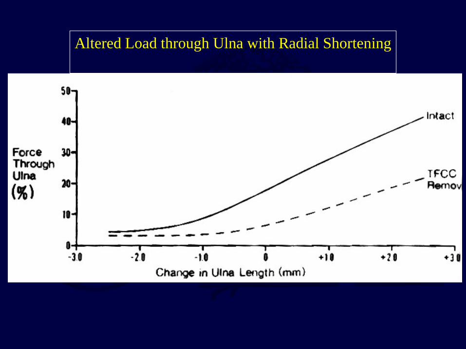
Altered Load through Ulna with Radial Shortening
Malunion of Distal Radius Fractures
Requires osteotomy, bone grafting and fixation
Dorsal plating traditionally done Volar plating now performed with indirection
reduction of fragment – +/- bone graft
– Cancellous or synthetic injectable grafts
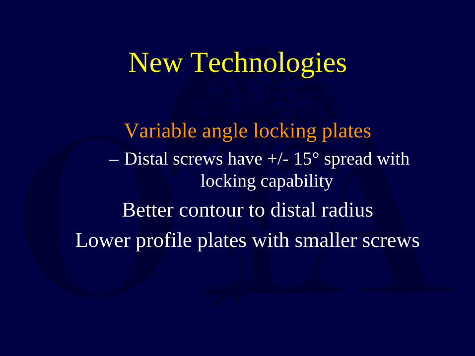
New Technologies
Variable angle locking plates – Distal screws have +/- 15° spread with
locking capability Better contour to distal radius
Lower profile plates with smaller screws

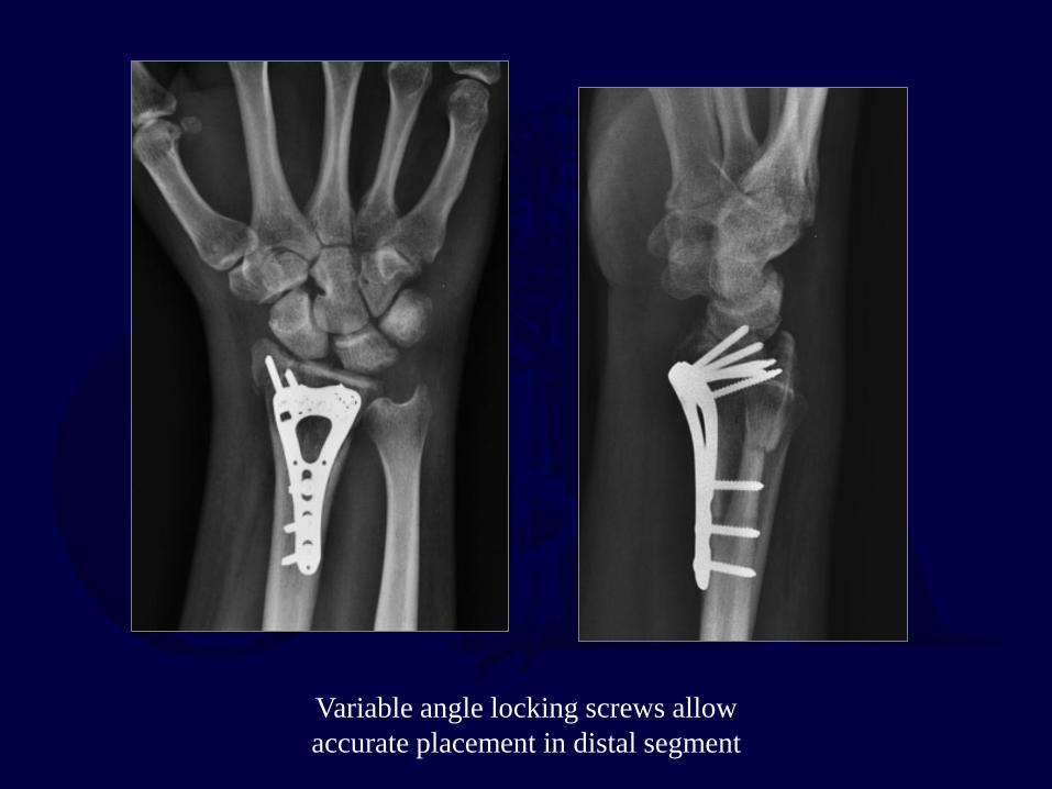
Variable angle locking screws allow accurate placement in distal segment
Conclusions
Need to be able to use all tools for treatment of distal radius fractures
Both external fixation and ORIF are useful. – ORIF better in high-energy fractures associated
with depression of articular surface – ORIF gives better anatomic restoration,
although not necessarily higher patient satisfaction.
Conclusions
External fixators still have a role in the treatment of distal radius fractures
Spanning ex fix unable to correct fracture deformity
by itself
Should usually combined with percutaneous pins (augmented fixation)

Bibliography
1: Ruch DS, Ginn TA, Yang CC, Smith BP, Rushing J, Hanel DP. Use of a distraction plate for distal radial fractures with metaphyseal and diaphyseal comminution. J Bone Joint Surg Am. 2005 May;87(5):945-54. 2: Sammer DM, Shah HM, Shauver MJ, Chung KC. The effect of ulnar styloid fractures on patient-rated outcomes after volar locking plating of distal radius fractures. J Hand Surg Am. 2009 Nov;34(9):1595-602. 3: Liporace FA, Adams MR, Capo JT, Koval KJ. Distal radius fractures. J Orthop Trauma. 2009 Nov-Dec;23(10):739-48. 4: Koenig KM, Davis GC, Grove MR, Tosteson AN, Koval KJ. Is early internal fixation preferred to cast treatment for well-reduced unstable distal radial fractures? J Bone Joint Surg Am. 2009 Sep;91(9):2086-93. 5: Rozental TD, Blazar PE, Franko OI, Chacko AT, Earp BE, Day CS. Functional outcomes for unstable distal radial fractures treated with open reduction and internal fixation or closed reduction and percutaneous fixation. A prospective randomized trial. J Bone Joint Surg Am. 2009 Aug;91(8):1837-46. 6: Wei DH, Raizman NM, Bottino CJ, Jobin CM, Strauch RJ, Rosenwasser MP. Unstable distal radial fractures treated with external fixation, a radial column plate, or a volar plate. A prospective randomized trial. J Bone Joint Surg Am. 2009 Jul;91(7):1568-77.

7: Berglund LM, Messer TM. Complications of volar plate fixation for managing distal radius fractures. J Am Acad Orthop Surg. 2009 Jun;17(6):369-77. 8: Casaletto JA, Machin D, Leung R, Brown DJ. Flexor pollicis longus tendon ruptures after palmar plate fixation of fractures of the distal radius. J Hand Surg Eur Vol. 2009 Aug;34(4):471-4. 9: Mirza A, Jupiter JB, Reinhart MK, Meyer P. Fractures of the distal radius treated with cross-pin fixation and a nonbridging external fixator, the CPX system: a preliminary report. J Hand Surg Am. 2009 Apr;34(4):603-16. 10: Souer JS, Ring D, Matschke S, Audige L, Marent-Huber M, Jupiter JB; AOCID Prospective ORIF Distal Radius Study Group. Effect of an unrepaired fracture of the ulnar styloid base on outcome after plate-and-screw fixation of a distal radial fracture. J Bone Joint Surg Am. 2009 Apr;91(4):830-8 11: Arora R, Gabl M, Gschwentner M, Deml C, Krappinger D, Lutz M. A comparative study of clinical and radiologic outcomes of unstable colles type distal radius fractures in patients older than 70 years: nonoperative treatment versus volar locking plating. J Orthop Trauma. 2009 Apr;23(4):237-42 12: Capo JT, Rossy W, Henry P, Maurer RJ, Naidu S, Chen L. External fixation of distal radius fractures: effect of distraction and duration. J Hand Surg Am. 2009 Nov;34(9):1605-11. 13: Jupiter JB, Marent-Huber M; LCP Study Group. Operative management of distal radial fractures with 2.4-millimeter locking plates. A multicenter prospective case series. J Bone Joint Surg Am. 2009 Jan;91(1):55-65.
14: Thomas AD, Greenberg JA. Use of fluoroscopy in determining screw overshoot in the dorsal distal radius: a cadaveric study. J Hand Surg Am. 2009 Feb;34(2):258-61. 15: Soong M, Got C, Katarincic J, Akelman E. Fluoroscopic evaluation of intra-articular screw placement during locked volar plating of the distal radius: a cadaveric study. J Hand Surg Am. 2008 Dec;33(10):1720-3. 16: Kreder HJ, Hanel DP, Agel J, McKee M, Schemitsch EH, Trumble TE, Stephen D. Indirect reduction and percutaneous fixation versus open reduction and internal fixation for displaced intra-articular fractures of the distal radius: a randomised, controlled trial. J Bone Joint Surg Br. 2005 Jun;87(6):829-36. 17: Orbay JL, Fernandez DL. Volar fixed-angle plate fixation for unstable distal radius fractures in the elderly patient. J Hand Surg Am. 2004 Jan;29(1):96-102. 18: Orbay JL, Fernandez DL. Volar fixation for dorsally displaced fractures of the distal radius: a preliminary report. J Hand Surg Am. 2002 Mar;27(2):205-15. 19: Kreder HJ, Agel J, McKee MD, Schemitsch EH, Stephen D, Hanel DP. A randomized, controlled trial of distal radius fractures with metaphyseal displacement but without joint incongruity: closed reduction and casting versus closed reduction, spanning external fixation, and optional percutaneous K-wires. J Orthop Trauma. 2006 Feb;20(2):115-21.

Conclusions New plating techniques allow for accurate and rigid
fixation of fragments
Plating allows early wrist ROM
Volar, smaller and more anatomic plates are better tolerated
Combination treatment is often needed
Return to
Upper Extremity Index
If you would like to volunteer as an author for the Resident Slide Project or recommend updates to any of the following slides, please send an e-mail to [email protected]