Embed Size (px)
DESCRIPTION
Trypanosomiasis This is a protozoan disease of animals and humans caused by parasites of the genus Trypanosoma, which are found in blood plasma, various body tissues and fluids.
Citation preview

Diseases caused by Diseases caused by protozoaprotozoa
Diseases caused by Diseases caused by protozoaprotozoa
Dr.Kedar KarkiDr.Kedar KarkiM.V.St.Preventive M.V.St.Preventive
Veterinary Medicine Veterinary Medicine PhilippinesPhilippines
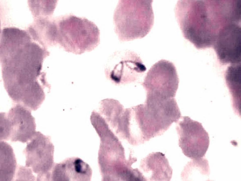
Trypanosomiasis• This is a protozoan disease of
animals and humans caused by parasites of the genus Trypanosoma, which are found in blood plasma, various body tissues and fluids.
Transmission:• Trypanosoma are transmitted
primarily by the Glossina spp., tsetse fly, Stomoxys, tabanid and reduviid bugs, and by venereal contact. Trypanosoma species in the insect vector undergo one or two cycles of development.
Ante mortem findings
• :• Intermittent fever • Anemia • Weight loss and weakness • Edema, particularly observed in the face and
legs • Enlarged body lymph nodes • Hemorrhage • Opacity of the cornea, keratitis and photophobia
• Chronic form of trypanosomiasis is sometimes manifested by progressive weakness, despite absent parasitemia, and death.
Postmortem findings:
• Enlarged lymph nodes • The enlargement of spleen, liver
and kidney may also occur. • Edematous and emaciated carcass • Mild icterus
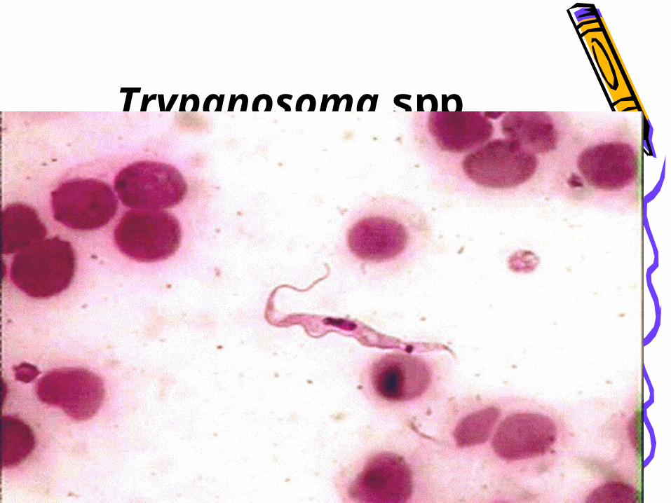
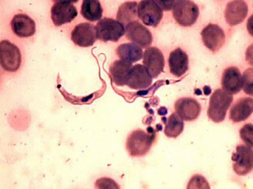
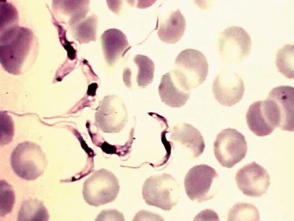
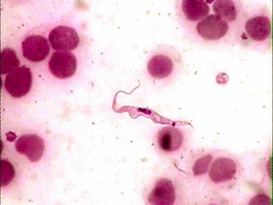
Trypanosoma spp
• Differential diagnosis: Helminthiasis, malnutrition and other chronic wasting diseases, equine infectious anemia, heart water, babesiosis and anaplasmosis
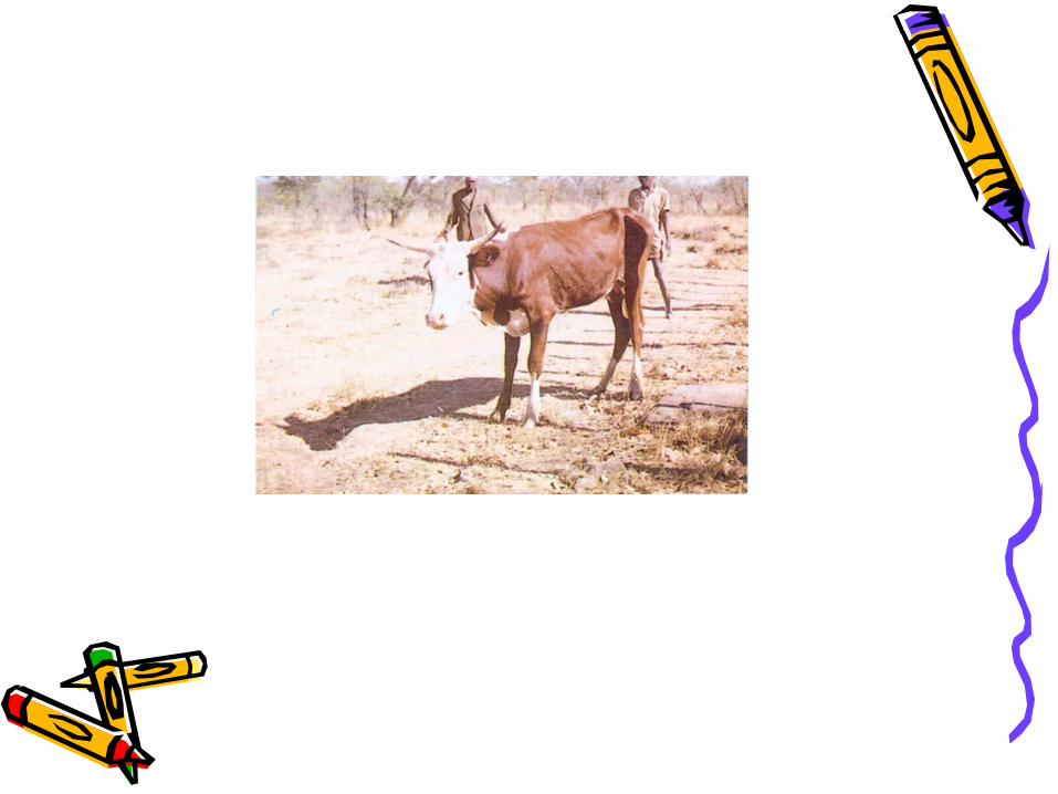
• Trypanosomiasis. This animal shows icteric mucous membranes, weakness in leg muscles and emaciation.
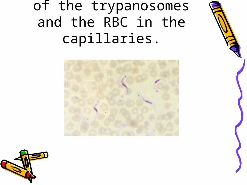
An impression smear of the trypanosomes and
the RBC in the capillaries.
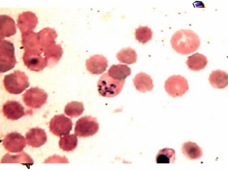
• Theileriosis (East cost fever)• East coast fever is a sub acute
haemoprotozoan disease of cattle caused by Theileria parva. Theileriosis is characterized by fever, enlarged lymph nodes, dyspnea and death. In chronic cases loss of condition, emaciation, diarrhea, blindness, etc. can be seen.

• Transmission: Vectors are ixodid ticks of the species Rhipicephalus.
• Ante mortem findings:1.Mortality up to 90 % 2.High temperature (up to 41 °C) 3.Difficult breathing and coughing 4.Nasal discharge, salivation and watery
eyes 5.Swelling of the lymph nodes draining the
area where the infected tick fed6.Cerebral signs manifested by circling to
one side, convulsions and death
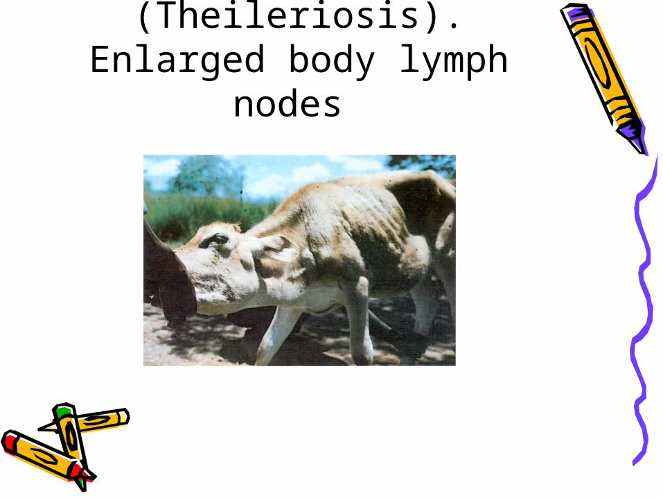
East Coast fever (Theileriosis). Enlarged
body lymph nodes
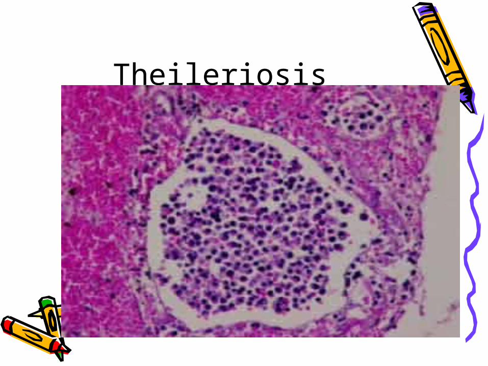
Theileriosis
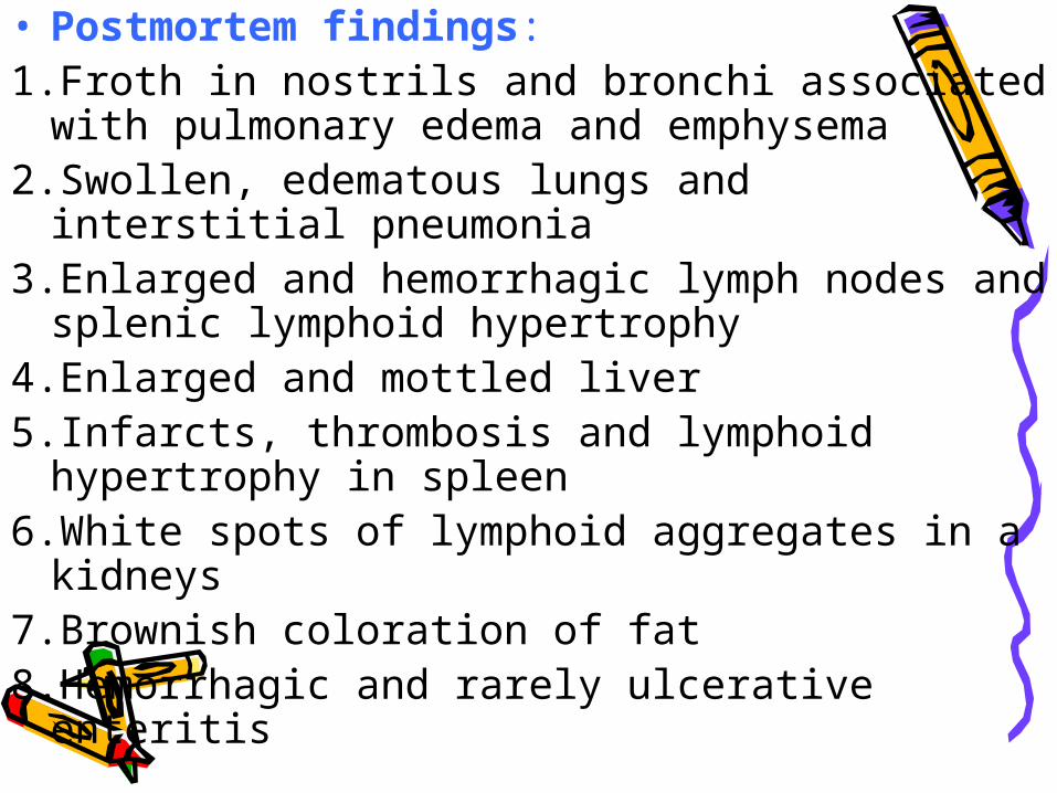
• Postmortem findings:1.Froth in nostrils and bronchi associated with
pulmonary edema and emphysema 2.Swollen, edematous lungs and interstitial
pneumonia 3.Enlarged and hemorrhagic lymph nodes and
splenic lymphoid hypertrophy 4.Enlarged and mottled liver 5.Infarcts, thrombosis and lymphoid hypertrophy
in spleen 6.White spots of lymphoid aggregates in a
kidneys 7.Brownish coloration of fat 8.Hemorrhagic and rarely ulcerative enteritis

• Differential diagnosis: Hemorrhagic septicemia, babesiosis, malignant catarrhal fever, trypanosomiasis, Rift Valley fever, heart water and bovine leucosis
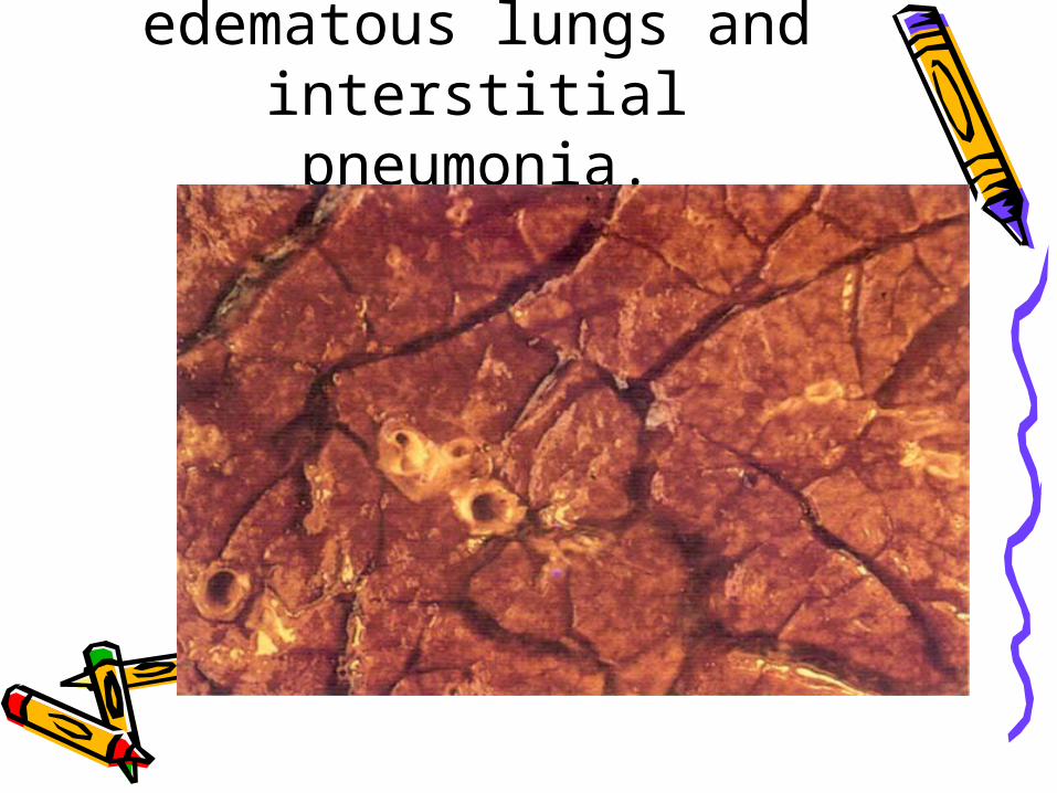
Theileriosis. Swollen edematous lungs and interstitial pneumonia.
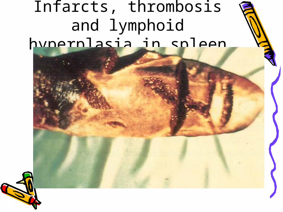
Theileriosis. Infarcts, thrombosis and lymphoid
hyperplasia in spleen
Anaplasmosis (gall sickness)
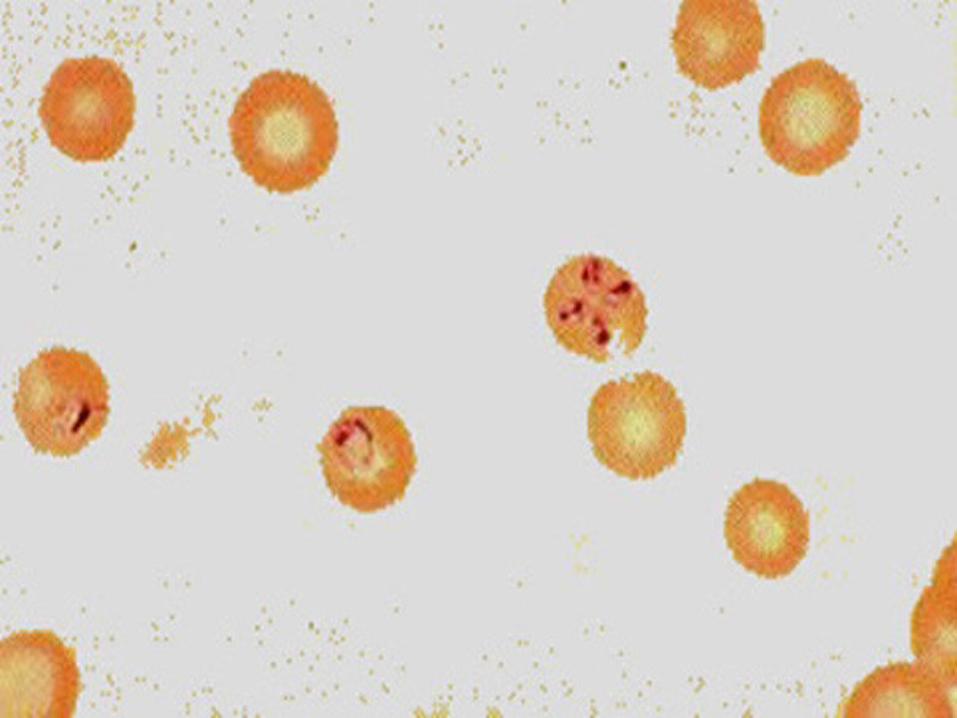
• Anaplasmosis is a rickettsial disease characterized by severe debility, emaciation, anemia and jaundice and is caused by Ana plasma spp... They are obligate intracellular parasites. Ana plasma marginal is the causative agent in cattle and wild ruminants.
• Transmission: Boophilus species of ticks transmit anaplasmosis. Mosquitoes and the horsefly are mechanical transmitters. Transmission is also possible through injection needles.

• Ante mortem findings:• Acute infection with A. marginal1.High fever 2.Jaundice and anemia demonstrated
by pale mucous membranes 3.Frequent urination and constipation
Chronic infection • Emaciation
• Postmortem findings:1.Enlarged and congested spleen
(splenomegaly) showing soft pulp 2.Distended gall bladder with dark tarry bile 3.Thin, watery blood, which clots poorly 4.Enlarged, icteric liver, deep orange in color
and distended bile ducts 5.Lemon yellow carcass and connective
tissue of the sclera of the eye, tendons, pleura, peritoneum, and attachments of diaphragm.
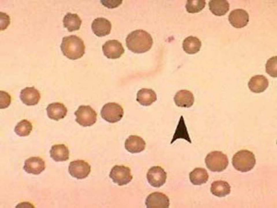
• Diagnosis can only be confirmed by detecting parasites in a blood smear stained with Giemsa.

• Differential diagnosis: Icterus and anemia of different causes, anthrax, leptospirosis, emaciation caused by parasitism and malignant lymphoma, babesiosis
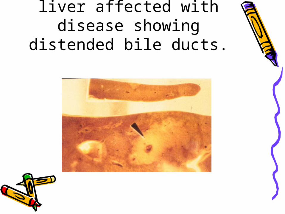
Anaplasmosis. Ox liver affected with disease
showing distended bile ducts.
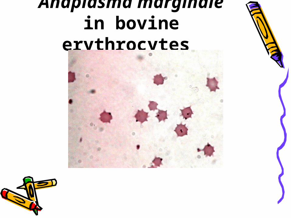
Anaplasma marginale in bovine
erythrocytes
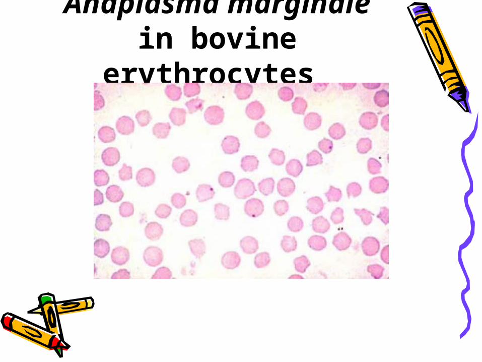
Anaplasma marginale in bovine
erythrocytes
Babesiosis (Piroplasmosis, Texas
fever, Red water fever, Tick fever)
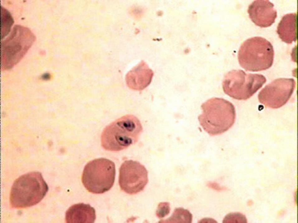
• Babesiosis of cattle, horses, sheep and swine is a febrile, tick borne disease caused by various species of the protozoan genus Babesia
• Transmission: Different species of ticks in the family Ixodidae serve as vectors in different locations. The Babesia parasites can be transmitted transstadially and transovarially within a tick species
• Ante mortem findings:1.Incubation 7–10 days 2.Mortality up to 50 % or over depending
on age, breed, etc. 3.High fever (41.5° C) 4.Dark reddish brown urine in the terminal
stage 5.Reddened and injected mucous
membranes at the early stages and later, anemic mucous membranes
6.Clinical signs may resemble rabies in cerebral form of babesiosis.
• Postmortem findings:1.Edema and congested lungs 2.Enlarged and yellow liver and distended gall
bladder with thick dark green bile. 3.Enlarged spleen 4.Anemia and pale muscles 5.Jaundice particularly noted in the connective
tissue 6.Edematous and hemorrhagic lymph nodes 7.Yellowish-orange color of musculature (mild
cases) 8.Occasionally dark kidneys with no other
findings 9.Pink hemorrhage of a bovine brain

• Differential diagnosis: Anaplasmosis, trypanosomiasis, theileriosis, leptospirosis and bacillary haemoglobinuria.
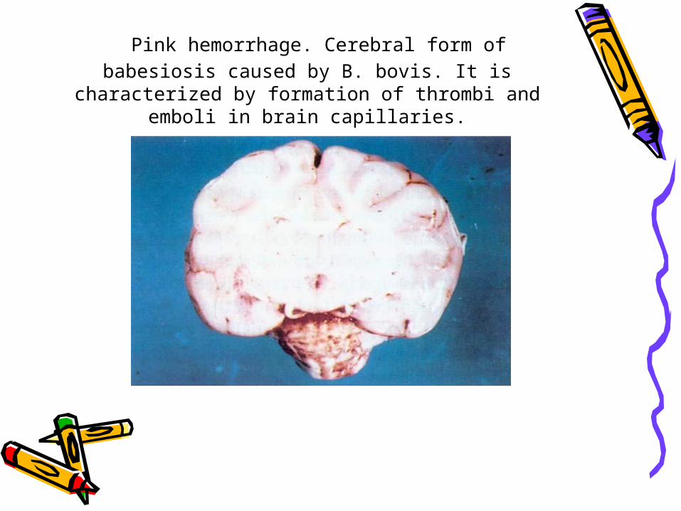
Pink hemorrhage. Cerebral form of babesiosis caused by B. bovis. It is characterized by formation
of thrombi and emboli in brain capillaries.
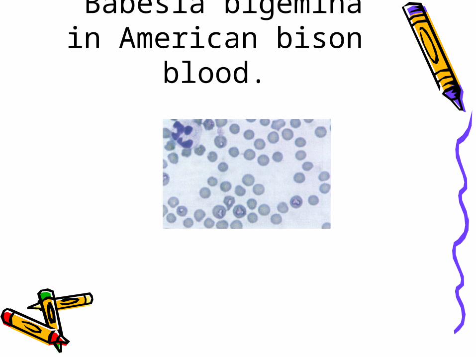
Babesia bigemina in American bison blood.