Embed Size (px)
Citation preview
RES. BULL. PL. PRoT. JAPAN No. 27 : 47-54 (1991)
Serological
Caused
Diagnosis of the Diseases
by Phytophthora spp.
Etsuo KIMISHIMA and Yoshinori KOBAYASHI
Research Division, Yokohama Plant Protection Station
Abstract : Naturally infected plants showing root and/or stem rot, gypsophira, white trumpet lily and pumpkin, were collected from the domestic growing fields. Diseased parts
of them reacted with Phytophthora antibodies by ELISA and DIBA. Aseptate hyphae with
fluorescence were observed by immunofluorescence staining method using with Phytophthora
antibodies. The pathogens isolated from gypsophila and white trumpet lily plants
were identified as P. nicotianae var. parasitica. The pathogen from pumpkin was identified
as P. capsici by the morphological and physiological characteristics. As a result, the
serological methods are considered to be very useful for the rapid and sensitive diagnosis of the diseases caused by Phytophthora spp.. Key words : Phytophthora, diagnosis, ELISA, immunofluorescence staining method, DIBA
Introduction
The identification of Phytophthora species is based on the morphological and physiological
characteristics, same as in the case of other fungi. However, it takes relatively long
time and needs skillful techniques to identify Phytophthora species. In plant quarantine,
quick diagnosis are requested. Therefore, the serological diagnostic methods have been
considered to be useful as rapid and sensitive techniques.
Naturally infected plants, gypsophila (Gypsophila elegans M.B.), white trumpet lily
(Lilium longlflorum THUNB.) and pumpkin (Cucurbita moschata DUCH.), were collected from
the domestic growing fields and used for this study. The diseased and healthy parts of
these plants were applied for the serological tests and the pathogens were identified. The
reliability of serological diagnosis for the disease by Phytophthora sp. was evaluated on this
experirnent. Parts of this work have been reported elsewhere (KIMISHIMA, et al., 1988a ;
KIMISHIMA, et al., 1989a).
Materials and Methods
Serological tests
It was tried to detect and diaguose the pathogen from the diseased plants directly by
the serological test. Rabbit antisera prepared for Phytophthora erythroseptica
PETHYBRIDGE and P. capsici LEONIAN (KIMISHIMA, et al., 1989b) were used. Three techniques
were used in this test : enzyme-1inked immunosorbent assay (ELISA) (CLARK and
ADAMS, 1977), the immunofiuorescent staining method (IFSM) and dot immunobinding
assay (DIBA) (HIBI and SAITO, 1985). The preparation of antigen samples and the procedures
of the serological techniques were performed as previously described (NISHIO, et al.,
1983 ; KIMISHIMA, et al., 1984 ; KIMISHIMA and KOBAYASHI, 1990a).
48 REs. BULL. PL. PROT. JAPAN No. 27
Isolation
Roots and/or stems were washed gently in running tap water. The discolored tissues
(0.5-1 cm in length) were plated onto a modified selective medium (MASAGO, et. al., 1977).
Plates were incubated at 25℃ for 2-4 days. The plugs cut from the advancing margin of
a colony growing on the selective medium were transferred to plates containing water agar
(WA). After a further incubation at 25℃ for 2-3 days, pure isolates of Phytophthora sp.
were obtained by the single hyphal tippings or single zoospore isolation.
ldentification of Phytophthora species
Representative isolates were used for describing the morphology on V-8 agar (200 ml
Campbell V-8 juice, 3.0 g calcium carbonate, 15g agar per liter). Isolates were identified
on the basis of mycelium characteristics, cardinal temperatures for vegetative growth,
morphology and dimensions of sporangia and production of sex organs. For the examines
of mycelium characteristics and cardinal temperatures, every isolates were cultured on V-
8 agar for 3-5days at various temperatures, ranging from 5 to 40℃. Sporangia were
produced on V-8 agar or eggplant (Solanum melongena L.), at room temperature (20-25℃)
for 5-7days. To study the production of sex organs, the isolates were grown on V-8 agar
to be paired individually with known A1 and A2 mating type cultures for 2 weeks at 25℃.
The identification was made by keys (WATERHOUSE, 1963 ; KATSURA, 1971) and previous
works (TASUGI and KUMAZAWA, 1938 ; WATERHOUSE and WATERSTON, 1964 ; STAMPS, 1985 ; KOBAYASHI, et al., 1987).
Pathogenicity tests
Representative isolates were selected and used for the following two pathogenicity
tests. Test a ; Mycelial mats together with V-8 agar were placed onto wounded and
unwounded leaves or fruit of each plant. The wounded plant surface was prepared by
injuring with a razor or a bundle of 20 needles. Inoculated plants were maintained in moist
condition at room temperature. The development of lesions was observed. Test b ; The
infested soil was prepared by mixing 400 ml of autoclaved soil with 10 ml of agar cultures.
The agar cultures were previously prepared by growing the test fungus on V-8 agar at 25℃
for 3-5days. The seedlings were planted in the infested and uninfested soils. These
inoculation experiments were carried out in a glasshouse (15-30℃) and observed for 3 weeks.
Results
Symptoms on plants
Gypsophila : In August 1986, root rot was found on gypsophila in Sizuoka prefecture.
Wilting of whole plant and discoloration of stem were also found on the affected plant.
Papillate sporangia were observed on the diseased parts of them.
White trumpet lily (Fig. 1-D) : In June 1987, stem rot was found on white trumpet lilies
at Okino-erabu island, Kagoshima prefecture. In infected plant the leaves and stem were
discolored, water-soaked, dark brown and rotted. Aseptate hyphae were observed in the
March, 1991 KIMISHIMA ・ KOBAYASHI : Serological Diagnosis 49
affected tissues, but no sporangia.
Pumpkin (Fig. 1-G) : In June 1988, root rot was found on pumpkins at Hitachi, Ibaraki
prefecture. The stem and root were water-soaked, discolored and rotted. Aseptate
hyphae and sporangia were observed in the affected tissue.
Serological tests
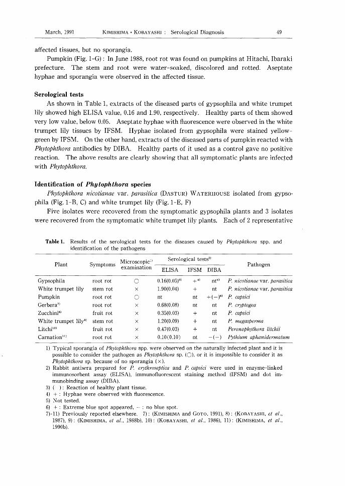
As shown in Table 1, extracts of the diseased parts of gypsophila and white trumpet
lily showed high ELISA value, 0.16 and 1.90, respectively. Healthy parts of them showed
very low value, below 0.05. Aseptate hyphae with fluorescence were observed in the white
trumpet lily tissues by IFSM. Hyphae isolated from gypsophila were stained yellow-
green by IFSM. On the other hand, extracts of the diseased parts of pumpkin reacted with
Phytophthora antibodies by DIBA. Healthy parts of it used as a control gave no positive
reaction. The above results are clearly showing that all symptomatic plants are infected
with Phytophthora.
ldentification of Phytophthora species
Phytophthora nicotianae var. parasitica (DASTUR) WATERHOUSE isolated from gypsophila
(Fig. 1-B, O and white trumpet lily (Fig. 1-E, F)
Five isolates were recovered from the symptomatic gypsophila plants and 3 isolates
were recovered from the symptomatic white trumpet lily plants. Each of 2 representative
Table 1. Results of the serological tests for the diseases caused
identification of the pathogens
by Phytophthora spp. and
Plant Symptoms Microscopic1) examination
Serological tests2)
ELISA IFSM DIBA Pathogen
Gypso phila
White trumpet lily
Pum pkin
Gerbera7)
Zucchini8)
White trumpet lily9)
Litchi10)
Carnation 11)
root rot
stem rot
root rot
root rot
fruit rot
stem rot
fruit rot
root rot
○
○ x
x
x
x
x
0.16(0.05)')
1.90(0.04)
nt
0.68(0.08)
0.35(0.03)
1.20(0.09)
0.47(0.03)
0.10(0.10)
+ ')
+ nt
nt
+ + + nt
nt5)
nt
+ ( - )6)
nt
nt
nt
nt
-(-)
P. nicotianae var, parasitica
P. nicotianae var, parasitica
P. capsici
P. cryptogea
P, capsici
P. megasperma
Peronophythora litchii
Pythium aphanidermatum
1) Typical sporangia of Phytophthora spp. were observed on the naturally infected plant and it is
possible to consider the pathogen as Phytophthora sp. (○), or it is impossible to consider it as Phytophthora sp. because of no sporangia (x).
2) Rabbit antisera prepared for P. erythroseptica and P. capsici were used in enzyme-linked
immunosorbent assay (ELISA), immunofluorescent staining method (IFSM) and dot im-munobinding assay (DIBA).
3) ( ) : Reaction of healthy plant tissue.
4) + : Hyphae were observed with fiuorescence. 5) Not tested.
6) + : Extreme blue spot appeared, - : no blue spot.
7)-11) Previously reported elsewhere. 7) : (KIMISHIMA and GoTO, 1991), 8) : (KOBAYASHI, et al.,
1987), 9) : (KIMISHIMA, et al., 1988b), 10) : (KOBAYASHI, et al., 1986), 11): (KIMISHIMA, et al.,
1990b).

50 RES. BULL. PL. PROT. JAPAN No. 27
Fig. 1. Symptoms of plant affected with Phytophthora sp. and morphological features of the pathogens. A-C, P, nicotiarlae var, parasitica : A, Symptoms of root rot on gypsophila
inoculated ; B, Papillate sporangia. C, Oogonium and amphigynous antheridium. D-F, P.
nicotianae var. parasitica : D, Symptoms of stem rot on white trumpet lily ; E, Papillate
sporangia ; F, Oogonium and amphigynous antheridium. G-1, P. c'apsici : G, Symptoms of
root rot on pumpkin ; H, Papillate sporangia ; I, Oogonium and amphigynous antheridium.
Bars on the figures represent 30 pm.
March, 1991 KIMISHIMA ・ KOBAYASHI Serological Diagnosis 51
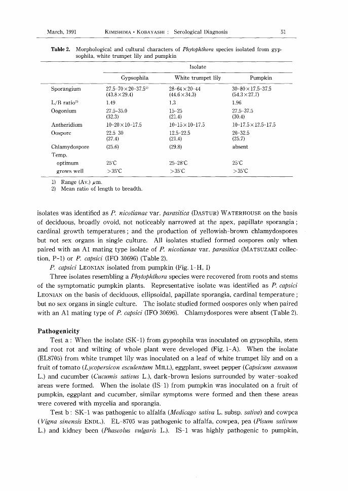
Table 2. Morphological and cultural characters of Phytophthora species isolated from gypsophira,
white trumpet lily and pumpkin
Isolate
Gypso phila White trumpet lily Pumpkin
Sporangium
L/B rati02)
Oogonium
Antheridium
Oospore
Chlamydospore
Temp.
optimum
grows well
27.5-70 x 20-37.5*)
(43.8 x 29.4)
1.49
27.5-35.0 (32.3)
10-20 x 10-17.5
22.5-30 (27.4)
(25.6)
25'C
> 35'C
28-64 x 20-44 (44.6 x 34.3)
1.3
15-25 (21.4)
10-15 x 10-17.5
12.5-22.5 (21.4)
(29.8)
25-28'C
> 35'C
30-80 x 17.5-37.5 (54.3 x 27.7)
1.96
27.5-37.5 (30.4)
10-17.5 x 12.5-17.5
20-32.5 (25.7)
absent
25'C
> 35'C
1)
2)
Range (Av.) pm. Mean ratio of length to breadth.
isolates was identified as P. nicotianae var. parasitica (DASTUR) WATERHOUSE On the basis
of deciduous, broadly ovoid, not noticeably narrowed at the apex, papillate sporangia ;
cardinal growth temperatures ; and the production of yellowish-brown chlamydospores
but not sex organs in single culture. All isolates studied formed oospores only when
paired with an A1 mating type isolate of P. nicotianae var. parasitica (MATSUZAKI collection
P-1) or P. capsici (IFO 30696) (Table 2).
P. capsici LEONIAN isolated from pumpkin (Fig. 1-H, I)
Three isolates resembling a Phytophthora species were recovered from roots and stems
of the symptomatic pumpkin plants. Representative isolate was identified as P. capsici
LEONIAN On the basis of deciduous, ellipsoidal, papillate sporangia, cardinal temperature ;
but no sex organs in single culture. The isolate studied formed oospores only when paired
with an A1 mating type of P. capsici (IFO 30696). Chlamydospores were absent (Table 2).
Pathogenicity
Test a : When the isolate (SK-1) from gypsophila was inoculated on gypsophila, stem
and root rot and wilting of whole plant were developed (Fig. 1-A). When the isolate
(EL8705) from white trumpet lily was inoculated on a leaf of white trumpet lily and on a
fruit of tomato (Lycopersicon esculentum MILL), eggplant, sweet pepper (Capsicum annuum
L.) and cucumber (Cucumis sativus L.), dark-brown lesions surrounded by water-soaked
areas were formed. When the isolate (IS-1) from pumpkin was inoculated on a fruit of
pumpkin, eggplant and cucumber, similar symptoms were formed and then these areas
were covered with mycelia and sporangia.
Test b : SK-1 was pathogenic to alfalfa (Medicago sativa L. subsp. sativa) and cowpea
( Vigna sinensis ENDL.). EL-8705 was pathogenic to alfalfa, cowpea, pea (Pisum sativum
L.) and kidney been (Phaseolus vulgaris L.). IS-1 was highly pathogenic to pumpkin,

52 REs. BULL. PL. PRoT. JAPAN No. 27
cucumber, sweet pepper and tomato. Root rot and damping-off were found on all affected
plants by the unwounded inoculation.
No symptom was found on the noninoculated plants. The causal fungi were consist-
ently reisolated from the diseased plants.
Discussion
It is known there are various methods to diaguose plant disease. The diagnosis of
fungal diseases is generally carried out by microscopically studying the morphology of its
mycelium, fruiting structures and spores. In case of Phytophthora diseases, if the typical
sporangia would not be confirmed by microscopic examination, it is very hard to decide the
pathogen to be Phytophthora sp.. It is said that the serological diagnosis is one of the rapid
and sensitive means, especially in plant virus diseases. We have prepared the antisera for
Phytophthora erythroseptica and P. capsici showing genus specific reactivities.
In this paper, it is examined about the reliability of serological test to diagnose the
Phytophthora diseases. Results of serological tests (ELISA, IFSM, DIBA) show that the
diseased plants, gypsophila, white trumpet lily and pumpkin showing root rot and stem rot,
were infected with Phytophthore species. Then, those pathogens were identified as P.
nicotianae var. parasitica for the former two plants and P. capsici for the last one by their
morphological and physiological characteristics (Table 2). Similar results have been
shown on gerbera (KIMISHIMA and GoTO, 1991), zucchini (KOBAYASHI, et al., 1987), white
trumpet lily (KIMISHIMA, et al., 1988b) and litchi (KOBAYASHI, et al., 1986) (Table 1).
However, carnation showing root rot caused by Pythium aphanidermatum did not reacted
with Phytophthora antibodies by ELISA and DIBA (KIMISHIMA. et al. 1990b).
Serological methods described here were found to be able to diagnose the diseases
caused by Phytophthora spp. or Peronophythora sp.. And they are considered to be very
useful as rapid and sensitive means for diagnosis of the Phytophthora diseases.
Acknowledgments
We would like to thank Dr. Takeshi NrsHro, plant protection division, for his valuable
advice. Thanks are also due to Mr. Masafumi MATSUZAKI, Saga agricultural experiment
station, for donation of the Phytophthora type cultures, and Mr. Makoto FUKUHARA, Daiichi
Seed Co., Ltd., for his aid in field sampling of symptomatic white trumpet lilies. We
are grateful to Mr. Masaaki GoTo, Yokohama plant protection station and Mr. H.A. van
KESTEREN, plant protection service, Wageningen, for critical reading of the manuscript.
Literature cited
CLARK, M.F. and A.N.ADAMS (1977) Characteristics of the microplate method of enzyme-linked immunosorbent assay for the detection of plant viruses. J. gen. Virol, 34 : 475-483.
HIBI.T. and Y.SAITO, (1985) A Dot Immunobinding Assay for the Detection of Tobacco Mosaic Virus in Infected Tissues. Ibid. 66 : 1191-1194.
KATSURA, K. (1971) Phytophthora Diseases of Plants - Theory and Practice. Seibundo Shinkosha,
Tokyo. pp 98-99.
KIMISHIMA, E., T. NrsHro, M. TAKAYAMA and N. NAGAO (1984) Studies on Serological Detection and
M arch , 1991 KIMISHIMA ・ KOBAYASHI : Serological Diagnosis 53
ldentification Methods for Species of Phytophthora. 111. Detection of Phytophthora syringae in
Plant Tissues by means of Enzyme-linked Immunosorbent Assay (ELISA). Res. Bull. Pl. Prot.
Japan 20 : 1-6.
KIMISHIMA,E., Y. KOBAYASHI and T. NISHIO (1988a) Serological Diagnosis of Phytophthora Diseases.
Ann. Phytopath. Soc. Japan 54 : 68 (Abstr.).
KIMISHIMA, E., Y. KOBAYASHI and T. NISHIO (1988b) Phytophthora megasperma DRECHSLER Newly Isolated from White Trumpet Lily. Ibid. 54 : 427-435.
KIMISHIMA, E., Y.KOBAYASHI and T. NISHIO (1989a) Serological studies on Phytophthora for devel-
oping rapid detection and identification methods. Phytophthora Newsletter 15 : 12-13.
KIMISHIMA,E., Y. KOBAYASHI and T. NISHIO (1989b) Serological Reactivities of Antisera Prepared
for Four Phytophthora spp.. Res. Bull. Pl. Prot. Japan 25 : 1-6.
KIMISHIMA, E. and Y. KOBAYASHI (1990a) Use of Dot Immunobinding Assay (DIBA) and Immunoen-
zymatic Staining Method (IESM) for Rapid Detection of Phytophthora spp.. Ibid. 26 : 7-13.
KIMISHIMA, E., Y.KOBAYASHI and T. NISHIO (1990b) The occurrence of root rot caused by Pythium
spp. in carnation. Ann. Phytopath. Soc. Japan 56 : 148 (Abstr.).
KIMISHIMA,E. and M. GOTO (1991) Root rot of Gerbera caused by Phytophthora cmptogea. Ibid. 57 :
77 (Abstr.).
KOBAYASHI, Y., E. KIMISHIMA. T. NISHIO and N. NAGAO (1986) Peronophythora litchii isolated from
litchi fruit imported from Taiwan. Res. Bull. Pl. Prot. Japan 22 : 55-60.
KOBAYASHI. Y., J. YASUTOMO and T. NISHIO (1987) Phytophthora capsici isolated from zucchini fruit
imported from the United States. Ibid. 23 : 45-48.
MASAGO, H., M. YOSHIKAWA, M. FUKADA and N. NAKANISHI (1977) Selective inhibition of Pythium
spp. on a medium for direct isolation of Phytophthora spp. from soils and plants. Phyto-
pathology 67 : 425-428.
NrsHro, T., E. KIMISHIMA, M.TAKAYAMA and T. SUETSUGU (1983) Studies on Serological Detection
and Identification Methods for Species of Phytophthora II. Use of Fluorescent Antibodies for
Detection of Phytophthora syringae. Res. Bull. Pl. Prot. Japan 19 : 55-62.
STAMPS, D.J. (1985) C.M.1. Descriptions of pathogenic fungi and bacteria. No. 836.
TASUGI, H. and M.KUMAZAWA (1938) Phytophthora rot of lily. Journal of Imp. Arg. Exp. Sta. 3 :
207-241.
WATERHOUSE, G.M. (1963) Key to the species of Phytophthora de BARY. C.M.1. Mycol. Pap. 92 : 1-
22.
WATERHOUSE G.M. and J.M. WATERSTON (1964) C.M.1. Descriptions of pathogenic fungi and bacteria. No. 35.

54 REs.BuLL.PL.PRoT.JAPAN No.27
和 文 摘 要
疫病の血清診断
君島悦夫・小林慶範 横浜植物防疫所調査研究部
根腐れ及び茎腐症状を示すカスミソウ,テッポ
ウユリ及びカボチャを国内の栽培圃場から採集し
た。これらの病植物を用い,疫病菌抗血清を用い
た血清診断を試みると共に,病原菌の分離・同定
を行い,血清診断の結果と比較した。診断方法は
ELISA,蛍光抗体法及びDIBAによった。試験の結果,病植物はELISA及びDIBAで疫病菌抗血清と顕著な反応を示した。また,蛍光抗体法によ
り無隔の菌糸が蛍光色に染色されて観察された。
形態的及び生理的特徴から,カスミソウ及びテッ
ポウユリからの分離菌は双方ともPhytophthora
nicotianae var. parasitica(DASTUR)
WATERHOUSEと同定され,カボチャからの分離菌は Phytophthora capsici LEONIAN と同定され
た。本試験の結果,疫病の診断に抗血清を用いる
方法は迅速で精度の高い方法と考えられた。