Embed Size (px)
Citation preview
i
Discovery of new chemicals with anthelmintic
activity against the barber’s pole worm and
other parasitic nematodes
Yaqing Jiao
ORCID ID: 0000-0002-7621-4166
Submitted in fulfilment of the requirements of the degree of
Doctor of Philosophy
November 2018
Faculty of Veterinary and Agricultural Sciences
The University of Melbourne
ii
SUMMARY
Parasitic nematodes cause a substantial disease burden on humans and animals worldwide.
A review of the literature (Chapter 1) showed that, on one hand, neglected tropical diseases
caused by parasitic nematodes have a devastating, long-term impact on human health; on the
other hand, gastrointestinal nematodes are a major constraint to the livestock industries,
causing subclinical infections and diseases in animals and leading to a substantial reduction
in meat, milk and fibre production. Currently, anthelmintic treatment remains the mainstay of
controlling parasitic nematode infections. However, the massive and widespread resistance to
the limited number of commercial anthelmintics, particularly in the veterinary and
agricultural contexts, demonstrates an urgency to discover new and effective anthelmintics to
sustain the economic and health benefits from the application of anthelmintics. Thus, the key
focus of this thesis was to discover new chemical entities and/or known drugs with
anthelmintic activities against Haemonchus contortus and/or other socioeconomically
important parasitic nematodes for subsequent development. Whole worm-based phenotypic
screening assays were employed, compound collections were obtained via product-
development-partnerships and/or collaborators, and active compounds were assessed for their
potential as anthelmintic candidates.
In this thesis, one new chemical entity (designated SN00797439), two human kinase
inhibitors (SNS-032 and AG-1295), 14 AG-1295 (tetrahydroquinoxaline) analogues, one
insecticide (tolfenpyrad) and two tolfenpyrad (pyrazole-5-carboxamide) derivatives (a-15 and
a-17) with anthelmintic activity in vitro were discovered following the screening of a total of
15,333 chemicals from five distinct compound collections against H. contortus. In Chapter 2,
a new chemical entity, SN00797439, was identified with activity against a range of parasitic
nematodes, including H. contortus, Ancylostoma ceylanicum, Brugia malayi, Dirofilaria
immitis and/or Trichuris muris in vitro, offering a novel, lead-like scaffold for the
development of a relatively broad-spectrum anthelmintic.
In Chapter 3, two human kinase inhibitors under pharmaceutical development, SNS-032
(piperidinecarboxamide) and AG-1295 (quinoxaline), were identified to have inhibitory
activity on the motility and development of parasitic larvae of H. contortus in vitro. AG-1295
had limited cytotoxicity against a normal mammalian epithelial cell line (designated
MCF10A).
In Chapters 4 and 5, three pyrazole-5-carboxamides (tolfenpyrad, a-15 and a-17) were
shown to possess significant inhibitory effects on H. contortus without detectable toxicity on
iii
a human neonatal foreskin fibroblast (NFF) cell line in vitro. All three of these chemicals
were shown to inhibit the oxygen consumption in H. contortus larvae, a finding that was
consistent with the known, specific inhibition of complex I of the respiratory electron
transport chain by selected pyrazole-5-carboxamides in arthropods. The evaluation of these
hit compounds using various technologies employed in parasitology, drug discovery,
chemistry, histology, toxicology, molecular biology and bioinformatics should offer data to
support their potential as leads for future drug development and to facilitate the exploration of
their mode(s) of action in this and related nematodes.
Encouraged by the findings in Chapter 3 and the detection of a non-wildtype phenotype in
treated worms in vitro, Chapter 6 investigated the activities of 14 additional
tetrahydroquinoxaline (AG-1295) analogues on H. contortus. Qualitative and quantitative
assessments of larval motility, development and morphological alterations showed that these
14 chemicals all affected the viability of parasitic larvae and, interestingly, induced an
eviscerated larval phenotype and led to cuticular damage and/or stunted growth in in vitro H.
contortus.
Taken together, Chapters 2 to 6 identified a series of 20 hit compounds, some of which
have selectivity against H. contortus compared with selected human cell lines tested. In
Chapter 7, the research achievements are summarised, and the next steps to be pursued in
future research are outlined, including (i) the chemical optimisation of representative
chemicals via structure-activity relationship (SAR) evaluations; (ii) assessment of the breadth
of spectrum of anthelmintic activity on other parasitic nematodes, such as other strongyloids,
ascaridoids, enoplids and filarioids; (iii) detailed investigations of the absorption, distribution,
metabolism, excretion and toxicity (ADMET) of optimised chemicals with broad nematocidal
or nematostatic activity; (iv) establishment of the modes of action of lead candidates. The
findings from the thesis are then put into a broad context and discussed. In conclusion, the
present thesis contributes to the fields of parasitology and anthelmintic discovery by
identifying compounds with in vitro anthelmintic activity that represent sound starting points
for ‘lead’ discovery.
iv
DECLARATION
The work described in the thesis was performed in the Faculty of Veterinary and Agricultural
Sciences of the University of Melbourne between September 2015 and November 2018. The
scientific work was performed by the author, with the exception of the assistance which has
been specifically acknowledged. The thesis is less than 100,000 words in length, exclusive of
tables, figures, references and appendices. No part of this thesis has been submitted for any
other degree or diploma.
…………………………
Yaqing Jiao
November 2018
v
ACKNOWLEDGEMENTS
I would like to express my deepest gratitude to my supervisor, Professor Robin B. Gasser,
for his invaluable guidance and generous support during my PhD candidature. He is a very
knowledgeable, highly supportive and well-organized scientist, who is dedicated to science. I
have been proud to be his student. I have to say, without him, I would not be able to achieve
the current insights that I have had into the biology of parasites. As a starting point of my
scientific research career, Robin helped me construct a solid foundation. I will do my best to
become an expert researcher in the “world of science”.
I would also like to thank my co-supervisors, Dr Sarah Preston and A/Professor Abdul
Jabbar, for their patient guidance, valuable discussions and constant encouragement. It is
Sarah who guided me in the field of drug discovery. During the past years, I benefited from
her, not only in science, but also personally. Her diligent attitude, comprehensive work and
good communication skills have helped me along the way. It is Abdul who always gave me
generous support and invaluable suggestions. Particularly the questions that he raised in
seminars, discussions and meetings always inspired me. I express my deepest gratitude to
their whole-hearted encouragement when I faced difficulties in developing my project.
I also would like to thank the members of the Gasser Laboratory, A/Prof Jose Garcia-
Bustos, Dr Anson V. Kohler, Dr Neil D. Young, Dr Clare Anstead, Dr Pasi Korhonen, Dr
Andreas J. Stroehlein, Dr Tao Wang, Mr Ross S. Hall, Mr Daxi Wang, Mr Guangxu Ma, Ms
Dilrukshi Herath, Ms Yan Zhang, and Prof Rebecca Traub. I consider myself so fortunate to
have met these happy, sincere, positive and kind-hearted people who made my study life so
bright.
I would also like to thank numerous collaborators including Prof Jonathan Baell, Prof
Andreas Hofmann, Prof Jennifer Keiser, Prof Qingmin Wang, A/Prof Kaylene J. Simpson,
Prof Ray M. Kaplan, Dr Gillian M. Fisher, Dr Michael J. Palmer, Dr Benoît Laleu, Dr Jeremy
N. Burrows, Dr Timothy N. C. Wells, Dr Paul Willis, Dr Dana Hutchinson, Dr Simon
Crawford and Dr Moana M. Simpson. I am also grateful to the China Scholarship Council
and The University of Melbourne Research for scholarship support. With these scholarships,
I was able to gain such skills and experience as a foundation for my future career.
Finally, I would like to thank my parents and my little sister for always being beside my
side. Their love and support make my life warm, and always motivated me to become a better
person. Thanks to my friends for sharing my feelings, emotions and experiences.
vi
PREFACE AND DISSEMINATION OF RESEARCH FINDINGS
Scientific papers published or submitted by the author in collaboration with supervisors
and other colleagues are listed in the following:
Peer-reviewed articles published in international scientific journals:
Preston, S.*, Jiao, Y.*, Baell, J.B., Keiser, J., Crawford, S., Koehler, A.V., Wang, T.,
Simpson, M.M., Kaplan, R.M., Cowley, K.J., Simpson, K.J., Hofmann, A., Jabbar, A.,
Gasser, R.B., 2017. Screening of the ‘Open Scaffolds’ collection from Compounds Australia
identifies a new chemical entity with selective anthelmintic activities against different
developmental stages of the barber’s pole worm and other parasitic nematodes. International
Journal for Parasitology: Drugs and Drug Resistance 7, 286-294 (Chapter 2). *Joint first
authorship.
Jiao, Y., Preston, S., Koehler, A.V., Stroehlein, A.J., Chang, B. C. H., Simpson, K.J., Palmer,
M., Willis, P., Wells, T.N.C., Jabbar, A., Gasser, R.B., 2017. Screening of the ‘Stasis Box’
identifies two kinase inhibitors under pharmaceutical development with activity against
Haemonchus contortus. Parasites & Vectors 10, 323 (Chapter 3).
Preston, S.*, Jiao, Y.*, Jabbar, A., McGee, S., Laleu, B., Willis, P., Wells, T.N.C., Gasser,
R.B., 2016. Screening of the ‘Pathogen Box’ identifies an approved pesticide with major
anthelmintic activity against the barber’s pole worm. International Journal for Parasitology:
Drugs and Drug Resistance 6, 329-334 (Chapter 4). *Joint first authorship.
Jiao, Y., Preston, S., Song, H., Jabbar, A., Liu, Y., Baell, J., Hofmann, A., Hutchinson, D.,
Wang, T., Koehler, A.V., Fisher, G.M., Andrews, K.T., Laleu, B., Palmer, M. J., Burrows,
J.N., Wells, T.N.C., Wang, Q., Gasser, R.B., 2017. Assessing the anthelmintic activity of
novel pyrazole-5-carboxamide derivatives against Haemonchus contortus. Parasites &
Vectors 10, 272 (Chapter 5).
Jiao, Y., Preston, S., Garcia-Bustos, J., Baell, J., Ventura, S., Le, T., McNamara, N., Nguyen,
N., Botteon, A., Skinner, C., Danne, J., Ellis, S., Koehler, A.V., Wang, T., Chang, B.C.H.,
vii
Hofmann, A., Jabbar, A., Gasser, R.B., 2018. Tetrahydroquinoxalines induce an lethal
evisceration phenotype in Haemonchus contortus larvae in vitro. International Journal for
Parasitology: Drugs and Drug Resistance 9, 59-71 (Chapter 6).
Conference proceedings and seminars given:
Jiao, Y., 2017. Discovery of pyrazole-5-carboxamide chemical class with anthelmintic
activity on Haemonchus contortus. Faculty of Veterinary and Agricultural Sciences, The
University of Melbourne, 2 February 2017 (PhD Confirmation Seminar).
Jiao, Y., 2017. Screening of the ‘Stasis Box’ identifies two kinase inhibitors under
pharmaceutical development with activity against Haemonchus contortus. Proceedings of
2017 FVAS Postgraduate Symposium, The University of Melbourne, 24-25 October 2017.
Jiao, Y., Preston, S., Baell, J.B., Keiser, J., Crawford, S., Koehler, A.V., Wang, T., Simpson,
M.M., Kaplan, R.M., Cowley, K.J., Simpson, K.J., Hofmann, A., Jabbar, A., Gasser, R.B.,
2018. Screening of the ‘Open Scaffolds’ collection from Compounds Australia identifies a
new chemical entity with selective anthelmintic activities against different developmental
stages of the barber’s pole worm and other parasitic nematodes. Proceedings of 2018
Australian Society for Parasitology Conference, Melbourne, Australia, 24-27 September
2018.
Jiao, Y., 2018. Discovery of new chemicals with anthelmintic activity against the barber’s
pole worm and/or other parasitic nematodes. Faculty of Veterinary and Agricultural Sciences,
The University of Melbourne, 20 September 2018 (PhD Completion Seminar).
viii
TABLE OF CONTENTS
SUMMARY .............................................................................................................................. ii
DECLARATION..................................................................................................................... iv
ACKNOWLEDGEMENTS .................................................................................................... v
PREFACE AND DISSEMINATION OF RESEARCH FINDINGS .................................. vi
Chapter 1 - Literature review ................................................................................................. 1
1.1 Introduction .......................................................................................................................... 1
1.2 Parasitic nematodes of socioeconomic importance ............................................................. 2
1.2.1 Parasitic nematodes of human health importance ......................................................... 2
1.2.2 Parasitic nematodes of veterinary importance .............................................................. 3
1.3 Treatment and control of parasitic nematodes ..................................................................... 5
1.3.1 Anthelmintic treatment of parasitic nematode infections ............................................. 5
1.3.2 Non-chemotherapeutic control of parasitic nematode infections .................................. 7
1.4 Anthelmintic resistance in parasitic nematodes ................................................................. 12
1.5 Anthelmintic drug discovery.............................................................................................. 15
1.5.1 Mechanism-based screening ....................................................................................... 16
1.5.2 Whole-worm screening ............................................................................................... 17
1.6 Selection of compound collections for anthelmintic drug discovery................................. 18
1.6.1 Discovery of new chemical entities ............................................................................ 19
1.6.2 Drug repurposing......................................................................................................... 20
1.7 The relevance of using advanced molecular and informatic technologies to assist
anthelmintic discovery ............................................................................................................. 21
1.8 Conclusions and research aims .......................................................................................... 24
1.9 References .......................................................................................................................... 25
Chapter 2 - Identification of a new chemical entity with selective anthelmintic activities
against different developmental stages of Haemonchus contortus and other parasitic
nematodes ............................................................................................................................... 54
2.1 Introduction ........................................................................................................................ 55
2.2 Materials and methods ....................................................................................................... 56
2.2.1 The compound library, and the selection and preparation of chemicals for screening
.............................................................................................................................................. 56
2.2.2 Screening and evaluation of the effects of compounds on H. contortus ..................... 57
2.2.3 Scanning electron microscopy .................................................................................... 59
2.2.4 Evaluating compound activity on filarial worms ........................................................ 59
ix
2.2.5 Assessing compound activity on adult A. ceylanicum and T. muris L1s .................... 61
2.2.6 Assessing compound cytotoxicity and selectivity ....................................................... 61
2.3 Results and discussion ....................................................................................................... 62
2.3.1 The identification of compound SN00797439 with activity against parasitic stages of
H. contortus .......................................................................................................................... 62
2.3.2 Compound SN00797439 also has inhibitory activity on the motility of different
developmental stages of other parasitic nematodes ............................................................. 63
2.3.3 Cytotoxicity and selectivity of SN00797439 .............................................................. 63
2.4 Conclusion ......................................................................................................................... 64
2.5 References .......................................................................................................................... 66
Chapter 3 - Identification of two kinase inhibitors under pharmaceutical development
with activity against Haemonchus contortus........................................................................ 77
3.1 Introduction ........................................................................................................................ 78
3.2 Methods.............................................................................................................................. 79
3.3 Results and discussion ....................................................................................................... 81
3.4 References .......................................................................................................................... 84
Chapter 4 - An approved pesticide with major anthelmintic activity against
Haemonchus contortus ........................................................................................................... 93
4.1 Introduction ........................................................................................................................ 94
4.2 Materials and methods ....................................................................................................... 95
4.2.1 Procurement of H. contortus ....................................................................................... 95
4.2.2 Screening of compounds ............................................................................................. 96
4.2.3 Dose-response assessments of active compounds on xL3 and L4 motility, and L4
growth and development ...................................................................................................... 97
4.2.4 Measurement of basal oxygen consumption ............................................................... 98
4.3 Results ................................................................................................................................ 98
4.4 Discussion .......................................................................................................................... 99
4.5 References ........................................................................................................................ 102
Chapter 5 - Assessing the anthelmintic activity of pyrazole-5-carboxamide derivatives
against Haemonchus contortus ............................................................................................ 110
5.1 Introduction ...................................................................................................................... 111
5.2 Materials and methods ..................................................................................................... 112
5.2.1 Procurement of H. contortus ..................................................................................... 112
5.2.2 Pyrazole-5-carboxamide compounds ........................................................................ 112
5.2.3 Screening of chemicals, inhibitory concentrations and cytotoxicity assessment ...... 113
5.2.4 Measuring the effects of compounds on respiration ................................................. 114
x
5.3 Results .............................................................................................................................. 115
5.4 Discussion ........................................................................................................................ 116
5.5 References ........................................................................................................................ 118
Chapter 6 - Tetrahydroquinoxalines induce a lethal evisceration phenotype in
Haemonchus contortus in vitro ........................................................................................... 126
6.1. Introduction ..................................................................................................................... 127
6.2 Materials and methods ..................................................................................................... 128
6.2.1 Procurement of H. contortus ..................................................................................... 128
6.2.2 AG-1295 analogues ................................................................................................... 129
6.2.3 Compound screening ................................................................................................. 129
6.2.4 Dose-response assessments of active compounds on xL3 and L4 motility, and L4
development ....................................................................................................................... 130
6.2.5 Assessment of cytotoxicity ....................................................................................... 130
6.2.6 Morphological examination of the eviscerated (Evi) phenotype induced by incubation
with individual test compounds.......................................................................................... 131
6.2.7 Scanning electron microscopy (SEM)....................................................................... 131
6.2.8 Transmission electron microscopy (TEM) ................................................................ 132
6.2.9 Evaluation of the dynamics of the compound-induced Evi phenotype in xL3s........ 132
6.2.10 Assessment of the reversibility of effect of test compounds on xL3s ..................... 133
6.2.11 Influence of external osmotic pressure on compound-induced Evi phenotype in xL3s
............................................................................................................................................ 133
6.3 Results .............................................................................................................................. 134
6.3.1 Inhibitory effects of AG-1295 analogues on xL3 motility, L4 motility and L4
development ....................................................................................................................... 134
6.3.2 Cytotoxicity of AG-1295 analogues on MCF10A cells ............................................ 135
6.3.3 Microscopic characterisation of the Evi phenotype in xL3s exposed to test
compounds ......................................................................................................................... 135
6.3.4 Determining the optimum compound concentrations to induce the Evi phenotype in
xL3s .................................................................................................................................... 137
6.3.5 Dynamics of evisceration in xL3s ............................................................................. 137
6.3.6 Reversibility of compound-induced effects and sensitivity time-window ................ 138
6.3.7 Influence of external osmotic pressure on the evisceration of xL3s ......................... 139
6.4 Discussion ........................................................................................................................ 139
6.5 References ........................................................................................................................ 143
Chapter 7 - General discussion ........................................................................................... 159
7.1. Screening technologies ................................................................................................... 159
7.2. Considerations regarding compound libraries ................................................................ 161
xi
7.3. Evaluation of ‘hit’ compounds........................................................................................ 162
7.4. Potential of identified compounds as ‘leads’ in future anthelmintic development ......... 163
7.5. Future work toward anthelmintic development .............................................................. 166
7.6. Concluding remarks ........................................................................................................ 168
7.7 References ........................................................................................................................ 169
LIST OF SUPPLEMENTARY FILES .............................................................................. 178
1
Chapter 1 - Literature review
1.1 Introduction
Parasitic nematodes cause diseases that adversely impact on human health and livestock
production (Fenwick, 2012; Fitzpatrick, 2013; Holden-Dye and Walker, 2014). On one hand,
more than one billion people in the world, particularly in underprivileged developing
countries, suffer from neglected tropical diseases, such as hookworm disease, lymphatic
filariasis and onchocerciasis (Hotez et al., 2007; Fenwick, 2012; Hotez et al., 2016a). On the
other hand, parasitic nematode infections severely constrain livestock production and
represent a major burden to livestock industries worldwide (McLeod, 1995, cf. Roeber et al.,
2013; Charlier et al., 2014; Sargison, 2016). Therefore, enormous health and economic
benefits can be gained from effectively controlling parasitic nematode infections and
associated diseases.
Focussing on economically important livestock production system, the control of parasitic
nematodes can be divided into strategies that use anthelmintics and those that do not (Waller,
2006; Sargison, 2012). In particular, anthelmintic treatment remains a dominant feature of the
control of parasitic nematode infections (Waller, 1993; Sargison, 2012; Preston et al., 2014).
However, the emergence of anthelmintic resistance to the current small numbers of
commercial anthelmintics is challenging the efficiency of this dominant control, emphasising
the need to discover new anthelmintics (Wolstenholme et al., 2004; Kaplan and
Vidyashankar, 2012; Miller et al., 2012).
With regard to anthelmintic discovery, two commonly used strategies are mechanism-
based screening and whole worm-based screening, with their individual advantages and
disadvantages that need to be considered (Geary et al., 1999; Geary et al., 2015). Besides the
screening strategies, the rational selection of compound collections and the application of
advanced -omics technologies for screening also play a key role in the early anthelmintic
discovery stage (Dandapani et al., 2012; Preston et al., 2016b).
The purpose of this literature review was to critically appraise the current literature on
parasitic nematodes, control of parasitic nematode infections, anthelmintic resistance and new
anthelmintic discovery, in order to identify knowledge gaps and then to define the research
aims of this thesis.
2
1.2 Parasitic nematodes of socioeconomic importance
Parasitic nematodes exert a heavy disease burden on human health and place a major
economic burden on livestock production (Fenwick, 2012; Fitzpatrick, 2013; Holden-Dye
and Walker, 2014). Based on various studies, neglected tropical diseases represent a burden
of 26 million disability-adjusted life years (DALYs) (Hotez et al., 2014; Hotez et al., 2016a).
The DALYs represent losses due to ill-health, disability and early death, which is a tool to
measure and compare disease burdens (Mathers et al., 2007). Neglected tropical diseases
including hookworm disease, ascariasis, trichuriasis, lymphatic filariasis and onchocerciasis,
caused by parasitic nematodes, represent a disease burden of 8.46 million DALYs (Hotez et
al., 2007; Hotez et al., 2016a).
1.2.1 Parasitic nematodes of human health importance
Hookworm disease, ascariasis and trichuriasis are three main soil-transmitted helminth
infections caused by intestinal nematodes (de Silva et al., 2003; Bethony et al., 2006).
Hookworm disease represents 3.23 million DALYs (Hotez et al., 2016a), which is mainly
caused by nematodes including Ancylostoma duodenale and Necator americanus (Crompton,
2000). The public health significance of hookworm disease relates to blood loss caused by
the worms’ feeding activity in the human gut, leading to the iron-deficiency anaemia
(Brooker et al., 2008; Smith and Brooker, 2010). The other two intestinal nematode diseases,
ascariasis and trichuriasis, represent 1.32 million and 0.64 million DALYs, respectively
(Hotez et al., 2016a). Ascariasis caused by Ascaris lumbricoides and trichuriasis caused by
Trichuris trichiura commonly co-infect people, together with hookworm infection (Bethony
et al., 2006), and mainly affect children, resulting in malnutrition, impaired growth, and
reduced physical fitness and intellectual development (WHO, 2005; Bethony et al., 2006).
The other two neglected tropical diseases, lymphatic filariasis and onchocerciasis, are
caused by filarial nematodes. For lymphatic filariasis (2.78 million DALYs), Brugia malayi,
Brugia timori and Wuchereria bancrofti are the causative agents in humans, with worms
developing in the lymphatic systems and microfilariae circulating in the blood system
(Molyneux, 2010; Taylor et al., 2010). The invasion and damage of the lymphatic system by
filarial worms accounts for a chronic suppression of host immunity, causing permanent
disability in patients (Taylor et al., 2010). It is also common for lymphatic filariasis to co-
occur with other diseases, in which the suppressive immunomodulatory mechanisms caused
by lymphatic filariasis have been identified to modulate protective immune responses for
3
malaria and tuberculosis (Babu et al., 2009a; Babu et al., 2009b; Metenou et al., 2009).
Onchocerciasis (0.49 million DALYs), which is linked to blindness, is caused by another
filarial nematode, Onchocerca volvulus, with adult worms occupying the subcutaneous layers
of the skin and deep tissues, and microfilariae migrating to the skin and the eyes (Burnham,
2010; Taylor et al., 2010).
Neglected tropical diseases commonly occur in underprivileged areas or countries, where
affected people live on less than 2 dollars per day (Bethony et al., 2006; Hotez et al., 2007;
Houweling et al., 2016; Stolk et al., 2016). The socioeconomic inequality has resulted in a
lack of commercial incentive for solutions, such that there is a 13-fold greater probability of a
drug being brought to market for the treatment of nervous system disorders or cancer than for
a neglected tropical disease (Trouiller et al., 2002), even though the latter diseases rank
amongst the world’s greatest global health problems (Hotez et al., 2016b; Houweling et al.,
2016; Stolk et al., 2016).
1.2.2 Parasitic nematodes of veterinary importance
Parasitic nematodes are amongst the major constraints in livestock production systems
(Fitzpatrick, 2013; McRae et al., 2015; Sargison, 2016). Particularly gastrointestinal
nematode infections cause reduced meat, milk and fibre production and even the death of
animals (Charlier et al., 2014; Preston et al., 2014), leading to annual economic losses
estimated at billions of dollars globally (cf. Roeber et al., 2013). Key gastrointestinal
nematodes of small ruminants responsible for the substantial economic losses include
Haemonchus contortus, Teladorsagia circumcincta and Trichostrongylus species (see Zajac,
2006; Cantacessi et al., 2012; Roeber et al., 2013). Other gastrointestinal nematodes include
Cooperia curticei, Nematodirus spathiger, N. fillicollis, N. abnormalis, Oesophagostomum
venulosum, Bunostomum trigonocephalum and Chabertia ovina (see Anderson, 2000; Zajac,
2006). Although some species can have relatively low pathogenicity alone, they can
contribute substantially to the overall problem of parasitic gastroenteritis in grazing small
ruminants in situations with mixed infections (Craig, 1986).
Teladorsagia circumcincta and Trichostrongylus spp. are prevalent in cool temperature
regions, including parts of Europe, Scandinavia, Asia, New Zealand, America and Australia,
with temperate climates being favourable for larval development (O'Connor et al., 2006). The
pre-patent period for T. circumcincta is 21 days; adult worms are relatively short lived,
surviving in their hosts for only a few months. Teladorsagia circumcincta inhabits the gastric
4
glands of the abomasum but does not feed on blood (Anderson et al., 1985). The main
pathogenic effects are caused by its larval stages (Zajac, 2006; Cantacessi et al., 2012; Roeber
et al., 2013). Larvae form nodules in the abomasal mucosa so as to damage parietal cells
which secrete hydrochloric acid to stimulate the conversion of pepsinogen to pepsin
(Anderson et al., 1985; McKellar, 1993). Pepsin is essential for protein digestion; thus,
heavily infected ruminants can develop anaemia, diarrhoea and death (Holmes, 1985).
Trichostrongylus colubriformis, T. rugatus and T. vitrinus are the three common
Trichostrongylus species (Beveridge et al., 1989). Trichostronglus vitrinus is considered to be
the most pathogenic one; the main pathogenic effects are caused by the exsheathed third-
stage larvae (xL3s) which induce villus atrophy by disrupting the integrity of the intestinal
epithelium and causing plasma loss from the gut (Beveridge et al., 1989). Trichostronglus
colubriformis is considered to be the intermediate pathogenic worm; heavy infections can
cause the enteritis, hypoalbuminaemia and hypoproteinaemia (Barker and Titchen, 1982).
Trichostronglus rugatus is considered to be the least pathogenic worm (Beveridge et al.,
1989).
Haemonchus contortus is recognized as the most highly pathogenic nematode, mainly in
tropical and subtropical areas (Miller and Horohov, 2006; O'Connor et al., 2006). Tropical
areas include parts of south-east Asia, southern India, central Africa and America, and
northern South America, and subtropical areas include eastern Australia, southern Africa,
southern North America, South America, and south-east China (cf. O'Connor et al., 2006;
Getachew et al., 2007). Various studies have showed that H. contortus is not suited to live in
cold climates, because of the negative effects of low temperature on the survival and
development of the eggs and larvae, and the migratory activities of free-living larvae (Rose,
1963; O'Connor et al., 2006). Individual females of H. contortus produce thousands of eggs
per day, which can contaminate pastures (Coyne et al., 1991). In sheep, the pre-patent period
is 17–21 days, and the life span of adult worms is a few months (Courtney et al., 1983).
Haemonchus contortus inhabits the mucosa of the abomasum (Parkins and Holmes, 1989).
The main pathogenic effect is caused by the fourth-stage larvae (L4s) and adults, which both
feed on blood, causing severe anaemia (Baker et al., 1959). Acute infection with large
numbers of H. contortus usually causes weight loss, with the clinical signs of tanned faeces,
anaemia, oedema, fatigue and/or sudden death (Zajac, 2006; Cantacessi et al., 2012; Roeber
et al., 2013). Chronic infection decreases small ruminants’ food intake, which causes
significant weight loss (Kassai, 1999; Taylor et al., 2016).
H. contortus is amongst the experimentally most tractable parasites because of its
5
fecundity and ease of production (Laing et al., 2013; Geary, 2016). Haemonchus contortus
has a direct and rapid life cycle which can be divided into free-living and parasitic phases
(Fig. 1.1). In the free-living phase, the eggs hatch to first-stage larvae (L1s) in faeces, then
moult through second-stage larvae (L2s) to third-stage larvae (L3s); L3s are infective, and
migrate from faeces on to pasture, and are then ingested by grazing ruminants, after which
the parasitic phase starts. In the parasitic phase, L3s exsheath predominantly in the
rumen/reticulum and moult to fourth-stage larvae (L4s) in the abomasum; L4s then develop
to adults, and female adults lay eggs in the abomasum, representing the end of the cycle
(Veglia, 1916). In addition, H. contortus has a close phylogenetic position to the model
organism Caenorhabditis elegans, and is well-placed for comparisons with C. elegans and
other related nematodes that infect humans and animals (e.g., Blaxter et al., 1998; Laing et
al., 2013; Schwarz et al., 2013).
1.3 Treatment and control of parasitic nematodes
Currently, anthelmintic treatment remains the mainstay of controlling nematode parasites
(Waller, 1993; Waller, 2006; Sargison, 2012; Preston et al., 2014). In addition, non-
chemotherapeutic means of control and management also play an essential role in livestock
production systems (Waller, 1999; Sargison, 2012; Besier et al., 2016; Selemon, 2018;
Vercruysse et al., 2018).
1.3.1 Anthelmintic treatment of parasitic nematode infections
Anthelmintic treatment remains a key approach to controlling parasitic nematode
infections (Sargison, 2012; Epe and Kaminsky, 2013; Preston et al., 2014; Besier et al.,
2016), and the extraordinary success and enormous benefits from using anthelmintic drugs
are undeniable (Sargison, 2012; Geary et al., 2015). Currently, five major anthelmintic classes
are applied in the field of veterinary medicine: benzimidazoles, imidazothiazoles,
macrocyclic lactones, monepantel and derquantel (McKellar and Jackson, 2004; Sargison,
2012; Besier et al., 2016; Harder, 2016). In addition, closantel is a very useful narrow
spectrum anthelmintic for only being used to control H. contortus (see Hall et al., 1981;
Dash, 1986; Besier et al., 2016). Here, individual anthelmintics are reviewed in a
chronological order based on their initial introduction to the market (Fig. 1.2).
In the early 1960s, the first benzimidazole drug, thiabendazole, was reported to have
broad-spectrum anthelmintic activity against gastrointestinal parasites of domestic animals
6
(Brown, 1961). Before that, many plant metabolites and chemical compounds with toxic
effects on parasitic nematodes, were used to control parasitic nematode infections, whereas
most of them were also toxic to mammalian hosts (Sargison, 2012). Benzimidazoles are
tubulin-binding anthelmintics, which bind to nematode tubulin in the cell cytoplasm,
resulting in the inhibition of the formation of microtubules (Borgers and De Nollin, 1975;
Lacey, 1988, 1990; Sargison, 2012). Microtubules are essential to many cellular activities by
transporting secretory granules or enzymes within the cell cytoplasm (Borgers et al., 1975;
Lacey, 1988; McKellar and Jackson, 2004; Sargison, 2012). Thus, nematodes die of
“starvation” after exposure to benzimidazoles (McKellar and Jackson, 2004; Sargison, 2012).
Followed by the introduction of benzimidazoles, the first imidazothiazole drug,
levamisole, was reported (Thienpont et al., 1966) and initially approved for use in the early
1970s (Vakil et al., 1970). Imidazothiazoles act as cholinergic agonists at nematode nicotinic
neuromuscular junctions, causing sustained muscle contraction and spastic paralysis of
nematodes (Van Neuten, 1972; Coles et al., 1975; McKellar and Jackson, 2004; Blanchard et
al., 2018). In addition, imidazothiazoles have a narrower therapeutic index than other broad-
spectrum anthelmintics, given that imidazothiazoles also act as nicotinic agonists in mammals
(Robertson and Martin, 1993; McKellar and Jackson, 2004).
The first macrocyclic lactone, ivermectin, as a natural fermentation product of
Streptomyces avermilitis, was launched on to the market in the early 1980s, and is a
representative of the avermectins (Sutherland and Campbell, 1990; Sargison, 2012). Later,
another natural fermentation product of Streptomyces cyanogriseus, moxidectin, became the
most commonly used drug of the milbemycins (Shoop et al., 1995). The macrocyclic lactones
act on ligand-gated ion channels, with the neurotransmitter gamma-aminobutyric acid
(GABA)-gated chloride ion channels and glutamate-gated chloride ion channels as the main
targets (Arena et al., 1992; Brownlee et al., 1997; Feng et al., 2002; Sargison, 2012). By
targeting the ion channels, macrocyclic lactones increase membrane permeability to chloride
ions, leading to reduced pharyngeal pumping, paralysis of body muscles and having adverse
effects on the uterus (Sutherland and Campbell, 1990; Gill et al., 1991; Geary et al., 1993;
Yates et al., 2003; Sargison, 2012). In addition, P-glycoproteins are considered to be essential
to the selective toxicity of macrocyclic lactones in nematodes. P-glycoproteins, representing a
cell-membrane efflux pump (Broeks et al., 1995), can exclude macrocyclic lactone drugs
from distributing into the central nervous system (Lankas et al., 1997; Kerboeuf et al., 2003;
McKellar and Jackson, 2004). There are studies showing that P-glycoprotein inhibitors can
improve the efficacy of macrocyclic lactones through oral absorption, because P-
7
glycoproteins are also present in the gut (Lifschitz et al., 2002; McKellar and Jackson, 2004).
A decade ago, the amino-acetonitrile derivative (AAD) drug, monepantel, was licensed in
Australia, Europe and Latin America (Kaminsky et al., 2008a; Kaminsky et al., 2008b;
Mason et al., 2009). Monepantel was shown to be an allosteric regulator, binding to nicotinic
acetylcholine receptor subunits (DEG-3 subfamily), including ACR-20 and ACR-23 in C.
elegans, and a ACR-23 homologous protein, MPTL-1, in H. contortus (see Kaminsky et al.,
2008a; Sargison, 2012; Baur et al., 2015), which are gated by betaine and choline (Rufener et
al., 2010; Peden et al., 2013; Baur et al., 2015). ACR-23/MPTL-1 is expressed in body wall
muscle cells; therefore, monepantel can cause paralysis in treated nematodes (Sargison, 2012;
Rufener et al., 2013). The reason for the selective toxicity of monepantel to nematodes but
not mammalian hosts is that ACR-23/MPTL-1 is nematode-specific (Rufener et al., 2010;
Lecová et al., 2014).
In 2011, as a semi-synthetic member of spiroindoles, derquantel, was introduced to the
animal health market, in combination with abamectin (Lee et al., 2002; Little et al., 2010;
Little et al., 2011). Derquantel also belongs to the nicotinic acetylcholine receptor
antagonists, causing rapid muscle paralysis and death in treated nematodes (Ruiz-Lancheros
et al., 2011; Sargison, 2012).
These commercial anthelmintics can assist in controlling infections of different parasitic
nematodes and do not compromise the performance of hosts when given at the recommended
doses (Sargison, 2012). Benefits to the livestock production system have been excellent,
which means that anthelmintic treatment has been and will likely remain a dominant feature
of nematode control.
1.3.2 Non-chemotherapeutic control of parasitic nematode infections
In addition to anthelmintic treatment, non-chemotherapeutic approaches also play an
important role in controlling parasitic nematode infections. Here, the advantages and
disadvantages of five non-chemotherapeutic methods commonly used in the livestock
production systems are appraised; these approaches include breeding of livestock for parasite
resistance, grazing management, vaccination, supplementary feeding and biological control
(Waller, 1997, 1999, 2006; Shalaby, 2013; Besier et al., 2016; Selemon, 2018).
Breeding approaches
Selecting genetically superior animals for breeding has been explored widely in the
8
livestock industry (e.g., Woolaston and Baker, 1996; Waller, 1997). Through continuous
exposure to infected larvae, based on Darwinian principle of ‘survival of the fittest’, it is
reasonable to assume that animals that are naturally resistant to parasitic nematodes exist
(Waller, 1997). For instance, the Red Maasai sheep has been reported to be resistant to
gastrointestinal nematode infections (Mugambi et al., 1996; Benavides et al., 2015). Other
examples of genetic resistant animals are Bos indicus cattle, which are resistant to the cattle
tick Rhipicephalus (Boophilid) microplus or australis (see Nabours, 1912; Riek, 1962;
Seifert, 1971; Wagland, 1978; Estrada-Pena et al., 2012; Jonsson et al., 2014), and Javanese
thin-tailed sheep, which are resistant to a trematode Fasciola gigantica (see Wiedosari and
Copeman, 1990). Moreover, given that the performance of an animal’s offspring can be
predicted according to their individual performance and genetic history, it is reasonable to
assume that the introduction of high performance genotypes and/or cross-breeding with
indigenous breeds would help improve productivity (Waller, 1997). It is logical to assume
that ruminants that are naturally resistant to parasitic nematodes can be selected by breeding,
which may be very useful to sustainable parasite control in livestock production system
(Waller, 1997).
However, introducing exotic breeds or cross-breeding with indigenous breeds may fail to
achieve worm resistance. The major problem encountered for exotic breeds is the adaptation
to new environments, which means that it could be difficult for exotic breeds to survive,
thrive and reproduce, leading to reduced tolerance to diseases in new environments (Waller,
1997, 1999). In addition to phenotypic traits, such as faecal egg counts and other quantitative
traits in sheep and cattle associated with resistance to gastrointestinal nematodes (e.g., Kim et
al., 2014; Benavides et al., 2015), many fundamental studies of animals need to be
undertaken to define genes or genomic elements responsible for such resistance.
Grazing management
The life cycle of parasitic nematodes of ruminant livestock can be divided into free-living
and parasitic phases. The control of parasitic nematode infections through grazing
management is based on controlling the free-living stages of parasitic nematodes on pasture
(Waller, 1997; Barger, 1999; Waller, 2006). Rotational grazing through subdivision of pasture
into small plots and alternation of grazing host species are key approaches of grazing
management (Barger, 1999).
Grazing management through interchanging pastures allows farmers to divide one pasture
into small plots and rotate animals grazing in these subdivided plots (Waller, 1997; Barger,
9
1999; Waller, 2006; Besier et al., 2016). The combination of grazing management with
anthelmintic treatment has been highly recommended in temperate regions (Levine et al.,
1975; Waller, 1997). This strategy not only helps reduce the usage of anthelmintics by
decreasing the intensity of infection of parasitic nematodes (Barger et al., 1994) but also
helps improve the efficiency of herbage conversion into animal products (Barger, 1997).
However, this strategy is affected by a number of factors including the environmental
conditions, seasonal conditions, parasite species, infection state and animal grazing behaviour
(Waller, 2006; Besier et al., 2016). Moreover, the grazing time and the number of subdivided
plots need to be well controlled and planned in order to keep efficacy as well as utilize
pastures economically. If the grazing time is too long and/or there are too many subdivided
plots, the grazed plot could be severely damaged while the ungrazed plots left senescent; if
the grazing time is too short and/or there are too few subdivided plots, rotation length may
not be long enough for grazed plots to regrow (Barger, 1999). In this instance, farmers often
consider themselves to take more effort in grazing management than simply drenching
animals with anthelmintics (Waller, 1997).
Interchanging different species of animals to graze on the same pasture has been stated as
a very effective grazing form in controlling parasite infections and increase the efficacy of
pasture utilisation (Waller, 1997; Besier et al., 2016). The principle of this method is based on
that the same species of nematodes are either usually not able to cross transmit to different
species of animals or less pathogenic to different species of animals (Waller, 1997; Barger,
1999; Waller 2006). For example, if sheep graze on one pasture first, the following year
would be considered safe for cattle to graze on the same pasture; alternative grazing between
cattle and sheep is effective for controlling infection of many parasites of both the cattle and
sheep (Waller, 1997).
Other reported grazing management strategies include concurrent grazing two different
animal species, increasing stocking rate and the use of bioactive forages, e. g., tanniferous
plants, in grazing (Waller, 1997, 2006; Besier et al., 2016). However, grazing mixed species
of animals did not necessarily reduce the requirement for anthelmintics (Nari et al., 1996;
Waller, 1997). Increasing the stocking rate was shown to fail to improve animal production;
there is no clear relationship between stocking rate and parasitism (cf. Waller, 2006).
Moreover, the efficacy and mechanism of bioactive forages needs further investigation (cf.
Waller, 2006; Hoste et al., 2016).
Vaccination
10
It would be of substantial benefit to use effective vaccines to control parasitic nematodes
of livestock (Hewitson and Maizels, 2014). The use of mathematical modelling to predict the
consequences of vaccination has revealed that substantial benefits are likely to be obtained
even with 80% efficacy achieved or more than 80% of the flock protected (Barnes et al.,
1995; Besier et al., 2016). Much work and enormous funds have been invested to attempt to
develop effective vaccines to control parasitic nematode infections.
Currently, no vaccines are available for human use (Hewitson and Maizels, 2014) and only
radiation-attenuated vaccines for lungworm Dictyocaulus viviparus of cattle (Jarrett et al.,
1959) and for hookworm A. caninum of dogs (see Miller, 1965) were/have been sold
commercially. After some 20 years of work, a promising H. contortus vaccine called
“Barbervax” containing hidden antigens, H-gal-GP and H11, has been developed and
commercialised by the Moredun Research Institute in Scotland (Smith, 2014; Besier et al.,
2016). The two vaccine antigens function in the digestion of blood taken up by H. contortus,
and thus the antigen specific antibodies produced after vaccination can bind to the functional
antigens to disturb digestive processes, resulting in worm starvation (Knox et al., 2003; Smith
and Zarlenga, 2006; VanHoy et al., 2018). Previous study of assessing H-gal-GP and H11 as
candidate vaccine antigens and vaccination trials have proven the efficacy of these two
vaccine antigens in sheep (Kabagambe et al., 2000; Smith et al., 2001; Knox et al., 2003;
LeJambre et al., 2008; Bassetto et al., 2014; Besier et al., 2016; Nisbet et al., 2016).
However, due to the vaccine antigens reside on the mucosal membrane of the worm’s
gastrointestinal tract that are hidden to the host immune system, a challenge infection could
not boost the immune response effectively and repeated vaccination is required (Smith et al.,
2001; LeJambre et al., 2008; Emery et al., 2016). To date, it remains unsuccessful to achieve
recombinant protective versions of the two vaccine antigens (Miller and Horohov, 2006;
Hewitson and Maizels, 2014; Besier et al., 2016; Gasser et al., 2016). The lack of a detailed
understanding of the complexity of the immune system also delays the development of
effective vaccines (Grencis et al., 2014; McRae et al., 2015). Despite the development of
Barbervax (Besier et al., 2016), much research needs to be done before effective vaccines are
developed against parasitic nematodes of livestock (Hewitson and Maizels, 2014; McRae et
al., 2015).
Supplementary feeding
Parasitic nematode infections have been stated to decrease voluntary feed intake, induce
substantial losses of endogenous proteins, redirect the nutrition to be used for immunological
11
responses and increase the synthesis of specific proteins to repair the damage caused by
infections in hosts (Knox et al., 2006). During parasitic nematode infections, the pain,
discomfort and the disrupted hormonal feedback mechanism(s) induced by infections are
considered to be reasons for decreased voluntary feed intake in infected hosts (Symons, 1985;
Knox et al., 2006). In addition, gastroenteritis caused by most infections could result in the
protein losses, leading to reduced productivity, or result in reabsorption and recycling of the
nutrients using additional energy (Knox et al., 2006). In addition, nematode infections cause
immunological responses which also consume the nutritional resources (McRae et al., 2015);
otherwise, nutritional resources could be used in other processes, such as meat growth, wool
production and milk production (Knox et al., 2006). The provision of additional protein to
infected hosts has also been observed to enhance the immunity to the parasitic nematode
infections (Brunsdon, 1964; Coop et al., 1995; Coop and Kyriazakis, 2001), and maintain
productivity in livestock (Knox et al., 2006).
The reality is that it is quite complex to apply a supplementary feeding program, with
many factors that need to be considered for implementation, including the species of hosts,
stages of growth and reproduction of hosts, the seasonal availability of forage, the species of
infected parasitic nematodes, and the intensity of infections (Knox et al., 2006). In addition,
the cost of supplementary feeding remains an essential consideration for farmers to decide
whether they are willing to employ this means to assist in controlling parasites or not (Knox
et al., 2006).
Biological control
The aim of biological control is to break the life cycles of parasites by targeting or
removing free-living worm stages (Waller, 1997, 2006). It is especially successful in
controlling the helminth infections of humans through the extensive use of “pit toilets”
(Waller, 1997). An example in the livestock production system is the use of coprophagous
invertebrates, e.g., dung beetles, scarabs or earthworms, by which faecal masses are removed
from pastures, so that the infective larvae are not able to be ingested by grazing ruminants
(Gronvold, 1987; Waller, 2006). In addition, particular birds seek out the coprophagous
invertebrates as food. Thus, faecal masses are broken down and dry rapidly, inhibiting the
development and survival of free-living larvae and/or faecal masses are dispersed,
contributing to the disruption of the life cycle(s) of parasitic nematodes (Waller, 1997, 2006).
Importantly, the use of nematode-destroying fungi to achieve the control of parasitic
nematode infections has shown considerable promise (Larsen et al., 1998; Larsen, 1999;
12
Chandrawathani et al., 2004; Waller, 2006; Waller et al., 2006). With the ability to survive in
the digestive system of livestock, a propensity to grow rapidly in fresh dung and a voracious
nematophagous capacity, Duddingtonia flagrans, attracted considerable attention as a
nematode control agent (Larsen et al., 1998; Larsen, 1999; Chandrawathani et al., 2004;
Waller et al., 2006), but has not yet been commercialised widely. In terms of the effects of
biological control, it has shown to be labile and daily supplementation of fungal material is
recommended, in order to achieve optimal results (Waller, 2006; Waller et al., 2006). Thus,
compared with anthelmintic treatment, biological control is more time-consuming and can be
inconsistent in controlling parasitic nematodes infections (Waller, 2003).
In spite of the development of non-chemotherapeutic means, effective anthelmintics are
still needed to assist the control of parasitic nematode infections (Knox et al., 2006).
Critically, strategic and integrated control programs incorporating non-chemotherapeutic
control and anthelmintic treatment are required (Waller, 1997; Sargison, 2012). Nonetheless,
livestock farmers usually consider that they need to invest more efforts and cost into the non-
chemotherapeutic methods compared with anthelmintic treatment, such that they prefer to
rely on the easiest, inexpensive and most effective anthelmintic treatment. Therefore,
anthelmintic treatment remains a predominant component of controlling parasitic nematodes
in livestock production systems (Sargison, 2012; Charlier et al., 2014; Preston et al., 2014;
Besier et al., 2016).
1.4 Anthelmintic resistance in parasitic nematodes
The irresponsible use of the limited number of anthelmintics has led to anthelmintic
resistance, which has become a major problem in veterinary medicine (Prichard et al., 1980;
Wolstenholme et al., 2004; Fleming et al., 2006; Miller et al., 2012; Kotze and Prichard,
2016). Currently, resistance to all the above anthelmintic classes (see section 1.3.1) has now
been reported, with the exception of resistance to derquantel (McRae et al., 2015). It is
noteworthy that the resistance to individual anthelmintics has been reported within a few
years after the introduction of these anthelmintics (Kaplan, 2004). The resistance to
benzimidazoles was first reported in 1964 (Conway, 1964), followed by imidazothiazoles in
1979 (Sangster et al., 1979) and the macrocyclic lactones in 1988 (van Wyk and Malan,
1988). As the first new anthelmintic on the commercial market in more than 25 years,
monepantel was also been reported to induce resistance in 2013 in the field (Scott et al.,
2013). Moreover, in the early 1980s, the problem of multiple resistances was identified
13
(Prichard et al., 1980), and by the 1990s, multiple-drug resistances were relatively widely
reported (Kaplan, 2004; Howell et al., 2008; Cezar et al., 2010; da Cruz et al., 2010; Kaplan
and Vidyashankar, 2012; Kotze and Prichard, 2016).
With regard to the mechanism of anthelmintic resistance, various mechanisms could be
involved, such as changes in the binding sites of drugs, changes in the metabolism of the
drugs, and changes in the distribution of drugs in worms (Kerboeuf et al., 2003;
Wolstenholme et al., 2004; Fleming et al., 2006). Selection pressure by repeated anthelmintic
treatment leads to genetic resistance, which is inherited (Prichard et al., 1980; Gilleard and
Beech, 2007). Resistance alleles can relate to pre-existing alleles, novel mutations and
recurrent mutations, with pre-existing alleles are suggested to be the main case (Gilleard and
Beech, 2007). The features of parasitic nematodes of veterinary importance, including a high
level of genetic diversity and rapid rates of evolution, favour the development of anthelmintic
resistance (Fleming et al., 2006; Gilleard and Beech, 2007). In terms of population genetics,
there are two likely effects on the genetic footprint of resistance (allele selection): gene
hitchhiking and meiotic recombination, which still need to be studied in detail (Gilleard and
Beech, 2007). In addition to gene mutations, P-glycoprotein as an ATP-binding cassette
transporter, which could extrude structurally and functionally unrelated agents, is also
reported to be sometimes involved in anthelmintic resistance (Broeks et al., 1995; Kerboeuf
et al., 2003; Kotze and Prichard, 2016).
In the following passage, respective mechanisms of anthelmintic resistance are reviewed.
Benzimidazole-resistance relates to mutations in the β-tubulin genes, preventing
benzimidazoles from binding to β-tubulin (Lubega and Prichard, 1990, 1991; Wolstenholme
et al., 2004). In H. contortus, utilising restriction map analysis, it was discovered that tyrosine
replaced phenylalanine (Phe-Tyr mutation) at codon 200 of isotype 1 ß-tubulin gene (Beech
et al., 1994; Kwa et al., 1994; Prichard, 2001; Silvestre and Humbert, 2002). Utilizing
transgenic C. elegans, the Phe-Tyr mutation at codon 200 of H. contortus isotype 1 ß-tubulin
gene was shown to confer benzimidazole-resistance, which provided more evidence that this
mutation was directly involved in the benzimidazole-resistance mechanism (Kwa et al., 1995;
Prichard, 2001). Moreover, the mutation was identified to be present in a range of parasitic
nematode species (Prichard, 2001; Silvestre and Humbert, 2002). In addition to the mutations
in isotype 1 ß-tubulin gene, mutations in isotype 2 ß-tubulin gene have also been identified to
confer benzimidazole-resistance for H. contortus in binding assays (Beech et al., 1994;
Prichard, 2001). Two other mutations, the Phe-Tyr mutation at codon 167 of the ß-tubulin
gene (isotypes 1 and 2) and the Glu-Ala mutation at codon 198 of this gene (isotype 1), have
14
also been reported to occur in benzimidazole-resistant H. contortus, with the Glu-Ala
mutation at codon 198 being less frequent (Prichard, 2001; Silvestre and Humbert, 2002;
Rufener et al., 2009a; Kotze et al., 2012; Chaudhry et al., 2015; Redman et al., 2015).
In the imidazothiazole class, levamisole is the most widely used compound, and
levamisole-resistance was reported in field strains of T. circumcincta and T. colubriformis as
early as 1979 (Sangster et al., 1979). Levamisole is a potent cholinergic agonist on nicotinic
acetylcholine receptors in nematode nicotinic neuromuscular junctions (Coles et al., 1975;
Harrow and Gration, 1985; Robertson and Martin, 1993; McKellar and Jackson, 2004). The
possible mechanism of levamisole-resistance is associated with changes in its binding to
nicotinic acetylcholine receptors (Lewis et al., 1980b; Wolstenholme et al., 2004). In early
studies of C. elegans, levamisole resistance was suggested to involve eleven genes (Lewis et
al., 1980a; Lewis et al., 1980b; Prichard, 2001), with five genes, lev-1, lev-8, unc-29, unc-38
and unc-63, encoding nicotinic acetylcholine receptor subunits (Fleming et al., 1997;
Blanchard et al., 2018). In H. contortus, using equilibrium binding studies, one high-affinity
binding component and one low-affinity binding component of nicotinic acetylcholine
receptors were revealed; using kinetic studies, the low-affinity binding component was shown
to be present more in levamisole-resistant isolates than in levamisole-susceptible isolates
(Sangster et al., 1998). In a recent study, through the measurement of gene expression of H.
contortus which survived exposure to different concentrations of levamisole, a biphasic
pattern of levamisole-resistance was shown, in which a non-specific P-glycoproteins-
mediated mechanism could confer low levels of resistance and altered nicotinic acetylcholine
receptor subunit composition could confer a higher level of resistance (Sarai et al., 2014). The
alteration in the nicotinic acetylcholine receptor subunit composition was shown to be
regulated by the changes in the gene expression of receptor subunits (Hco-unc-63a, -63b, -
unc-29 and -acr-8) as well as changes in the expression of proteins involved in receptor
assembly (Hco-unc-74, -unc-50, -ric-3.1 and -ric-3.2) (Sarai et al., 2014; Blanchard et al.,
2018).
Regarding resistance to the macrocyclic lactones, ivermectin-resistance has become
widespread, and moxidectin-resistance is increasing (Prichard et al., 2012). Ivermectin-
resistance and moxidectin-resistance are not the same (Prichard et al., 2012; Bygarski et al.,
2014). The mechanism of ivermectin-resistance is considered to be regulated mainly by an
overexpression of P-glycoproteins (Kerboeuf et al., 2003; Wolstenholme et al., 2004;
Prichard et al., 2012; Ardelli and Prichard, 2013). In a study using ivermectin-resistant C.
elegans, P-glycoprotein inhibitors significantly reduced motility and pharyngeal pumping,
15
indicating a key role of P-glycoproteins in ivermectin-resistance (Ardelli and Prichard, 2013).
Recent studies of C. elegans have shown that the moxidectin-resistance is mediated in part by
P-glycoproteins, with other complex mechanisms still being explored (Bygarski et al., 2014).
Resistance to monepantel seems to be relatively rare, but has been reported recently (Scott
et al. 2013; Mederos et al. 2014; Van den Brom et al. 2015; Sales and Love, 2016; Lamb et
al., 2017; Martins et al., 2017). Through artificial selection under anthelmintic pressure, the
resistance to monepantel has been reported to develop rapidly in T. circumcincta (Bartley et
al., 2015) and in H. contortus (de Albuquerque et al., 2017). The mechanism of monepantel-
resistance has not yet been confirmed, but mutations in the acr-23 gene in monepantel-
resistant C. elegans, and in the Hco-MPTL-1 and Hco-DES-2H genes in monepantel-resistant
H. contortus have been identified (cf. Rufener et al., 2009b; Bagnall et al., 2017; de
Albuquerque et al., 2017). Clearly, studies need to be conducted to provide an insight into the
mechanism(s) involved in monepantel-resistance.
1.5 Anthelmintic drug discovery
The problem of anthelmintic resistance, particularly in gastrointestinal nematodes of small
ruminants, is prevalent worldwide, threatening the economic development of livestock
industry and animal welfare (Wolstenholme et al., 2004; Kaplan and Vidyashankar, 2012;
Miller et al., 2012; Kotze and Prichard, 2016). As reviewed above, there are many reports of
anthelmintic resistance and studies of resistance (except to derquantel). It seems that the rapid
emergence of resistance is likely to outpace the development of new anthelmintics. In the
face of widespread anthelmintic resistance, there is an urgency to discover new anthelmintic
candidates, and a necessity to make continual efforts to discover new anthelmintic drugs. The
discovery and development of new anthelmintics will preserve economic and health
advantages (Geary et al., 1999; Geary et al., 2015).
However, as the starting point, drug discovery has many challenges, which mainly relate
to cost, limited available technologies and resources for screening, and cooperation among
different areas (including parasitology, drug discovery, medicinal chemistry and safety
evaluation) (Geary et al., 2015; Preston et al., 2016b). Generally, from drug discovery
through to approval by the United States Food and Drug Administration (FDA), it usually
takes 10 -17 years to develop a new medicine at considerable financial cost (an average of
$2.9 billion) (DiMasi et al., 2016). Up till now, the pharmaceutical industry has delivered
approximately 1,400 new drugs (Munos, 2009; Kinch et al., 2014).
16
Anthelmintic discovery has been restrained to some extent in large companies due to
limited economic returns and limited progress, despite some success through the discovery
of, for example, emodepside (Harder et al., 2003; Martin et al., 2012), tribendimidine (Xiao
et al., 2005; Steinmann et al., 2008), monepantel (Kaminsky et al., 2008b) and derquantel
(Little et al., 2011). Currently, with investments from governmental and non-governmental
organisations (e.g., Bill and Melinda Gates Foundation), drug discovery in the parasite realm
has evolved through devoted resources (Geary et al., 2015). It is also practicable to adapt
veterinary drugs to human health realm, which allows the discovery and development of new
drugs for both human and veterinary applications (Geary et al., 2015).
On the other hand, drug-screening technology also plays a key role in discovery. In the
past, anthelmintic discovery has been conducted using animal models (Geary et al., 1999;
Geary et al., 2015), with the successful discovery of anthelmintics such as levamisole
(Thienpont et al., 1966) and ivermectin (Campbell et al., 1983). The strategy was to screen
compounds in infected animals to identify which ones were effective, in other words, reduced
or eliminated parasite burdens (Geary and Thompson, 2003). However, this approach is too
costly and time- and labour-intensive (Gosai et al., 2010; Geary, 2016). Currently, screening
in animal models has been abandoned due to these mentioned constraints, with only the most
promising compounds being tested in animals (Geary et al., 1999; Geary et al., 2015; Geary,
2016). With the economic pressures to reduce labour and time, and to minimise the amounts
of compounds used for primary screening and changes in animal ethics, various in vitro
screening strategies have been established (Gosai et al., 2010; Geary et al., 2015). In the
following sessions, two current commonly used screening strategies are reviewed.
1.5.1 Mechanism-based screening
The principle of mechanism-based anthelmintic screening is to measure the interaction of
the drug with a specific target protein (Kotze, 2012). The advantage of this strategy is that the
targets of identified active compounds have already been identified or are already known
(Kotze, 2012; Geary et al., 2015). Caenorhabditis elegans has been extensively studied and
provides a major resource for understanding the biology of parasitic nematodes (Holden-Dye
and Walker, 2014; Britton et al., 2016). Thus, this worm can help define potential targets for
drug discovery based on the understanding of molecular pathways in nematodes, and
chemists feel more confident about designing compounds that selectively bind to a defined
target (Geary et al., 1999; Holden-Dye and Walker, 2014). In addition, minimum amounts of
17
compounds are routinely synthesised in medicinal chemistry, making mechanism-based
screening amenable to high through-put screening, so as to improve the efficiency of drug
discovery (Geary et al., 1999; Kotze, 2012; Geary, 2016).
However, mechanism-based screening can miss specific targets, and ‘unassayable’ ones
may represent the great majority of potential anthelmintic targets in a worm (Kotze, 2012).
Compounds may work on predicted targets, but may not work on whole worms if the
compounds are not bioavailable (Kotze, 2012). In addition, the complex biological system
also challenges and questions the ‘one drug - one target’ paradigm as it is rare for drugs to
bind to just a single molecular target, with most compounds being involved in off-target
interactions in vivo (Hopkins et al., 2006). Moreover, the limited understanding of the
biology of parasitic nematodes represents a barrier to the mechanism-based anthelmintic
screen (Geary et al., 1999; Kotze, 2012; Geary et al., 2015). Indeed, no compound discovered
in mechanism-based screen has yet been commercialised as an anthelmintic (Geary et al.,
2015).
1.5.2 Whole-worm screening
Whole-worm screening offers a means of discovering novel drugs for which the target is
unknown but ensures that compounds (‘hits’) identified are active against the whole organism
in vitro (Kotze, 2012; Schenone et al., 2013; Geary et al., 2015). Unknown targets might
undertake greater potential for being used as new anthelmintic targets than already known,
predicted targets (see Kotze, 2012).
Whole-worm anthelmintic screening relies on measuring the viability or behaviour
(phenotype) of live parasites in vitro (Geary et al., 2015). Conventional whole-worm
screening assays have been utilised for screening anthelmintics and detecting anthelmintic
resistance. Most assays use free-living larval stages, including the egg hatch assay (Le
Jambre, 1976; Dobson et al., 1986), larval development assay (Kotze et al., 2006), larval
feeding assay (Alvarez-Sanchez et al., 2005), larval migration inhibition assay and larval
motility assays (Lorimer et al., 1996). Indeed, the first benzimidazole anthelmintic,
thiabendazole, was discovered in a trichostrongyloid larval assay (Brown, 1961). However,
most of these assays are labour- and time-consuming, and are not suitable to efficiently
screen large amounts of compounds; moreover, the subjective and manual recording of
nematocidal activity (e.g., motility or larval development) can also influence the
measurement of the activity of compounds (Paveley and Bickle, 2013; Buckingham et al.,
18
2014; Preston et al., 2015).
Nonetheless, there have been some recent improvements in the development of screening
assays for parasitic nematodes, mainly focusing on the automated recording of nematode
phenotypes (Gosai et al., 2010; Smout et al., 2010; Marcellino et al., 2012; Paveley and
Bickle, 2013; Storey et al., 2014; Preston et al., 2015). Particularly, the application of video-
imaging system and computer software technology have advanced the development of whole-
worm anthelmintic screening, which has become easier, less labour- and time-consuming
(Geary et al., 2015).
In a recent study, Preston et al. developed a low-cost whole-organism motility screen using
parasitic stages of H. contortus (Preston et al., 2015). In this assay, the first step is to dilute
experimental compounds and prepare parasitic stages of H. contortus; diluted compounds are
then added to worms in 96-well flat-bottom plates; the plates will be incubated in a CO2
incubator at 38 °C and 10% (v/v) CO2 for 72 h; before plates are imaged, each plate is
agitated for 20 min on an orbital shaker at 37 °C; a 5 sec video recording is taken of each
well on each plate and the video-recordings are then analyzed by quantifying the changes in
light intensity over time to get motility index; the motility index of each well will be
normalised by comparison to positive control and negative control; a compound will be
recorded as active if it reduces xL3 motility by ≥ 70% (Preston et al., 2016a). This assay can
also be adapted to using other parasitic nematodes which can be maintained in vitro (Preston
et al., 2015). Given that most anthelmintic drugs are considered to be more potent against
parasitic stages than free-living stages (see Kotze, 2012), the whole-organism motility
screening assay using parasitic larval stages of H. contortus might help identify active
compounds that are missed in other assays using free-living larval stages. It is worthy to note
that several compounds with in vitro anthelmintic activity have been identified using this
assay (Preston et al., 2015; Preston et al., 2016b; Preston et al., 2016c).
1.6 Selection of compound collections for anthelmintic
drug discovery
The rational selection of a representative subsets of compounds for screening is a critical
aspect of drug discovery and remains a challenge for drug discovery (Dandapani et al., 2012;
Preston et al., 2016b). Crafting and curating a successful collection of compounds for
screening assists in minimizing false-positive and maximizing true-positive ‘hit’-rates
(Huggins et al., 2011; Preston et al., 2016b). Generally, there are three parts in assembling a
19
collection of compounds for screening: (i) compound sourcing, (ii) compound filtering and
(iii) compound selection (Huggins et al., 2011). For compound sourcing, chemical libraries,
including natural products and synthetic compounds, are acquired based on their drug-like
and lead-like properties (Baurin et al., 2004; Monge et al., 2006; Chuprina et al., 2010;
Simpson and Poulsen, 2014). The majority of available compounds are synthetic compounds,
with a much lower percentage of natural products currently approved for commercial use
(Huggins et al., 2011). For compound filtering, compounds with undesirable physicochemical
properties, e.g., low levels of drug absorption, and specific structures that are unsuitable for
screening or cause problems later in drug development (e.g., aldehydes, epoxides and/or α-
halo ketones) are removed (Walters and Murcko, 2000; Huggins et al., 2011). Following
compound filtering, the diversity of chemicals is assessed and compared with those that have
known biological activities (Martin et al., 2002; Huggins et al., 2011). Clearly, in primary
drug screens, it is important to test panels of compounds with diverse chemical structures
(Matter, 1997). Following these steps, a compound collection is established, and chemicals
can be selected for screening depending on the drug discovery aims (i.e. discovery of new
chemical entities or repurposing of known drugs).
1.6.1 Discovery of new chemical entities
A new chemical entity is a drug that contains no active moiety that has been approved
previously by the U.S. FDA for any application (www.fda.com). The discovery of new
chemical entities as new anthelmintics, is hoped to overcome (in the first instance) current
anthelmintic resistance problems and widen the current chemical spaces (Geary et al., 2015).
The available access to large compound collections, collaborations with chemists and the
availability of high-throughput screening technology all favour the discovery of new
chemical entities (Hergenrother, 2006). For the access to large compound collections,
commercial industries, such as ChemBridge, ChemDiv, Enamine Screening Compounds,
Maybridge and eMolecules, all offer access to compound collections (Hergenrother, 2006),
and some academic facilities and universities, such as Compounds Australia, also offer
comprehensive compound collections for screening (Simpson and Poulsen, 2014). In
addition, the application of high-throughput screening technology (both phenotypic and
mechanism-based screening) has decreased instrumentation and running costs of new
chemical entity screening (Archer, 2004; Hergenrother, 2006).
Despite the current advantages of discovering new chemical entities, the fact is, only
20
approximately 1400 new chemical entities have been approved by FDA for use as
therapeutics (Trouiller et al., 2002; Munos, 2009; Kinch et al., 2014). It is a one to two-
decade process to bring a new chemical entity to the market, which will go through discovery
and lead optimisation (2-5 years), preclinical development (1-2 years), clinical development
(6-7 years), and registration and marketing (1-2 years) (e.g., Ashburn and Thor, 2004) (Fig.
1.3), at major financial cost (on average: $2.9 billion) (DiMasi et al., 2016). Therefore,
repurposing known drugs has become attractive, particularly when safety and bioavailability
are known, and the cost of production of a chemical is low (O'Connor and Roth, 2005).
1.6.2 Drug repurposing
Drug repurposing is the process of identifying new indications for existing, failed or
abandoned drugs, or advanced clinical candidates apart from their original indicated uses
(Ashburn and Thor, 2004; Allarakhia, 2013; Sekhon, 2013). Particularly approved drugs with
well-defined in vivo pharmacokinetic parameters, toxicity data and dosing information, could
be rapidly tested in validation studies using suitable animal models and/or pilot clinical trials,
and then proceed through to clinical trials faster than new chemical entities (Ashburn and
Thor, 2004; O'Connor and Roth, 2005). Moreover, the original indications would be helpful
in predicting and studying the targets of the repurposed drugs to new indications (O'Connor
and Roth, 2005). Obviously, compared with the discovery of new chemical entities,
repurposing known drugs has the advantage of reducing safety risk and pharmacokinetic
uncertainty, and saving time and cost in development (Ashburn and Thor, 2004;
Hergenrother, 2006; Corsello et al., 2017). Therefore, drug repurposing via screening is
becoming increasingly popular for drug discovery and development (Ashburn and Thor,
2004; Sekhon, 2013). There are numerous successful examples of repurposed drugs (Ashburn
and Thor, 2004; Hergenrother, 2006; Sekhon, 2013), such as the repurposing of the antifungal
drug amphotericin for the treatment of leishmaniasis (Mondal et al., 2010), the anti-
Parkinson’s disease drug ropinirole for treating restless leg syndrome (Dusek et al., 2010),
and the anti-hypertension drug propranolol for migraine prophylaxis (Bidabadi and Mashouf,
2010).
Drug repurposing is fostering collaborations between academic scientists and
pharmaceutical companies (Hergenrother, 2006; Allarakhia, 2013). In the field of discovering
new therapeutics for neglected tropical diseases, product development partnerships (PDPs)
represent a major advance as a non-profit organisational structure that play a key role in
21
offering compound resources for repurposing investigation. PDPs that was emerged in the
late 1990s, target neglected tropical diseases, including a number of organisations, such as
Medicines for Malaria Venture (MMV), Drugs for Neglected Diseases Initiative (DNDi) and
Global Alliance for TB Drug Development (TB Alliance) (Grace, 2010; Moran et al., 2010;
Hussaarts et al., 2017; Preston and Gasser, 2018). The significant role of the PDP model has
been demonstrated through the delivery of commercial products, such as paromomycin
against leishmaniasis - developed by the Institute of One World Health (Davidson et al.,
2009), artemether-lumefantrine (Coartem dispersible) - a child-friendly treatment against
malaria - developed by PDP between Novartis and MMV (Premji, 2009), and a new vaccine
called MenAfriVac against the bacterial meningitis by the Meningitis Vaccine Project (Butler,
2010; Bishai et al., 2011). Through PDPs, compound collections containing specific
compounds that have been explored for use against some neglected disease pathogens, could
be accessible and tested on other pathogens. For example, in 2013, a compound collection
termed the Malaria Box containing 400 key malaria phenotypic ‘hits’ was launched by MMV
for being tested on other related parasites by researchers around the world (Spangenberg et
al., 2013). However, the amounts of known and/or approved drugs are relatively small, such
as that there are only ~1200 ‘small molecule drugs’ and ~160 ‘biological’ drugs approved by
FDA (Trouiller et al., 2002; Overington et al., 2006; Kinch et al., 2014), which seems to
confine a relatively small chemical space for drug repurposing (Hergenrother, 2006).
1.7 The relevance of using advanced molecular and
informatic technologies to assist anthelmintic discovery
In the era of “systems biology” (cf. Pujol et al., 2010; Cantacessi et al., 2012; Zuck et al.,
2017), the application of genomic, transcriptomic, proteomic and/or metabolomic
technologies is furthering our understanding of fundamental biology and the pathogenesis of
diseases, and is underpinning the design or development of new interventions against
infectious and non-infectious diseases (e.g., Hood and Rowen, 2013; Wasmuth, 2014;
Matthews et al., 2016; Sotillo et al., 2017). In particular, the integrative use of such
technologies can assist in drug discovery by addressing the challenges relating to target
validation, modes of action and the definition of endpoints in clinical studies (Matthews et
al., 2016).
Genome-guided drug discovery (McCarthy et al., 2013; Preston et al., 2016) has particular
promise and can complement traditional approaches to drug target validation (Debouck and
22
Metcalf, 2000; Shanmugam et al., 2012; Eder et al., 2014). For instance, the availability of
genomic and transcriptomic data sets and information for model organisms, such as
Caenorhabditis elegans and Drosophila melanogaster, allows inferences or predictions to be
made about gene function and, importantly, the essentiality of particular genes and their
orthologues in other metazoans including parasites (e.g., Stroehlein et al., 2015a; Stroehlein
et al., 2015b; Korhonen et al., 2016; Stroehlein et al., 2017). In recent years, numerous draft
genomes of parasitic helminths have been characterised (cf. Sotillo et al., 2017; Stroehlein et
al., 2018), including those of Ascaris suum (see Jex et al., 2011), Trichuris suis (see Jex et al.,
2014) and Opithorchis viverrini (see Young et al., 2014). In 2013, H. contortus was the first
strongylid nematode to have its genome and transcriptomes sequenced; this now provides an
important resource for genetic, biological, ecological and epidemiological investigations and
also a solid foundation for research in drug discovery and drug resistance (Laing et al., 2013;
Schwarz et al., 2013; Gasser et al., 2016).
Haemonchus contortus is relatively closely related to C. elegans, facilitating comparative
and functional genomic studies of this and related strongylid nematodes (Blaxter et al., 1998;
Gilleard, 2004; Laing et al., 2011; Laing et al., 2013; Cantacessi et al., 2015; Britton et al.,
2016). The mining of genomic and transcriptomic data of H. contortus, predictions of gene
essentiality from functional genomic information for C. elegans and the identification of
enzymatic chokepoints all assist in identifying target candidates for new anthelmintics
(Schwarz et al., 2013). In addition, available genomic and transcriptomic information,
together with post-genomic explorations using proteomic, lipidomic and glycomic tools (Ju et
al., 2010; Schwarz et al., 2013; Matthews et al., 2016; Sotillo et al., 2017) should open up
new avenues for drug target and drug discovery.
The genome and transcriptome of a parasite represent only the first step in understanding
its molecular biology, and an examination of the genome may guide biological investigations
of parasitism and parasitic diseases, or assist in finding new ways of disrupting such
processes (Banks et al., 2000; Dhingra et al., 2005; Viney, 2014; Popara et al., 2015). The
proteome is subject to a relatively high degree of post-translational modification, such as
phosphorylation with functional implications, and is able to provide information for focused
investigations of specific biological pathways that enable cross-corroboration and validation
with genomic data sets (Banks et al., 2000; Matthews et al., 2016). For the discovery of new
anthelmintics, unknown (orphan) proteins of parasitic helminths are now receiving increased
attention (Sotillo et al., 2017). Proteomic investigations are thus particularly pertinent to drug
discovery (Page et al., 1999; Dhingra et al., 2005; Sotillo et al., 2017). Recently, the
23
deployment of advanced technologies (e.g., high performance liquid chromatographic and
mass spectrometric methods as well as novel techniques for sample preparation and the
development of tailored and cutting-edge bioinformatic tools) and the availability of large
data sets in high quality and public databases, such as at WormBase ParaSite
(parasite.wormbase.org), are improving the way we approach drug discovery (e.g., Harris et
al., 2009; Yook et al., 2011; Hood et al., 2012; Howe et al., 2012; Harris et al., 2013; Wang et
al., 2014; Howe et al., 2016; Matthews et al., 2016; Howe et al., 2017; Lee et al., 2017).
Taken together, the integrative use of these advanced technologies will likely aid significantly
in identifying biological pathways affected by anthelmintic treatment and the modes of action
of new (and old) chemical entities (Duke et al., 2013; Wasmuth, 2014; Dos Santos et al.,
2016; Matthews et al., 2016; Preston et al., 2016; Sotillo et al., 2017).
24
1.8 Conclusions and research aims
This literature has shown that parasitic nematodes cause diseases (nematodiases) of major
animal and human health importance worldwide. As no highly effective vaccines are readily
available to prevent these diseases, anthelmintic treatment remains a cornerstone of
controlling these parasites. However, emerging or established resistance to most drugs
represents a serious risk to future control, such that there is an urgency to discover and
develop new and effective anthelmintics. Although target-based screening has not been
successful for discovering novel anthelmintics recently, whole-worm screening shows major
promise in the following context. First, an important advance has been the development of a
screening method that uses parasitic stages of a nematode (e.g., H. contortus) - rather than
free-living stages that have been traditionally utilised. Second, numerous well-curated
collections of synthetic and natural compounds in different countries provide an untapped
resource for the discovery of new chemicals with selective nematocidal or nematostatic
properties. Third, knowledge of the genome and developmental transcriptome of H. contortus
underpins drug discovery, as it allows the mechanisms of action of chemicals in the nematode
to be studied at the molecular level. Therefore, the availability of a solid whole-organism
screening method, access to banks of curated chemicals and the availability of new molecular
tools provide a solid basis for the present thesis.
The research aims of this thesis were: (i) to screen compounds from well-defined, curated
collections of chemicals, from Australia and overseas, for inhibitory activity against H.
contortus and/or other parasitic nematodes; (ii) to identify “hit” compounds and then (iii) to
assess the potential of such “hits” as possible “lead” compounds for optimisation and
subsequent development (Chapters 2-6) and, finally, to discuss the findings in relation to
current knowledge and propose areas of future research (Chapter 7).
25
1.9 References
Allarakhia, M., 2013. Open-source approaches for the repurposing of existing or failed
candidate drugs: learning from and applying the lessons across diseases. Drug Des. Dev.
Ther. 7, 753-766.
Alvarez-Sanchez, M.A., Perez Garcia, J., Bartley, D., Jackson, F., Rojo-Vazquez, F.A., 2005.
The larval feeding inhibition assay for the diagnosis of nematode anthelmintic resistance.
Exp. Parasitol. 110, 56-61.
Anderson, N., Hansky, J., Titchen, D.A., 1985. Effects on plasma pepsinogen, gastrin and
pancreatic polypeptide of Ostertagia spp. transferred directly into the abomasum of
sheep. Int. J. Parasitol. 15, 159-165.
Anderson, R.C., 2000. Nematode Parasites of Vertebrates : Their Development and
Transmission, 2nd edition. CAB International, Wallingford, Oxfordshire, UK. ISBN:
0851994210.
Archer, J.R., 2004. History, evolution, and trends in compound management for high
throughput screening. Assay Drug Dev. Technol. 2, 675-681.
Ardelli, B.F., Prichard, R.K., 2013. Inhibition of P-glycoprotein enhances sensitivity of
Caenorhabditis elegans to ivermectin. Vet. Parasitol. 191, 264-275.
Arena, J.P., Liu, K.K., Paress, P.S., Schaeffer, J.M., Cully, D.F., 1992. Expression of a
glutamate-activated chloride current in Xenopus oocytes injected with Caenorhabditis
elegans RNA: evidence for modulation by avermectin. Brain Res. Mol. Brain Res. 15,
339-348.
Ashburn, T.T., Thor, K.B., 2004. Drug repositioning: identifying and developing new uses
for existing drugs. Nat. Rev. Drug Discov. 3, 673-683.
Babu, S., Bhat, S.Q., Kumar, N.P., Anuradha, R., Kumaran, P., Gopi, P.G., Kolappan, C.,
Kumaraswami, V., Nutman, T.B., 2009a. Attenuation of toll-like receptor expression and
function in latent tuberculosis by coexistent filarial infection with restoration following
antifilarial chemotherapy. PLoS Negl. Trop. Dis. 3, e489.
Babu, S., Bhat, S.Q., Kumar, N.P., Jayantasri, S., Rukmani, S., Kumaran, P., Gopi, P.G.,
Kolappan, C., Kumaraswami, V., Nutman, T.B., 2009b. Human type 1 and 17 responses
in latent tuberculosis are modulated by coincident filarial infection through cytotoxic T
lymphocyte antigen-4 and programmed death-1. J. Infect. Dis. 200, 288-298.
26
Bagnall, N.H., Ruffell, A., Raza, A., Elliott, T.P., Lamb, J., Hunt, P.W., Kotze, A.C., 2017.
Mutations in the Hco-mptl-1 gene in a field-derived monepantel-resistant isolate of
Haemonchus contortus. Int. J. Parasitol. Drugs Drug Resist. 7, 236-240.
Baker, N.F., Cook, E.F., Douglas, J.R., Cornelius, C.E., 1959. The pathogenesis of
Trichostrongyloid parasites. III. some physiological observations in lambs suffering from
acute parasitic gastroenteritis. American Society of Parasitologists, p. 643.
Banks, R.E., Dunn, M.J., Hochstrasser, D.F., Sanchez, J.C., Blackstock, W., Pappin, D.J.,
Selby, P.J., 2000. Proteomics: new perspectives, new biomedical opportunities. Lancet
356, 1749-1756.
Barger, I., 1997. Control by management. Vet. Parasitol. 72, 493-500; discussion 500-496.
Barger, I.A., 1999. The role of epidemiological knowledge and grazing management for
helminth control in small ruminants. Int. J. Parasitol. 29, 41-47; discussion 49-50.
Barger, I.A., Siale, K., Banks, D.J., Le Jambre, L.F., 1994. Rotational grazing for control of
gastrointestinal nematodes of goats in a wet tropical environment. Vet. Parasitol. 53,
109-116.
Barker, I.K., Titchen, D.A., 1982. Gastric dysfunction in sheep infected with
Trichostrongylus colubriformis, a nematode inhabiting the small intestine. Int. J.
Parasitol. 12, 345-356.
Barnes, E.H., Dobson, R.J., Barger, I.A., 1995. Worm control and anthelmintic resistance:
adventures with a model. Parasitol. Today 11, 56-63.
Bartley, D.J., Devin, L., Nath, M., Morrison, A.A., 2015. Selection and characterisation of
monepantel resistance in Teladorsagia circumcincta isolates. Int. J. Parasitol. Drugs
Drug Resist. 5, 69-76.
Bassetto, C., Picharillo, M., Newlands, G., Smith, W., Fernandes, S., Siqueira, E., Amarante,
A., 2014. Attempts to vaccinate ewes and their lambs against natural infection with
Haemonchus contortus in a tropical environment. Int. J. Parasitol. 44, 1049-1054.
Baur, R., Beech, R., Sigel, E., Rufener, L., 2015. Monepantel irreversibly binds to and opens
Haemonchus contortus MPTL-1 and Caenorhabditis elegans ACR-20 receptors. Mol.
Pharmacol. 87, 96-102.
Baurin, N., Baker, R., Richardson, C., Chen, I., Foloppe, N., Potter, A., Jordan, A., Roughley,
S., Parratt, M., Greaney, P., Morley, D., Hubbard, R.E., 2004. Drug-like annotation and
duplicate analysis of a 23-supplier chemical database totalling 2.7 million compounds. J.
Chem. Inf. Comput. Sci. 44, 643-651.
27
Beech, R.N., Prichard, R.K., Scott, M.E., 1994. Genetic variability of the beta-tubulin genes
in benzimidazole-susceptible and -resistant strains of Haemonchus contortus. Genetics
138, 103-110.
Benavides, M.V., Sonstegard, T.S., Kemp, S., Mugambi, J.M., Gibson, J.P., Baker, R.L.,
Hanotte, O., Marshall, K., Van Tassell, C., 2015. Identification of novel loci associated
with gastrointestinal parasite resistance in a Red Maasai x Dorper backcross population.
PLoS One 10, e0122797.
Besier, R., Kahn, L., Sargison, N., Van Wyk, J., 2016. Diagnosis, treatment and management
of Haemonchus contortus in small ruminants, Adv. Parasitol. 93, 181-238.
Bethony, J., Brooker, S., Albonico, M., Geiger, S.M., Loukas, A., Diemert, D., Hotez, P.J.,
2006. Soil-transmitted helminth infections: ascariasis, trichuriasis, and hookworm.
Lancet 367, 1521-1532.
Beveridge, I., Pullman, A.L., Martin, R.R., Barelds, A., 1989. Effects of temperature and
relative humidity on development and survival of the free-living stages of
Trichostrongylus colubriformis, T. rugatus and T. vitrinus. Vet. Parasitol. 33, 143-153.
Bidabadi, E., Mashouf, M., 2010. A randomized trial of propranolol versus sodium valproate
for the prophylaxis of migraine in pediatric patients. Pediatr. Drugs 12, 269-275.
Bishai, D.M., Champion, C., Steele, M.E., Thompson, L., 2011. Product development
partnerships hit their stride: lessons from developing a meningitis vaccine for Africa.
Health Aff. 30, 1058-1064.
Blanchard, A., Guegnard, F., Charvet, C.L., Crisford, A., Courtot, E., Sauve, C., Harmache,
A., Duguet, T., O'Connor, V., Castagnone-Sereno, P., Reaves, B., Wolstenholme, A.J.,
Beech, R.N., Holden-Dye, L., Neveu, C., 2018. Deciphering the molecular determinants
of cholinergic anthelmintic sensitivity in nematodes: When novel functional validation
approaches highlight major differences between the model Caenorhabditis elegans and
parasitic species. PLoS Pathog. 14, e1006996.
Blaxter, M.L., 2003. Nematoda: genes, genomes and the evolution of parasitism. Adv.
Parasitol. 54, 101-195.
Blaxter, M.L., De Ley, P., Garey, J.R., Liu, L.X., Scheldeman, P., Vierstraete, A.,
Vanfleteren, J.R., Mackey, L.Y., Dorris, M., Frisse, L.M., Vida, J.T., Thomas, W.K.,
1998. A molecular evolutionary framework for the phylum Nematoda. Nature 392, 71-
75.
Borgers, M., De Nollin, S., 1975. Ultrastructural changes in Ascaris suum intestine after
mebendazole treatment in vivo. J. Parasitol. 61, 110-122.
28
Borgers, M., De Nollin, S., De Brabander, M., Thienpont, D., 1975. Influence of the
anthelmintic mebendazole on microtubules and intracellular organelle movement in
nematode intestinal cells. Am. J. Vet. Res. 36, 1153-1166.
Britton, C., Roberts, B., Marks, N., 2016. Chapter fourteen-functional genomics tools for
Haemonchus contortus and lessons from other helminths. Adv. Parasitol. 93, 599-623.
Broeks, A., Janssen, H.W., Calafat, J., Plasterk, R.H., 1995. A P-glycoprotein protects
Caenorhabditis elegans against natural toxins. EMBO J. 14, 1858-1866.
Brooker, S., Akhwale, W., Pullan, R., Estambale, B., Clarke, S.E., Snow, R.W., Hotez, P.J.,
2007. Epidemiology of plasmodium-helminth co-infection in Africa: populations at risk,
potential impact on anemia, and prospects for combining control. Am. J. Trop. Med.
Hyg. 77, 88-98.
Brooker, S., Clements, A.C., Hotez, P.J., Hay, S.I., Tatem, A.J., Bundy, D.A., Snow, R.W.,
2006. The co-distribution of Plasmodium falciparum and hookworm among African
schoolchildren. Malar. J. 5, 99.
Brooker, S., Hotez, P.J., Bundy, D.A., 2008. Hookworm-related anaemia among pregnant
women: a systematic review. PLoS Negl. Trop. Dis. 2, e291.
Brown, H., 1961. Antiparasitic drugs. IV. 2-(4'thiazolyl)-benzimidazole, a new anthelmintic.
J. Am. Chem. Soc. 83, 1764-1765.
Brownlee, D.J., Holden-Dye, L., Walker, R.J., 1997. Actions of the anthelmintic ivermectin
on the pharyngeal muscle of the parasitic nematode, Ascaris suum. Parasitology 115 ( Pt
5), 553-561.
Brunsdon, R.V., 1964. The effect of nutrition on the establishment and persistence of
trichostrongyle infestation. N. Z. Vet. J. 12, 108-111.
Buckingham, S.D., Partridge, F.A., Sattelle, D.B., 2014. Automated, high-throughput,
motility analysis in Caenorhabditis elegans and parasitic nematodes: applications in the
search for new anthelmintics. Int. J. Parasitol. Drugs Drug Resist. 4, 226-232.
Burnham, G., 2010. Cutaneous filariasis. In: Warrell, D., Cox, T., Firth, J. (Ed.), Oxford
Textbook of Medicine, 5th edition. Oxford University Press, Oxford, pp. 1145-1153.
ISBN: 9780199204854.
Butler, D., 2010. Vaccine offers meningitis hope. Nature 468, 143.
Bygarski, E.E., Prichard, R.K., Ardelli, B.F., 2014. Resistance to the macrocyclic lactone
moxidectin is mediated in part by membrane transporter P-glycoproteins: Implications
for control of drug resistant parasitic nematodes. Int. J. Parasitol. Drugs Drug Resist. 4,
143-151.
29
Campbell, W.C., Fisher, M.H., Stapley, E.O., Albers-Schonberg, G., Jacob, T.A., 1983.
Ivermectin: a potent new antiparasitic agent. Science 221, 823-828.
Cantacessi, C., Campbell, B.E., Gasser, R.B., 2012. Key strongylid nematodes of animals -
Impact of next-generation transcriptomics on systems biology and biotechnology.
Biotechnol. Adv. 30, 469-488.
Cantacessi, C., Hofmann, A., Campbell, B.E., Gasser, R.B., 2015. Impact of next-generation
technologies on exploring socioeconomically important parasites and developing new
interventions. In: Cunha, M. and Inacio, J. (Ed.), Veterinary Infection Biology: Molecular
Diagnostics and High-Throughput Strategies. Springer, pp. 437-474. ISBN: 978-1-4939-
2003-7.
Cezar, A.S., Toscan, G., Camillo, G., Sangioni, L.A., Ribas, H.O., Vogel, F.S., 2010.
Multiple resistance of gastrointestinal nematodes to nine different drugs in a sheep flock
in southern Brazil. Vet. Parasitol. 173, 157-160.
Chandrawathani, P., Jamnah, O., Adnan, M., Waller, P.J., Larsen, M., Gillespie, A.T., 2004.
Field studies on the biological control of nematode parasites of sheep in the tropics,
using the microfungus Duddingtonia flagrans. Vet. Parasitol. 120, 177-187.
Charlier, J., van der Voort, M., Kenyon, F., Skuce, P., Vercruysse, J., 2014. Review: Chasing
helminths and their economic impact on farmed ruminants. Trends Parasitol. 30, 361-
367.
Chaudhry, U., Redman, E.M., Raman, M., Gilleard, J.S., 2015. Genetic evidence for the
spread of a benzimidazole resistance mutation across southern India from a single origin
in the parasitic nematode Haemonchus contortus. Int. J. Parasitol. 45, 721-728.
Chuprina, A., Lukin, O., Demoiseaux, R., Buzko, A., Shivanyuk, A., 2010. Drug- and lead-
likeness, target class, and molecular diversity analysis of 7.9 million commercially
available organic compounds provided by 29 suppliers. J. Chem. Inf. Model 50, 470-479.
Coles, G.C., East, J.M., Jenkins, S.N., 1975. The mechanism of action of the anthelmintic
levamisole. Gen. Pharmac. 6, 309-313.
Conway, D.P., 1964. Variance in the effectiveness of thiabendazole against Haemonchus
contortus in Sheep. Am. J. Vet. Res. 25, 844-846.
Coop, R.L., Huntley, J.F., Smith, W.D., 1995. Effect of dietary protein supplementation on
the development of immunity to Ostertagia circumcincta in growing lambs. Res. Vet.
Sci. 59, 24-29.
Coop, R.L., Kyriazakis, I., 2001. Influence of host nutrition on the development and
consequences of nematode parasitism in ruminants. Trends Parasitol. 17, 325-330.
30
Corsello, S.M., Bittker, J.A., Liu, Z., Gould, J., McCarren, P., Hirschman, J.E., Johnston,
S.E., Vrcic, A., Wong, B., Khan, M., Asiedu, J., Narayan, R., Mader, C.C., Subramanian,
A., Golub, T.R., 2017. The Drug Repurposing Hub: a next-generation drug library and
information resource. Nat. Med. 23, 405-408.
Courtney, C.H., Parker, C.F., McClure, K.E., Herd, R.P., 1983. Population dynamics of
Haemonchus contortus and Trichostrongylus spp. in sheep. Int. J. Parasitol. 13, 557-560.
Coyne, M.J., Smith, G., Johnstone, C., 1991. Fecundity of gastrointestinal trichostrongylid
nematodes of sheep in the field. Am. J. Vet. Res. 52, 1182-1188.
Craig, T.M., 1986. Epidemiology and control of gastrointestinal nematodes and cestodes in
small ruminants: Southern United States. Vet. Clin. North Am. Food Anim. Pract. 2,
367-372.
Crompton, D.W., 2000. The public health importance of hookworm disease. Parasitology 121
Suppl, S39-50.
da Cruz, D.G., da Rocha, L.O., Arruda, S.S., Palieraqui, J.G., Cordeiro, R.C., Santos, E., Jr.,
Molento, M.B., Santos Cde, P., 2010. Anthelmintic efficacy and management practices
in sheep farms from the state of Rio de Janeiro, Brazil. Vet. Parasitol. 170, 340-343.
Dandapani, S., Rosse, G., Southall, N., Salvino, J.M., Thomas, C.J., 2012. Selecting,
acquiring, and using small molecule libraries for high-throughput screening. Curr.
Protoc. Chem. Biol. 4, 177-191.
Dash, K.M., 1986. Control of helminthosis in lambs by strategic treatment with closantel and
broad-spectrum anthelmintics. Aust. Vet. J. 63, 4-7.
Davidson, R.N., den Boer, M., Ritmeijer, K., 2009. Paromomycin. Trans. R. Soc. Trop. Med.
Hyg. 103, 653-660.
de Albuquerque, A.C.A., Bassetto, C.C., de Almeida, F.A., Amarante, A.F.T., 2017.
Development of Haemonchus contortus resistance in sheep under suppressive or targeted
selective treatment with monepantel. Vet. Parasitol. 246, 112-117.
de Silva, N.R., Brooker, S., Hotez, P.J., Montresor, A., Engels, D., Savioli, L., 2003. Soil-
transmitted helminth infections: updating the global picture. Trends Parasitol. 19, 547-
551.
Debouck, C., Metcalf, B., 2000. The impact of genomics on drug discovery. Annu. Rev.
Pharmacol. Toxicol. 40, 193-207.
Dhingra, V., Gupta, M., Andacht, T., Fu, Z.F., 2005. New frontiers in proteomics research: a
perspective. Int. J. Pharm. 299, 1-18.
31
DiMasi, J.A., Grabowski, H.G., Hansen, R.W., 2016. Innovation in the pharmaceutical
industry: new estimates of R&D costs. J. Health Econ. 47, 20-33.
Dobson, R.J., Donald, A.D., Waller, P.J., Snowdon, K.L., 1986. An egg-hatch assay for
resistance to levamisole in trichostrongyloid nematode parasites. Vet. Parasitol. 19, 77-
84.
Dos Santos, B.S., da Silva, L.C., da Silva, T.D., Rodrigues, J.F., Grisotto, M.A., Correia,
M.T., Napoleao, T.H., da Silva, M.V., Paiva, P.M., 2016. Application of Omics
technologies for evaluation of antibacterial mechanisms of action of plant-derived
products. Front. Microbiol. 7, 1466.
Duke, S.O., Bajsa, J., Pan, Z., 2013. Omics methods for probing the mode of action of natural
and synthetic phytotoxins. J. Chem. Ecol. 39, 333-347.
Dusek, P., Buskova, J., Ruzicka, E., Majerova, V., Srp, A., Jech, R., Roth, J., Sonka, K.,
2010. Effects of ropinirole prolonged-release on sleep disturbances and daytime
sleepiness in Parkinson disease. Clin. Neuropharmacol. 33, 186-190.
Eder, J., Sedrani, R., Wiesmann, C., 2014. The discovery of first-in-class drugs: origins and
evolution. Nat. Rev. Drug Discov. 13, 577.
Emery, D.L., Hunt, P.W., Le Jambre, L.F., 2016. Haemonchus contortus: the then and now,
and where to from here? Int. J. Parasitol. 46, 755-769.
Emilien, G., Ponchon, M., Caldas, C., Isacson, O., Maloteaux, J.M., 2000. Impact of
genomics on drug discovery and clinical medicine. Qjm. 93, 391-423.
Epe, C., Kaminsky, R., 2013. New advancement in anthelmintic drugs in veterinary
medicine. Trends Parasitol. 29, 129-134.
Estrada-Pena, A., Venzal, J.M., Nava, S., Mangold, A., Guglielmone, A.A., Labruna, M.B.,
de la Fuente, J., 2012. Reinstatement of Rhipicephalus (Boophilus) australis (Acari:
Ixodidae) with redescription of the adult and larval stages. J. Med. Entomol. 49, 794-802.
Feng, X.P., Hayashi, J., Beech, R.N., Prichard, R.K., 2002. Study of the nematode putative
GABA type-A receptor subunits: evidence for modulation by ivermectin. J. Neurochem.
83, 870-878.
Fenwick, A., 2012. The global burden of neglected tropical diseases. Public Health 126, 233-
236.
Fitzpatrick, J.L., 2013. Global food security: the impact of veterinary parasites and
parasitologists. Vet. Parasitol. 195, 233-248.
Fleming, J.T., Squire, M.D., Barnes, T.M., Tornoe, C., Matsuda, K., Ahnn, J., Fire, A.,
Sulston, J.E., Barnard, E.A., Sattelle, D.B., Lewis, J.A., 1997. Caenorhabditis elegans
32
levamisole resistance genes lev-1, unc-29, and unc-38 encode functional nicotinic
acetylcholine receptor subunits. J. Neurosci. 17, 5843-5857.
Fleming, S.A., Craig, T., Kaplan, R.M., Miller, J.E., Navarre, C., Rings, M., 2006.
Anthelmintic resistance of gastrointestinal parasites in small ruminants. J. Vet. Intern.
Med. 20, 435-444.
Gasser, R., Schwarz, E., Korhonen, P., Young, N., 2016. Understanding Haemonchus
contortus better through genomics and transcriptomics, Adv. Parasitol. 93, 519-567.
Geary, T.G., 2016. Haemonchus contortus: applications in drug discovery. Adv. Parasitol. 93,
429-463.
Geary, T.G., Sakanari, J.A., Caffrey, C.R., 2015. Anthelmintic drug discovery: into the
future. J. Parasitol. 101, 125-133.
Geary, T.G., Sims, S.M., Thomas, E.M., Vanover, L., Davis, J.P., Winterrowd, C.A., Klein,
R.D., Ho, N.F., Thompson, D.P., 1993. Haemonchus contortus: ivermectin-induced
paralysis of the pharynx. Exp. Parasitol. 77, 88-96.
Geary, T.G., Thompson, D.P., 2001. Caenorhabditis elegans: how good a model for
veterinary parasites? Vet. Parasitol. 101, 371-386.
Geary, T.G., Thompson, D.P., 2003. Development of antiparasitic drugs in the 21st century.
Vet. Parasitol. 115, 167-184.
Geary, T.G., Thompson, D.P., Klein, R.D., 1999. Mechanism-based screening: discovery of
the next generation of anthelmintics depends upon more basic research. Int. J. Parasitol.
29, 105-112.
Getachew, T., Dorchies, P., Jacquiet, P., 2007. Trends and challenges in the effective and
sustainable control of Haemonchus contortus infection in sheep. Review. Parasite 14, 3-
14.
Gill, J.H., Redwin, J.M., van Wyk, J.A., Lacey, E., 1991. Detection of resistance to
ivermectin in Haemonchus contortus. Int. J. Parasitol. 21, 771-776.
Gilleard, J.S., 2004. The use of Caenorhabditis elegans in parasitic nematode research.
Parasitology 128, S49-S70.
Gilleard, J.S., 2006. Understanding anthelmintic resistance: the need for genomics and
genetics. Int. J. Parasitol. 36, 1227-1239.
Gilleard, J.S., Beech, R.N., 2007. Population genetics of anthelmintic resistance in parasitic
nematodes. Parasitology 134, 1133-1147.
Gosai, S.J., Kwak, J.H., Luke, C.J., Long, O.S., King, D.E., Kovatch, K.J., Johnston, P.A.,
Shun, T.Y., Lazo, J.S., Perlmutter, D.H., Silverman, G.A., Pak, S.C., 2010. Automated
33
high-content live animal drug screening using C. elegans expressing the aggregation
prone serpin alpha1-antitrypsin Z. PLoS One 5, e15460.
Grace, C., 2010. Product Development Partnerships (PDPs): Lessons from PDPs established
to develop new health technologies for neglected diseases. Great Britain, Department for
International Development, Human Development Resource Centre London.
Greeff, J., Karlsson, L., 2006. Breeding for worm resistance-whole farm benefits. Int. J.
Sheep Wool Sci. 54, 102-108.
Grencis, R.K., Humphreys, N.E., Bancroft, A.J., 2014. Immunity to gastrointestinal
nematodes: mechanisms and myths. Immunol. Rev. 260, 183-205.
Gronvold, J., 1987. Field experiment on the ability of earthworms (Lumbricidae) to reduce
the transmission of infective larvae of Cooperia oncophora (Trichostrongylidae) from
cow pats to grass. J. Parasitol. 73, 1133-1137.
Hall, C., Kelly, J., Whitlock, H., Ritchie, L., 1981. Prolonged anthelmintic effect of closantel
and disophenol against a thiabendazole selected resistant strain of Haemonchus contortus
in sheep. Res. Vet. Sci. 31, 104-106.
Harder, A., 2016. The biochemistry of Haemonchus contortus and other parasitic nematodes.
Adv. Parasitol. 93, 69-94.
Harder, A., Schmitt-Wrede, H.P., Krucken, J., Marinovski, P., Wunderlich, F., Willson, J.,
Amliwala, K., Holden-Dye, L., Walker, R., 2003. Cyclooctadepsipeptides--an
anthelmintically active class of compounds exhibiting a novel mode of action. Int. J.
Antimicrob. Agents 22, 318-331.
Harris, T.W., Antoshechkin, I., Bieri, T., Blasiar, D., Chan, J., Chen, W.J., De La Cruz, N.,
Davis, P., Duesbury, M., Fang, R., 2009. WormBase: a comprehensive resource for
nematode research. Nucleic Acids Res. 38, D463-D467.
Harris, T.W., Baran, J., Bieri, T., Cabunoc, A., Chan, J., Chen, W.J., Davis, P., Done, J.,
Grove, C., Howe, K., 2013. WormBase 2014: new views of curated biology. Nucleic
Acids Res. 42, D789-D793.
Harrow, I.D., Gration, K.A., 1985. Mode of action of the anthelmintics morantel, pyrantel
and levamisole on muscle cell membrane of the nematode Ascaris suum. Pestic. Sci. 16,
662-672.
Hergenrother, P.J., 2006. Obtaining and screening compound collections: a user's guide and a
call to chemists. Curr. Opin. Chem. Biol. 10, 213-218.
Hewitson, J.P., Maizels, R.M., 2014. Vaccination against helminth parasite infections. Expert
Rev. Vaccines 13, 473-487.
34
Holden-Dye, L., Walker, R.J., 2014. Anthelmintic drugs and nematicides: studies in
Caenorhabditis elegans. Wormbook, 1-29.
Holmes, P.H., 1985. Pathogenesis of trichostrongylosis. Vet. Parasitol. 18, 89-101.
Hood, L.E., Omenn, G.S., Moritz, R.L., Aebersold, R., Yamamoto, K.R., Amos, M., Hunter‐
Cevera, J., Locascio, L., Participants, W., 2012. New and improved proteomics
technologies for understanding complex biological systems: addressing a grand
challenge in the life sciences. Proteomics 12, 2773-2783.
Hood, L., Rowen, L., 2013. The human genome project: big science transforms biology and
medicine. Genome Med. 5, 79.
Hopkins, A.L., Mason, J.S., Overington, J.P., 2006. Can we rationally design promiscuous
drugs? Curr. Opin. Struct. Biol. 16, 127-136.
Hoste, H., Torres-Acosta, J., Quijada, J., Chan-Perez, I., Dakheel, M., Kommuru, D.,
Mueller-Harvey, I., Terrill, T., 2016. Interactions between nutrition and infections with
Haemonchus contortus and related gastrointestinal nematodes in small ruminants. Adv.
Parasitol. 93, 239-351.
Hotez, P.J., Alvarado, M., Basáñez, M.G., Bolliger I, Bourne R, Boussinesq M, Brooker S.J.,
Brown, A.S., Buckle, G., Budke, C.M., Carabin, H., Coffeng, L.E., Fèvre, E.M., Fürst,
T., Halasa, Y.A., Jasrasaria, R., Johns, N.E., Keiser, J., King, C.H., Lozano, R.,
Murdoch, M.E., O'Hanlon, S., Pion S.D.S., Pullan, R.L., Ramaiah, K.D., Roberts, T.,
Shepard, D.S., Smith, J.L., Stolk, W.A., Undurraga, E.A., Utzinger, J., Wang, M.,
Murray, C.J.L., Naghavi, M. 2014. The global burden of disease study 2010:
interpretation and implications for the neglected tropical diseases. PLoS Negl. Trop. Dis.
8, e2865.
Hotez, P.J., Bottazzi, M.E., Strych, U., 2016a. New vaccines for the world's poorest people.
Annu. Rev. Med. 67, 405-417.
Hotez, P.J., Molyneux, D.H., Fenwick, A., Kumaresan, J., Sachs, S.E., Sachs, J.D., Savioli,
L., 2007. Control of neglected tropical diseases. N. Engl. J. Med. 357, 1018-1027.
Hotez, P.J., Pecoul, B., Rijal, S., Boehme, C., Aksoy, S., Malecela, M., Tapia-Conyer, R.,
Reeder, J.C., 2016b. Eliminating the neglected tropical diseases: translational science
and new technologies. PLoS Negl. Trop. Dis. 10, e0003895.
Houweling, T.A., Karim-Kos, H.E., Kulik, M.C., Stolk, W.A., Haagsma, J.A., Lenk, E.J.,
Richardus, J.H., de Vlas, S.J., 2016. Socioeconomic inequalities in neglected tropical
diseases: a systematic review. PLoS Negl. Trop. Dis. 10, e0004546.
Howe, K.L., Bolt, B.J., Cain, S., Chan, J., Chen, W.J., Davis, P., Done, J., Down, T., Gao, S.,
35
Grove, C., Harris, T.W., Kishore, R., Lee, R., Lomax, J., Li, Y., Muller, H.-M.,
Nakamura, C., Nuin, P., Paulini, M., Raciti, D., Schindelman, G., Stanley, E., Tuli, M.A.,
Van Auken, K., Wang, D., Wang, X., Williams, G., Wright, A., Yook, K., Berriman, M.,
Kersey, P., Schedl, T., Stein, L., Sternberg, P.W., 2016. WormBase 2016: expanding to
enable helminth genomic research. Nucleic Acids Res. 44, D774-D780.
Howe, K.L., Bolt, B.J., Shafie, M., Kersey, P., Berriman, M., 2017. WormBase ParaSite− a
comprehensive resource for helminth genomics. Mol. Biochem. Parasitol. 215, 2-10.
Howe, K., Davis, P., Paulini, M., Tuli, M.A., Williams, G., Yook, K., Durbin, R., Kersey, P.,
Sternberg, P.W., 2012. WormBase: annotating many nematode genomes. Worm 1, 15-21.
Howell, S.B., Burke, J.M., Miller, J.E., Terrill, T.H., Valencia, E., Williams, M.J.,
Williamson, L.H., Zajac, A.M., Kaplan, R.M., 2008. Prevalence of anthelmintic
resistance on sheep and goat farms in the southeastern United States. J. Am. Vet. Med.
Assoc. 233, 1913-1919.
Huggins, D.J., Venkitaraman, A.R., Spring, D.R., 2011. Rational methods for the selection of
diverse screening compounds. ACS Chem. Biol. 6, 208-217.
Hussaarts, L., van der Weijde, K., Dome, P., Kourany-Lefoll, E., Reinhard-Rupp, J., de
Vrueh, R., Consortium, P.P., 2017. Product development programs for neglected tropical
diseases: a crucial role for expert meetings. PLoS Negl. Trop. Dis. 11, e0005183.
Jarrett, W.F., Jennings, F.W., Mc, I.W., Mulligan, W., Thomas, B.A., Urquhart, G.M., 1959.
Immunological studies on Dictyocaulus viviparus infection: the immunity resulting from
experimental infection. Immunology 2, 252-261.
Jex, A.R., Nejsum, P., Schwarz, E.M., Hu, L., Young, N.D., Hall, R.S., Korhonen, P.K.,
Liao, S., Thamsborg, S., Xia, J., Xu, P., Wang, S., Scheerlinck, J.P., Hofmann, A.,
Sternberg, P.W., Wang, J., Gasser, R.B., 2014. Genome and transcriptome of the porcine
whipworm Trichuris suis. Nat. Genet. 46, 701-706.
Jex, A.R., Liu, S., Li, B., Young, N.D., Hall, R.S., Li, Y., Yang, L., Zeng, N., Xu, X., Xiong,
Z., 2011. Ascaris suum draft genome. Nature 479, 529.
Jonsson, N., Piper, E., Constantinoiu, C., 2014. Host resistance in cattle to infestation with
the cattle tick Rhipicephalus microplus. Parasite Immunol. 36, 553-559.
Ju, C., Feng, Z., Brindley, P.J., McManus, D.P., Han, Z., Peng, J.-x., Hu, W., 2010. Our
wormy world: genomics, proteomics and transcriptomics in East and Southeast Asia.
Adv. Parasitol. 73, 327-371.
36
Kabagambe, E.K., Barras, S.R., Li, Y., Pena, M.T., Smith, W.D., Miller, J.E., 2000. Attempts
to control haemonchosis in grazing ewes by vaccination with gut membrane proteins of
the parasite. Vet. Parasitol. 92, 15-23.
Kaminsky, R., Ducray, P., Jung, M., Clover, R., Rufener, L., Bouvier, J., Weber, S.S.,
Wenger, A., Wieland-Berghausen, S., Goebel, T., Gauvry, N., Pautrat, F., Skripsky, T.,
Froelich, O., Komoin-Oka, C., Westlund, B., Sluder, A., Maser, P., 2008a. A new class
of anthelmintics effective against drug-resistant nematodes. Nature 452, 176-180.
Kaminsky, R., Gauvry, N., Schorderet Weber, S., Skripsky, T., Bouvier, J., Wenger, A.,
Schroeder, F., Desaules, Y., Hotz, R., Goebel, T., Hosking, B.C., Pautrat, F., Wieland-
Berghausen, S., Ducray, P., 2008b. Identification of the amino-acetonitrile derivative
monepantel (AAD 1566) as a new anthelmintic drug development candidate. Parasitol.
Res. 103, 931-939.
Kaplan, R.M., 2004. Drug resistance in nematodes of veterinary importance: a status report.
Trends Parasitol. 20, 477-481.
Kaplan, R.M., Vidyashankar, A.N., 2012. An inconvenient truth: global worming and
anthelmintic resistance. Vet. Parasitol. 186, 70-78.
Kassai, T., 1999. Veterinary helminthology. Boston : Butterworth-Heinemann, Oxford.
ISBN: 0750635630 9780750635639.
Kerboeuf, D., Blackhall, W., Kaminsky, R., von Samson-Himmelstjerna, G., 2003. P-
glycoprotein in helminths: function and perspectives for anthelmintic treatment and
reversal of resistance. Int. J. Antimicrob. Agents 22, 332-346.
Kim, E.S., Sonstegard, T.S., Silva, M.V., Gasbarre, L.C., Van Tassell, C.P., 2014.
Identification of quantitative trait loci affecting gastrointestinal parasite resistance in an
experimental Angus population. Anim. Genet. 45, 117-121.
Kinch, M.S., Haynesworth, A., Kinch, S.L., Hoyer, D., 2014. An overview of FDA-approved
new molecular entities: 1827–2013. Drug Discov. Today 19, 1033-1039.
Klein, R.D., Favreau, M.A., Alexander-Bowman, S.J., Nulf, S.C., Vanover, L., Winterrowd,
C.A., Yarlett, N., Martinez, M., Keithly, J.S., Zantello, M.R., Thomas, E.M., Geary,
T.G., 1997. Haemonchus contortus: cloning and functional expression of a cDNA
encoding ornithine decarboxylase and development of a screen for inhibitors. Exp.
Parasitol. 87, 171-184.
Klein, R.D., Olson, E.R., Favreau, M.A., Winterrowd, C.A., Hatzenbuhler, N.T., Shea, M.H.,
Nulf, S.C., Geary, T.G., 1991. Cloning of a cDNA encoding phosphofructokinase from
Haemonchus contortus. Mol. Biochem. Parasitol. 48, 17-26.
37
Klein, R.D., Winterrowd, C.A., Hatzenbuhler, N.T., Shea, M.H., Favreau, M.A., Nulf, S.C.,
Geary, T.G., 1992. Cloning of a cDNA encoding phosphoenolpyruvate carboxykinase
from Haemonchus contortus. Mol. Biochem. Parasitol. 50, 285-294.
Knox, D.P., Redmond, D.L., Newlands, G.F., Skuce, P.J., Pettit, D., Smith, W.D., 2003. The
nature and prospects for gut membrane proteins as vaccine candidates for Haemonchus
contortus and other ruminant trichostrongyloids. Int. J. Parasitol. 33, 1129-1137.
Knox, M.R., Torres-Acosta, J.F., Aguilar-Caballero, A.J., 2006. Exploiting the effect of
dietary supplementation of small ruminants on resilience and resistance against
gastrointestinal nematodes. Vet. Parasitol. 139, 385-393.
Korhonen, P.K., Pozio, E., La Rosa, G., Chang, B.C., Koehler, A.V., Hoberg, E.P., Boag, P.R.,
Tan, P., Jex, A.R., Hofmann, A., Sternberg, P.W., Young, N.D., Gasser, R.B., 2016.
Phylogenomic and biogeographic reconstruction of the Trichinella complex. Nat.
Commun. 7, 10513.
Kotze, A.C., 2012. Target-based and whole-worm screening approaches to anthelmintic
discovery. Vet. Parasitol. 186, 118-123.
Kotze, A.C., Cowling, K., Bagnall, N.H., Hines, B.M., Ruffell, A.P., Hunt, P.W., Coleman,
G.T., 2012. Relative level of thiabendazole resistance associated with the E198A and
F200Y SNPs in larvae of a multi-drug resistant isolate of Haemonchus contortus. Int. J.
Parasitol. Drugs Drug Resist. 2, 92-97.
Kotze, A.C., Le Jambre, L.F., O'Grady, J., 2006. A modified larval migration assay for
detection of resistance to macrocyclic lactones in Haemonchus contortus, and drug
screening with Trichostrongylidae parasites. Vet. Parasitol. 137, 294-305.
Kotze, A.C., Prichard, R.K., 2016. Anthelmintic resistance in Haemonchus contortus: history,
mechanisms and diagnosis. Adv. Parasitol. 93, 397-428.
Kwa, M.S., Veenstra, J.G., Roos, M.H., 1994. Benzimidazole resistance in Haemonchus
contortus is correlated with a conserved mutation at amino acid 200 in beta-tubulin
isotype 1. Mol. Biochem. Parasitol. 63, 299-303.
Kwa, M.S., Veenstra, J.G., Van Dijk, M., Roos, M.H., 1995. Beta-tubulin genes from the
parasitic nematode Haemonchus contortus modulate drug resistance in Caenorhabditis
elegans. J. Mol. Biol. 246, 500-510.
Lacey, E., 1988. The role of the cytoskeletal protein, tubulin, in the mode of action and
mechanism of drug resistance to benzimidazoles. Int. J. Parasitol. 18, 885-936.
Lacey, E., 1990. Mode of action of benzimidazoles. Parasitol. Today 6, 112-115.
Laing, R., Hunt, M., Protasio, A.V., Saunders, G., Mungall, K., Laing, S., Jackson, F., Quail,
38
M., Beech, R., Berriman, M., 2011. Annotation of two large contiguous regions from the
Haemonchus contortus genome using RNA-seq and comparative analysis with
Caenorhabditis elegans. PLoS One 6, e23216.
Laing, R., Kikuchi, T., Martinelli, A., Tsai, I.J., Beech, R.N., Redman, E., Holroyd, N.,
Bartley, D.J., Beasley, H., Britton, C., Curran, D., Devaney, E., Gilabert, A., Hunt, M.,
Jackson, F., Johnston, S.L., Kryukov, I., Li, K., Morrison, A.A., Reid, A.J., Sargison, N.,
Saunders, G.I., Wasmuth, J.D., Wolstenholme, A., Berriman, M., Gilleard, J.S., Cotton,
J.A., 2013. The genome and transcriptome of Haemonchus contortus, a key model
parasite for drug and vaccine discovery. Genome Biol. 14, R88.
Lamb, J., Elliott, T., Chambers, M., Chick, B., 2017. Broad spectrum anthelmintic resistance
of Haemonchus contortus in Northern NSW of Australia. Vet. Parasitol. 241, 48-51.
Lankas, G.R., Cartwright, M.E., Umbenhauer, D., 1997. P-glycoprotein deficiency in a
subpopulation of CF-1 mice enhances avermectin-induced neurotoxicity. Toxicol. Appl.
Pharmacol. 143, 357-365.
Larsen, M., 1999. Biological control of helminths. Int. J. Parasitol. 29, 139-146.
Larsen, M., Faedo, M., Waller, P.J., Hennessy, D.R., 1998. The potential of nematophagous
fungi to control the free-living stages of nematode parasites of sheep: studies with
Duddingtonia flagrans. Vet. Parasitol. 76, 121-128.
Le Jambre, L.F., 1976. Egg hatch as an in vitro assay of thiabendazole resistance in
nematodes. Vet. Parasitol. 2, 385-391.
LeJambre, L.F., Windon, R.G., Smith, W.D., 2008. Vaccination against Haemonchus
contortus: performance of native parasite gut membrane glycoproteins in Merino lambs
grazing contaminated pasture. Vet. Parasitol. 153, 302-312.
Lecová, L., Stuchlíková, L., Prchal, L., Skálová, L., 2014. Monepantel: the most studied new
anthelmintic drug of recent years. Parasitology 141, 1686-1698.
Lee, B.H., Clothier, M.F., Dutton, F.E., Nelson, S.J., Johnson, S.S., Thompson, D.P., Geary,
T.G., Whaley, H.D., Haber, C.L., Marshall, V.P., Kornis, G.I., McNally, P.L., Ciadella,
J.I., Martin, D.G., Bowman, J.W., Baker, C.A., Coscarelli, E.M., Alexander-Bowman,
S.J., Davis, J.P., Zinser, E.W., Wiley, V., Lipton, M.F., Mauragis, M.A., 2002.
Marcfortine and paraherquamide class of anthelmintics: discovery of PNU-141962. Curr.
Top. Med. Chem. 2, 779-793.
Lee, R.Y.N., Howe, K.L., Harris, T.W., Arnaboldi, V., Cain, S., Chan, J., Chen, W.J., Davis,
P., Gao, S., Grove, C., 2017. WormBase 2017: molting into a new stage. Nucleic Acids
Res. 46, D869-D874.
39
Levine, N.D., Clark, D.T., Bradley, R.E., Kantor, S., 1975. Relationship of pasture rotation to
acquisition of gastrointestinal nematodes by sheep. Am. J. Vet. Res. 36, 1459-1464.
Lewis, J.A., Wu, C.H., Berg, H., Levine, J.H., 1980a. The genetics of levamisole resistance in
the nematode Caenorhabditis elegans. Genetics 95, 905-928.
Lewis, J.A., Wu, C.H., Levine, J.H., Berg, H., 1980b. Levamisole-resistant mutants of the
nematode Caenorhabditis elegans appear to lack pharmacological acetylcholine
receptors. Neuroscience 5, 967-989.
Lifschitz, A., Virkel, G., Sallovitz, J., Imperiale, F., Pis, A., Lanusse, C., 2002. Loperamide-
induced enhancement of moxidectin availability in cattle. J. Vet. Pharmacol. Ther. 25,
111-120.
Little, P.R., Hodge, A., Maeder, S.J., Wirtherle, N.C., Nicholas, D.R., Cox, G.G., Conder,
G.A., 2011. Efficacy of a combined oral formulation of derquantel-abamectin against the
adult and larval stages of nematodes in sheep, including anthelmintic-resistant strains.
Vet. Parasitol. 181, 180-193.
Little, P.R., Hodges, A., Watson, T.G., Seed, J.A., Maeder, S.J., 2010. Field efficacy and
safety of an oral formulation of the novel combination anthelmintic, derquantel-
abamectin, in sheep in New Zealand. N. Z. Vet. J. 58, 121-129.
Lorimer, S.D., Perry, N.B., Foster, L.M., Burgess, E.J., Douch, P.G., Hamilton, M.C.,
Donaghy, M.J., McGregor, R.A., 1996. A nematode larval motility inhibition assay for
screening plant extracts and natural products. J. Agric. Food Chem. 44, 2842-2845.
Lubega, G.W., Prichard, R.K., 1990. Specific interaction of benzimidazole anthelmintics with
tubulin: high-affinity binding and benzimidazole resistance in Haemonchus contortus.
Mol. Biochem. Parasitol. 38, 221-232.
Lubega, G.W., Prichard, R.K., 1991. Specific interaction of benzimidazole anthelmintics with
tubulin from developing stages of thiabendazole-susceptible and -resistant Haemonchus
contortus. Biochem. Pharmacol. 41, 93-101.
Maizels, R.M., Holland, M.J., Falcone, F.H., Zang, X.X., Yazdanbakhsh, M., 1999.
Vaccination against helminth parasites--the ultimate challenge for vaccinologists?
Immunol. Rev. 171, 125-147.
Marcellino, C., Gut, J., Lim, K.C., Singh, R., McKerrow, J., Sakanari, J., 2012. WormAssay:
a novel computer application for whole-plate motion-based screening of macroscopic
parasites. PLoS Negl. Trop. Dis. 6, e1494.
Martin, R.J., Buxton, S.K., Neveu, C., Charvet, C.L., Robertson, A.P., 2012. Emodepside and
SL0-1 potassium channels: a review. Exp. Parasitol. 132, 40-46.
40
Martin, Y.C., Kofron, J.L., Traphagen, L.M., 2002. Do structurally similar molecules have
similar biological activity? J. Med. Chem. 45, 4350-4358.
Martins, A.C., Bergamasco, P.L.F., Felippelli, G., Tebaldi, J.H., Moraes, M.F.D., Testi,
A.J.P., Lapera, I.M., Hoppe, E.G.L., 2017. Haemonchus contortus resistance to
monepantel in sheep: fecal egg count reduction tests and randomized controlled trials.
Semin. Ciênc. Agrár. 38, 231-238.
Mason, P.C., Hosking, B.C., Nottingham, R.M., Cole, D.J., Seewald, W., McKay, C.H.,
Griffiths, T.M., Kaye-Smith, B.G., Chamberlain, B., 2009. A large-scale clinical field
study to evaluate the efficacy and safety of an oral formulation of the amino-acetonitrile
derivative ( AAD ), monepantel , in sheep in New Zealand. N. Z. Vet. J. 57, 3-9.
Mathers, C.D., Ezzati, M., Lopez, A.D., 2007. Measuring the burden of neglected tropical
diseases: the global burden of disease framework. PLoS Negl. Trop. Dis. 1, e114.
Matter, H., 1997. Selecting optimally diverse compounds from structure databases: a
validation study of two-dimensional and three-dimensional molecular descriptors. J.
Med. Chem. 40, 1219-1229.
Matthews, H., Hanison, J., Nirmalan, N., 2016. "Omics"-informed drug and biomarker
discovery: opportunities, challenges and future perspectives. Proteomes 4, 28.
McCarthy, J.J., McLeod, H.L., Ginsburg, G.S., 2013. Genomic medicine: a decade of
successes, challenges, and opportunities. Sci. Transl. Med. 5, 189sr4.
McKellar, Q.A., 1993. Interactions of Ostertagia species with their bovine and ovine hosts.
Int. J. Parasitol. 23, 451-462.
McKellar, Q.A., Jackson, F., 2004. Veterinary anthelmintics: old and new. Trends Parasitol.
20, 456-461.
McRae, K.M., Stear, M.J., Good, B., Keane, O.M., 2015. The host immune response to
gastrointestinal nematode infection in sheep. Parasite Immunol. 37, 605-613.
Mederos, A.E., Ramos, Z., Banchero, G.E., 2014. First report of monepantel Haemonchus
contortus resistance on sheep farms in Uruguay. Parasit. Vectors 7, 598.
Metenou, S., Dembele, B., Konate, S., Dolo, H., Coulibaly, S.Y., Coulibaly, Y.I., Diallo,
A.A., Soumaoro, L., Coulibaly, M.E., Sanogo, D., Doumbia, S.S., Wagner, M., Traore,
S.F., Klion, A., Mahanty, S., Nutman, T.B., 2009. Patent filarial infection modulates
malaria-specific type 1 cytokine responses in an IL-10-dependent manner in a
filaria/malaria-coinfected population. J. Immunol. 183, 916-924.
41
Miller, C.M., Waghorn, T.S., Leathwick, D.M., Candy, P.M., Oliver, A.M., Watson, T.G.,
2012. The production cost of anthelmintic resistance in lambs. Vet. Parasitol. 186, 376-
381.
Miller, J.E., Horohov, D.W., 2006. Immunological aspects of nematode parasite control in
sheep. J. Anim. Sci. 84 Suppl, E124-132.
Miller, T.A., 1965. Effect of route of administration of vaccine and challenge on the
immunogenic efficiency of double vaccination with irradiated Ancylostoma caninum
larvae. J. Parasitol. 51, 200-206.
Molyneux, D.H., 2010. Lymphatic filariasis. [electronic resource]. London : Henry Stewart
Talks.
Mondal, S., Bhattacharya, P., Rahaman, M., Ali, N., Goswami, R.P., 2010. A curative
immune profile one week after treatment of Indian kala-azar patients predicts success
with a short-course liposomal amphotericin B therapy. PLoS Negl. Trop. Dis. 4, e764.
Monge, A., Arrault, A., Marot, C., Morin-Allory, L., 2006. Managing, profiling and
analyzing a library of 2.6 million compounds gathered from 32 chemical providers. Mol.
Divers. 10, 389-403.
Moran, M., Guzman, J., Ropars, A., Illmer, A., 2010. The role of Product Development
Partnerships in research and development for neglected diseases. Int. Health 2, 114-122.
Mugambi, J.M., Wanyangu, S.W., Bain, R.K., Owango, M.O., Duncan, J.L., Stear, M.J.,
1996. Response of Dorper and red Maasai lambs to trickle Haemonchus contortus
infections. Res. Vet. Sci. 61, 218-221.
Munos, B., 2009. Lessons from 60 years of pharmaceutical innovation. Nat. Rev. Drug
Discov. 8, 959-968.
Murray, C.J., Vos, T., Lozano, R., Naghavi, M., Flaxman, A.D., Michaud, C. et al., 2012.
Disability-adjusted life years (DALYs) for 291 diseases and injuries in 21 regions, 1990-
2010: a systematic analysis for the global burden of disease study 2010. Lancet 380,
2197-2223.
Nabours, R.K., 1912. Evidence of alternative inheritance in the F2 generation from crosses of
Bos indicus on Bos taurus. Am. Nat. 46, 428-436.
Nisbet, A.J., Meeusen, E.N., Gonzalez, J.F., Piedrafita, D.M., 2016. Immunity to
Haemonchus contortus and vaccine development. Adv. Parasitol. 93, 353-396.
O'Connor, K.A., Roth, B.L., 2005. Finding new tricks for old drugs: an efficient route for
public-sector drug discovery. Nat. Rev. Drug Discov. 4, 1005-1014.
42
O'Connor, L.J., Walkden-Brown, S.W., Kahn, L.P., 2006. Ecology of the free-living stages of
major trichostrongylid parasites of sheep. Vet. Parasitol. 142, 1-15.
Overington, J.P., Al-Lazikani, B., Hopkins, A.L., 2006. How many drug targets are there?
Nat. Rev. Drug Discov. 5, 993-996.
Page, M.J., Amess, B., Rohlff, C., Stubberfield, C., Parekh, R., 1999. Proteomics: a major
new technology for the drug discovery process. Drug Discov. Today 4, 55-62.
Parkins, J.J., Holmes, P.H., 1989. Effects of gastrointestinal helminth parasites on ruminant
nutrition. Nutr. Res. Rev. 2, 227-246.
Paveley, R.A., Bickle, Q.D., 2013. Automated imaging and other developments in whole-
organism anthelmintic screening. Parasite Immunol. 35, 302-313.
Peden, A.S., Mac, P., Fei, Y.J., Castro, C., Jiang, G., Murfitt, K.J., Miska, E.A., Griffin, J.L.,
Ganapathy, V., Jorgensen, E.M., 2013. Betaine acts on a ligand-gated ion channel in the
nervous system of the nematode C. elegans. Nat. Neurosci. 16, 1794-1801.
Popara, M., Villar, M., de la Fuente, J., 2015. Proteomics characterization of tick-host-
pathogen interactions. In: Cunha, M. and Inacio, J. (Ed.), Veterinary Infection Biology:
Molecular Diagnostics and High-Throughput Strategies. Springer, pp. 513-527. ISBN:
978-1-4939-2003-7.
Premji, Z.G., 2009. Coartem: the journey to the clinic. Malar. J. 8 Suppl 1, S3.
Preston, S., Gasser, R.B., 2018. Working towards new drugs against parasitic worms in a
Public-Development Partnership. Trends Parasitol. 34, 4-6.
Preston, S., Jabbar, A., Gasser, R.B., 2016. A perspective on genomic-guided anthelmintic
discovery and repurposing using Haemonchus contortus. Infect. Genet. Evol. 40, 368-
373.
Preston, S., Jabbar, A., Nowell, C., Joachim, A., Ruttkowski, B., Baell, J., Cardno, T.,
Korhonen, P.K., Piedrafita, D., Ansell, B.R., Jex, A.R., Hofmann, A., Gasser, R.B.,
2015. Low cost whole-organism screening of compounds for anthelmintic activity. Int. J.
Parasitol. 45, 333-343.
Preston, S., Jabbar, A., Nowell, C., Joachim, A., Ruttkowski, B., Cardno, T., Hofmann, A.,
Gasser, R.B., 2016a. Practical and low cost whole-organism motility assay: A step-by-
step protocol. Mol. Cell. Probes 30, 13-17.
Preston, S., Jiao, Y., Jabbar, A., McGee, S.L., Laleu, B., Willis, P., Wells, T.N., Gasser, R.B.,
2016b. Screening of the 'Pathogen Box' identifies an approved pesticide with major
anthelmintic activity against the barber's pole worm. Int. J. Parasitol. Drugs Drug Resist.
6, 329-334.
43
Preston, S., Luo, J., Zhang, Y., Jabbar, A., Crawford, S., Baell, J., Hofmann, A., Hu, M.,
Zhou, H.B., Gasser, R.B., 2016c. Selenophene and thiophene-core estrogen receptor
ligands that inhibit motility and development of parasitic stages of Haemonchus
contortus. Parasit. Vectors 9, 346.
Preston, S.J., Sandeman, M., Gonzalez, J., Piedrafita, D., 2014. Current status for
gastrointestinal nematode diagnosis in small ruminants: where are we and where are we
going? J. Immunol. Res. 2014, 12.
Prichard, R., 2001. Genetic variability following selection of Haemonchus contortus with
anthelmintics. Trends Parasitol. 17, 445-453.
Prichard, R., Menez, C., Lespine, A., 2012. Moxidectin and the avermectins: consanguinity
but not identity. Int. J. Parasitol. Drugs Drug Resist. 2, 134-153.
Prichard, R.K., Hall, C.A., Kelly, J.D., Martin, I.C., Donald, A.D., 1980. The problem of
anthelmintic resistance in nematodes. Aust. Vet. J. 56, 239-251.
Pujol, A., Mosca, R., Farrés, J., Aloy, P., 2010. Unveiling the role of network and systems
biology in drug discovery. Trends Pharmacol. Sci.31, 115-123.
Redman, E., Whitelaw, F., Tait, A., Burgess, C., Bartley, Y., Skuce, P.J., Jackson, F.,
Gilleard, J.S., 2015. The emergence of resistance to the benzimidazole anthlemintics in
parasitic nematodes of livestock is characterised by multiple independent hard and soft
selective sweeps. PLoS Negl. Trop. Dis. 9, e0003494.
Riek, R., 1962. Studies on the reactions of animals to infestation with ticks. VI. Resistance of
cattle to infestation with the tick Boophilus microplus (Canestrini). Aust. J. Agric. Res.
13, 532-550.
Righetti, A.A., Glinz, D., Adiossan, L.G., Koua, A.Y., Niamke, S., Hurrell, R.F., Wegmuller,
R., N'Goran, E.K., Utzinger, J., 2012. Interactions and potential implications of
Plasmodium falciparum-hookworm coinfection in different age groups in south-central
Cote d'Ivoire. PLoS Negl. Trop. Dis. 6, e1889.
Robertson, S.J., Martin, R.J., 1993. Levamisole-activated single-channel currents from
muscle of the nematode parasite Ascaris suum. Br. J. Pharmacol. 108, 170-178.
Roeber, F., Jex, A.R., Gasser, R.B., 2013. Impact of gastrointestinal parasitic nematodes of
sheep, and the role of advanced molecular tools for exploring epidemiology and drug
resistance - an Australian perspective. Parasit. Vectors 6, 153.
Rose, J.H., 1963. Observations on the free-living stages of the stomach worm Haemonchus
contortus. Parasitology 53, 469.
44
Rufener, L., Bedoni, N., Baur, R., Rey, S., Glauser, D.A., Bouvier, J., Beech, R., Sigel, E.,
Puoti, A., 2013. acr-23 Encodes a monepantel-sensitive channel in Caenorhabditis
elegans. PLoS Pathog. 9, e1003524.
Rufener, L., Kaminsky, R., Maser, P., 2009a. In vitro selection of Haemonchus contortus for
benzimidazole resistance reveals a mutation at amino acid 198 of beta-tubulin. Mol.
Biochem. Parasitol. 168, 120-122.
Rufener, L., Keiser, J., Kaminsky, R., Maser, P., Nilsson, D., 2010. Phylogenomics of ligand-
gated ion channels predicts monepantel effect. PLoS Pathog. 6, e1001091.
Rufener, L., Mäser, P., Roditi, I., Kaminsky, R., 2009b. Haemonchus contortus acetylcholine
receptors of the DEG-3 subfamily and their role in sensitivity to monepantel. PLoS
Pathog. 5, e1000380.
Ruiz-Lancheros, E., Viau, C., Walter, T.N., Francis, A., Geary, T.G., 2011. Activity of novel
nicotinic anthelmintics in cut preparations of Caenorhabditis elegans. Int. J. Parasitol.
41, 455-461.
Sales, N., Love, S., 2016. Resistance of Haemonchus sp. to monepantel and reduced efficacy
of a derquantel/abamectin combination confirmed in sheep in NSW, Australia. Vet.
Parasitol. 228, 193-196.
Sangster, N., Riley, F., Wiley, L., 1998. Binding of [3 H] m-aminolevamisole to receptors in
levamisole-susceptible and-resistant Haemonchus contortus. Int. J. Parasitol. 28, 707-
717.
Sangster, N.C., Whitlock, H.V., Russ, I.G., Gunawan, M., Griffin, D.L., Kelly, J.D., 1979.
Trichostrongylus colubriformis and Ostertagia circumcincta resistant to levamisole,
morantel tartrate and thiabendazole: occurrence of field strains. Res. Vet. Sci. 27, 106-
110.
Sarai, R.S., Kopp, S.R., Coleman, G.T., Kotze, A.C., 2014. Drug-efflux and target-site gene
expression patterns in Haemonchus contortus larvae able to survive increasing
concentrations of levamisole in vitro. Int. J. Parasitol. Drugs Drug Resist. 4, 77-84.
Sargison, N.D., 2012. Pharmaceutical treatments of gastrointestinal nematode infections of
sheep--future of anthelmintic drugs. Vet. Parasitol. 189, 79-84.
Sargison, N.D., 2016. Keys to solving health problems in small ruminants: anthelmintic
resistance as a threat to sustainable nematode control. Small Ruminant Res. 142, 11-15.
Schenone, M., Dancik, V., Wagner, B.K., Clemons, P.A., 2013. Target identification and
mechanism of action in chemical biology and drug discovery. Nat. Chem. Biol. 9, 232-
240.
45
Schwarz, E.M., Korhonen, P.K., Campbell, B.E., Young, N.D., Jex, A.R., Jabbar, A., Hall,
R.S., Mondal, A., Howe, A.C., Pell, J., Hofmann, A., Boag, P.R., Zhu, X.Q., Gregory,
T., Loukas, A., Williams, B.A., Antoshechkin, I., Brown, C., Sternberg, P.W., Gasser,
R.B., 2013. The genome and developmental transcriptome of the strongylid nematode
Haemonchus contortus. Genome Biol. 14, R89.
Scott, I., Pomroy, W.E., Kenyon, P.R., Smith, G., Adlington, B., Moss, A., 2013. Lack of
efficacy of monepantel against Teladorsagia circumcincta and Trichostrongylus
colubriformis. Vet. Parasitol. 198, 166-171.
Seifert, G., 1971. Variations between and within breeds of cattle in resistance to field
infestations of the cattle tick (Boophilus microplus). Aust. J. Agric. Res. 22, 159-168.
Selemon, M., 2018. Review on control of Haemonchus contortus in sheep and goat. J. Vet.
Med. Res. 5(5), 1139.
Sekhon, B.S., 2013. Repositioning drugs and biologics: retargeting old/existing drugs for
potential new therapeutic applications. J. Pharm. Educ. Res. 4, 1-15.
Shalaby, H.A., 2013. Anthelmintics resistance; how to overcome it? Iran. J. Parasitol. 8, 18-
32.
Shanmugam, D., Ralph, S.A., Carmona, S.J., Crowther, G.J., Roos, D.S., Agüero, F., 2012.
Integrating and mining helminth genomes to discover and prioritize novel therapeutic
targets. In: Caffrey, C.R. (Ed.), Parasitic Helminths: Targets, Screens, Drugs & Vaccines.
Wiley-VCH, Weinheim, pp. 43-59. ISBN: 9783527652969
Shoop, W.L., Mrozik, H., Fisher, M.H., 1995. Structure and activity of avermectins and
milbemycins in animal health. Vet. Parasitol. 59, 139-156.
Silvestre, A., Humbert, J.F., 2002. Diversity of benzimidazole-resistance alleles in
populations of small ruminant parasites. Int. J. Parasitol. 32, 921-928.
Simpson, M., Poulsen, S.A., 2014. An overview of Australia's compound management
facility: the Queensland Compound Library. ACS Chem. Biol. 9, 28-33.
Smith, D., 2014. Barbervax: the first commercially available subunit vaccine for a nematode
parasite. Moredun Research Institute, Edinburgh.
Smith, J.L., Brooker, S., 2010. Impact of hookworm infection and deworming on anaemia in
non-pregnant populations: a systematic review. Trop. Med. Int. Health 15, 776-795.
Smith, W.D., Zarlenga, D.S., 2006. Developments and hurdles in generating vaccines for
controlling helminth parasites of grazing ruminants. Vet. Parasitol. 139, 347-359.
46
Smith, W.D., van Wyk, J.A., van Strijp, M.F., 2001. Preliminary observations on the
potential of gut membrane proteins of Haemonchus contortus as candidate vaccine
antigens in sheep on naturally infected pasture. Vet. Parasitol. 98, 285-297.
Smout, M.J., Kotze, A.C., McCarthy, J.S., Loukas, A., 2010. A novel high throughput assay
for anthelmintic drug screening and resistance diagnosis by real-time monitoring of
parasite motility. PLoS Negl. Trop. Dis. 4, e885.
Sotillo, J., Toledo, R., Mulvenna, J., Loukas, A., 2017. Exploiting helminth-host interactomes
through big data. Trends Parasitol. 33, 875-888.
Spangenberg, T., Burrows, J.N., Kowalczyk, P., McDonald, S., Wells, T.N., Willis, P., 2013.
The open access malaria box: a drug discovery catalyst for neglected diseases. PLoS One
8, e62906.
Steinmann, P., Zhou, X.N., Du, Z.W., Jiang, J.Y., Xiao, S.H., Wu, Z.X., Zhou, H., Utzinger,
J., 2008. Tribendimidine and albendazole for treating soil-transmitted helminths,
Strongyloides stercoralis and Taenia spp.: open-label randomized trial. PLoS Negl. Trop.
Dis. 2, e322.
Stolk, W.A., Kulik, M.C., le Rutte, E.A., Jacobson, J., Richardus, J.H., de Vlas, S.J.,
Houweling, T.A., 2016. Between-country inequalities in the neglected tropical disease
burden in 1990 and 2010, with projections for 2020. PLoS Negl. Trop. Dis. 10,
e0004560.
Storey, B., Marcellino, C., Miller, M., Maclean, M., Mostafa, E., Howell, S., Sakanari, J.,
Wolstenholme, A., Kaplan, R., 2014. Utilization of computer processed high definition
video imaging for measuring motility of microscopic nematode stages on a quantitative
scale: "The Worminator". Int. J. Parasitol. Drugs Drug Resist. 4, 233-243.
Stroehlein, A.J., Young, N.D., Gasser, R.B., 2018. Advances in kinome research of parasitic
worms - implications for fundamental research and applied biotechnological outcomes.
Biotechnol. Adv. 36, 915-934.
Stroehlein, A.J., Young, N.D., Jex, A.R., Sternberg, P.W., Tan, P., Boag, P.R., Hofmann, A.,
Gasser, R.B., 2015a. Defining the Schistosoma haematobium kinome enables the
prediction of essential kinases as anti-schistosome drug targets. Sci. Rep. 5, 17759.
Stroehlein, A.J., Young, N.D., Korhonen, P.K., Chang, B.C., Nejsum, P., Pozio, E., La Rosa,
G., Sternberg, P.W., Gasser, R.B., 2017. Whipworm kinomes reflect a unique biology and
adaptation to the host animal. Int. J. Parasitol. 47, 857-866.
Stroehlein, A.J., Young, N.D., Korhonen, P.K., Jabbar, A., Hofmann, A., Sternberg, P.W.,
Gasser, R.B., 2015b. The Haemonchus contortus kinome-a resource for fundamental
47
molecular investigations and drug discovery. Parasit. Vectors 8, 623.
Sutherland, I.H., Campbell, W.C., 1990. Development, pharmacokinetics and mode of action
of ivermectin. Acta Leiden. 59, 161-168.
Symons, L.E., 1985. Anorexia: occurrence, pathophysiology, and possible causes in parasitic
infections. Adv. Parasitol. 24, 103-133.
Taylor, M.A., Coop, R.L., Wall, R., 2016. Veterinary parasitology, 4th edition. Chichester,
West Sussex, UK ; Ames, Iowa, USA : John Wiley & Sons, Ltd. ISBN: 9781119073673.
Taylor, M.J., Hoerauf, A., Bockarie, M., 2010. Lymphatic filariasis and onchocerciasis.
Lancet 376, 1175-1185.
Thienpont, D., Vanparijs, O.F., Raeymaekers, A.H., Vandenberk, J., Demoen, J.A., Allewijn,
F.T., Marsboom, R.P., Niemegeers, C.J., Schellekens, K.H., Janssen, P.A., 1966.
Tetramisole (R 8299), a new, potent broad spectrum anthelmintic. Nature 209, 1084-
1086.
Trouiller, P., Olliaro, P., Torreele, E., Orbinski, J., Laing, R., Ford, N., 2002. Drug
development for neglected diseases: a deficient market and a public-health policy failure.
Lancet 359, 2188-2194.
Vakil, B.J., Dalal, N.J., Sapre, M.M., Kulkarni, H.J., 1970. Clinical trials with a new
anthelminthic, tetramisole. Trans. R. Soc. Trop. Med. Hyg 64, 717-722.
Van den Brom, R., Moll, L., Kappert, C., Vellema, P., 2015. Haemonchus contortus
resistance to monepantel in sheep. Vet. Parasitol. 209, 278-280.
Van Neuten, J., 1972. Pharmacological aspects of tetramisole. Comp. Biochem. Parasites,
101-115.
van Wyk, J.A., Malan, F.S., 1988. Resistance of field strains of Haemonchus contortus to
ivermectin, closantel, rafoxanide and the benzimidazoles in South Africa. Vet. Rec. 123,
226-228.
VanHoy, G., Carman, M., Habing, G., Lakritz, J., Hinds, C.A., Niehaus, A., Kaplan, R.M.,
Marsh, A.E., 2018. Safety and serologic response to a Haemonchus contortus vaccine in
alpacas. Vet. Parasitol. 252, 180-186.
Veglia, F., 1916. The anatomy and life history of Haemonchus (Strongylus) contortus (Rud.).
J. Comp. Pathol. Ther. 29, 265-277.
Vercruysse, J., Charlier, J., Van Dijk, J., Morgan, E.R., Geary, T., von Samson-
Himmelstjerna, G., Claerebout, E., 2018. Control of helminth ruminant infections by
2030. Parasitology, 1-10.
Viney, M., 2014. The failure of genomics in biology. Trends Parasitol. 30, 319-321.
48
Vos, T., Barber, R.M., Bell, B., Bertozzi-Villa, A., Biryukov, S., Bolliger, I., Charlson, F.,
Davis, A., Degenhardt, L., Dicker, D., 2015. Global, regional, and national incidence,
prevalence, and years lived with disability for 301 acute and chronic diseases and injuries
in 188 countries, 1990-2013: a systematic analysis for the global burden of disease study
2013. Lancet 386, 743-800.
Wagland, B., 1978. Host resistance to cattle tick (Boophilus microplus) in Brahman (Bos
indicus) cattle. III.* Growth on previously unexposed animals. Aust. J. Agric. Res. 29,
401-409.
Waller, P.J., 1993. Towards sustainable nematode parasite control of livestock. Vet. Parasitol.
48, 295-309.
Waller, P.J., 1997. Sustainable helminth control of ruminants in developing countries. Vet.
Parasitol. 71, 195-207.
Waller, P.J., 1999. International approaches to the concept of integrated control of nematode
parasites of livestock. Int. J. Parasitol. 29, 155-164.
Waller, P.J., 2003. Global perspectives on nematode parasite control in ruminant livestock:
the need to adopt alternatives to chemotherapy, with emphasis on biological control.
Anim. Health Res. Rev. 4, 35-43.
Waller, P.J., 2006. Sustainable nematode parasite control strategies for ruminant livestock by
grazing management and biological control. Anim. Feed Sci. Technol. 126, 277-289.
Waller, P.J., Ljungstrom, B.L., Schwan, O., Martin, L.R., Morrison, D.A., Rydzik, A., 2006.
Biological control of sheep parasites using Duddingtonia flagrans: trials on commercial
farms in Sweden. Acta Vet. Scand. 47, 23-32.
Walters, W.P., Murcko, M.A., 2000. Library filtering systems and prediction of drug-like
properties. Meth. Principles Med. Chem. 10, 15-32.
Wang, J.H., Hewick, R.M., 1999. Proteomics in drug discovery. Drug Discov. Today 4, 129-
133.
Wang, K., Huang, C., Nice, E., 2014. Recent advances in proteomics: towards the human
proteome. Biomed. Chromatogr. 28, 848-857.
Wasmuth, J.D., 2014. Realizing the promise of parasite genomics. Trends Parasitol. 30, 321-
323.
WHO, 2005. Deworming for health and development: report of the third global meeting of
the partners for parasite control. Geneva, 29–30 November 2004. Geneva: World Health
Organization.
49
Wiedosari, E., Copeman, D.B., 1990. High resistance to experimental infection with Fasciola
gigantica in Javanese thin-tailed sheep. Vet. Parasitol. 37, 101-111.
Wolstenholme, A.J., Fairweather, I., Prichard, R., von Samson-Himmelstjerna, G., Sangster,
N.C., 2004. Drug resistance in veterinary helminths. Trends Parasitol. 20, 469-476.
Woolaston, R.R., Baker, R.L., 1996. Prospects of breeding small ruminants for resistance to
internal parasites. Int. J. Parasitol. 26, 845-855.
Xiao, S.H., Hui-Ming, W., Tanner, M., Utzinger, J., Chong, W., 2005. Tribendimidine: a
promising, safe and broad-spectrum anthelmintic agent from China. Acta Trop. 94, 1-14.
Yates, D.M., Portillo, V., Wolstenholme, A.J., 2003. The avermectin receptors of
Haemonchus contortus and Caenorhabditis elegans. Int. J. Parasitol. 33, 1183-1193.
Yook, K., Harris, T.W., Bieri, T., Cabunoc, A., Chan, J., Chen, W.J., Davis, P., De La Cruz,
N., Duong, A., Fang, R., 2011. WormBase 2012: more genomes, more data, new website.
Nucleic Acids Res. 40, D735-D741.
Young, N.D., Nagarajan, N., Lin, S.J., Korhonen, P.K., Jex, A.R., Hall, R.S., Safavi-Hemami,
H., Kaewkong, W., Bertrand, D., Gao, S., 2014. The Opisthorchis viverrini genome
provides insights into life in the bile duct. Nat. Commun. 5, 4378-4388.
Zajac, A.M., 2006. Gastrointestinal nematodes of small ruminants: life cycle, anthelmintics,
and diagnosis. Vet. Clin. North Am. Food Anim. Pract. 22, 529-541.
Zuck, M., Austin, L.S., Danziger, S.A., Aitchison, J.D., Kaushansky, A., 2017. The promise
of systems biology approaches for revealing host pathogen interactions in malaria. Front.
Microbiol. 8, 2183.
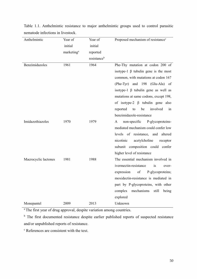
50
Table 1.1. Anthelmintic resistance to major anthelmintic groups used to control parasitic
nematode infections in livestock.
Anthelmintic Year of
initial
marketinga
Year of
initial
reported
resistanceb
Proposed mechanism of resistancec
Benzimidazoles 1961 1964 Phe-Thy mutation at codon 200 of
isotype-1 β tubulin gene is the most
common, with mutations at codon 167
(Phe-Tyr) and 198 (Glu-Ala) of
isotype-1 β tubulin gene as well as
mutations at same codons, except 198,
of isotype-2 β tubulin gene also
reported to be involved in
benzimidazole-resistance
Imidazothiazoles 1970 1979 A non-specific P-glycoproteins-
mediated mechanism could confer low
levels of resistance, and altered
nicotinic acetylcholine receptor
subunit composition could confer
higher level of resistance
Macrocyclic lactones 1981 1988 The essential mechanism involved in
ivermectin-resistance is over-
expression of P-glycoproteins;
moxidectin-resistance is mediated in
part by P-glycoproteins, with other
complex mechanisms still being
explored
Monepantel 2009 2013 Unknown
a The first year of drug approval, despite variation among countries.
b The first documented resistance despite earlier published reports of suspected resistance
and/or unpublished reports of resistance.
c References are consistent with the text.
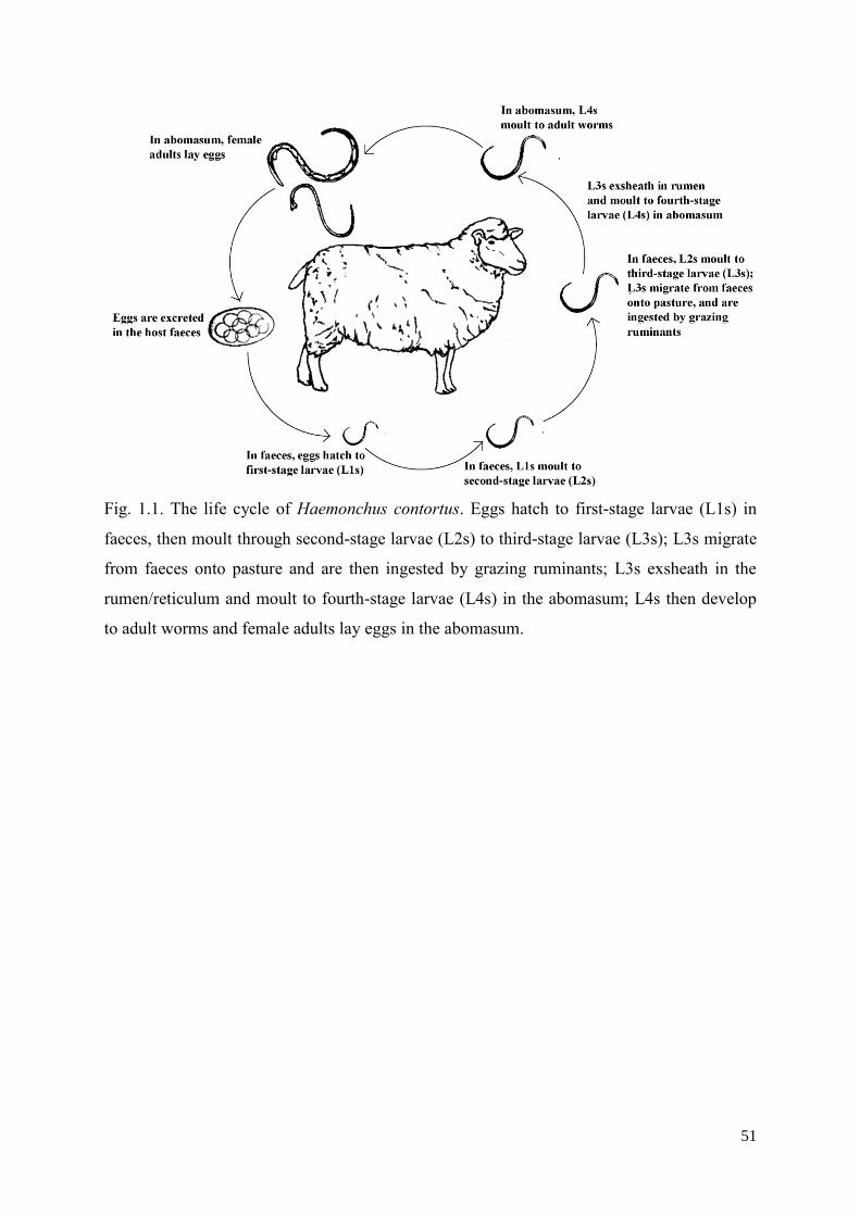
51
Fig. 1.1. The life cycle of Haemonchus contortus. Eggs hatch to first-stage larvae (L1s) in
faeces, then moult through second-stage larvae (L2s) to third-stage larvae (L3s); L3s migrate
from faeces onto pasture and are then ingested by grazing ruminants; L3s exsheath in the
rumen/reticulum and moult to fourth-stage larvae (L4s) in the abomasum; L4s then develop
to adult worms and female adults lay eggs in the abomasum.
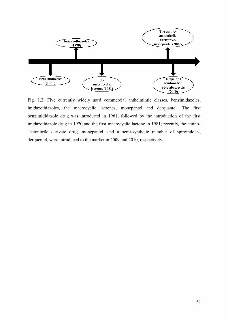
52
Fig. 1.2. Five currently widely used commercial anthelmintic classes, benzimidazoles,
imidazothiazoles, the macrocyclic lactones, monepantel and derquantel. The first
benzimididazole drug was introduced in 1961, followed by the introduction of the first
imidazothiazole drug in 1970 and the first macrocyclic lactone in 1981; recently, the amino-
acetonitrile derivate drug, monepantel, and a semi-synthetic member of spiroindoles,
derquentel, were introduced to the market in 2009 and 2010, respectively.
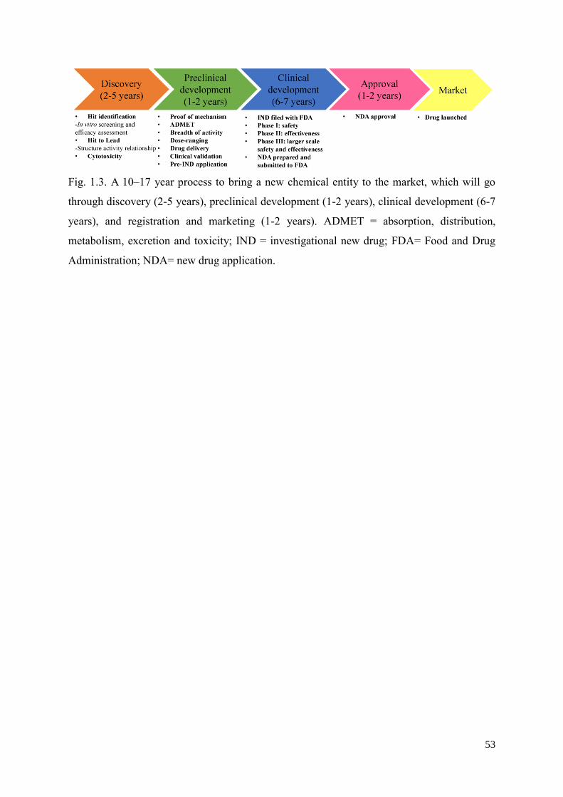
53
Fig. 1.3. A 10–17 year process to bring a new chemical entity to the market, which will go
through discovery (2-5 years), preclinical development (1-2 years), clinical development (6-7
years), and registration and marketing (1-2 years). ADMET = absorption, distribution,
metabolism, excretion and toxicity; IND = investigational new drug; FDA= Food and Drug
Administration; NDA= new drug application.
54
Chapter 2 - Identification of a new chemical entity with
selective anthelmintic activities against different
developmental stages of Haemonchus contortus and other
parasitic nematodes
Abstract
The discovery and development of novel anthelmintic classes is essential to sustain the
control of socioeconomically important parasitic worms of humans and animals. With the
aim of offering novel, lead-like scaffolds for drug discovery, Compounds Australia released
the ‘Open Scaffolds’ collection containing 33,999 compounds, with extensive information
available on the physicochemical properties of these chemicals. In the present study, we
screened 14,464 prioritised compounds from the ‘Open Scaffolds’ collection against the
exsheathed third-stage larvae (xL3s) of Haemonchus contortus using recently developed
whole-organism screening assays. We identified a hit compound, called SN00797439, which
was shown to reproducibly reduce xL3 motility by ≥70%; this compound induced a
characteristic, “coiled” xL3 phenotype (IC50 = 3.46-5.93 µM), inhibited motility of fourth-
stage larvae (L4s; IC50 = 0.31-12.5 µM) and caused considerable cuticular damage to L4s in
vitro. When tested on other parasitic nematodes in vitro, SN00797439 was shown to inhibit
(IC50 = 3-50 µM) adults of Ancylostoma ceylanicum (hookworm) and first-stage larvae of
Trichuris muris (whipworm) and eventually kill (>90%) these stages. Furthermore, this
compound completely inhibited the motility of female and male adults of Brugia malayi (50-
100 µM) as well as microfilariae of both B. malayi and Dirofilaria immitis (heartworm).
Overall, these results show that SN00797439 acts against genetically (evolutionarily) distant
parasitic nematodes (i.e. H. contortus and A. ceylanicum [strongyloids] vs. B. malayi and D.
immitis [filarioids] vs. T. muris [enoplid]), and, thus, might offer a novel, lead-like scaffold
for the development of a relatively broad-spectrum anthelmintic. Our future work will focus
on assessing the activity of SN00797439 against other pathogens that cause neglected tropical
diseases, optimising analogues with improved biological activities and characterising their
targets.
55
2.1 Introduction
Parasitic worms (helminths) of animals and humans cause chronic and often deadly
diseases that have a major socioeconomic impact worldwide (Fenwick, 2012; Fitzpatrick,
2013). On one hand, in humans, the disease burden due to parasitic worms represents ~14
million disability-adjusted life years (DALYs) globally, representing half of the DALYs
caused by major neglected tropical diseases (Hotez et al., 2014, 2016). On the other hand, in
agricultural animals, the annual economic losses due to death, poor health and reduced
productivity caused by parasitic worms are estimated at billions of dollars per annum
worldwide (cf. Knox et al., 2012; Roeber et al., 2013). In Australia alone, worms of cattle and
sheep cause economic losses estimated at 500 million dollars per annum (Lane et al., 2015).
As no vaccines are available for the vast majority of these parasites, their control relies
predominantly on the use of (usually) one of a small number of medicines (anthelmintics).
These anthelmintics are only, at best, partially effective, and their excessive and widespread
use, particularly in livestock animals, has led to serious drug resistance problems around the
world (Kaplan and Vidyashankar, 2012; Wolstenholme and Kaplan, 2012; Kotze and
Prichard, 2016). Therefore, the ongoing development of novel treatments is crucial for the
control of parasitic worms of animals.
Despite some success through the discovery of, for example, monepantel (Kaminsky et al.,
2008; Prichard and Geary, 2008) and derquantel (Little et al., 2011), limited progress has
been made in discovering new drugs against parasitic roundworms (nematodes) (Geary et al.,
2015). Major hurdles include achieving anthelmintic efficacy with an acceptable therapeutic
index, the ability to develop formulations that deliver the pharmacokinetic profile necessary
for efficacy, while maintaining the therapeutic index to support safety, as well as achieving
human food safety requirements linked to the use of food animal products and a low cost of
manufacturing the drugs (cf. Geary et al., 2015; Campbell, 2016).
In 2008, the Queensland Compound Library (QCL), now called Compounds Australia,
established a dedicated compound management facility to augment capability in chemical
screening and biomedical research, including the discovery of new anti-parasite drugs
(Simpson and Poulsen, 2014). A key role of Compounds Australia has been to source small
molecules, to consolidate them into a central repository that facilitates subsequent screening
and to provide these molecules to laboratories around the world to boost drug discovery
efforts.
56
Currently, Compounds Australia maintains three main collections: ‘Open Scaffolds’ (with
~33,400 compounds), ‘Open Academic’ (~19,500) and ‘Open Drugs’ (~2,500; Food and
Drug Administration [FDA]-approved) (Simpson and Poulsen, 2014). Each collection is well
curated and characterized, with extensive information available on the physicochemical
properties of compounds, including "rule-of-five" descriptors (chirality, hydrogen bonding
acceptor and donors, logP and molecular weight), and chemical fingerprints. The ready
availability of these resources provided us with a unique opportunity to evaluate selected
compound groups for activity against parasitic nematodes using recently developed whole-
organism screening assays. Therefore, we elected to screen prioritised compounds from the
largest library, ‘Open Scaffolds’, against parasitic stages of the barber’s pole worm,
Haemonchus contortus (of ruminants), to identify hit compounds, and then characterise and
assess them as nematocidal candidates.
2.2 Materials and methods
2.2.1 The compound library, and the selection and preparation of chemicals for
screening
We purchased a prioritised set of compounds from the ‘Open Scaffolds’ collection from
Compounds Australia, which contains a total of 33,999 chemicals representing 1,226
scaffolds (Simpson and Poulsen, 2014). This collection contains novel, lead-like scaffolds (or
chemotypes), with an average of 28 compounds per scaffold to allow meaningful structure-
activity relationships (SAR) to be explored. For each compound, the simplified molecular-
input line-entry system (SMILES) string was converted to SYBYL line notation (SLN;
Homer et al., 2008) using SYBL software (www.certara.com/software/molecular-modeling-
and-simulation/sybyl-x-suite/); using this approach, 33,949 compounds were annotated.
Then, we selected a subset of representative compounds from this library using the following
steps. First, we removed 213 pan assay interference compounds (PAINs) (Baell et al., 2010,
2016). Second, we subjected the remaining chemicals to stringent physicochemical and
structural filtering, in order to select compounds with the highest probability of permeating
cells, being soluble and being chemically optimised as drugs, using the following criteria:
mixtures, metals, isotopes: 0, minimum number of rings: 1, maximum number of rings: 4;
minimum molecular weight: 150 Da; maximum molecular weight: 400 Da; hydrogen bond
donors: ≤ 3; hydrogen bond acceptors: ≤ 6; minimum number of hydrogen bond acceptors: 1;
maximum number of chiral centers: 3; maximum number of rotatable bonds: 10; this process
57
removed 14,328 compounds, leaving 19,408 compounds. Third, compounds that were > 90%
similar to each other in structure were removed, leaving a final set of 14,464 compounds.
These 14,464 compounds (Supplementary file 2.1) were individually solubilised in dimethyl
sulfoxide (DMSO; cat no. 2225; Ajax Finechem, Australia) to achieve a stock concentration
of 5 mM, and then diluted and tested for activity against H. contortus (see Subsection 2.2.2).
2.2.2 Screening and evaluation of the effects of compounds on H. contortus
Production of parasitic larvae
Haemonchus contortus (Haecon-5 strain) was maintained in experimental sheep in
accordance with institutional animal ethics guidelines (permit no. 1413429; The University of
Melbourne) as described previously (Preston et al., 2015). To produce exsheathed third-stage
larvae (xL3s), L3s were exposed to 0.15% (v/v) of sodium hypochlorite (NaClO) for 20 min
at 37 °C (Preston et al., 2015), washed five times in sterile saline (0.9%) and cultured in Luria
Bertani medium (LB) and supplemented with final concentrations of 100 IU/ml of penicillin,
100 µg/ml of streptomycin and 0.25 µg/ml of amphotericin (antibiotic-antimycotic, cat. no.
15240-062; Gibco, Life Technologies, USA) (LB*). To produce fourth-stage larvae (L4s),
xL3s were incubated in a water-jacketed CO2 incubator (model no. 2406 Shel Lab, USA) for
7 days at 38 °C and 10% v/v CO2 or until ≥ 80% of L3s had developed to the L4 stage.
Preparation of compounds for screening
Compounds were supplied from Compounds Australia at a concentration of 5 mM.
Individual compounds were diluted to 40 µM in LB* containing 1% DMSO and dispensed in
50 µl into the wells of sterile 96-well flat bottom microplates (Corning 3596, Life Sciences,
USA) using an automated, multi-channel pipetting platform (Viaflow Assist, Switzerland).
LB* containing 1% DMSO and LB* were both included as negative controls in screening
assays. The anthelmintics moxidectin (Cydectin®, Virbac, France) and monepantel (Zolvix®,
Novartis Animal Health, Switzerland) were used as positive-control compounds in screening
assays.
Screening of compounds for their effect on xL3 motility
The whole-organism screening assay developed by Preston et al. (2016a) was used to
evaluate the effect of compounds on the motility of xL3s of H. contortus. On each 96-well
plate, the positive-control compounds (moxidectin and/or monepantel) were arrayed in
triplicate. Six wells were used for each negative control (LB* + 1% DMSO and LB* alone).
58
Test compounds were arrayed in individual wells. Following dispensing into the plates, 300
xL3s in 50 µl of LB* were transferred to each well of each plate (with the exception of
perimeter wells) using a multi-channel pipette (Finnpipette, Thermo Scientific). During
dispensing, xL3s were kept in a homogenous suspension by bubbling air through the solution
using an air pump (Airpump-S100; Aquatrade, Australia). Thus, following the addition of
xL3s to individual wells, the final concentrations were 20 µM (compound) and 0.5%
(DMSO).
Plates were incubated in a water-jacketed CO2 incubator at 38 °C and 10% v/v CO2. After
72 h, the plates were agitated (126 rotations per min) using an orbital shaker (model EOM5,
Ratek, Australia) for 20 min at 38 °C. In order to capture the motility of xL3s at 72 h, a video
recording (5 sec) was taken of each well on each plate as described previously (Preston et al.,
2016a). Each 5 second-video capture of each well was processed using a custom macro in the
program Image J (1.47v, imagej.nih.gov/ij), which indirectly measured larvae motility by
quantifying the changes in light intensity over time (cf. Preston et al., 2016a).
Screening of compounds for their effect on the motility and development/growth of
fourth-stage larvae (L4s)
The motility of L4s was evaluated using the same protocol as for xL3 (Preston et al.,
2016a). Following the measurement of larval motility, plates were re-incubated for four more
days in a humidified environment (water-jacketed CO2 incubator) at 38 °C and 10% v/v CO2.
Then, worms were fixed with 50 µl of 1% iodine, and 30 worms from each well were
examined at 20-times magnification (DP26 camera, Olympus, Japan) to assess their
development based on the presence of a well-developed mouth/pharynx in H. contortus L4s
(see Sommerville, 1966). The number of L4s was expressed as a percentage of the total worm
number (n = 30). Compounds were tested in triplicate on three different days.
Analyses of results from bioassays using H. contortus
Raw data were normalised against values of the positive and negative controls to remove
plate-to-plate variation by calculating the percentage of motility using the program GraphPad
Prism (v.6 GraphPad Software, USA). A compound was recorded as having anti-xL3 activity
if it reduced motility by ≥ 70% at 72 h, and was re-screened at 20 µM to confirm its
inhibitory properties on motility.
For a compound that consistently reduced xL3 motility by ≥ 70%, an 18-point dose-
response curve (two-fold serial dilutions; from 100 µM to 0.00076 µM) was produced for
59
xL3 and L4, to establish its half maximal inhibitory concentration (IC50). For xL3 and L4,
motility was measured at 24 h, 48 h and 72 h, and L4 development at 7 days of incubation
with each active compound (triplicate) (Subsection 2.2.3). Compound concentrations were
transformed using the equation (x = log10 (concentration in nM)) and a log (agonist) versus
response -- variable slope (four parameter) equation, in GraphPad prism v.6.07 was used to
calculate IC50 values. For L4 development, IC50 values were calculated using the same
approach. If a IC50 value could not be accurately calculated by the log (agonist) versus
response -- variable slope (four parameter) equation, a range for the IC50 value was given.
2.2.3 Scanning electron microscopy
This microscopy technique was used to assess whether compounds that reduced motility
by ≥ 70% caused structural damage to the larval stages of H. contortus, as described
previously (Preston et al., 2016b). The xL3s and L4s were produced and cultured as
described previously (Preston et al., 2015; Subsections 2.2.1-2.2.2). Six replicates of 300
xL3s or L4s were incubated in 100 μM of each compound in LB* for 24 h at 38 °C and 10%
v/v CO2. Larvae were pooled, washed 3 times in 0.9% saline at 9,000 xg and resuspended in
1 ml of phosphate-buffered saline (PBS). Subsequently, the larvae were fixed and processed
as described previously (Preston et al., 2016b). Larvae were imaged using a field-emission
scanning electron microscope (XL30 Philips, Netherlands); six representative images were
taken of each sample.
2.2.4 Evaluating compound activity on filarial worms
Procurement of filarial nematodes
The Filariasis Research Reagent Resource Center (Athens, Georgia, USA;
http://www.filariasiscenter.org/) has official ethics approval to produce filarial nematodes in
experimental animals. From this centre, we obtained fresh, live adults of Brugia malayi,
produced in Meriones unguiculatus (jird) (Michalski et al., 2011; Mutafchiev 2014). Fresh,
live microfilariae of Dirofilaria immitis (Missouri strain) obtained from the bloods from dogs
with patent infection (cf. Michalski et al., 2011; Mutafchiev 2014) were maintained in RPMI-
1640 medium (Gibco, Life Technologies, USA).
60
Assessing compound activity against adults and microfilariae of B. malayi
The compound with known activity against H. contortus was purchased from ChemDiv
(USA) and then dissolved in DMSO to reach a stock concentration of 30 mM, and then tested
for its effect on the motility of adult female and male B. malayi (see Storey et al., 2014).
First, adults were manually separated from each other with forceps, and individuals
transferred to single wells of 24-well plates containing 500 µl of RPMI-1640 (Gibco, Life
Technologies, USA) supplemented with 10 µg/ml of gentamycin and 2.5 µg/ml amphotericin
B (designated RPMI*) at 37 °C. Two-fold dilutions (100 µM to 0.97 µM) of each compound
in 500 µl of RPMI* were tested in duplicate (adding the same volume to the wells). RPMI*
plus 1% DMSO, but without compound, was added to four negative-control wells. The plates
were then incubated at 37°C and 5% v/v CO2. Subsequently, using the Worminator image
analysis system (Storey et al., 2014), the motility of individual adult worms was recorded
after 24 h and 48 h of incubation. In brief, at each time point, a video recording (1 min) was
taken of each well on each plate until the standard deviation reached zero.
In addition, the effect of compounds on the production and motility of microfilariae
originating from individual gravid females of B. malayi after 72 h of incubation was assessed.
To do this, 200 µl of medium (RPMI* plus 1% DMSO) containing ~100 microfilariae from
individual wells were transferred to individual wells of a 96-well plate and recorded for 30
sec (in the same manner as for adult worms) using the Worminator. All assays were repeated
thrice, and IC50 values were calculated using the log (agonist) versus response -- variable
slope (four parameter) equation -- in GraphPad Prism (v.6 GraphPad Software).
Evaluating compound activity against microfilariae of D. immitis
To assess the effects on the microfilariae of D. immitis, compounds were serially diluted
two-fold (100 µM to 0.39 µM) in 96-well plates; RPMI* was used as the medium, and each
compound dilution was tested in triplicate. Six wells contained RPMI* plus 1% DMSO
(negative controls). In test wells, 50 µl of RPMI* containing ~100 microfilariae were added
to each well using a multi-channel pipette. Plates were then incubated at 37 °C and 5% v/v
CO2. At 24 h and 48 h, plates were imaged for microfilarial motility using the Worminator
(see Subsection 2.5.2). All assays were repeated thrice on different days, and IC50 values were
determined using GraphPad prism (Subsection 2.2.5).
61
2.2.5 Assessing compound activity on adult A. ceylanicum and T. muris L1s
Adults of A. ceylanicum were collected from the small intestine of hamsters, which had
been infected orally with 150 A. ceylanicum L3s for three weeks. For each compound, three
worms were placed in each well of a 24-well plate, using 2 wells per compound. Levamisole
(50 µM) was used as a positive-control compound. Worms were incubated in the presence of
50 µM of each compound, and culture medium, which was composed of Hanks' Balanced
Salt Solution (HBSS) supplemented with 10% foetal calf serum, 25 µg/ml of amphotericin B,
100 U/ml of penicillin and 100 µg/ml of streptomycin. Worms were kept in an incubator at
37°C and 5% v/v CO2 for 72 h. Thereafter, the condition of the worms was microscopically
evaluated.
For T. muris, 40 L1s were placed in each well of a 96-well plate and incubated for 24 h at
37°C and 5% v/v CO2 in the presence of 100 µl RPMI-1640 medium with amphotericin B
(12.5 µg/ml), penicillin (500 U/ml), streptomycin (500 µg/ml) and 100 µM of the compound
to be tested. Levamisole (100 µM) was used as a positive-control compound. Each compound
was tested in duplicate. At 24 h, the total number of L1s per well was counted. The larvae
were then stimulated with 100 µl hot water and motile L1s were counted.
2.2.6 Assessing compound cytotoxicity and selectivity
Cell toxicity was assessed in a mammary epithelial cell line (MCF10A), essentially as
described previously (Kumarasingha et al., 2016). In brief, MCF10A cells were seeded in
black walled, flat bottom 384 well black walled plates (Corning, USA) at 700 cells/well using
a BioTek 406 automated liquid handling dispenser (BioTek, Vermont, USA) in a total
volume of 40 μl/well. Cells were cultured in DMEM-F12 containing 5% horse serum (Life
Technologies, Australia), 20 ng/ml human epidermal growth factor (EGF, Life Technologies,
Australia), 100 ng/ml Cholera toxin (Sigma, Australia), 0.5 μg/ml hydrocortisone (Sigma,
Australia) and 10 μg/ml insulin (human; Novo Nordisk Pharmaceuticals Pty Ltd, Australia).
After an incubation for 24 h at 37 °C, 5% v/v CO2, the growth medium was aspirated and the
cells were treated with test compounds starting at 100 µM, and positive and negative controls
(media ± 1% DMSO, monepantel, moxidectin). The chemotherapeutic compound
doxorubicin, starting at 10 µM, was also used as a positive control. Compounds were titrated
to generate a five-point dose-response curve, in quadruplicate, using an automated liquid
handling robot (SciClone ALH3000 Lab Automation Liquid Handler, Caliper Lifesciences,
USA) and incubated for a further 48 h. Matched DMSO concentrations for each compound
62
concentration were also tested separately to account for DMSO induced cell toxicity. To
measure cell proliferation, cells were fixed and stained with 4',6-diamidino-2-phenylindole
(DAPI; 1:1000), and individual wells imaged at 10-times magnification, covering 16 fields
(~90% of well) using a high content imager (Cellomics CellInsight Personal Cell Imager,
ThermoFisher Scientific, USA) at a fixed exposure time of 0.12 seconds. Viable cells were
counted using the Target Activation BioApplication within the Cellomics Scan software
(v.6.5.0, Thermo Scientific, USA) and normalised to the cell density in wells without
compound. Toxicity due to DMSO was removed from the normalised cell density counts and
IC50 values calculated from the variable slope four-parameter equation (v.6 GraphPad
Software). Experiments were repeated twice on two different days using four technical
replicates for each compound.
2.3 Results and discussion
2.3.1 The identification of compound SN00797439 with activity against parasitic
stages of H. contortus
In the primary screen, we tested all 14,464 prioritised compounds from the ‘Open
Scaffolds’ collection against H. contortus xL3s (Fig. 2.1A). Any compounds that consistently
reduced xL3 motility (in independent screens) by > 70% at 72 h was recorded as a “hit” (Fig.
2.1B). Of all compounds tested, a chemical, designated SN00797439 (IUPAC name: N-(4-
chlorophenyl)-2-(3-phenyl-1,2,4-oxadiazol-5-yl)-1-pyrrolidinecarboxamide) reduced xL3
motility by ≥ 70% in both the primary and subsequent confirmatory screens (Fig. 2.1B); this
compound caused a “coiled” xL3 phenotype (Supplementary File 2.2). The chemical
structure and predicted physicochemical properties of SN00797439 are given in Fig. 2.1.
Dose-response curves (two-fold serial dilutions from 100 µM to 0.00076 µM) showed that
SN00797439 inhibited xL3 motility, with IC50 values of 3.46 ± 0.82 µM (24 h), 10.08 ± 2.06
µM (48 h), and 5.93 ± 1.38 µM (72 h) (Fig. 2.2; Table 2.1); these IC50 values were
comparable with those of monepantel and moxidectin (positive-control compounds).
Dose-response curves (two-fold serial dilutions from 100 µM to 0.00076 µM) revealed
that SN00797439 inhibited the motility of L4s, with IC50 values of 6.25 - 12.5 μM (24 h),
0.78 - 1.5 μM (48 h) and 0.31 - 0.78 μM (72 h) (Fig. 2.2; Table 2.1). Subsequently,
SN00797439 was tested for its ability to inhibit growth/development from xL3 to L4. The
dose-response curves revealed that this compound inhibited L4 development, with an IC50
value of 11.04 ± 2.16 µM (Fig. 2.2; Table 2.1). Upon SEM analysis, SN00797439 caused
63
only minor morphological damage to xL3, but resulted in considerable cuticle alterations to
the L4 stage, including multiple, cuticle-‘embossed’ rings around the worm along its length
and a ‘scaly’ appearance of the worm surface (Fig. 2.3).
2.3.2 Compound SN00797439 also has inhibitory activity on the motility of
different developmental stages of other parasitic nematodes
In order to assess whether compound SN00797439 would act against nematodes that are
genetically (evolutionarily) very distant from H. contortus (order Strongylida), we tested
whether these compounds would inhibit the motility of adults and microfilariae of B. malayi
as well as microfilariae of D. immitis in vitro. SN00797439 inhibited the motility of female
and male adults of B. malayi (Fig. 2.4; Table 2.2), with movement ceasing at 100 µM (24 h)
and 50 µM (48 h). This result compares with 1.3 µM (24 h) and 2.9 µM (48 h) for moxidectin
(cf. Storey et al., 2014). SN00797439 was then tested for its effect on microfilariae of B.
malayi (released from females after 72 h in vitro), and inhibited the motility of the
microfilariae, with an IC50 of ~ 3 µM (24 h) compared with ~ 6 µM for moxidectin (cf. Storey
et al., 2014).
Subsequently, SN00797439 was tested against microfilariae of D. immitis, a filarial
nematode that is related to B. malayi, and inhibited the motility of microfilariae, with
complete inhibition at 100 µM (24 h and 48 h). For both of these time points, an IC50 value of
~ 50 µM was achieved (Table 2.2), which compares with IC50 values of 43 µM and 9.3 µM
published for ivermectin and moxidectin, respectively (Storey et al., 2014). In addition, when
tested on A. ceylanicum and T. muris, SN00797439 killed adult A. ceylanicum at 50 µM and
displayed a high nematocidal activity against T. muris L1s (90.1% of L1s dead at 100 µM).
The positive-control compound (levamisole) killed both nematode species.
2.3.3 Cytotoxicity and selectivity of SN00797439
Using an established proliferation assay, SN00797439 was assessed for its toxicity on
mammary epithelial cells (Table 2.3) and was shown to be selective for parasitic larvae of H.
contortus, with a selectivity index (SI) of ~9 to 128, compared with ~ 67 to 332 (monepantel)
and ~ 2 to 300 (moxidectin) for the positive-control compounds (Table 2.3). For filarial
worms, SN00797439 was selective only for microfilariae of B. malayi (but not D. immitis)
with an SI of 33.34.
64
2.4 Conclusion
In the past three decades, only four ‘new’ anthelmintic drugs, emodepside (Martin et al.,
2012; Harder, 2016), tribendimidine (Xiao et al., 2005; Steinmann et al., 2008), monepantel
(Kaminsky et al., 2008; Prichard and Geary, 2008) and derquantel (Little et al., 2011), have
been commercialised. Due the rapid emergence of anthelmintic resistance, even to some
recently commercialised compounds (Scott et al., 2013; Van den Brom et al., 2105; Sales and
Love, 2016; Cintra et al., 2016), there is an urgent need to identify, validate, optimise and
develop novel chemical entities for the treatment of parasitic worms of humans and
agricultural animals (Geary et al., 2015).
With this focus in mind, we screened 14,464 prioritised compounds (representing a
collection of 33,999 chemicals) to identify potential starting points for the design of new
anthelmintics. Under the conditions used in this screening platform, compound SN00797439
was identified as a hit, and exhibited considerable anthelmintic activity against H. contortus
and induced a “coiled” phenotype (Supplementary File 2.2). SN00797439 was also shown to
have anthelmintic activity against a species of hookworm (A. ceylanicum; order Strongylida)
and two filarioid nematodes (D. immitis and B. malayi; order Spirurida). Additionally, this
compound was also shown to have activity against the mouse whipworm, T. muris, which is
used as a model to study the genetically related human whipworm (T. trichiura; order
Enoplida) (cf. Foth et al., 2014) These findings, for multiple representatives of
taxonomically, biologically and genetically distinct orders (i.e. Strongylida vs. Spirurida vs.
Enoplida), indicate a relatively broad spectrum of activity of this chemotype against
nematodes.
Compound SN00797439 is made up of an oxadiazole and pyrrolidine core. An appraisal of
the intellectual property surrounding the sub-structures of this compound (Li and Zhong,
2011, 2010) reveals that similar compounds have been explored as therapeutics for the
control of hepatitis C, but not yet as anthelmintics. Following the identification of
SN00797439 in the primary screen against H. contortus, an evaluation revealed differences in
its potency to inhibit the motility, growth and/or development of the parasitic larval stages of
this nematode. The results showed that SN00797439 was more potent on L4s than xL3s. This
difference in potency might be due to variation in expression of its target(s) in the nematode,
a distinction in physiology between these two developmental stages of the worm and/or the
nature and extent of the uptake of the compound by the worm. An integrated use of
transcriptomic, proteomic and metabolomic techniques (Mikami et al., 2012; Schwarz et al.,
65
2013; Preston et al, 2016c) might be able to elucidate the physiological and biological
pathways in H. contortus and/or other worms affected by SN00797439.
In our opinion, identifying a compound with activity against taxonomically distinct
nematodes provides a starting point for designing and optimising a lead compound. Thus, we
are now eager to critically assess the activity of SN00797439 on various developmental
stages of a range of parasitic nematodes of humans, including Ascaris sp. (large roundworm)
and other common species of hookworm (Necator americanus and Ancylostoma duodenale)
as well as additional filarial worms (Onchocerca volvulus and Loa Loa) which cause some of
the most neglected tropical diseases and collectively affect ~ 1.8 billion people, resulting in a
loss of 8.5 million DALYs worldwide (e.g., Hotez et al., 2016). Importantly, future work
should also focus on (i) studying the structure-activity relationship (SAR) of derivatives of
compounds SN00797439 via medicinal chemistry optimization, to attain a new entity with
broad-spectrum and enhanced activity; (ii) on assessing their pharmacological properties
(absorption, distribution, metabolism, excretion and toxicity; ADMET) of the compound
series; and ultimately (iii) on assessing the anthelmintic efficacy of active compounds in vivo
in animals.

66
2.5 References
Baell, J.B., 2016. Feeling nature's PAINS: natural products, natural product drugs, and pan
assay interference compounds (PAINs). J. Nat. Prod. 79, 616-628.
Baell, J.B., Holloway, G.A., 2010. New substructure filters for removal of pan assay
interference compounds (PAINS) from screening libraries and for their exclusion in
bioassays. J. Med. Chem. 53, 2719-2740.
Campbell, W.C., 2016. Lessons from the history of ivermectin and other antiparasitic agents.
Annu. Rev. Anim. Biosci. 4,1-14.
Cintra, M.C., Teixeira, V.N., Nascimento, L.V., Sotomaior, C.S., 2016. Lack of efficacy of
monepantel against Trichostrongylus colubriformis in sheep in Brazil. Vet. Parasitol.
216, 4-6.
Fenwick, A., 2012. The global burden of neglected tropical diseases. Public Health, 126, 233-
236.
Fitzpatrick, J.L., 2013. Global food security: the impact of veterinary parasites and
parasitologists. Vet. Parasitol. 195, 233-248.
Foth, B.J., Tsai, I.J., Reid, A.J., Bancroft, A.J., Nichol, S., Tracey, A., Holroyd, N, Cotton,
J.A., Stanley, E.J., Zarowiecki, M., Liu, J.Z., Huckvale, T., Cooper, P.J., Grencis, R.K.,
Berriman, M.L., 2014. Whipworm genome and dual-species transcriptome analyses
provide molecular insights into an intimate host-parasite interaction. Nat. Genet. 46, 693-
702.
Geary, T.G., Sakanari, J.A., Caffrey, C.R., 2015. Anthelmintic drug discovery: into the
future. J. Parasitol. 101, 125-133.
Harder, A., 2016. The biochemistry of Haemonchus contortus and other parasitic nematodes.
Adv. Parasitol. 93, 69-94.
Homer, R.W., Swanson, J., Jilek, R.J., Hurst, T., Clark, R.D., 2008. SYBYL line notation
(SLN): a single notation to represent chemical structures, queries, reactions, and virtual
libraries. J. Chem. Inf. Model. 48, 2294-2307.
Hotez, P.J., Bottazzi, M.E., Strych, U., 2016. New vaccines for the world’s poorest people.
Annu. Rev. Med. 67, 405-417.
Hotez, P.J., Alvarado, M., Basáñez, M.G., Bolliger I, Bourne R, Boussinesq M, Brooker S.J.,
Brown, A.S., Buckle, G., Budke, C.M., Carabin, H., Coffeng, L.E., Fèvre, E.M., Fürst,
T., Halasa, Y.A., Jasrasaria, R., Johns, N.E., Keiser, J., King, C.H., Lozano, R.,
Murdoch, M.E., O'Hanlon, S., Pion S.D.S., Pullan, R.L., Ramaiah, K.D., Roberts, T.,
67
Shepard, D.S., Smith, J.L., Stolk, W.A., Undurraga, E.A., Utzinger, J., Wang, M.,
Murray, C.J.L., Naghavi, M. 2014. The global burden of disease study 2010:
interpretation and implications for the neglected tropical diseases. PLoS Negl. Trop. Dis.
8, e2865.
Kaplan, R.M., Vidyashankar, A.N., 2012. An inconvenient truth: global warming and
anthelmintic resistance. Vet. Parasitol. 186, 70-78.
Kaminsky, R., Ducray, P., Jung, M., Clover, R., Rufener, L., Bouvier, J., Wenger, A.,
Schroeder, F., Desaules, Y., Hotz, R., Goebel, T., Hosking, B.C., Pautrat, F., Wieland-
Berghausen, S., Ducray, P., 2008. A new class of anthelmintics effective against drug-
resistant nematodes. Nature 452, 176-180.
Knox, M.R., Besier, R.B., Le Jambre, L.F., Kaplan, R.M., Torres-Acosta, J.F., Miller, J.,
Sutherland, J., 2012. Novel approaches for the control of helminth parasites of livestock
VI: Summary of discussions and conclusions. Vet. Parasitol. 186, 143-149.
Kotze, A.C., Prichard, R.K., 2016. Anthelmintic resistance in Haemonchus contortus: history,
mechanisms and diagnosis. Adv. Parasitol. 93, 397-428.
Kumarasingha, R., Preston, S., Yeo, T.C., Lim, D.S.L., Tu, C.L., Palombo, E.A., Shaw, J.M.,
Gasser, R.B., Boag, P.R., 2016. Anthelmintic activity of selected ethno-medicinal plant
extracts on parasitic stages of Haemonchus contortus. Parasit. Vectors 9, 187.
Lane, J., Jubb, T., Shephard, R., Webb-Ware, J., Fordyce, G., 2015. MLA Final Report:
Priority list of endemic diseases for the red meat industries. MLA Available from:
www.mla.com.au/download/finalreports?itemId=2877, Accessed 7th September 2016.
Li, L., Zhong, M., 2011. Preparation of biarylacetylenes, arheteroarylacetylenes and
biheteroarylacetylenes end-capped with amino acid or peptide derivatives as inhibitors of
HCV NS5A. Patent WO 2011150243 A1 20111201.
Li L, Zhong M., 2010. Preparation of biarylacetylenes, arheteroarylacetylenes and
biheteroarylacetylenes end-capped with amino acid or peptide derivatives as inhibitors of
HCV NS5A. Patent WO 2010065668 A1 20100610.
Little, P.R., Hodge, A., Maeder, S.J., Wirtherle, N.C., Nicholas, D.R., Cox, G.G., Conder
G.A., 2011. Efficacy of a combined oral formulation of derquantel–abamectin against the
adult and larval stages of nematodes in sheep, including anthelmintic-resistant strains.
Vet. Parasitol. 181, 180-193.
Martin, R.J., Buxton, S.K., Neveu, C., Charvet, C.L., Robertson, A.P., 2012. Emodepside and
SL0-1 potassium channels: a review. Exp. Parasitol. 132, 40-46.
68
Michalski, M.L., Griffiths, K.G., Williams, S.A., Kaplan, R.M., Moorhead, A.R., 2011. The
NIH-NIAID Filariasis Research Reagent Resource Center. PLoS Negl. Trop. Dis. 5,
e1261.
Mikami, T., Aoki, M., Kimura, T., 2012. The application of mass spectrometry to proteomics
and metabolomics in biomarker discovery and drug development. Curr. Mol. Pharmacol.
5, 301-316.
Mutafchiev, Y., Bain, O., Williams, Z., McCall, J.W., Michalski, M.L., 2014. Intraperitoneal
development of the filarial nematode Brugia malayi in the Mongolian jird (Meriones
unguiculatus). Parasitol. Res. 113, 1827-18235.
Preston, S., Jabbar, A., Gasser, R.B., 2016a. A perspective on genomic-guided anthelmintic
discovery and repurposing using Haemonchus contortus. Infect. Genet. Evol. 40, 368-
373.
Preston, S., Jabbar, A., Nowell, C., Joachim, A., Ruttkowski, B., Baell, J., Cardno, T.,
Korhonen, P.K., Piedrafita, D., Ansell, B.R., Jex, A.R., Hofmann, A., Gasser, R.B.,
2015. Low cost whole-organism screening of compounds for anthelmintic activity. Int. J.
Parasitol. 45, 333-343.
Preston, S., Jabbar, A., Nowell, C., Joachim, A., Ruttkowski, B., Cardno, T., Hofmann, A.,
Gasser, R.B., 2016b. Practical and low cost whole-organism motility assay: A step-by-
step protocol. Mol. Cell. Probes 30, 13-17.
Preston, S., Luo, J., Zhang, Y., Jabbar, A., Crawford, S., Baell, J., Hofmann, A., Hu, M.,
Zhou, H.B., Gasser, R.B., 2016c. Selenophene and thiophene-core estrogen receptor
ligands that inhibit motility and development of parasitic stages of Haemonchus
contortus. Parasit. Vectors 9, 346.
Prichard, R.K., Geary, T.G., 2008. Drug discovery: fresh hope to can the worms. Nature. 452,
157-158.
Roeber, F., Jex, A.R., Gasser, R.B., 2013. Impact of gastrointestinal parasitic nematodes of
sheep, and the role of advanced molecular tools for exploring epidemiology and drug
resistance - an Australian perspective. Parasit. Vectors 6, 153.
Sales, N., Love, S., 2016. Resistance of Haemonchus sp. to monepantel and reduced efficacy
of a derquantel abamectin combination confirmed in sheep in NSW, Australia. Vet.
Parasitol. 15, 193-196.
Schwarz, E.M., Korhonen, P.K., Campbell, B.E., Young, N.D., Jex, A.R., Jabbar, A., Hall,
R.S., Mondal, A., Howe, A.C., Pell, J., Hofmann, A., Boag, P.R., Zhu, X.Q., Gregory, T.,
Loukas, A., Williams, B.A., Antoshechkin, I., Brown, C., Sternberg, P.W., Gasser, R.B.,
69
2013. The genome and developmental transcriptome of the strongylid
nematode Haemonchus contortus. Genome Biol. 14, R89.
Scott, I., Pomroy, W.E., Kenyon, P.R., Smith, G., Adlington, B., Moss, A., 2013. Lack of
efficacy of monepantel against Teladorsagia circumcincta and Trichostrongylus
colubriformis. Vet. Parasitol. 198, 166-171.
Simpson, M., Poulsen, S.A., 2014. An overview of Australia's compound management
facility: the Queensland Compound Library. ACS Chem. Biol. 9, 28-33.
Sommerville, R.I., 1966. The development of Haemonchus contortus to the fourth stage in
vitro. J. Parasitol. 52, 127-136.
Steinmann, P., Zhou, X.N., Du, Z.W., Jiang, J.Y., Xiao, S.H., Wu, Z.X., Zhou, H., Utzinger,
J., 2008. Tribendimidine and albendazole for treating soil-transmitted helminths,
Strongyloides stercoralis and Taenia spp.: open-label randomized trial. PLoS Negl. Trop.
Dis. 2, e322
Storey, B., Marcellino, C., Miller, M., Maclean, M., Mostafa, E., Howell, S., Sakanari, J.,
Wolstenholme, A., Kaplan, R., 2014. Utilization of computer processed high definition
video imaging for measuring motility of microscopic nematode stages on a quantitative
scale: "The Worminator". Int. J. Parasitol. Drugs Drug Resist. 4, 233-243.
Van den Brom, R., Moll, L., Kappert, C., Vellema, P., 2015. Haemonchus contortus
resistance to monepantel in sheep. Vet. Parasitol. 209, 278-280.
Wolstenholme, A.J., Kaplan, R.M., 2012. Resistance to macrocyclic lactones. Curr. Pharm.
Biotechnol. 13, 873-887.
Xiao, S.H., Hui-Ming, W., Tanner, M., Utzinger, J., Chong, W., 2005. Tribendimidine: a
promising, safe and broad-spectrum anthelmintic agent from China. Acta Trop. 94, 1-14.
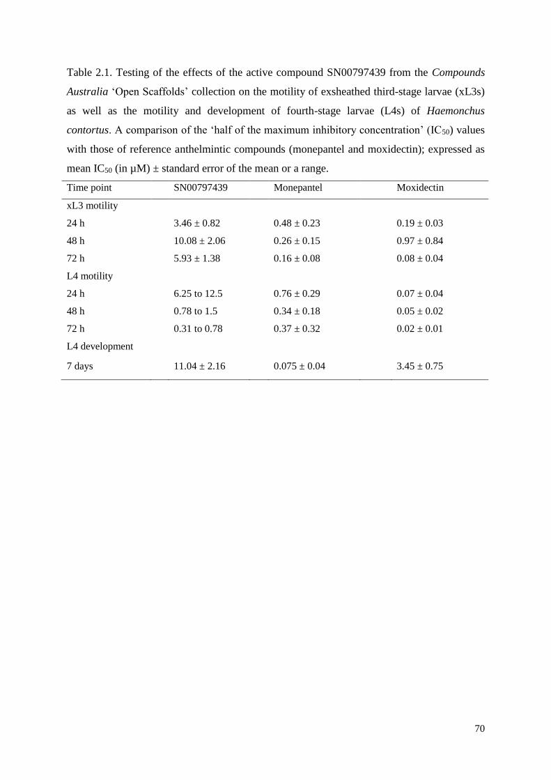
70
Table 2.1. Testing of the effects of the active compound SN00797439 from the Compounds
Australia ‘Open Scaffolds’ collection on the motility of exsheathed third-stage larvae (xL3s)
as well as the motility and development of fourth-stage larvae (L4s) of Haemonchus
contortus. A comparison of the ‘half of the maximum inhibitory concentration’ (IC50) values
with those of reference anthelmintic compounds (monepantel and moxidectin); expressed as
mean IC50 (in µM) ± standard error of the mean or a range.
Time point SN00797439 Monepantel Moxidectin
xL3 motility
24 h 3.46 ± 0.82 0.48 ± 0.23 0.19 ± 0.03
48 h 10.08 ± 2.06 0.26 ± 0.15 0.97 ± 0.84
72 h 5.93 ± 1.38 0.16 ± 0.08 0.08 ± 0.04
L4 motility
24 h 6.25 to 12.5 0.76 ± 0.29 0.07 ± 0.04
48 h 0.78 to 1.5 0.34 ± 0.18 0.05 ± 0.02
72 h 0.31 to 0.78 0.37 ± 0.32 0.02 ± 0.01
L4 development
7 days 11.04 ± 2.16 0.075 ± 0.04 3.45 ± 0.75
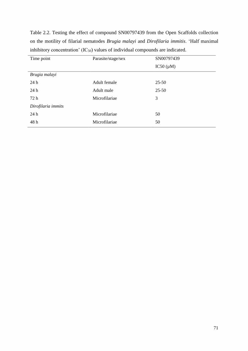
71
Table 2.2. Testing the effect of compound SN00797439 from the Open Scaffolds collection
on the motility of filarial nematodes Brugia malayi and Dirofilaria immitis. ‘Half maximal
inhibitory concentration’ (IC50) values of individual compounds are indicated.
Time point Parasite/stage/sex SN00797439
IC50 (µM)
Brugia malayi
24 h Adult female 25-50
24 h Adult male 25-50
72 h Microfilariae 3
Dirofilaria immits
24 h Microfilariae 50
48 h Microfilariae 50
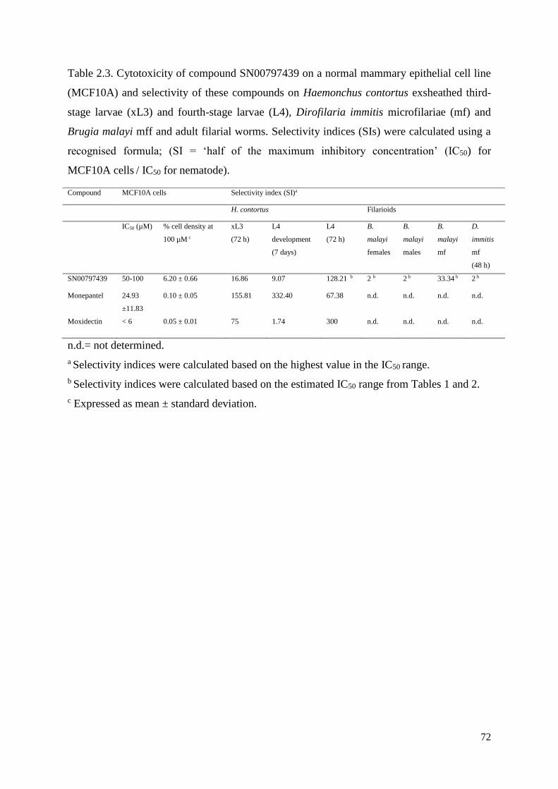
72
Table 2.3. Cytotoxicity of compound SN00797439 on a normal mammary epithelial cell line
(MCF10A) and selectivity of these compounds on Haemonchus contortus exsheathed third-
stage larvae (xL3) and fourth-stage larvae (L4), Dirofilaria immitis microfilariae (mf) and
Brugia malayi mff and adult filarial worms. Selectivity indices (SIs) were calculated using a
recognised formula; (SI = ‘half of the maximum inhibitory concentration’ (IC50) for
MCF10A cells / IC50 for nematode).
n.d.= not determined.
a Selectivity indices were calculated based on the highest value in the IC50 range.
b Selectivity indices were calculated based on the estimated IC50 range from Tables 1 and 2.
c Expressed as mean ± standard deviation.
Compound MCF10A cells Selectivity index (SI)a
H. contortus Filarioids
IC50 (µM) % cell density at
100 µM c
xL3
(72 h)
L4
development
(7 days)
L4
(72 h)
B.
malayi
females
B.
malayi
males
B.
malayi
mf
D.
immitis
mf
(48 h)
SN00797439 50-100 6.20 ± 0.66 16.86 9.07 128.21 b 2 b 2 b 33.34 b 2 b
Monepantel 24.93
±11.83
0.10 ± 0.05 155.81 332.40 67.38 n.d. n.d. n.d. n.d.
Moxidectin < 6 0.05 ± 0.01 75 1.74 300 n.d. n.d. n.d. n.d.
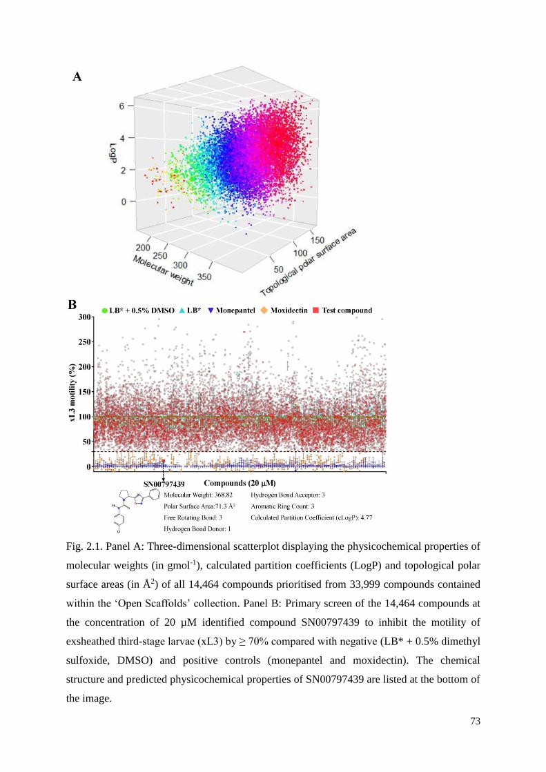
73
Fig. 2.1. Panel A: Three-dimensional scatterplot displaying the physicochemical properties of
molecular weights (in gmol-1), calculated partition coefficients (LogP) and topological polar
surface areas (in Å2) of all 14,464 compounds prioritised from 33,999 compounds contained
within the ‘Open Scaffolds’ collection. Panel B: Primary screen of the 14,464 compounds at
the concentration of 20 µM identified compound SN00797439 to inhibit the motility of
exsheathed third-stage larvae (xL3) by ≥ 70% compared with negative (LB* + 0.5% dimethyl
sulfoxide, DMSO) and positive controls (monepantel and moxidectin). The chemical
structure and predicted physicochemical properties of SN00797439 are listed at the bottom of
the image.
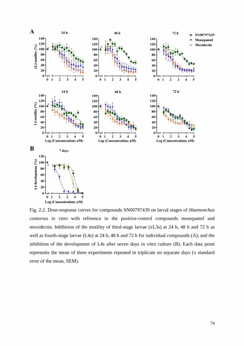
74
Fig. 2.2. Dose-response curves for compounds SN00797439 on larval stages of Haemonchus
contortus in vitro with reference to the positive-control compounds monepantel and
moxidectin. Inhibition of the motility of third-stage larvae (xL3s) at 24 h, 48 h and 72 h as
well as fourth-stage larvae (L4s) at 24 h, 48 h and 72 h for individual compounds (A); and the
inhibition of the development of L4s after seven days in vitro culture (B). Each data point
represents the mean of three experiments repeated in triplicate on separate days (± standard
error of the mean, SEM).
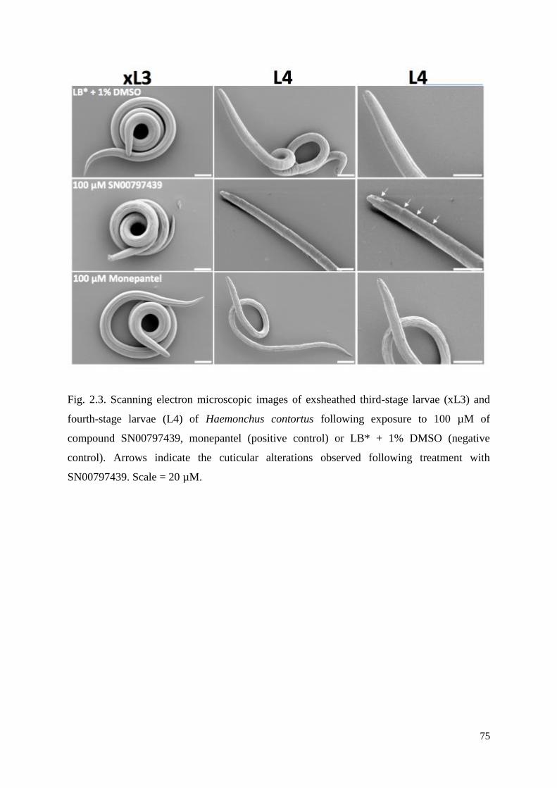
75
Fig. 2.3. Scanning electron microscopic images of exsheathed third-stage larvae (xL3) and
fourth-stage larvae (L4) of Haemonchus contortus following exposure to 100 µM of
compound SN00797439, monepantel (positive control) or LB* + 1% DMSO (negative
control). Arrows indicate the cuticular alterations observed following treatment with
SN00797439. Scale = 20 µM.
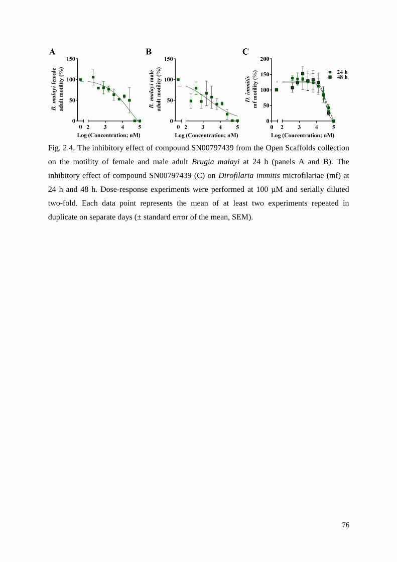
76
Fig. 2.4. The inhibitory effect of compound SN00797439 from the Open Scaffolds collection
on the motility of female and male adult Brugia malayi at 24 h (panels A and B). The
inhibitory effect of compound SN00797439 (C) on Dirofilaria immitis microfilariae (mf) at
24 h and 48 h. Dose-response experiments were performed at 100 µM and serially diluted
two-fold. Each data point represents the mean of at least two experiments repeated in
duplicate on separate days (± standard error of the mean, SEM).
77
Chapter 3 - Identification of two kinase inhibitors under
pharmaceutical development with activity against
Haemonchus contortus
Abstract
In partnership with the Medicines for Malaria Venture (MMV), we screened a collection
(‘Stasis Box’) of 400 compounds (which are under pharmaceutical development for illnesses
other than infectious diseases) for inhibitory activity against Haemonchus contortus, in order
to attempt to repurpose some of the compounds to parasitic nematodes. We assessed the
inhibition of compounds on the motility and/or development of exsheathed third-stage (xL3s)
and fourth-stage (L4) larvae of H. contortus using a whole-organism screening assay. In the
primary screen, we identified compound MMV690767 (also known as SNS-032) that
inhibited xL3 motility by ~ 70% at a concentration of 20 µM after 72 h as well as compound
MMV079840 (also known as AG-1295), which induced a coiled xL3 phenotype, with ~ 50%
inhibition on xL3 motility. Subsequently, we showed that SNS-032 (IC50 = 12.4 µM) and
AG-1295 (IC50 = 9.92 ± 1.86 µM) had a similar potency to inhibit xL3 motility. Although
neither SNS-032 nor AG-1295 had a detectable inhibitory activity on L4 motility, both
compounds inhibited L4 development (IC50 values = 41.24 µM and 7.75 ± 0.94 µM for SNS-
032 and AG-1295, respectively). The assessment of the two compounds for toxic effects on
normal human breast epithelial (MCF10A) cells revealed that AG-1295 had limited
cytotoxicity (IC50 > 100 µM), whereas SNS-032 was quite toxic to the epithelial cells (IC50 =
1.27 µM). Although the two kinase inhibitors, SNS-032 and AG-1295, had moderate
inhibitory activity on the motility or development of xL3s or L4s of H. contortus in vitro,
further work needs to be undertaken to chemically alter these entities to achieve the potency
and selectivity required for them to become nematocidal or nematostatic candidates.
78
3.1 Introduction
Parasites of animals cause diseases of major socioeconomic importance globally
(Anderson, 2000; Beveridge, 2014). For example, gastrointestinal nematodes of livestock
cause subclinical infections and diseases that lead to reductions in meat, milk and fibre
production (Charlier et al., 2014; Preston et al., 2014), with ~ AUD $500 million losses per
annum in Australia alone (Lane et al., 2015). Currently, anthelmintic treatment remains the
mainstay of control for parasitic nematodes (Epe and Kaminsky, 2013; Geary et al., 2015).
The occurrence of anthelmintic resistance, together with the limited number of anthelmintics
being commercialised, indicates an urgency to discover new and effective anthelmintic
compounds (Kaplan and Vidyashankar, 2012; Epe and Kaminsky, 2013; Whittaker et al.,
2017).
Product development partnerships (PDPs) are playing a significant role in the development
of new medicines for neglected diseases (e.g., Moran et al., 2010; Reeder and Mpanju-
Shumbusho, 2016). In the present context, a PDP is usually a collaboration between a non-
for-profit organisation, such as the Medicine of Malaria Venture (MMV), Drugs for
Neglected Diseases Initiative (DNDi) and Global Alliance for TB Drug Development (TB
Alliance), industry and/or academic partners to collectively combat infectious/parasitic
diseases (Grace, 2010; Moran et al., 2010). The significant role of the PDP model is
demonstrated through the delivery of commercial products, such as paromomycin against
leishmaniasis, developed by the Institute of One World Health (Davidson et al., 2009),
artemether-lumefantrine (Coartem dispersible) - a child-friendly treatment against malaria -
developed by PDP between Novartis and MMV (Premji, 2009), and a new vaccine called
MenAfriVac against the bacterial meningitis by the Meningitis Vaccine Project (Butler, 2010;
Bishai et al., 2011).
Recently, in a PDP with MMV, we screened compounds in the ‘Pathogen Box’ with
known activities against one or more pathogens that cause neglected diseases (including
tuberculosis, malaria, sleeping sickness, leishmaniasis, schistosomiasis, hookworm disease,
toxoplasmosis and cryptosporidiosis) against Haemonchus contortus (see Preston et al.,
2016b), an economically important parasitic nematode of ruminants that represents a large
order of nematodes called the Strongylida (Schwarz et al., 2013). We identified tolfenpyrad,
an approved pesticide with known activity against some kinetoplastid protists (Witschel et al.,
2012), which has anthelmintic activity against H. contortus (see Preston et al., 2016b). Within
this collaborative framework, we were able to source another library, called the ‘Stasis Box’,
79
from MMV, which contains 400 compounds that have been in clinical development but have
not been approved for illnesses; none of them were developed against neglected tropical
diseases. The ‘Stasis Box’ contains compounds that have been developed against disorders
such as atherosclerosis, restenosis, pulmonary fibrosis, selected cancers, urinary incontinence
or depression (Mike Palmer, personal communication). Here, we screened all of these
compounds against H. contortus using an established whole-organism motility assay (Preston
et al., 2016a), with the aim of repurposing some of them as nematocides.
3.2 Methods
Procurement of H. contortus
In accordance with institutional animal ethics guidelines (permit no. 1413429; The
University of Melbourne), H. contortus (Haecon-5 strain) was maintained in experimental
sheep as described previously (Preston et al., 2015). To produce exsheathed third-stage larvae
(xL3s), infective L3s were exposed to 0.15% (v/v) of sodium hypochlorite (NaClO) for 20
min at 37 °C (Preston et al., 2015), washed five times in sterile saline (0.9%) and cultured in
Luria Bertani medium (LB) supplemented with final concentrations of 0.25 µg/ml of
amphotericin B, 100 µg/ml of streptomycin and 100 IU/ml of penicillin (antibiotic-
antimycotic, cat. no. 15240-062; Life Technologies, USA) (LB*). To produce fourth-stage
larvae (L4s), xL3s were incubated in a water-jacketed CO2 incubator (model no. 2406 Shel
Lab, USA) for 7 days at 38 °C and 10% v/v CO2 or until ≥ 70% of L3s had developed to the
L4 stage.
Compound library and screening
From MMV we obtained the 'Stasis Box', which contains 400 compounds that have been
under drug development. These compounds were individually solubilised in 10 µl of
dimethyl sulfoxide (DMSO) to achieve a stock concentration of 10 mM, and then diluted and
tested for activity against H. contortus. The compounds were tested using a previously
described method (Preston et al., 2016a). In brief, using 96-well flat bottom plates, individual
compounds (40 µM) in LB* (50 µl) were added to individual wells in triplicate, with LB*+
0.5% DMSO as a negative control and a commercial anthelmintic, monepantel (Zolvix,
Novartis Animal Health, Switzerland) as a positive control. Subsequently, 300 xL3s in 50 µl
LB* were added to individual wells. The plates were then incubated in a 38 °C water-
jacketed CO2 incubator for 72 h. Then, a video recording (5 sec) was made of each well using
80
a grey-scale camera (Rolera bolt, Q imaging Scientific Coms, Canada) and a motorised X-Y
axis stage (BioPoint 2; Ludl Electronics Products, USA). The motility of worms in each well
was calculated in a pixel-based algorithm, called motility index (Mi), based on the light
intensity changes caused by the worm movement (Preston et al., 2015). For each compound,
Mi values were normalized against the positive and negative controls using the program
GraphPad Prism (v.6 GraphPad Software, USA). A compound was identified as a “hit” if it
reduced worm motility by ≥ 70% or induced a phenotype that differed from wild-type xL3
(i.e. LB* + 0.5% DMSO control). For each compound, each data point represented the mean
of a triplicate (± standard error of the mean, SEM).
Dose-response assay
Active compounds (99.9% purity; purchased from Selleck Chemicals or Cayman
Chemicals, USA), were serially (two-fold) diluted from 100 µM to 0. 76 nM in triplicate in a
96-well flat bottom plate and ~300 xL3s (in 50 µl LB*) added to each well. The plates were
then incubated in a 38°C water-jacketed CO2 incubator and the Mi values of worms measured
(Preston et al., 2016a). In addition, following the measurement of xL3 motility, L4
development rates were measured, with 30 worms from each well being examined at 20-
times magnification following the addition of 50 µl of 1% iodine to each well after seven
days of incubation prior to light microscopic examination at 100-times magnification. Half
the maximum inhibitory concentration (IC50) on xL3 motility, L4 motility and L4
development were determined from the dose-response curves using a variable slope four-
parameter equation in Graphpad Prism by constraining the top value to 100% and using a
least squares (ordinary) fit model. For each curve, each data point represented the mean of
two to five experiments repeated in triplicate on different days (± standard error of the mean,
SEM).
Assessing cytotoxicity and selectivity
Compounds with activity on H. contortus were tested for cell toxicity properties on a non-
cancerous (‘normal’) mammary epithelial cell line (MCF10A; Kumarasingha et al., 2016). In
brief, MCF10A cells were dispensed into wells of flat bottom 384-well, black walled plates
(Corning, USA) at 700 cells per well (40 μl) using a liquid handling dispenser (BioTek,
Vermont, USA). Cells were cultured in DMEM-F12 containing 100 ng/ml cholera toxin
(Sigma, Australia), 20 ng/ml human epidermal growth factor (EGF, Life Technologies,
Australia), 10 μg/ml insulin (human; Novo Nordisk Pharmaceuticals Pty Ltd, Australia), 5%

81
horse serum (Life Technologies, Australia) and 0.5 μg/ml Hydrocortisone (Sigma, Australia).
Following incubation (24 h at 37 °C and 5% v/v CO2), the growth medium was aspirated and
the cells were treated with test compounds starting at 100 µM as well as positive-
(monepantel or moxidectin) and negative- (medium ± 1% DMSO) controls. Compounds were
titrated to generate a 5-point dose-response curve (in quadruplicate) employing an automated
liquid handling robot (SciClone ALH3000 Lab Automation Liquid Handler, Caliper
Lifesciences, USA) and incubated for a further 48 h. For each compound concentration,
matched DMSO concentrations were also tested separately to account for DMSO-induced
cytotoxicity. To measure cell proliferation, cells were fixed and stained with 4',6-diamidino-
2-phenylindole (DAPI; 1:1000) and individual wells imaged at 10-times magnification,
covering 16 fields (~90% of well) using a high content imager (Cellomics Cell Insight
Personal Cell Imager, ThermoFisher Scientific, USA) at a fixed exposure time of 0.12 sec.
Viable cells were counted using the Target Activation BioApplication within the Cellomics
Scan software and normalized to the cell density in wells without compound. Toxicity due to
DMSO was removed from the normalised cell density counts, and IC50 calculated from the
variable slope four-parameter equation in GraphPad Prism. Experiments were repeated twice
on two different days. The selectivity indices of active compounds were calculated as
follows: selectivity index = human fibroblast (MCF10A) cells IC50 / H. contortus IC50 (for
xL3 motility, L4 motility and L4 development).
3.3 Results and discussion
In the primary screen of the 400 compounds from the ‘Stasis Box’ (Fig. 3.1), one
compound, MMV690767 (also known as SNS-032), inhibited xL3 motility by ~70% and
another compound, MMV079840 (also known as tyrphostin AG-1295 or NSC 380341),
induced a coiled larval phenotype and inhibited motility by 50% (Additional file 3.1). No
other compound inhibited motility by ≥ 70% or induced a non-wildtype phenotype. The
chemical structures and predicted physicochemical properties of SNS-032 and AG-1295 are
given in Fig. 3.1.
Compound SNS-032, an N-(5-{[(5-tert-butyl-1,3-oxazol-2-yl)methyl]sulfanyl}-1,3-
thiazol-2-yl)-4-piperidinecarboxamide was developed as a cyclin dependent kinase (CDK)-2,
-7 and -9 inhibitor for the treatment of B-cell lymphoma by the company Sunesis
Pharmaceuticals (California, USA) and entered phase I clinical trials. Compound AG-1295, a
6,7-dimethyl-2-phenylquinoxaline, is a protein tyrosine kinase (PTK) inhibitor targeting the
82
platelet-derived growth factor (PDGF) receptor kinase. Given their activity against H.
contortus, compounds SNS-032 and AG-1295 were selected for further evaluation. Dose-
response curves on xL3 motility showed that SNS-032 (IC50 = 12.4 µM at 72 h) and AG-
1295 (IC50 = 9.92 ±1.86 µM at 72 h) had a similar potency at inhibiting xL3 motility, without
a statistically significant difference (see Table 3.1; Fig. 3.2A). Although neither SNS-032 nor
AG-1295 had any detectable inhibitory activity on L4 motility (Table 3.1; Fig. 3.2B), both
compounds had considerable potency at inhibiting L4 development, with SNS-032 being less
potent at inhibiting larval development than AG-1295 (IC50 = 41.24 µM versus 7.75 ± 0.94
µM for SNS-032 and AG-1295, respectively) (Table 3.1; Fig. 3.2C). Comparative IC50 values
for monepantel (control compound) against xL3 motility, L4 motility and L4 development
were 0.16 ± 0.08 µM (72 h), 0.37 ± 0.32 µM (72 h) and 0.075 ± 0.04 µM (7 days),
respectively (Table 3.1). The testing of the two compounds for toxic effects on breast
epithelial (MCF10A) cells revealed AG-1295 to have limited cytotoxicity (IC50 >100 µM),
whereas SNS-032 was quite toxic to these epithelial cells (IC50 = 1.27 µM) and not selective
for the parasite (Table 3.2; Fig. 3.2D). The limited inhibitory effect of AG-1295 on the
proliferation of MCF10A cells seems to associate with limited expression/transcriptional of
genes encoding PDGFR-β in this non-tumorigenic cell line (cf. Kadivar et al., 2017).
SNS-032 is an anti-cancer protein kinase inhibitor that acts as an apoptosis stimulator, cell
cycle inhibitor and radio-sensitizer (Gojo et al., 2002; Ali et al., 2007; Kodym et al., 2009).
Based on the current literature (Chen et al., 2009; Tong et al., 2010; Mariaule and Belmont,
2014), SNS-032 selectively targets human cyclin-dependent kinases (CDKs), including
CDK2, CDK7 and CDK9, suggesting that one or more CDKs of H. contortus are target(s) for
this compound. This statement is also supported by a recent prediction and prioritization that
CDK-7 and CDK-9 homologs (designated Hc-PK-002.1 and Hc-PK-236.1, respectively) of
H. contortus are amongst the top-ten kinase drug targets for this nematode (Stroehlein et al.,
2015b). The relatively close sequence (79.4%) and structural homologies (root-mean-square
deviation, RMSD: 1.79 Å) in the catalytic domain of the latter CDK homologs between H.
contortus and human (Fig. 3.3) appear to reflect the toxicity of SNS-032 to cells of both
organisms and its limited selectivity. In addition, the subtle conformational difference
predicted within the ATP-binding site of Hc-PK-236.1 and human CDK9 (Fig. 3.3) might
explain a reduced potency of SNS-032 in H. contortus with respect to human cells (Tables
3.1 and 3.2). This information indicates that SNS-032 would need to undergo medicinal
chemistry optimization to achieve high potency and selectivity for H. contortus and/or related
nematodes before it could be considered as an anthelmintic candidate.
83
On the other hand, AG-1295 had more selective and better anthelmintic activity against H.
contortus than SNS-032 (Tables 3.1 and 3.2), achieving IC50 values of 9.92 ± 1.86 µM (xL3
motility) and of 7.75 ± 0.94 µM (L4 development). This selectivity likely relates to limited
expression of the target in (normal) MCF10A cells compared with a distinct and moderate
activity in developing larvae of H. contortus proposed to be associated with relatively high
levels of PTK expression (cf. Stroehlein et al., 2015b). AG-1295 is a quinoxaline compound
that acts as a platelet-derived growth factor (PDGF) receptor kinase inhibitor (Kovalenko et
al., 1994; Gazit et al., 1996; Kovalenko et al., 1997) which has been shown to attenuate
porcine and human smooth muscle cell growth in vitro and to possess considerable anti-
restenosis effects in pigs (Banai et al., 1998; Levitzki, 2013); this chemical has also been
reported to significantly inhibit aortic allograft vasculopathy in rats (Karck et al., 2002) and
attenuates the proliferation of rat hepatic stellate cells (Iwamoto et al., 2000). Current
evidence shows that AG-1295 selectively inhibits PDGF receptor tyrosine kinase activity
apparently without interacting with other protein kinases (Kovalenko et al., 1994; Banai et
al., 1998; Levitzki, 2013), and inhibits PDGF-stimulated DNA synthesis with an IC50 value
of 2.5 µM, without affecting the activity of the epidermal growth factor (EGF) receptor
(Kovalenko et al., 1994). Interestingly, although AG-1295 inhibited xL3 motility and L4
development in H. contortus, there is presently no evidence of a PDGF receptor kinase in H.
contortus (cf. Stroehlein et al., 2015b), suggesting an alternative kinase target. Possible
targets of AG-1295 in this nematode might be one or more of five related kinases, namely
Hc-PK-144.1 within the fibroblast growth factor receptor (FGFR) tyrosine kinase family, Hc-
PK-185.1 and Hc-PK-200.1 within the growth factor receptor tyrosine kinase-like family
KIN16 (similar to human vascular endothelial growth factor, VEGFR) and Hc-PK-319.1 and
Hc-PK-319.2 within the EGF receptor tyrosine kinase family (cf. Stroehlein et al., 2015b).
Further work needs to be done to test these proposals using an integrated experimental-
structural biology approach. In conclusion, although two kinase inhibitors (SNS-032 and AG-
1295) were shown to have moderate inhibitory activity on the motility or development of
xL3s or L4s of H. contortus in vitro, substantial further work would need to be undertaken to
chemically modify these entities to achieve the potency and selectivity needed for them to
become viable nematocidal or nematostatic candidates.
84
3.4 References
Ali, M.A., Choy, H., Habib, A.A., Saha, D., 2007. SNS-032 prevents tumor cell-induced
angiogenesis by inhibiting vascular endothelial growth factor. Neoplasia 9, 370-381.
Anderson, R.C., 2000. Nematode parasites of vertebrates : their development and
transmission, Second ed. Wallingford, Oxfordshire, UK : CABI, 2000.
Banai, S., Wolf, Y., Golomb, G., Pearle, A., Waltenberger, J., Fishbein, I., Schneider, A.,
Gazit, A., Perez, L., Huber, R., Lazarovichi, G., Rabinovich, L., Levitzki, A., Gertz,
S.D., 1998. PDGF-receptor tyrosine kinase blocker AG1295 selectively attenuates
smooth muscle cell growth in vitro and reduces neointimal formation after balloon
angioplasty in swine. Circulation 97, 1960-1969.
Beveridge, I., Emery, D., 2014. Australian Animal Parasites - Inside and Out. The Australian
Society for Parasitology Inc. Australia, pp. 8-19. ISBN 978-0-646-93560-7.
Bishai, D.M., Champion, C., Steele, M.E., Thompson, L., 2011. Product development
partnerships hit their stride: lessons from developing a meningitis vaccine for Africa.
Health Aff. 30, 1058-1064.
Butler, D., 2010. Vaccine offers meningitis hope. Nature 468, 143.
Charlier, J., van der Voort, M., Kenyon, F., Skuce, P., Vercruysse, J., 2014. Review: Chasing
helminths and their economic impact on farmed ruminants. Trends Parasitol. 30, 361-
367.
Chen, R., Wierda, W.G., Chubb, S., Hawtin, R.E., Fox, J.A., Keating, M.J., Gandhi, V.,
Plunkett, W., 2009. Mechanism of action of SNS-032, a novel cyclin-dependent kinase
inhibitor, in chronic lymphocytic leukemia. Blood 113, 4637-4645.
Davidson, R.N., den Boer, M., Ritmeijer, K., 2009. Paromomycin. Trans. R. Soc. Trop. Med.
Hyg. 103, 653-660.
Edgar, R.C., 2004. MUSCLE: a multiple sequence alignment method with reduced time and
space complexity. BMC Bioinformatics 5, 113.
Epe, C., Kaminsky, R., 2013. New advancement in anthelmintic drugs in veterinary
medicine. Trends Parasitol. 29, 129-134.
Fisher, G.M., Tanpure, R.P., Douchez, A., Andrews, K.T., Poulsen, S.A., 2014. Synthesis
and evaluation of antimalarial properties of novel 4-aminoquinoline hybrid compounds.
Chem. Biol. Drug Des. 84, 462-472.
Gazit, A., App, H., McMahon, G., Chen, J., Levitzki, A., Bohmer, F.D., 1996. Tyrphostins. 5.
Potent inhibitors of platelet-derived growth factor receptor tyrosine kinase: structure-
85
activity relationships in quinoxalines, quinolines, and indole tyrphostins. J. Med. Chem.
39, 2170-2177.
Geary, T.G., Sakanari, J.A., Caffrey, C.R., 2015. Anthelmintic drug discovery: into the
future. J. Parasitol. 101, 125-133.
Gojo, I., Zhang, B., Fenton, R.G., 2002. The cyclin-dependent kinase inhibitor flavopiridol
induces apoptosis in multiple myeloma cells through transcriptional repression and
down-regulation of Mcl-1. Clin. Cancer Res. 8, 3527-3538.
Grace, C., 2010. Product Development Partnerships (PDPs): Lessons from PDPs established
to develop new health technologies for neglected diseases. Great Britain, Department for
International Development, Human Development Resource Centre London.
Iwamoto, H., Nakamuta, M., Tada, S., Sugimoto, R., Enjoji, M., Nawata, H., 2000. Platelet-
derived growth factor receptor tyrosine kinase inhibitor AG1295 attenuates rat hepatic
stellate cell growth. J. Lab. Clin. Med. 135, 406-412.
Kadivar, A., Kamalidehghan, B., Akbari Javar, H., Karimi, B., Sedghi, R., Noordin, M.I.,
2017. Antiproliferation effect of imatinib mesylate on MCF7, T-47D tumorigenic and
MCF 10A nontumorigenic breast cell lines via PDGFR-beta, PDGF-BB, c-Kit and SCF
genes. Drug Des. Devel. Ther. 11, 469-481.
Kaplan, R.M., Vidyashankar, A.N., 2012. An inconvenient truth: global worming and
anthelmintic resistance. Vet. Parasitol. 186, 70-78.
Karck, M., Meliss, R., Hestermann, M., Mengel, M., Pethig, K., Levitzki, A., Banai, S.,
Golomb, G., Fishbein, I., Chorny, M., Haverich, A., 2002. Inhibition of aortic allograft
vasculopathy by local delivery of platelet-derived growth factor receptor tyrosine-kinase
blocker AG-1295. Transplantation 74, 1335-1341.
Kodym, E., Kodym, R., Reis, A.E., Habib, A.A., Story, M.D., Saha, D., 2009. The small-
molecule CDK inhibitor, SNS-032, enhances cellular radiosensitivity in quiescent and
hypoxic non-small cell lung cancer cells. Lung Cancer 66, 37-47.
Kotze, A.C., Prichard, R.K., 2016. Anthelmintic resistance in Haemonchus contortus: history,
mechanisms and diagnosis. Adv. Parasitol. 93, 397-428.
Kovalenko, M., Gazit, A., Bohmer, A., Rorsman, C., Ronnstrand, L., Heldin, C.H.,
Waltenberger, J., Bohmer, F.D., Levitzki, A., 1994. Selective platelet-derived growth
factor receptor kinase blockers reverse sis-transformation. Cancer Res. 54, 6106-6114.
Kovalenko, M., Ronnstrand, L., Heldin, C.H., Loubtchenkov, M., Gazit, A., Levitzki, A.,
Bohmer, F.D., 1997. Phosphorylation site-specific inhibition of platelet-derived growth
86
factor beta-receptor autophosphorylation by the receptor blocking tyrphostin AG1296.
Biochemistry 36, 6260-6269.
Kumarasingha, R., Karpe, A.V., Preston, S., Yeo, T.C., Lim, D.S., Tu, C.L., Luu, J.,
Simpson, K.J., Shaw, J.M., Gasser, R.B., Beale, D.J., Morrison, P.D., Palombo, E.A.,
Boag, P.R., 2016. Metabolic profiling and in vitro assessment of anthelmintic fractions
of Picria fel-terrae Lour. Int. J. Parasitol. Drugs Drug Resist. 6, 171-178.
Lane, J., Jubb, T., Shepherd, R., Webb-Ware, J., Fordyce, G., 2015. Priority list of endemic
diseases for the red meat industries. Final Report. Meat & Livestock Australia L.
Levitzki, A., 2013. Tyrosine kinase inhibitors: views of selectivity, sensitivity, and clinical
performance. Annu. Rev. Pharmacol. Toxicol. 53, 161-185.
Mariaule, G., Belmont, P., 2014. Cyclin-dependent kinase inhibitors as marketed anticancer
drugs: where are we now? A short survey. Molecules 19, 14366-14382.
Moran, M., Guzman, J., Ropars, A., Illmer, A., 2010. The role of Product Development
Partnerships in research and development for neglected diseases. Int. Health 2, 114-122.
Premji, Z.G., 2009. Coartem: the journey to the clinic. Malar. J. 8 Suppl 1, S3.
Preston, S., Jabbar, A., Nowell, C., Joachim, A., Ruttkowski, B., Baell, J., Cardno, T.,
Korhonen, P.K., Piedrafita, D., Ansell, B.R., Jex, A.R., Hofmann, A., Gasser, R.B.,
2015. Low cost whole-organism screening of compounds for anthelmintic activity. Int. J.
Parasitol. 45, 333-343.
Preston, S., Jabbar, A., Nowell, C., Joachim, A., Ruttkowski, B., Cardno, T., Hofmann, A.,
Gasser, R.B., 2016a. Practical and low cost whole-organism motility assay: A step-by-
step protocol. Mol. Cell. Probes 30, 13-17.
Preston, S., Jiao, Y., Jabbar, A., McGee, S.L., Laleu, B., Willis, P., Wells, T.N., Gasser, R.B.,
2016b. Screening of the 'Pathogen Box' identifies an approved pesticide with major
anthelmintic activity against the barber's pole worm. Int. J. Parasitol. Drugs Drug Resist.
6, 329-334.
Preston, S.J., Sandeman, M., Gonzalez, J., Piedrafita, D., 2014. Current status for
gastrointestinal nematode diagnosis in small ruminants: where are we and where are we
going? J. Immunol. Res. 2014, 12.
Reeder, J.C., Mpanju-Shumbusho, W., 2016. Building research and development on poverty-
related diseases. Bull. World Health Organ. 94, 78.
Schwarz, E.M., Korhonen, P.K., Campbell, B.E., Young, N.D., Jex, A.R., Jabbar, A., Hall,
R.S., Mondal, A., Howe, A.C., Pell, J., Hofmann, A., Boag, P.R., Zhu, X.Q., Gregory,
T., Loukas, A., Williams, B.A., Antoshechkin, I., Brown, C., Sternberg, P.W., Gasser,
87
R.B., 2013. The genome and developmental transcriptome of the strongylid nematode
Haemonchus contortus. Genome Biol. 14, R89.
Stroehlein, A.J., Young, N.D., Jex, A.R., Sternberg, P.W., Tan, P., Boag, P.R., Hofmann, A.,
Gasser, R.B., 2015a. Defining the Schistosoma haematobium kinome enables the
prediction of essential kinases as anti-schistosome drug targets. Sci. Rep. 5, 17759.
Stroehlein, A.J., Young, N.D., Korhonen, P.K., Jabbar, A., Hofmann, A., Sternberg, P.W.,
Gasser, R.B., 2015b. The Haemonchus contortus kinome--a resource for fundamental
molecular investigations and drug discovery. Parasit. Vectors 8, 623.
Tong, W.G., Chen, R., Plunkett, W., Siegel, D., Sinha, R., Harvey, R.D., Badros, A.Z.,
Popplewell, L., Coutre, S., Fox, J.A., Mahadocon, K., Chen, T., Kegley, P., Hoch, U.,
Wierda, W.G., 2010. Phase I and pharmacologic study of SNS-032, a potent and
selective Cdk2, 7, and 9 inhibitor, in patients with advanced chronic lymphocytic
leukemia and multiple myeloma. J. Clin. Oncol. 28, 3015-3022.
Whittaker, J., Carlson, S., Jones, D., Brewer, M., 2017. Molecular mechanisms for
anthelmintic resistance in strongyle nematode parasites of veterinary importance. J. Vet.
Pharmacol. Ther. 40, 105-115.
Witschel, M., Rottmann, M., Kaiser, M., Brun, R., 2012. Agrochemicals against malaria,
sleeping sickness, leishmaniasis and Chagas disease. PLoS Negl. Trop. Dis. 6, e1805.
Yang, J., Zhang, Y., 2015. Protein Structure and Function Prediction Using I-TASSER. Curr.
Protoc. Bioinformatics 52, 581-15.
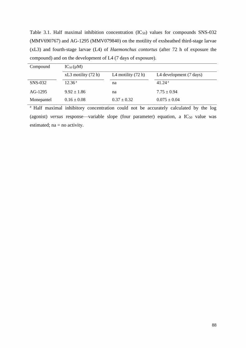
88
Table 3.1. Half maximal inhibition concentration (IC50) values for compounds SNS-032
(MMV690767) and AG-1295 (MMV079840) on the motility of exsheathed third-stage larvae
(xL3) and fourth-stage larvae (L4) of Haemonchus contortus (after 72 h of exposure the
compound) and on the development of L4 (7 days of exposure).
Compound IC50 (µM)
xL3 motility (72 h) L4 motility (72 h) L4 development (7 days)
SNS-032 12.36 a na 41.24 a
AG-1295 9.92 ± 1.86 na 7.75 ± 0.94
Monepantel 0.16 ± 0.08 0.37 ± 0.32 0.075 ± 0.04
a Half maximal inhibitory concentration could not be accurately calculated by the log
(agonist) versus response—variable slope (four parameter) equation, a IC50 value was
estimated; na = no activity.
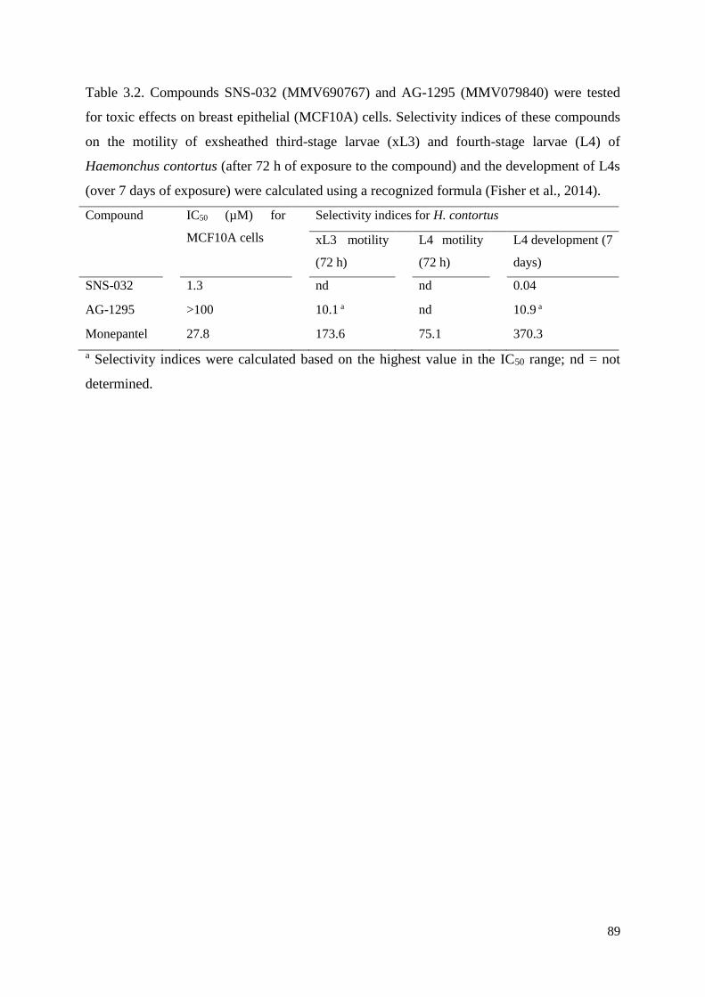
89
Table 3.2. Compounds SNS-032 (MMV690767) and AG-1295 (MMV079840) were tested
for toxic effects on breast epithelial (MCF10A) cells. Selectivity indices of these compounds
on the motility of exsheathed third-stage larvae (xL3) and fourth-stage larvae (L4) of
Haemonchus contortus (after 72 h of exposure to the compound) and the development of L4s
(over 7 days of exposure) were calculated using a recognized formula (Fisher et al., 2014).
Compound IC50 (µM) for
MCF10A cells
Selectivity indices for H. contortus
xL3 motility
(72 h)
L4 motility
(72 h)
L4 development (7
days)
SNS-032 1.3 nd nd 0.04
AG-1295 >100 10.1 a nd 10.9 a
Monepantel 27.8 173.6 75.1 370.3
a Selectivity indices were calculated based on the highest value in the IC50 range; nd = not
determined.
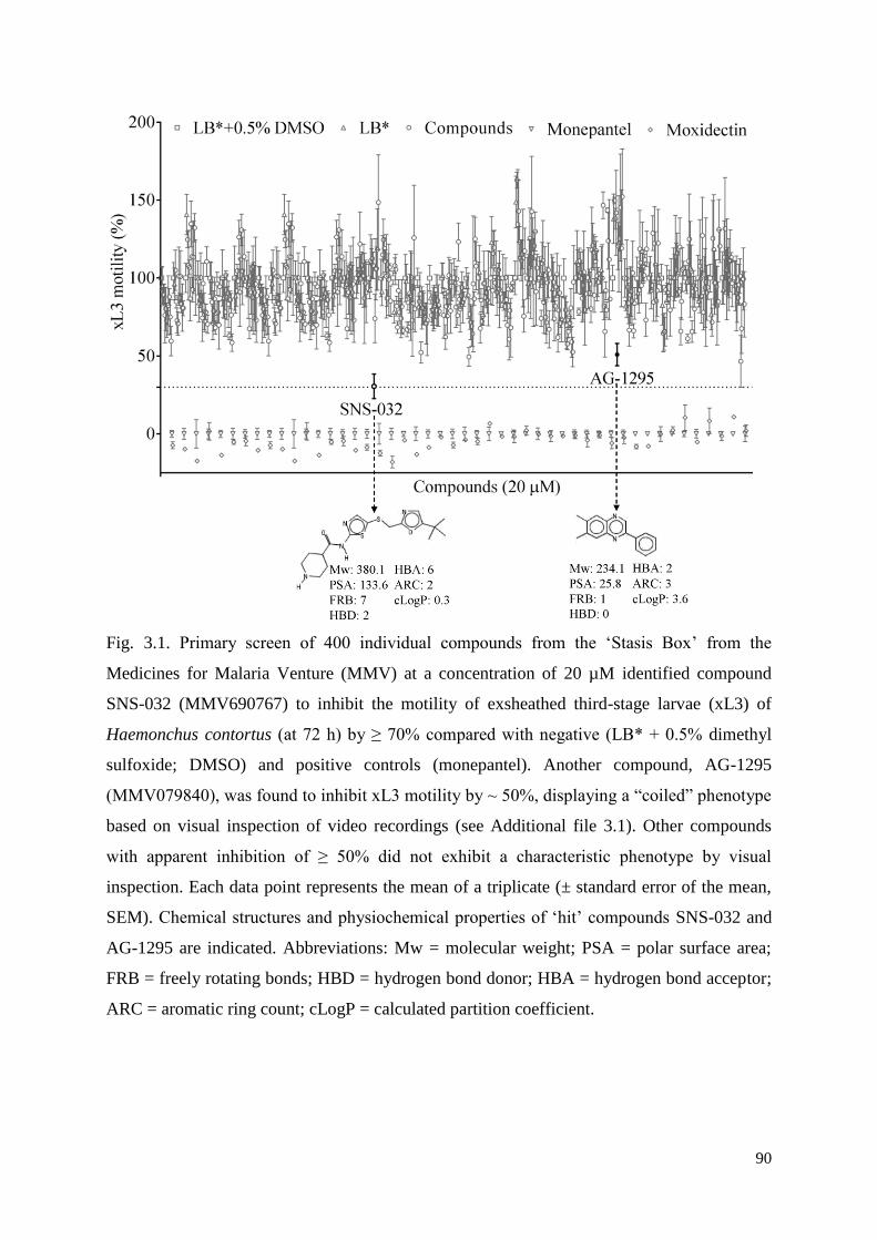
90
Fig. 3.1. Primary screen of 400 individual compounds from the ‘Stasis Box’ from the
Medicines for Malaria Venture (MMV) at a concentration of 20 µM identified compound
SNS-032 (MMV690767) to inhibit the motility of exsheathed third-stage larvae (xL3) of
Haemonchus contortus (at 72 h) by ≥ 70% compared with negative (LB* + 0.5% dimethyl
sulfoxide; DMSO) and positive controls (monepantel). Another compound, AG-1295
(MMV079840), was found to inhibit xL3 motility by ~ 50%, displaying a “coiled” phenotype
based on visual inspection of video recordings (see Additional file 3.1). Other compounds
with apparent inhibition of ≥ 50% did not exhibit a characteristic phenotype by visual
inspection. Each data point represents the mean of a triplicate (± standard error of the mean,
SEM). Chemical structures and physiochemical properties of ‘hit’ compounds SNS-032 and
AG-1295 are indicated. Abbreviations: Mw = molecular weight; PSA = polar surface area;
FRB = freely rotating bonds; HBD = hydrogen bond donor; HBA = hydrogen bond acceptor;
ARC = aromatic ring count; cLogP = calculated partition coefficient.
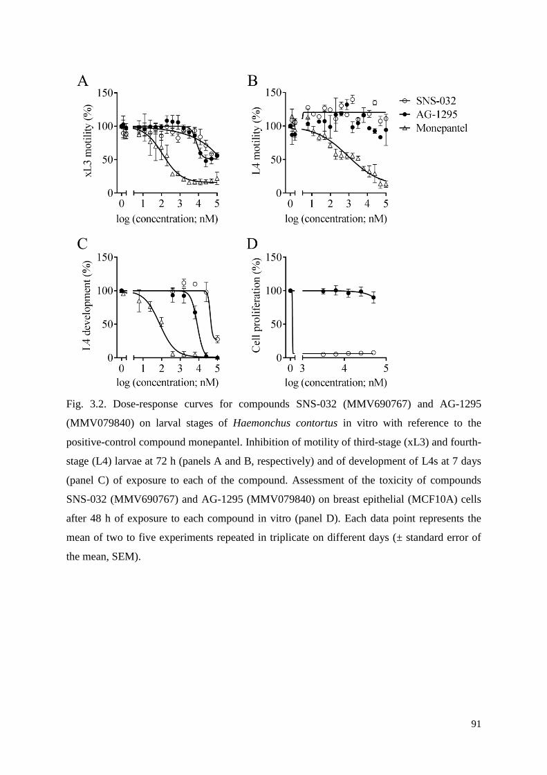
91
Fig. 3.2. Dose-response curves for compounds SNS-032 (MMV690767) and AG-1295
(MMV079840) on larval stages of Haemonchus contortus in vitro with reference to the
positive-control compound monepantel. Inhibition of motility of third-stage (xL3) and fourth-
stage (L4) larvae at 72 h (panels A and B, respectively) and of development of L4s at 7 days
(panel C) of exposure to each of the compound. Assessment of the toxicity of compounds
SNS-032 (MMV690767) and AG-1295 (MMV079840) on breast epithelial (MCF10A) cells
after 48 h of exposure to each compound in vitro (panel D). Each data point represents the
mean of two to five experiments repeated in triplicate on different days (± standard error of
the mean, SEM).
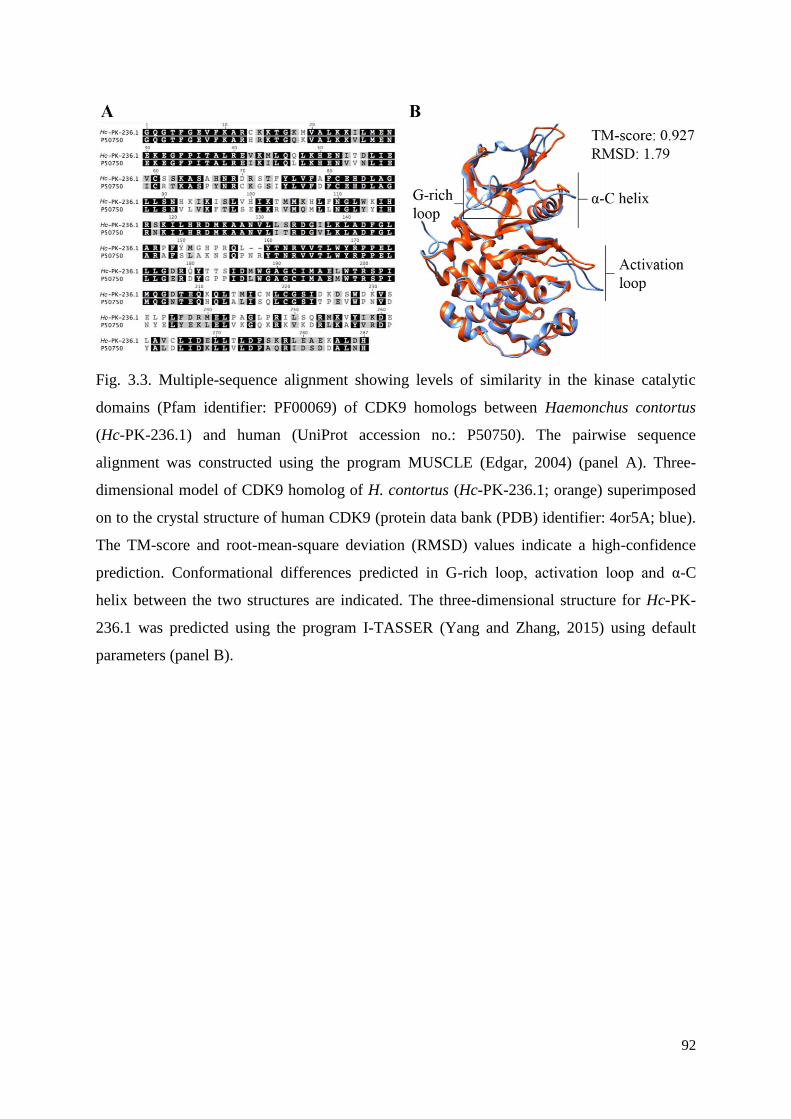
92
Fig. 3.3. Multiple-sequence alignment showing levels of similarity in the kinase catalytic
domains (Pfam identifier: PF00069) of CDK9 homologs between Haemonchus contortus
(Hc-PK-236.1) and human (UniProt accession no.: P50750). The pairwise sequence
alignment was constructed using the program MUSCLE (Edgar, 2004) (panel A). Three-
dimensional model of CDK9 homolog of H. contortus (Hc-PK-236.1; orange) superimposed
on to the crystal structure of human CDK9 (protein data bank (PDB) identifier: 4or5A; blue).
The TM-score and root-mean-square deviation (RMSD) values indicate a high-confidence
prediction. Conformational differences predicted in G-rich loop, activation loop and α-C
helix between the two structures are indicated. The three-dimensional structure for Hc-PK-
236.1 was predicted using the program I-TASSER (Yang and Zhang, 2015) using default
parameters (panel B).
93
Chapter 4 - An approved pesticide with major
anthelmintic activity against Haemonchus contortus
Abstract
There is a substantial need to develop new medicines against parasitic diseases via public-
private partnerships. Based on high throughput phenotypic screens of largely protozoal
pathogens and bacteria, the Medicines for Malaria Venture (MMV) has recently assembled
an open-access ‘Pathogen Box’ containing 400 well-curated chemical compounds. In the
present study, we tested these compounds for activity against parasitic stages of the nematode
Haemonchus contortus (barber’s pole worm). In an optimised, whole-organism screening
assay, using exsheathed third-stage (xL3) and fourth-stage (L4) larvae, we measured the
inhibition of larval motility, growth and development of H. contortus. We also studied the
effect of the ‘hit’ compound on mitochondrial function by measuring oxygen consumption.
Among the 400 ‘Pathogen Box’ compounds, we identified one chemical, called tolfenpyrad
(compound identification code: MMV688934) that reproducibly inhibits xL3 motility as well
as L4 motility, growth and development, with IC50 values ranging between 0.02 and 3 µM.
An assessment of mitochondrial function showed that xL3s treated with tolfenpyrad
consumed significantly less oxygen than untreated xL3s, which was consistent with specific
inhibition of complex I of the respiratory electron transport chain in arthropods. Given that
tolfenpyrad was developed as a pesticide and has already been tested for absorption,
distribution, excretion, biotransformation, toxicity and metabolism, it shows considerable
promise for hit-to-lead optimisation and/or repurposing for use against H. contortus and other
parasitic nematodes. Future work should assess its activity against hookworms and other
pathogens that cause neglected tropical diseases.
94
4.1 Introduction
Compounded by massive global food and water shortages and climate change, parasitic
illnesses, including neglected tropical diseases (NTDs; WHO, 2015), have a devastating,
long-term impact on human and animal health and welfare worldwide, and thus represent a
major global challenge. Together, NTDs infect more than one billion people worldwide,
resulting in an estimated loss of 26 million disability-adjusted life years (Hotez et al., 2014).
Despite their adverse socioeconomic impact, there are major limitations in the diagnosis,
treatment and control of NTDs. Currently, there are no commercial vaccines available against
most of these diseases (Pedrique et al., 2013; Hotez et al., 2016), diagnostic methods
frequently suffer from insufficient specificity and sensitivity (Utzinger et al., 2012; Assefa et
al., 2014), and treatments are often not highly effective and/or are toxic (Castro et al., 2006;
Witschel et al., 2012; Molina et al., 2015). In addition, often the small numbers of drugs (or
drug classes) frequently used, limited use of combination drug therapies and the
implementation of mass drug administration programs bear the risk of drug resistance
emerging in some groups of target pathogens (Humphries et al., 2012; Witschel et al., 2012;
Webster et al., 2014). Therefore, the development of new drugs is crucial to ensure effective
and sustained treatment and control into the future.
In spite of some success through the discovery of, for example, monepantel (Kaminsky et
al. 2008; Prichard and Geary 2008) and derquantel (Little et al., 2011), progress in
discovering new drugs against parasitic worms of animal health importance has been
relatively poor. Likely reasons for limited success beyond the lack of resources include an
over-confidence in the validation of molecular targets (enzymes and receptors) and in
studying an inappropriate developmental stage of a pathogen. However, key gaps include a
lack of readily available curated sets of compounds for targeted screening and subsequent
evaluation, limited cooperation among different areas (including parasitology, drug
discovery, medicinal chemistry and safety evaluation) which are essential to find starting
points for drug discovery, and to bring them to tangible and translational outcomes and
outputs.
In the late 1990s, an innovative collaboration model for research and development for
neglected diseases emerged in the form of public-private partnerships (PPPs) that came to be
known as product development partnerships (PDPs). A key example is the Medicines for
Malaria Venture (MMV), created from a desire to catalyse the discovery, development and
delivery of new medicines against malaria. Over the last decade, almost seven million
95
compounds have been tested in phenotypic assays against malaria, and this has resulted in a
solid pipeline of new preclinical and clinical candidates. In addition, an open science
initiative has made many of these structures available, and a collection of 400 key malaria
phenotypic ‘hits’, called the ‘Malaria Box’, was launched in 2013. Building on this model, in
December 2015, MMV took this a stage further, with an initiative to stimulate the discovery
of drugs for neglected parasitic diseases. The ‘Pathogen Box’ (www.pathogenbox.org),
contains 400 diverse drug-like molecules, and is provided at no cost to research groups.
Each of the 400 compounds in the ‘Pathogen Box’ has confirmed activity against one or
more key pathogens that cause some of the most socioeconomically important diseases
worldwide, including tuberculosis, malaria, sleeping sickness, leishmaniasis, schistosomiasis,
hookworm disease, toxoplasmosis and cryptosporidiosis. In addition, all compounds have
been tested for cytotoxicity, with compounds included in the library being at least 5-fold
more selective for the pathogen than its mammalian host. The complete set of compounds is
dispatched to laboratories around the world to boost drug discovery efforts. This initiative
provided us with a unique opportunity to assess these curated compounds for nematocidal
activity in a recently developed whole-organism screening assay (Preston et al., 2015, 2016).
Our aim was to rapidly screen all 400 compounds against parasitic stages of the barber’s pole
worm, Haemonchus contortus, and to identify hit compounds and characterise/assess them
for further evaluation as nematocidal candidates. This worm was used because it is one of the
best-studied members of a large order (Strongylida) of socioeconomically important
nematodes of animals, including humans, because there is extensive information available on
its biology and molecular biology, and because its genome and developmental transcriptome
have been characterised in detail (Gasser and von Samson-Himmelstjerna, 2016), providing a
foundation for drug discovery efforts.
4.2 Materials and methods
4.2.1 Procurement of H. contortus
The Haecon-5 strain of Haemonchus contortus, which is partially resistant to
benzimidazoles (Dr Jody Zawadzki, personal communication), was maintained in
experimental sheep as described previously (Schwarz et al., 2013; Preston et al., 2015) and in
accordance with the institutional animal ethics guidelines (permit no. 1413429; The
University of Melbourne, Australia). L3s were produced from H. contortus eggs by
incubating faeces from infected sheep at 27 °C for 1 week (Preston et al., 2015), sieved
96
through nylon mesh (pore size: 20 µm) to remove debris or dead larvae and then stored at 10
°C for a maximum of 3 months. For screening and basal oxygen consumption measurements
(see following sub-sections), L3s were exsheathed and sterilised in 0.15% v/v sodium
hypochlorite (NaClO) at 37 °C for 20 min (Preston et al., 2015). Thereafter, xL3s were
washed five times in sterile physiological saline by centrifugation at 1700 g (5 min) at 22–24
°C. Then, xL3s were immediately suspended in Luria Bertani medium [LB: 10 g of tryptone
(cat no. LP0042; Oxoid, England), 5 g of yeast extract (cat no. LP0042; Oxoid) and 5 g of
NaCl (cat. no. K43208004210; Merck, Denmark)] in 1 l of reverse-osmosis deionised water).
LB was autoclaved and supplemented with 100 IU/ml of penicillin, 100 µg/ml of
streptomycin and 2.5 µg/ml of amphotericin (Fungizone, antibiotic – antimycotic; cat. no.
15240-062; Gibco, USA); this supplemented LB was designated LB*. Fourth-stage larvae
(L4s) were produced from xL3s in vitro for 7 days at 38 °C and 10% CO2, as described by
Preston et al. (2015, 2016).
4.2.2 Screening of compounds
The Pathogen Box contains 400 compounds representing compounds that are active
against one or more of 12 distinct pathogens (http://www.pathogenbox.org/about-pathogen-
box/supporting-information). Individual compounds had only been tested to confirm activity
against the pathogen for which the compounds were first reported to be active, and have not
been tested against the other pathogens represented in the ‘Pathogen Box’. All compounds
have been tested for cytotoxicity; typically, they are five-fold less potent against a human
fibroblast cell line (MRC-5) than the pathogen (cf. Table 4.1); toxicity values are within
levels considered acceptable for an initial drug discovery programme
(www.pathogenbox.org/about-pathogen-box/supporting-information). Each of the 400
compounds was prepared as described previously (Preston et al., 2015) and screened (in
triplicate) at a concentration of 20 µM on xL3s of H. contortus in 96-well microculture plates
using two reference-control compounds, moxidectin and monepantel (Preston et al., 2015,
2016). In brief, compounds were dissolved to a stock concentration of 10 mM in dimethyl
sulfoxide (DMSO, Ajax Finechem, Australia). Compounds were individually diluted to the
final concentration of 20 µM using LB*, and dispensed (in triplicate) into wells of the
microculture plates using a multichannel pipette. In addition, the negative-controls (LB* and
LB* + 0.5% DMSO; six wells each), and positive-controls (final concentration of 20 µM of
monepantel [Zolvix, Novartis Animal Health, Switzerland] and 20 µM of moxidectin
97
[cydectin, Virbac, France]) were dispensed in triplicate wells. Then, xL3s (~300/well) were
dispensed into wells of the plate using an automated multichannel pipette (Viaflo Assist/II,
Integra Biosciences, Switzerland). Following an incubation for 72 h at 38 °C and 10% CO2, a
video recording (5 sec) was taken of each well of the 96-well microculture plate (containing
xL3s) using a grey-scale camera (Rolera bolt, Q imaging Scientific Coms, Canada), and a
motorised X-Y axis stage (BioPoint 2, Ludl Electronics Products, USA). Individual videos
were processed to calculate a motility index (MI) using an algorithm described previously
(Preston et al., 2015, 2016). MIs were normalised to the positive- and negative-controls (to
remove plate-to-plate variation) using the program Prism (v.6 GraphPad Software, USA). A
compound was recorded as having activity if it reduced xL3 motility by ≥ 70% after 72 h of
incubation.
4.2.3 Dose-response assessments of active compounds on xL3 and L4 motility,
and L4 growth and development
Anti-xL3 activity of any ‘hit’ compound was confirmed, and half maximum inhibitory
concentration (IC50) values estimated from dose-response curves (24 h, 48 h and 72 h).
Compounds that reduced the motility of xL3s were also tested for their ability to inhibit the
development of xL3s to L4s, the motility of L4s and/or their ability to retard L4 growth as
described previously (Preston et al., 2015, 2016). In brief, growth retardation and
morphological alterations in L4s exposed for 48 h to LB* containing either 1% DMSO
(negative-control), 100 µM of each tolfenpyrad (test compound), moxidectin (positive-
control) or monepantel (positive-control) were assessed microscopically (20-100 x
magnification). For each treatment, the mean width of 30 L4s ± the standard error of the
mean (SEM) was calculated, and a non-parametric (Kruskal-Wallis) and Dunn’s multiple
comparisons test was used to calculate statistical difference between treatments. All assays
(xL3 motility, and L4 development, growth and motility) were performed in triplicate,
between 3-5 times on different days. To determine IC50 values, the data from each assay (xL3
motility, L4 motility and development) were converted to a percentage with reference to the
negative-control (LB* + 0.5% DMSO), and IC50 values determined using a variable slope
four-parameter equation, constraining the top value to 100% and using a least squares
(ordinary) fit model (v.6 GraphPad Software). Selectivity indices (SIs) were calculated using
a recognised formula (SI = human fibroblast (MRC-5) cells IC50 / H. contortus IC50; Fisher et
al., 2014) employing cytotoxicity data linked to the Pathogen Box compounds.
98
4.2.4 Measurement of basal oxygen consumption
Following standardisation using an established protocol, the basal oxygen consumption
(‘respiratory’) rate in xL3s was measured using the Seahorse XF24 flux analyser (Seahorse
Biosciences, USA) (McGee et al., 2011). In brief, 450 µl of LB* + 1% DMSO containing 100
µM of tolfenpyrad (‘hit’ compound) or 100 µM moxidectin (control; not known to inhibit
respiration), or LB* + 1% DMSO alone (untreated control) were transferred in quadruplicate
to the wells of a 24-well plate (Seahorse XF24). Then, 1000 xL3s in 50 µl LB* were added to
the wells, and the plates pre-incubated for 2 h, equilibrated for 30 min and oxygen
consumption measured using the flux analyser. Seven measurements were taken over a 1 h-
period (protocol: 2 min-mix, 2 min-pause and 4 min-measure; McGee et al., 2011).
Experiments were repeated twice. A two-way repeated measures ANOVA with a Dunnett’s
multiple comparisons test (v.6 GraphPad Prism) was used to assess the statistical difference
in oxygen consumption between treated and untreated xL3s.
4.3 Results
In the primary screen of the 400 compounds (Fig. 4.1; Supplementary file 4.1), one
compound (tolfenpyrad; Compound ID: MMV688934; batch: MMV688934-01;
pubchem.ncbi.nlm.nih.gov/compound/10110536) was recorded to inhibit xL3 motility by ≥
70%. Although there are benzimidazole-based compounds in the Pathogen Box, nematocidal
activity was not detected using this ≥ 70% threshold, because the Haecon-5 strain of H.
contortus is partially resistant to this class of chemicals. Subsequent assays using xL3s and
L4s of H. contortus showed that the potency of tolfenpyrad, measured as IC50 values, ranged
from 0.02 to 3 μM (Fig. 4.1; Table 4.1). In comparison to moxidectin and monepantel,
tolfenpyrad was able to reduce motility earlier than monepantel, with inhibition occurring
after 2 h of exposure and an IC50 value of 2.3 μM at 24 h compared with 52.1 μM for
monepantel (Fig. 4.3). Tolfenpyrad and moxidectin were found to have a similar inhibitory
effect on xL3 motility at the time points tested (Table 4.1). Furthermore, when examining the
inhibitory activity of the compounds on L4 motility, tolfenpyrad and moxidectin had lower
IC50 values at 24 h, 48 h and 72 h compared with that of monepantel (Table 4.1). In the L4
development assay, tolfenpyrad had a greater inhibitory effect on the development of xL3 to
L4 (IC50 of 0.06 μM) than did moxidectin and monepantel (IC50 of 12.3 μM and 0.4 μM,
respectively). Light microscopic examination of parasitic larvae revealed morphological
99
damage (shrivelled and granulated appearance) in L4s following exposure in vitro (48 h) to
tolfenpyrad and monepantel (Fig. 4.2), but not in xL3s (data not shown). L4s exposed to
tolfenpyrad were significantly thinner than ‘untreated’ controls, and had a similar width to
moxidectin-exposed, but not as pronounced as monepantel-exposed larvae (Fig. 4.2). Finally,
it was assessed whether tolfenpyrad would inhibit respiration in H. contortus xL3s, as it does
in arthropods by targeting complex I of the respiratory electron transport chain (Song et al.,
2013), by measuring oxygen consumption over time (Fig. 4.3). The results showed that
tolfenpyrad-treated xL3s consumed substantially (P<0.05) less oxygen than both moxidectin-
treated and untreated xL3s (Fig. 4.3).
4.4 Discussion
The screening of the Pathogen Box compounds identified one chemical, tolfenpyrad, with
major activity against parasitic larval stages (xL3 and L4) of H. contortus in vitro; IC50 values
were comparable with those of two commercially available anthelmintics, monepantel and
moxidectin. Tolfenpyrad, a pyrazole-5-carboxamide insecticide, is the International
Organization for Standardization – approved name for 4-chloro-3-ethyl-1-methyl-N-[4-(p-
tolyloxy)benzyl]pyrazole-5-carboxamide (International Union of Pure and Applied
Chemistry, IUPAC), which has the Chemical Abstracts Service number 129558-76-5.
Tolfenpyrad was included in the MMV ‘Pathogen Box’ based on results from a compound
screen of agro-chemicals, which showed that tolfenpyrad has potent and selective in vitro
activity against Trypanosoma spp. (Witschel et al., 2012). As a pesticide, tolfenpyrad has
relatively broad activity against egg, larval, nymphal and adult stages of various arthropods
(including Hemiptera, Coleoptera, Diptera, Lepidoptera, Thysanoptera and Acarina), and has
been applied to various infested crops (Song et al., 2012; Yamaguchi et al., 2012). This
chemical was developed in Japan and was first approved in 2002; it has been registered for
commercial use in several countries other than Japan, including the Dominican Republic,
Thailand, the United Arab Emirates, Indonesia and the USA (Yamaguchi et al., 2012).
In the present study, tolfenpyrad was shown to be more potent against L4s than xL3s
based on IC50 values (motility and development), with substantial cuticular damage to the L4
but not to the xL3 stage. Compared with untreated-control L4s, the width of tolfenpyrad-
treated L4s were significantly reduced as were moxidectin- and monepantel-treated control
L4s, indicating that treatment suppresses parasite growth and development. It is unclear
whether the difference in potency of tolfenpyrad between the two larval stages tested is due
100
to variation in drug uptake, as at the L4 stage has a functioning pharynx (Sommerville, 1966),
or the mode of action. The mode of action of this chemical (as a contact insecticide and
fungicide) is likely linked to the specific inhibition of complex I of the respiratory electron
transport chain in mitochondria (Lummen, 1998; Song et al., 2013), such that it is effective
against various pests that are resistant to insecticides, including organophosphates and
carbamates, which have modes of action that are entirely distinct from tolfenpyrad.
To explore whether tolfenpyrad might act as an inhibitor of the electron transport chain in
H. contortus, oxygen consumption was measured in xL3s in the presence or absence of
tolfenpyrad following a pre-exposure for 2 h to the compound. This approach is routinely
used to evaluate mitochondrial function in Caenorhabditis elegans and mammalian tissue by
detecting oxygen consumption in real-time using oxygen-sensitive probes (McGee et al.,
2011; Andreux et al., 2014). In the present study, we elected to pre-treat the xL3s of H.
contortus for 2 h prior to the initial measurement of oxygen consumption, as both tolfenpyrad
and moxidectin significantly inhibit motility following immediate exposure. The results
revealed a significant decrease in oxygen consumption in xL3s exposed to tolfenpyrad
compared with untreated controls. Similarly, oxygen consumption was reduced in
tolfenpyrad-treated than moxidectin-treated xL3s, which was also associated with reduced
motility following the exposure for 2 h. This discrepancy in oxygen consumption between
these treatments indicates that tolfenpyrad is also a complex I mitochondrial electron
transport inhibitor in H. contortus.
Although mainly used against agricultural pests, tolfenpyrad had not been assessed
previously for use against endoparasites, such as parasitic worms, of animals. A report was
prepared by the Joint FAO/WHO Meeting on Pesticide Residues at the request of the Codex
Committee on Pesticide Residues in 2013 (FAO, 2013). This report extensively reviewed and
appraised many aspects of tolfenpyrad, including: (i) acceptable daily intake; (ii) absorption,
distribution, excretion and biotransformation; (iii) toxicity studies (acute; short and long-
term; carcinogenicity; genotoxicity; reproductive, developmental and neural toxicity), and
(iv) studies of metabolites. The main conclusions from this report were: (i) the acceptable
daily intake of tolfenpyrad is 0-0.006 mg/kg in mammals; (ii) following oral administration,
tolfenpyrad is rapidly absorbed, widely distributed and metabolized by the liver, with 88-93%
being excreted in the faeces, and (iii) tolfenpyrad was not found to be carcinogenic, genotoxic
or neurotoxic, but was found to have some reproductive and developmental toxicity.
Although some intoxications (with excessive exposure/ingestion) have been recorded in
humans (e.g., Yamaguchi et al., 2012; Hikiji et al., 2013), well-controlled risk assessment
101
studies in monogastric mammals (mouse, rat, rabbit and dog) have shown that the no-
observed-adverse-effect levels (NOAELs) of tolfenpyrad are between 1-1.5 mg/kg body
weight per day, with lowest-observed-adverse-effect levels (LOAEL) of 5-21 mg/kg body
weight per day (FAO, 2013). Therefore, based on the evidence presented in this report and
associated literature (see FAO, 2013) as well as the high selectivity of tolfenpyrad for H.
contortus (see Table 4.1), we propose to test tolfenpyrad in vivo in sheep against H. contortus
and/or related parasitic nematodes at dosages between 1 and 5 mg/kg body weight. We also
propose to assess its activity in vitro against the hookworms Ancylostoma ceylanicum and
Necator americanus of humans and other NTD pathogens (London Declaration, 2012).
Nonetheless, it will be important to also extend medicinal chemistry work to establish
whether a safer and more effective derivative of this chemical might be synthesized. Given
that tolfenpyrad was developed as a pesticide and has already been tested for absorption,
distribution, excretion, biotransformation, toxicity and metabolism, there is considerable
promise for the repurposing of this chemical for use against H. contortus and other parasitic
nematodes. Further work should now focus on assessing the activity of tolfenpyrad against
hookworms and other worms that cause NTDs.
102
4.5 References
Andreux, P.A., Mouchiroud, L., Wang, X., Jovaisaite, V., Mottis, A., Bichet., S, Moullan N.,
Houtkooper, R.H., Auwerx, J., 2014. A method to identify and validate mitochondrial
modulators using mammalian cells and the worm C. elegans. Sci. Rep. 4, 5285.
Assefa, L.M., Crellen, T., Kepha, S., Kihara, J.H., Njenga S.M., Pullan R.L., Brooker, S.J.,
2014. Diagnostic accuracy and cost-effectiveness of alternative methods for detection of
soil-transmitted helminths in a post-treatment setting in Western Kenya. PLoS. Negl.
Trop. Dis. 8, e2843.
Castro, J.A., de Mecca, M.M., Bartel, L.C., 2006. Toxic side effects of drugs used to treat
Chagas' disease (American trypanosomiasis). Hum. Exp. Toxicol. 25, 471-479.
FAO, 2013. Pesticide residues in food 2013. Report from the joint meeting of the FAO panel
of experts on pesticide residues in food and the environment and the WHO core
assessment group on pesticide residues Geneva, Switzerland (17 to 26 September 2013;
http://www.fao.org/publications/card/en/c/299ca869-ae51-5093-8407-9cb30782b9f5),
2013. ISBN: 9789251080436. Accessed 19 March 2016.
Fisher, G.M., Tanpure, A.D., Andrews, K.T., Poulsen, S.A., 2014. Synthesis and evaluation
of antimalarial properties of novel 4-aminoquinoline hybrid compounds. Chem. Biol.
Drug Des. 84, 462-472.
Gasser, R.B., von Samson-Himmelstjerna, G., 2016. Haemonchus contortus and
Haemonchosis Past, Present and Future Trends. Adv. Parasitol. Volume 93.
Hikiji, W., Yamaguchi, K., Saka, K., Hayashida, M., Ohno, Y., Fukunaga, T., 2013. Acute
fatal poisoning with tolfenpyrad. J. Forensic Leg. Med. 8, 962-964.
Hotez, P.J., Bottazzi, M.E., Strych, U., 2016. New vaccines for the world's poorest people.
Annu. Rev. Med. 67, 405-417.
Hotez, P.J., Alvarado, M., Basáñez, M.G., Bolliger, I., Bourne, R., Boussinesq, M., Brooker,
S.J., Brown, A.S., Buckle, G., Budke, C.M., Carabin, H., Coffeng, L.E., Fèvre, E.M.,
Fürst, T., Halasa, Y.A., Jasrasaria, R., Johns, N.E., Keiser, J., King, C.H., Lozano, R.,
Murdoch, M.E., O'Hanlon, S., Pion, S.D.S., Pullan, R.L., Ramaiah, K.D., Roberts, T.,
Shepard, D.S., Smith, J.L., Stolk, W.A., Undurraga, E.A., Utzinger, J., Wang, M.,
Murray, C.J.L., Naghavi, M., 2014. The global burden of disease study 2010:
interpretation and implications for the neglected tropical diseases. PLoS Negl. Trop. Dis.
8, e2865.
103
Humphries, D., Nguyen, S., Boakye, D., Wilson, M., Cappello, M., 2012. The promise and
pitfalls of mass drug administration to control intestinal helminth infections. Curr. Opin.
Infect. Dis. 25, 584-589.
Kaminsky, R., Ducray, P., Jung, M., Clover, R., Rufener, L., Bouvier, J., Weber, S.S.,
Wenger, A., Wieland-Berghausen, S., Goebel, T., Gauvry, N., Pautrat, F., Skripsky, T.,
Froelich, O., Komoin-Oka, C., Westlund, B., Sluder, A., Maser, P., 2008. A new class of
anthelmintics effective against drug-resistant nematodes. Nature 452, 176-180.
Little, P.R., Hodge, A., Maeder, S.J., Wirtherle, N.C., Nicholas, D.R., Cox, G.G., Conder,
G.A., 2011. Efficacy of a combined oral formulation of derquantel–abamectin against the
adult and larval stages of nematodes in sheep, including anthelmintic-resistant strains.
Vet. Parasitol. 181, 180-193.
London Declaration, 2012. Uniting to combat neglected tropical diseases.
http://unitingtocombatntds.org/resource/london-declaration. Accessed 27 July 2016.
Lummen, P., 1998. Complex I inhibitors as insecticides and acaricides. Biochem. Biophys.
Acta 1364, 287-296.
McGee, S.L., Sadli, N., Morrison, S., Swinton, C., Suphioglu, C., 2011. DHA protects
against zinc mediated alterations in neuronal cellular bioenergetics. Cell. Physiol.
Biochem. 28, 157-162.
Molina, I., Salvador, F., Sanchez-Montalva, A., Trevino, B., Serre, N., Sao Aviles, A.,
Almirante, B., 2015. Toxic profile of benznidazole in patients with chronic chagas
disease: risk factors and comparison of the product from two different manufacturers.
Antimicrob. Agents Chemother. 59, 6125-6131.
Pedrique, B., Strub-Wourgaft, N., Some, C., Olliaro, P., Trouiller, P., Ford, N., Pecoul, B.,
Bradol, J.H., 2013. The drug and vaccine landscape for neglected diseases (2000-11): a
systematic assessment. Lancet Glob. Health 1, e371-e379.
Preston, S., Jabbar, A., Nowell, C., Joachim, A., Ruttkowski, B., Baell, J., Cardno, T.,
Korhonen, P.K., Piedrafita, D., Ansell, B.R., Jex, A.R., Hofmann, A., Gasser, R.B.,
2015. Low cost whole-organism screening of compounds for anthelmintic activity. Int. J.
Parasitol. 45, 333-343.
Preston, S., Jabbar, A., Nowell, C., Joachim, A., Ruttkowski, B., Cardno, T., Hofmann, A.,
Gasser, R.B., 2016. Practical and low cost whole-organism motility assay: A step-by-step
protocol. Mol. Cell. Probes 30, 13-17.
Prichard, R.K., Geary, T.G., 2008. Drug discovery: fresh hope to can the worms. Nature 452,
157-158.
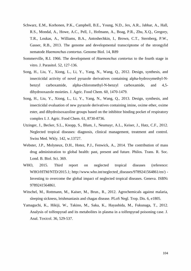
104
Schwarz, E.M., Korhonen, P.K., Campbell, B.E., Young, N.D., Jex, A.R., Jabbar, A., Hall,
R.S., Mondal, A., Howe, A.C., Pell, J., Hofmann, A., Boag, P.R., Zhu, X.Q., Gregory,
T.R., Loukas, A., Williams, B.A., Antoshechkin, I., Brown, C.T., Sternberg, P.W.,
Gasser, R.B., 2013. The genome and developmental transcriptome of the strongylid
nematode Haemonchus contortus. Genome Biol. 14, R89
Sommerville, R.I. 1966. The development of Haemonchus contortus to the fourth stage in
vitro. J. Parasitol. 52, 127-136.
Song, H., Liu, Y., Xiong, L., Li, Y., Yang, N., Wang, Q., 2012. Design, synthesis, and
insecticidal activity of novel pyrazole derivatives containing alpha-hydroxymethyl-N-
benzyl carboxamide, alpha-chloromethyl-N-benzyl carboxamide, and 4,5-
dihydrooxazole moieties. J. Agric. Food Chem. 60, 1470-1479.
Song, H., Liu, Y., Xiong, L., Li, Y., Yang, N., Wang, Q., 2013. Design, synthesis, and
insecticidal evaluation of new pyrazole derivatives containing imine, oxime ether, oxime
ester, and dihydroisoxazoline groups based on the inhibitor binding pocket of respiratory
complex I. J. Agric. Food Chem. 61, 8730-8736.
Utzinger, J., Becker, S.L., Knopp, S., Blum, J., Neumayr, A.L., Keiser, J., Hatz, C.F., 2012.
Neglected tropical diseases: diagnosis, clinical management, treatment and control.
Swiss Med. Wkly. 142, w.13727.
Webster, J.P., Molyneux, D.H., Hotez, P.J., Fenwick, A., 2014. The contribution of mass
drug administration to global health: past, present and future. Philos. Trans. R. Soc.
Lond. B. Biol. Sci. 369.
WHO, 2015. Third report on neglected tropical diseases (reference:
WHO/HTM/NTD/2015.1; http://www.who.int/neglected_diseases/9789241564861/en/) -
Investing to overcome the global impact of neglected tropical diseases. Geneva. ISBN:
9789241564861.
Witschel, M., Rottmann, M., Kaiser, M., Brun., R., 2012. Agrochemicals against malaria,
sleeping sickness, leishmaniasis and chagas disease. PLoS Negl. Trop. Dis. 6, e1805.
Yamaguchi, K., Hikiji, W., Takino, M., Saka, K., Hayashida, M., Fukunaga, T., 2012.
Analysis of tolfenpyrad and its metabolites in plasma in a tolfenpyrad poisoning case. J.
Anal. Toxicol. 36, 529-537.
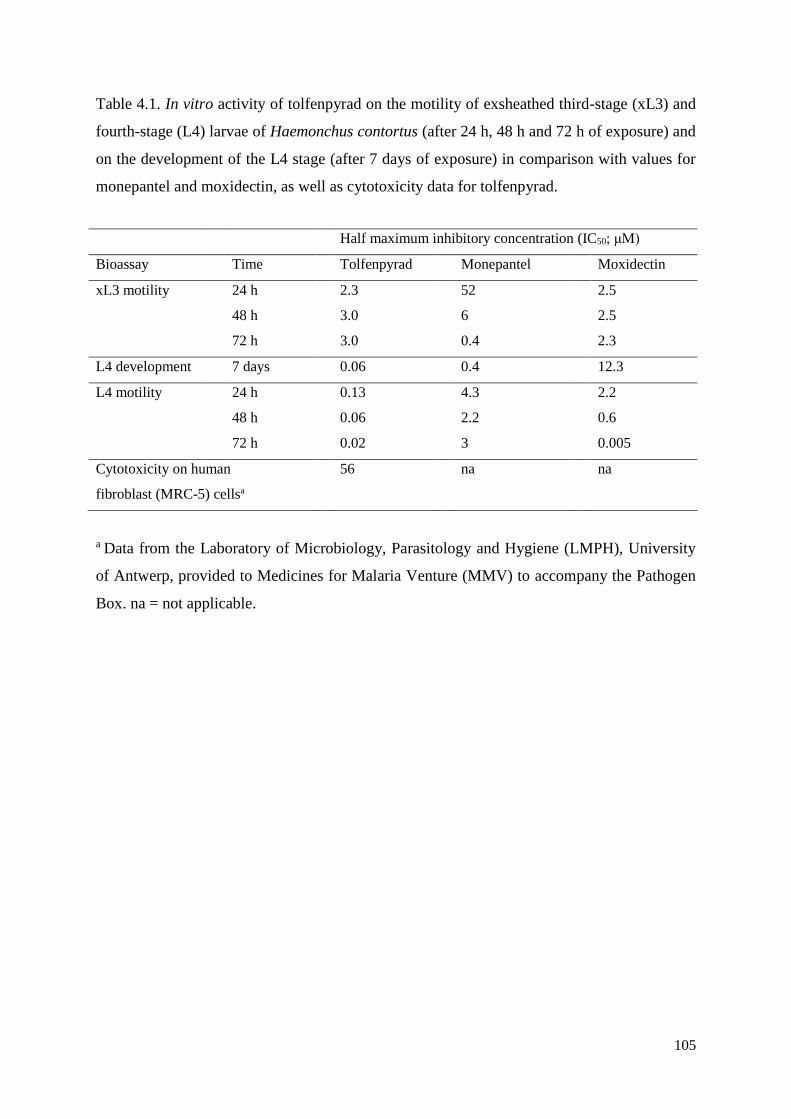
105
Table 4.1. In vitro activity of tolfenpyrad on the motility of exsheathed third-stage (xL3) and
fourth-stage (L4) larvae of Haemonchus contortus (after 24 h, 48 h and 72 h of exposure) and
on the development of the L4 stage (after 7 days of exposure) in comparison with values for
monepantel and moxidectin, as well as cytotoxicity data for tolfenpyrad.
a Data from the Laboratory of Microbiology, Parasitology and Hygiene (LMPH), University
of Antwerp, provided to Medicines for Malaria Venture (MMV) to accompany the Pathogen
Box. na = not applicable.
Half maximum inhibitory concentration (IC50; μM)
Bioassay Time Tolfenpyrad Monepantel Moxidectin
xL3 motility 24 h 2.3 52 2.5
48 h 3.0 6 2.5
72 h 3.0 0.4 2.3
L4 development 7 days 0.06 0.4 12.3
L4 motility 24 h 0.13 4.3 2.2
48 h 0.06 2.2 0.6
72 h 0.02 3 0.005
Cytotoxicity on human
fibroblast (MRC-5) cellsa
56 na na
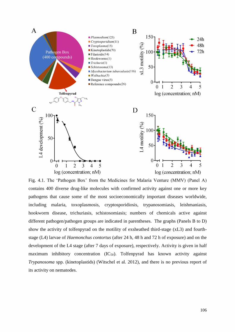
106
Fig. 4.1. The ‘Pathogen Box’ from the Medicines for Malaria Venture (MMV) (Panel A)
contains 400 diverse drug-like molecules with confirmed activity against one or more key
pathogens that cause some of the most socioeconomically important diseases worldwide,
including malaria, toxoplasmosis, cryptosporidiosis, trypanosomiasis, leishmaniasis,
hookworm disease, trichuriasis, schistosomiasis; numbers of chemicals active against
different pathogen/pathogen groups are indicated in parentheses. The graphs (Panels B to D)
show the activity of tolfenpyrad on the motility of exsheathed third-stage (xL3) and fourth-
stage (L4) larvae of Haemonchus contortus (after 24 h, 48 h and 72 h of exposure) and on the
development of the L4 stage (after 7 days of exposure), respectively. Activity is given in half
maximum inhibitory concentration (IC50). Tolfenpyrad has known activity against
Trypanosoma spp. (kinetoplastids) (Witschel et al. 2012), and there is no previous report of
its activity on nematodes.
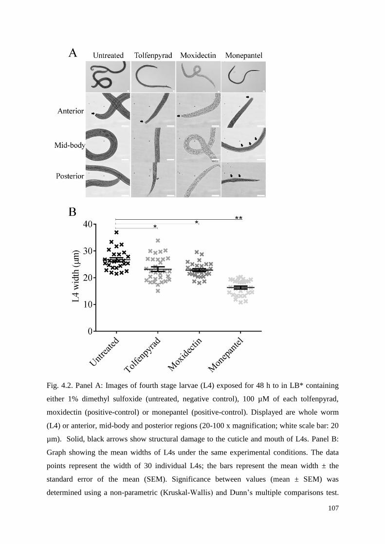
107
Fig. 4.2. Panel A: Images of fourth stage larvae (L4) exposed for 48 h to in LB* containing
either 1% dimethyl sulfoxide (untreated, negative control), 100 µM of each tolfenpyrad,
moxidectin (positive-control) or monepantel (positive-control). Displayed are whole worm
(L4) or anterior, mid-body and posterior regions (20-100 x magnification; white scale bar: 20
µm). Solid, black arrows show structural damage to the cuticle and mouth of L4s. Panel B:
Graph showing the mean widths of L4s under the same experimental conditions. The data
points represent the width of 30 individual L4s; the bars represent the mean width ± the
standard error of the mean (SEM). Significance between values (mean ± SEM) was
determined using a non-parametric (Kruskal-Wallis) and Dunn’s multiple comparisons test.

108
*/** indicate which values are significantly different from one another (P<0.05 and P<0.01,
respectively).
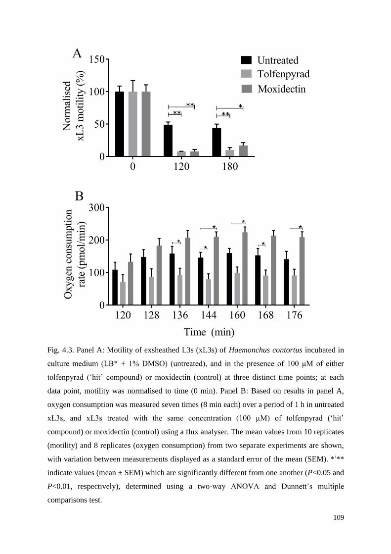
109
Fig. 4.3. Panel A: Motility of exsheathed L3s (xL3s) of Haemonchus contortus incubated in
culture medium (LB* + 1% DMSO) (untreated), and in the presence of 100 μM of either
tolfenpyrad (‘hit’ compound) or moxidectin (control) at three distinct time points; at each
data point, motility was normalised to time (0 min). Panel B: Based on results in panel A,
oxygen consumption was measured seven times (8 min each) over a period of 1 h in untreated
xL3s, and xL3s treated with the same concentration (100 µM) of tolfenpyrad (‘hit’
compound) or moxidectin (control) using a flux analyser. The mean values from 10 replicates
(motility) and 8 replicates (oxygen consumption) from two separate experiments are shown,
with variation between measurements displayed as a standard error of the mean (SEM). */**
indicate values (mean ± SEM) which are significantly different from one another (P<0.05 and
P<0.01, respectively), determined using a two-way ANOVA and Dunnett’s multiple
comparisons test.
110
Chapter 5 - Assessing the anthelmintic activity of
pyrazole-5-carboxamide derivatives against Haemonchus
contortus
Abstract
In this study, we tested five series of pyrazole-5-carboxamide compounds (n = 55) for
activity against parasitic stages of the nematode Haemonchus contortus (barber’s pole worm).
In an optimised, whole-organism screening assay, using exsheathed third-stage (xL3) and
fourth-stage (L4) larvae, we measured the inhibition of larval motility and development of H.
contortus. Amongst the 55 compounds, we identified two compounds (designated a-15 and a-
17) that reproducibly inhibit xL3 motility as well as L4 motility and development, with IC50
values ranging between ~ 3.4 and 55.6 µM. We studied the effect of these two ‘hit’
compounds on mitochondrial function by measuring oxygen consumption. This assessment
showed that xL3s exposed to each of these compounds consumed significantly less oxygen
and had less mitochondrial activity than untreated xL3s, which was consistent with specific
inhibition of complex I of the respiratory electron transport chain in arthropods. The present
findings provide a sound basis for future work aiming to identify the targets of compounds a-
15 and a-17, and to establish the modes of action of these chemicals in H. contortus.
111
5.1 Introduction
Synthetic pyrazole-5-carboxamide derivatives, such as tebufenpyrad and tolfenpyrad, are
important pesticides which are recognized to inhibit complex I of the mitochondrial electron
transport (respiratory) chain (Miyoshi, 1998). Tebufenpyrad, which was first discovered by
Mitsubishi Kasei Co., Ltd., in 1987, has known activity against selected Homoptera and
phytophagous mites (Okada et al., 1991). Tolfenpyrad was discovered by Mitsubishi
Chemical Corporation (now Nihon Nohyaku Co., Ltd.) and developed for the control of
various agricultural pests, including Acarina, Coleoptera, Diptera, Hemiptera, Lepidoptera
and Thysanoptera (Arthropoda); the latter chemical is active mainly upon contact with egg,
larval, nymphal and/or adult stages (Okada et al., 1999). Because of the effectiveness of these
two pyrazole-5-carboxamides in controlling such agricultural pests (Okada et al., 1996,
1999), there has been a considerable commercial interest in synthesizing various derivatives
based on their structures, with changes being made to the pyrazole and/or benzene rings
(Okada et al., 1989; Okada et al., 1990; Natsume et al., 1991; Okada et al., 1992; Okada et
al., 1996; Okada et al., 1997; Kano et al., 1999; Okada et al., 2002), but little work on altering
the chemical bridge between the pyrazole and benzene rings.
Although these pyrazole-5-carboxamide derivatives have been developed to kill arthropod
pests, we recently showed, in a compound screen of the ‘Pathogen Box’
(www.pathogenbox.org) from the Medicines for Malaria Ventures (MMV; www.mmv.org),
that tolfenpyrad has an exquisite in vitro activity against parasitic stages of the barber’s pole
worm, Haemonchus contortus (Nematoda: Strongylida; Preston et al., 2016). Indeed,
tolfenpyrad reproducibly and irreversibly inhibits the motility of exsheathed third-stage
(xL3s) and fourth-stage larvae (L4s) of this parasitic nematode, and also the growth and
development of L4s, with IC50 values ranging between 0.03 and 3.1 µM after 72 h of
exposure. We demonstrated that xL3s exposed to tolfenpyrad consumed significantly less
oxygen than unexposed xL3s, which was consistent with a specific inhibition of complex I of
the respiratory electron transport chain in the mitochondrion (cf. Miyoshi, 1998). In addition,
this compound is estimated to be ≥ 18-fold more selective for this nematode than a
mammalian cell line (Preston et al., 2016), indicating its potential for repurposing against (at
least some) parasitic nematodes and/or hit-to-lead optimisation.
In general terms, this evidence suggested that some pyrazole-5-carboxamide compounds
developed as agricultural pesticides (against arthropods of plants) might also be able to be
repurposed to other ecdysozoans, such as nematodes, provided that they are sufficiently safe
112
for application/administration to animals and/or the environment. Therefore, we undertook a
search for panels of two published series of novel pyrazole-5-carboxamides (Song et al.,
2012, 2013) and three other series of new pyrazole-5-carboxamides analogues. The
availability of this small library provided us with an opportunity to extend our recent study
(Preston et al., 2016). Here, we evaluated the activity of these pyrazole-5-carboxamide
derivatives (n = 55) against parasitic stages of the nematode H. contortus and compared their
potency with the two original, commercially available chemicals, tolfenpyrad and
tebufenpyrad. We used this whole-organism screening assay to measure the inhibition of
larval motility and development of H. contortus and then investigated the effect of any active
compound on mitochondrial function by measuring oxygen consumption in this nematode.
5.2 Materials and methods
5.2.1 Procurement of H. contortus
The Haecon-5 strain of H. contortus was maintained in experimental sheep as described
previously (Preston et al., 2015) and in accordance with the institutional animal ethics
guidelines (permit no. 1413429; The University of Melbourne, Australia). L3s were produced
from H. contortus eggs by incubating humidified faeces from infected sheep at 27 °C for 1
week (Preston et al., 2015), sieved through nylon mesh (pore size: 20 µm) to remove debris
or dead larvae and then stored at 10 °C for a maximum of 3 months. For screening and basal
oxygen consumption measurements (see following sub-sections), L3s were exsheathed and
sterilised in 0.15% v/v sodium hypochlorite (NaClO) at 37 °C for 20 min (Preston et al.,
2015). Thereafter, xL3s were washed five times in sterile physiological saline by
centrifugation at 1700 g (5 min) at 22–24 °C. Then, xL3s were immediately suspended in
Luria Bertani medium [LB: 10 g of tryptone (cat no. LP0042; Oxoid, Hampshire, England), 5
g of yeast extract (cat no. LP0021; Oxoid) and 5 g of NaCl (cat. no. 1064045000; Merck,
Kenilworth, NJ, USA)] in 1 l of reverse-osmosis deionised water). LB was autoclaved and
supplemented with 100 IU/ml of penicillin, 100 µg/ml of streptomycin and 0.25 µg/ml of
amphotericin (Fungizone, antibiotic – antimycotic; cat. no. 15240-062; Carlsbad, CA, USA);
this supplemented LB was designated LB*.
5.2.2 Pyrazole-5-carboxamide compounds
A total of 55 analogues of tebufenpyrad and tolfenpyrad were derivatised (Supplementary
file 5.1). In brief, pyrazole derivatives containing α-hydroxymethyl-N-benzyl carboxamide,
113
α-chloromethyl-N-benzyl carboxamide and 4,5-dihydrooxazole moieties (a-1 to a-23) (Song
et al., 2012), carbohydrazide (b-1 to b-7), imine, oxime ether, oxime ester, and
dihydroisoxazoline (c-1 to c-15) (Song et al., 2013), oxazole (d-1 to d-8) and e-1 to e-2 (this
study) were designed and synthesized (Supplementary file 5.1). In addition, tolfenpyrad
(IUPAC name: 4-chloro-N-[[4-(1,1-dimethylethyl)phenyl]methyl]-3-ethyl-1-methyl-1H-
pyrazole-5-carboxamide, cat. no. T535325, Toronto Research Chemicals, Toronto, Ontario,
Canada) and tebufenpyrad (IUPAC name: 4-chloro-3-ethyl-1-methyl-N-[4-(p-tolyloxy)
benzyl]pyrazole-5-carboxamide, cat. no. T013500, Toronto Research Chemicals, Canada)
(99.9% purity) were purchased from commercial suppliers; the former chemical was used as
a positive-reference compound (cf. Preston et al., 2016) (Sub-sections 5.2.3 and 5.2.4).
5.2.3 Screening of chemicals, inhibitory concentrations and cytotoxicity
assessment
All compounds (Supplementary file 5.1) were prepared as described previously (Preston et
al., 2016) and screened (in triplicate) at a concentration of 100 µM on xL3s of H. contortus in
96-well microculture plates using two assay-control compounds, moxidectin (Cydectin,
Virbac, France) and monepantel (Zolvix, Novartis Animal Health, Basel, Switzerland). In
brief, compounds were dissolved to a stock concentration of 10 mM in dimethyl sulfoxide
(DMSO, Ajax Finechem, Melbourne, Australia), individually diluted to the final
concentration of 100 µM using LB*, and dispensed (in triplicate) into wells of the 96-well
microculture plates (cat no. 3596, Corning Life Sciences, Corning, NY, USA) using a
multichannel pipette. In addition, the negative-controls (LB* and LB* + 0.5% DMSO; six
wells each), and positive-controls (final concentration of 100 µM of monepantel and 100 µM
of moxidectin in triplicate wells) were dispensed. Then, xL3s (~300/well) were dispensed
into wells of the plate using an automated multichannel pipette (Viaflo Assist/II, Integra
Biosciences, Switzerland). Following an incubation for 72 h at 38 °C and 10% CO2, a video
recording (5 sec) was taken of each well of the 96-well microculture plate (containing xL3s)
using a grey-scale camera (Rolera bolt, Q imaging Scientific Coms, Canada), and a motorised
X-Y axis stage (BioPoint 2, Ludl Electronics Products, Hawthorne, NY, USA). Individual
videos were processed to calculate a motility index (MI) using an algorithm described
previously (Preston et al., 2015, 2016). MIs were normalised to the positive- and negative-
controls (to remove plate-to-plate variation) using the program Prism (v.6 GraphPad
114
Software, USA). A compound was recorded as having activity if it reduced xL3 motility by ≥
70% after 72 h of incubation.
Anti-xL3 activity of individual compounds was confirmed, and half maximum inhibitory
concentration (IC50) values estimated from dose-response curves (24 h, 48 h and 72 h).
Compounds that reduced the motility of xL3s were also tested for their ability to inhibit the
development of xL3s to L4s, and the motility of L4s. All assays (for xL3 motility, and L4
development and motility) were performed in triplicate, between 3-5 times on different days.
To determine IC50 values, the data from each assay (xL3 motility, L4 motility and
development) were converted to a percentage with reference to the negative-control (LB* +
0.5% DMSO), and IC50 values determined using a variable slope four-parameter equation,
constraining the top value to 100% and using a least squares (ordinary) fit model (v.6
GraphPad Software). The toxicity of selected compounds was measured by assessing their
inhibition of the proliferation of human neonatal foreskin fibroblast (NFF) cells as described
previously (Fisher et al., 2014). Selectivity indices (SIs) were calculated using a recognised
formula (SI = human fibroblast (MRC-5) cells IC50 / H. contortus IC50; Fisher et al., 2014).
5.2.4 Measuring the effects of compounds on respiration
Oxygen consumption (reflecting oxidative phosphorylation) was measured in the medium
containing H. contortus xL3s (n = 600 per well) in the presence (50 μM or 100 μM) or
absence of individual chemical compounds using the Seahorse XFe96 analyzer (Seahorse
Biosciences, Agilent Technologies, Santa Clara, CA, USA) as described previously with
minor modification (Preston et al., 2016). In brief, xL3s were dispensed into XFe96 cell
culture microplates (Seahorse Biosciences, Aligent Technologies) at a density of 600 xL3s
per well in 150 µl of XF Base Medium (Seahorse Bioscience, USA), supplemented with 4.5
g/l of glucose, 0.5 mM of sodium pyruvate and 2 mM of glutamine (Sigma-Adlrich, St.
Louis, MI, USA) (pH = 7.4). Four wells contained Seahorse medium alone and served as
normalisation controls. Subsequently, compounds dissolved in 25 µl of seahorse medium
were individually loaded into the injection ports (in quadruplicate), and programmed to
dispense into the XFe96 microplate after 5 measurements of respiration at 6 min intervals (2
min-mix; 4 min-measure). Using this approach, xL3s were exposed to compound
concentrations of 100 μM, 50 μM and 0 μM. Respiration rates were measured every 6 min for
a period of 180 min. Assays were repeated three times on separate days.
115
5.3 Results
In the primary screen of the 55 test compounds (Fig. 5.1; Supplementary file 5.1), two
pyrazole-5-carboxamide derivatives, a-15 and a-17, were recorded to inhibit xL3 motility by
≥70% at concentrations of 50 µM or 100 µM, both revealing a straight phenotype (Fig. 5.1;
Supplementary File 5.2). Subsequent assays using xL3s of H. contortus showed that the
potency of these two test compounds at 72 h of incubation (IC50 values = 55.63 ± 0.18 µM
and 51.60 ± 1.41 µM, respectively) was considerably less than tolfenpyrad (IC50 value = 3.05
± 0.47 μM) (Table 5.1).
In the L4 motility assay, test compounds a-15, a-17 and tolfenpyrad all had significant
inhibitory activities, but IC50 values of the test compounds (~ 15-26 μM) at 24 h, 48 h and 72
h were considerably greater than those of the reference control (tolfenpyrad; 0.03 ± 0.02 µM)
at the 72 h time point (Table 5.1). In the L4 development assay, a-15 (IC50 of 3.97 ± 0.35
μM) and a-17 (IC50 of 3.42 ± 0.50 μM) had significantly less inhibitory effect on the L4
development than did tolfenpyrad (IC50 of 0.08 ± 0.01 μM) at 7 days using a one-way
ANOVA (F (2, 6) = 35.52, P = 0.0005) and Dunnett's multiple comparisons test (P = 0.0004
for a-15 and P = 0.001 for a-17). Interestingly, while tolfenpyrad had the expected inhibitory
effect on the motility of xL3 and L4 stages (cf. Preston et al., 2016), tebufenpyrad reduced
the motility and development of L4s, but not of xL3s. Using available cytotoxicity
information (cf. Table 5.2), selective indices (SIs) of a-15 (1.2, 2.5 and 16.8), a-17 (i.e. 1.4,
4.6 and 21.1) and tolfenpyrad (i.e. ~ 33, 3,333 and 1,250) for xL3 motility, L4 motility and
L4 development, respectively, were selective and relatively high for the L4s. Subsequently, it
was assessed whether a-15 and a-17 would inhibit mitochondrial respiration in xL3s of H.
contortus by measuring oxygen consumption over time (Fig. 5.3). The results showed that a-
15-, a-17- and tolfenpyrad-treated xL3s consumed significantly less oxygen than untreated
xL3s at 50 μM and 100 μM using a one-way ANOVA (F(5, 12) = 30.18, P < 0.0001 at 50
μM; F(5, 12) = 34.57, P < 0.0001 at 100 μM) and Dunnett's multiple comparisons test (P =
0.0001, P = 0.0001 and P = 0.0012 for a-15, a-17 and tolfenpyrad at 50 μM compared to
untreated, respectively; P = 0.0001, P = 0.0001 and P = 0.0016 for a-15, a-17 and
tolfenpyrad at 100 μM compared to untreated, respectively) (Fig. 5.3). As expected, based on
its distinct mode of action (cf. Shoop et al., 1995), monepantel resulted in an inhibition of
larval motility and development, but it did not significantly reduce oxygen consumption at
the time-point measured. Similarly, tebufenpyrad did not inhibit respiration.
116
5.4 Discussion
The screening of 55 novel pyrazole-5-carboxamide derivatives identified two compounds
(designated a-15 and a-17) with major activity against parasitic larval stages (xL3 and L4) of
H. contortus in vitro; IC50 values were compared with tolfenpyrad, a pyrazole-5-carboxamide
insecticide, which was recently shown, for the first time, to have substantial anthelmintic
activity (Preston et al., 2016).
In the present study, compounds a-15 and a-17, like tolfenpyrad, were shown to be more
potent against L4s than xL3s based on IC50 values (larval motility and development).
Although it is not clear whether the potency difference of each of these two compounds
between xL3s and L4s is due to variation in drug uptake, metabolism and/or mode of action,
but it is likely that uptake of the chemicals is considerably greater in the L4 stage, as it has a
functional pharynx and has a substantial nutrient requirement at this stage of development
(Sommerville, 1966). It may also be that the target(s) of these chemicals is/are expressed at a
higher level in L4s than xL3s, achieving greater binding and inhibitory effects in the
nematode. Although compounds a-15 and a-17 have less anthelmintic effect than tolfenpyrad
on H. contortus (Table 5.1), these compounds, which contain α-hydroxymethyl-N-benzyl
carboxamide, still have considerable anthelmintic activity and appear to have a reasonable
level of selectivity. A comparison with results from a previous study (Song et al., 2012)
shows that both of the test compounds with anti-H. contortus effect(s) also have considerable
activity against some plant-parasitic insects. This finding is in accord with that of
tolfenpyrad, which is reported to have a relatively broad spectrum of activity against some
arthropods of plants (Hollingworth, 2001) and nematodes (Preston et al., 2016).
Published information has implied that the mode of action of some pyrazole derivatives,
such as tolfenpyrad (fungicide or insecticide/acaricide), also relates to a specific inhibition of
complex I in the respiratory electron transport chain in mitochondria (e.g., Lümmen, 1998;
Song et al., 2013). Therefore, to provide support for the hypothesis that compounds a-15 and
a-17 act to disrupt or interrupt mitochondrial function, resulting in a loss of parasite motility
and viability, oxygen consumption (via oxidative phosphorylation) was measured in real-time
in xL3s of H. contortus using oxygen-sensitive probes (cf. McGee et al., 2011; Andreux et
al., 2014). Both compounds were shown (within 180 min) to reduce, in a dose-dependent
manner, oxygen consumption in H. contortus. This reduction in oxygen consumption
preceded the inhibition of motility and development in this nematode, indicating that each of
the two compounds significantly decreases oxidative phosphorylation, resulting (directly
117
and/or indirectly) in a substantial inhibition of larval motility and development. That these
two compounds induce a similar phenotype to that caused by tolfenpyrad (Preston et al.,
2016) appears to provide a basis for future studies to confirm that the electron transport chain
is indeed the target. Such future studies might be conducted in the free-living nematode C.
elegans (which is related to H. contortus), because it is very amenable to gene knockdown or
knockout experiments, in contrast to H. contortus (see Britton et al., 2012, 2016; Holden-Dye
and Walker, 2014). In addition, other investigations could be conducted in C. elegans to
establish whether any other pathways or (e.g., stress) responses are involved in enabling or
exacerbating the anthelmintic effects of these compounds.
Interestingly, a previous study (Omura et al., 2001) had identified nafuredin as a
nematocide against H. contortus in vivo in sheep, which selectively targets mitochondrial
complex I. Moreover, nafuredin has been shown to selectively inhibit mitochondrial complex
I of Ascaris by > 1000-fold than the same complex in rat liver cells (Omura et al., 2001;
Murai and Miyoshi, 2016). However, the latter drug was not of practical use or
commercialised as a nematocide, because it had been shown chemically unstable in air
(Shiomi et al., 2005). Adapting to the developmental and environmental alterations, parasitic
worms use mitochondria complex I in respiratory chain with rhodoquinone and ubiquinone as
electron receptors in parasitic stages (anaerobic) and free-living larvae stages (aerobic),
respectively, but not mammalian complex I, indicating that structure features of mitochondria
complex I in worms differ considerably from those of the same complex in mammalian
mitochondria (Kita et al., 2003; Kita et al., 2007; Murai and Miyoshi, 2016; Harder, 2016).
Thus, based on evidence from the present and previous studies, it appears that elements of
mitochondrial respiratory chain in parasitic worms seem to have considerable potential as
targets for nematocides.
Conclusions
The present findings provide a sound basis for future work, aimed at identifying the targets
of compounds a-15, a-17 and tolfenpyrad, and establishing the modes of action of these
chemicals in H. contortus. In addition, medicinal chemistry-based structure activity
relationship (SAR) studies of pyrazole-5-carboxamides will enhance understanding of which
features of the pyrazole-5-carboxamides skeleton are vital for anthelmintic activity.
118
5.5 References
Andreux, P.A., Mouchiroud, L., Wang, X., Jovaisaite, V., Mottis, A., Bichet, S., Moullan, N.,
Houtkooper, R.H., Auwerx, J., 2014. A method to identify and validate mitochondrial
modulators using mammalian cells and the worm C. elegans. Sci. Rep. 4, 5285.
Britton, C., Samarasinghe, B., Knox, D.P., 2012. Ups and downs of RNA interference in
parasitic nematodes. Exp. Parasitol. 132, 56-61.
Britton, C., Roberts, B., Marks, ND., 2016. Functional genomics tools for Haemonchus
contortus and lessons from other helminths. Adv. Parasitol. 93, 599-623.
Fisher, G.M., Tanpure, R.P., Douchez, A., Andrews, K.T., Poulsen, S.A., 2014. Synthesis
and evaluation of antimalarial properties of novel 4-aminoquinoline hybrid compounds.
Chem. Biol. Drug Des. 84, 462-472.
Harder, A., 2016. The biochemistry of Haemonchus contortus and other parasitic nematodes.
Adv. Parasitol. 93, 69-94.
Holden-Dye, L., Walker, R.J., 2014. Anthelmintic drugs and nematicides: studies in
Caenorhabditis elegans. Wormbook, 1-29.
Hollingworth, R.M., 2001. New insecticides: modes of action and selective toxicity, in:
Baker, D.R., Umetsu, N.K. (Eds.). Washington DC, American Chemical Society, pp.
238-255.
Kano, H., Ikeda, Y., Kyomura, N., Tomita, H., Fukuchi, T., 1999. Preparation of
pyrazolecarboxamides as insecticides, acaricides, and fungicides. WO Patent 9946247.
Kita, K., Nihei, C., Tomitsuka, E., 2003. Parasite mitochondria as drug target: diversity and
dynamic changes during the life cycle. Curr. Med. Chem. 10, 2535-2548.
Kita, K., Shiomi, K., Omura, S., 2007. Advances in drug discovery and biochemical studies.
Trends Parasitol. 23, 223-229.
Lümmen, P., 1998. Complex I inhibitors as insecticides and acaricides. BBA-Bioenergetics
1364, 287-296.
McGee, S.L., Sadli, N., Morrison, S., Swinton, C., Suphioglu, C., 2011. DHA protects
against zinc mediated alterations in neuronal cellular bioenergetics. Cell. Physiol.
Biochem. 28, 157-162.
Miyoshi, H., 1998. Structure-activity relationships of some complex I inhibitors. Biochim.
Biophys. Acta 1364, 236-244.
Murai, M., Miyoshi, H., 2016. Current topics on inhibitors of respiratory complex I. Biochim.
Biophys. Acta 1857, 884-891.
119
Natsume, B., Kyomura, N., Kikutake, K., Fukuchi, T., 1991. Preparation of
pyrazolecarboxamides as pesticides. EP Patent 462573.
Okada, I., Suzuki, S., Okui, S., Takahashi, Y., Fukuchi, T., Nakajima, T., 1989. Preparation
of [(5-pyrazolylcarboxamido) alkyl] pyridine derivatives and insecticidal, miticidal, and
fungicidal compositions containing them as active ingredients. EP Patent 329020.
Okada, I., Suzuki, S., Takahashi, Y., 1990. Preparation of pyrazole derivatives as insecticides
and acaricides. JP Patent 02264760.
Okada, I., Okui, S., Takahashi, Y., Fukuchi, T., 1991. Synthesis and acaricidal activity of
pyrazole-5-carboxamide derivatives. J. Pesti. Sci. 16, 623-629.
Okada, I. Okui, S., Sekine, M., Takahashi, Y., Fukuchi, T., 1992. Synthesis and acaricidal
activity of bicyclic pyrazole-3-carboxamide derivatives. J. Pestic. Sci. 17, 69-73.
Okada, I., Okui, S., Wada, M., Takahashi, Y., 1996. Synthesis and insecticidal activity of N-
(4-aryloxybenzyl)-pyrazolecarboxamide derivatives. J. Pestic. Sci. 21, 305-310.
Okada, I., Suzuki, S., Okui, S., Takahashi, Y., 1997. Synthesis and acaricidal activity of N-
(3-pyridylmethyl) pyrazolecarboxamide derivatives. J. Pestic. Sci. 22, 230-232.
Okada, I., Okui, S., Fukuchi, T., Yoshiya, K., 1999. Synthesis and insecticidal activity of N-
(tolyloxybenzyl)-pyrazolecarboxamide derivatives. J. Pestic. Sci. 24, 393-396.
Okada, I., Takizawa, E., Kikutake, K., Fukuchi, T., 2002. Preparation of
pyrazolecarboxamide derivatives as pest control agents, fungicides, and acaricides. WO
Patent 2002083647.
Omura, S., Miyadera, H., Ui, H., Shiomi, K., Yamaguchi, Y., Masuma, R., Nagamitsu, T.,
Takano, D., Sunazuka, T., Harder, A., Kolbl, H., Namikoshi, M., Miyoshi, H., Sakamoto,
K., Kita, K., 2001. An anthelmintic compound, nafuredin, shows selective inhibition of
complex I in helminth mitochondria. Proc. Natl. Acad. Sci. U. S. A. 98, 60-62.
Preston, S., Jabbar, A., Nowell, C., Joachim, A., Ruttkowski, B., Baell, J., Cardno, T.,
Korhonen, P.K., Piedrafita, D., Ansell, B.R., Jex, A.R., Hofmann, A., Gasser, R.B.,
2015. Low cost whole-organism screening of compounds for anthelmintic activity. Int. J.
Parasitol. 45, 333-343.
Preston, S., Jiao, Y., Jabbar, A., McGee, S.L., Laleu, B., Willis, P., Wells, T.N., Gasser, R.B.,
2016. Screening of the 'Pathogen Box' identifies an approved pesticide with major
anthelmintic activity against the barber's pole worm. Int. J. Parasitol. Drugs Drug Resist.
6, 329-334.
Shiomi, K., Ui, H., Suzuki, H., Hatano, H., Nagamitsu, T., Takano, D., Miyadera, H.,
Yamashita, T., Kita, K., Miyoshi, H., Harder, A., Tomoda, H., Omura, S., 2005. A
120
gamma-lactone form nafuredin, nafuredin-gamma, also inhibits helminth complex I. J.
Antibiot. (Tokyo) 58, 50-55.
Shoop, W.L., Mrozik, H., Fisher, M.H., 1995. Structure and activity of avermectins and
milbemycins in animal health. Vet. Parasitol. 59, 139-156.
Song, H., Liu, Y., Xiong, L., Li, Y., Yang, N., Wang, Q., 2012. Design, synthesis, and
insecticidal activity of novel pyrazole derivatives containing alpha-hydroxymethyl-N-
benzyl carboxamide, alpha-chloromethyl-N-benzyl carboxamide, and 4,5-
dihydrooxazole moieties. J. Agric. Food Chem. 60, 1470-1479.
Song, H., Liu, Y., Xiong, L., Li, Y., Yang, N., Wang, Q., 2013. Design, synthesis, and
insecticidal evaluation of new pyrazole derivatives containing imine, oxime ether, oxime
ester, and dihydroisoxazoline groups based on the inhibitor binding pocket of respiratory
complex I. J. Agric. Food Chem. 61, 8730-8736.
Sommerville, R.I., 1996. The development of Haemonchus contortus to the fourth stage in
vitro. J. Parasitol. 52, 127-136.
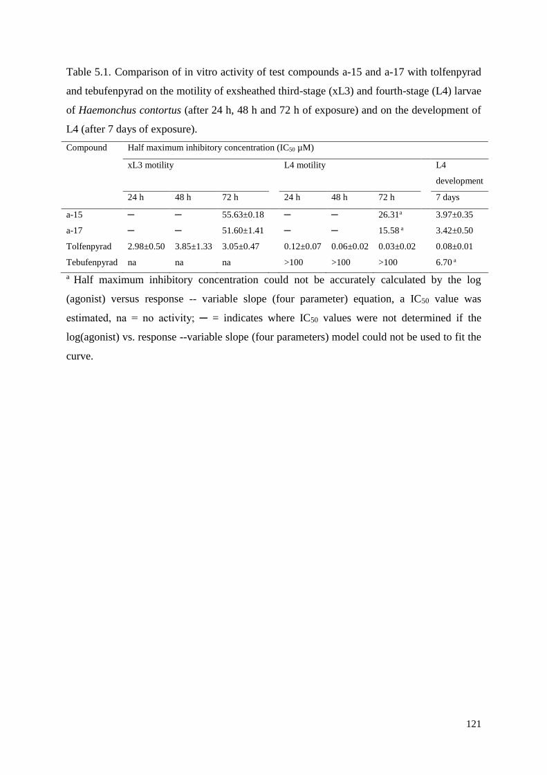
121
Table 5.1. Comparison of in vitro activity of test compounds a-15 and a-17 with tolfenpyrad
and tebufenpyrad on the motility of exsheathed third-stage (xL3) and fourth-stage (L4) larvae
of Haemonchus contortus (after 24 h, 48 h and 72 h of exposure) and on the development of
L4 (after 7 days of exposure).
Compound Half maximum inhibitory concentration (IC50 µM)
xL3 motility L4 motility L4
development
24 h 48 h 72 h 24 h 48 h 72 h 7 days
a-15 ─ ─ 55.63±0.18 ─ ─ 26.31a 3.97±0.35
a-17 ─ ─ 51.60±1.41 ─ ─ 15.58 a 3.42±0.50
Tolfenpyrad 2.98±0.50 3.85±1.33 3.05±0.47 0.12±0.07 0.06±0.02 0.03±0.02 0.08±0.01
Tebufenpyrad na na na >100 >100 >100 6.70 a
a Half maximum inhibitory concentration could not be accurately calculated by the log
(agonist) versus response -- variable slope (four parameter) equation, a IC50 value was
estimated, na = no activity; ─ = indicates where IC50 values were not determined if the
log(agonist) vs. response --variable slope (four parameters) model could not be used to fit the
curve.
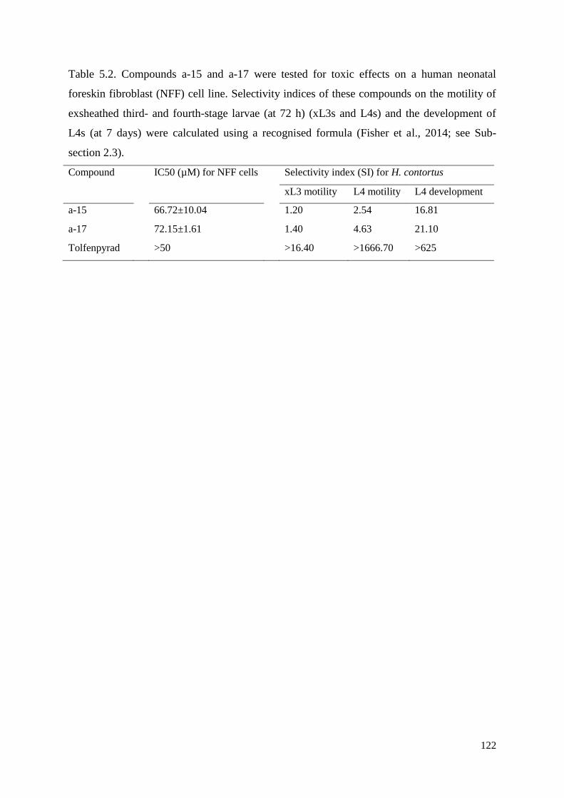
122
Table 5.2. Compounds a-15 and a-17 were tested for toxic effects on a human neonatal
foreskin fibroblast (NFF) cell line. Selectivity indices of these compounds on the motility of
exsheathed third- and fourth-stage larvae (at 72 h) (xL3s and L4s) and the development of
L4s (at 7 days) were calculated using a recognised formula (Fisher et al., 2014; see Sub-
section 2.3).
Compound IC50 (µM) for NFF cells Selectivity index (SI) for H. contortus
xL3 motility L4 motility L4 development
a-15 66.72±10.04 1.20 2.54 16.81
a-17 72.15±1.61 1.40 4.63 21.10
Tolfenpyrad >50 >16.40 >1666.70 >625

123
Fig. 5.1. Primary screening of 55 synthetic pyrazole-5-carboxamides for inhibition of motility
of exsheathed third-stage larvae (xL3) of Haemonchus contortus at a concentration of 100
µM (after 72 h of exposure). Three commercial compounds, tolfenpyrad, monepantel and
moxidectin, were also included as control compounds. Tolfenpyrad was used as the positive
reference-control compound, as it has known activity against xL3s and L4s of H. contortus
(see Preston et al., 2016). Two active compounds, a-15 and a-17, inhibited xL3 motility by ≥
70%. Supplementary file 5.2 shows the “straight” phenotype in larvae exposed to a-15 and a-
17, similar to that of the tolfenpyrad control.
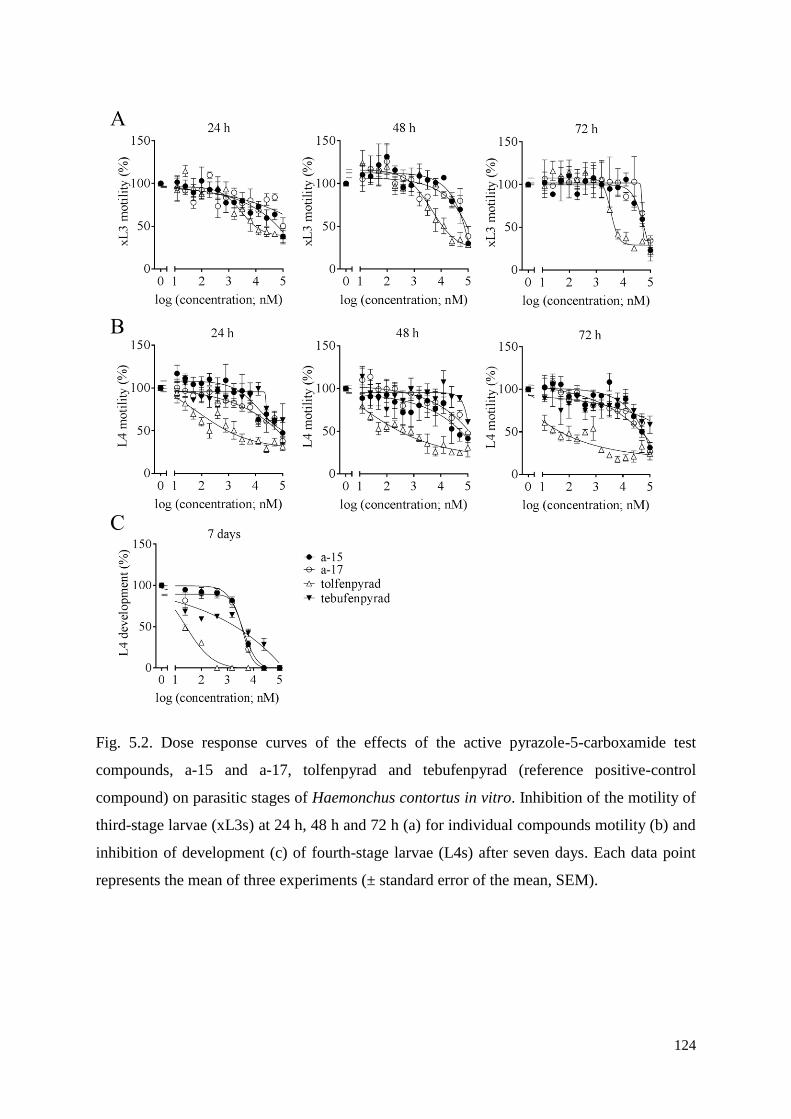
124
Fig. 5.2. Dose response curves of the effects of the active pyrazole-5-carboxamide test
compounds, a-15 and a-17, tolfenpyrad and tebufenpyrad (reference positive-control
compound) on parasitic stages of Haemonchus contortus in vitro. Inhibition of the motility of
third-stage larvae (xL3s) at 24 h, 48 h and 72 h (a) for individual compounds motility (b) and
inhibition of development (c) of fourth-stage larvae (L4s) after seven days. Each data point
represents the mean of three experiments (± standard error of the mean, SEM).
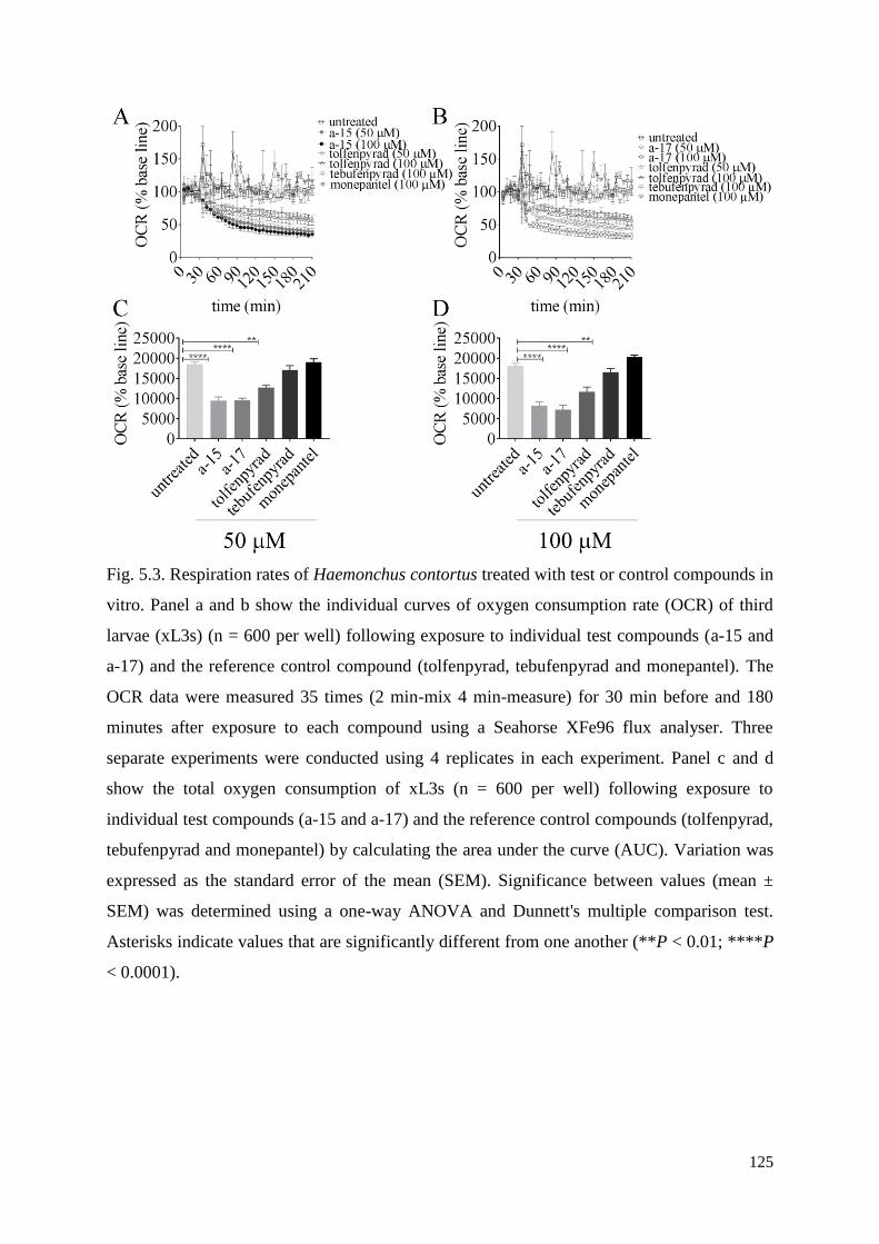
125
Fig. 5.3. Respiration rates of Haemonchus contortus treated with test or control compounds in
vitro. Panel a and b show the individual curves of oxygen consumption rate (OCR) of third
larvae (xL3s) (n = 600 per well) following exposure to individual test compounds (a-15 and
a-17) and the reference control compound (tolfenpyrad, tebufenpyrad and monepantel). The
OCR data were measured 35 times (2 min-mix 4 min-measure) for 30 min before and 180
minutes after exposure to each compound using a Seahorse XFe96 flux analyser. Three
separate experiments were conducted using 4 replicates in each experiment. Panel c and d
show the total oxygen consumption of xL3s (n = 600 per well) following exposure to
individual test compounds (a-15 and a-17) and the reference control compounds (tolfenpyrad,
tebufenpyrad and monepantel) by calculating the area under the curve (AUC). Variation was
expressed as the standard error of the mean (SEM). Significance between values (mean ±
SEM) was determined using a one-way ANOVA and Dunnett's multiple comparison test.
Asterisks indicate values that are significantly different from one another (**P < 0.01; ****P
< 0.0001).
126
Chapter 6 - Tetrahydroquinoxalines induce a lethal
evisceration phenotype in Haemonchus contortus in vitro
Abstract
In the present study, the anthelmintic activity of a human tyrosine kinase inhibitor, AG-1295,
and 14 related tetrahydroquinoxaline analogues against Haemonchus contortus was explored.
These compounds were screened against parasitic larvae - exsheathed third-stage (xL3) and
fourth-stage (L4) - using a whole-organism screening assay. All compounds were shown to
have inhibitory effects on larval motility, development and growth, and induced evisceration
through the excretory pore in xL3s. The estimated IC50 values ranged from 3.5 to 52.0 μM for
inhibition of larval motility or development. Cytotoxicity IC50 against human MCF10A cells
was generally higher than 50 μM. Microscopic studies revealed that this eviscerated (Evi)
phenotype occurs rapidly (< 20 min) and relates to a protrusion of internal tissues and organs
(evisceration) through the excretory pore in xL3s; severe pathological damage in L4s as well
as a suppression of larval growth in both stages were also observed. Using a relatively low
concentration (12.5 μM) of compound m10, it was established that the inhibitor has to be
present for a relatively short time (between 30 h and 42 h) during in vitro development from
xL3 to L4, to induce the Evi phenotype. Increasing external osmotic pressure prevented
evisceration and moulting, and xL3s remained unaffected by the test compound. These results
point to a mode of action involving a dysregulation of morphogenetic processes during a
critical time-frame, in agreement with the expected behaviour of a tyrosine kinase inhibitor,
and suggest potential for development of this compound class as nematocidal drugs.
127
6.1. Introduction
Parasitic worms cause substantial morbidity and mortality in humans and animals, and
major losses to food production globally (cf. Roeber et al., 2013; Hotez et al., 2014). In
particular, some parasitic roundworms (nematodes) of the order Strongylida, also called
strongylids, or bursate nematodes, cause some of the most important gastrointestinal diseases
of livestock worldwide, affecting hundreds of millions of animals (including sheep, goats,
cattle and/or pigs), with economic losses estimated at tens of billions of dollars per annum
(Roeber et al., 2013; Thamsborg et al., 2013; Lane et al., 2015). These pathogens can cause
gastroenteritis, anaemia and/or associated complications (depending on species), and death in
severely affected animals, and many of them cause marked productivity losses through
subclinical infections. These nematodes are transmitted orally from contaminated pastures to
the host through a direct life cycle (Beveridge and Emery, 2014): eggs are excreted in host
faeces; the first-stage larvae (L1s) develop inside the egg, usually hatch within 1 day at
environmental temperatures of ≥ 10°C and then develop to the second- and third-stage
larvae (L2s and L3s) in about one week; the infective L3s are then ingested by the host,
exsheath (xL3) and develop through fourth-stage larvae (L4) to dioecious adults (often within
3-4 weeks) in the gastrointestinal tract of the animal.
Currently, the control of these nematodes relies largely on the use of a limited number of
anti-parasitic drug classes. However, drug resistance is now widespread (Kotze and Prichard,
2016), and no vaccines are available for the vast majority of these worms (Hewitson and
Maizels, 2014), such that the development of new drugs is crucial to ensure sustained and
effective control into the future. Although the development of monepantel (Kaminsky et al.,
2008; Prichard and Geary, 2008) and derquantel (Little et al., 2011) has introduced novel
classes of nematocides, success in discovering new anthelmintic drugs has been limited.
We have been focusing on identifying synthetic chemical entities with nematocidal or
nematostatic activity (Preston et al., 2016b; Jiao et al., 2017a; Jiao et al., 2017b; Preston et
al., 2017), working toward optimising their potency and selectivity. Recently, we obtained a
collection of compounds (‘Stasis Box’) from Medicine for Malaria Venture (MMV); this
collection contains 400 compounds which were under pharmaceutical development, and
about which there is published information in the scientific literature or patents. Utilizing a
whole-worm motility screening assay (Preston et al., 2015), we identified in the ‘Stasis Box’
a platelet-derived growth factor (PDGF) receptor kinase inhibitor (Kovalenko et al., 1994;
Gazit et al., 1996; Kovalenko et al., 1997) called AG-1295 (6,7-dimethyl-2-
128
phenylquinoxaline) with relatively potent anthelmintic activity against H. contortus in vitro
(Jiao et al., 2017a). Dose-response evaluations showed that AG-1295 inhibits the motility of
exsheathed third-stage larvae (xL3s) and the development of fourth-stage larvae (L4s), with
half maximum inhibition concentration (IC50) values of 9.9 ± 1.9 µM and 7.8 ± 0.9 µM,
respectively (Jiao et al., 2017a). Moreover, AG-1295 was not toxic (IC50>100 µM) to normal
human breast epithelial (MCF10A) cells when tested in vitro (Jiao et al., 2017a).
Interestingly, AG-1295 had been under development as a medicine for restenosis
treatment (Levitzki, 2013). This kinase inhibitor has been reported to (i) attenuate porcine
and human smooth muscle cell growth in vitro (Banai et al., 1998), (ii) block the proliferation
of rat hepatic stellate cells in vitro (Iwamoto et al., 2000) and (iii) inhibit aortic allograft
vasculopathy in rats (Karck et al., 2002). However, currently nothing is known about how
AG-1295 affects or acts on H. contortus or any other nematode. Considering that AG-1295
has a well-defined drug-like structure (Gazit et al., 1996), has suitable pharmacokinetic
properties (Banai et al., 1998; Levitzki, 2001, 2010) and has dramatic effects on both motility
and development of H. contortus in vitro (Jiao et al., 2017a), this compound or one or more
analogues thereof might have potential for being repurposed as anthelmintics. In the present
study, we extend previous work (Jiao et al., 2017a) to (i) explore the anthelmintic activities of
14 AG-1295 analogues (tetrahydroquinoxalines) on motility and development of larval stages
of H. contortus in vitro, (ii) start investigating their mode of action by qualitatively and
quantitatively assessing the morphological alterations that occur in larval stages of H.
contortus following exposure to these analogues in vitro, and (iii) discuss how this worm is
affected by representative compounds.
6.2 Materials and methods
6.2.1 Procurement of H. contortus
Haemonchus contortus (Haecon-5 strain; partially resistant to benzimidazoles) was
maintained in Merino sheep in approved facilities in The University of Melbourne with
institutional animal ethics approval (permit no. 1413429). Larval stages of H. contortus were
produced as described previously (Preston et al., 2015). In brief, faeces were collected from
infected sheep four weeks following inoculation and were then incubated at 27 °C for one
week. Then, L3s were isolated from faeces and sieved through two layers of nylon mesh
(pore size: 20 µm; Rowe Scientific, Australia). Exsheathed L3s (xL3s) were produced by
incubating L3s in 0.15% (v/v) sodium hypochlorite (NaClO) at 37 °C for 20 min, and
129
immediately washed five-times in sterile physiological saline by centrifugation (1700 xg, 5
min). The xL3s were resuspended in LB* [10 g of tryptone (cat. no. LP0042; Oxoid,
England), 5 g of yeast extract (cat. no. LP0021; Oxoid, England) and 5 g of NaCl (cat. no.
1064045000; Merck, Denmark) in 1 l of reverse-osmosis deionized water; supplemented with
100 IU/ml of penicillin, 100 µg/ml of streptomycin and 0.25 µg/ml of amphotericin
(antibiotic-antimycotic; cat. no. 15240062; Gibco, USA)]. L4s were produced by incubating
xL3s in LB* at 38 °C and 10% (v/v) CO2 for 7 days; larvae were not used unless ≥ 80% of
xL3s developed to L4s - identified based on the appearance of a mouth and pharynx (Veglia,
1916; Sommerville et al., 1966).
6.2.2 AG-1295 analogues
A total of 14 tetrahydroquinoxaline compounds whose structures are closely related to
AG-1295 were sourced from two co-authors (JBB and SV) (Supplementary file 6.1). All
compounds were synthesized to form the arylated 5,6,7,8- tetrahydroquinoxaline N-oxide
intermediates by palladium catalyzation, and the N-oxide moiety was reduced to yield the
final chemicals (Supplementary file 6.1). AG-1295 was purchased from a commercial
company (IUPAC name: 6,7-dimethyl-2-phenylquinoxaline, cat. no. A425200, 99.9% purity,
Toronto Research Chemicals, Canada).
6.2.3 Compound screening
Compounds were screened against xL3s of H. contortus using an established approach
(Preston et al., 2016a). In brief, compounds were individually dissolved in dimethyl sulfoxide
(DMSO, Ajax Finechem, Australia) to a stock concentration of 20 mM. To test their activity
against H. contortus, individual compounds were diluted in LB* to the final concentration of
100 µM. All compounds were tested in triplicate; a commercial anthelmintic, monepantel
(Zolvix, Elanco Animal Health, Switzerland) was used as a positive control (in triplicate),
and LB*+0.5% DMSO was used as a negative control (in sextuplicate). All compounds were
tested using in a 96-well plate (Corning 3596; Life Sciences, USA) with 300 xL3s per well
for 72 h at 38 °C and 10% (v/v) CO2. Following incubation, worms were agitated using an
orbital shaker at 37 °C for 20 min, and five-second video recordings were taken of individual
wells. Each video capture of each well was processed using a custom macro in the program
Image J (1.47v, imagej.nih.gov/ij); larval motility in each well was expressed as a motility
index (Mi), calculated as described previously (Preston et al., 2015). A compound was
130
identified as active against H. contortus (‘hit’) if it reduced the Mi value of xL3s by ≥ 70%,
or if a phenotype was observed that differed from wildtype xL3 (i.e. LB* + 0.5% DMSO
control).
6.2.4 Dose-response assessments of active compounds on xL3 and L4 motility,
and L4 development
Active compounds were tested against H. contortus larvae at a range of concentrations to
determine the half maximum inhibitory concentration (IC50) values of each compound as
described earlier (Preston et al., 2015). Stocks of individual compounds (20 mM) were
diluted in LB* (50 µl) to 200 µM, and then serially titrated (in two-fold steps) across a 96-
well flat bottom plate. Subsequently, 300 worms (either xL3s or L4s) in 50 µl of LB* were
added to individual wells and the plates were incubated in a water-jacketed CO2 incubator at
38 °C and 10% (v/v) CO2. After 24 h, 48 h and 72 h of incubation, worm motility index (Mi)
values were measured and processed to generate an 18-point dose-response curve using v.7
GraphPad Prism software. Following motility measurement, the xL3 plates were re-incubated
for four more days to assess the L4 development. IC50 values for each dose-response curve
(xL3 motility, L4 motility and L4 development) were calculated by fitting a variable slope
four-parameter equation, constraining the top value to 100%.
6.2.5 Assessment of cytotoxicity
A ‘normal’ human breast epithelial cell line (MCF10A) was used to test the cytotoxic
activity of compounds (Jiao et al., 2017a). In brief, in a 384-well flat bottom, black walled
plate (Corning, USA), 700 MCF10A cells in 40 µl DMEM-F12 [100 ng/ml cholera toxin
(Sigma, Australia), 20 ng/ml human epidermal growth factor (EGF, Life Technologies,
Australia), 10 µg/ml insulin (human; Novo Nordisk Pharmaceuticals Pty Ltd, Australia), 5%
horse serum (Life Technologies, Australia) and 0.5 µg/ml hydrocortisone (Sigma, Australia)],
were dispensed into wells using a liquid handling dispenser (BioTek, Vermont, USA). Plates
were incubated at 37 °C and 5% (v/v) CO2 for 24 h. Meanwhile, test compounds were diluted
in the cell growth media from 250 µM to 15.6 µM in a two-fold serial way using an
automated liquid handling robot (SciClone ALH3000 Lab Automation Liquid Handler,
Caliper Lifesciences, USA). The diluted compounds (10 µl) were then added to cultured
cells, with doxorubicin as a positive control and DMEM-F12 + 0.5% DMSO as a negative
control. Following an incubation for 48 h, cells were fixed and stained with 4’6-diamidino-2-
131
phenylindole (DAPI; 1:1000). Subsequently, cell nuclei present in each well were quantified
using a high content imager (Cellomics Cell Insight Personal Cell Imager, Thermofisher
Scientific, USA) at 10-times magnification (with a fixed exposure time of 0.12 sec) and the
Target Activation BioApplication within the Cellomics software. IC50 values for inhibition of
MCF10A cell proliferation were calculated from dose-response curves, using a variable slope
four-parameter equation in GraphPad Prism. Each assay was repeated two times in
quadruplicate on different days. The selectivity indices (Si) of hit compounds were then
calculated using the formula: Si = IC50 for MCF10A cells / IC50 for H. contortus.
6.2.6 Morphological examination of the eviscerated (Evi) phenotype induced by
incubation with individual test compounds
Light microscopy was used to examine morphological alteration(s) in larvae caused by test
compounds after 7 days (Subsection 6.2.4). At each compound concentration tested, 30 larvae
(fixed with 1% iodine) were classified as either ‘normal xL3s’, ‘eviscerated xL3s’ or ‘L4s’,
using a light microscope (DP26 camera, Olympus, USA). The concentration at which each
test compound induced maximum numbers of eviscerated larvae was recorded. Compounds
were also tested for their ability to inhibit larval growth by measuring worm length and width
microscopically (20-times magnification), using a corresponding concentration of DMSO as
a negative control. For each treatment, the mean length and width of ≥ 30 larvae ± standard
error of mean were calculated, and a one-way ANOVA with Tukey’s test was used to
measure the statistical differences between different treatments.
6.2.7 Scanning electron microscopy (SEM)
Larvae were also examined by SEM. Briefly, in a 96-well flat bottom plate, 300 worms
(xL3s or L4s) in 50 µl of LB* were exposed to the concentration of test compound that
induced the greatest number of larvae with an Evi phenotype, with a corresponding
concentration of DMSO as a negative control (in sextuplicate). Subsequently, the plate was
incubated at 38 °C and 10% (v/v) CO2 for 72 h. For each treatment, worms were then pooled
together and washed three times in 0.9% saline at 9,000 xg. For SEM analysis, worms were
fixed in 2.5% glutaraldehyde and 2% paraformaldehyde for 2 h, rinsed three times in 0.1 M
sodium cacodylate trihydrate for 10 min, and then post-fixed in 1% osmium tetroxide at room
temperature for 2 h. Fixed worms were rinsed again three times in distilled water for 15 min.
Following fixation, 200 μl of concentrated worms were incubated on poly-L-lysine coated
132
glass coverslips; the slips were pre-prepared by immersing them in poly-L-lysine solution for
10 min, drying in an oven at 60 °C for 10 min, immersing again in poly-L-lysine solution for
10 min and then drying them at room temperature. The coverslips with adhered nematodes
were dehydrated in a graded ethanol series (30%, 50%, 70%, 90% and 100%) for 17 min,
dried in a critical point dryer (EM CPD300, Leica, Wetzlar, Germany), mounted on to
aluminium stubs (25 mm) with double-sided carbon tabs and then coated with gold. Worms
were imaged using a JEOL JCM-6000 Plus NeoScope scanning electron microscope (Jeol,
Tokyo, Japan); at least six worms were examined for each treatment group.
6.2.8 Transmission electron microscopy (TEM)
Individual xL3s and L4s were sliced twice to give three even (anterior, mid and posterior)
body portions in 3.2% paraformaldehyde and 0.2% glutaraldehyde with 0.15 M sodium
cacodylate trihydrate (pH 7.2) (cf. Kovacs, 2015), and ruthenium red (RR) solution was
added to stain individual pieces. After 2 h of primary fixation, worm portions were rinsed
three times in 0.125 M sodium cacodylate trihydrate for 2 h, and then post-fixed in 1%
osmium tetroxide and 1.5% potassium ferrocyanide in the dark for 2 h. Fixed worm portions
were rinsed again three times in 0.125 M sodium cacodylate trihydrate for 2 h and then
embedded in 1.5% agar while keeping them straight. The agar blocks containing worm
portions were washed two times in distilled water for 2 h and dehydrated in a graded ethanol
series (50%, 70%, 90%, 95% and 100%) for 2 h on a platform rocker, rinsed two times in
100% acetone for 45 min, rocking, and then embedded in Spurrs resin. Ultrathin sections
were cut with an EM UC7 ultramicrotome (Leica Microsystems GmbH, Wetzlar, Germany)
and contrasted with lead citrate and aqueous uranyl acetate. Sections were imaged using a
transmission electron microscope (JEOL 1011, Tokyo, Japan).
6.2.9 Evaluation of the dynamics of the compound-induced Evi phenotype in
xL3s
To determine the kinetics of the anterior protrusion and evisceration in xL3s, a time course
experiment was performed using the compound concentration that caused maximum numbers
of worms with the Evi phenotype. At various time-points (every 6 h for the first 4 days and
then every 24 h for the next 3 days) after treatment, aliquots (50 µl) of 150 larvae were fixed
in 1% iodine, and 30 worms from each aliquot were scored as either ‘normal xL3s’,
‘eviscerated xL3s’ or ‘L4s’ under a compound microscope using 100-times magnification.
133
For each time point, the percentage of larvae in each category was calculated, and a two-way
ANOVA with Tukey’s test was used to calculate statistical differences between groups. This
assay was repeated at least two times in triplicate on different days.
6.2.10 Assessment of the reversibility of effect of test compounds on xL3s
To determine whether the effect of test compounds on xL3 was reversible, a previously
published method was used (Kumarasingha et al., 2016). In brief, xL3s were exposed for 18
h, 24 h, 27 h, 30 h, 36 h and 48 h to 12.5 µM of test compound in triplicate in 96-well plates.
Following exposure, at each of these time points, compounds were removed by washing
larvae three times in 0.9% saline, 100 μl of LB* were added to individual wells, and the plate
was then re-incubated in a water-jacketed CO2 incubator at 38 °C and 10% (v/v) CO2. After
72 h, the percentages of the eviscerated xL3s and the L4s were calculated. Controls included
xL3s continually incubated with 12.5 µM of test compound (in triplicate) for 72 h and xL3s
incubated in LB* alone. This assay was repeated two times on different days. Differences in
percentage of eviscerated xL3s between treatments were evaluated using a one-way ANOVA
with Tukey's test.
Subsequently, xL3s were cultured first in LB*, and test compound (to 12.5 µM) was
added to individual wells at two different time-points (39 h and 42 h). Again, worms were
scored microscopically at 72 h. A matched compound concentration, added at 0 h, was used
as a positive control, and the corresponding concentration of DMSO was used as a negative
control. This assay was repeated two times on different days. A two-way ANOVA with
Tukey's test was used to measure statistical differences between treatments in the percentages
of eviscerated xL3s and developed L4s.
6.2.11 Influence of external osmotic pressure on compound-induced Evi
phenotype in xL3s
To examine whether an osmotic pressure gradient was involved in the induction of the Evi
phenotype in xL3s, compound-treated and untreated xL3s were cultured in the presence of
increasing concentrations of sorbitol (0 M, 0.1 M, 0.2 M, 0.3 M, 0.4 M, 0.5 M, 0.6 M, 0.7 M,
0.8 M, 0.9 M and 1.0 M in LB*). After 72 h of incubation at 38 °C and 10% (v/v) CO2, the
percentages of the eviscerated xL3s and the developed L4s were calculated at each
concentration. This assay was repeated two times on different days.
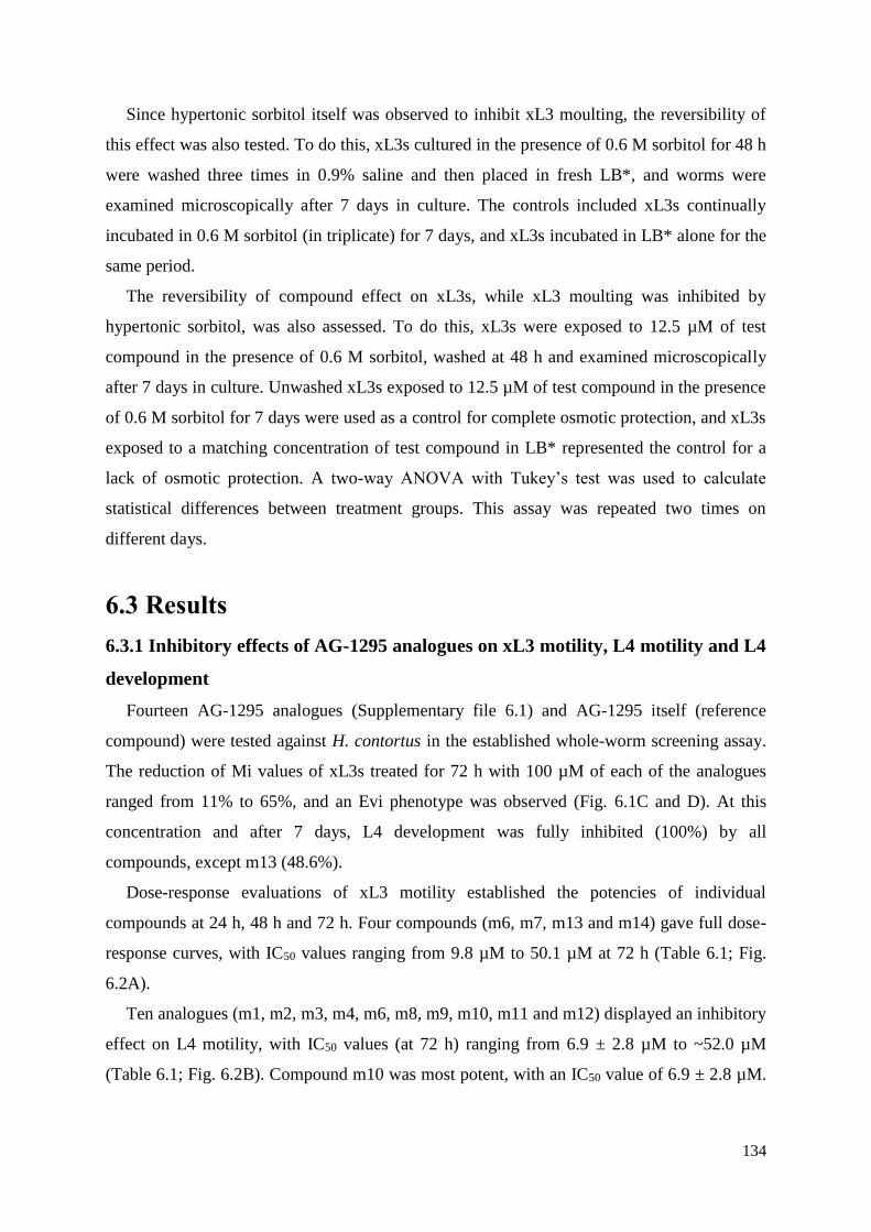
134
Since hypertonic sorbitol itself was observed to inhibit xL3 moulting, the reversibility of
this effect was also tested. To do this, xL3s cultured in the presence of 0.6 M sorbitol for 48 h
were washed three times in 0.9% saline and then placed in fresh LB*, and worms were
examined microscopically after 7 days in culture. The controls included xL3s continually
incubated in 0.6 M sorbitol (in triplicate) for 7 days, and xL3s incubated in LB* alone for the
same period.
The reversibility of compound effect on xL3s, while xL3 moulting was inhibited by
hypertonic sorbitol, was also assessed. To do this, xL3s were exposed to 12.5 µM of test
compound in the presence of 0.6 M sorbitol, washed at 48 h and examined microscopically
after 7 days in culture. Unwashed xL3s exposed to 12.5 µM of test compound in the presence
of 0.6 M sorbitol for 7 days were used as a control for complete osmotic protection, and xL3s
exposed to a matching concentration of test compound in LB* represented the control for a
lack of osmotic protection. A two-way ANOVA with Tukey’s test was used to calculate
statistical differences between treatment groups. This assay was repeated two times on
different days.
6.3 Results
6.3.1 Inhibitory effects of AG-1295 analogues on xL3 motility, L4 motility and L4
development
Fourteen AG-1295 analogues (Supplementary file 6.1) and AG-1295 itself (reference
compound) were tested against H. contortus in the established whole-worm screening assay.
The reduction of Mi values of xL3s treated for 72 h with 100 µM of each of the analogues
ranged from 11% to 65%, and an Evi phenotype was observed (Fig. 6.1C and D). At this
concentration and after 7 days, L4 development was fully inhibited (100%) by all
compounds, except m13 (48.6%).
Dose-response evaluations of xL3 motility established the potencies of individual
compounds at 24 h, 48 h and 72 h. Four compounds (m6, m7, m13 and m14) gave full dose-
response curves, with IC50 values ranging from 9.8 µM to 50.1 µM at 72 h (Table 6.1; Fig.
6.2A).
Ten analogues (m1, m2, m3, m4, m6, m8, m9, m10, m11 and m12) displayed an inhibitory
effect on L4 motility, with IC50 values (at 72 h) ranging from 6.9 ± 2.8 µM to ~52.0 µM
(Table 6.1; Fig. 6.2B). Compound m10 was most potent, with an IC50 value of 6.9 ± 2.8 µM.
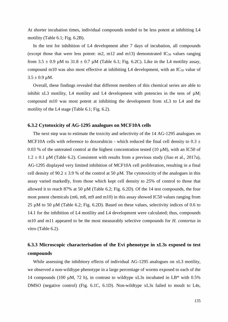
135
At shorter incubation times, individual compounds tended to be less potent at inhibiting L4
motility (Table 6.1; Fig. 6.2B).
In the test for inhibition of L4 development after 7 days of incubation, all compounds
(except those that were less potent: m2, m12 and m13) demonstrated IC50 values ranging
from 3.5 ± 0.9 µM to 31.8 ± 0.7 µM (Table 6.1; Fig. 6.2C). Like in the L4 motility assay,
compound m10 was also most effective at inhibiting L4 development, with an IC50 value of
3.5 ± 0.9 µM.
Overall, these findings revealed that different members of this chemical series are able to
inhibit xL3 motility, L4 motility and L4 development with potencies in the tens of µM;
compound m10 was most potent at inhibiting the development from xL3 to L4 and the
motility of the L4 stage (Table 6.1; Fig. 6.2).
6.3.2 Cytotoxicity of AG-1295 analogues on MCF10A cells
The next step was to estimate the toxicity and selectivity of the 14 AG-1295 analogues on
MCF10A cells with reference to doxorubicin - which reduced the final cell density to 0.3 ±
0.03 % of the untreated control at the highest concentration tested (10 µM), with an IC50 of
1.2 ± 0.1 µM (Table 6.2). Consistent with results from a previous study (Jiao et al., 2017a),
AG-1295 displayed very limited inhibition of MCF10A cell proliferation, resulting in a final
cell density of 90.2 ± 3.9 % of the control at 50 µM. The cytotoxicity of the analogues in this
assay varied markedly, from those which kept cell density to 25% of control to those that
allowed it to reach 87% at 50 µM (Table 6.2; Fig. 6.2D). Of the 14 test compounds, the four
most potent chemicals (m6, m8, m9 and m10) in this assay showed IC50 values ranging from
25 µM to 50 µM (Table 6.2; Fig. 6.2D). Based on these values, selectivity indices of 0.6 to
14.1 for the inhibition of L4 motility and L4 development were calculated; thus, compounds
m10 and m11 appeared to be the most measurably selective compounds for H. contortus in
vitro (Table 6.2).
6.3.3 Microscopic characterisation of the Evi phenotype in xL3s exposed to test
compounds
While assessing the inhibitory effects of individual AG-1295 analogues on xL3 motility,
we observed a non-wildtype phenotype in a large percentage of worms exposed to each of the
14 compounds (100 µM, 72 h), in contrast to wildtype xL3s incubated in LB* with 0.5%
DMSO (negative control) (Fig. 6.1C, 6.1D). Non-wildtype xL3s failed to moult to L4s,

136
whereas wildtype xL3s in LB* containing 0.5% DMSO developed unhindered to L4s with
discernible mouthparts and pharynx (Fig. 6.1C).
Detailed light microscopic examination (100-times magnification) with phase-contrast
optics revealed that the Evi phenotype in xL3s was characterised by a protrusion of the
alimentary tract and surrounding tissues through or around the excretory pore (Fig. 6.3A),
while the excretory canal was clearly visible in untreated xL3s in the same area
(Supplementary file 6.2A). An examination of timed video recordings revealed that
evisceration was a gradual but relatively rapid process (< 20 min) (Supplementary file 6.2B).
Initially, a few globular structures emerged from the excretory pore (109.5 ± 1.9 µm from the
anterior tip of the xL3), followed by an expulsion of the anterior and, sometimes, the
posterior part of the intestine, forming an external ‘balloon’, with ingesta and body fluids
flowing to the exterior of the worm (Fig. 6.3A; Supplementary file 6.2B). At this later stage,
the breach in the body wall seemed to expand, and the eviscerated mass ended up being
located about 80-90 µm from the anterior tip of the worm (Fig. 6.1C; Fig. 6.3A;
Supplementary file 6.2B). This Evi phenotype was observed exclusively in xL3s (but not in
L4s) exposed to any of the 14 compounds.
A SEM study provided a detailed insight into the protrusion of the alimentary tract and
surrounding tissues, and into the wrinkled cuticular surface of treated xL3s (Fig. 6.3B).
Although the Evi phenotype was not observed in L4s, detailed SEM examination revealed a
shrivelling in the cuticular surface of such larvae, compared with the smooth cuticular surface
of untreated L4s (Fig. 6.3B); xL3s or L4s exposed to LB* containing the corresponding
concentration of DMSO (negative control) were unaffected. A TEM examination revealed a
release of the hypodermis from the undulated cuticle of treated xL3s, with pathological
alterations in internal organs and tissues, including excretory canals, whereas in untreated
xL3s the hypodermis was consistently connected to the cuticle, and no anatomical changes
were evident (Fig. 6.3C). The cuticular shrivelling seen in L4s by SEM is likely related to the
pathological alterations observed by TEM in compound-treated larvae (detached hypodermis;
myodegeneration; cell lysis; and the appearance of electron-dense vesicles) (Fig. 6.3C). Such
alterations were not observed in untreated L4s maintained in culture for the same time.
Members of this compound series were also shown to inhibit larval growth. After
measuring 30 xL3s treated with 12.5 µM m10 or LB* + 0.5% DMSO for 72 h, it was shown
that compound treatment reduced xL3 length from 641 ± 4.6 µm to 456 ± 11.2 µm. Width
was similarly reduced from 18 ± 0.2 µm to 16 ± 0.4 µm (Supplementary file 6.3). Statistical
analyses, using a one-way ANOVA with Tukey’s test, showed the differences to be
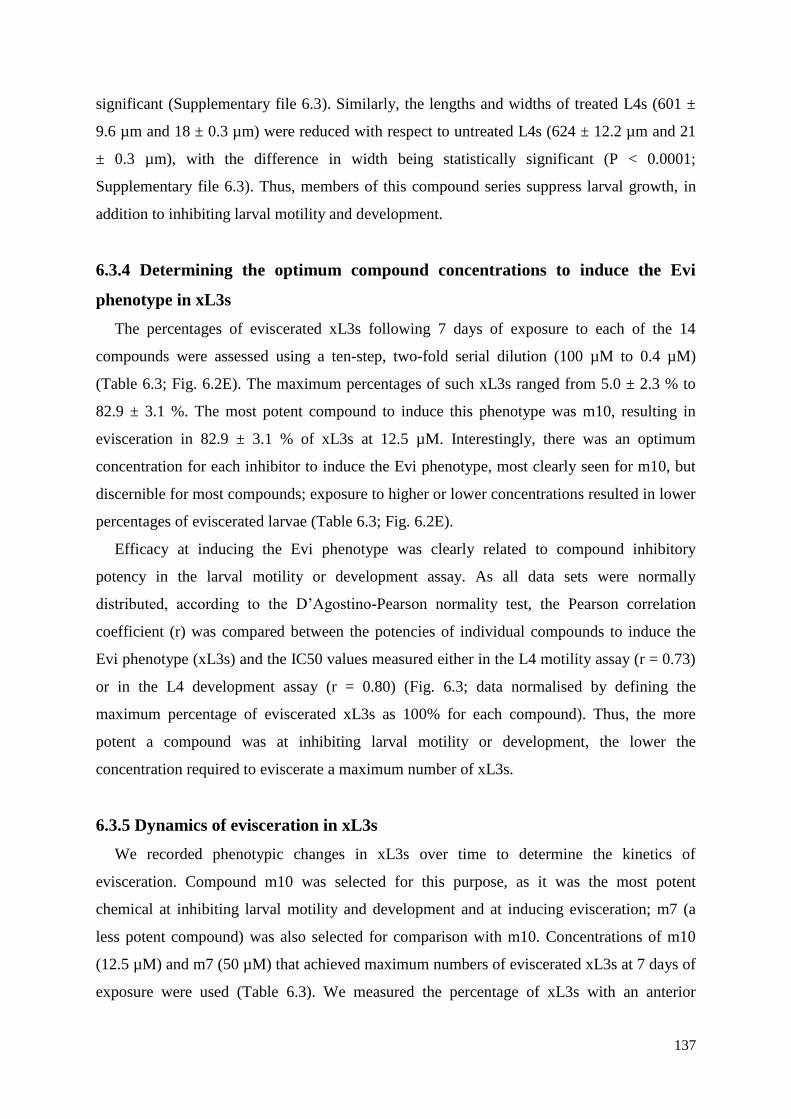
137
significant (Supplementary file 6.3). Similarly, the lengths and widths of treated L4s (601 ±
9.6 µm and 18 ± 0.3 µm) were reduced with respect to untreated L4s (624 ± 12.2 µm and 21
± 0.3 µm), with the difference in width being statistically significant (P < 0.0001;
Supplementary file 6.3). Thus, members of this compound series suppress larval growth, in
addition to inhibiting larval motility and development.
6.3.4 Determining the optimum compound concentrations to induce the Evi
phenotype in xL3s
The percentages of eviscerated xL3s following 7 days of exposure to each of the 14
compounds were assessed using a ten-step, two-fold serial dilution (100 µM to 0.4 µM)
(Table 6.3; Fig. 6.2E). The maximum percentages of such xL3s ranged from 5.0 ± 2.3 % to
82.9 ± 3.1 %. The most potent compound to induce this phenotype was m10, resulting in
evisceration in 82.9 ± 3.1 % of xL3s at 12.5 µM. Interestingly, there was an optimum
concentration for each inhibitor to induce the Evi phenotype, most clearly seen for m10, but
discernible for most compounds; exposure to higher or lower concentrations resulted in lower
percentages of eviscerated larvae (Table 6.3; Fig. 6.2E).
Efficacy at inducing the Evi phenotype was clearly related to compound inhibitory
potency in the larval motility or development assay. As all data sets were normally
distributed, according to the D’Agostino-Pearson normality test, the Pearson correlation
coefficient (r) was compared between the potencies of individual compounds to induce the
Evi phenotype (xL3s) and the IC50 values measured either in the L4 motility assay (r = 0.73)
or in the L4 development assay (r = 0.80) (Fig. 6.3; data normalised by defining the
maximum percentage of eviscerated xL3s as 100% for each compound). Thus, the more
potent a compound was at inhibiting larval motility or development, the lower the
concentration required to eviscerate a maximum number of xL3s.
6.3.5 Dynamics of evisceration in xL3s
We recorded phenotypic changes in xL3s over time to determine the kinetics of
evisceration. Compound m10 was selected for this purpose, as it was the most potent
chemical at inhibiting larval motility and development and at inducing evisceration; m7 (a
less potent compound) was also selected for comparison with m10. Concentrations of m10
(12.5 µM) and m7 (50 µM) that achieved maximum numbers of eviscerated xL3s at 7 days of
exposure were used (Table 6.3). We measured the percentage of xL3s with an anterior
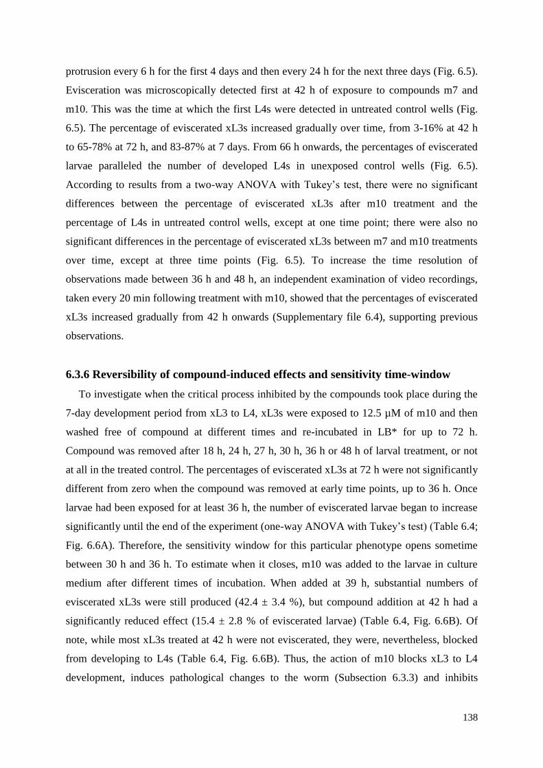
138
protrusion every 6 h for the first 4 days and then every 24 h for the next three days (Fig. 6.5).
Evisceration was microscopically detected first at 42 h of exposure to compounds m7 and
m10. This was the time at which the first L4s were detected in untreated control wells (Fig.
6.5). The percentage of eviscerated xL3s increased gradually over time, from 3-16% at 42 h
to 65-78% at 72 h, and 83-87% at 7 days. From 66 h onwards, the percentages of eviscerated
larvae paralleled the number of developed L4s in unexposed control wells (Fig. 6.5).
According to results from a two-way ANOVA with Tukey’s test, there were no significant
differences between the percentage of eviscerated xL3s after m10 treatment and the
percentage of L4s in untreated control wells, except at one time point; there were also no
significant differences in the percentage of eviscerated xL3s between m7 and m10 treatments
over time, except at three time points (Fig. 6.5). To increase the time resolution of
observations made between 36 h and 48 h, an independent examination of video recordings,
taken every 20 min following treatment with m10, showed that the percentages of eviscerated
xL3s increased gradually from 42 h onwards (Supplementary file 6.4), supporting previous
observations.
6.3.6 Reversibility of compound-induced effects and sensitivity time-window
To investigate when the critical process inhibited by the compounds took place during the
7-day development period from xL3 to L4, xL3s were exposed to 12.5 µM of m10 and then
washed free of compound at different times and re-incubated in LB* for up to 72 h.
Compound was removed after 18 h, 24 h, 27 h, 30 h, 36 h or 48 h of larval treatment, or not
at all in the treated control. The percentages of eviscerated xL3s at 72 h were not significantly
different from zero when the compound was removed at early time points, up to 36 h. Once
larvae had been exposed for at least 36 h, the number of eviscerated larvae began to increase
significantly until the end of the experiment (one-way ANOVA with Tukey’s test) (Table 6.4;
Fig. 6.6A). Therefore, the sensitivity window for this particular phenotype opens sometime
between 30 h and 36 h. To estimate when it closes, m10 was added to the larvae in culture
medium after different times of incubation. When added at 39 h, substantial numbers of
eviscerated xL3s were still produced (42.4 ± 3.4 %), but compound addition at 42 h had a
significantly reduced effect (15.4 ± 2.8 % of eviscerated larvae) (Table 6.4, Fig. 6.6B). Of
note, while most xL3s treated at 42 h were not eviscerated, they were, nevertheless, blocked
from developing to L4s (Table 6.4, Fig. 6.6B). Thus, the action of m10 blocks xL3 to L4
development, induces pathological changes to the worm (Subsection 6.3.3) and inhibits
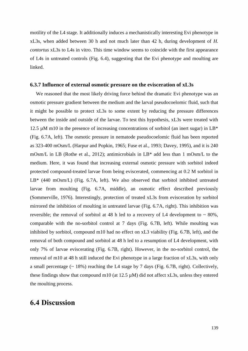
139
motility of the L4 stage. It additionally induces a mechanistically interesting Evi phenotype in
xL3s, when added between 30 h and not much later than 42 h, during development of H.
contortus xL3s to L4s in vitro. This time window seems to coincide with the first appearance
of L4s in untreated controls (Fig. 6.4), suggesting that the Evi phenotype and moulting are
linked.
6.3.7 Influence of external osmotic pressure on the evisceration of xL3s
We reasoned that the most likely driving force behind the dramatic Evi phenotype was an
osmotic pressure gradient between the medium and the larval pseudocoelomic fluid, such that
it might be possible to protect xL3s to some extent by reducing the pressure differences
between the inside and outside of the larvae. To test this hypothesis, xL3s were treated with
12.5 µM m10 in the presence of increasing concentrations of sorbitol (an inert sugar) in LB*
(Fig. 6.7A, left). The osmotic pressure in nematode pseudocoelomic fluid has been reported
as 323-400 mOsm/L (Harpur and Popkin, 1965; Fuse et al., 1993; Davey, 1995), and it is 240
mOsm/L in LB (Rothe et al., 2012); antimicrobials in LB* add less than 1 mOsm/L to the
medium. Here, it was found that increasing external osmotic pressure with sorbitol indeed
protected compound-treated larvae from being eviscerated, commencing at 0.2 M sorbitol in
LB* (440 mOsm/L) (Fig. 6.7A, left). We also observed that sorbitol inhibited untreated
larvae from moulting (Fig. 6.7A, middle), an osmotic effect described previously
(Sommerville, 1976). Interestingly, protection of treated xL3s from evisceration by sorbitol
mirrored the inhibition of moulting in untreated larvae (Fig. 6.7A, right). This inhibition was
reversible; the removal of sorbitol at 48 h led to a recovery of L4 development to ~ 80%,
comparable with the no-sorbitol control at 7 days (Fig. 6.7B, left). While moulting was
inhibited by sorbitol, compound m10 had no effect on xL3 viability (Fig. 6.7B, left), and the
removal of both compound and sorbitol at 48 h led to a resumption of L4 development, with
only 7% of larvae eviscerating (Fig. 6.7B, right). However, in the no-sorbitol control, the
removal of m10 at 48 h still induced the Evi phenotype in a large fraction of xL3s, with only
a small percentage (~ 18%) reaching the L4 stage by 7 days (Fig. 6.7B, right). Collectively,
these findings show that compound m10 (at 12.5 µM) did not affect xL3s, unless they entered
the moulting process.
6.4 Discussion
140
Extending a study (Jiao et al., 2017a) that identified a quinoxaline compound, AG-1295,
as being inhibitory to H. contortus motility, we sought to investigate whether the anthelmintic
activity of AG-1295 was a unique occurrence to this particular compound or a property of the
quinoxaline chemical series that could be chemically modulated. The results of the present
investigation show that AG-1295 is not a singleton and that a series of 14 analogues have
varying degrees of activity, paving the way to structure-activity relationships through future
chemical optimisation. We recorded marked inhibitory effects of individual test compounds
on larval motility or development and a striking Evi phenotype in xL3s in vitro.
In the primary screen, these compounds were identified as ‘hits’ based on their induction
of a unique larval phenotype and their ability to inhibit L4 development, unlike in previous
screens, in which ‘hit’ compounds were identified by a ≥ 70% reduction in xL3 motility
(Preston et al., 2015). Subsequent compound evaluation confirmed a relatively limited
potency of these compounds at inhibiting xL3 motility, with only four of them achieving IC50
values in the range of 10 µM to 50 µM. These findings indicated that other complementary
endpoints, such as morphological phenotype, in addition to xL3 motility, could be employed
to refine the detection of ‘hits’ through in vitro screening.
Most (n = 11) of the test compounds were shown to be more potent at inhibiting the
motility of L4s than xL3s (Table 6.1). Differences in compound penetration, target
expression level, metabolism, detoxification and/or efflux (cf. Gill et al., 1991; George et al.,
2017) between xL3s and L4s might explain the different potencies of these compounds
between these two larval stages. Importantly, L4s possess a pronounced mouth and functional
pharynx (for blood feeding in vivo) which likely facilitate drug uptake. Nonetheless, three
select compounds (m7, m13 and m14) as well as AG-1295 itself all reduced xL3 motility, but
not L4 motility. To fully understand the reasons for varying potencies, a study of structure-
activity relationships by chemical modification and drug target identification should be
undertaken in this or model nematodes.
Cytotoxicity was tested against a human, non-malignant mammary epithelial cell line
(MCF10A), and different members of this series exhibited varied, generally low
cytotoxicities (Table 6.2). Furthermore, there was no correlation between the potency against
H. contortus and the cytotoxicity against MCF10A cells (Tables 6.2 and 6.3). Although the
present selectivity (0.6 to 14.1) is lower than desired, variation among the different
compounds indicates that selectivity can be chemically modulated during lead optimisation.
In later stages, in vivo toxicity should be investigated to establish the relationship between in
141
vitro and in vivo systems, as they may not correlate perfectly (cf. McKim, 2010; Sayes,
2014).
The initial screening hit and parent compound, AG-1295, has been described as a receptor
tyrosine kinase inhibitor (Kovalenko et al., 1994; Gazit et al., 1996; Kovalenko et al., 1997).
Whether this is also the mode of anthelmintic action needs to be investigated, but here we
conducted a detailed morphological analysis of the effects of the most potent analogue, m10,
against xL3s and L4s, using light and electron microscopy. At the relatively low
concentration of 12.5 µM, the m10 analogue was observed to cause widespread
morphological damage in both larval stages (Fig. 6.3; Supplementary file 6.2), but treated
xL3s, in particular, suffered from a lethal process (i.e. evisceration), by which an initial small
protrusion, located at the level of the excretory pore, rapidly progressed to an extrusion of the
intestinal tube plus other pseudocoelomic components (Fig. 6.3A). Interestingly, all
analogues were able to induce this phenotype at an optimum concentration, related to their
potency in the anthelmintic assays (Table 6.3; Fig. 6.2E; Fig. 6.4), suggesting that the
inhibition of larval motility or development and the induction of the Evi phenotype all result
from an inhibition of the same target(s) in H. contortus. Nevertheless, while the degree of
inhibition of motility or development increased monotonically with compound concentration,
the Evi phenotype became less pronounced above an optimum concentration, indicating that
some physiological processes have to be disrupted, but not fully inhibited, for evisceration to
take place. This supports a mode of action involving alterations in signalling mechanisms, in
agreement with the described activity of AG-1295 and the multiple protein kinases involved
in nematode moulting (see Lazetic and Fay, 2017). Another explanation could be compound
insolubility above a particular concentration, but this would have also reduced the inhibition
of motility or development at the highest concentrations tested, and it did not (Fig. 6.2A and
B).
We also found that there was a particular time window in which the Evi phenotype could
be induced in xL3s. Larvae exposed to test compounds for the first 30 h of culture only,
before ecdysis started, or to compound added after 42 h, once the first L4s appeared, did not
show eviscerations. However, while early treatment, followed by compound removal, did not
seem to affect larvae, worms treated after 42 h in culture appeared as morphologically
damaged xL3s and L4s. One possible interpretation is that test compounds can only gain
access to the larvae once ecdysis starts and the cuticle sloughs, and the morphogenetic
process that makes larvae vulnerable to evisceration occurs in a time window of 30 h to 42 h
of in vitro culture. An alternative explanation might be that the target(s) for this chemical
142
series is essential only after the beginning of ecdysis. We found that xL3s do survive
compound exposure when placed in hypertonic medium, but we also found that, as
previously published (Sommerville, 1976), hypertonic media prevent larval moulting, so it is
still unclear whether the target(s) first appears in L4s, or whether it is always present but the
compounds only access the larval interior after ecdysis commences. In support of this latter
hypothesis, AG-1295 and analogues m6, m7, m13 and m14 seem able to affect xL3 motility
after prolonged exposure (72 h) (Table 6.1); this might also explain why the kinetics of
emergence of the Evi phenotype in xL3s parallel those of moulting (Subsection 6.3.5).
From a fundamental biological perspective, it would be significant to elucidate the
signalling mechanism required to produce the intestinal prolapse through the excretory pore.
Supplementary file 6.2 shows intestinal content moving but being diverted to the pore, not to
the mouth or the anus, as if, at this stage, these two openings would not allow passage of
material. Clearly, morphogenetic processes are being disrupted, with catastrophic
consequences for the worm, indicating that the compounds will have nematocidal activity
against larval stages, although activity against adult worms needs to be assessed.
In conclusion, we have established the in vitro anthelmintic activity of this
tetrahydroquinoxaline chemical series, a family of compounds derived from a protein
tyrosine kinase inhibitor, AG-1295, a known inhibitor of PDGF receptor kinase in humans
(Kovalenko et al., 1994; Gazit et al., 1996; Kovalenko et al., 1997). This tyrosine kinase
might have been of interest as a drug target in this nematode, but the PDGF receptor kinase
family had not been identified in H. contortus (Stroehlein et al., 2015; Jiao et al., 2017a).
However, other related tyrosine kinase families may serve as a target for the quinoxalines
and, thus, have potential as anthelmintic drug targets. If this turns out to be the case, a useful
anthelmintic could be developed with a totally novel mode of action, and its further
development would benefit from the wealth of knowledge accumulated for compounds
targeting protein kinases.

143
6.5 References
Banai, S., Wolf, Y., Golomb, G., Pearle, A., Waltenberger, J., Fishbein, I., Schneider, A.,
Gazit, A., Perez, L., Huber, R., Lazarovichi, G., Rabinovich, L., Levitzki, A., Gertz,
S.D., 1998. PDGF-receptor tyrosine kinase blocker AG1295 selectively attenuates
smooth muscle cell growth in vitro and reduces neointimal formation after balloon
angioplasty in swine. Circulation 97, 1960-1969.
Beveridge, I., Emery, D., 2014. Australian Animal Parasites - Inside and Out. The Australian
Society for Parasitology Inc., Australia. ISBN: 978-0-646-93560-7.
Davey, K.G., 1995. Water, water compartments and water regulation in some nematodes
parasitic in vertebrates. J. Nematol. 27, 433-440.
Fuse, M., Davey, K.G., Sommerville, R.I., 1993. Osmoregulation in the parasitic nematode
Pseudoterranova decipiens. J. Exp. Biol. 175, 127-142.
Gazit, A., App, H., McMahon, G., Chen, J., Levitzki, A., Bohmer, F.D., 1996. Tyrphostins. 5.
Potent inhibitors of platelet-derived growth factor receptor tyrosine kinase: structure-
activity relationships in quinoxalines, quinolines, and indole tyrphostins. J. Med.
Chem. 39, 2170-2177.
George, M.M., Lopez-Soberal, L., Storey, B.E., Howell, S.B., Kaplan, R.M., 2017. Motility
in the L3 stage is a poor phenotype for detecting and measuring resistance to
avermectin/milbemycin drugs in gastrointestinal nematodes of livestock. Int. J.
Parasitol. Drugs Drug Resist. 8, 22-30.
Gill, J.H., Redwin, J.M., van Wyk, J.A., Lacey, E., 1991. Detection of resistance to
ivermectin in Haemonchus contortus. Int. J. Parasitol. 21, 771-776.
Harpur, R.P., Popkin, J.S., 1965. Osmolality of blood and intestinal contents in the pig,
guinea pig, and Ascaris lumbricoides. Can. J. Biochem. 43, 1157-1169.
Hewitson, J.P., Maizels, R.M., 2014. Vaccination against helminth parasite infections. Expert
Rev. Vaccines 13, 473-487.
Hotez, P.J., Alvarado, M., Basáñez, M.G., Bolliger I, Bourne R, Boussinesq M, Brooker S.J.,
Brown, A.S., Buckle, G., Budke, C.M., Carabin, H., Coffeng, L.E., Fèvre, E.M.,
Fürst, T., Halasa, Y.A., Jasrasaria, R., Johns, N.E., Keiser, J., King, C.H., Lozano, R.,
Murdoch, M.E., O'Hanlon, S., Pion S.D.S., Pullan, R.L., Ramaiah, K.D., Roberts, T.,
Shepard, D.S., Smith, J.L., Stolk, W.A., Undurraga, E.A., Utzinger, J., Wang, M.,
Murray, C.J.L., Naghavi, M. 2014. The global burden of disease study 2010:
144
interpretation and implications for the neglected tropical diseases. PLoS Negl. Trop.
Dis. 8, e2865.
Iwamoto, H., Nakamuta, M., Tada, S., Sugimoto, R., Enjoji, M., Nawata, H., 2000. Platelet-
derived growth factor receptor tyrosine kinase inhibitor AG1295 attenuates rat hepatic
stellate cell growth. J. Lab. Clin. Med. 135, 406-412.
Jiao, Y., Preston, S., Koehler, A.V., Stroehlein, A.J., Chang, B.C.H., Simpson, K.J., Cowley,
K.J., Palmer, M.J., Laleu, B., Wells, T.N.C., Jabbar, A., Gasser, R.B., 2017a.
Screening of the 'Stasis Box' identifies two kinase inhibitors under pharmaceutical
development with activity against Haemonchus contortus. Parasit. Vectors 10, 323.
Jiao, Y., Preston, S., Song, H., Jabbar, A., Liu, Y., Baell, J., Hofmann, A., Hutchinson, D.,
Wang, T., Koehler, A.V., Fisher, G.M., Andrews, K.T., Laleu, B., Palmer, M.J.,
Burrows, J.N., Wells, T.N.C., Wang, Q., Gasser, R.B., 2017b. Assessing the
anthelmintic activity of pyrazole-5-carboxamide derivatives against Haemonchus
contortus. Parasit. Vectors 10, 272.
Kaminsky, R., Gauvry, N., Schorderet Weber, S., Skripsky, T., Bouvier, J., Wenger, A.,
Schroeder, F., Desaules, Y., Hotz, R., Goebel, T., Hosking, B.C., Pautrat, F.,
Wieland-Berghausen, S., Ducray, P., 2008. Identification of the amino-acetonitrile
derivative monepantel (AAD 1566) as a new anthelmintic drug development
candidate. Parasitol. Res. 103, 931-939.
Karck, M., Meliss, R., Hestermann, M., Mengel, M., Pethig, K., Levitzki, A., Banai, S.,
Golomb, G., Fishbein, I., Chorny, M., Haverich, A., 2002. Inhibition of aortic
allograft vasculopathy by local delivery of platelet-derived growth factor receptor
tyrosine-kinase blocker AG-1295. Transplantation 74, 1335-1341.
Kotze, A.C., Prichard, R.K., 2016. Anthelmintic resistance in Haemonchus contortus: history,
mechanisms and diagnosis. Adv. Parasitol. 93, 397-428.
Kovacs, A.L., 2015. The application of traditional transmission electron microscopy for
autophagy research in Caenorhabditis elegans. Biophys. Rep. 1, 99-105.
Kovalenko, M., Gazit, A., Bohmer, A., Rorsman, C., Ronnstrand, L., Heldin, C.H.,
Waltenberger, J., Bohmer, F.D., Levitzki, A., 1994. Selective platelet-derived growth
factor receptor kinase blockers reverse sis-transformation. Cancer Res. 54, 6106-
6114.
Kovalenko, M., Ronnstrand, L., Heldin, C.H., Loubtchenkov, M., Gazit, A., Levitzki, A.,
Bohmer, F.D., 1997. Phosphorylation site-specific inhibition of platelet-derived
145
growth factor beta-receptor autophosphorylation by the receptor blocking tyrphostin
AG1296. Biochemistry 36, 6260-6269.
Kumarasingha, R., Preston, S., Yeo, T.C., Lim, D.S., Tu, C.L., Palombo, E.A., Shaw, J.M.,
Gasser, R.B., Boag, P.R., 2016. Anthelmintic activity of selected ethno-medicinal
plant extracts on parasitic stages of Haemonchus contortus. Parasit. Vectors 9, 187.
Lane, J., Jubb, T., Shephard, R., Webb-Ware, J., Fordyce, G., 2015. MLA Final Report:
Priority list of endemic diseases for the red meat industries. Meat & Livestock
Australia Limited (MLA), pp. 209-219. ISBN: 9781741918946.
Lazetic, V., Fay, D.S., 2017. Molting in C. elegans. Worm 6, e1330246.
Levitzki, A., 2001. Protein tyrosine kinase inhibitors as therapeutic agents. In: Waldmann, H.
(Ed.), Bioorganic Chemistry of Biological Signal Transduction. Springer, Berlin and
Heidelberg, pp. 1-15. ISBN: 978-3-662-14697-2.
Levitzki, A., 2010. Chapter 65: Protein kinase inhibitors. In: Bradshaw, R. A. and Dennis, E.
A. (Ed.), Handbook of Cell Signaling, 2nd ed. Elsevier, Amsterdam and Boston, pp.
481-490. ISBN: 978-0-12-374145-5.
Levitzki, A., 2013. Tyrosine kinase inhibitors: views of selectivity, sensitivity, and clinical
performance. Annu. Rev. Pharmacol. Toxicol. 53, 161-185.
Little, P.R., Hodge, A., Maeder, S.J., Wirtherle, N.C., Nicholas, D.R., Cox, G.G., Conder
G.A., 2011. Efficacy of a combined oral formulation of derquantel–abamectin against
the adult and larval stages of nematodes in sheep, including anthelmintic-resistant
strains. Vet. Parasitol. 181, 180-193.
McKim, J.M., Jr., 2010. Building a tiered approach to in vitro predictive toxicity screening: a
focus on assays with in vivo relevance. Comb. Chem. High Throughput Screen 13,
188-206.
Preston, S., Jabbar, A., Nowell, C., Joachim, A., Ruttkowski, B., Baell, J., Cardno, T.,
Korhonen, P.K., Piedrafita, D., Ansell, B.R., Jex, A.R., Hofmann, A., Gasser, R.B.,
2015. Low cost whole-organism screening of compounds for anthelmintic activity.
Int. J. Parasitol. 45, 333-343.
Preston, S., Jabbar, A., Nowell, C., Joachim, A., Ruttkowski, B., Cardno, T., Hofmann, A.,
Gasser, R.B., 2016a. Practical and low cost whole-organism motility assay: A step-
by-step protocol. Mol. Cell. Probes 30, 13-17.
Preston, S., Jiao, Y., Baell, J.B., Keiser, J., Crawford, S., Koehler, A.V., Wang, T., Simpson,
M.M., Kaplan, R.M., Cowley, K.J., Simpson, K.J., Hofmann, A., Jabbar, A., Gasser,
R.B., 2017. Screening of the ‘Open Scaffolds’ collection from Compounds Australia
146
identifies a new chemical entity with anthelmintic activities against different
developmental stages of the barber's pole worm and other parasitic nematodes. Int. J.
Parasitol. Drugs Drug Resist. 7, 286-294.
Preston, S., Jiao, Y., Jabbar, A., McGee, S.L., Laleu, B., Willis, P., Wells, T.N., Gasser, R.B.,
2016b. Screening of the 'Pathogen Box' identifies an approved pesticide with major
anthelmintic activity against the barber's pole worm. Int. J. Parasitol. Drugs Drug
Resist. 6, 329-334.
Prichard, R.K., Geary, T.G., 2008. Drug discovery: fresh hope to can the worms. Nature 452,
157-158.
Roeber, F., Jex, A.R., Gasser, R.B., 2013. Impact of gastrointestinal parasitic nematodes of
sheep, and the role of advanced molecular tools for exploring epidemiology and drug
resistance - an Australian perspective. Parasit. Vectors 6, 153.
Rothe, M., Alpert, C., Engst, W., Musiol, S., Loh, G., Blaut, M., 2012. Impact of nutritional
factors on the proteome of intestinal Escherichia coli: induction of OxyR-dependent
proteins AhpF and Dps by a lactose-rich diet. Appl. Environ. Microbiol. 78, 3580-
3591.
Sayes, C.M., 2014. The relationships among structure, activity, and toxicity of engineered
nanoparticles. KONA Powder Part. J. 31, 10-21.
Sommerville, R.I., 1966. The development of Haemonchus contortus to the fourth stage in
vitro. J. Parasitol. 52.
Sommerville, R.I., 1976. Influence of potassium ion and osomtic pressure on development of
Haemonchus contortus in vitro. J. Parasitol. 62, 242-246.
Stroehlein, A.J., Young, N.D., Korhonen, P.K., Jabbar, A., Hofmann, A., Sternberg, P.W.,
Gasser, R.B., 2015. The Haemonchus contortus kinome--a resource for fundamental
molecular investigations and drug discovery. Parasit. Vectors 8, 623.
Thamsborg, S.M., Nejsum, P., Mejer, H., 2013. Impact of Ascaris suum in Livestock. In:
Holland, C. (Ed.), Ascaris: The neglected parasite, 1st ed. Elsevier, Amsterdam and
Boston, pp. 363–381. ISBN: 978-0-12-397285-9.
Veglia, F., 1916. The Anatomy and Life History of Haemonchus (Strongylus) contortus
(Rud.). J. Comp. Pathol. Ther. 29, 265-277.
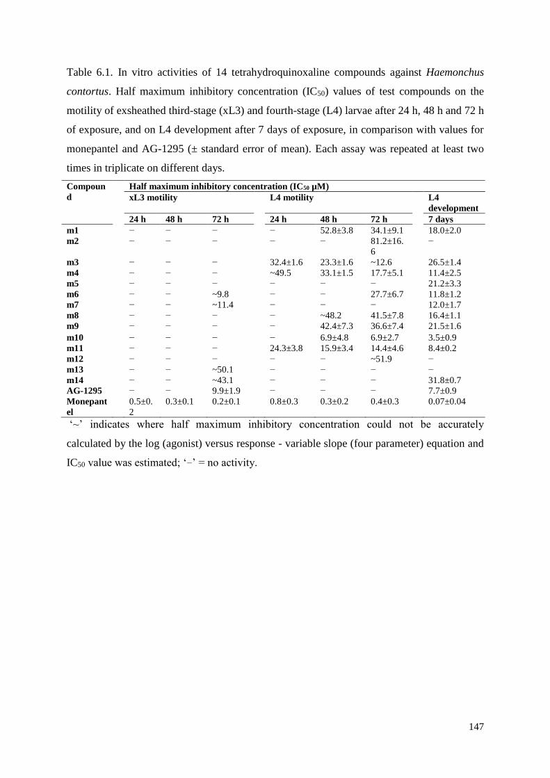
147
Table 6.1. In vitro activities of 14 tetrahydroquinoxaline compounds against Haemonchus
contortus. Half maximum inhibitory concentration (IC50) values of test compounds on the
motility of exsheathed third-stage (xL3) and fourth-stage (L4) larvae after 24 h, 48 h and 72 h
of exposure, and on L4 development after 7 days of exposure, in comparison with values for
monepantel and AG-1295 (± standard error of mean). Each assay was repeated at least two
times in triplicate on different days.
Compoun
d
Half maximum inhibitory concentration (IC50 µM)
xL3 motility L4 motility L4
development
24 h 48 h 72 h 24 h 48 h 72 h 7 days
m1 − − − − 52.8±3.8 34.1±9.1 18.0±2.0
m2 − − − − − 81.2±16.
6
−
m3 − − − 32.4±1.6 23.3±1.6 ~12.6 26.5±1.4
m4 − − − ~49.5 33.1±1.5 17.7±5.1 11.4±2.5
m5 − − − − − − 21.2±3.3
m6 − − ~9.8 − − 27.7±6.7 11.8±1.2
m7 − − ~11.4 − − − 12.0±1.7
m8 − − − − ~48.2 41.5±7.8 16.4±1.1
m9 − − − − 42.4±7.3 36.6±7.4 21.5±1.6
m10 − − − − 6.9±4.8 6.9±2.7 3.5±0.9
m11 − − − 24.3±3.8 15.9±3.4 14.4±4.6 8.4±0.2
m12 − − − − − ~51.9 −
m13 − − ~50.1 − − − −
m14 − − ~43.1 − − − 31.8±0.7
AG-1295 − − 9.9±1.9 − − − 7.7±0.9
Monepant
el
0.5±0.
2
0.3±0.1 0.2±0.1 0.8±0.3 0.3±0.2 0.4±0.3 0.07±0.04
‘~’ indicates where half maximum inhibitory concentration could not be accurately
calculated by the log (agonist) versus response - variable slope (four parameter) equation and
IC50 value was estimated; ‘−’ = no activity.
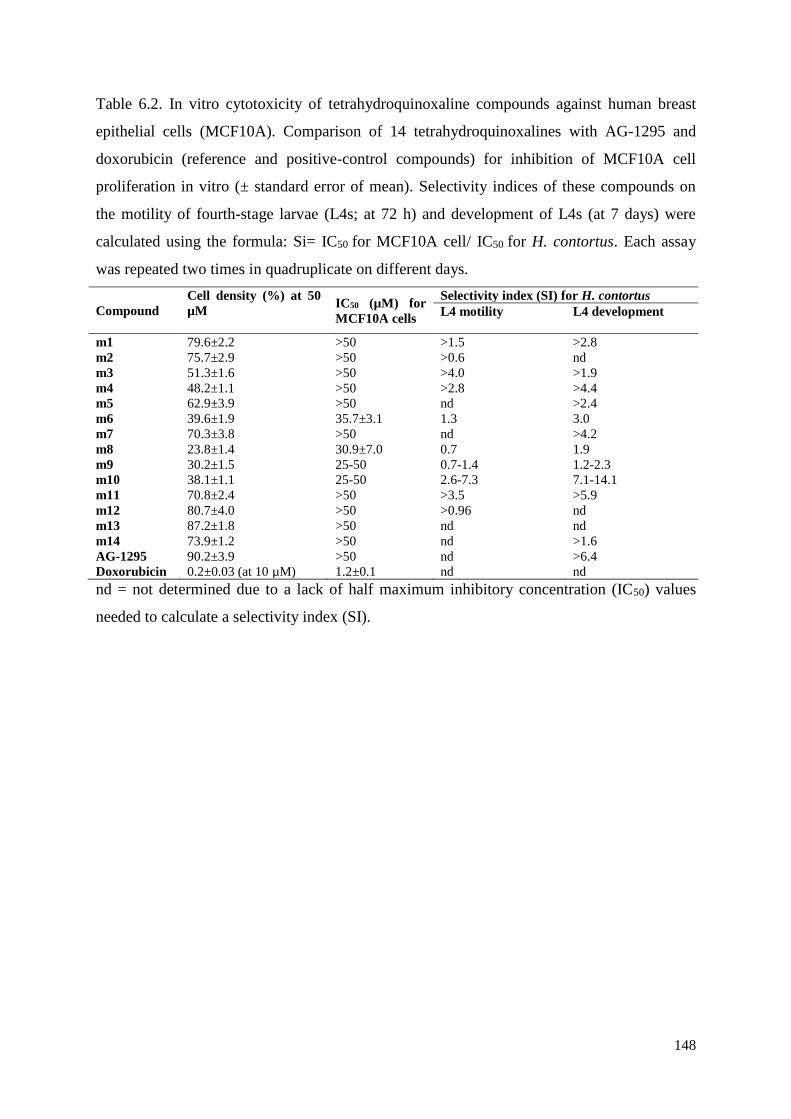
148
Table 6.2. In vitro cytotoxicity of tetrahydroquinoxaline compounds against human breast
epithelial cells (MCF10A). Comparison of 14 tetrahydroquinoxalines with AG-1295 and
doxorubicin (reference and positive-control compounds) for inhibition of MCF10A cell
proliferation in vitro (± standard error of mean). Selectivity indices of these compounds on
the motility of fourth-stage larvae (L4s; at 72 h) and development of L4s (at 7 days) were
calculated using the formula: Si= IC50 for MCF10A cell/ IC50 for H. contortus. Each assay
was repeated two times in quadruplicate on different days.
Compound
Cell density (%) at 50
µM
IC50 (µM) for
MCF10A cells
Selectivity index (SI) for H. contortus
L4 motility L4 development
m1 79.6±2.2 >50 >1.5 >2.8
m2 75.7±2.9 >50 >0.6 nd
m3 51.3±1.6 >50 >4.0 >1.9
m4 48.2±1.1 >50 >2.8 >4.4
m5 62.9±3.9 >50 nd >2.4
m6 39.6±1.9 35.7±3.1 1.3 3.0
m7 70.3±3.8 >50 nd >4.2
m8 23.8±1.4 30.9±7.0 0.7 1.9
m9 30.2±1.5 25-50 0.7-1.4 1.2-2.3
m10 38.1±1.1 25-50 2.6-7.3 7.1-14.1
m11 70.8±2.4 >50 >3.5 >5.9
m12 80.7±4.0 >50 >0.96 nd
m13 87.2±1.8 >50 nd nd
m14 73.9±1.2 >50 nd >1.6
AG-1295 90.2±3.9 >50 nd >6.4
Doxorubicin 0.2±0.03 (at 10 µM) 1.2±0.1 nd nd
nd = not determined due to a lack of half maximum inhibitory concentration (IC50) values
needed to calculate a selectivity index (SI).
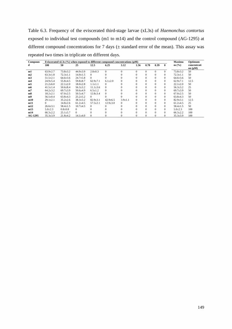
149
Table 6.3. Frequency of the eviscerated third-stage larvae (xL3s) of Haemonchus contortus
exposed to individual test compounds (m1 to m14) and the control compound (AG-1295) at
different compound concentrations for 7 days (± standard error of the mean). This assay was
repeated two times in triplicate on different days.
Compoun
d
Eviscerated xL3s (%) when exposed to different compound concentrations (µM) Maximu
m (%)
Optimum
concentrati
on (µM) 100 50 25 12.5 6.25 3.12 1.56 0.78 0.39 0
m1 63.9±2.7 73.8±3.2 44.9±3.9 2.8±0.3 0 0 0 0 0 0 73.8±3.2 50
m2 63.3±1.8 72.3±1.1 14.9±1.5 0 0 0 0 0 0 0 72.3±1.1 50
m3 51.5±2.1 64.6±3.6 24.7±5.8 0 0 0 0 0 0 0 64.6±3.6 50
m4 24.9±5.4 55.8±4.5 59.8±8.7 62.9±7.1 6.1±2.0 0 0 0 0 0 62.9±7.1 12.5
m5 21.2±6.0 22.1±2.0 18.0±2.8 1.1±1.1 0 0 0 0 0 0 22.1±2.0 50
m6 41.5±1.4 50.6±8.4 56.3±3.2 11.1±3.6 0 0 0 0 0 0 56.3±3.2 25
m7 64.2±3.2 69.7±3.9 50.6±4.9 6.5±1.2 0 0 0 0 0 0 69.7±3.9 50
m8 18.2±2.1 67.0±2.5 50.5±4.7 12.8±3.4 0 0 0 0 0 0 67.0±2.5 50
m9 56.5±0.4 63.8±4.3 25.2±5.2 0 0 0 0 0 0 0 63.8±4.3 50
m10 29.1±2.1 35.2±2.6 38.3±3.2 82.9±3.1 42.9±6.5 3.9±1.1 0 0 0 0 82.9±3.1 12.5
m11 0 14.8±2.6 61.2±4.5 57.5±2.1 12.9±3.0 0 0 0 0 0 61.2±4.5 25
m12 26.6±3.1 58.4±1.5 10.7±4.5 0 0 0 0 0 0 0 58.4±1.5 50
m13 5.0±2.3 0.8±0.8 0 0 0 0 0 0 0 0 5.0±2.3 100
m14 66.3±2.2 25.1±5.7 0 0 0 0 0 0 0 0 66.3±2.2 100
AG-1295 35.3±3.9 21.8±4.2 14.1±4.0 0 0 0 0 0 0 0 35.3±3.9 100
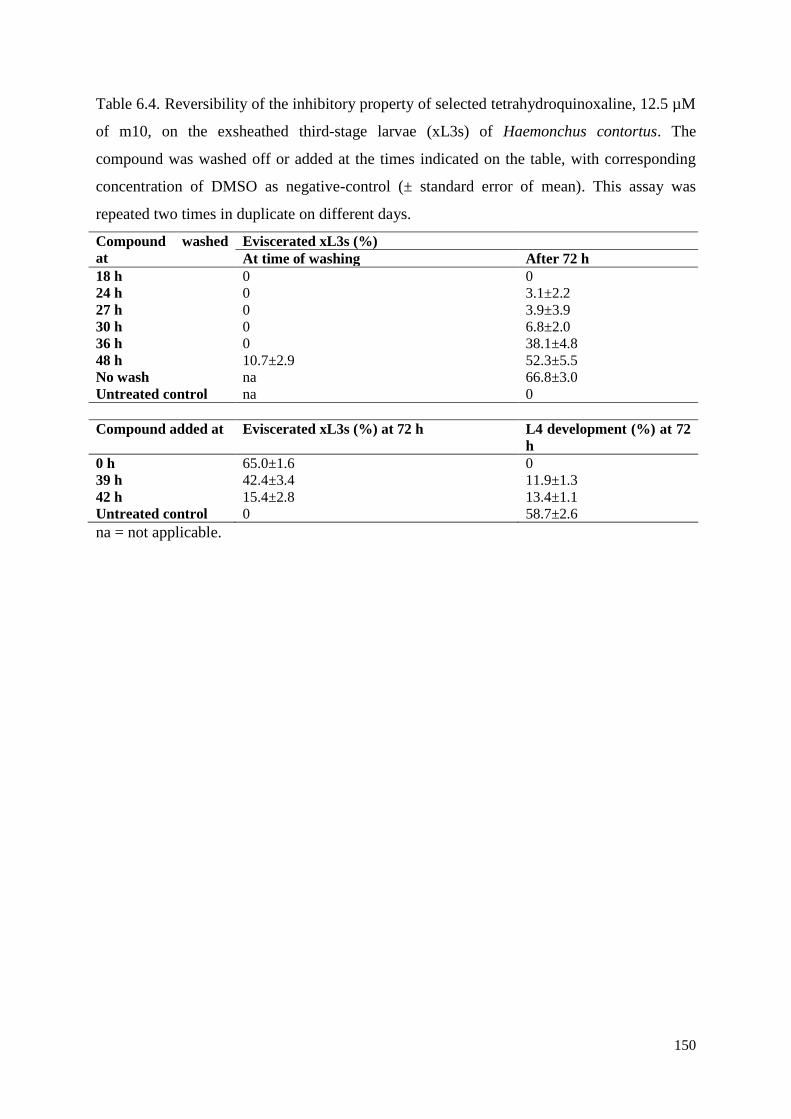
150
Table 6.4. Reversibility of the inhibitory property of selected tetrahydroquinoxaline, 12.5 µM
of m10, on the exsheathed third-stage larvae (xL3s) of Haemonchus contortus. The
compound was washed off or added at the times indicated on the table, with corresponding
concentration of DMSO as negative-control (± standard error of mean). This assay was
repeated two times in duplicate on different days.
Compound washed
at
Eviscerated xL3s (%)
At time of washing After 72 h
18 h 0 0
24 h 0 3.1±2.2
27 h 0 3.9±3.9
30 h 0 6.8±2.0
36 h 0 38.1±4.8
48 h 10.7±2.9 52.3±5.5
No wash na 66.8±3.0
Untreated control na 0
Compound added at Eviscerated xL3s (%) at 72 h L4 development (%) at 72
h
0 h 65.0±1.6 0
39 h 42.4±3.4 11.9±1.3
42 h 15.4±2.8 13.4±1.1
Untreated control 0 58.7±2.6
na = not applicable.
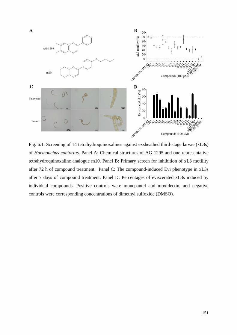
151
Fig. 6.1. Screening of 14 tetrahydroquinoxalines against exsheathed third-stage larvae (xL3s)
of Haemonchus contortus. Panel A: Chemical structures of AG-1295 and one representative
tetrahydroquinoxaline analogue m10. Panel B: Primary screen for inhibition of xL3 motility
after 72 h of compound treatment. Panel C: The compound-induced Evi phenotype in xL3s
after 7 days of compound treatment. Panel D: Percentages of eviscerated xL3s induced by
individual compounds. Positive controls were monepantel and moxidectin, and negative
controls were corresponding concentrations of dimethyl sulfoxide (DMSO).
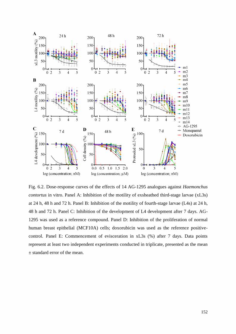
152
Fig. 6.2. Dose-response curves of the effects of 14 AG-1295 analogues against Haemonchus
contortus in vitro. Panel A: Inhibition of the motility of exsheathed third-stage larvae (xL3s)
at 24 h, 48 h and 72 h. Panel B: Inhibition of the motility of fourth-stage larvae (L4s) at 24 h,
48 h and 72 h. Panel C: Inhibition of the development of L4 development after 7 days. AG-
1295 was used as a reference compound. Panel D: Inhibition of the proliferation of normal
human breast epithelial (MCF10A) cells; doxorubicin was used as the reference positive-
control. Panel E: Commencement of evisceration in xL3s (%) after 7 days. Data points
represent at least two independent experiments conducted in triplicate, presented as the mean
± standard error of the mean.
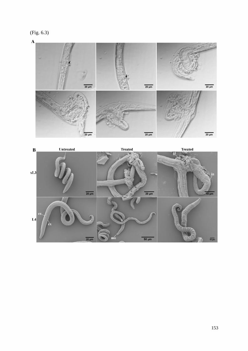
153
(Fig. 6.3)
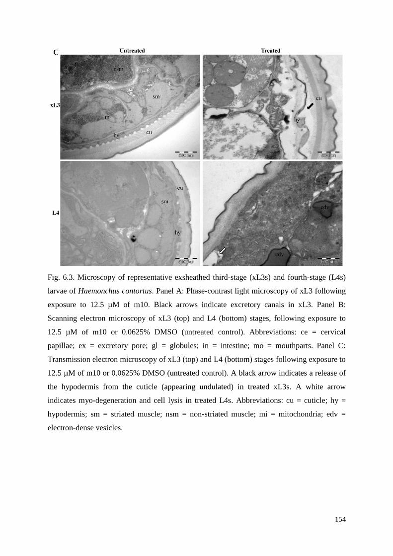
154
Fig. 6.3. Microscopy of representative exsheathed third-stage (xL3s) and fourth-stage (L4s)
larvae of Haemonchus contortus. Panel A: Phase-contrast light microscopy of xL3 following
exposure to 12.5 µM of m10. Black arrows indicate excretory canals in xL3. Panel B:
Scanning electron microscopy of xL3 (top) and L4 (bottom) stages, following exposure to
12.5 µM of m10 or 0.0625% DMSO (untreated control). Abbreviations: ce = cervical
papillae; ex = excretory pore; gl = globules; in = intestine; mo = mouthparts. Panel C:
Transmission electron microscopy of xL3 (top) and L4 (bottom) stages following exposure to
12.5 µM of m10 or 0.0625% DMSO (untreated control). A black arrow indicates a release of
the hypodermis from the cuticle (appearing undulated) in treated xL3s. A white arrow
indicates myo-degeneration and cell lysis in treated L4s. Abbreviations: cu = cuticle; hy =
hypodermis; sm = striated muscle; nsm = non-striated muscle; mi = mitochondria; edv =
electron-dense vesicles.
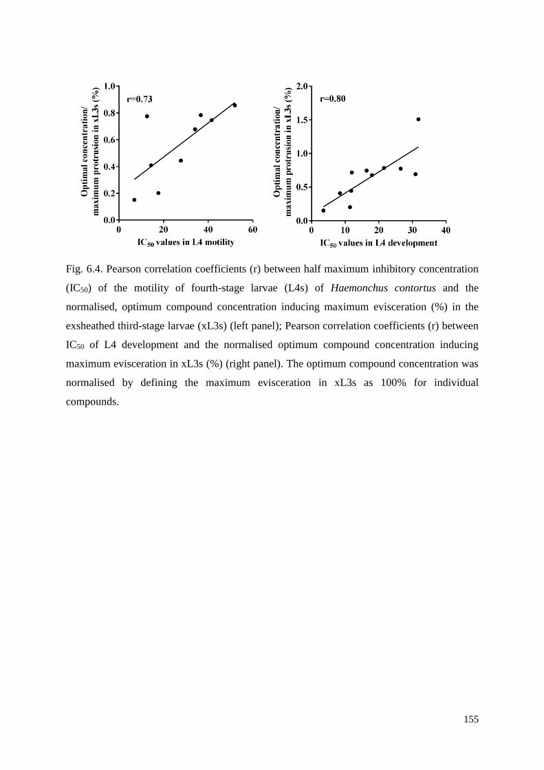
155
Fig. 6.4. Pearson correlation coefficients (r) between half maximum inhibitory concentration
(IC50) of the motility of fourth-stage larvae (L4s) of Haemonchus contortus and the
normalised, optimum compound concentration inducing maximum evisceration (%) in the
exsheathed third-stage larvae (xL3s) (left panel); Pearson correlation coefficients (r) between
IC50 of L4 development and the normalised optimum compound concentration inducing
maximum evisceration in xL3s (%) (right panel). The optimum compound concentration was
normalised by defining the maximum evisceration in xL3s as 100% for individual
compounds.
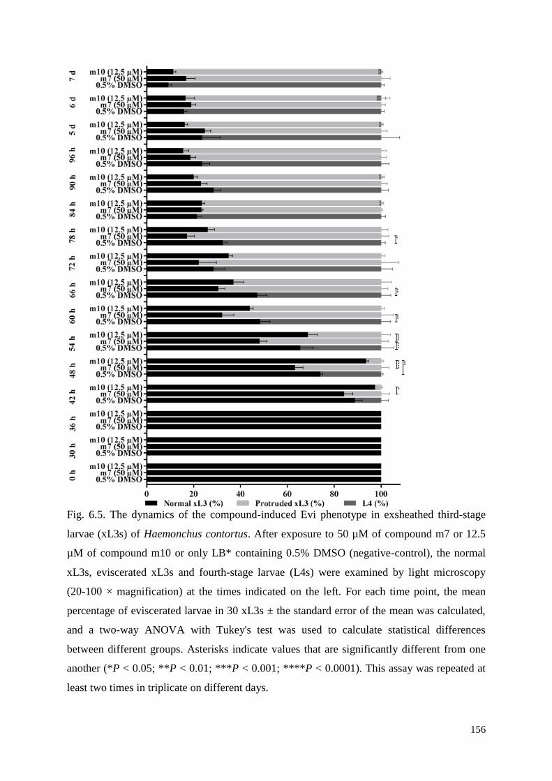
156
Fig. 6.5. The dynamics of the compound-induced Evi phenotype in exsheathed third-stage
larvae (xL3s) of Haemonchus contortus. After exposure to 50 µM of compound m7 or 12.5
µM of compound m10 or only LB* containing 0.5% DMSO (negative-control), the normal
xL3s, eviscerated xL3s and fourth-stage larvae (L4s) were examined by light microscopy
(20-100 × magnification) at the times indicated on the left. For each time point, the mean
percentage of eviscerated larvae in 30 xL3s ± the standard error of the mean was calculated,
and a two-way ANOVA with Tukey's test was used to calculate statistical differences
between different groups. Asterisks indicate values that are significantly different from one
another (*P < 0.05; **P < 0.01; ***P < 0.001; ****P < 0.0001). This assay was repeated at
least two times in triplicate on different days.
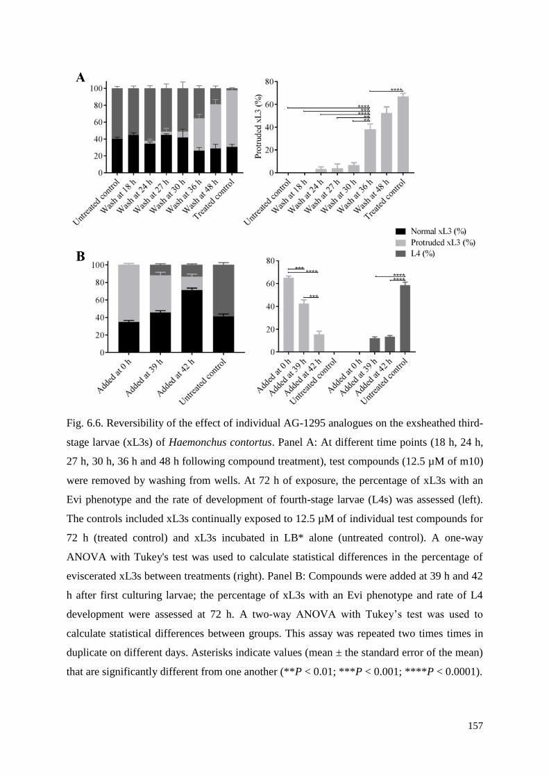
157
Fig. 6.6. Reversibility of the effect of individual AG-1295 analogues on the exsheathed third-
stage larvae (xL3s) of Haemonchus contortus. Panel A: At different time points (18 h, 24 h,
27 h, 30 h, 36 h and 48 h following compound treatment), test compounds (12.5 µM of m10)
were removed by washing from wells. At 72 h of exposure, the percentage of xL3s with an
Evi phenotype and the rate of development of fourth-stage larvae (L4s) was assessed (left).
The controls included xL3s continually exposed to 12.5 µM of individual test compounds for
72 h (treated control) and xL3s incubated in LB* alone (untreated control). A one-way
ANOVA with Tukey's test was used to calculate statistical differences in the percentage of
eviscerated xL3s between treatments (right). Panel B: Compounds were added at 39 h and 42
h after first culturing larvae; the percentage of xL3s with an Evi phenotype and rate of L4
development were assessed at 72 h. A two-way ANOVA with Tukey’s test was used to
calculate statistical differences between groups. This assay was repeated two times times in
duplicate on different days. Asterisks indicate values (mean ± the standard error of the mean)
that are significantly different from one another (**P < 0.01; ***P < 0.001; ****P < 0.0001).
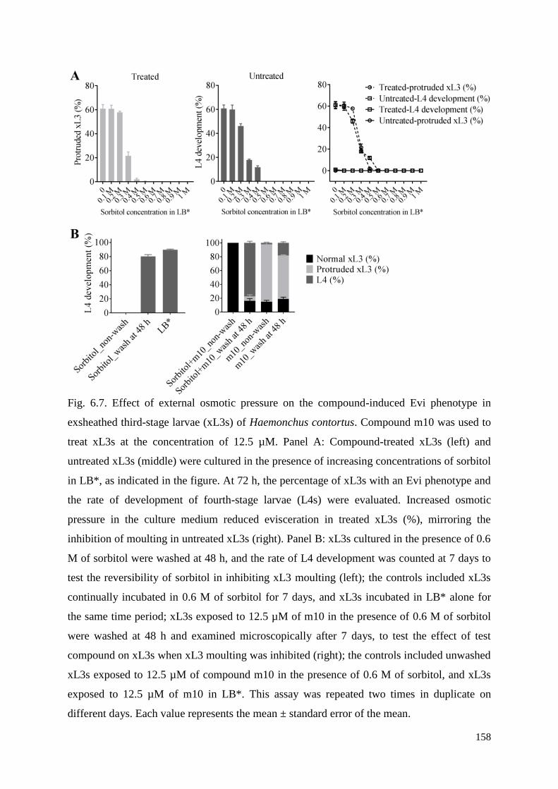
158
Fig. 6.7. Effect of external osmotic pressure on the compound-induced Evi phenotype in
exsheathed third-stage larvae (xL3s) of Haemonchus contortus. Compound m10 was used to
treat xL3s at the concentration of 12.5 µM. Panel A: Compound-treated xL3s (left) and
untreated xL3s (middle) were cultured in the presence of increasing concentrations of sorbitol
in LB*, as indicated in the figure. At 72 h, the percentage of xL3s with an Evi phenotype and
the rate of development of fourth-stage larvae (L4s) were evaluated. Increased osmotic
pressure in the culture medium reduced evisceration in treated xL3s (%), mirroring the
inhibition of moulting in untreated xL3s (right). Panel B: xL3s cultured in the presence of 0.6
M of sorbitol were washed at 48 h, and the rate of L4 development was counted at 7 days to
test the reversibility of sorbitol in inhibiting xL3 moulting (left); the controls included xL3s
continually incubated in 0.6 M of sorbitol for 7 days, and xL3s incubated in LB* alone for
the same time period; xL3s exposed to 12.5 µM of m10 in the presence of 0.6 M of sorbitol
were washed at 48 h and examined microscopically after 7 days, to test the effect of test
compound on xL3s when xL3 moulting was inhibited (right); the controls included unwashed
xL3s exposed to 12.5 µM of compound m10 in the presence of 0.6 M of sorbitol, and xL3s
exposed to 12.5 µM of m10 in LB*. This assay was repeated two times in duplicate on
different days. Each value represents the mean ± standard error of the mean.

159
Chapter 7 - General discussion
The control of parasitic nematodes is seriously challenged by widespread anthelmintic
resistance (Kaplan and Vidyashankar, 2012; Wolstenholme and Kaplan, 2012; Geary et al.,
2015; Kotze and Prichard, 2016). Therefore, the discovery and development of new and
effective anthelmintics are essential to sustain the economic and health benefits from their
application to control parasites (Geary et al., 2015; Campbell, 2016).
The research aims of this thesis were (i) to use whole-organism screening of compounds
from well-defined, curated collections of chemicals for inhibitory activity against H.
contortus and/or other parasitic nematodes; (ii) to identify ‘hit’ compounds, and (iii) to assess
these compounds as possible ‘leads’ for optimisation and subsequent development. These
aims were achieved in that 20 compounds with in vitro-activity against H. contortus,
including one new chemical entity (designated SN00797439), two human kinase inhibitors
(SNS-032 and AG-1295), 14 AG-1295 (tetrahydroquinoxaline) analogues, one insecticide
(tolfenpyrad) and two tolfenpyrad (pyrazole-5-carboxamide) derivatives (a-15 and a-17)
(Chapters 2-6). Collectively, these achievements offer a sound starting point for the
development of new anthelmintics.
The purpose of the present chapter is to discuss the achievements in a broader context,
considering aspects of screening technologies (Subsection 7.1), compound libraries (7.2), the
evaluation of ‘hit’ compounds (7.3) and their potential as ‘lead’ compounds in future
anthelmintic development (7.4), and to propose lines of future research (7.5 and 7.6).
7.1. Screening technologies
In this project, established phenotypic screening assays employing parasitic stages of H.
contortus were used (Preston et al., 2015), with the advantage of being able to assess
nematostatic or nematocidal effects of chemicals on whole worms in real time. This approach
circumvented some of the hurdles of mechanism-based screens (Geary et al., 1999), where
compounds only bind to selected targets and are often not assessed on other molecular targets
or whole organisms (cf. Kotze, 2012; Geary et al., 2015). However, compared with
mechanism-based screens, where the target/s is/are defined, in whole-worm screens, the
molecular targets are usually not known in the first instance. Nonetheless, the modes of
action of active compounds identified in whole-worm screening assays can be studied at the
molecular level, for instance, using knowledge of the genome, transcriptome and proteome of
the target organism (e.g., Laing et al., 2013; Schwarz et al., 2013; Geary, 2016; Harder,

160
2016). In addition, for known drugs destined for repurposing to parasitic worms, their modes
of action might be extrapolated to the ‘new’ organism to which the drug is being repurposed,
although an emphasis needs to be placed on critically evaluating any possible side effects on
the host organism (O'Connor and Roth, 2005).
Motility reduction as a measure to detect hits and its limitations. The assays employed in
this thesis were designed to measure a reduction in ‘motility’ of larvae in order to identify
active compounds (Preston et al., 2015). However, some compounds, such as AG-1295 and
its analogues (Chapter 6), did not reduce motility, but rather caused a phenotypic change in
the larvae. Thus, motility reduction is not always a suitable measure and can lead to ‘false-
negative’ results. In the present study, close inspection of treated larvae by microscopy
identified a number of ‘hit’ compounds based on their induction of non-wildtype phenotypes,
such as the ‘coiled’, ‘straight’ and ‘anterior protrusion’ phenotypes, following exposure of H.
contortus to SN00797439, tolfenpyrad and AG-1295 analogues. In addition, scanning
electron microscopy (SEM) and transmission electron microscopy (TEM) sometimes
provided useful insights into compound-induced morphological changes at the tissue and
cellular levels.
Therefore, in the future, the phenotype of treated worms should be recorded as a
complementary endpoint to improve the identification of ‘hit’ compounds. Although the
current assays allow for worm phenotypes to be recorded by visual inspection of five-second
videos, the workload associated with this step, particularly when screening large numbers of
compounds, such as the ‘Open Scaffolds’ library, restricts screening throughput. Hence, it
will be important to develop an assay that can quantitatively assess both worm motility and
worm phenotype to avoid ‘false-negative’ results and to increase screening efficiency. It is
worth mentioning that quantitative tracking of phenotypes has already been applied to worms
such as C. elegans (see Geng et al., 2004) and Schistosoma mansoni (see Singh et al., 2018),
utilizing mathematic algorithms and automatic imaging technology. As the motility dynamics
of parasitic nematodes is distinct from C. elegans and S. mansoni, new algorithms will need
to be developed and assessed to ensure reliability for hit discovery.
The importance of extending screening to adult worms. Traditionally, conventional whole-
worm screening assays, such as the egg hatch assay (Le Jambre, 1976; Dobson et al., 1986)
and larval development assay (Kotze et al., 2006), have been applied to free-living larval
stages. Compared with such conventional assays, the present motility assays (Preston et al.,
161
2015) were an improvement through the use of ‘parasitic’ larvae, xL3s and L4s, both
produced in vitro. A substantial advantage was also the ability to store L3s for long periods
(up to 6 months). The ability to directly exsheath L3s in vitro and raise xL3s and then L4s
circumvented the need for experimental animals to produce L4 and adult stages in vivo in
sheep. Nonetheless, considering that the adult stages of gastrointestinal (strongylid)
nematodes are usually the target stage for most anthelmintics, the inclusion of adults in future
in vitro screening of chemical libraries would be advantageous, and would allow a
comparative assessment of potency among developmental stages.
7.2. Considerations regarding compound libraries
Numerous well-curated compound collections in different countries provide an untapped
resource with potential for the discovery of new anthelmintics. In the present project, five
different compound collections were obtained from different sources. The ‘Open Scaffolds’
library was sourced from Compounds Australia, the ‘Stasis Box’ and the ‘Pathogen Box’
were both acquired through a product-development partnership (PDP) with Medicine for
Malaria Venture (MMV), and the pyrazole-5-carboxamide collections and
tetrahydroquinoxaline collections were both obtained from academic institutions.
Compounds Australia sources small molecules and consolidates them into a central
repository that facilitates subsequent screening and provides these molecules to laboratories
around the world, in order to boost drug discovery efforts (Simpson and Poulsen, 2014). The
‘Open Scaffolds’ library, screened in the present project, is one of the main collections
maintained by Compounds Australia (Chapter 2). This collection, containing 33,999
compounds (Simpson and Poulsen, 2014), is much larger than other compound collections
that were screened, e.g., the ‘Stasis Box’ and the ‘Pathogen Box’, each with 400 compounds.
Generally, the relatively small number of known compounds is considered to limit the
chemical space for drug repurposing (Hergenrother, 2006). However, the ‘hit’ rate from the
latter two boxes (0.5% for ‘Stasis Box’; 0.25% for ‘Pathogen Box’) for possible repurposing
was higher than that for the discovery of new chemical entities (0.0029%) from the ‘Open
Scaffolds’ library. This finding can be explained by the smaller compound collections
containing advanced compounds (cf. Sekhon, 2013), e.g., with known pharmacokinetic
parameters and/or biological activities. Thus, careful consideration needs to be given to the
selection of compound libraries for screening on parasites.
162
Fortunately, 14 tetrahydroquinoxaline analogues of the active compound from the ‘Stasis
Box’ (AG-1295) could be sourced from a collaborator, Professor Jonathan Baell, which
enabled early investigations of potency on xL3 and L4 stages of H. contortus (Chapter 6) and
a foundation for future mode of action studies. Similarly, 55 tolfenpyrad (pyrazole-5-
carboxamide) analogues were sourced through collaborators in China from the State Key
Laboratory of Elemento-organic Chemistry in Nankai University, China (Chapter 5),
allowing potency and cytotoxicity evaluations. Clearly, further medicinal chemistry
optimisation is required to enable further assessment and development of these chemical
entities.
7.3. Evaluation of ‘hit’ compounds
Following the identification of hits through screening and dose-response experiments,
compound evaluation can guide the identification of lead candidates (cf. Goldstein et al.,
2008).
Inhibition of larval motility and development. In this project, the assessment of inhibition
of larval motility and development in H. contortus was consistently used to evaluate the
potency of ‘hit’ compounds, offering essential information for the selection of compounds for
further study. Of the twenty ‘hit’ compounds, the IC50 values for xL3 motility inhibition (72
h) ranged from 3 µM to 56 µM, the IC50 values of L4 motility reduction (72 h) ranged from
0.03 µM to 26 µM, and the IC50 values of L4 development inhibition (7 d) ranged from 0.08
µM to 41 µM. The in vitro potency of some of these ‘hit’ compounds, e.g., tolfenpyrad, was
comparable with that of the commercial anthelmintic monepantel (IC50 values of 0.2 µM, 0.4
µM and 0.07 µM, respectively). Based on IC50 values, tolfenpyrad was identified as the most
potent compound at inhibiting larval motility and development (Chapter 4) and, thus, may be
a candidate for further optimisation. Nevertheless, other hit compounds (i.e. SN00797439, a-
15, a-17, SNS-032, AG-1295 and 14 tetrahydroquinoxaline compounds) also show promise
for hit-to-lead optimisation, given that they have lead-like scaffolds and/or have already been
developed or proposed as drugs.
The kinase inhibitors SNS-032 and AG-1295 did not show any detectable inhibitory
activity on L4 motility, even though the L4 stage has a functional pharynx, which allows the
uptake of chemicals (Chapter 3). This information suggests that the expression of the kinase
target(s) in L4s is markedly lower than that in xL3s, and may explain the difference in
163
potency between the two stages. In contrast, for the other hit compounds with more potency
in L4s than in xL3s, it remains unclear whether the difference in potency between the two
stages is due to the level of expression of target(s) and/or variation in drug uptake and/or
metabolism.
Cytotoxicity testing. With regard to cytotoxicity and selectivity of ‘hit’ compounds,
mammalian cell proliferation was used as an ‘endpoint assay’ - which has been used
extensively for the screening of drugs for use in humans (Li et al., 2006; McKim, 2010;
Fisher et al., 2014). The in vitro cytotoxicity test provides important information for selecting
compounds with (apparent) low risk profiles for further development and for the ‘de-
prioritisation’ of compounds with high risk profiles prior to expensive preclinical and clinical
trials, thus reducing early risks and improving the probability of success in the development
pipeline (McKim, 2010; Sayes, 2014). However, in vitro cytotoxicity tests may be unreliable
in predicting in vivo toxicity due to the pronounced differences between in vitro and in vivo
systems relating, for example, to differences in metabolic activities, protein binding and/or
compound solubility (McKim, 2010). Therefore, results from an in vitro cytotoxicity test
cannot be used to make a final decision about the toxicity of a compound in vivo, such that
extensive animal toxicity testing must be undertaken to verify the safety of a drug under
consideration as a lead candidate, and is a requirement for registration.
7.4. Potential of identified compounds as ‘leads’ in future
anthelmintic development
Pyrazole-5-carboxamides. Of the ‘hit’ compounds identified in this project, the pyrazole-
5-carboxamide insecticide, tolfenpyrad, has the highest in vitro anthelmintic potency against
H. contortus. As insecticides or acaricides, pyrazole-5-carboxamides are considered to target
complex I of the electron transport chain in arthropods (Hollingworth, 2001). Here, through
measuring the mitochondrial oxygen consumption rate (OCR), tolfenpyrad and its two
pyrazole-5-carboxamide derivatives, a-15 and a-17, were shown to significantly reduce OCR
in H. contortus (Chapters 4 and 5), indicating that this compound series might also target
complex I of the electron transport chain in H. contortus. In the electron transport chain,
parasitic larvae and adult worms of H. contortus are predicted to employ a NADH-fumarate
reductase system, in which complex I is the NADH-rhodoquinone reductase that is different
from mammalian animals using NADH-ubiquinone reductase as complex I (Kita et al., 1997;
164
Harder, 2016). The difference in complex I between H. contortus and mammals suggests that
it may be a suitable and selective drug target for anthelmintic discovery (Kita et al., 2003;
Harder, 2016), a proposal supported by cytotoxicity data showing that tolfenpyrad is more
selective for H. contortus than mammalian cells (Chapters 4 and 5).
The energy production in adult worms via mitochondria appears to be mainly linked to
reproduction and a complex regulation of the neuromuscular system (Harder, 2016). Indeed,
there are only few anthelmintics targeting the energy production system in nematodes, with
most currently used anthelmintics targeting the nematode neuromuscular system (Kotze,
2012; Holden-Dye and Walker, 2014). For instance, imidazothiazoles act as cholinergic
agonists at nicotinic neuromuscular junctions (Coles et al., 1975; Prichard, 1990; McKellar
and Jackson, 2004), macrocyclic lactones target neurotransmitter gamma-aminobutyric acid-
gated chloride ion channels and glutamate-gated chloride ion channels (Arena et al., 1992;
Brownlee et al., 1997), monepantel binds to nicotinic acetylcholine receptor subunits
(Kaminsky et al., 2008; Sargison, 2012; Baur et al., 2015) and derquantel represents nicotinic
acetylcholine receptor antagonists (Ruiz-Lancheros et al., 2011; Sargison, 2012). Here,
tolfenpyrad shows some promise as a new anthelmintic chemical, but needs to be explored
further, optimised and tested for safety and efficacy in animals.
Kinase inhibitors. By screening compound collections obtained through a PDP with
MMV, besides tolfenpyrad, two human kinase inhibitors (SNS-032 and AG-1295) were
identified to have in vitro anthelmintic activity but showed less motility inhibition than
tolfenpyrad. The discovery of kinase inhibitors as therapeutics is a relatively ‘hot’ topic in the
field of drug discovery, particularly in relation to anti-cancer drugs (Sebolt-Leopold and
English, 2006; Zhang et al., 2009). Clearly, kinases play a crucial role in all cellular
processes, and the dysregulation of kinase activities is related to many diseases (Goldstein et
al., 2008). With the success of imatinib (Manley et al., 2002; Baselga, 2006; Collins and
Workman, 2006; Larson et al., 2008), the discovery of kinase inhibitors has attracted
considerable attention (Sebolt-Leopold and English, 2006; Verweij and de Jonge, 2007;
Zhang et al., 2009). Moreover, for drug repurposing, imatinib has been evaluated for activity
against S. mansoni, and has pronounced schistosomicidal activity in vitro but not in vivo
(Katz et al., 2013). Kinome studies of socioeconomically important parasitic worms,
including H. contortus, S. haematobium, Trichinella spiralis, T. pseudospiralis and Trichuris
suis (see Cantacessi et al., 2010; Beckmann et al., 2014; Preston et al., 2015; Stroehlein et al.,
2015a; Stroehlein et al., 2015b; Stroehlein et al., 2016), can deliver important resources for
165
genome- or transcriptome-guided anthelmintic discovery. In this context, the identification of
SNS-032 with anthelmintic activity against H. contortus indicates the potential of
bioinformatic-guided prioritisation of targets and chemicals via kinome investigations,
because the targets of SNS-032, CDK-7 and CDK-9 have been predicted in silico to be
prioritised kinase targets in H. contortus (see Stroehlein et al., 2015b).
SNS-032 has been developed as an anti-cancer drug (Ali et al., 2007; Kodym et al., 2009)
and AG-1295 has been developed as an anti-restenosis drug (Banai et al., 1998). Therefore,
the two human drugs were specifically designed to bind to human kinase targets rather than
those of H. contortus. Marked differences in sequences and three-dimensional structures
between the human kinase targets of SNS-032 and their corresponding H. contortus
homologs (Jiao et al., 2017) appear to help explain variations in potency and selectivity of
this kinase inhibitor at inhibiting worms versus mammalian cells, and suggest that there is an
opportunity to chemically optimise SNS-032 and AG-1295 (tetrahydroquinoxaline)
analogues to bind specifically to targets in H. contortus.
Compound SN00797439. The new chemical entity, SN00797439, has a novel, lead-like
scaffold containing a 1, 2, 4-oxadiazole and a 1-pyrrolidinecarboxamide. The 1, 2, 4-
oxadiazole has been relatively widely used as a drug for different applications (Bora et al.,
2014), such as an anti-inflammatory agent (Dalhamn, 1969), an analgesic (Kumar et al.,
2012) and a muscarinic receptor agonist (Street et al., 1990). The 1-pyrrolidinecarboxamide
has been rarely used, except as cisanilide ((2R, 5S)-rel-2, 5-dimethyl-N-phenyl-1-
pyrrolidinecarboxamide), a now obsolete herbicide (Frear and Swanson, 1975; Frear and
Swanson, 1976).
SN00797439 achieved IC50 values of 0.3 µM in inhibiting larval motility, and is more
potent than other ‘hit’ compounds, except tolfenpyrad. In particular, the efficacy of this
compound was also characterised by a ‘coiled’ xL3 phenotype in H. contortus and
considerable cuticular damage in L4s in vitro. Evaluations on other parasitic nematodes
showed that SN00797439 possesses relatively broad-spectrum anthelmintic activity, and has
potential to be developed as a new anthelmintic. In this project, several genetically
(evolutionarily) distant and socioeconomically important parasitic nematodes (i.e. H.
contortus and A. ceylanicum [strongyloids] vs. B. malayi and D. immitis [filarioids] vs. T.
muris [enoplid]) were tested (Chapter 2). In the future, the activity of SN00797439 might be
assessed against other worms that cause neglected tropical diseases.
166
7.5. Future work toward anthelmintic development
Although in vitro nematocidal/nematostatic activity and cytotoxicity data for the ‘hit’
compounds identified in this project provide some promise for their development as new
anthelmintics, future work needs to focus on detailed investigations of absorption,
distribution, metabolism, excretion and toxicity (ADMET), following their optimisation via
structure-activity relationship (SAR) assessments. This step is essential for compounds to
enter further preclinical and then clinical development stages prior to registration and
commercialisation (Ashburn and Thor, 2004; Lombardino and Lowe, 2004; McKim, 2010;
Hughes et al., 2011; Geary et al., 2015; Andrade et al., 2016; Campbell, 2016; Matthews et
al., 2016). This is an involved process, with many risks along the way, which explains why
many anthelmintic candidates have not reached commercialisation (Hughes et al., 2011;
Geary, 2016). Indeed, drug development is extremely challenging, with only 1 in 10,000
chemicals in the discovery process ever reaching the market (McKim, 2010; Matthews et al.,
2016). ‘Chemical death’ occurs particularly in the latter stages of the development pipeline
(Kola, 2008). Clearly, late-stage failures contribute substantially to the cost of bringing new
drugs to market (Kola, 2008; McKim, 2010).
Given the challenges facing the pharmaceutical industry, it is crucial to prevent such
attrition (Kola and Landis, 2004; Bowes et al., 2012; Jakovljevic and Ogura, 2016). Factors
that can lead to drug development attrition are indicated in Fig. 7.1, with the two principal
reasons for this failure being inefficacy and toxicity (cf. Kola and Landis, 2004; Kola, 2008;
Hughes et al., 2011; Waring et al., 2015). In compiled attrition data for small molecule drug
candidates destined for development in four large pharmaceutical companies (AstraZeneca,
Eli Lilly, GlaxoSmithKline and Pfizer) between 2000 and 2010 (Fig. 7.1), toxicology
terminations were most prominent in the preclinical phase (59.27%) and clinical phase I
(25.48%), and a lack of efficacy was most prominent in clinical phase II (34.83%) (Waring et
al., 2015). Thus, given the major pressure that pharmaceutical companies are under to
circumvent drug development failures (Kola, 2008; Waring et al., 2015), the focus should be
on overcoming the two ‘key’ factors/challenges (Kola and Landis, 2004; Bowes et al., 2012).
In addition, drug formulation, bioavailability, manufacturing costs and commercial viability
are also important aspects that need to be considered in the drug development process (Kola
and Landis, 2004; Waring et al., 2015). For example, formulation is essential for the
compound to be dissolved and absorbed at a particular pH.
167
Although drug discovery and development are long, complex, expensive and risky
processes (Ashburn and Thor, 2004; Lombardino and Lowe, 2004; McKim, 2010; Geary et
al., 2015; Campbell, 2016), there is an urgency to develop new and effective anthelmintics to
circumvent established and emergent resistance problems in parasitic nematodes of animals
(particularly ruminant livestock), to sustain parasite control into the future. In the present
project, the identification and validation of 20 ‘hit’ compounds offer a sound starting point
for the translation of these chemicals into new anthelmintics (Fig. 7.2).
Chemical optimisation. In the drug development stage, chemical optimisation plays an
essential role in assisting in maximising potency and minimising side effects of ‘hit’
compounds (cf. Lombardino and Lowe, 2004; Hughes et al., 2011). Medicinal chemists will
decide on which analogues should be synthesised to explore structure-activity relationships,
in an effort to maximise the desired activity (Lombardino and Lowe, 2004). In preliminary
work, pyrazole-5-carboxamide derivatives have been synthesised and tested in SAR for
enhancing potency and reducing toxicity. In the first instance, with the application of
medium- to high-throughput screening, chemical optimisation relies heavily on data obtained
from in vitro testing. Further optimisation and validation are required to assess whether the
desired biological effect in vivo, e.g. in sheep, is achieved or not, and detect any side effects
(cf. Lombardino and Lowe, 2004; Hughes et al., 2011).
Pharmacokinetic properties. In order to assess the compounds in vivo, the
pharmacokinetic properties (absorption, distribution, metabolism, excretion and toxicity;
ADMET) of test compounds would need to be defined (Hughes et al., 2011). In particular,
the assessment of toxicity should be conducted once active analogues are obtained, and the
next screen should eliminate any compounds with a high-risk profile (cf. Lombardino and
Lowe, 2004; McKim, 2010). In the present project, all ‘hit’ compounds, except SNS-032,
showed limited toxicity to normal breast epithelial cells (MCF10A) in vitro; however, further
work needs to assess these compounds and their active analogues for toxicity in vivo as well
as non-clinical toxicity, e.g., genotoxicity and mutagenicity.
Spectrum of activity. In order to develop a new anthelmintic with broad-spectrum activity,
the compounds with most promise should be critically assessed for their activity on a range of
socioeconomic-important parasitic nematodes (e.g., hookworms, ascaridoids, filarioids and
whipworms). Although there is a lack of commercial incentives (by companies) to develop,
168
for example, chemotherapies against neglected tropical diseases affecting 1.8 billion people
in developing countries (Hotez et al., 2016; Houweling et al., 2016), this step obviously
would have major philanthropic and societal importance. SN00797439 has already shown
potential for this purpose, considering that it acts against a range of parasitic nematodes
including A. ceylanicum (hookworm of dogs, cats and humans), T. muris (whipworm of
mice) and B. malayi (filarial worm of humans and some animals) as well as D. immitis
(heartworm of canids).
Mode of action and resistance development. Other critical steps will be studies of the
mode of action of any new anthelmintic entity and its potential to induce drug resistance
(Vanden Bossche, 1990; Harder, 2016; Kotze and Prichard, 2016). Investigating modes of
action will be important to support further work on lead compounds (Swinney and Anthony,
2011; Duke et al., 2013; Dos Santos et al., 2016). The use of advanced molecular, informatic
and functional genomic (e.g., gene knockdown and knockout methods) technologies will be
important to assist in exploring drug targets of new anthelmintics in the future.
7.6. Concluding remarks
The present thesis makes a contribution to the field of anthelmintic drug discovery and
offers prospects for anthelmintic development, in light of the massive and widespread
anthelmintic resistance problems. This general discussion shows that the future focus should
now be on key areas including: (i) chemical optimisation of representative chemicals via
structure-activity relationship (SAR) evaluations; (ii) assessment of the breadth of spectrum
of anthelmintic activity on various parasitic nematodes, such as other strongyloids,
ascaridoids, enoplids and filarioids; (iii) detailed investigations of the absorption, distribution,
metabolism, excretion and toxicity (ADMET) of optimised chemicals with (relatively) broad
nematocidal or nematostatic activity; (iv) establishment of the modes of action of lead
candidates; (v) assessment of drug resistance development to lead compounds; and (vi)
passage through pre- and clinical development phases, in which the dosage, bioavailability,
therapeutic index, safety, formulation and other parameters of lead candidates need to be
established. Clearly, collaborations among scientists from different fields (including
parasitology, drug discovery, medicinal chemistry, clinical sciences and pharmacy and/or
biotechnology) will be central to achieving these goals and toward the translation and
subsequent commercialisation of effective new anthelmintic compounds.
169
7.7 References
Ali, M.A., Choy, H., Habib, A.A., Saha, D., 2007. SNS-032 prevents tumor cell-induced
angiogenesis by inhibiting vascular endothelial growth factor. Neoplasia 9, 370-381.
Andrade, E.L., Bento, A.F., Cavalli, J., Oliveira, S.K., Schwanke, R.C., Siqueira, J.M.,
Freitas, C.S., Marcon, R., Calixto, J.B., 2016. Non-clinical studies in the process of new
drug development - Part II: Good laboratory practice, metabolism, pharmacokinetics,
safety and dose translation to clinical studies. Braz. J. Med. Biol. Res. 49, e5646.
Arena, J.P., Liu, K.K., Paress, P.S., Schaeffer, J.M., Cully, D.F., 1992. Expression of a
glutamate-activated chloride current in Xenopus oocytes injected with Caenorhabditis
elegans RNA: evidence for modulation by avermectin. Mol. Brain Res. 15, 339-348.
Ashburn, T.T., Thor, K.B., 2004. Drug repositioning: identifying and developing new uses
for existing drugs. Nat. Rev. Drug Discov. 3, 673-683.
Banai, S., Wolf, Y., Golomb, G., Pearle, A., Waltenberger, J., Fishbein, I., Schneider, A.,
Gazit, A., Perez, L., Huber, R., Lazarovichi, G., Rabinovich, L., Levitzki, A., Gertz,
S.D., 1998. PDGF-receptor tyrosine kinase blocker AG1295 selectively attenuates
smooth muscle cell growth in vitro and reduces neointimal formation after balloon
angioplasty in swine. Circulation 97, 1960-1969.
Baselga, J., 2006. Targeting tyrosine kinases in cancer: the second wave. Science 312, 1175-
1178.
Baur, R., Beech, R., Sigel, E., Rufener, L., 2015. Monepantel irreversibly binds to and opens
Haemonchus contortus MPTL-1 and Caenorhabditis elegans ACR-20 receptors. Mol.
Pharmacol. 87, 96-102.
Beckmann, S., Long, T., Scheld, C., Geyer, R., Caffrey, C.R., Grevelding, C.G., 2014. Serum
albumin and alpha-1 acid glycoprotein impede the killing of Schistosoma mansoni by the
tyrosine kinase inhibitor Imatinib. Int. J. Parasitol. Drugs Drug Resist. 4, 287-295.
Bora, R.O., Dar, B., Pradhan, V., Farooqui, M., 2014. [1, 2, 4]-oxadiazoles: synthesis and
biological applications. Mini Rev. Med. Chem. 14, 355-369.
Bowes, J., Brown, A.J., Hamon, J., Jarolimek, W., Sridhar, A., Waldron, G., Whitebread, S.,
2012. Reducing safety-related drug attrition: the use of in vitro pharmacological
profiling. Nat. Rev. Drug Discov. 11, 909-922.
Brownlee, D.J., Holden-Dye, L., Walker, R.J., 1997. Actions of the anthelmintic ivermectin
on the pharyngeal muscle of the parasitic nematode, Ascaris suum. Parasitology 115 (Pt
5), 553-561.
170
Campbell, W.C., 2016. Lessons from the history of ivermectin and other antiparasitic agents.
Annu. Rev. Anim. Biosci. 4, 1-14.
Cantacessi, C., Mitreva, M., Jex, A.R., Young, N.D., Campbell, B.E., Hall, R.S., Doyle,
M.A., Ralph, S.A., Rabelo, E.M., Ranganathan, S., Sternberg, P.W., Loukas, A., Gasser,
R.B., 2010. Massively parallel sequencing and analysis of the Necator americanus
transcriptome. PLoS Negl. Trop. Dis. 4, e684.
Coles, G.C., East, J.M., Jenkins, S.N., 1975. The mechanism of action of the anthelmintic
levamisole. Gen. Pharmac. 6, 309-313.
Collins, I., Workman, P., 2006. New approaches to molecular cancer therapeutics. Nat.
Chem. Biol. 2, 689-700.
Dalhamn, T., 1969. The anticiliostatic effect of cigarettes treated with oxolamine citrate. Am.
Rev. Respir. Dis. 99, 447-448.
Dobson, R.J., Donald, A.D., Waller, P.J., Snowdon, K.L., 1986. An egg-hatch assay for
resistance to levamisole in trichostrongyloid nematode parasites. Vet. Parasitol. 19, 77-
84.
Dos Santos, B.S., da Silva, L.C., da Silva, T.D., Rodrigues, J.F., Grisotto, M.A., Correia,
M.T., Napoleao, T.H., da Silva, M.V., Paiva, P.M., 2016. Application of omics
technologies for evaluation of antibacterial mechanisms of action of plant-derived
products. Front Microbiol. 7, 1466
Duke, S.O., Bajsa, J., Pan, Z., 2013. Omics methods for probing the mode of action of natural
and synthetic phytotoxins. J. Chem. Ecol. 39, 333-347.
Fisher, G.M., Tanpure, R.P., Douchez, A., Andrews, K.T., Poulsen, S.A., 2014. Synthesis
and evaluation of antimalarial properties of novel 4-aminoquinoline hybrid compounds.
Chem. Biol. Drug Des. 84, 462-472.
Frear, D., Swanson, H., 1975. Metabolism of cisanilide (cis-2, 5-dimethyl-1-
pyrrolidinecarboxanilide) by excised leaves and cell suspension cultures of carrot and
cotton. Pest Biochem. Physiol. 5, 73-80.
Frear, D.S., Swanson, H.R., 1976. Metabolism of cisanilide (Cis-2, 5-dimethyl-1-
pyrrolidinecarboxanilide) by rat liver microsomes. Pest Biochem. Physiol. 6, 52-57.
Geary, T.G., 2016. Haemonchus contortus: applications in drug discovery. Adv. Parasitol. 93,
429-463.
Geary, T.G., Sakanari, J.A., Caffrey, C.R., 2015. Anthelmintic drug discovery: into the
future. J. Parasitol. 101, 125-133.
171
Geary, T.G., Thompson, D.P., Klein, R.D., 1999. Mechanism-based screening: discovery of
the next generation of anthelmintics depends upon more basic research. Int. J. Parasitol.
29, 105-112.
Geng, W., Cosman, P., Berry, C.C., Feng, Z., Schafer, W.R., 2004. Automatic tracking,
feature extraction and classification of C. elegans phenotypes. IEEE Trans. Biomed. Eng.
51, 1811-1820.
Goldstein, D.M., Gray, N.S., Zarrinkar, P.P., 2008. High-throughput kinase profiling as a
platform for drug discovery. Nat. Rev. Drug Discov. 7, 391-397.
Harder, A., 2016. The biochemistry of Haemonchus contortus and other parasitic nematodes.
Adv. Parasitol. 93, 69-94.
Hergenrother, P.J., 2006. Obtaining and screening compound collections: a user's guide and a
call to chemists. Curr. Opin. Chem. Biol. 10, 213-218.
Holden-Dye, L., Walker, R.J., 2014. Anthelmintic drugs and nematicides: studies in
Caenorhabditis elegans. WormBook, 1-29.
Hollingworth, R.M., 2001. New insecticides: modes of action and selective toxicity, in:
Baker, D.R., Umetsu, N.K. (Eds.). Washington DC, American Chemical Society, pp.
238-255.
Hotez, P.J., Bottazzi, M.E., Strych, U., 2016. New vaccines for the World's Poorest People.
Annu. Rev. Med. 67, 405-417.
Houweling, T.A., Karim-Kos, H.E., Kulik, M.C., Stolk, W.A., Haagsma, J.A., Lenk, E.J.,
Richardus, J.H., de Vlas, S.J., 2016. Socioeconomic inequalities in neglected tropical
diseases: A Systematic Review. PLoS Negl. Trop. Dis. 10, e0004546.
Hughes, J.P., Rees, S., Kalindjian, S.B., Philpott, K.L., 2011. Principles of early drug
discovery. Br. J. Pharmacol. 162, 1239-1249.
Jakovljevic, M.M., Ogura, S., 2016. Health economics at the crossroads of centuries–from the
past to the future. Front. Public Health 4, 115-119.
Jiao, Y., Preston, S., Koehler, A.V., Stroehlein, A.J., Chang, B.C.H., Simpson, K.J., Cowley,
K.J., Palmer, M.J., Laleu, B., Wells, T.N.C., Jabbar, A., Gasser, R.B., 2017. Screening of
the 'Stasis Box' identifies two kinase inhibitors under pharmaceutical development with
activity against Haemonchus contortus. Parasit. Vectors 10, 323.
Kaminsky, R., Gauvry, N., Schorderet Weber, S., Skripsky, T., Bouvier, J., Wenger, A.,
Schroeder, F., Desaules, Y., Hotz, R., Goebel, T., Hosking, B.C., Pautrat, F., Wieland-
Berghausen, S., Ducray, P., 2008. Identification of the amino-acetonitrile derivative
172
monepantel (AAD 1566) as a new anthelmintic drug development candidate. Parasitol.
Res. 103, 931-939.
Kaplan, R.M., Vidyashankar, A.N., 2012. An inconvenient truth: global warming and
anthelmintic resistance. Vet. Parasitol. 186, 70-78.
Katz, N., Couto, F.F., Araujo, N., 2013. Imatinib activity on Schistosoma mansoni. Mem.
Inst. Oswaldo Cruz 108, 850-853.
Kita, K., Hirawake, H., Takamiya, S., 1997. Cytochromes in the respiratory chain of helminth
mitochondria. Int. J. Parasitol. 27, 617-630.
Kita, K., Nihei, C., Tomitsuka, E., 2003. Parasite mitochondria as drug target: diversity and
dynamic changes during the life cycle. Curr. Med. Chem. 10, 2535-2548.
Kodym, E., Kodym, R., Reis, A.E., Habib, A.A., Story, M.D., Saha, D., 2009. The small-
molecule CDK inhibitor, SNS-032, enhances cellular radiosensitivity in quiescent and
hypoxic non-small cell lung cancer cells. Lung Cancer 66, 37-47.
Kola, I., Landis, J., 2004. Can the pharmaceutical industry reduce attrition rates? Nat. Rev.
Drug Discov. 3, 711-716.
Kola, I., 2008. The state of innovation in drug development. Clin. Pharmacol. Ther. 83, 227-
230.
Kotze, A.C., 2012. Target-based and whole-worm screening approaches to anthelmintic
discovery. Vet. Parasitol. 186, 118-123.
Kotze, A.C., Le Jambre, L.F., O'Grady, J., 2006. A modified larval migration assay for
detection of resistance to macrocyclic lactones in Haemonchus contortus, and drug
screening with Trichostrongylidae parasites. Vet. Parasitol. 137, 294-305.
Kotze, A.C., Prichard, R.K., 2016. Anthelmintic resistance in Haemonchus contortus: history,
mechanisms and diagnosis. Adv. Parasitol. 93, 397-428.
Kumar, D., Patel, G., Vijayakrishnan, L., Dastidar, S.G., Ray, A., 2012. Design and synthesis
of 3,5-disubstituted-1,2,4-oxadiazoles as potent inhibitors of phosphodiesterase4b2.
Chem. Biol. Drug Des. 79, 810-818.
Laing, R., Kikuchi, T., Martinelli, A., Tsai, I.J., Beech, R.N., Redman, E., Holroyd, N.,
Bartley, D.J., Beasley, H., Britton, C., Curran, D., Devaney, E., Gilabert, A., Hunt, M.,
Jackson, F., Johnston, S.L., Kryukov, I., Li, K., Morrison, A.A., Reid, A.J., Sargison, N.,
Saunders, G.I., Wasmuth, J.D., Wolstenholme, A., Berriman, M., Gilleard, J.S., Cotton,
J.A., 2013. The genome and transcriptome of Haemonchus contortus, a key model
parasite for drug and vaccine discovery. Genome Biol. 14, R88.
173
Larson, R.A., Druker, B.J., Guilhot, F., O'Brien, S.G., Riviere, G.J., Krahnke, T., Gathmann,
I., Wang, Y., Group, I.S., 2008. Imatinib pharmacokinetics and its correlation with
response and safety in chronic-phase chronic myeloid leukemia: a subanalysis of the
IRIS study. Blood 111, 4022-4028.
Le Jambre, L.F., 1976. Egg hatch as an in vitro assay of thiabendazole resistance in
nematodes. Vet. Parasitol. 2, 385-391.
Li, W., Lam, M.S., Birkeland, A., Riffel, A., Montana, L., Sullivan, M.E., Post, J.M., 2006.
Cell-based assays for profiling activity and safety properties of cancer drugs. J.
Pharmacol. Toxicol. Methods 54, 313-319.
Lombardino, J.G., Lowe, J.A., 3rd, 2004. The role of the medicinal chemist in drug
discovery--then and now. Nat. Rev. Drug Discov. 3, 853-862.
Manley, P.W., Cowan-Jacob, S.W., Buchdunger, E., Fabbro, D., Fendrich, G., Furet, P.,
Meyer, T., Zimmermann, J., 2002. Imatinib: a selective tyrosine kinase inhibitor. Eur. J.
Cancer 38 Suppl 5, S19-27.
Matthews, H., Hanison, J., Nirmalan, N., 2016. “Omics”-informed drug and biomarker
discovery: opportunities, challenges and future perspectives. Proteomes 4, 28.
McKellar, Q.A., Jackson, F., 2004. Veterinary anthelmintics: old and new. Trends Parasitol.
20, 456-461.
McKim, J.M., Jr., 2010. Building a tiered approach to in vitro predictive toxicity screening: a
focus on assays with in vivo relevance. Comb. Chem. High Throughput Screen 13, 188-
206.
O'Connor, K.A., Roth, B.L., 2005. Finding new tricks for old drugs: an efficient route for
public-sector drug discovery. Nat. Rev. Drug Discov. 4, 1005-1014.
Preston, S., Jabbar, A., Nowell, C., Joachim, A., Ruttkowski, B., Baell, J., Cardno, T.,
Korhonen, P.K., Piedrafita, D., Ansell, B.R., Jex, A.R., Hofmann, A., Gasser, R.B.,
2015. Low cost whole-organism screening of compounds for anthelmintic activity. Int. J.
Parasitol. 45, 333-343.
Preston, S., Jabbar, A., Nowell, C., Joachim, A., Ruttkowski, B., Cardno, T., Hofmann, A.,
Gasser, R.B., 2016a. Practical and low cost whole-organism motility assay: A step-by-
step protocol. Mol. Cell Probes 30, 13-17.
Prichard, R.K., 1990. Anthelmintic resistance in nematodes: extent, recent understanding and
future directions for control and research. Int. J. Parasitol. 20, 515-523.
174
Ruiz-Lancheros, E., Viau, C., Walter, T.N., Francis, A., Geary, T.G., 2011. Activity of novel
nicotinic anthelmintics in cut preparations of Caenorhabditis elegans. Int. J. Parasitol.
41, 455-461.
Sargison, N.D., 2012. Pharmaceutical treatments of gastrointestinal nematode infections of
sheep--future of anthelmintic drugs. Vet. Parasitol. 189, 79-84.
Sayes, C.M., 2014. The relationships among structure, activity, and toxicity of engineered
nanoparticles. KONA Powder Part. J. 31, 10-21.
Schwarz, E.M., Korhonen, P.K., Campbell, B.E., Young, N.D., Jex, A.R., Jabbar, A., Hall,
R.S., Mondal, A., Howe, A.C., Pell, J., Hofmann, A., Boag, P.R., Zhu, X.Q., Gregory,
T., Loukas, A., Williams, B.A., Antoshechkin, I., Brown, C., Sternberg, P.W., Gasser,
R.B., 2013. The genome and developmental transcriptome of the strongylid nematode
Haemonchus contortus. Genome Biol. 14, R89.
Sebolt-Leopold, J.S., English, J.M., 2006. Mechanisms of drug inhibition of signalling
molecules. Nature 441, 457-462.
Sekhon, B.S., 2013. Repositioning drugs and biologics: retargeting old/existing drugs for
potential new therapeutic applications. J. Pharm. Educ. Res. 4, 1-15.
Simpson, M., Poulsen, S.A., 2014. An overview of Australia's compound management
facility: the Queensland Compound Library. ACS Chem. Biol. 9, 28-33.
Singh, R., Beasley, R., Long, T., Caffrey, C.R., 2018. Algorithmic mapping and
characterization of the drug-induced phenotypic-response space of parasites causing
schistosomiasis. IEEE/ACM Trans. Comput. Biol. Bioinform. 15, 469-481.
Street, L.J., Baker, R., Book, T., Kneen, C.O., MacLeod, A.M., Merchant, K.J., Showell,
G.A., Saunders, J., Herbert, R.H., Freedman, S.B., et al., 1990. Synthesis and biological
activity of 1,2,4-oxadiazole derivatives: highly potent and efficacious agonists for
cortical muscarinic receptors. J. Med. Chem. 33, 2690-2697.
Stroehlein, A.J., Young, N.D., Jex, A.R., Sternberg, P.W., Tan, P., Boag, P.R., Hofmann, A.,
Gasser, R.B., 2015a. Defining the Schistosoma haematobium kinome enables the
prediction of essential kinases as anti-schistosome drug targets. Sci. Rep. 5, 17759.
Stroehlein, A.J., Young, N.D., Korhonen, P.K., Chang, B.C., Sternberg, P.W., La Rosa, G.,
Pozio, E., Gasser, R.B., 2016. Analyses of compact Trichinella kinomes reveal a MOS-
like protein kinase with a unique N-terminal domain. G3 (Bethesda) 6, 2847-2856.
Stroehlein, A.J., Young, N.D., Korhonen, P.K., Jabbar, A., Hofmann, A., Sternberg, P.W.,
Gasser, R.B., 2015b. The Haemonchus contortus kinome--a resource for fundamental
molecular investigations and drug discovery. Parasit. Vectors 8, 623.
175
Swinney, D.C., Anthony, J., 2011. How were new medicines discovered? Nat. Rev. Drug
Discov. 10, 507-519.
Vanden Bossche, H., 1990. Studies of the mode of action of anthelmintic drugs: tools to
investigate the biochemical peculiarities of helminths. Ann. Parasitol. Hum. Comp. 65
Suppl 1, 99-102.
Verweij, J., de Jonge, M., 2007. Multitarget tyrosine kinase inhibition: “and the winner is...”.
J. Clin. Oncol. 25, 2340-2342.
Waring, M.J., Arrowsmith, J., Leach, A.R., Leeson, P.D., Mandrell, S., Owen, R.M.,
Pairaudeau, G., Pennie, W.D., Pickett, S.D., Wang, J., Wallace, O., Weir, A., 2015. An
analysis of the attrition of drug candidates from four major pharmaceutical companies.
Nat. Rev. Drug Discov. 14, 475-486.
Wolstenholme, A.J., Kaplan, R.M., 2012. Resistance to macrocyclic lactones. Curr. Pharm.
Biotechnol. 13, 873-887.
Zhang, J., Yang, P.L., Gray, N.S., 2009. Targeting cancer with small molecule kinase
inhibitors. Nat. Rev. Cancer 9, 28-39.
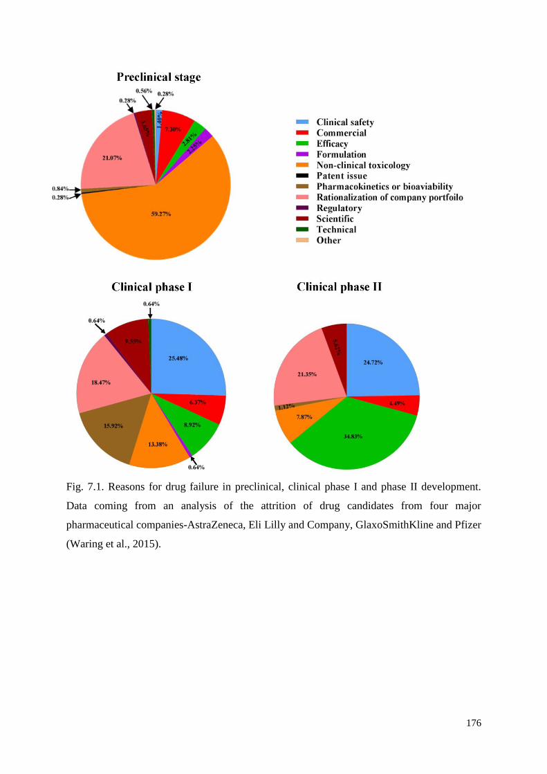
176
Fig. 7.1. Reasons for drug failure in preclinical, clinical phase I and phase II development.
Data coming from an analysis of the attrition of drug candidates from four major
pharmaceutical companies-AstraZeneca, Eli Lilly and Company, GlaxoSmithKline and Pfizer
(Waring et al., 2015).
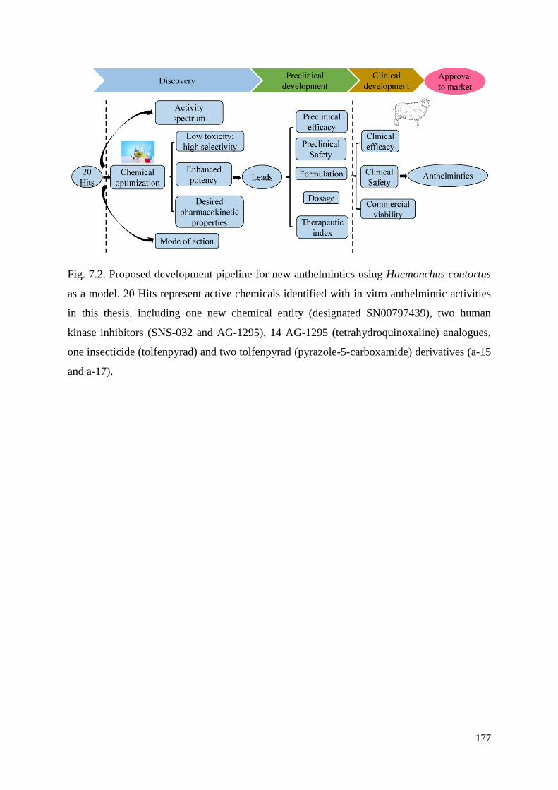
177
Fig. 7.2. Proposed development pipeline for new anthelmintics using Haemonchus contortus
as a model. 20 Hits represent active chemicals identified with in vitro anthelmintic activities
in this thesis, including one new chemical entity (designated SN00797439), two human
kinase inhibitors (SNS-032 and AG-1295), 14 AG-1295 (tetrahydroquinoxaline) analogues,
one insecticide (tolfenpyrad) and two tolfenpyrad (pyrazole-5-carboxamide) derivatives (a-15
and a-17).
178
LIST OF SUPPLEMENTARY FILES
CHAPTER 2
Supplementary file 2.1. Physicochemical properties of chemicals compounds in the ‘Open
Scaffolds’ collection screened against exsheathed third-stage larvae (xL3s) of Haemonchus
contortus.
Supplementary file 2.2. Five-second digital movies of live exsheathed third-stage larvae
exposed to 20 μM of SN00797439 and 20 μM of each of the two positive-control compounds
(i.e. monepantel and moxidectin); the negative control (LB* + 0.5% DMSO) does not contain
compound.
CHAPTER 3
Supplementary file 3.1. Five-second video recordings of exsheathed third-stage larvae (xL3s)
of Haemonchus contortus displaying the ‘coiled’ phenotype induced by exposure to AG-1295
(MMV079840; 100 μM). Videos of xL3s exposed to the same concentration of SNS-032
(MMV690767), monepantel, or no-compound were also included for comparison.
CHAPTER 4
Supplementary file 4.1. The 400 compounds in the ‘Pathogen Box’.
CHAPTER 5
Supplementary file 5.1. The 55 pyrazole-5-carboxamide derivatives synthesized de novo.
Supplementary file 5.2. Five-second video recordings of Haemonchus contortus exsheathed
third-stage larvae (xL3s) showing the “straight” phenotype induced by exposure to the
pyrazole-5-carboxamides a-15, a-17 (test compounds) or tolfenpyrad (100 μM). Videos of
xL3s exposed to the same concentration of tebufenpyrad, monepantel or moxidectin, or not
exposed to any compound (LB* + 0.5% DMSO) were also included for comparison.
179
CHAPTER 6
Supplementary file 6.1. Organic synthesis and characterization of 14 tetrahydroquinoxaline
derivatives of compound AG-1295.
Supplementary file 6.2. Video recordings of functional excretory canal (black arrow;
pumping ~2.4 times per second) in untreated exsheathed third-stage larvae (xL3s.) of
Haemonchus contortus (Panel A) and the Evi phenotype in xL3s treated with 12.5 µM of
m10 (Panel B), employing phase-contrast light microscopy.
Supplementary file 6.3. Mean lengths (left) and widths (right) of exsheathed third-stage
(xL3s) and fourth-stage (L4s) larvae of Haemonchus contortus under the same experimental
conditions. Untreated xL3s and treated xL3s were measured at 0 h and after 7 days of
exposure to 12.5 µM of m10, respectively; untreated L4s and treated L4s were measured at 0
h and after 72 h of exposure to 12.5 µM of m10, respectively. The data points represent the
mean lengths or widths of 30 individual larvae ± the standard error of the mean. Significance
of differences between values (mean ± standard error of the mean) was determined using a
one-way ANOVA with Tukey’s test. Asterisks indicate values that are significantly different
from one another (*P < 0.05; **P < 0.01; ***P < 0.001; ****P < 0.0001). This assay was
repeated two times on different days.
Supplementary file 6.4. Detailed dynamics study of compound-induced Evi phenotype in the
exsheathed third-stage larvae (xL3s) of Haemonchus contortus between 36 h and 48 h
following compound exposure. Larvae were counted microscopically every 20 min.
Corresponding concentration of DMSO was used as a negative control.

Minerva Access is the Institutional Repository of The University of Melbourne
Author/s:
Jiao, Yaqing
Title:
Discovery of new chemicals with anthelmintic activity against the barber’s pole worm and
other parasitic nematodes
Date:
2018
Persistent Link:
http://hdl.handle.net/11343/220756
File Description:
PhD thesis
Terms and Conditions:
Terms and Conditions: Copyright in works deposited in Minerva Access is retained by the
copyright owner. The work may not be altered without permission from the copyright owner.
Readers may only download, print and save electronic copies of whole works for their own
personal non-commercial use. Any use that exceeds these limits requires permission from
the copyright owner. Attribution is essential when quoting or paraphrasing from these works.