Embed Size (px)
Citation preview
A SURVEY OF ANTHELMINTIC RESISTANCE IN RELATION TO MANAGEMENT OF SMALL RUMINANTS IN PENINSULAR MALAYSIA
KHADIJAH BINTI SAAD
UNIVERSITI SAINS MALAYSIA
2007
A SURVEY OF ANTHELMINTIC RESISTANCE IN RELATION TO MANAGEMENT OF SMALL RUMINANTS IN PENINSULAR MALAYSIA
by
KHADIJAH BINTI SAAD
Thesis submitted in fulfilment of the requirements for the degree
of Master of Science
APRIL 2007
ii
ACKNOWLEDGEMENTS
I wish to extend my sincere thanks and appreciation to my main supervisor,
Professor Abdul Wahab Abdul Rahman, Parasitology lecturer of Universiti Sains
Malaysia for his encouragement and extensive assistance throughout this
research.
I would also like to thank my field supervisor, Dr. Chandrawathani
Panchadcharam, Head of Parasitology Department, Veterinary Research
Institute, Ipoh who warmly accepted me to be a part of her team in this
nationwide research. Being in her team had not only improved my knowledge in
laboratory techniques but also enabled me to learn to be a successful
researcher in the field of veterinary parasitology. The never-ending motivation
and encouragement from a great leader and a wonderful woman like her had
greatly improved my self-confidence throughout this research.
My deepest appreciation also goes to these individuals who have made this
research possible;
• The Director General, Department of Veterinary Services, Ministry of
Agriculture for the permission to conduct this study and to publish a
scientific paper on this topic.
• The Director of Veterinary Research Institute, Ipoh; Dr. Sharifah Syed
Hassan for the permission to use the institute’s equipment, materials and
transportation during the research
iii
• The Directors, Department of Veterinary Services in all states of
Malaysia who generously gave us the permission to conduct this
research in their areas.
• Staff of Parasitology Department, Veterinary Research Institute, my ever
dear friend and team mate; Ms. Nurulaini Raimy, my mentors; Madam
Jamnah Omar, Mr. Adnan Musbah and Mr. Zaini Che Mamat and not
forgetting Mr. Ramachandran. Their guidance and assistance especially
in laboratory work in this research is highly appreciated. Being a part of
this team has improved my laboratory skills as well as social skills, and
also taught me the meaning of teamwork and friendship.
• Dr. Peter J. Waller, a world-renowned Parasitology lecturer of Swedish
Agriculture University for his ideas, stimulating suggestions and
motivation. His inspiring encouragement has made the publication of this
research possible.
• Dr. Mohd Naheed Hussein, Madam Vasuge Marimuthu and Mr. Hasan
Gulap Din who unselfishly taught the skills of proper handling and
management of animals on the field.
• Prof Chan Lai Keng, lecturer of Universiti Sains Malaysia for the guide
and advice in statistical analysis.
• Officers from Department of Veterinary Services in all States of
Peninsular Malaysia and officers from the District Veterinary Department
for the assistance in the field throughout this research.
• The staff of Veterinary Research Institute; especially Dr. Maria Jamli
(head of Bacteriology Department), Mr. Ibrahim, Mr. Shuhaili, Mr.
iv
Roszeman, Mr. Adnan, Mr. Aziz for the guidance and help during my
stay in the institute.
• The farmers involved in this research for their cooperation during the
study in their farms.
Most of all, to my dear parents, brothers and my best friend Norasmah
Basari for their never-ending love, moral support and help throughout this
research. Thank you very much!
いつもありがとうございます!
v
TABLE OF CONTENTS
Page ACKNOWLEDGEMENTS ii
TABLE OF CONTENTS v
LIST OF TABLES ix
LIST OF FIGURES x
LIST OF PLATES xi
LIST OF APPENDICES xiii
LIST OF PUBLICATIONS & SEMINARS xiv
ABSTRAK xv
ABSTRACT xvi
1.0 INTRODUCTION 1
2.0 LITERATURE REVIEW
2.1 Helminth infection in small ruminants 4
2.1.1 Haemonchus contortus 5
2.1.2 Oesophagostomum spp. 7
2.1.3 Bunostomum spp. 8
2.1.4 Cooperia spp. 8
vi
2.1.5 Trichostrongylus spp. 9
2.2 Anthelmintics for helminth control 10
2.2.1 Benzimidazole anthelmintics 11
2.2.2 Imidazothiazole anthelmintics 13
2.2.3 Macrocyclic lactone anthelmintics 15
2.2.4 Salicylanilides anthelmintics 16
2.3 Anthelmintic resistance 18
2.4 Anthelmintic resistance in small ruminants 19
3.0 MATERIALS AND METHODS
3.1 Experimental animals 23
3.2 Field and laboratory testing 28
3.3 Data analysis 41
3.4 Interviews 42
3.5 Statistical analysis 42
vii
4.0 RESULTS
4.1 Government farms
4.1.1 Animals and management 43
4.1.2 De-worming history and mortality rate 46
4.1.3 Faecal Egg Count Reduction Test (FECRT) 47
4.1.4 Faecal Cultures 50
4.2 Private farms
4.2.1 Animals and management 55
4.2.2 De-worming history and mortality rate 61
4.2.3 Faecal Egg Count Reduction Test (FECRT) 63
4.2.4 Faecal Cultures 69
4.3 Statistical analysis 74
5.0 DISCUSSION
5.1 Resistance to the four different anthelmintic groups
5.1.1 Resistance to Benzimidazole anthelmintics
(oxfendazole) 75
5.1.2 Resistance to Macrocyclic Lactone anthelmintics
(moxidectin) 76
viii
5.1.3 Resistance to Imidazothiazole anthelmintics
(levamisole) 78
5.1.4 Resistance to Salicylanilides anthelmintics
(closantel) 79
5.2 Resistance to all anthelmintics in government farms 80
5.3 Resistance to all anthelmintics in private farms 84
5.4 Resistance to three anthelmintics tested 84
5.5 Resistance to two anthelmintics tested 85
5.6 Resistance to one anthelmintic tested 85
5.7 Faecal cultures 86
5.8 Statistical analysis 87
6.0 SUMMARY AND CONCLUSION 89
BIBLIOGRAPHY 93
APPENDICES 104
ix
LIST OF TABLES
Page
3.1 Anthelmintic drugs used in Faecal Egg Count Reduction
Tests (FECRT) 29
4.1 Brief information of the government farms 43
4.2 Breed of animals, management, grazing areas, type of grass,
size of grazing areas and grazing time of the government
farms
44
4.3 Information on supplements in each government farm 45
4.4 Information on de-worming history, mortality rate and cause
of mortality in the government farms 46
4.5 Results of FECRT on 5 government farms 48
4.6 Brief information of the private farms 56
4.7 Breed of animals, management, grazing areas and size of
grazing areas of the private farms 58
4.8 Water supply, grazing time, supplements and type of grass in
each private farm 60
4.9 De-worming history and mortality rate on private farms 62
4.10 Anthelmintic resistance on small ruminant private farms in
Peninsular Malaysia (n=21) 64
x
LIST OF FIGURES
Page
2.1 Chemical structure of oxfendazole 12
2.2 Chemical structure of levamisole 14
2.3 Chemical structure of moxidectin 16
2.4 Chemical structure of closantel 17
3.1 Location of small ruminant farms involved in FECRT
between October 2004 and August 2006 25
4.1 Faecal Egg Count Reduction (%) of the government farms 49
4.2 Mean percentage of third stage larvae on government farms 50
4.3 Faecal Egg Count Reduction (%) on 3 farms (resistant to all
anthelmintics) 65
4.4 Faecal Egg Count Reduction (%) on 5 farms (resistant to 3
anthelmintics) 66
4.5 Faecal Egg Count Reduction (%) on 11 farms (resistant to 2
anthelmintics) 67
4.6 Faecal Egg Count Reduction (%) on 1 farm (resistant to 1
anthelmintic) 68
4.7 Mean percentage of third stage larva on private farms 69
xi
LIST OF PLATES
Page
3.1 Animal’s shed in a government farm 26
3.2 Animal’s shed in a private farm 27
3.3 Oral administration of anthelmintics using a drenching gun 30
3.4 Oral administration of anthelmintics using a syringe 31
3.5 Subcutaneous injection for anthelmintic administration 32
3.6 Ear tagging on a goat 33
3.7 Neck tagging on a goat 34
3.8 Rectal faecal sampling 35
3.9 Preparation for faecal culture 39
3.10 Faecal culture for larvae collection after 7 days 40
4.1 A strongyle egg (magnification X40) 47
4.2 Third stage larvae of Haemonchus contortus (magnification
X10) 51
4.3 Tail of Haemonchus contortus (magnification X40) 52
4.4 Third stage larvae of Trichostrongylus sp. (magnification
X10) 53
xii
4.5 Tail of Trichostrongylus sp. (magnification X40) 54
4.6 Third stage larvae of Oesophagostomum sp. (magnification
X10) 70
4.7 Tail of Oesophagostomum sp. (magnification X40) 71
4.8 Third stage larvae of Cooperia sp. (magnification X10) 72
4.9 Tail of Cooperia sp. (magnification X40) 73
xiii
LIST OF APPENDICES
Page
Appendix 1. Standard questionnaire used in the survey 104
Appendix 2. Faecal Egg Count Reduction Test (FECRT)
results of 26 farms 106
Appendix 3. Third stage larvae identification in the farms 115
Appendix 4. Statistical analysis results 116
xiv
LIST OF PUBLICATIONS & SEMINARS
Page
1 Proceedings of the 17th Veterinary Association Malaysia
Congress. Kuala Lumpur 117
2
Proceedings of the 20th International Conference of the
World Association for the Advancement of Veterinary
Parasitology. New Zealand.
118
3
Proceedings of the 42nd Annual Scientific Seminar,
Malaysian Society of Parasitology and Tropical Medicine.
Ipoh.
120
4 Proceedings of the Second ASEAN Congress of Tropical
Medicine and Parasitology. Indonesia. 122
5
Proceedings of the 18th Annual General Meeting and
Scientific Congress 2006 Veterinary Association Malaysia.
Kuala Lumpur.
124
6
Nematode Anthelmintic Resistance on Government Small
Ruminant Farms in Peninsular Malaysia. Jurnal Veterinar
Malaysia (In press). 2007.
125
7
Short communications: Small ruminants on private farms in
Peninsular Malaysia: Nematode resistance to anthelmintics.
Jurnal Veterinar Malaysia (In press). 2007.
126
xv
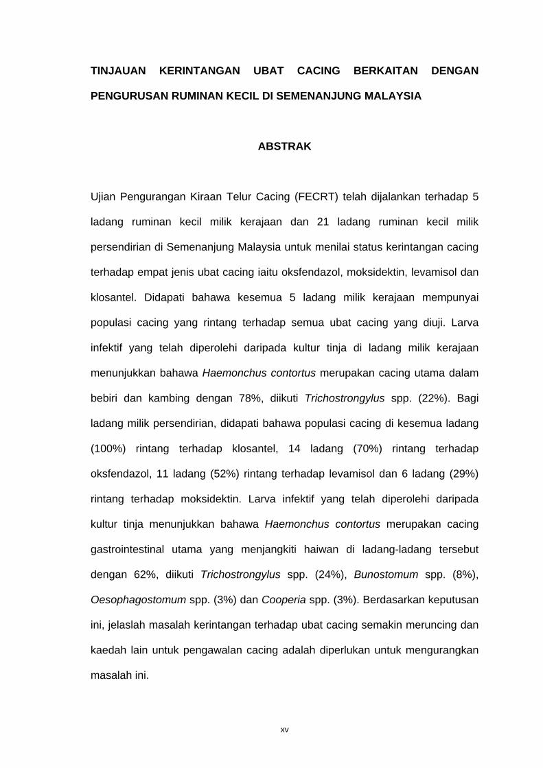
TINJAUAN KERINTANGAN UBAT CACING BERKAITAN DENGAN
PENGURUSAN RUMINAN KECIL DI SEMENANJUNG MALAYSIA
ABSTRAK
Ujian Pengurangan Kiraan Telur Cacing (FECRT) telah dijalankan terhadap 5
ladang ruminan kecil milik kerajaan dan 21 ladang ruminan kecil milik
persendirian di Semenanjung Malaysia untuk menilai status kerintangan cacing
terhadap empat jenis ubat cacing iaitu oksfendazol, moksidektin, levamisol dan
klosantel. Didapati bahawa kesemua 5 ladang milik kerajaan mempunyai
populasi cacing yang rintang terhadap semua ubat cacing yang diuji. Larva
infektif yang telah diperolehi daripada kultur tinja di ladang milik kerajaan
menunjukkan bahawa Haemonchus contortus merupakan cacing utama dalam
bebiri dan kambing dengan 78%, diikuti Trichostrongylus spp. (22%). Bagi
ladang milik persendirian, didapati bahawa populasi cacing di kesemua ladang
(100%) rintang terhadap klosantel, 14 ladang (70%) rintang terhadap
oksfendazol, 11 ladang (52%) rintang terhadap levamisol dan 6 ladang (29%)
rintang terhadap moksidektin. Larva infektif yang telah diperolehi daripada
kultur tinja menunjukkan bahawa Haemonchus contortus merupakan cacing
gastrointestinal utama yang menjangkiti haiwan di ladang-ladang tersebut
dengan 62%, diikuti Trichostrongylus spp. (24%), Bunostomum spp. (8%),
Oesophagostomum spp. (3%) dan Cooperia spp. (3%). Berdasarkan keputusan
ini, jelaslah masalah kerintangan terhadap ubat cacing semakin meruncing dan
kaedah lain untuk pengawalan cacing adalah diperlukan untuk mengurangkan
masalah ini.
xvi
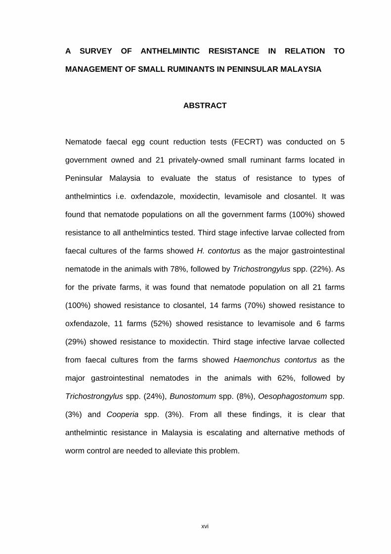
A SURVEY OF ANTHELMINTIC RESISTANCE IN RELATION TO
MANAGEMENT OF SMALL RUMINANTS IN PENINSULAR MALAYSIA
ABSTRACT
Nematode faecal egg count reduction tests (FECRT) was conducted on 5
government owned and 21 privately-owned small ruminant farms located in
Peninsular Malaysia to evaluate the status of resistance to types of
anthelmintics i.e. oxfendazole, moxidectin, levamisole and closantel. It was
found that nematode populations on all the government farms (100%) showed
resistance to all anthelmintics tested. Third stage infective larvae collected from
faecal cultures of the farms showed H. contortus as the major gastrointestinal
nematode in the animals with 78%, followed by Trichostrongylus spp. (22%). As
for the private farms, it was found that nematode population on all 21 farms
(100%) showed resistance to closantel, 14 farms (70%) showed resistance to
oxfendazole, 11 farms (52%) showed resistance to levamisole and 6 farms
(29%) showed resistance to moxidectin. Third stage infective larvae collected
from faecal cultures from the farms showed Haemonchus contortus as the
major gastrointestinal nematodes in the animals with 62%, followed by
Trichostrongylus spp. (24%), Bunostomum spp. (8%), Oesophagostomum spp.
(3%) and Cooperia spp. (3%). From all these findings, it is clear that
anthelmintic resistance in Malaysia is escalating and alternative methods of
worm control are needed to alleviate this problem.
1
1.0 INTRODUCTION
The 2004 figures from the Department of Veterinary Services, Malaysia
recorded the total number of sheep and goat populations as 109,511 and
225,520 respectively (Department of Veterinary Services,
http://agrolink.moa.my/jph).
These numbers, which constitute the small ruminant industry of Malaysia,
is an important component to the overall livestock sector for the country.
Furthermore, the objectives of the government in the National Agriculture Policy
(NAP3) are: to increase small ruminant production through the importation of
exotic breeds of sheep and goats, increasing the number of female animals and
conducting intensive research in small ruminants and their diseases
(Department of Veterinary Services, http://agrolink.moa.my/jph).
Infestation by parasitic worms (helminthiasis) in small ruminants has
been identified as one of the major limiting factors of animal productivity in
farming enterprises which rely on grazing animals on pasture (Waller, 1985).
Trichostrongyle worms, from the superfamily of Trichostrongyloidea are
responsible for considerable mortality and widespread morbidity. The most
important genera in Trichostrongyles are Ostertagia, Haemonchus,
Trichostrongylus, Cooperia, Nematodirus, Hyostrongylus, Marshallagia and
Mecistocirrus (Urquhart et al., 1996).
2
Humid climate throughout the year in Malaysia is very favourable for the
development of free living stages of trichostrongyle nematodes and it is likely
that all times of the year, infective larvae are available on the pasture for
grazing animals (Ikeme et al., 1987).
With this in mind, farmers depend heavily on anthelmintic treatments to
control helminthiasis problem. Benzimidazoles, Imidothiazoles,
Tetrahydropyrimidines, Avermectins and Salicylanilides (Bogan and Armour,
1987) are examples of anthelmintic groups that are available in the market – to
be chosen by farmers or veterinarians to treat animals with helminthiasis.
Unfortunately, irresponsible usage of the anthelmintics (not used in a
recommended manner, being often overused, misused or applied incorrectly)
has led to the development of resistance nematode against anthelmintics in
small ruminants (Waller, 2006).
Anthelmintic resistance is a major problem faced by livestock farmers
throughout the world. Countries such as South Africa, Australia and Uruguay
reported million dollars of annual losses due to helminthiasis (Waller, 2006). As
anthelmintic resistance will result in failure of anthelmintics to control helminth,
the losses are expected to increase.
Based on this, a lot of interests are shown by researchers, mainly
veterinarians and parasitologists, to study this problem and explore the possible
alternative measures to control the problem.
3
Thus, the objective of this study is to report on;
1. The prevalence of anthelmintic resistance in government and also private
small ruminant farms in Peninsular Malaysia.
2. The management of animals in government and private small ruminant
farms in Peninsular Malaysia.
3. The prevalent species of nematode existing in government and private
small ruminant farms in Peninsular Malaysia.
4
2.0 LITERATURE REVIEW
2.1 Helminth infection in small ruminants
Helminth infection causes problems and financial losses to the livestock
industry (Waller, 2006) as it is one of the most prominent causes of mortality
and morbidity in Malaysia (Fatimah et al., 1985, Sani and Rajamanickam, 1990,
Sani et al., 2004). This is due to the grazing activities of the livestock in
pastures contaminated with third stage infective larvae of parasitic nematodes.
Fatimah et al. (1985) reported that parasitic gastroenteritis was the main cause
of mortality in Dorset Horn Sheep hogs while for the adults, parasitic
gastroenteritis was one of the causes of mortality besides pneumonia, bacterial
enteritis, trauma and septicaemia. Sani et al. (1985) reported that from 72 goats
necropsied in Universiti Putra Malaysia, Haemonchus contortus was found to be
the major helminth found (67%), followed by Moniezia expansa (51%),
Oesophagostomum columbianum (42%) and Trichostrongylus colubriformis
(38%). Work done by Dorny et al. (1995) on sheep and goats from three farms
in Peninsular Malaysia showed that H. contortus and Trichostrongylus spp.
were the most important strongyles in sheep and goats.
Helminth infection (helminthiasis or helminthiasis) is mainly caused by
trichostrongyle nematodes such as H. contortus, Oesophagostomum spp.,
Cooperia spp. and Trichostrongylus spp. The parasite that causes the most
problems to small ruminants is the bloodsucker nematode; H. contortus, better
known as the "barber’s pole" worm, which is generally considered as the most
5
pathogenic parasite of small ruminants (Soulsby, 1982). This parasite is a
problem to small ruminants, not only in countries with warm and wet climates
such as Malaysia but also in temperate countries such as Sweden (Waller and
Chandrawathani, 2005).
2.1.1 Haemonchus contortus
This blood-sucking abomasal nematode is responsible for the extensive
productivity losses in sheep and cattle industry, especially in tropical areas. The
nematode can be found worldwide, but it is most important in tropical and
subtropical areas (Urquhart et al., 1996). The infective larvae of H. contortus
can survive dessication as the second stage cuticle becomes less permeable as
it dries (Ellenby, 1968).
The eggs of Haemonchus spp. hatch to first stage larvae (L1) on the
pasture and may develop to third stage infective larvae (L3) in a period of five
days but development may be delayed for weeks or months under cool
conditions (Urquhart et al., 1996). Four days after infection, all the worms are at
the fourth stage larvae (L4) and the third stage larvae (L3) are totally absent. The
worms spent between 7 to 11 days after infection in the mucosa before
emerging, as late L4 larvae and establishing themselves in the lumen (Rahman
and Collins, 1990a).
6
The late L4 larvae developed piercing lancet before the final moulting
which enables them to obtain blood from the mucosal vessels. As adults, they
move freely on the surface of the mucosa. The prepatent period is 2-3 weeks in
sheep and four weeks in cattle (Urquhart et al., 1996).
The main feature of Haemonchus spp. infection is anaemia as both the
adult and the fourth larval stage (L4) sucks blood, moves and leaves wounds
which haemorrhage into the abomasum. Infection by an extremely large number
of Haemonchus spp. results in a rapidly developing severe anaemia, dark-
coloured faeces and sudden death due to acute blood loss. There is also a
severe haemorrhagic gastritis and death may occur in the prepatent period of
such heavy infections (Soulsby, 1982).
When young susceptible animals become heavily infected, the anaemia
may develop rapidly, accompanied by hypoproteinaemia and oedema (i.e.
bottle jaw) and death will occur. The most common clinical signs in infected
animals are weakness, unthriftiness and emaciation (Soulsby, 1982).
Rahman and Collins (1991) reported that infections caused by goat-
derived strain of H. contortus in goats caused greater weight loss compared to
infections by sheep–derived strain. The study also reported that the reduction in
packed cell volume (PCV) values and haemoglobin concentration was
significant at day 14-28 after infection. This coincides with worm maturation and
active feeding of the worms at that particular period (Rahman and Collins,
1990a).
7
It was reported that each worm removes about 0.05 ml of blood per day
by ingestion and leakage from the lesions (Urquhart et al., 1996). Thus, an
amount of 5000 larvae of H. contortus can cause death in goats (Rahman and
Collins, 1991) with an estimation of 250 ml blood loss daily (Urquhart et al.,
1996).
2.1.2 Oesophagostomum spp.
Oesophagostomum spp. are parasites in the small and large intestines of
cattle, sheep, pigs and primates. These nematodes are often referred to as
nodular worms, owing to the fact that several species cause nodule formation
on the wall of the intestine (Soulsby, 1982). The prepatent period is about 45
days (Urquhart et al., 1996).
Oesophagostomum columbianum is a serious pathogen of sheep where
extensive nodular formation seriously interferes with absorption, bowel
movement and digestion. The nodules may rupture on the peritoneal surface,
causing peritonitis and multiple adhesions. Though the adult worms do not suck
blood as in H. contortus, they can cause marked thickenings of the bowel wall,
cause massive congestion and produce large amount of mucous. In infected
lamb, the first sign of infection is marked by persistent diarrhoea, which results
in exhaustion and death. This diarrhoea begins on the sixth day after a severe
infection and coincides with the time when the larvae leave the nodules
(Soulsby, 1982).
8
2.1.3 Bunostomum spp.
Bunostomum trigonocephalum is a hookworm which occurs in the small
intestine (ileum and jejunum) of sheep and goats in many parts of the world.
The parasite is more important in warmer climates like Malaysia than colder
climates. Infection usually occurs along with other gastrointestinal strongyles
and the hookworms contribute to the general effects of parasitism (Soulsby,
1982). The adult worms attach themselves to the intestinal mucosa and suck
blood. The main clinical signs when substantial number of hookworms occurs
are progressive anaemia, hydraemia and oedema especially in the
intermandibular region (i.e. bottle jaw). Diarrhoea is infrequent and the faeces
may be dark in colour due to the presence of altered blood pigments (Soulsby,
1982).
2.1.4 Cooperia spp.
In temperate areas, members of the genus Cooperia usually play a
secondary role in the pathogenesis of parasitic gastroenteritis of ruminants,
although they may be the most numerous trichostrongyle present. However, in
some tropical and subtropical areas, Cooperia spp. are responsible for severe
enteritis in calves (Urquhart et al., 1996).
The adult worms penetrate the mucosa of the small intestine. There is no
consequence from a light infection of the worms, but young cattle and sheep
9
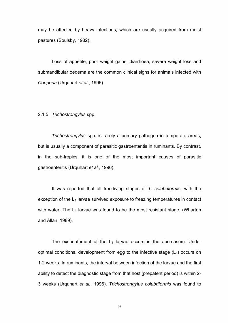
may be affected by heavy infections, which are usually acquired from moist
pastures (Soulsby, 1982).
Loss of appetite, poor weight gains, diarrhoea, severe weight loss and
submandibular oedema are the common clinical signs for animals infected with
Cooperia (Urquhart et al., 1996).
2.1.5 Trichostrongylus spp.
Trichostrongylus spp. is rarely a primary pathogen in temperate areas,
but is usually a component of parasitic gastroenteritis in ruminants. By contrast,
in the sub-tropics, it is one of the most important causes of parasitic
gastroenteritis (Urquhart et al., 1996).
It was reported that all free-living stages of T. colubriformis, with the
exception of the L1 larvae survived exposure to freezing temperatures in contact
with water. The L3 larvae was found to be the most resistant stage. (Wharton
and Allan, 1989).
The exsheathment of the L3 larvae occurs in the abomasum. Under
optimal conditions, development from egg to the infective stage (L3) occurs on
1-2 weeks. In ruminants, the interval between infection of the larvae and the first
ability to detect the diagnostic stage from that host (prepatent period) is within 2-
3 weeks (Urquhart et al., 1996). Trichostrongylus colubriformis was found to
10
stay in the mucosa of goats until about 11 days after infection, and the
immature adults emerge and mature in the lumen (Rahman and Collins, 1990b).
Twenty one days after infection, it was found that about 57% of the female
worms had eggs in their uteri and around this time the eggs were first detected
in the faeces (Rahman and Collins, 1990b).
In sheep and goats, young animals are susceptible to the infection of this
worm. When a severe infection is acquired within a short time, the disease may
be acute and may rapidly lead to death. Animals usually do not show
emaciation or anaemia, but they become weak in the legs and are unable to
stand shortly before they die. Mild anaemia, emaciation, alternating constipation
and diarrhoea are visible in chronic cases (Soulsby, 1982).
2.2 Anthelmintics for helminth control
As helminth infections cause serious problems to small ruminants and
sometimes death, farmers depend on anthelmintics to control the infections. A
wide variety of anthelmintics from various chemical groups, are used for the
treatment of helminth parasites. These chemical groups include: the
benzimidazoles, imidothiazoles (levamisole), salicylanilides (closantel) and
macrocyclic lactones (ivermectin) (Chandrawathani et al., 1994).
11
According to Behm and Bryant (1985) anthelmintics can be divided into
four major categories according to their mode of action:
1. drugs that interfere with neurophysiology or neuromuscular coordination
and exploit the differences in neurophysiology between host and parasite (e.g.
ivermectin and levamisole),
2. drugs that interfere with essential energy metabolism (e.g. salicylanilides)
3. drugs that interfere with essential biosynthetic pathways and
4. drugs that interfere with essential cellular processes (e.g. substitutes of
benzimidazole).
2.2.1 Benzimidazole anthelmintics
All benzimidazole anthelmintics remove most of the adult parasites which
include Bunostomum spp., Chabertia spp., Cooperia spp., Haemonchus spp.,
Nematodirus spp., Oesophagostomum spp., Ostertagia spp., Strongyloides spp.
and Trichostrongylus spp. (Brander et al., 1982).
The benzimidazole anthelmintics bind to the nematode tubulin, thus
inhibiting the formation of microtubules. Subsequently, the nematode is unable
to transport secretory granules or secrete enzymes within the cell cytoplasm.
12
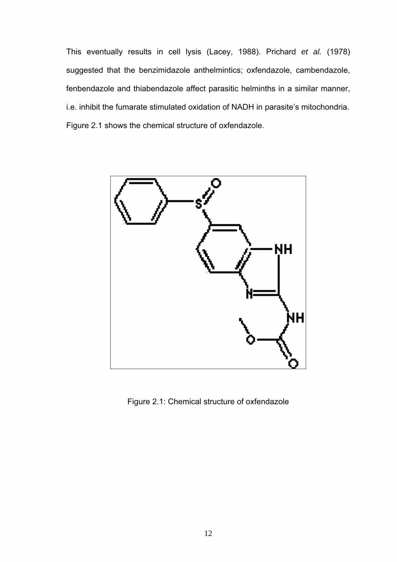
This eventually results in cell lysis (Lacey, 1988). Prichard et al. (1978)
suggested that the benzimidazole anthelmintics; oxfendazole, cambendazole,
fenbendazole and thiabendazole affect parasitic helminths in a similar manner,
i.e. inhibit the fumarate stimulated oxidation of NADH in parasite’s mitochondria.
Figure 2.1 shows the chemical structure of oxfendazole.
Figure 2.1: Chemical structure of oxfendazole
13
2.2.2 Imidazothiazole anthelmintics
Levamisole, the major drug used in this group is a broad spectrum
anthelmintic and is effective against the adult and larval stages of Haemonchus
spp., Ostertagia spp., Trichostrongylus spp., Cooperia spp., Nematodirus spp.,
Bunostomum spp., Oesophagostomum spp., Metastrongylus spp., Ascaris spp.,
and Trichuris spp., and the lungworm Dictyocaulus spp. This anthelmintic has
been used against both artificial and natural infections, and has the advantage
of being active at a low dosage. Because of its activity against the larval stage,
it has widened the whole field of the strategic dosing of cattle and sheep to
prevent larval infestation. It is also active against benzimidazole-resistant H.
contortus and T. colubriformis (Brander et al., 1982).
Low levels of the levamisole anthelmintic act as ganglion stimulants and
give rise to muscular paralysis in parasites, whereas higher levels interfere with
carbohydrate metabolism. The blockage occurs at the site of fumarate reduction
and succinate oxidation (Brander et al., 1982). Figure 2.2 shows the chemical
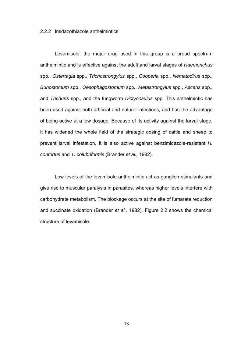
structure of levamisole.
14
Figure 2.2: Chemical structure of levamisole
15
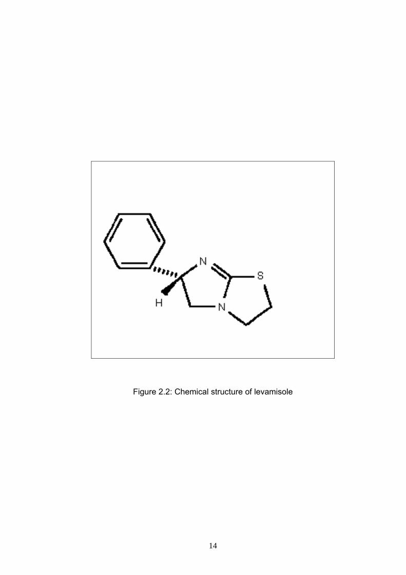
2.2.3 Macrocyclic lactone anthelmintics
Anthelmintics in this group gate the opening of glutamate gated chloride
channels found only in invertebrates (Martin et al., 1997) and cause inhibition of
the motility and development of the free living stages of trichostrongylid
nematode parasites (Le Jambre, 1996). Macrocyclic lactone anthelmintics are
used for the treatment and control of gastrointestinal nematodes, lungworms,
warbles, mange and sucking lice, and has been claimed to be effective against
nematodes and external parasites (Brander et al., 1982).
Moxidectin (a member of macrocyclic lactone anthelmintics) was found to
be 100% effective in eliminating trichostrongyle egg counts by day 3 of
treatment and the egg counts remained negative until 31 days post treatment
(Miller et al., 1994). Besides that, it was found that horn flies (Haematobina
irritants) feeding on the blood of moxidectin-treated cattle drawn on day 21 of
daily treatment, showed a decline in survival and egg production (Miller et al.,
1994). Moxidectin was also found to have larvacidal activity against the
immature stages of the horn fly in the manure of treated cattle (Miller et al.,
1994).
Initial studies carried out on a government sheep farm in Malaysia
showed that moxidectin was 100% efficacious against sheep infected with
benzimidazole-resistant nematodes for up to 6 weeks. As an alternative to
resistant anthelmintics, this drug was suggested to be used for the control of
16
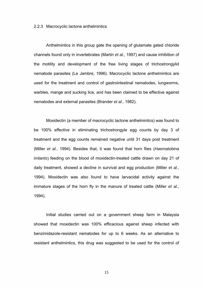
helminthiasis in sheep (Chandrawathani et al., 1998). Figure 2.3 shows the
chemical structure of oxfendazole.
Figure 2.3: Chemical structure of moxidectin
2.2.4 Salicylanilides anthelmintics
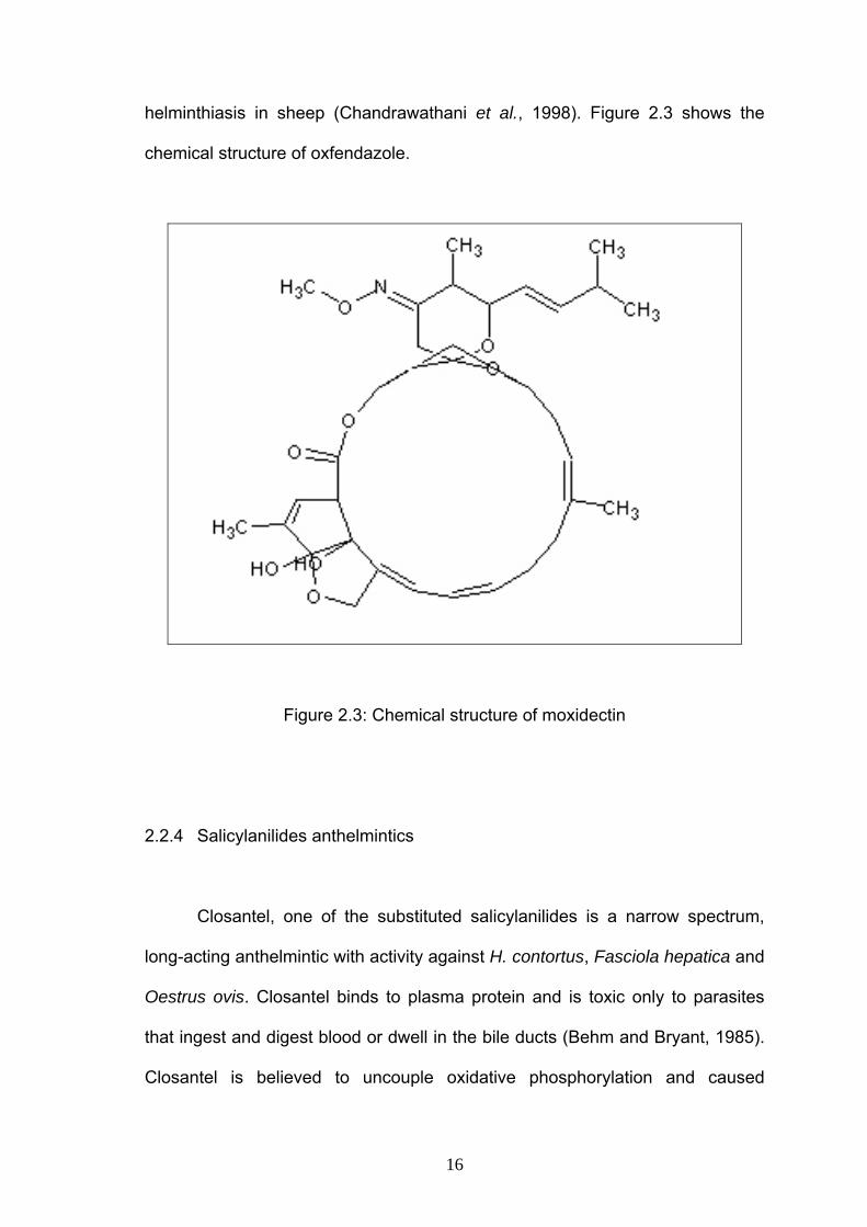
Closantel, one of the substituted salicylanilides is a narrow spectrum,
long-acting anthelmintic with activity against H. contortus, Fasciola hepatica and
Oestrus ovis. Closantel binds to plasma protein and is toxic only to parasites
that ingest and digest blood or dwell in the bile ducts (Behm and Bryant, 1985).
Closantel is believed to uncouple oxidative phosphorylation and caused
17
poisoning in the mitochondria (Martin et al., 1997). However, Rothwell and
Sangster (1996) suggested that closantel is a molecule with the ability to affect
a number of membrane-associated processes including ion and fluid transport,
and the uncoupling of oxidative phosphorylation is secondary to such a
membrane effect.
This drug has been used widely for H. contortus control as a result of the
widespread occurrence of parasite resistance to broad spectrum anthelmintics
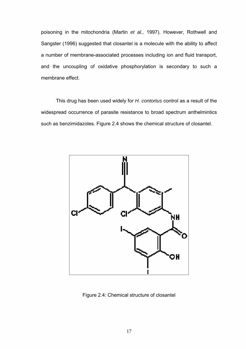
such as benzimidazoles. Figure 2.4 shows the chemical structure of closantel.
Figure 2.4: Chemical structure of closantel
18
2.3 Anthelmintic resistance
As defined by Prichard et al. (1980), anthelmintic resistance is present
when there is a greater frequency of individuals within a population able to
tolerate doses of a compound than in a normal population of the same species.
Evolutionary theory explains the appearance of a resistant strain by proposing
that the original population of worms contained a few rare individuals that
possessed the ability to survive the anthelmintic. Since anthelmintics kill all
susceptible worms, the next generation will consist of offsprings of the resistant
minority (Le Jambre, 1985). Resistance is a heritable character (Prichard et al.,
1980). Therefore, many of these worms inherited their parent’s ability to survive
the drugs. If the character that provides resistance is controlled by a single
major gene, then the resistant population will build up very rapidly (Le Jambre,
1985).
Prichard (1994) reviewed anthelmintic resistance to levamisole,
benzimidazoles and ivermectin. Levamisole resistance appears to be
associated with alterations in cholinergic receptors in resistant nematodes. On
the other hand, benzimidazoles resistance in nematodes appear to be
associated with an alteration in ß-tubulin genes which reduces or abolishes the
high affinity binding of benzimidazoles for tubulin in these organisms.
Alterations in the glutamate/ivermectin chloride channel receptor in nematodes
were suggested as the mechanism of ivermectin resistance.
19
2.4 Anthelmintic resistance in small ruminants
Anthelmintic resistance is a problem for livestock industries especially in
countries with hot and humid climates, an ideal condition for the development
and survival of free-living stages of nematode parasites on pasture.
In South America, more than 70% of the total sheep population is found
in Argentina, Brazil, Paraguay and Uruguay. Within these countries is the sub-
tropical humid zone which encompasses Northern Argentina, Southern Brazil
and all of Paraguay and Uruguay. The environment is ideal for parasite
development and transmission and this may occur more or less continuously
throughout the year. The 45 million sheep in this region are therefore exposed
to frequent use of anthelmintic, resulting in anthelmintic resistance in parasite
populations of the sheep (Waller et al., 1996). Many reports were published on
the prevalence of anthelmintic resistance in Argentina, Brazil, Paraguay and
Uruguay (Eddi et al., 1996, Echevarria et al., 1996, Maciel et al., 1996, Nari et
al., 1996).
According to Eddie et al. (1996), from 65 sheep farms in Argentina, it
was found that 40% of the farms showed resistance to benzimidazoles, 22% to
levamisole, 11% to the combination anthelmintics (benzimidazole and
levamisole) and 6% to ivermectin. A similar situation was also observed in
Brazil where 90% of 182 sheep farms selected showed resistance to
albendazole, 84% to levamisole, 73% to the combination anthelmintics
(benzimidazole and levamisole) and 13% to ivermectin (Echevarria et al., 1996).
20
According to them, the results demonstrated that the parasite control in this
country is rapidly reaching a state of crisis (Echevarria et al., 1996)
The level of anthelmintic resistance in sheep in Paraguay has also
reached a crisis situation. A report in 1996 showed that the levels of resistance
were 73% to benzimidazoles, 68% to levamisole, 73% to oral ivermectin and
47% to injectable ivermectin (Maciel et al., 1996).
A study conducted on 252 sheep farms in Uruguay showed that 86% of
the sheep flocks were resistant to benzimidazoles, 71% to levamisole and 1.2%
to ivermectin (Nari et al., 1996).
In 1993, it was reported that albendazole and oxfendazole caused a very
low reduction in faecal egg counts after treatment in a goat farm in Malaysia,
indicating the failure of both anthelmintics. Highly benzimidazole-resistant strain
of H. contortus was isolated from that farm. This was the first resistance to
Benzimidazole in H. contortus reported in Malaysia (Dorny et al., 1993).
Rahman (1993) reported thiabendazole resistance cases in 19% of 48
goat farms in northern Peninsular Malaysia. Later in 1994, he also reported H.
contortus resistance to fenbendazole in West Malaysia. In addition, he also
reported that levamisole was highly effective against trichostrongyle nematodes
in goats and in some farms, there was total removal of the worms (Rahman,
1994a). In another study, Rahman (1994b) reported that levamisole significantly
21
reduced the mean worm burdens compared to reductions in animals treated
with albendazole, fenbendazole, oxfendazole and mebendazole.
The first benzimidazole resistance in H. contortus in sheep from
Southeast Asia was reported by Pandey and Sivaraj (1994) showing high
nematode resistance albendazole, oxfendazole and fenbendazole in 4 sheep
farms in Malaysia. In this study, no resistance to closantel and levamisole was
observed (Pandey and Sivaraj, 1994).
A first case of multiple anthelmintic resistance in a sheep farm in
Malaysia was reported by Sivaraj et al. in 1994. The study demonstrated
simultaneous resistance of H. contortus against benzimidazoles and levamisole
and of T. colubriformis against benzimidazoles and levamisole in the same farm.
It was also reported that moxidectin was effective against the ivermectin
resistant H. contortus (Sivaraj et al., 1994).
With the expectation that the anthelmintic resistance status was
developing rapidly on sheep and goat farms in Malaysia, a nationwide
anthelmintic resistance survey involving 48 farms, was conducted in the 1990’s.
This survey reported the presence of nematode resistance to benzimidazole in
16 farms, levamisole in 7 farms, closantel in 1 farm, ivermectin in 1 farm and
also resistance to the combination of levamisole and closantel in 1 farm
(Chandrawathani et al., 1999).
22
Since then, further studies of the anthelmintic resistance status on
government small ruminant breeding farms have showed high levels of multiple
resistance in one government sheep farm in Peninsular Malaysia
(Chandrawathani et al., 2003) and total anthelmintic failure of all five
commercially available anthelmintic drug classes in five small ruminant
government farms in Sabah, East Malaysia (Chandrawathani et al., 2004).
Since government small ruminant farms in Malaysia distribute their
animals to small-holder farmers throughout the country, the severity of this
problem is likely to have been widely dispersed.
23
3.0 MATERIALS AND METHODS
3.1 Experimental animals
Faecal samples from 100 small ruminant farms in Peninsular Malaysia
were collected from October 2004 to August 2006. Faecal egg counts were
estimated using the modified McMaster method (Ministry of Agriculture, Food
and Fisheries, 1986).
Faecal Egg Count Reduction Test (FECRT) was also carried out but they were
confined to farms which meet the following criteria:
1. The farm had more than 30 animals of age, ranging from 6 months to 3
years.
2. When screening for nematode faecal egg counts, at least 10% of the
animals showed mean counts exceeding 120 eggs per gram (e.p.g.)
faeces.
3. The animals were generally healthy and in good condition based on
gross clinical examination.
Twenty six farms were selected, of which 15 were exclusively goat farms, 9
sheep and two mixed. They were randomly distributed in the states of Perak (14
farms), Kelantan (2 farms), Terengganu (2 farms), Johor (2 farms), Penang (1

24
farm), Kedah (1 farm), Selangor (1 farm), Pahang (1 farm), Negeri Sembilan (1
farm) and Melaka (1 farm) (Figure 3.1).