Embed Size (px)
Citation preview
1
Discovery and Validation of Novel Peptide Agonists for G-Protein Coupled Receptors
Ronen Shemesh, Amir Toporik, Zurit Levine, Iris Hecht, Galit Rotman, Assaf Wool, Dvir Dahary, Eyal
Gofer, Yossef Kliger, Michal Ayalon Soffer, Avi Rosenberg, Dani Eshel and Yossi Cohen
From: Compugen Ltd. 72 Pinchas Rosen st., Tel Aviv, 69512 ISRAEL.
Running Head: Novel GPCR peptide agonists
Address correspondence to: Ronen Shemesh Compugen Ltd. 72 Pinchas Rosen st., Tel Aviv, 69512
ISRAEL. Tel: +972-3-7658138 Fax: +972-3-7658555 Email: [email protected]
Summary:
G-protein coupled receptors (GPCRs)
represent an important group of targets for
pharmaceutical therapeutics. The completion of
the human genome revealed a large number of
putative GPCRs. However, the identification of
their natural ligands, and especially peptides,
suffers from low discovery rates, thus impeding
development of therapeutics based on these
potential drug targets.
We describe the discovery of novel
GPCR ligands encrypted in the human
proteome. Hundreds of potential peptide
ligands were predicted by machine learning
algorithms. In vitro screening of selected 33
peptides on a set of 152 GPCRs, including a
group of designated orphan receptors, was
conducted by intracellular calcium
measurements and cAMP assays. The screening
revealed eight novel peptides as potential
agonists that specifically activated six different
receptors in a dose dependent manner. Most of
the peptides showed distinct stimulatory
patterns targeted at designated and orphan
GPCRs. Further analysis demonstrated a
significant in-vivo effect for one of the peptides
in a mouse inflammation model.
Introduction:
G-protein coupled receptors (GPCRs)
represent a large group of receptors which are
directly involved in cellular signaling networks
and are considered to be an important family of
targets for pharmacological intervention (reviewed
in [1]). It is estimated that approximately one third
of therapeutic development in the pharmaceutical
industry is targeted towards drug discovery in the
area of GPCRs [2]. Recent analysis of the human
genome identified over 800 putative members of
the GPCR super family, over half of which are
thought to be involved in sensory mechanisms and
are not considered to be drug targets. Among the
known GPCRs, approximately 300 are considered
potential pharmaceutical targets [3]. Out of this
group around 150 GPCRs have no designated
natural/endogenous ligand or activator. These
receptors are termed “orphan GPCRs” and have
recently become very attractive to many industrial
and academic researchers as they hold great
potential as targets for novel drug discovery [4].
Lately, a major effort was made by many
academic institutes as well as biotech and
pharmaceutical companies to identify both natural
and surrogate ligands for both known and orphan
GPCRs [5, 6] . The most common method to
screen for novel GPCR activators is through large
scale screening of hundreds and thousands of
compounds on many GPCRs, testing for in vitro
activation of any given receptor by assessing its
signaling response of an expressing cell to binding
of a specific compound or tissue extract. This
method, called “reverse pharmacology” [7, 8] gave
rise in the past decade to over 30 new
receptor/ligand pairs, and many previously known
or novel ligands, most of them peptides [5].
However, this method is very expensive, time
consuming, and might lead to many potential
ligands either being missed or falsely annotated [6,
9]. In order to increase the feasibility of this type
of testing, it is necessary to pre-screen any
potential group of ligands and focus on high
chance, low risk candidates. This might allow
more thorough screening and validation of true
novel activators with better chances of becoming
therapies.
Computational screening can supplement
experimental efforts as a preliminary screening
tool for efficient identification of natural ligands,
http://www.jbc.org/cgi/doi/10.1074/jbc.M805181200The latest version is at JBC Papers in Press. Published on October 9, 2008 as Manuscript M805181200
Copyright 2008 by The American Society for Biochemistry and Molecular Biology, Inc.
by guest on May 11, 2019
http://ww
w.jbc.org/
Dow
nloaded from
2
especially peptides [10]. Bioinformatics can serve
as a powerful tool that provides reliable predictive
measures to select for the high potential candidates
and provide a spotlight pointed at potential new
candidates for experimental discovery, thereby
enabling higher success rates in identification of
novel GPCR ligands [11].
In this work, we describe the experimental
validation of computationally discovered novel
GPCR activating peptides. A subgroup of 33
potential candidate peptides was experimentally
screened. Herein we report the in-vitro validation
of eight novel GPCR peptide activators (hit rate of
approximately 25%) as potential candidates for
related clinical utilities. One of the examined
peptides was tested in-vivo and was found
effective in a mouse inflammation model.
Experimental Procedures:
Data set preparation for the proteolytic
site predictor: All mammalian proteins (28,780)
were downloaded from Swiss-Prot [12] (Release
43). Of these, 11,705 proteins were classified as
secreted or membrane proteins according to Swiss-
Prot annotation. From these 11,705 proteins, 553
experimentally-validated 18-mer peptide
convertase (PC) proteolytic sites were extracted
based on Swiss-Prot FT annotation lines. This
subset of sequences was used as a learning set for
Breiman's random forest classifier algorithm [13],
whereas the negative set (non proteolytic sites)
consisted of all extracellular 18-mers, excluding
the boundaries of pro-peptides, chains, & peptides,
according to Swiss-prot annotations.
Creation of a predicted Human Peptidome
dataset: A predicted human secretome was
generated by analyzing human proteins from two
sources: (i) the Uniprot/Swissprot protein database
(version 47) and (ii) Refseq sequences from the
NCBI nr protein database (Genebank version 149).
There were 29,530 proteins from NCBI nr,
including proteins with various validation
annotations, reviewed, predicted and modeled. The
annotation for Uniprot proteins was used to filter
out proteins that do not have a signal peptide,
validated or predicted. For NCBI nr proteins the
SignalP 3.0 software [14] was used to predict the
existence and location of a signal peptide.
For each protein precursor sequence, the
following procedure was used to create the
potential peptide products of the precursor (The
full cleavage prediction concept is detailed in
Kliger, et al [15]). The predicted signal peptide
was removed, and potential cleavage sites were
scored based on the outcome of the machine
learning algorithm used for cleavage site
prediction. Based on precision/recall analysis,
each putative cleavage site was assigned a score
value between 0 and 10, where 0 represents a
known cleavage site (as annotated on UniProt) and
10 corresponds to unlikely cleavage sites. Sites
with a score of 4 or less, and sites that contained
two consecutive basic amino acids (e.g. Arg or
Lys), regardless of their score, were selected.
Peptides whose endpoints were either a selected
cleavage site or an endpoint of the precursor
(Signal peptide cleavage site at the N-terminus or
the precursor’s C-terminus) were created. Peptides
longer than 100 amino acids were discarded. It
should be noted that different peptides from the
same precursor may have an overlapping
sequence, and one can be a subsequence of
another. The peptide set was extended by
sequentially removing C-terminal basic amino
acids and removing an additional C-terminal
Glycine, leaving an Amide attached to the carboxy
terminus of the peptide (this mimics a known
natural peptide maturation process called “glycine-
directed peptide amidation”). All the intermediate
peptides resulting from this extension were
considered as separate peptide products. The
complete list of predicted peptides (the
“peptidome”) contained hundreds of thousands of
peptides, which were then analyzed as a source
data by the GPCR activating ligand predictor.
GPCR ligand predictor: The set of known
GPCR peptide ligands was extracted from
Uniprot, using the Uniprot annotation (SRS from
Swiss-Prot and by relevant keywords from
GPCRBD - [16]) and was manually curated based
on literature search. The peptides’ sequence
lengths and positions were extracted from the
Swiss-Prot feature table. The learning data set
contained 64 precursors and 123 peptides (See
Table S1 in Supplemental data - SD). Of these, 87
did not overlap with another peptide of the same
precursor. Several known GPCR ligands were
removed from the learning set (for example, the
chemokine family and the hormone proteins that
are larger than 100 amino acids) giving an average
peptide size of 26 amino acids. The learning
by guest on May 11, 2019
http://ww
w.jbc.org/
Dow
nloaded from

3
process also required a negative set of peptides
that are not known as GPCR peptide ligands. Two
negative sets of peptides were constructed. The
first was extracted based on Uniprot using
annotations to filter peptides which are not GPCR
peptide ligands. The second set was a collection of
3000 randomly chosen peptides from the novel
peptide database (peptidome) described above.
Using the generated learning set, a
classifier based on the “Random Forest” algorithm
[13] was implemented to select peptides that are
likely to be GPCR ligands. Following a thorough
optimization process, the classifier was
programmed to analyze the following index
parameters:
• Length of the peptide (in amino acids).
• The frequency of each amino acid in the
peptide relative to the peptide length (21
parameters, one for each amino acid and
an extra parameter for those which are not
conventional amino acids).
• The amino acids in proximity to the N-
terminal cleavage site from both sides: the
first 4 amino acids in the N terminus of
the peptide and the last 4 amino acids of
the precursor preceding the cleavage site
(8 parameters, each can be one of 21
values, one for each amino acid and an
extra value for other letters or for amino
acids beyond the signal peptide cleavage
site). This parameter (4 amino acids) was
found most effective in the optimization
process.
• The amino acids in proximity to the C-
terminal cleavage site from both sides:
The last 3 amino acids in the C terminus
of the peptide and the first 3 amino acids
of the precursor following the cleavage
site (6 parameters, each can be one of 21
values, one for each amino acid and an
extra value for other letters or for amino
acids beyond the precursor end). This
parameter (3 amino acids) was found most
effective in the optimization process.
• The number of coding exons in the gene
coding for of the precursor.
Based on these numeric parameters the
classifying program was trained to distinguish
between the learning set of known ligands (true
set) and the negative (false) set. This program was
then used to give each predicted peptide a score
signifying the likelihood of being a GPCR
activating ligand. At the end of the process, a
dataset of 100 peptides with the highest classifier
scores was created. These peptides underwent a
manual expert analysis process and literature
review by a few different biologists. The selection
of GPCR activating peptides was based on the
following criteria:
• Expression profile and tissue specificity of
the precursor, with relation to the receptor.
• Comparison of the cleavage and GPCR
classifier scores of the candidate peptides
and their Mouse orthologs.
• Conservation of the peptide’s sequence
and cleavage sites in all orthologues
precursor proteins.
• Peptide’s position within the precursor
with respect to known domains and
features (as a negative rule).
• Number of Cysteine residues and disulfide
bridge annotations.
The peptides were also checked for their
novelty status based upon the comparison to
sequences appearing in patent databases with very
strict exclusion parameters. This process resulted
in a subset of 35 confirmed novel peptides most
likely to be GPCR ligands.
Peptide synthesis: For each peptide, from
the subset of 35 peptides, a number of
modifications were made in order to enhance
stability and functionality. In peptides containing a
single Cysteine residue, the Cysteine was replaced
by either a Serine or a Valine depending on the
hydrophobicity and amino acid content of the
peptide. For peptides containing a Glycine residue
at the C-terminus, the Glycine was replaced by an
amide. Peptides were synthesized (10mg) by
Pepscan Inc. (The Netherlands) and purified by
HPLC to >90% (confirmed by MS). Apart from
one peptide that failed synthesis, and one insoluble
peptide, which was discarded, all remaining 33
peptides were soluble in PBS or in Lab-grade
purified water.
G-Protein Coupled Receptor selection:
The list of receptors was generated by ranking all
human (GPCRDB – [14]) receptors according to
their clinical relevance. All known odorant and
taste receptors were omitted. GPCRs known to be
activated by non-peptide ligands and small
by guest on May 11, 2019
http://ww
w.jbc.org/
Dow
nloaded from
4
molecules (such as Dopamine, Serotonin, Purines,
etc.) were removed from the list together with
receptors for proteins (size over 50 amino acids)
such as chemokines. The final list of receptors (see
S2 in Supplemental Data) contained mainly
peptide activated GPCRs together with receptors
annotated as orphan, with predicted high
probability of being activated by a peptide (based
upon sequence similarity and evolutionary relation
to GPCRs activated by peptides and literature
searches). Each receptor was also assessed for its
most potent known ligand as a positive control for
the screening experiments.
GPCR screening for Ca2+ activation by
the predicted peptides: The experimental
screening was done by Applied Cell Sciences INC
(ACS) on all the selected GPCRs by utilizing the
promiscuous Gα16 to divert signaling to the Gq
pathway, thus enabling readout of GPCR
activation by testing for Ca2+
flux as described
[17]. Peptides were diluted in PBS containing
0.1% BSA. All plates were stored at -800C until
used. cDNA clones of the GPCRs were
commercially obtained in one of the following
expression vectors: pcDNA3.1, pCMV6, or MO2.
Transient transfections were performed
using CHO-K1 cells as host cells. Cells (12
millions) were plated into T75 flasks on the day
preceding transfection. Cells were transfected
with a GPCR DNA and Gα16 using a lipid
technique according to the manufacturer’s
recommendation. Cells were transfected for 5
hours, then re-plated into 96-well dishes (60,000
cells per well) and grown overnight.
On the day of the experiment, cells were
loaded with Fluo4-NW (Invitrogen) according to
the manufacturer’s recommendation. Plates were
loaded into a FlexStationTM
(Molecular Devices)
plate reader and fluorescence was monitored.
Seventeen seconds following initiation of reading,
cells were stimulated with the indicated
agonist/compound at final concentration of 1 µM.
Each 96 well plate contained each of the examined
peptides in triplicate.
We defined a hit as a peptide which
elicited a clear and distinct increase in intracellular
calcium that is clearly visible and statistically
significant (Using a t-test comparing the levels of
calcium before and after peptide addition, with p-
value lower than 0.001) upon examination of the
calcium trace for at least two repeats.
GPCR screening for cyclic-AMP
accumulation with candidate peptides: Applied
Cell Sciences (ACS) transiently transfected 13
different GPCRs (included within the list of
receptors in S2) into CHO-K1 cells as previously
described. Transfected cells were plated into 24
wells of a 96-well plate. Cells were pre-treated
with 0.5mM IBMX (stimulation buffer) for 10 min
at 37oC, then stimulated with either a positive
control or a candidate peptide (for Gs functional
examination), followed by stimulation or a pre-
incubation with 10 µM forskolin (for Gi functional
examination). Following a 20 min stimulation with
1µM of either positive control or the tested
peptides, either with or without forskolin,
intracellular cAMP was assayed using the Hit
Hunter cAMP kit (DiscoveRx Corporation),
according to the manufacturer’s recommended
protocol. Data was converted to nmol of cAMP
by running a standard cAMP curve.
Dose response of GPCR activation and
affinity (EC50) measurements: Cells were
transfected as described above. Each peptide was
added, in triplicates, according to functional assay
selected (either Ca2+
or cAMP) in final
concentrations of 1nM, 3nM, 10nM, 30nM,
100nm, 300nM, 1µM and 3µM. All peptides were
examined and compared to similar concentrations
of a selected positive control (as described in
Table 1). EC50 best fit values (representing
affinity) were calculated by non-linear regression
of sigmoidal dose-response curves, using Prism
version 4 (GraphPad Software Inc., San Diego,
CA). The formulae for the sigmoidal dose-
response curves was defined as Y=Bottom + (Top
- Bottom)/(1+ 10^ (LogEC50 - X) *HillSlope)).
In vivo activity of P58 and its derivates:
Male out-bred Swiss albino mice were purchased
from Harlan, UK (T.O. strain). All animals were
housed for 7 days prior to experimentation to
allow body weight to reach ~25 g on the day of the
experiment. Air pouches were formed by injecting
2.5 ml of sterile air s.c. to the back of the mice 3
and 6 days before the experiment. Before use,
peptides were dissolved with pyrogen free PBS
(Gibco, cat no. 14190-094). Peptides or vehicle
were administered i.v. at a final volume of 200-µl,
at the indicated doses immediately before intra-
pouch injection of 1 mg zymosanA (Sigma). Four
by guest on May 11, 2019
http://ww
w.jbc.org/
Dow
nloaded from
5
hrs post treatment, air pouches were washed with
2 ml of ice cold PBS containing 3 mM EDTA and
25U/mL of Heparin, and kept on ice. PMNs
content was determined by staining an aliquot of
the lavage with PE-conjugated anti-GR-1
monoclonal antibody (eBiosciences). An irrelevant
PE-conjugated rat IgG2b Ab served as isotype
control. Samples were analyzed by FACScan
(Becton Dickinson, Cowley, UK). For
determination of total leukocytes, lavages diluted
1:10 in Turk's solution (0.01% crystal violet in 3%
acetic acid) were analyzed using Neubauer
haematocymometer and light microscope
(Olympus B061). Statistical differences were
determined by ANOVA, plus Student Newman
Keuls test. A Pvalue <0.05 was taken as
significant.
Results:
Generation of in silico predicted human GPCR
activating peptides:
A list of the 100 highest scoring peptides
as potential GPCR ligands were manually
examined and ranked according to additional
peptide characteristics (see methods). Finally, 33
peptides were synthesized and screened against
152 GPCRs (see S2 in Supplemental Data - SD).
Screening results, Ca2+ flux assay:
Screening was carried out on all types of
GPCRs by utilizing the promiscuous Gα16 to divert
signaling to the Gq pathway, thus enabling
readouts of Gs, Gi and Gq GPCR activation by
testing for Ca2+
flux [17]. The efficiency of this
method in diverting either cAMP signaling into
Ca2+
flux was estimated at ~70% for Gi/Gs
activating receptors.
Of the 152 receptors screened (see SD), 54
were orphan receptors (according to the literature)
and 98 GPCRs had known activating ligands
(non–orphan). Of the 98 non-orphan receptors that
were screened, 68 (~70 %) showed a response to a
known agonist stimulation.
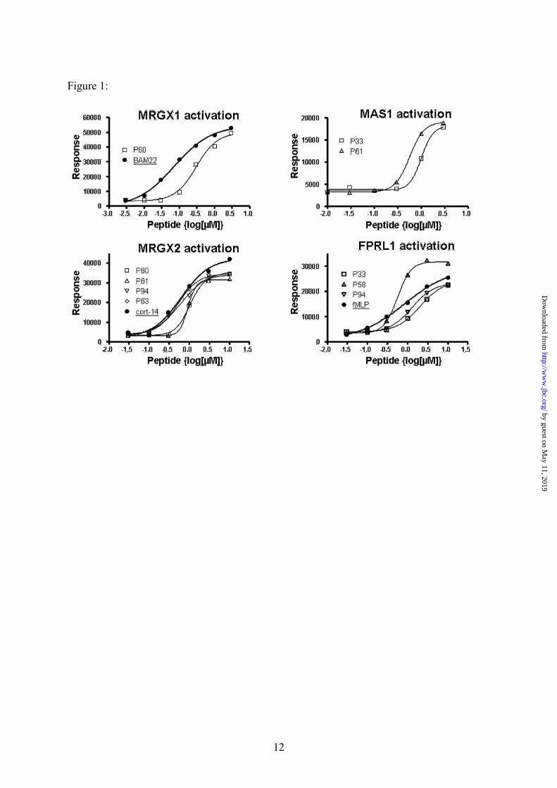
Out of the 152 receptors screened by the Ca2+
flux
activation assay, four receptors (MRGX1,
MRGX2, MAS1 & FPRL1) showed distinct
activation with at least one of the novel peptides
added at a concentration of 1 µM. Six peptides
(peptides P33, P58, P60, P61, P63 and P94 – See
Table 1) that showed either a moderate or a strong
hit for either receptor were further examined by
dose response on the specific GPCRs they
activated. Curve analysis and affinity (EC50)
calculation of the dose response experiments were
compared to the receptor’s most potent activator
as described in the literature and the results are
illustrated in Table 2 and Figure 1. In all cases, we
were able to demonstrate a dose dependent
activation of Ca2+
flux with good correlation to the
positive control used (Table 2).
We observed activation of MRGX1 with
our peptide P60 (Tables 1 and 2). According to the
dose response experiments we conducted (Figure
1), P60 activates MRGX1 in a dose dependent
manner, with affinity estimated within the
nanomolar range, slightly lower than that of the
positive control used (BAM22) [18]. In addition,
further experimental results indicate that P60 and
BAM22 differ in their activation of Opioid related
receptors [19]. While BAM22 activates the latter,
P60 is specific to MRGXs (activating MRGX1
and weakly activating MRGX2) and it did not
activate any of the known Opioid receptors (D1,
M1, L1 and K1) which were included in the screen
(S2).
For MRGX2, we identified several
potential peptides (Table 2). According to dose
response experiments, the affinity of two of the
novel peptides, P94 & P63 (Table 1), was similar
to that of the positive control, Cortistatin-14 [18]
(Figure 1). However, unlike Cortistatin-14, none
of the peptides had any affect upon the receptors
of the somatostatin family (SSTR 1-5) that are
known to be activated by cortistatin-14.
The FPRL1 (ALX) receptor is involved in
regulation of inflammation by controlling
leukocytes activities such as chemotaxis and
phagocytosis [20, 21]. We identified several
peptides that activated this receptor, P58, P94 and
P33 (Tables 1 and 2). The leading hit, peptide P58,
exhibited in dose response experiments, higher
potency and higher affinity than fMLP, which was
used as a positive control, although it is not the
most potent agonist for this receptor [22] (Figure
1). Importantly, P58, as well as none of the other
peptides examined, did not activate another family
member, FPR1. However, a recent experiment
(Hecht et al. – In preparation) indicates a dose
dependent activation of FPRL2, a closer family
member (data not shown).
by guest on May 11, 2019
http://ww
w.jbc.org/
Dow
nloaded from

6
We also identified two peptides, P61 and
P33 (Table 1) that dose-dependently activated the
MAS1 receptor (Table 2 and Figure 1). In
agreement with previous publications, we were
unable to demonstrate calcium flux induction by
the positive control (Ang 1-7), a known ligand for
MAS1, [23]. However, our peptides did activate
the receptor at concentrations similar to that found
for Dynorphine A (an established surrogate, lower
affinity activator of MAS1). Our peptides were
specific to MAS1 and did not activate neither AT1
nor AT2, Angiotensin II receptors which are
known to be weakly activated by Ang (1-7) (Table
2).
Cyclic AMP screening assay results:
Since the promiscuous Gα16 does not
always divert a cAMP-related signal to Ca2+
accumulation, a subset of 13 GPCRs with very
low activation by either the positive controls or
any of the novel examined peptides were selected
to be tested for Gi/Gs activation by two of the 33
examined peptides. These receptors were screened
for cAMP accumulation (Gs) or inhibition (Gi) as
described below.
Two of the peptides (P59 and P74 – Table
1) showed distinct Gi dependent activation of two
related GPCRs (LGR7 (RXFP1) and LGR8
(RXFP2) – Table 2, Figure 2). Both these
receptors were recently identified as being
activated by Relaxin and INSL3 respectively [24].
A much weaker hit, on LGR4, an orphan family
member (data not shown), was also observed.
Specificity was examined by GPR135 (RXFP3), a
receptor for Relaxin 3, which was not activated by
peptide P59 (Table 2).
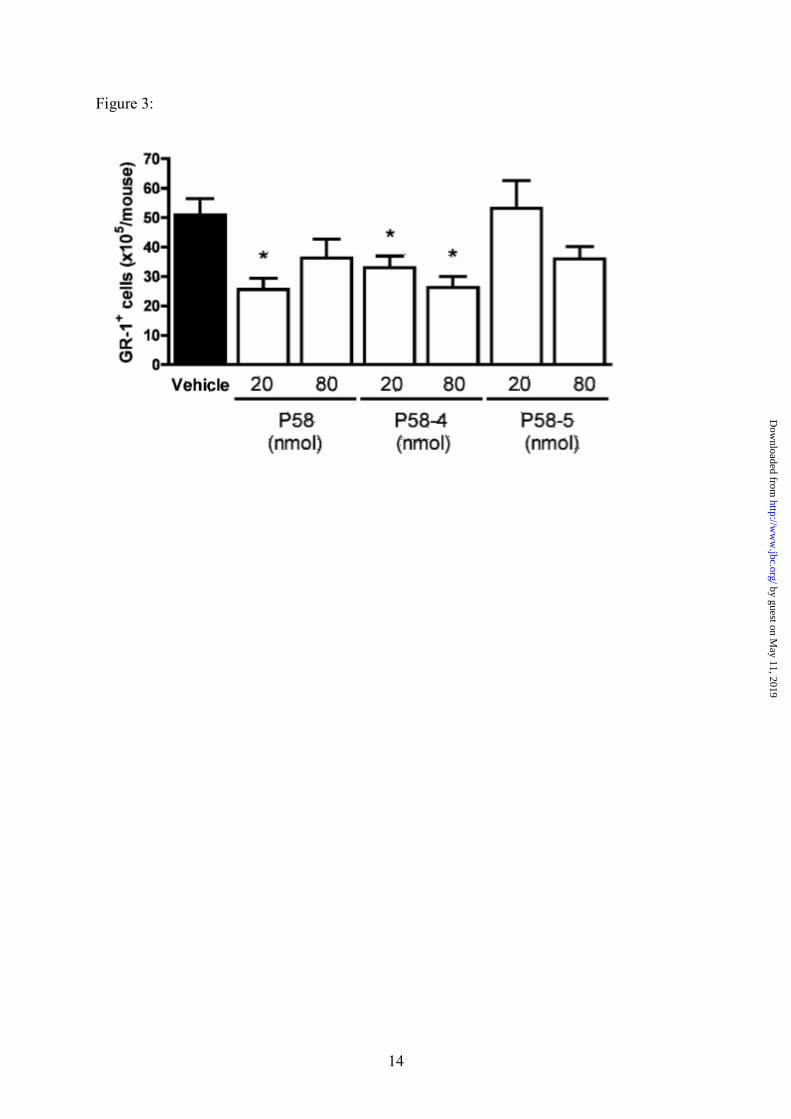
In-vivo activation of the FPRL1 receptor by P58:
FPRL1 activation promotes resolution of
inflammation- an active and tightly synchronized
process, involving counter-regulation of
leukocytes, which leads to a prominent anti-
inflammatory effect [21]. To study the in vivo
activity of our novel FPRL1 peptide agonist, P58,
and two of its derivatives (P58-4 and P58-5), we
used a model of acute inflammation, involving
zymosan-induced leukocyte recruitment into
murine dorsal air pouch (See methods). The novel
peptide P58 as well as P58-4 and P58-5, were
tested at doses of 20 and 80 nmoles per mouse,
and were given intravenously at the time of
zymosan A injection (time zero). These doses
correspond to 50 and 200 µg of P58 per mouse,
respectively; and equivalent doses were used for
the shorter peptides. Cell recruitment into the air-
pouches was determined at the 4 hrs time-point,
using both light microscopy (for total leukocyte
count) and FACS analysis for Gr1-stained cells
(for assessment of PMNs). A significant inhibition
of about 50% was observed in the infiltration of
PMNs into the air-pouch with P58 at
20nmole/mouse, (Figure 3). Out of the two
derivatives tested, P58-4 displayed a 47%
inhibition of Gr1+ cell migration into air-pouches
at the highest dose tested (80 nmol/mouse). P58-5
produced a discrete effect at the highest dose that
was not statistically significant. Similar effects
were observed on total leukocyte counts (data not
shown).
Discussion:
In this work we describe the results of a
screen for novel peptide GPCR agonists. A subset
of the predicted candidate peptides was screened
on a large group of GPCRs and eight peptide
agonists were identified for six different GPCRs.
The peptides activity was further validated
experimentally using established biochemical
assays such as dose dependant induction of
calcium flux, and by assessing the efficacy in-vivo
in one case.
By utilizing bioinformatic capabilities and
machine learning, tools both related to identifying
all potential secreted proteins (the secretome), and
predicting all potential Arg/Lys (R/K) cleavage
sites, we were able to create a comprehensive
peptidome which includes hundreds of thousands
of potential peptides, ranked and prioritized by
computational scores. Sequences of the predicted
peptidome were further analyzed for features and
assigned a probability score of being GPCR
ligands by a machine learning algorithm. This
collection contained hundreds of candidate
peptides that were scored and ranked by distinct
parameters and expert examination. The highest
scoring candidates were checked for novelty and
synthesized for further screening for activation on
a group of target GPCRs. The screening included
33 peptides which were screened for activation of
by guest on May 11, 2019
http://ww
w.jbc.org/
Dow
nloaded from

7
152 GPCRs selected according to relevance and
availability.
Out of the 33 peptides screened, eight
(~25%) showed a distinct activation of at least one
receptor. The ratio of successful candidates is
remarkable when compared to the average ratio of
novel GPCR peptide ligand discovery rate, which
is on average around two to three new candidates
per year in the past decade [25]. All of these
peptides are novel and most of them are derived
from hypothetical protein precursors. Most of the
peptides that showed initial hits also displayed
dose response activation. Furthermore, one of the
peptides, designated peptide P58 (now named
CGEN855A), which showed a dose-dependent
activation of the FPRL1 receptor, was further
examined for in-vivo activation of the FPRL1
receptor using a murine model of acute
inflammation. The activation of FPRL1, as
determined by inhibition of PMNs infiltration, was
found to be significant. Moreover, out of two
subsequent peptides derived from P58, P58-4
(now CGEN-855B) and P58-5 (now CGEN-
855C), at least one (P58-4) was found to invoke an
in-vivo response mediated by the FPRL1 receptor,
similar to the original P58 peptide. An
examination of the activation of the receptors by
the novel peptides shows cross hits of the same
peptides on several different GPCRs. This does
not seem surprising since some of the receptors,
such as the two relaxin related GPCRs, namely
RXFP1 (LGR7) and RXFP2 (LGR8), belong to
the same family while the others (MAS1,
MRGX1, MRGX2 and FPRL1) belong to the same
evolutionary branch (according to [26]).
Even though no sequence homology was
found between the newly discovered peptides and
the known GPCR ligands, we predict that other
structural properties (such as secondary structure)
might show a mechanistic resemblance between
the novel and known peptides. A preliminary
computational structural prediction of the eight
novel peptides by using the PSIPRED [27]
secondary structure predictor, revealed that all of
them contain at least one helical stretch, a feature
common in many known peptide GPCR ligands
(data not shown).
We conclude that we were able to show
efficacy and a proof of concept for our discovery
platform of GPCR peptide agonists. We believe
that we will be able to broaden our analysis and
discover additional novel peptide GPCR ligands
by this method. We further predict that more of the
newly discovered and validated activators found
for these GPCRs will be further investigated for
their potential as therapeutic compounds targeted
at receptors involved in distinct diseases and
conditions.
by guest on May 11, 2019
http://ww
w.jbc.org/
Dow
nloaded from
8
References: 1. Imming, P., Sinning, C., and Meyer, A. (2006) Nat Rev Drug Discov,. 5(10), 821-34.
2. Hill, S.J. (2006) Br J Pharmacol. 147 Suppl 1, S27-37.
3. Lin, S.H. and Civelli, O. (2004) Ann. Med. 36(3), 204-14.
4. Leifert, W.R., et al. (2005) J. Biomol. Screen. 10(8), 765-79.
5. Kutzleb, C., et al. (2005) Curr. Protein. Pept. Sci. 6(3), 265-78.
6. Jiang, Z. and Zhou, Y. (2006) Curr. Protein. Pept. Sci. 7(5) 459-64.
7. Howard, A.D., et al. (2001) Trends Pharmacol. Sci. 22(3) 132-40.
8. Civelli, O. (2005) Trends Pharmacol. Sci. 26(1) 15-9.
9. Eglen, R.M. (2005) Comb. Chem. High Throughput Screen. 8(4) 311-8.
10. Bock, J.R., and Gough D.A. (2005) J. Chem. Inf. Model. 45(5) 1402-14.
11. Huang, E.S. (2005) Drug Discov Today. 10(1) 69-73.
12. Bairoch, A., et al. (2005) Nucleic Acids Res. 33(Database issue) D154-9.
13. Breiman, L. (2001) Machine Learning. 45(1) 5-32.
14. Bendtsen, J.D., et al (2004) J. Mol. Biol. 340(4) 783-95.
15. Kliger Y., et al (2008) Bioinformatics. 24(8) 1049-55.
16. Horn, F., et al. (2003) Nucleic Acids Res. 31(1) 294-7.
17. Liu, A.M., et al. (2003) J. Biomol. Screen.. 8(1) 39-49.
18. Burstein, E.S., et al. (2006) Br. J. Pharmacol. 147(1) 73-82.
19. Wollemann, M., and Benyhe, S. (2004) Life Sci. 75(3) 257-70.
20. Iribarren, P., et al. (2005) Immunol. Res. 31(3) 165-76.
21. Perretti, M., and D'Acquisto, F. (2006) Inflamm. Allergy Drug Targets. 5(2) 107-14.
22. VanCompernolle, S.E., et al. (2003) J. Immunol. 171(4) 2050-6.
23. Santos, R.A., et al. (2003) Proc. Natl. Acad. Sci. U. S. A. 100(14) 8258-63.
24. Halls, M.L., Bathgate, R.A., and Summers, R.J. (2006) Mol. Pharmacol. 70(1) 214-26.
25. Katugampola, S., and Davenport, A. (2003) Trends Pharmacol. Sci. 24(1) 30-5.
26. Joost, P., and. Methner, A. (2002) Genome Biol. 3(11) RESEARCH0063.
27. McGuffin, L.J., Bryson, K. and Jones, D.T. (2000) Bioinformatics. 16(4):404-5
by guest on May 11, 2019
http://ww
w.jbc.org/
Dow
nloaded from
9
Figure legends:
Figure 1: Dose response activation measured for Ca2+ flux activating peptides: Peptides that
showed significant results in the screening assay were tested for dose dependent activation on the
activated receptors (MRGX1, MRGX2, FPRL1 and MAS1 – counter clockwise). Dose response
activation for positive control peptides are indicated with a thick line and solid circles. Dose response
curves for the tested peptides are indicated by thin lines and empty points. The best fit curves and
EC50 were calculated as described in the methods section and are presented in Table 1. For the MAS1
receptor, no activation was observed by the suggested positive control (Ang 1-7).
Figure 2: Inhibition of cyclic AMP (cAMP) in forskolin stimulated cells by activation of LGR7
and LGR8: cAMP was stimulated by incubation of ten minutes with the presence of 10µM of
forskolin, as described above. The peptide was added at a final concentration of 1µM and the sample
was incubated for 20 min following the readout of the endpoint cAMP concentration (measured in
Arbitrary Fluorescence Units – AFU). NT – represents addition of the buffer only as a negative
control. Forskolin with no added peptide serves as a positive control.
Figure 3: Effects of P58 and its derivatives on zymosan-induced PMNs infiltration into murine air-pouch. Mice were treated i.v. with the peptides (20 or 80 nmoles per mouse) or vehicle,
immediately followed by an intra pouch challenge with zymosan A at time 0. Four hours after the
zymosan A injection, pouch cavities were washed, and the leukocytes recovered in the lavage fluids
were stained with an anti-GR-1 antibody and analyzed by FACS. Data are shown as GR-1+ cells
(x105) per mouse (mean ± SE). * P<0.05 vs. vehicle group.
by guest on May 11, 2019
http://ww
w.jbc.org/
Dow
nloaded from
10
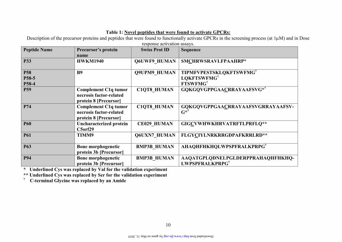
Table 1: Novel peptides that were found to activate GPCRs:
Description of the precursor proteins and peptides that were found to functionally activate GPCRs in the screening process (at 1µM) and in Dose
response activation assays.
Peptide Name
Precursor’s protein
name
Swiss Prot ID Sequence
P33 HWKM1940 Q6UWF9_HUMAN
SMCHRWSRAVLFPAAHRP*
P58
P58-5
P58-4
B9 Q9UPM9_HUMAN
TIPMFVPESTSKLQKFTSWFMG†
LQKFTSWFMG†
FTSWFMG†
P59 Complement C1q tumor
necrosis factor-related
protein 8 [Precursor]
C1QT8_HUMAN
GQKGQVGPPGAACRRAYAAFSVG*†
P74 Complement C1q tumor
necrosis factor-related
protein 8 [Precursor]
C1QT8_HUMAN
GQKGQVGPPGAACRRAYAAFSVGRRAYAAFSV-
G*†
P60 Uncharacterized protein
C5orf29
CE029_HUMAN
GIGCVWHWKHRVATRFTLPRFLQ**
P61 TIMM9 Q6UXN7_HUMAN
FLGYCIYLNRKRRGDPAFKRRLRD**
P63 Bone morphogenetic
protein 3b [Precursor]
BMP3B_HUMAN
AHAQHFHKHQLWPSPFRALKPRPG†
P94 Bone morphogenetic
protein 3b [Precursor]
BMP3B_HUMAN
AAQATGPLQDNELPGLDERPPRAHAQHFHKHQ-
LWPSPFRALKPRPG†
* Underlined Cys was replaced by Val for the validation experiment
** Underlined Cys was replaced by Ser for the validation experiment † C-terminal Glycine was replaced by an Amide
by guest on May 11, 2019 http://www.jbc.org/ Downloaded from

11
Table2: GPCRs activation and specificity by screened peptides:
Positive hits and calculated EC50 for Compugen’s peptides and receptor specific positive controls. Each hit is indicated by a “+” mark as well as a
calculated EC50 value (µM). The observed affinity (EC50) value is also indicated for the positive control peptides (with the exception of Ang 1-7,
where no activation of the MAS1 receptor was observed - NA). ND = No data
Receptor
Positive
controls
(EC50 µµµµM)
P60
P61 P94 P63 P58 P33 P59 P74 Other GPCRs**
activated by same
positive control
MRGX1 BAM22
(0.08)
+
(0.3)
- - - - - - - OPRM, OPRK,
OPRL & OPRD
MRGX2 Cort14
(0.61)
+
(1.0)
+
(0.9)
+
(0.6)
+
(0.5)
- - - - SSTR (1-5)
MAS1 (Ang1-7)
(NA)
- +
(0.57)
- - - +
(1.0)
- - AGTR1, AGTR 2
FPRL1 fMLP
(0.85)
- - +
(1.3)
- +
(0.55)
+
(2.0)
- - FPR1
LGR7 (RXFP1)* Relaxin
(ND)
- - - - - - +
(ND)
+
(ND)
GPR135 (RXFP3)
LGR8 (RXFP2)* Relaxin
(ND)
- - - - - - +
(ND)
+
(ND)
GPR135 (RXFP3)
* Examined by cAMP inhibition assay. No dose response was performed (ND).
** Receptors were included in the screening. No activation by Compugen peptides was observed. Activation by positive control is indicated
from the literature.
by guest on May 11, 2019 http://www.jbc.org/ Downloaded from
Eshel and Yossi CohenDvir Dahary, Eyal Gofer, Yossef Kliger, Michal Ayalon Soffer, Avi Rosenberg, Dani Ronen Shemesh, Amir Toporik, Zurit Levine, Iris Hecht, Galit Rotman, Assaf Wool,
Discovery and validation of novel peptide agonists for G-protein coupled receptors
published online October 9, 2008J. Biol. Chem.
10.1074/jbc.M805181200Access the most updated version of this article at doi:
Alerts:
When a correction for this article is posted•
When this article is cited•
to choose from all of JBC's e-mail alertsClick here
by guest on May 11, 2019
http://ww
w.jbc.org/
Dow
nloaded from