Embed Size (px)
Citation preview
Direct and widespread role for the nuclear receptorEcR in mediating the response to ecdysonein DrosophilaChristopher M. Uyeharaa,b,c,d and Daniel J. McKaya,b,d,1
aDepartment of Biology, The University of North Carolina at Chapel Hill, Chapel Hill, NC 27599; bDepartment of Genetics, The University of North Carolinaat Chapel Hill, Chapel Hill, NC 27599; cCurriculum in Genetics and Molecular Biology, The University of North Carolina at Chapel Hill, Chapel Hill, NC 27599;and dIntegrative Program for Biological and Genome Sciences, The University of North Carolina at Chapel Hill, Chapel Hill, NC 27599
Edited by Lynn M. Riddiford, University of Washington, Friday Harbor, WA, and approved April 5, 2019 (received for review January 10, 2019)
The ecdysone pathway was among the first experimental systemsemployed to study the impact of steroid hormones on the genome.In Drosophila and other insects, ecdysone coordinates developmen-tal transitions, including wholesale transformation of the larva intothe adult during metamorphosis. Like other hormones, ecdysonecontrols gene expression through a nuclear receptor, which func-tions as a ligand-dependent transcription factor. Although it is clearthat ecdysone elicits distinct transcriptional responses within its dif-ferent target tissues, the role of its receptor, EcR, in regulating tar-get gene expression is incompletely understood. In particular, EcRinitiates a cascade of transcription factor expression in response toecdysone, making it unclear which ecdysone-responsive genes aredirect EcR targets. Here, we use the larval-to-prepupal transition ofdeveloping wings to examine the role of EcR in gene regulation.Genome-wide DNA binding profiles reveal that EcR exhibits wide-spread binding across the genome, including at many canonical ec-dysone response genes. However, the majority of its binding sitesreside at genes with wing-specific functions. We also find that EcRbinding is temporally dynamic, with thousands of binding siteschanging over time. RNA-seq reveals that EcR acts as both a tempo-ral gate to block precocious entry to the next developmental stageas well as a temporal trigger to promote the subsequent program.Finally, transgenic reporter analysis indicates that EcR regulates notonly temporal changes in target enhancer activity but also spatialpatterns. Together, these studies define EcR as a multipurpose, di-rect regulator of gene expression, greatly expanding its role in co-ordinating developmental transitions.
hormone | transcription factor | CUT&RUN | ecdysone | temporal generegulation
Hormones function as critical regulators of a diverse set ofphysiological and developmental processes, including reproduc-
tion, immune system function, and metabolism. During development,hormones act as long-range signals to coordinate the timing of eventsbetween distant tissues. The effects of hormone signaling are medi-ated by nuclear receptors, which function as transcription factorsthat differentially regulate gene expression in a hormone-dependentmanner. Whereas many of the coregulators that contribute to nu-clear receptor function have been identified, the mechanisms usedby these factors to generate distinct, yet appropriate, transcriptionalresponses in different target tissues are incompletely understood.Ecdysone signaling has long served as a paradigm to understand
how hormones generate spatial and temporal-specific biologicalresponses. In Drosophila, ecdysone is produced by the ring glandand secreted into the hemolymph, where it is converted into itsactive form, 20-hydroxyecdysone (20E), before reaching target tis-sues (1, 2). Pulses of ecdysone are required for transitions betweendevelopmental stages, such as the larval molts. A high-titer pulse ofecdysone triggers the end of larval development and the beginningof metamorphosis (1, 2). Ecdysone effects transcriptional changesthrough binding to its receptor, a heterodimer of the proteins ec-dysone receptor (EcR) (homolog of the mammalian farnesoid X
receptor) and ultraspiracle (Usp) (homolog of mammalian RXR)(3). In the absence of ecdysone, EcR/Usp is nuclear localized andbound to DNA where it is thought to act as a transcriptional re-pressor (4, 5). Upon ecdysone binding, EcR/Usp switches to atranscriptional activator (4). Consistent with the dual regulatorycapacity of EcR/Usp, a variety of coactivator and corepressorcomplexes have been shown to function with this heterodimer toregulate gene expression (5–8).Understanding how ecdysone exerts its effects on the genome has
been heavily influenced by the work of Ashburner and colleagues inthe 1970s. By culturing larval salivary glands in vitro, Ashburner (9)described a sequence of visible puffs that appear in the giantpolytene chromosomes upon addition of ecdysone. A small numberof puffs appeared immediately after ecdysone addition, followed bythe appearance of more than 100 additional puffs over the nextseveral hours (9). The appearance of early puffs was found to beindependent of protein synthesis, suggesting direct action by EcR/Usp, whereas the appearance of late puffs was not, suggesting theyrequire the protein products of early genes for activation (9). Thesefindings, and decades of subsequent work elucidating the molecularand genetic details, have led to a hierarchical model of ecdysonesignaling in which EcR/Usp directly induces expression of a smallnumber of early response genes. Many of these early response genes
Significance
Nuclear receptors (NRs) are sequence-specific DNA bindingproteins that act as intracellular receptors for small moleculessuch as hormones. Prior work has shown that NRs function asligand-dependent switches that initiate a cascade of gene ex-pression changes. The extent to which NRs function as directregulators of downstream genes in these hierarchies remainsincompletely understood. Here, we study the role of the NR EcRin metamorphosis of the Drosophila wing. We find that EcRdirectly regulates many genes at the top of the hierarchy aswell as at downstream genes. Furthermore, we find that EcRbinds distinct sets of target genes at different developmentaltimes. This work helps inform how hormones elicit tissue- andtemporal-specific responses in target tissues.
Author contributions: C.M.U. and D.J.M. designed research; C.M.U. performed research;C.M.U. and D.J.M. analyzed data; and C.M.U. and D.J.M. wrote the paper.
The authors declare no conflict of interest.
This article is a PNAS Direct Submission.
This open access article is distributed under Creative Commons Attribution-NonCommercial-NoDerivatives License 4.0 (CC BY-NC-ND).
Data deposition: The data reported in this paper have been deposited in the Gene Ex-pression Omnibus (GEO) database, https://www.ncbi.nlm.nih.gov/geo (accession no.GSE124254).1To whom correspondence should be addressed. Email: [email protected].
This article contains supporting information online at www.pnas.org/lookup/suppl/doi:10.1073/pnas.1900343116/-/DCSupplemental.
Published online April 24, 2019.
www.pnas.org/cgi/doi/10.1073/pnas.1900343116 PNAS | May 14, 2019 | vol. 116 | no. 20 | 9893–9902
DEV
ELOPM
ENTA
LBIOLO
GY
Dow
nloa
ded
by g
uest
on
Nov
embe
r 26
, 202
1

encode transcription factors, such as the zinc finger protein Broad,the nuclear receptor Ftz-f1, and the pipsqueak domain factor E93(2). The early response transcription factors are required, in turn, toinduce expression of the late response genes, which encode proteinsthat impart temporal and tissue-specific responses in target tissues.Although the framework of the ecdysone pathway was estab-
lished through work in salivary glands, additional studiesaffirmed an essential role for ecdysone signaling in many othertissues. Similar to other hormones, the physiological response toecdysone is often profoundly specific to each target tissue. Forexample, ecdysone signaling triggers proliferation, changes in celland tissue morphology, and eventual differentiation of larval tissuesfated to become part of the adult fly, such as the imaginal discs (2,10). By contrast, ecdysone signaling initiates the wholesale elimi-nation of obsolete tissues, such as the larval midgut and salivaryglands through programmed cell death (1, 2, 10). Ecdysone is alsoessential for remodeling larval neurons that persist until adulthoodand specifying the temporal identity of neural stem cell progenyborn during this time (11). While it is clear that ecdysone signalingtriggers the gene expression cascades that underlie these events, themolecular mechanisms by which ecdysone elicits diverse tran-scriptional responses in target tissues remains poorly understood.A key step in delineating the mechanisms by which ecdysone
signaling regulates target gene expression involves identificationof EcR/Usp DNA binding sites. Given the hierarchical structureof the ecdysone pathway, it is unclear whether EcR acts primarilyat the top of the transcriptional cascade, or whether it also actsdirectly on downstream effector genes. Several early responsegenes such as br, Eip74EF, and the glue genes have been shownto be directly bound by EcR in vivo (12, 13). At the genome-widelevel, polytene chromosome staining revealed ∼100 sites boundby EcR in larval salivary glands (14). DamID and ChIP-seq ex-periments have identified roughly 500 sites directly bound byEcR in Drosophila cell lines (15, 16). Thus, the available evi-dence, albeit limited, indicates that EcR binds to a limitednumber of target genes, consistent with hierarchical modelswherein the response to ecdysone is largely driven by early re-sponse genes and other downstream factors.We recently identified the ecdysone-induced transcription fac-
tor E93 as being essential for the proper temporal sequence ofenhancer activation during pupal wing development (17). In theabsence of E93, early-acting enhancers fail to turn off, and late-acting enhancers fail to turn on. Moreover, ChIP-seq identifiedthousands of E93 binding sites across the genome. These datasupport the hierarchical model of ecdysone signaling in whichearly response transcription factors like E93 directly regulate asignificant fraction of ecdysone-responsive genes in target tissues.Here, we sought to determine the role that EcR performs in
temporal gene regulation during the larval-to-prepupal transi-tion of the wing. Using wing-specific RNAi, we find that EcR isrequired for proper morphogenesis of prepupal wings, althoughit is largely dispensable for wing disc patterning at earlier stagesof development. RNA-seq profiling reveals that EcR functions asboth a temporal gate to prevent the precocious transition toprepupal development as well as a temporal trigger to promoteprogression to next stage. Using CUT&RUN, we map bindingsites for EcR genome-wide before and after the larval-to-prepupal transition. Remarkably, we find that EcR binds ex-tensively throughout the genome, including at many genes withwing-specific functions that are not part of the canonical ecdy-sone signaling cascade. Moreover, EcR binding is highly dy-namic, with thousands of binding sites gained and lost over time.Finally, transgenic reporter analyses demonstrate that EcR isrequired not only for temporal regulation of enhancer activitybut also for spatial regulation of target enhancers. Together,these findings indicate that EcR does not control gene expres-sion solely through induction of a small number of downstream
transcription factors, but instead plays a direct and widespreadrole in regulating tissue-specific transcriptional programs.
ResultsTemporal Changes in Gene Expression During the Larval-to-PrepupalTransition. In Drosophila, the end of larval development marksthe beginning of metamorphosis. Over a 5-d period, larval tissuesare destroyed, and the progenitors of adult tissues, such as wingimaginal discs, undergo a series of progressive morphologicaland cell differentiation events to acquire their final shapes andsizes. By the end of larval development, the wing disc is com-prised of a largely undifferentiated array of columnar epithelialcells (18, 19). The first 12 h after puparium formation (APF) istermed the prepupal stage. During this period, cell division isarrested, and the pouch of the wing disc everts outward, causingthe dorsal and ventral surfaces of the wing to appose one an-other, forming the presumptive wing blade (Fig. 1 A and B) (18,19). At the same time, the notum of the wing disc extends dor-solaterally and eventually fuses with the contralateral wing discto form the back of the adult fly (Fig. 1 A and B). Additionalevents occurring during this time period include secretion of theprepupal cuticle and migration of muscle progenitor cells.To understand EcR’s role in promoting the larval-to-prepupal
transition, we began by identifying global changes in gene ex-pression that occur in wild-type (WT) wings before and after theonset of pupariation. We collected wing tissue from wandering,third-instar larvae, approximately 6 h before puparium formation(hereafter, −6hAPF) and from prepupae, approximately 6 h af-ter puparium formation (hereafter, +6hAPF), and performedRNA-seq, aligning our reads to the dm3 reference sequence(20). As described previously (19), WT gene expression is highlydynamic during this time period. Using a conservative definitionfor differential expression (false-diskovery rate < 0.05, ≥2-foldchange in expression), we identified over 1,300 genes increasingin expression and nearly 800 genes decreasing in expression (Fig.1C). The observed gene expression changes are consistent withdevelopmental events occurring at this time. For example, genesthat increase over time are involved in cuticle deposition, cellularmetabolism, and muscle development (Fig. 1C). By contrast,genes that decrease over time are involved in cell cycle regula-tion and DNA replication. Thus, the morphological changes thatdefine the larval-to-prepupal transition are rooted in thousandsof changes in gene expression.
EcR Is Required for the Larval-to-Prepupal Transition in Wings. Theonset of pupariation is induced by a high-titer ecdysone pulse. Atthe genetic level, ecdysone acts through its receptor, EcR. Nullmutations in EcR are embryonic lethal. Therefore, to investigatethe role that EcR plays in wing development, we used a wing-specific GAL4 driver in combination with an RNAi construct toknock down EcR expression throughout wing development (21).EcR-RNAi driven in wing discs diminished protein levels by∼95% (SI Appendix, Fig. S1 A–C).In agreement with previous work suggesting that EcR does not
appear to be required for wing development during the first- andsecond-instar stages (22, 23), EcR-RNAi wings appear mor-phologically similar to WT wing imaginal discs at −6hAPF (Fig.1B). However, EcR-RNAi wing discs are noticeably larger thanWT wing discs, consistent with the proposed role for ecdysonesignaling in cell cycle inhibition in third-instar larvae (22, 23). Bycontrast, EcR-RNAi wings at +6hAPF appear morphologicallydissimilar to both −6hAPF EcR-RNAi wings and to WT wings at+6hAPF. The pouch fails to properly evert and larval folds re-main visible. Similarly, the notum fails to extend appropriatelyand appears more similar to the larval notum than the notum at+6hAPF (Fig. 1B). These findings suggest that wings fail toproperly progress through the larval-to-prepupal transition inthe absence of EcR. Notably, this failure is likely not due to a
9894 | www.pnas.org/cgi/doi/10.1073/pnas.1900343116 Uyehara and McKay
Dow
nloa
ded
by g
uest
on
Nov
embe
r 26
, 202
1
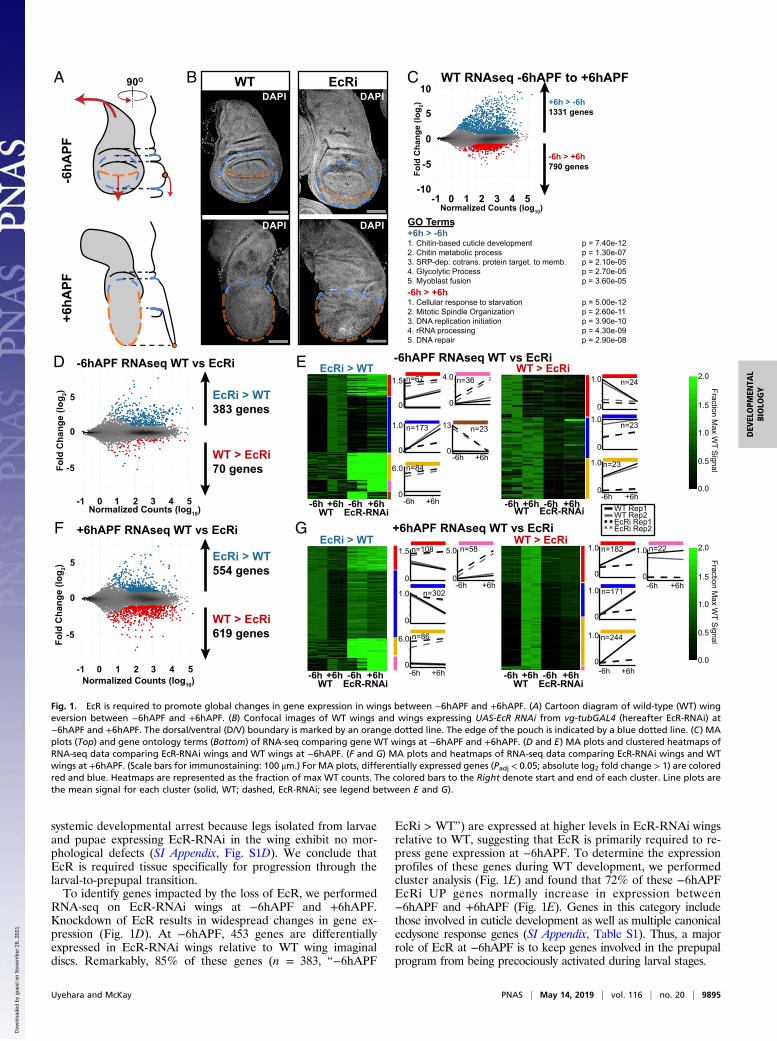
systemic developmental arrest because legs isolated from larvaeand pupae expressing EcR-RNAi in the wing exhibit no mor-phological defects (SI Appendix, Fig. S1D). We conclude thatEcR is required tissue specifically for progression through thelarval-to-prepupal transition.To identify genes impacted by the loss of EcR, we performed
RNA-seq on EcR-RNAi wings at −6hAPF and +6hAPF.Knockdown of EcR results in widespread changes in gene ex-pression (Fig. 1D). At −6hAPF, 453 genes are differentiallyexpressed in EcR-RNAi wings relative to WT wing imaginaldiscs. Remarkably, 85% of these genes (n = 383, “−6hAPF
EcRi > WT”) are expressed at higher levels in EcR-RNAi wingsrelative to WT, suggesting that EcR is primarily required to re-press gene expression at −6hAPF. To determine the expressionprofiles of these genes during WT development, we performedcluster analysis (Fig. 1E) and found that 72% of these −6hAPFEcRi UP genes normally increase in expression between−6hAPF and +6hAPF (Fig. 1E). Genes in this category includethose involved in cuticle development as well as multiple canonicalecdysone response genes (SI Appendix, Table S1). Thus, a majorrole of EcR at −6hAPF is to keep genes involved in the prepupalprogram from being precociously activated during larval stages.
DAPI
DAPI
WTDAPI
DAPI
EcRiB WT RNAseq -6hAPF to +6hAPF
-6h > +6h1. Cellular response to starvation p = 5.00e-122. Mitotic Spindle Organization p = 2.60e-113. DNA replication initiation p = 3.90e-104. rRNA processing p = 4.30e-095. DNA repair p = 2.90e-08
GO Terms+6h > -6h1. Chitin-based cuticle development p = 7.40e-12 2. Chitin metabolic process p = 1.30e-073. SRP-dep. cotrans. protein target. to memb. p = 2.10e-054. Glycolytic Process p = 2.70e-055. Myoblast fusion p = 3.60e-05
C
-6h > +6h790 genes
+6h > -6h1331 genes
Normalized Counts (log10)
Fold
Cha
nge
(log 2)
-1 0 1 2 3 4 5
-5
-10
0
5
10
+6hA
PF-6
hAPF
A 90O
D
WT > EcRi619 genes
EcRi > WT554 genes
Normalized Counts (log10)-1 0 1 2 3 4 5
Fold
Cha
nge
(log 2)
-5
0
5
+6hAPF RNAseq WT vs EcRiF
WT > EcRi70 genes
EcRi > WT383 genes
Fold
Cha
nge
(log 2)
-5
0
5
Normalized Counts (log10)-1 0 1 2 3 4 5
-6hAPF RNAseq WT vs EcRi
WT Rep1WT Rep2EcRi Rep1EcRi Rep2
E -6hAPF RNAseq WT vs EcRiEcRi > WT
WT EcR-RNAi-6h +6h -6h +6h
1.5
0
n=67
1.0
0
n=173
4.0
0
n=36
13
-6h +6h0
n=23
6.0
0
n=84
WT > EcRi1.0
0
n=24
1.0
0-6h +6h
n=23
1.0
0
n=23
WT EcR-RNAi-6h +6h -6h +6h
1.0
0.5
0.0
1.5
2.0
Fraction Max W
T Signal
-6h +6h
G +6hAPF RNAseq WT vs EcRi
WT EcR-RNAi-6h +6h -6h +6h
EcRi > WT1.5
0
n=108
1.0
0
n=302
5.0
0-6h +6h
n=58
6.0
0
n=86
WT EcR-RNAi-6h +6h -6h +6h
WT > EcRi1.0
0
n=182
1.0
0
n=171
1.0
0
n=244
1.0
0-6h +6h
n=22
1.0
0.5
0.0
1.5
2.0
Fraction Max W
T Signal
-6h +6h -6h +6h
Fig. 1. EcR is required to promote global changes in gene expression in wings between −6hAPF and +6hAPF. (A) Cartoon diagram of wild-type (WT) wingeversion between −6hAPF and +6hAPF. (B) Confocal images of WT wings and wings expressing UAS-EcR RNAi from vg-tubGAL4 (hereafter EcR-RNAi) at−6hAPF and +6hAPF. The dorsal/ventral (D/V) boundary is marked by an orange dotted line. The edge of the pouch is indicated by a blue dotted line. (C) MAplots (Top) and gene ontology terms (Bottom) of RNA-seq comparing gene WT wings at −6hAPF and +6hAPF. (D and E) MA plots and clustered heatmaps ofRNA-seq data comparing EcR-RNAi wings and WT wings at −6hAPF. (F and G) MA plots and heatmaps of RNA-seq data comparing EcR-RNAi wings and WTwings at +6hAPF. (Scale bars for immunostaining: 100 μm.) For MA plots, differentially expressed genes (Padj < 0.05; absolute log2 fold change > 1) are coloredred and blue. Heatmaps are represented as the fraction of max WT counts. The colored bars to the Right denote start and end of each cluster. Line plots arethe mean signal for each cluster (solid, WT; dashed, EcR-RNAi; see legend between E and G).
Uyehara and McKay PNAS | May 14, 2019 | vol. 116 | no. 20 | 9895
DEV
ELOPM
ENTA
LBIOLO
GY
Dow
nloa
ded
by g
uest
on
Nov
embe
r 26
, 202
1
We next examined the impact of EcR knockdown in +6hAPFwings. In contrast to the effect at −6hAPF, wherein genes pri-marily increased in the absence of EcR, we observed approxi-mately equal numbers of up- and down-regulated genes relativeto WT wings at +6hAPF (Fig. 1F). Clustering of EcR-RNAi andWT RNA-seq data revealed distinct differences in the inferredregulatory role of EcR at +6hAPF relative to −6hAPF (Fig. 1G).Seventy-four percent of the genes expressed at higher levels inEcR-RNAi wings relative to WT normally decrease in expressionbetween −6hAPF and +6hAPF (Fig. 1G). Genes in this categoryinclude factors that promote sensory organ development andcell cycle genes (SI Appendix, Table S2). The increased levels ofthese “+6hAPF EcRi > WT” genes suggest that, in addition topreventing precocious activation of the prepupal gene expres-sion program, EcR is also required to shut down the larval geneexpression program. However, we also observe a role for EcRin gene activation. For genes that are expressed at lower levelsin EcR-RNAi wings (n = 619, “+6hAPF WT > EcRi”), 96% ofthese genes normally increase between −6hAPF and +6hAPF.Genes in this category include those involved in muscle de-velopment, metabolic genes, and regulators of cell and tissuemorphology (SI Appendix, Table S2). We conclude that EcR isrequired not only for gene repression but also for gene acti-vation, consistent with the demonstrated interaction of EcRwith both activating and repressing gene-regulatory complexes(5–8). Collectively, these data demonstrate that the failure ofEcR-RNAi wings to progress through the larval-to-prepupaltransition coincides with widespread failures in temporal geneexpression changes.The transcriptional response to ecdysone has recently been
examined in a set of 41 different Drosophila cell lines (24),including several wing disc-derived cell lines. To determine theextent to which these responses mirror ecdysone-triggeredgene expression changes in a developing tissue, we comparedthem to our EcR-RNAi wings (SI Appendix, Fig. S2). In gen-eral, the overlap between differentially expressed genes for anygiven cell line and EcR-dependent genes in the wing was low(e.g., median of 3.97% of EcR-dependent genes at −6hAPFoverlap an ecdysone-responsive gene in cell lines) (SI Appen-dix, Fig. S2D). A subset of wing disc-derived cell lines exhibitedmodestly greater similarity (e.g., median of 8.39% of ecdysone-responsive genes in wing disc-derived cell lines are categorizedas EcR-dependent in −6hAPF wings); however, the overlapremained low overall. Cumulatively, only 16–21% of EcR-dependent genes in the wing were identified as ecdysone re-sponsive in any given cell line (SI Appendix, Fig. S2 C and E).Conversely, only 6–16% of ecdysone-responsive genes in anygiven cell line were identified as EcR dependent in the wing(SI Appendix, Fig. S2 C and E). Thus, the transcriptional re-sponse to ecdysone is highly specific to both cell and devel-opmental state.
EcR Directly Binds Thousands of Sites Genome-Wide. The experi-ments described above reveal that ecdysone triggers thousands ofgene expression changes in wings during the larval-to-prepupaltransition. Because ecdysone signaling initiates a cascade oftranscription factor expression, it is unclear which of thesechanges are mediated directly by EcR. Therefore, we sought todetermine the genome-wide DNA binding profiles of EcR indeveloping wings. For these experiments, we utilized a fly strainin which the endogenous EcR gene product has been epitope-tagged by a transposon inserted into an intron of EcR (25). Thisepitope tag is predicted to be incorporated into all EcR proteinisoforms (hereafter EcRGFSTF) (SI Appendix, Fig. S3A). Geneticcomplementation tests determined that EcRGFSTF flies are viableat the expected frequency (SI Appendix, Fig. S3B), indicating thatepitope-tagged EcR proteins are fully functional. Supporting thisinterpretation, Western blotting demonstrated that EcRGFSTF
protein levels are equivalent to untagged EcR, and immunoflu-orescence experiments revealed nuclear localization of EcRGFSTF
as well as binding of EcRGFSTF to DNA in polytene chromosomespreads (SI Appendix, Fig. S3 C–E).To generate genome-wide DNA binding profiles for EcR, we
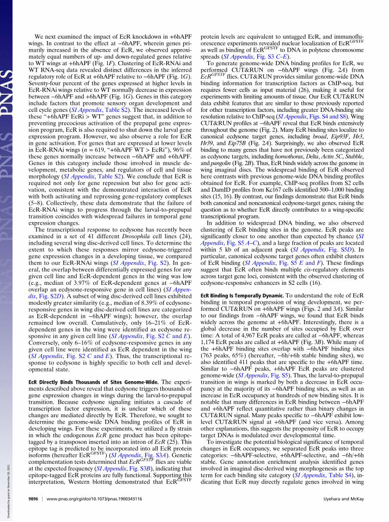
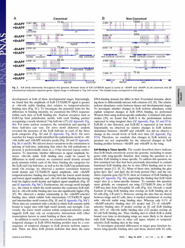
performed CUT&RUN on −6hAPF wings (Fig. 2A) fromEcRGFSTF flies. CUT&RUN provides similar genome-wide DNAbinding information for transcription factors as ChIP-seq, butrequires fewer cells as input material (26), making it useful forexperiments with limiting amounts of tissue. Our EcR CUT&RUNdata exhibit features that are similar to those previously reportedfor other transcription factors, including greater DNA-binding siteresolution relative to ChIP-seq (SI Appendix, Figs. S4 and S8). WingCUT&RUN profiles at −6hAPF reveal that EcR binds extensivelythroughout the genome (Fig. 2). Many EcR binding sites localize tocanonical ecdysone target genes, including broad, Eip93F, Hr3,Hr39, and Eip75B (Fig. 2A). Surprisingly, we also observed EcRbinding to many genes that have not previously been categorizedas ecdysone targets, including homothorax, Delta, Actin 5C, Stubble,and pangolin (Fig. 2B). Thus, EcR binds widely across the genome inwing imaginal discs. The widespread binding of EcR observedhere contrasts with previous genome-wide DNA binding profilesobtained for EcR. For example, ChIP-seq profiles from S2 cellsand DamID profiles from Kc167 cells identified 500–1,000 bindingsites (15, 16). By contrast, our findings demonstrate that EcR bindsboth canonical and noncanonical ecdysone-target genes, raising thequestion as to whether EcR directly contributes to a wing-specifictranscriptional program.In addition to widespread DNA binding, we also observed
clustering of EcR binding sites in the genome. EcR peaks aresignificantly closer to one another than expected by chance (SIAppendix, Fig. S5 A–C), and a large fraction of peaks are locatedwithin 5 kb of an adjacent peak (SI Appendix, Fig. S5D). Inparticular, canonical ecdysone target genes often exhibit clustersof EcR binding (SI Appendix, Fig. S5 E and F). These findingssuggest that EcR often binds multiple cis-regulatory elementsacross target gene loci, consistent with the observed clustering ofecdysone-responsive enhancers in S2 cells (16).
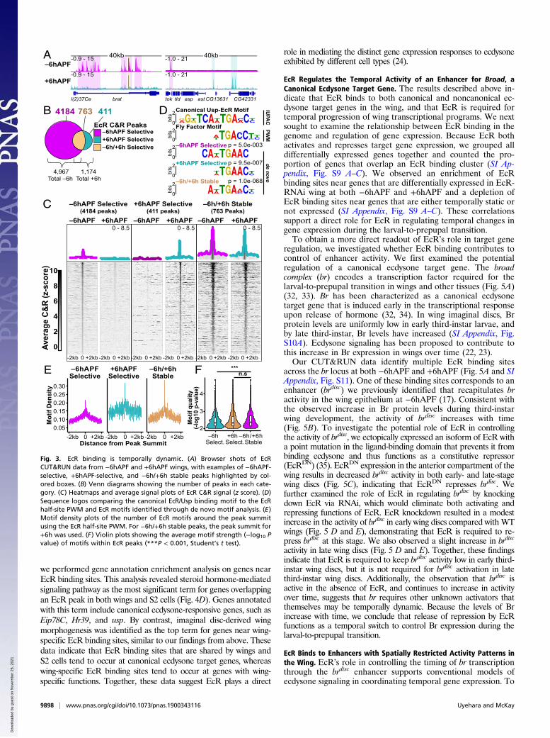
EcR Binding Is Temporally Dynamic. To understand the role of EcRbinding in temporal progression of wing development, we per-formed CUT&RUN on +6hAPF wings (Figs. 2 and 3A). Similarto our findings from −6hAPF wings, we found that EcR bindswidely across the genome at +6hAPF. Interestingly, there is aglobal decrease in the number of sites occupied by EcR overtime: A total of 4,967 EcR peaks are called at −6hAPF, whereas1,174 EcR peaks are called at +6hAPF (Fig. 3B). While many ofthe +6hAPF binding sites overlap with −6hAPF binding sites(763 peaks, 65%) (hereafter, −6h/+6h stable binding sites), wealso identified 411 peaks that are specific to the +6hAPF time.Similar to −6hAPF peaks, +6hAPF EcR peaks are clusteredgenome-wide (SI Appendix, Fig. S5). Thus, the larval-to-prepupaltransition in wings is marked by both a decrease in EcR occu-pancy at the majority of its −6hAPF binding sites, as well as anincrease in EcR occupancy at hundreds of new binding sites. It isnotable that many differences in EcR binding between −6hAPFand +6hAPF reflect quantitative rather than binary changes inCUT&RUN signal. Many peaks specific to −6hAPF exhibit low-level CUT&RUN signal at +6hAPF (and vice versa). Amongother explanations, this suggests the propensity of EcR to occupytarget DNAs is modulated over developmental time.To investigate the potential biological significance of temporal
changes in EcR occupancy, we separated EcR peaks into threecategories: −6hAPF-selective, +6hAPF-selective, and −6h/+6hstable. Gene annotation enrichment analysis identified genesinvolved in imaginal disc-derived wing morphogenesis as the topterm for each binding site category (SI Appendix, Table S4), in-dicating that EcR may directly regulate genes involved in wing
9896 | www.pnas.org/cgi/doi/10.1073/pnas.1900343116 Uyehara and McKay
Dow
nloa
ded
by g
uest
on
Nov
embe
r 26
, 202
1
development at both of these developmental stages. Interestingly,we found that the amplitude of EcR CUT&RUN signal is greaterat −6h/+6h stable binding sites relative to temporal-selectivebinding sites (Fig. 3C). To investigate the potential basis for thedifference in binding intensity, we examined the DNA sequencewithin each class of EcR binding site. Nuclear receptors such asEcR/Usp bind palindromic motifs, with each binding partnerrecognizing a nearly identical 7-bp half-site (27) (SI Appendix, Fig.S6). For some nuclear receptors, the orientation and spacing ofthese half-sites can vary. De novo motif diskovery analysisrevealed the presence of the EcR half-site in each of the threepeak categories (Fig. 3D and SI Appendix, Fig. S6A). De novosearches for longer motifs identified the palindromic motif in −6h/+6h stable and +6hAPF-selective peaks (Fig. 3D and SI Appendix,Fig. S6 A and B). We did not detect variations in the orientation orspacing of half-sites, indicating that when the full palindrome ispresent, it preferentially exists in a 13-bp inverted repeat confor-mation. To determine whether differences in signal amplitude be-tween −6h/+6h stable EcR binding sites could be caused bydifferences in motif content, we examined motif density aroundpeak summits within each of the three binding site categories forthe EcR and Usp half-sites, as well as for the EcR/Usp palindromicmotif. On average, we observed a positive correlation betweenmotif density and CUT&RUN signal amplitude, with −6hAPFtemporal-selective binding sites having both the lowest motif densityand lowest signal amplitude, and −6h/+6h stable binding sites havingboth the highest motif density and highest signal amplitude (Fig. 3Eand SI Appendix, Fig. S6C). Furthermore, the average motif strength(i.e., the extent to which the motif matches the consensus EcR half-site) in −6h/+6h stable binding sites was also significantly higher (Fig.3F). We observed a similar relationship in the +6hAPF-selectivebinding sites, which exhibit both intermediate CUT&RUN signaland intermediate motif content (Fig. 3E and SI Appendix, Fig. S6C).These data are consistent with a model in which EcR remains stablybound to target sites with high motif density and strength. Con-versely, the lower motif content within temporal-selective peakssuggests EcR may rely on cooperative interactions with othertranscription factors to assist binding at these sites.In addition to motif content, we considered the possibility that
temporal changes in EcR DNA-binding profiles may be a con-sequence of temporal changes in EcR protein isoform expres-sion. There are three EcR protein isoforms that share the same
DNA-binding domain but differ in their N-terminal domains, allow-ing them to differentially interact with cofactors (10, 28). The relativeisoform abundance varies between tissues and developmental stages.To investigate whether changes in EcR isoform abundance couldexplain temporal changes in EcR DNA binding, we performedWestern blots using isoform-specific antibodies. Consistent with priorstudies (29), we found that EcR-A is the predominant isoformexpressed in wing imaginal discs (SI Appendix, Figs. S3 and S7A).EcR-B1 is also detected, and EcR-B2 is expressed at low levels.Importantly, we observed no relative changes in EcR isoformabundance between −6hAPF and +6hAPF, nor did we observe achange in the overall levels of EcR over time (SI Appendix, Fig.S7A). Therefore, we conclude that changes in EcR isoform ex-pression are not responsible for the observed changes in EcRbinding profiles between −6hAPF and +6hAPF in the wing.
EcR Binding Is Tissue Specific. The results described above indicatethat EcR binds extensively across the genome, including to manygenes with wing-specific function, thus raising the question as towhether EcR binding is tissue specific. To address this question, wefirst examined loci that had been previously determined to containfunctional EcR binding sites by in vitro DNA binding and in vivoreporter assays (13, 30, 31). Many of these sites, including the gluegenes Sgs3, Sgs7, and Sgs8, the fat body protein Fbp1, and the oxi-dative response gene Eip71CD, show no evidence of EcR binding inwings (SI Appendix, Fig. S8), supporting the finding that EcR bindstarget sites in a tissue-specific manner. To examine this questionmore globally, we compared our wing CUT&RUN data to EcRChIP-seq data from Drosophila S2 cells (Fig. 4A). Overall, a smallfraction of wing EcR binding sites overlap an EcR binding site inS2 cells (Fig. 4 B and C). However, among the sites that are sharedbetween wings and S2 cells, there is marked enrichment of overlapwith −6h/+6h stable wing binding sites. Whereas only 0.1% of−6hAPF-selective binding sites (41 peaks) and 2% of +6hAPF-selective binding sites (9 peaks) overlap an S2 cell EcR bindingsite, 16% of −6h/+6h stable binding sites (122 peaks) overlap anS2 cell EcR binding site. Thus, binding sites to which EcR is stablybound over time in developing wings are more likely to be sharedwith EcR binding sites in other cell types, relative to temporal-selective EcR binding sites in the wing.To investigate potential differences in target gene function be-
tween wing-specific binding sites and those shared with S2 cells,
A Canonical
60 kb-6hAPF+6hAPF
-0.899 - 32
Hr39 l(2)k14505CG31626 CG8671
brCG14795
75 kb-6hAPF+6hAPF
-0.841 - 66
150 kb-6hAPF+6hAPF
-0.989 - 17
Eip93FCG6690CG667875 kb-6hAPF
+6hAPF-0.979 - 63
Hr3 Hdc CG12209CG34221KCNQ
-6hAPF+6hAPF
-0.715 - 77
Eip75B CG32192
150kb
B Non-Canonical-0.989 - 10
hth
150kb
Dl
-0.989 - 12 50kb
Act5CCG16721mof CG3016 CG4020 CG3726CG3011
-0.841 - 18 75kb
-0.989 - 14
Sb
50 kb
-1.006 - 21
panCi RpS3A
75 kb
Fig. 2. EcR binds extensively throughout the genome. Browser shots of EcR CUT&RUN signal (z score) at −6hAPF and +6hAPF at (A) canonical and (B)noncanonical ecdysone response genes. Signal range is indicated in Top Left corner. The shaded areas correspond to EcR peaks.
Uyehara and McKay PNAS | May 14, 2019 | vol. 116 | no. 20 | 9897
DEV
ELOPM
ENTA
LBIOLO
GY
Dow
nloa
ded
by g
uest
on
Nov
embe
r 26
, 202
1
we performed gene annotation enrichment analysis on genes nearEcR binding sites. This analysis revealed steroid hormone-mediatedsignaling pathway as the most significant term for genes overlappingan EcR peak in both wings and S2 cells (Fig. 4D). Genes annotatedwith this term include canonical ecdysone-responsive genes, such asEip78C, Hr39, and usp. By contrast, imaginal disc-derived wingmorphogenesis was identified as the top term for genes near wing-specific EcR binding sites, similar to our findings from above. Thesedata indicate that EcR binding sites that are shared by wings andS2 cells tend to occur at canonical ecdysone target genes, whereaswing-specific EcR binding sites tend to occur at genes with wing-specific functions. Together, these data suggest EcR plays a direct
role in mediating the distinct gene expression responses to ecdysoneexhibited by different cell types (24).
EcR Regulates the Temporal Activity of an Enhancer for Broad, aCanonical Ecdysone Target Gene. The results described above in-dicate that EcR binds to both canonical and noncanonical ec-dysone target genes in the wing, and that EcR is required fortemporal progression of wing transcriptional programs. We nextsought to examine the relationship between EcR binding in thegenome and regulation of gene expression. Because EcR bothactivates and represses target gene expression, we grouped alldifferentially expressed genes together and counted the pro-portion of genes that overlap an EcR binding cluster (SI Ap-pendix, Fig. S9 A–C). We observed an enrichment of EcRbinding sites near genes that are differentially expressed in EcR-RNAi wing at both −6hAPF and +6hAPF and a depletion ofEcR binding sites near genes that are either temporally static ornot expressed (SI Appendix, Fig. S9 A–C). These correlationssupport a direct role for EcR in regulating temporal changes ingene expression during the larval-to-prepupal transition.To obtain a more direct readout of EcR’s role in target gene
regulation, we investigated whether EcR binding contributes tocontrol of enhancer activity. We first examined the potentialregulation of a canonical ecdysone target gene. The broadcomplex (br) encodes a transcription factor required for thelarval-to-prepupal transition in wings and other tissues (Fig. 5A)(32, 33). Br has been characterized as a canonical ecdysonetarget gene that is induced early in the transcriptional responseupon release of hormone (32, 34). In wing imaginal discs, Brprotein levels are uniformly low in early third-instar larvae, andby late third-instar, Br levels have increased (SI Appendix, Fig.S10A). Ecdysone signaling has been proposed to contribute tothis increase in Br expression in wings over time (22, 23).Our CUT&RUN data identify multiple EcR binding sites
across the br locus at both −6hAPF and +6hAPF (Fig. 5A and SIAppendix, Fig. S11). One of these binding sites corresponds to anenhancer (brdisc) we previously identified that recapitulates bractivity in the wing epithelium at −6hAPF (17). Consistent withthe observed increase in Br protein levels during third-instarwing development, the activity of brdisc increases with time(Fig. 5B). To investigate the potential role of EcR in controllingthe activity of brdisc, we ectopically expressed an isoform of EcR witha point mutation in the ligand-binding domain that prevents it frombinding ecdysone and thus functions as a constitutive repressor(EcRDN) (35). EcRDN expression in the anterior compartment of thewing results in decreased brdisc activity in both early- and late-stagewing discs (Fig. 5C), indicating that EcRDN represses brdisc. Wefurther examined the role of EcR in regulating brdisc by knockingdown EcR via RNAi, which would eliminate both activating andrepressing functions of EcR. EcR knockdown resulted in a modestincrease in the activity of brdisc in early wing discs compared with WTwings (Fig. 5 D and E), demonstrating that EcR is required to re-press brdisc at this stage. We also observed a slight increase in brdisc
activity in late wing discs (Fig. 5 D and E). Together, these findingsindicate that EcR is required to keep brdisc activity low in early third-instar wing discs, but it is not required for brdisc activation in latethird-instar wing discs. Additionally, the observation that brdisc isactive in the absence of EcR, and continues to increase in activityover time, suggests that br requires other unknown activators thatthemselves may be temporally dynamic. Because the levels of Brincrease with time, we conclude that release of repression by EcRfunctions as a temporal switch to control Br expression during thelarval-to-prepupal transition.
EcR Binds to Enhancers with Spatially Restricted Activity Patterns inthe Wing. EcR’s role in controlling the timing of br transcriptionthrough the brdisc enhancer supports conventional models ofecdysone signaling in coordinating temporal gene expression. To
–6hAPF
+6hAPF
40kb-0.9 - 15
-0.9 - 15
l(2)37Ce brat tok tld asp ast CG13631 CG42331
-1.0 - 21
-1.0 - 21
40kb
0
2
bits
0
2
bits
D
0
2
bits
0
2bi
ts+6hAPF Selective
0
2
bits
p = 1.0e-068
p = 9.5e-007
p = 5.0e-003–6hAPF Selective
–6h/+6h Stable
Fly Factor Motif
Canonical Usp-EcR Motif IUPACde novo
PWM
763 4114184
–6hAPF Selective
–6h/+6h Selective+6hAPF Selective
EcR C&R Peaks
4,967Total –6h
1,174 Total +6h
A
B
C
E
0
2
4
6
8
10
Aver
age
C&R
(z-s
core
)
0 - 8.5 0 - 8.5 0 - 8.5
–6hAPF Selective(4184 peaks)
–6hAPF +6hAPF
+6hAPF Selective(411 peaks)
–6hAPF +6hAPF
–6h/+6h Stable(763 Peaks)
–6hAPF +6hAPF
***F n.s
2
3
4
–6hSelect.
+6hSelect.
–6h/+6hStable
Mot
if qu
ality
(-log
10 p
-val
ue)
Distance from Peak Summit+2kb-2kb 0 +2kb -2kb 0 +2kb -2kb 0
0.050.100.150.200.250.30
Mot
if De
nsity
–6hAPFSelective
+6hAPFSelective
–6h/+6hStable
0 +2kb-2kb0 +2kb-2kb 0 +2kb-2kb 0 +2kb-2kb 0 +2kb-2kb0 +2kb-2kb
Fig. 3. EcR binding is temporally dynamic. (A) Browser shots of EcRCUT&RUN data from −6hAPF and +6hAPF wings, with examples of −6hAPF-selective, +6hAPF-selective, and −6h/+6h stable peaks highlighted by col-ored boxes. (B) Venn diagrams showing the number of peaks in each cate-gory. (C) Heatmaps and average signal plots of EcR C&R signal (z score). (D)Sequence logos comparing the canonical EcR/Usp binding motif to the EcRhalf-site PWM and EcR motifs identified through de novo motif analysis. (E)Motif density plots of the number of EcR motifs around the peak summitusing the EcR half-site PWM. For −6h/+6h stable peaks, the peak summit for+6h was used. (F) Violin plots showing the average motif strength (−log10 Pvalue) of motifs within EcR peaks (***P < 0.001, Student’s t test).
9898 | www.pnas.org/cgi/doi/10.1073/pnas.1900343116 Uyehara and McKay
Dow
nloa
ded
by g
uest
on
Nov
embe
r 26
, 202
1

determine whether EcR plays a similar role at noncanonicalecdysone target genes, we focused on the Delta (Dl) gene, whichencodes the ligand for the Notch (N) receptor. Notch-Deltasignaling is required for multiple cell fate decisions in the wing(36, 37). In late third-instar wing discs, Dl is expressed at highlevels in cells adjacent to the dorsal/ventral (D/V) boundary,along each of the four presumptive wing veins, and in proneuralclusters throughout the wing (37). Remarkably, despite the re-quirement of Notch-Delta signaling in each of these areas, no
enhancers active in wing discs have been described for the Dlgene. The Dl locus contains multiple sites of EcR binding (Fig. 6Aand SI Appendix, Fig. S11). Using open chromatin data fromwing imaginal discs to identify potential Dl enhancers (38), wecloned two EcR-bound regions for use in transgenic reporterassays. The first of these enhancers exhibits a spatially restrictedactivity pattern in late third-instar wing discs that is highlyreminiscent of sensory organ precursors (SOPs) (Fig. 6B).Immunostaining for the proneural factor Achaete (Ac) revealed
EcR S2 ChIPseq
EcR WingC&R
+6h
-6h
A
p = 4.0e-05p = 5.6e-05
D GO Terms
Overlap S2 ChIP Peak1) Steroid hormone mediate signaling pathway2) Regulation of development, heterochronic
1) Imaginal disc-derived wing morphogenesis2) Negative regulation of transcription from RNA polymerase II promoter
No Overlap w/ S2 ChIP Peakp = 2.3e-22p = 1.0e-19
-6hAPF Select.
-6h/+6h Stable +6hAPF Select.
EcR -6hAPF C&R Signal (log10)
EcR
GFS
TF +
6hA
PF C
&R
Sig
nal (
log 10
)
-3 -2 -1 0 1-3
-2
-1
0
1Ec
RG
FSTF
+6h
APF
C&
R S
igna
l (lo
g 10)
EcR -6hAPF C&R Signal (log10)-3 -2 -1 0 1
-3
-2
-1
0
1
Overlap S2 ChIP Peak
No Overlap
Wing CUT&RUN Peaks C
B1.00
0.75
0.50
0.25
0.00-6hAPF Selective
+6hAPF Selective
-6h/+6hStable
Overlaps S2 PeakNo Overlap
EcR C&R PeaksOverlap w/ S2 Peaks30kb
Sb
-0.631 - 14
-0.631 - 14
-1.591 - 19
-0.841 - 66
-0.841 - 66
-0.747 - 6.64
60kb
br
6
66
66
47 -
41 -
41 -
-0.
-0.
-0.
Wing C&R and S2 ChIP S2 ChIP Only Wing C&R OnlyHr3
-0.979 - 63
-0.979 - 63
-1.284 - 14
35kb
Fig. 4. EcR binding is tissue specific. (A) Browser shots comparing EcR CUT&RUN to EcR ChIP-seq in S2 cells (16). Colored boxes highlight examples of shared(red), S2-specific (yellow), and wing-specific peaks (gray). (B) Bar plots showing the proportion of EcR C&R peaks that overlap an S2 ChIP peak in each category.(C) A comparison of the average signal within EcR C&R peaks colored by how they behave temporally (Left) and whether they overlap an S2 ChIP peak (Right).(D) GO terms of the closest gene to a wing EcR peak stratified by whether they overlap an S2 ChIP peak.
A 130kb-0.841 - 66
CG14796 brdisc broad dor hfw
EcRC&R
-6h
EcRiWT
Pos
B WT, brdisc::tdTomato
96A
EL12
0AEL
(-6h
APF
)
brDiscDAPIbrdisc
brDiscDAPIbrdisc
CCi>EcRDN, brdisc::tdTomatobrdiscDAPI
brdisc
GFP
brdiscDAPIbrdisc
GFP
D Ci>EcR RNAi, brdisc::tdTomatoDAPIbrdisc
GFP
DAPIbrdisc
GFP
brdisc
brdisc
Ant Pos Ant40
60
80
40
20
60
80n.s
n.s
***
*
E
Fig. 5. EcR regulates the temporal activity of an enhancer for the gene broad. (A) Browser shots of the br locus, with the location of the brdisc highlighted bya shaded gray region. (B) brdisc activity in WT wings (red) at 96 h after egg laying (96AEL) and 120AEL (−6hAPF). (C) The effect expressing EcR-B2W650A (EcRDN)in the anterior compartment of the wing marked by GFP (green) on brdisc activity. (D and E) Comparison of brdisc activity between the anterior (Ant) andposterior (Pos) compartments of the wing in WT and EcR-RNAi wings (*P < 0.05; ***P < 0.005, paired Student’s t test). The dotted yellow boxes indicate thelocation of Insets. (Scale bars: 100 μm.)
Uyehara and McKay PNAS | May 14, 2019 | vol. 116 | no. 20 | 9899
DEV
ELOPM
ENTA
LBIOLO
GY
Dow
nloa
ded
by g
uest
on
Nov
embe
r 26
, 202
1
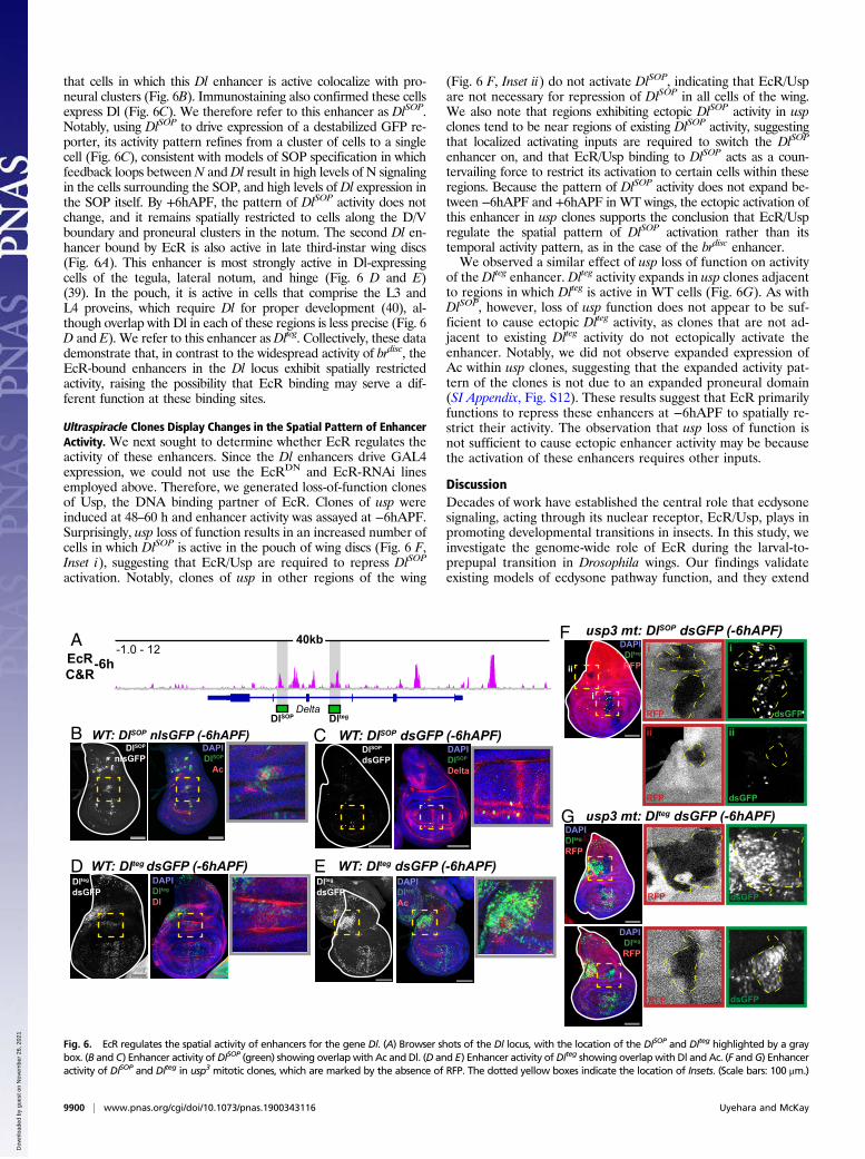
that cells in which this Dl enhancer is active colocalize with pro-neural clusters (Fig. 6B). Immunostaining also confirmed these cellsexpress Dl (Fig. 6C). We therefore refer to this enhancer as DlSOP.Notably, using DlSOP to drive expression of a destabilized GFP re-porter, its activity pattern refines from a cluster of cells to a singlecell (Fig. 6C), consistent with models of SOP specification in whichfeedback loops betweenN andDl result in high levels of N signalingin the cells surrounding the SOP, and high levels of Dl expression inthe SOP itself. By +6hAPF, the pattern of DlSOP activity does notchange, and it remains spatially restricted to cells along the D/Vboundary and proneural clusters in the notum. The second Dl en-hancer bound by EcR is also active in late third-instar wing discs(Fig. 6A). This enhancer is most strongly active in Dl-expressingcells of the tegula, lateral notum, and hinge (Fig. 6 D and E)(39). In the pouch, it is active in cells that comprise the L3 andL4 proveins, which require Dl for proper development (40), al-though overlap with Dl in each of these regions is less precise (Fig. 6D and E). We refer to this enhancer as Dlteg. Collectively, these datademonstrate that, in contrast to the widespread activity of brdisc, theEcR-bound enhancers in the Dl locus exhibit spatially restrictedactivity, raising the possibility that EcR binding may serve a dif-ferent function at these binding sites.
Ultraspiracle Clones Display Changes in the Spatial Pattern of EnhancerActivity. We next sought to determine whether EcR regulates theactivity of these enhancers. Since the Dl enhancers drive GAL4expression, we could not use the EcRDN and EcR-RNAi linesemployed above. Therefore, we generated loss-of-function clonesof Usp, the DNA binding partner of EcR. Clones of usp wereinduced at 48–60 h and enhancer activity was assayed at −6hAPF.Surprisingly, usp loss of function results in an increased number ofcells in which DlSOP is active in the pouch of wing discs (Fig. 6 F,Inset i), suggesting that EcR/Usp are required to repress DlSOP
activation. Notably, clones of usp in other regions of the wing
(Fig. 6 F, Inset ii) do not activate DlSOP, indicating that EcR/Uspare not necessary for repression of DlSOP in all cells of the wing.We also note that regions exhibiting ectopic DlSOP activity in uspclones tend to be near regions of existing DlSOP activity, suggestingthat localized activating inputs are required to switch the DlSOP
enhancer on, and that EcR/Usp binding to DlSOP acts as a coun-tervailing force to restrict its activation to certain cells within theseregions. Because the pattern of DlSOP activity does not expand be-tween −6hAPF and +6hAPF in WT wings, the ectopic activation ofthis enhancer in usp clones supports the conclusion that EcR/Uspregulate the spatial pattern of DlSOP activation rather than itstemporal activity pattern, as in the case of the brdisc enhancer.We observed a similar effect of usp loss of function on activity
of the Dlteg enhancer. Dlteg activity expands in usp clones adjacentto regions in which Dlteg is active in WT cells (Fig. 6G). As withDlSOP, however, loss of usp function does not appear to be suf-ficient to cause ectopic Dlteg activity, as clones that are not ad-jacent to existing Dlteg activity do not ectopically activate theenhancer. Notably, we did not observe expanded expression ofAc within usp clones, suggesting that the expanded activity pat-tern of the clones is not due to an expanded proneural domain(SI Appendix, Fig. S12). These results suggest that EcR primarilyfunctions to repress these enhancers at −6hAPF to spatially re-strict their activity. The observation that usp loss of function isnot sufficient to cause ectopic enhancer activity may be becausethe activation of these enhancers requires other inputs.
DiscussionDecades of work have established the central role that ecdysonesignaling, acting through its nuclear receptor, EcR/Usp, plays inpromoting developmental transitions in insects. In this study, weinvestigate the genome-wide role of EcR during the larval-to-prepupal transition in Drosophila wings. Our findings validateexisting models of ecdysone pathway function, and they extend
EcRC&R
-6h-1.0 - 12A
DeltaDlSOP Dlteg
B WT: DlSOP nlsGFP (-6hAPF)DlSOP
nlsGFPDAPIDlSOP
Ac
DAPIDlteg
Dl
Dlteg
dsGFP
WT: Dlteg dsGFP (-6hAPF)D
WT: DlSOP dsGFP (-6hAPF)CDAPIDlSOP
Delta
DlSOP
dsGFP
DAPIDlteg
Ac
WT: Dlteg dsGFP (-6hAPF)EEDlteg
dsGFP
F
i
ii
usp3 mt: DlSOP dsGFP (-6hAPF)
ii
RFP
dsGFP
i
ii
dsGFP
DAPIDlteg
RFP
G usp3 mt: Dlteg dsGFP (-6hAPF)
RFP
RFP
dsGFP
dsGFP
DAPIDlteg
RFP
DAPIDlteg
RFP
40kbi
RFP
Fig. 6. EcR regulates the spatial activity of enhancers for the gene Dl. (A) Browser shots of the Dl locus, with the location of the DlSOP and Dlteg highlighted by a graybox. (B and C) Enhancer activity ofDlSOP (green) showing overlap with Ac and Dl. (D and E) Enhancer activity ofDlteg showing overlapwith Dl and Ac. (F andG) Enhanceractivity of DlSOP and Dlteg in usp3 mitotic clones, which are marked by the absence of RFP. The dotted yellow boxes indicate the location of Insets. (Scale bars: 100 μm.)
9900 | www.pnas.org/cgi/doi/10.1073/pnas.1900343116 Uyehara and McKay
Dow
nloa
ded
by g
uest
on
Nov
embe
r 26
, 202
1
understanding of the direct role played by EcR in coordinatingdynamic gene expression programs.
The Role of EcR in Promoting Gene Expression Changes DuringDevelopmental Transitions. Our RNA-seq data reveal that EcRcontrols the larval-to-prepupal transition by activating andrepressing distinct sets of target genes. In larval wing imaginaldiscs, we find that EcR is primarily required to prevent pre-cocious activation of the prepupal gene expression program. Thisfinding is consistent with previous work that demonstrated pre-cocious differentiation of sensory neurons in the absence of ec-dysone receptor function (41). Since ecdysone titers remain lowduring most of the third larval instar, these data are also con-sistent with prior work that demonstrated that EcR functions as atranscriptional repressor in the absence of hormone (4, 31).Later in prepupal wings, we find that loss of EcR results infailure to activate the prepupal gene expression program. In-deed, many of the genes that become precociously activated inwing discs fail to reach their maximum level in prepupae. Sincerising ecdysone titers at the end of third larval instar trigger thetransition to the prepupal stage, this finding is consistent with ahormone-induced switch in EcR from a repressor to an activator(4, 31). We also find that loss of EcR results in persistent acti-vation of the larval gene expression program in prepupal wings.This finding is not clearly explained by a hormone-inducedswitch in EcR’s regulatory activity. However, it is possible thatEcR activates a downstream transcription factor, which repressesgenes involved in larval wing development. Overall, these find-ings indicate that EcR functions both as a temporal gate to en-sure accurate timing of the larval-to-prepupal transition and as atemporal switch to simultaneously shut down the preceding de-velopmental program and initiate the subsequent program. Fi-nally, it is of particular note that these genome-wide results fitremarkably well with the model of ecdysone pathway functionpredicted by Ashburner (9) 45 y ago.
Widespread Binding of EcR Across the Genome. Existing modelsdescribe EcR as functioning at the top of a transcriptional cascade,in which it binds a relatively small number of primary-responsegenes. These factors then activate downstream effectors thatmediate the physiological response to ecdysone. Consistent withthis model, attempts to assay EcR binding genome-wide in S2 cellsand Kc167 cells identified relatively few EcR binding sites. How-ever, this model does not adequately explain how ecdysone elicitsdistinct transcriptional responses from different target tissues. Ourdata reveal that EcR binds to thousands of sites genome-wide.While many genes bound by EcR have been previously identifiedas direct targets, the majority of EcR binding we observe occursnear genes with essential roles in wing development. These datasupport a model in which EcR directly mediates the response toecdysone both at the top of the hierarchy and at many of thedownstream effectors. Interestingly, comparison of our wingbinding profiles with ChIP-seq from S2 cells revealed that sharedEcR binding sites are enriched in canonical ecdysone-responsegenes, suggesting that the top tier of genes in the ecdysone hier-archy are direct targets of EcR across multiple tissues, while thedownstream effectors are direct EcR targets only in specific tis-sues. These data neatly account for the observation that parts ofthe canonical ecdysone transcriptional response are shared be-tween tissues, even as many other responses are tissue specific.Aside from assay-specific issues, it is possible that the greaternumber of EcR binding sites identified in the wing relative to celllines is due to the presence of multiple cell types in the wing thatpossess distinct EcR binding profiles. Additionally, the extent ofEcR binding may directly scale with the magnitude of the physi-ological response to ecdysone, which in wing imaginal discs isarguably greater (i.e., transformation into pupal wings) than inKc167 cells (i.e., change in cell shape) (19). In any case, it will be
important to identify the factors that contribute to EcR’s tissue-specific DNA targeting in future work. It is possible that tissue-specific transcription factors facilitate EcR binding, as suggestedby recent DNA-binding motif analysis of ecdysone-responsiveenhancers in S2 and OSC cell lines (16).
Temporally Dynamic Binding of EcR. Pulses of ecdysone mediatedistinct transcriptional responses at different times in develop-ment. Some of this temporal selectivity is mediated by the se-quential activation of transcription factors that form the core ofthe ecdysone cascade (32, 42, 43). Our data suggest that changesin EcR binding over time may also be involved. The mechanismsresponsible for these changes remain unclear. One potentialexplanation is that changes in the expression of EcR isoformscould allow recruitment to new sites in the genome. However, wedo not observe changes in protein isoform abundance, indicatingthat this is unlikely to account for changes in EcR DNA-bindingprofiles. An alternative possibility is that ecdysone titers couldinduce ligand-dependent changes in EcR structure or affectligand-dependent interactions with coregulator proteins that in-fluence EcR’s DNA binding. It is also possible that overall EcRlevels or the nuclear-to-cytoplasmic ratio of EcR changes withtime, as has been previously proposed (44). However, we do notobserve changes in EcR protein levels, and while nuclear exportof EcR could explain the global reduction in the number of EcRbinding sites, it cannot explain the appearance of new EcRbinding sites at +6hAPF. For this reason, it is notable thattemporal-selective binding sites contain lower motif content onaverage relative to temporally stable EcR binding sites. Thissuggests that temporal-selective binding may be more dependenton external factors. An intriguing possibility is that stage-specifictranscription factors activated as part of the canonical ecdysonecascade may contribute to recruitment or inhibition of EcRbinding at temporal-selective sites.
EcR Controls both Temporal and Spatial Patterns of Gene Expression.EcR has been shown to act as both a transcriptional activatorand repressor. This dual functionality confounded our attemptsto draw genome-wide correlations between EcR binding andchanges in gene expression. Therefore, we sought to examine theeffect of EcR binding on individual target enhancers. We findthat EcR regulates the temporal activity of an enhancer for theearly-response gene, br. In WT wings, the activity of this en-hancer increases between early and late third-instar stages, as doBr protein levels. Ectopic expression of a dominant-repressorisoform of EcR decreased activity of brdisc. Surprisingly, RNAiknockdown of EcR increased brdisc activity, indicating that EcR isnot required for brdisc activation. Instead, these findings indicatethat EcR represses brdisc in early third-instar wings, consistentwith our RNA-seq data which demonstrated that EcR preventsprecocious activation of the prepupal gene expression programbefore the developmental transition. It is not known what factorsactivate br or other prepupal genes.Temporal control of gene expression by EcR is expected given
its role in governing developmental transitions. However, ourexamination of EcR-bound enhancers from the Dl locus dem-onstrates that it also directly controls spatial patterns of geneexpression. Loss-of-function clones for EcR’s DNA bindingpartner Usp exhibited ectopic activation of two Dl enhancers.However, we did not detect ectopic enhancer activity in all uspmutant clones, indicating that EcR is required to restrict activity oftarget enhancers only at certain locations within the wing. Exami-nation of +6hAPF wings revealed no changes in the spatial patternof Dl enhancer activity relative to −6hAPF, indicating that ectopicenhancer activation in usp clones does not reflect incipient changesin enhancer activity. Recently, EcR binding sites were shown tooverlap with those for the Notch regulator, Hairless, supporting apotential role of EcR in regulating spatial patterns of gene expression
Uyehara and McKay PNAS | May 14, 2019 | vol. 116 | no. 20 | 9901
DEV
ELOPM
ENTA
LBIOLO
GY
Dow
nloa
ded
by g
uest
on
Nov
embe
r 26
, 202
1
(45). We conclude that EcR regulates both temporal and spatialpatterns of gene expression. Given the widespread binding of EcRacross the genome, our findings suggest that EcR plays a direct rolein temporal and spatial patterning of many genes.Hormones and other small molecules act through nuclear re-
ceptors to initiate transcriptional cascades that continue for ex-tended periods of time. For example, thyroid hormone triggersmetamorphosis in frogs and other chordates, a process that cantake weeks for completion (46). Our work raises the possibilitythat nuclear receptors play a direct role in regulating the activityof many response genes. In particular, the widespread and tem-porally dynamic binding of EcR that we observed over a short in-terval of wing development suggests that the complete repertoireof EcR targets is vastly larger than previously appreciated.
MethodsDetailed experimental materials and methods can be found in SI Appendix,Supplementary Materials and Methods.
RNA-Seq. RNA from a minimum of 60 wings was extracted as describedpreviously (38). Total RNA-seq was performed with the Ovation RNA-seqsystem. Reads were aligned to the dm3 reference genome. DESeq2 was used togenerate normalized count matrices and identify differentially expressed genes
(Padj < 0.05, absolute log2 fold change > 1). Gene clustering was performed usingk-medoids. Gene ontology terms used expressed genes as a background.
CUT&RUN. A minimum of 100 wings was dissected from w; EcRGFSTF/Df(2R)BSC889. Intact wings were permeabalized using digitonin as previously de-scribed (26). MNase was activated and digestion was performed for 45 s. Sol-uble DNA fragments were used as input for the Rubicon Thruplex 12s DNaseqkit. Fragments between 20 and 120 bp were identified and used throughout.Peaks called in a merged file that overlap a peak from at least one replicatewere used for analysis. Coverage files were z-normalized per chromosome arm.Gene ontology terms used all genes as a background. Peak clusters were cre-ated by resizing each peak to 5,000 bp, and then taking the furthest start andend coordinates of peaks that fell within each overlapping region.
Motif Analysis. De novomotifs were identified using DREME using FAIRE peaksas a background. FIMO was used to identify EcR and Usp motifs genome-wideusing position weight matrices (PWMs) from bacterial one-hybrids.
ACKNOWLEDGMENTS. We thank Peter J. Skene and Steven Henikoff forreagents and advice on the CUT&RUN protocol. Stocks obtained from theBloomington Drosophila Stock Center (NIH Grant P40OD018537) were usedin this study. C.M.U. was supported in part by NIH Grant T32GM007092. Thiswork was supported in part by Research Scholar Grant RSG-17-164-01-DDC(to D.J.M.) from the American Cancer Society, and in part by Grant R35-GM128851 (to D.J.M.) from the National Institute of General Medical Sci-ences of the NIH (https://www.nigms.nih.gov/).
1. Thummel CS (2001) Molecular mechanisms of developmental timing in C. elegans andDrosophila. Dev Cell 1:453–465.
2. Yamanaka N, Rewitz KF, O’Connor MB (2013) Ecdysone control of developmentaltransitions: Lessons from Drosophila research. Annu Rev Entomol 58:497–516.
3. Yao TP, et al. (1993) Functional ecdysone receptor is the product of EcR and Ultra-spiracle genes. Nature 366:476–479.
4. Dobens L, Rudolph K, Berger EM (1991) Ecdysterone regulatory elements function asboth transcriptional activators and repressors. Mol Cell Biol 11:1846–1853.
5. Tsai C-C, Kao H-Y, Yao T-P, McKeown M, Evans RM (1999) SMRTER, a Drosophilanuclear receptor coregulator, reveals that EcR-mediated repression is critical for de-velopment. Mol Cell 4:175–186.
6. Badenhorst P, et al. (2005) The Drosophila nucleosome remodeling factor NURF isrequired for ecdysteroid signaling and metamorphosis. Genes Dev 19:2540–2545.
7. Carbonell A, Mazo A, Serras F, Corominas M (2013) Ash2 acts as an ecdysone receptorcoactivator by stabilizing the histone methyltransferase Trr. Mol Biol Cell 24:361–372.
8. Kreher J, et al. (2017) EcR recruits dMi-2 and increases efficiency of dMi-2-mediated re-modelling to constrain transcription of hormone-regulated genes. Nat Commun 8:14806.
9. Ashburner M (1990) Puffs, genes, and hormones revisited. Cell 61:1–3.10. Talbot WS, Swyryd EA, Hogness DS (1993) Drosophila tissues with different metamorphic
responses to ecdysone express different ecdysone receptor isoforms. Cell 73:1323–1337.11. SyedMH,Mark B, Doe CQ (2017) Steroid hormone induction of temporal gene expression in
Drosophila brain neuroblasts generates neuronal and glial diversity. eLife 6:e26287.12. Hitrik A, et al. (2016) Combgap promotes ovarian niche development and chromatin
association of EcR-binding regions in BR-C. PLoS Genet 12:e1006330.13. Lehmann M, Wattler F, Korge G (1997) Two new regulatory elements controlling the
Drosophila Sgs-3 gene are potential ecdysone receptor and fork head binding sites.Mech Dev 62:15–27.
14. Fisk GJ, Thummel CS (1998) The DHR78 nuclear receptor is required for ecdysteroidsignaling during the onset of Drosophila metamorphosis. Cell 93:543–555.
15. Gauhar Z, et al. (2009) Genomic mapping of binding regions for the ecdysone re-ceptor protein complex. Genome Res 19:1006–1013.
16. Shlyueva D, et al. (2014) Hormone-responsive enhancer-activity maps reveal predictivemotifs, indirect repression, and targeting of closed chromatin. Mol Cell 54:180–192.
17. Uyehara CM, et al. (2017) Hormone-dependent control of developmental timingthrough regulation of chromatin accessibility. Genes Dev 31:862–875.
18. Fristrom D, Wilcox M, Fristrom J (1993) The distribution of PS integrins, laminin A and F-actin during key stages in Drosophila wing development. Development 117:509–523.
19. Guo Y, Flegel K, Kumar J, McKay DJ, Buttitta LA (2016) Ecdysone signaling inducestwo phases of cell cycle exit in Drosophila cells. Biol Open 5:1648–1661.
20. Uyehara CM, McKay DJ (2019) Direct and widespread role for the nuclear receptor EcR inmediating the response to ecdysone in Drosophila. Gene Expression Omnibus. Availableat https://www.ncbi.nlm.nih.gov/geo/query/acc.cgi?acc=GSE124254. Deposited December21, 2018.
21. Colombani J, et al. (2005) Antagonistic actions of ecdysone and insulins determinefinal size in Drosophila. Science 310:667–670.
22. Herboso L, et al. (2015) Ecdysone promotes growth of imaginal s through the regu-lation of Thor in D. melanogaster. Sci Rep 5:12383.
23. Mirth CK, Truman JW, Riddiford LM (2009) The ecdysone receptor controls the post-critical weight switch to nutrition-independent differentiation in Drosophila wingimaginal s. Development 136:2345–2353.
24. Stoiber M, Celniker S, Cherbas L, Brown B, Cherbas P (2016) Diverse hormone responsenetworks in 41 independent Drosophila cell lines. G3 (Bethesda) 6:683–694.
25. Nagarkar-Jaiswal S, et al. (2015) A genetic toolkit for tagging intronic MiMIC con-taining genes. eLife 4:e08469.
26. Skene PJ, Henikoff JG, Henikoff S (2018) Targeted in situ genome-wide profiling withhigh efficiency for low cell numbers. Nat Protoc 13:1006–1019.
27. D’Avino PP, Crispi S, Cherbas L, Cherbas P, Furia M (1995) The moulting hormoneecdysone is able to recognize target elements composed of direct repeats. Mol CellEndocrinol 113:1–9.
28. Cherbas L, Hu X, Zhimulev I, Belyaeva E, Cherbas P (2003) EcR isoforms in Drosophila:Testing tissue-specific requirements by targeted blockade and rescue. Development130:271–284.
29. Schubiger M, Tomita S, Sung C, Robinow S, Truman JW (2003) Isoform specific controlof gene activity in vivo by the Drosophila ecdysone receptor. Mech Dev 120:909–918.
30. Antoniewski C, Laval M, Dahan A, Lepesant JA (1994) The ecdysone response en-hancer of the Fbp1 gene of Drosophila melanogaster is a direct target for the EcR/USPnuclear receptor. Mol Cell Biol 14:4465–4474.
31. Cherbas L, Lee K, Cherbas P (1991) Identification of ecdysone response elements byanalysis of the Drosophila Eip28/29 gene. Genes Dev 5:120–131.
32. Karim FD, Guild GM, Thummel CS (1993) The Drosophila Broad-Complex plays a keyrole in controlling ecdysone-regulated gene expression at the onset of metamorphosis.Development 118:977–988.
33. Kiss I, Beaton AH, Tardiff J, Fristrom D, Fristrom JW (1988) Interactions and de-velopmental effects of mutations in the Broad-Complex of Drosophila melanogaster.Genetics 118:247–259.
34. von Kalm L, Crossgrove K, Von Seggern D, Guild GM, Beckendorf SK (1994) The Broad-Complex directly controls a tissue-specific response to the steroid hormone ecdysoneat the onset of Drosophila metamorphosis. EMBO J 13:3505–3516.
35. Brown HLD, Cherbas L, Cherbas P, Truman JW (2006) Use of time-lapse imaging anddominant negative receptors to dissect the steroid receptor control of neuronal re-modeling in Drosophila. Development 133:275–285.
36. de Celis JF, Garcia-Bellido A, Bray SJ (1996) Activation and function of Notch at thedorsal-ventral boundary of the wing imaginal. Development 122:359–369.
37. Kooh PJ, Fehon RG, Muskavitch MA (1993) Implications of dynamic patterns of Delta andNotch expression for cellular interactions during Drosophila development. Development117:493–507.
38. McKay DJ, Lieb JD (2013) A common set of DNA regulatory elements shapes Dro-sophila appendages. Dev Cell 27:306–318.
39. Huang F, Dambly-Chaudière C, Ghysen A (1991) The emergence of sense organs in thewing of Drosophila. Development 111:1087–1095.
40. Huppert SS, Jacobsen TL, MuskavitchMA (1997) Feedback regulation is central to Delta-Notchsignalling required for Drosophila wing vein morphogenesis. Development 124:3283–3291.
41. Schubiger M, Carré C, Antoniewski C, Truman JW (2005) Ligand-dependent de-repression via EcR/USP acts as a gate to coordinate the differentiation of sensoryneurons in the Drosophila wing. Development 132:5239–5248.
42. Woodard CT, Baehrecke EH, Thummel CS (1994) A molecular mechanism for the stagespecificity of the Drosophila prepupal genetic response to ecdysone. Cell 79:607–615.
43. Agawa Y, et al. (2007)Drosophila Blimp-1 is a transient transcriptional repressor that controlstiming of the ecdysone-induced developmental pathway. Mol Cell Biol 27:8739–8747.
44. Wang S, Wang J, Sun Y, Song Q, Li S (2012) PKC-mediated USP phosphorylation at Ser35modulates 20-hydroxyecdysone signaling in Drosophila. J Proteome Res 11:6187–6196.
45. Chan SKK, et al. (2017) Role of co-repressor genomic landscapes in shaping the Notchresponse. PLoS Genet 13:e1007096.
46. Wen L, Shi Y-B (2016) Regulation of growth rate and developmental timing byXenopus thyroid hormone receptor α. Dev Growth Differ 58:106–115.
9902 | www.pnas.org/cgi/doi/10.1073/pnas.1900343116 Uyehara and McKay
Dow
nloa
ded
by g
uest
on
Nov
embe
r 26
, 202
1