Embed Size (px)
Citation preview

C O N C E R N I N G A S E R U M - T H E R A P Y FOR E X P E R I - M E N T A L I N F E C T I O N W I T H DIPLOCOCCUS
I N T R A C E L L U L A R I S .
BY SIMON FLEXNER, M.D.
(From the Rocke[eIler Institute [or Medical Research, New York.)
The high mortality of epidemic meningitis and the deplorable deformities caused by it demand that incessant effort be made to dis- cover therapeutic measures which may mitigate the consequences of the disease. The epidemic through which the city of New York has recently passed, and the almost co-incident Silesian epidemic, have been scientifically fruitful in establishing more firmly the belief in the spread of the disease through immediate or mediate contact with the sick, and in tracing the common occurrence of Diplococcus intraceIlularis in the nasal and pharyngeal secretions of the sick, and the exceptional occurrence of the micro-organism in these secretions in the well who have been in contact with the sick. This mode of spread of the disease through directly and in- directly infected persons must come to exercise an important in- fluence on the hygienic measures which will be enforced hereafter to limit the dissemination of the disease. 1
1Goodwin and Sholly: The frequent occurrence of meningococci in the nasal cavities of meningitis patients and of those in direct contact with them. Journal o[ In[ectious Diseases, 19o6, Supplement No. 2, p. 21.
Flatten: Die iibertragbarre Genickstarre im Regierungsbezirk Oppeln, im Jahre 19o 5 und ihre BekS.mpfung. Klinisches Jahrbuch, 19o6, xv, 211.
Schneider: Idem im Regierungsbezirk Breslau, ibid., p. 3oo. Rieger: Idem im Kreise Brieg, ibid., p. 321. Sehmidt: Idem im Regierungsbezirk Leignitz, ibid., p. 341. Fliigge: Die im hygienischen Institut der k6nigl. Universitiit Breslau
wS.hrend der Genickstarre-epidemie im Jahre 19o 5 ausgefuhrten Untersuchungen, ibid., p. 353.
v. Lingelsheim: Die bacteriolog. Arbeiten der kgl. hyg. Station zu Beuthen, etc., ibid., p. 373.
G6ppert : Zur Kenntnis der Meningitis cerebro-spinalis-Epidemica mit besonderer Berficksichtigung des Kindersalters, ibid., p. 313.
168
on April 12, 2019jem.rupress.org Downloaded from http://doi.org/10.1084/jem.9.2.168Published Online: 14 March, 1907 | Supp Info:
Simon Flexner. 169
A less certain advance has been made in the therapeutics of epidemic meningitis. The one therapeutic measure growing out of the study of the epidemics in America and Germany which offers any hope is an antiserum for the diplococcus. It is true that the experience of the past is not favorable to the hope of achieving remarkable success by the employment of antibacterial immune
e .
sera. All indications point to the pathological effects of the diplo- coccus as being caused by endotoxic constituents; and thus far; according to many investigators, these endotoxins have failed to yield, by methods of immunization, active antisera which have proved valuable in the treatment of infectious diseases. Opinion is, however, considerably divided on this subject; 2 and in the absence of more certain methods of reaching the desired goal tests of anti- sera for the diplococcus are certainly justified. These tests can in the preliminary stages be carried out on certain animals, since the course of infection in them with the diplococcus is now fairly well understood, s
The main question which would seem to be involved in the search for an active antiserum against meningitis is whether the quantity of antibody which can be produced will suffice to neutralize such a quantity of the poison of the diplococcus as to influence the result of the infection. In fact, the problem may not be so simple, or, indeed, so hopeless, as this proposition indicates. It is, of course, important that neutralization of the poison should if pos- sible be secured, but the effect of the restraint of growth and multi- plication of the diplococcus may, at some periods of the disease, be of greater significance than the neutralization of free endotoxin. Fortunately, many agents, some of them quite indifferent, are able to affect the power of multiplication in the body of the diplococcus. It has been shown, indeed, that serum in the fresh state and after
Meyer: Bericht fiber rhinolog. Beobaehtung bei der Genickstarre-epidemie, 19o5, ibid., p. 427 .
Westenhoeffer: Pathologisch-anatomische Ergebnisse der oberschlesischen Genickstarre-epidemie ~¢on 19o5, ibid., p. 447.
Jehle: Entstehung der Genickstarre-epidemie, Wien. klin. Woch., 19o5, xix, 25. 2 Besredka, A n n a l e s FIns t i tu t Pas teur , 19o6, xx, 4. SAttention is directed to the two previous papers of this series published
in this number of the Journal.

170 Experimental Infection with Diplococcus.
heating to 6o ° C., preserves the power todes t roy in test-tubes large numbers of the diplococcus, and sterile fluid inflammatory exudates possess this power in even greater degree. An antiserum, there- fore, even though it contain relatively small amounts of antibodies, as indicated by neutralization experiments, may be effective beyond this calculated value by restraining the multiplication of the diplo- cocci, possibly by reducing outright their number, and by support- ing the power of resistance normally at the dispdsal of the body.
The conditions are made theoretically less discouraging, perhaps, because-the main pathological lesions are limited to the cavity of the cerebro-spinal axis. They can, therefore, be brought directly under the influence of the antisera by injecting the latter into the spinal canal. A large advantage is gained by this circumstance. It is, on the other hand, discouraging to reflect that in monkeys infected with the diplococcus, severe cortical lesions already exist at the end of ten or twelve hours. The question arises whether these deeper lesions tend to appear as early in the human infections. In respect to this question it should be stated that observation is against the occurrence of any such development of the diplococcus in the early stages of the human disease as is represented by the prodigious number of diplococci required to be injected into monkeys to produce the rapidly lethal effect with which the cortical lesions are associated. It is worthy of note that the more slowly developed lesions in the monkey remain more superficial, agreeing in this respect with the more common lesions present in fatal cases of the human infection. Hence, some encouragement may be taken from the power of the antiserum to influence favorably the course of meningitis in the monkey, although it has been injected as late as six hours after the inoculation.
I'f we are at all permitted to apply test-tube experiments to what may happen in the body, it would not be remarkable if the normal serum of animals, and perhaps of human beings, proved to be beneficial to a degree when brought into direct relation with the focus of development of the diplococcus. Aic first sight, judg- ing from test-tube experiments, it would appear as if the exudate, called out by the inflammation, should suffice to destroy the diplo- cocci; this manifestly does not happen in many cases. Indeed, it

Simon Flexner. 171
is found that incubation outside the body will even increase the number of diplococci in the inflammatory fluid withdrawn from the spinal canal. It is safe to assume, therefore, that the exudate withdrawn has been exhausted of its power to destroy the diplo- coccus. It is quite possible that the introduction of fresh serum, of the same species of animal, may be helpful by bringing quickly into contact with the diplococci a quantity of actively destructive serum. The results of some of my experiments show that normal serum reduces appreciably the toxic effect of given doses of the diplococcus.
In experiments upon the monkey there is a definite low limit, beyond which it is not safe to go, for injection of fluids into th~ spinal canal. The species which I studied contain a small amount only of free spinal fluid. I f one attempts to inject several cubic centimeters of fluid, symptoms of pressure may develop. In this respect the monkey is far less satisfactory to treat by intraspinal injections than are human beings.
I am far from having any conviction that cerebro-spinal menin- gitis in man can be influenced favorably by injections of immune sera into the spinal canal, or elsewhere in the body. The experi- ments to be described merely show that guinea-pigs and monkeys, in which the conditions of infection can be controlled, can be saved from the otherwise fatal effects of the diplococcus by the use of antisera, and to a less extent by the use of normal sera and other fluids. A preliminary note on this subject has already been made. ~ The protocols show that the experiments on immunity were begun during the spring of I9o 5. While the work was in progress two papers on the same subject appeared in Germany. 5 The use of monkeys for testing the antisera by direct injections into the in- fected and inflamed cerebro-spinal canal has not been made by the other investigators whose experimental studies were confined to guinea-pigs. Jochmann injected an antiserum prepared in the horse into the spinal canal of several human subjects of epidemic menin- gitis. The number of cases was too few to permit any conclusion of the value of the injections; but they showed that the injection of
4]our. of Amer. Med. Assoc., 19o6, xlvii, 560. 5Kolle and Wassermann, Deutsch. Med. [/Vochenschrift, 19o6, xxxii, I6.
Jochmann, ibid., p. 20.

172 Experimental Infection with Diplococcus.
horse's serum into the inflamed canal is not attended with special danger.
My first experiments on guinea-pigs were made with goat's sera. A female goat had been injected twice with cultures from several sources (12) of the diplococcus within a period of two weeks. The injections were made subcutaneously and gave rise to tumefaction which soon disappeared. After the second injection the goat aborted. The first bleeding was made two weeks after the second injection. As the table shows the serum at this time had little or no immunizing power. The experiment was designed to test "the effect of an injection ( I ) previous to the injection of the dip- lococci, and (2) after the inoculation of the diplococci. The serum in the first instance was injected at 5 P. M. the day before, and in the second instance two hours after the inoculation with the diplo- coccus. All the injections, except bouillon in one pig, were intra- peritoneal. The emulsion of the diplococcus was injected at I I A. M., November 29, I9O 5.
I5 o t5I I52 *53 154 I55 156
W e i g h t Series in Grams N o . _ _ - - -
144 I89 x45 189 I46 / I82 147 I97 I48 197 I49 22o
199 I9O I85 192 I9O 19o 2IO
P r o t e c t i v e S u b s t a n c e I n j e c t e d .
" I m m u n e " serum o o3 c.c. 5 P .M. Nov. 28 ,, 6, 0 . 0 4 C.C. ~' '~ ~
~ " 0.0 5 C.C. 6~ ~, ~
" N o r m a l " " 0.05 c.c. " " "
Boui l lonintraper . I.O c.c. " " "
" subcut. I . O C.C. " " "
" I m m u n e " serum I . o c.c. I P .M. Nov. 29 ~ ~ 2 . 0 C.C. ~ ~ ~
" N o r m a l " " 1.o c.c. " " " '~ '~ 2.0 C.C. ~ ~ ~
Nothing : control Idem.
R e s u l t .
Died 9 A . M . Nov. 3 o. Died 8 A.M. Dec. 2. Survived. Survived. Lost weight .
" We igh t 12/x t 182 grams.
Died morning , Dec. 3. Survived. Lost weight .
Died 5 P .M. Nov. 3 ° . Died in night , Nov. 29 . Died morning, Dec. I . Died in night, Nov. 29.
The preceding experiment is of interest in showing the irregular action of the serum injections, and especially, as bringing out the fact that such an indifferent substance as bouillon can, if it is injected in advance of the diplococcus, impart power of successful resistance to the guinea-pig. It developed subsequently that the bouillon need not be injected into the peritoneal cavity to achieve this effect; one cubic centimeter injected subcutaneously in pigs of 225 to 25o gram weight, the day before inoculation, frequently

Simon Flexner. 173
saves the animals. Using the goat serum of low protective value, the fact was determined that the protecting power of the serum has a definite limit (about o.o 5 c.c. for pigs of 25o grams weight) under the most favorable conditions, namely, intraperitoneal injection the day before the infection. By using larger quantities (up to I.O c.c.) of serum simultaneously with the injection of the diplococcus the pigs can also be saved; subcutaneous injections of the serum (I.O c.c.) in advance are effective. The smallest number of suc- cessful results is obtained in pigs in which the serum injections follow, after one or more hours, the infection. Several experi- ments were carried out in order to determine the fate of the diplo- coccus in the peritoneal cavity in the "pro tec ted" and "unpro- tected" animals. The plan was to withdraw fluid, after a suitable interval, by means of capillary tubes. The results were not wholly uniform, but in the main showed more rapid disappearance of the
Guinea-pigs weighing 230 to 260 grams, received o.I c.e. " i m m u n e " goat 's s e rum intraperitoneally and subcutaneously, 1.0 c.e. bouillon intraperi toneally and subcutaneously, followed the next day by an emulsion of the diplococeus injected into the peritoneal cavity.
Series No. I How Injected. Examination During Life. Autopsy Findings in Peritoneum.
119 Goat's serum intraperi- 4 P.M. Many leucocytes Survived. I toneal; e m u l s i o n containing a few diplo-
cocci at I 1:3 ° A.M. cocci ; no extracellular diplococci.
I2o ! Goat's serum subcutan- 4 P.M. Enormous hum- Died during the night. 2 eous ; emulsion cocci ber of diplococci ; very c.c. fluid exudate ; more at 1I:3o A.M. few leucocytes overlaid pus present than usual ;
with diplococci, many leucocytes includ- ing diplococci.
121 Bouillon i n t r a p e r i - 4 P.M. Diplococci more Died during the night. 3 toneal; e m u l s i o n numerous than in 12o; c.c. fluid exudate; no cocci at I 1:3o A.M, no leucocytes, leucocytes ; many diplo-
cocci.
122 Bouillon subcutaneous; 4 P.M. Many leucocytes; Survived. emulsion cocci at some intracellular diplo- 1I:3O A.M. cocci ; very few extra-
cellular ones.
125 Control ; e m u l s i o n cocci at I I :30 A.M.
4 P.M. Enormous num- ber of diplococci; al- most no leucocytes.
Died during the night. Usual p. m. appearances ; very large number of diplococci free; almost no leucocytes. The omen- tum contains some leuco- cytes including cocci.
174 Experimental Infection with Diplococcus.
diplococcus from the peritoneum of the treated as compared with the untreated pigs. Exudation of cells was more abundant in the former animals.
The injections of the goat were carried on subsequently, during and after lactation, until the end of February, 19o6. Blood was withdrawn from time to time and the serum tested for its pro- tective and therapeutic value. The results were never uniform, but the general indication was that the protective properties were in- creased measurably. The animal fell ill on March 15, 19o6, and was bled to death. The serum obtained from this bleeding was used for many subsequent experiments. It was found by simul- taneous injection to protect small pigs (19o to 200 grams) against a twelve hour fatal dose of the diplococcus in quantities varying from o. 5 to o.oi c.c., but not regularly. On the whole, as the next experiment given in detail will tend to show, the serum had acquired protective and therapeutic properties.
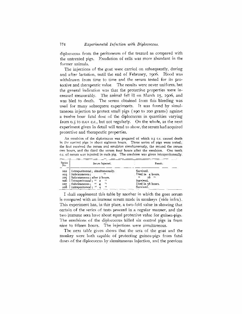
An emulsion of the diplocoecus was prepared of which 0.5 c.e. caused death in the control pigs in about eighteen hours. Th ree series of pigs were t es ted ; the first received the se rum and emulsion simultaneously, the seeond the se rum two hours, and the third the serum four hours a f te r the emulsion. One tenth c.c. o f se rum was injected in each pig. T h e emulsion was given intraperitoneally.
SerieSNo. Intraperitoneal Serum Injected.
102 Subcuttan;o°U: ;; ; simultane°uslY'after 2 hours.
Result.
Survived. to 3 Died in 4 hours. 10 5 " I6 " IO6 I Intraperitoneal ; " 2 " ' Survived. x°7 i Subcutaneous; " 4 " Died in 5 6 hours. xo8 Intraperitoneal ; " 4 " Survived.
I shall supplement this table by another in which the goat serum is compared with an immune serum made in monkeys (vide infra) . This experiment has, in this place, a two-fold value in showing that certain of the series of tests proceed in a regular manner, and the two immune sera have about equal protective value for guinea-pigs. The emulsions of the diplococeus killed six control pigs in from nine to fifteen hours. The injections were simultaneous.
The next table given shows that the sera of the goat and the monkey were both capable of protecting guinea-pigs from fatal doses of the diplococcus by simultaneous injection, and the previous
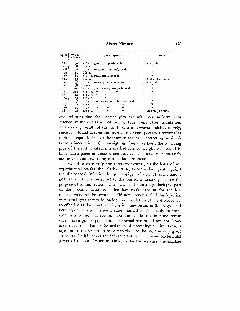
Simon Flexner. 175
Se r i e s W e i g h t N o . in G r a m s .
I O 6 I 9 0 O. I c . c .
xo 7 158 Idem. Io8 I8o o.I c.c. to9 I85 Idem. I I O 1 8 5 O . i e . c ,
I t 2 I 7 5 l d e m .
xt4 I95 o.I c.c. XI 7 I78 Idem. I 7 5 2 1 0 O . I C.C,
I78 205 0.2 c.c. z8I XTO 0. 5 c.c. I 8 8 I 8 5 1 . 0 C.C. I82 205 o.I c.c. 1 8 3 1 8 2 0 . 2 C.C.
1 8 6 1 7 5 0 . 5 C.C. 187 I72 I.O C.c.
Serum Injected.
goat, interperitoneal.
monkey, intraperitoneal.
goat, subcutaneous.
monkey, subcutaneous.
goat serum, intraperitoneal.
6C ~t 6t
monkey serum, intraperitoneal.
R e s u l t .
Survived. 6t g6
Died in I2 hours. Survived.
t t l~ ~c
6~
i~
t l
i~
Died in 3 ° hours.
one indicates that the infected pigs can with less uniformity be rescued at the expiration of two to four hours after inoculation. The striking results of the last table are, however, relative merely, since it is found that certain normal goat sera possess a power that it almost equal to that of the immune serum in protecting by simul- taneous inoculation. On reweighing, four days later, the surviving pigs of the last tabulation a marked loss of weight was found to have taken place in those which received the sera subcutaneously and not in those receiving it into the peritoneum.
It would be extremely hazardous to express, on the basis of my experimental results, the relative value, as protective agents against the diplococcal infection in guinea-pigs, of normal and immune goat sera. I was restricted to the use of a female goat for the purpose of immunization, which was, unfortunately, during a part of the process, lactating. This fact could account for the low relative value of the serum. I did not, however, find the injection of normal goat serum following the inoculation of the diplococeus, so effective as the injection of the immune serum in this way. But here again, I was, I should state, limited in this study to three specimens of normal serum. On the whole, the immune serum saved more guinea-pigs than the normal serum. I am not, how- ever, convinced that in the instances of preceding or simultaneous injection of the serum, in respect to the inoculation, any very great stress can be laid upon the inherent antitoxic, or even bactericidal power of the specific serum, since, in the former case, the exudam

176 Experimental Infection with Diplococcus.
caused to be poured in the peritoneal cavity has itself, in vitro, the power of suppressing large numbers of diplococci, and, in the latter, the sera possess this power only in less degree than the exu- date. If, however, it can be shown that the immune sera are more effective as therapeutic agents than normal sera, and by simul- taneous and preceding injections, also, then it will have to be con- ceded that they contain some useful elements absent from the normal serum or present in it in less amount.
In the course of these experiments the effort was made to pro- duce an immune serum in the horse. For this purpose, recent cultures were injected by Dr. Jobling, first subcutaneously and later intravenously. The doses had to be carefully chosen on account of the high sensibility of the horse to the diplococcus. The sub- cutaneous injections produced local swellings which frequently softened and discharged externally. After a period, intravenous injections of autolysates were begun; but the alarming symptoms which followed almost immediately the injection of a few cubic centimeters of this fluid caused a return to the use of cultures and autolysates by the subcutaneous method. After about five months of intermittent injection the serum was collected and compared for protective value on small guinea-pigs. I employed as controls several samples of normal horse serum kindly supplied by Dr. Park of the Board of Health'. I shall give a few typical experi- ments from which it would appear that on the whole the serum of the treated horse has greater protective value than the serum of normal horses.
Two strains of the diplococcus of moderate virulence were em- ployed in the tests. The suspensions were made in the morning, control pigs being inoculated with them immediately. About four hours later, the rectal temperatures indicated the probably fatal doses. It was subsequently found that o.I c.c. of the suspensions caused death in less than twelve hours and represented, for sus- ceptible pigs of about two hundred grams, two or three fatal doses. The inoculation of the pigs for the experiments on immunity was made in the afternoon of the same day. This procedure is ren- dered necessary by the rapid deterioration of the suspensions in salt solution and Ringer's fluid which sometimes takes place at
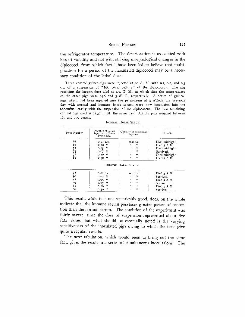
Simon Flexner. 177
the refrigerator temperature. The deterioration is associated with loss of viability and not with striking morphological changes in the diplococci, from which fact I have been led to believe that multi- plication for a period of the inoculated diplococci may be a neces- sary condition of the lethal dose.
Three control guinea-pigs were injected at io A. M. with o.I, 0.2, and 0.3 c.c. of a suspension of " Mt. Sinai cul ture" of the diplococcus. The pig receiving the largest dose died at 4.30 P. M., at which time the temperatures of the other pigs were 34.6 and 34.8 ° C., respectively. A series of guinea- pigs which had been injected into the peritoneum at 4 o'clock the previous day with normal and immune horse serum, were now inoculated into the abdominal cavity with the suspension of the diplococcus. The two remaining control pigs died at II.3O P. M. the same day. All the pigs weighed between 165 and 19o grams.
NORMAL HORSE SERUM.
Quantity of Serum Quantity of Suspension Series Number Injected 2 4 Hours Injected. Result.
Previously.
68 69 72 75 78 82
001 C.C. 0.02 ~t 0 .05 t~ 0.0 7 " 0 IO t t
0 . 3 0 -
0,2 C.C.
4 ( (Ig
Died midnight. Died 3 A.M. Died midnight. Survived. Died midnight. Died I A.M.
IMMUNE HORSE SERUM.
47 [ O.OI C.C. 50 0.02 ~
58 0.05 " 59 I 0 . 0 7 " 61 ~ O. I0 " 66 ! 0.30 "
0.2 C.C. 4 6 ~ (
Died 3 A.M. Survived. Died 9 A.M. Survived. Died 3 A.M. Survived.
This result, while it is not remarkably good, does, on the whole indicate that the immune serum possesses greater power of protec- tion than the normal serum. The condition of the experiment was fairly severe, since the dose of suspension represented about five fatal doses; but what should be especially noted is the varying sensitiveness of the inoculated pigs owing to which the tests give quite irregular results.
The next tabulation, which would seem to bring out the same fact, gives the result in a series of simultaneous inoculations. The

178 Experimental Infection with Diplococcus.
mixtures of suspension and serum were permitted to stand at the warm room temperature for half an hour before injectionfi
~N]'ORMAL S E R U M N O . I .
Ser ies N u m b e r .
5 I 54 46
Serum In jec ted .
O.OI C.C.
0 . 0 2 "
O. IO ~
Suspens ion Injec ted .
O , I C,C.
0 . 2 ~
0 . 5 ~
Resu l t .
Died after 8 hours.
Survived.
N O R M A L S E R U M N O . 2.
49 o.oI c.c. o.I c c. Survived. 43 o.o2 " o.2 " "
38 o . Io " o. 5 " Died after 8 hours.
I M M U N E S E R U M .
4 I
37 34 44 42 39
O.OI C.C.
O OI "
0 . 0 2 ~
0 . 0 2 ~
O . I O ~c
O. IO ~¢
O . I C.C.
O . I ~
0 . 2 ~
0 . 2 ~¢
0 . 5 ~
0 . 5 ~
Survived.
Two other kinds of antisera were prepared and tested upon guinea-pigs. The first was made by injecting large rabbits with the peritoneal exudate of guinea-pigs which succumbed to intra- peritoneal inoculation with the diplococcus. The exudates were toluolized, freed from cells, the toluol was removed by evaporation at a low temperature, and injected into rabbits. The antisera obtained from the rabbit exercised a degree of protection which can be expressed as follows: simultaneous injections of the diplo- coccus and the antiserum into the peritoneal cavity tend to give protection; separate simultaneous injections, diplococcus into the peritoneum and serum under the skin, are effective in proportion to the quantity injected. Small doses of the serum (o.I c.c.) do not protect, but they delay the lethal effect; larger doses (o. 5 c.c.) pre- vent the lethal effect. Dosage of rabbit serum proved to be im- portant; too little failed to protect, and too much (o. 5 c.c. into the peritoneal cavity) prejudices the result by reason of its own tox- icity. The pigs which received the culture and this dose of serum died soon after the controls. The second was an homologous serum
s i t is my in ten t ion to make a l a t e r report , when the immun iza t i on shal l
have gone far ther , upon the effects of the s e rum of the immun ized horse as
r ega rds its power of pro tec t ion for gu inea -p igs and for monkeys .

Simon Flexner. 179
yielded by large guinea-pigs which were injected at intervals for several months with cultures of the diplococcus, the peritoneal exu- date from other guinea-pigs, and the autolysate, which proved not to have greater protective value than normal guinea-pig's serum.
I f the result of these attempts to produce an antiserum for Diplo- coccus intracellularis, which should be effective in the experimental infection with the diplococcus in the guinea-pig are reviewed, they cannot be held to be particularly promising. Under the severe con- ditions of the experiments, the most that can be said is that various agents--bouillon, normal sera, immune sera--ean at times affect favorably the course of the experimental infection; the lead in respect to this influence being taken rather by the immune sera. It is to be recalled that in small guinea-pigs the experimental infec- tion is rapidly fatal; that the prostration of the pigs develops very quickly, and the animals are often moribund in six to eight hours after inoculation. To influence very favorably and systematically a pathological process which progresses as rapidly as this, would be, perhaps, an achievement. As the experiments show, this can be done, although not wholly in this degree, by appropriate dosage of certain antisera.
The next experiments to be reported relate to monkeys inoculated with the diploeoceus and treated with anti-diplococcus serum made in the monkey. Two large monkeys (Macacus nemestrinus) were immunized, for the production of an homologous serum, by inject- ing them subcutaneously with exudates from the peritoneal cavity of guinea-pigs succumbing to diplococcus infection, and with emul- sions of the diplococcus. The injections were made at intervals for a period of nine months, after which the animals were bled to death, and the sera tested. Before giving the protocols of these experiments, the chief facts of a much earlier experiment to in- fluence the course of meningitis in a monkey by means of the anti- serum of the goat should be given.
December 2, I905, each of two spider monkeys (Atales ater) was inoculated with one agar slant culture of the diplococcus. Fluid flowed from the needles before injection. 3 P. M., control sick; mate to this perhaps not quite so sick. Into the spinal canal of the latter 2 c.c. of goat's immune serum were injected. The immediate effect was alarming: animal relaxed, heart's action tumultuous,

180 Experimental Infection with Diplococcus.
respiration sighing. The symptoms passed off in IO to 15 minutes. L . p . be- fore injection of serum showed in each many diplococci; no leucocytes. 9 P. M., control in stupor from which he could be aroused; easily handled. Responded to introduction of needle for 1. p., no fluid obtained; point of needle carried a small amount of exudate which showed many leucocytes and a small number of intracellular cocci. Rectal temperature 4 °0 C. Serum treated, less stupid than control ; could be handled alone. No fluid obtained by I. p. The small amount of exudate on needle showed leucocytes some of which contained diplococci. Rec- tal temperature 360 C. December 3, 6 A. M., both still very sick; 8 A, M., serum treated animal very much depressed; IO A. M., serum treated animal dead; control brighter. The latter animal finally recovered. Autopsy on serum treated monkey. The spinal canal showed h~emorrhagic imbibition of the mem- branes of the lower third of the cord. The pia-arachnoid of the cord was infil- trated with gelatinous-cedematous exuda te . Small h~emorrhages beset the pia- arachnoid of the cortex, and the meninges of the brain were infiltrated with an e.xudate similar to that of the spinal cord. The ventricles contained turbid fluid in small amount. Microscopical examination of sections of the brain and cord show the exudate to be moderate in amount, and to be thicker over the convexity than over the base of the brain. The h~emorrhages in the membranes of the spinal cord are large, and of the brain small. The most str iking lesion is an acute en- darteritis which effects all sized branches of the arteries of the brain and cord. Very few diplococci can be found.
The symptoms in this animal and the lesions found at autopsy were taken to indicate that goat serum cannot be injected with impunity into the inflamed spinal canal of monkeys.
Preliminary to the tests of the antisera prepared in the two mon- keys, the lethal dose of the diplococcus had to be established. A recently isolated culture (Mt. Sinai 596), which had proven viru- lent for guinea-pigs, was lated ('I) with 0.5 c.c., culture. Brief histories
chosen. Twocontrol monkeys were inocu- (2) with I.O c.c. of a suspension of the follow :
Control No. I. Macacus rhesus. June 27, 19o6, II A. M., given 0.5 c.c. suspension. 6 P. M., very sick; June 28, 9 A. M., br ighter ; 12 M,, 1. p. small amount of thin fluid obtained; animal very weak. June 29, 8 A. M., mor ibund; chloroformed at I P . M . Autopsy: The membranes of the cord and brain were pale; little visible exudate. C. s, show leucocytes in small numbers over cord and bra in ; and a few intracellular diplococci. Sections of the tissues confirm these findings; the inflammatory exudate is small in quanti ty; no marked lesions of the nervous tissue itself are to be seen.
Control No. 2. Macacus rhesus. June 28, 12 M., I.O c.e. of same emulsion injected (in refrigerator over n ight ) . 6 P. M., monkey sick; 9 P. M., very sick; June 29, 9 A. M., died. Autopsy: The meninges were injected and con- tained small h~emorrhages. The fluid in the pia of the cord was increased, and turbid. The meninges of the brain and the ventricles contained similar fluid in excess; the basal meninges the largest quantity. C. s. show leucocytes and

Simon Flexner. 181
diplococi throughout the membranes. The spinal membranes contain the largest number of free diplococci. Cultures positive. Sections of the tissues show the lesions of an acute infla~nmation of the meninges and of the super- ficial portion of the cortex of the brain.
In making the serum tests, the larger dose of emulsion was always employed. Brief protocols of the experiments follow:
Exper iment I, July 3, I9O6: Medium sized Macacus rhesus given at II A. M. I.O c.c. emulsion of diplococcus " 5 9 6 " together with I.O c.e. of monkey anti- serum No. I. Flui4 flowed from the needle before injection. July 4, 9 A. M., monkey appeared normal ; active; on perch. L.p . yielded several drops of faintly turbid fluid, which showed on cover-slips free cocci, and leucocytes, some of which contained diploeocci in moderate numbers staining feebly. Cultures from the fluid negative. July 5, II A. M., monkey apparently well. Lumbar puncture gave a small amount of clear fluid, which contained neither leucocytes nor cocci. Cultures negative. January IO, I9O7, the monkey remained well.
Exper iment 2, July 5, I9O6: Medium-sized Macacus rhesus given I.O e.c. suspension of culture " 5 9 6 " at 11:45 A. M. Fluid flowed from needle before injection. At 2 P. M., the monkey was sick. L. p. gave a small quantity of ra ther thick, opaque fluid. One cubic centimeter of immune serum (monkey No. 2) injected rapidly. Immediately at conclusion of injection pressure symptoms of an a larming character developed. The animal was prostrated for two hours, af ter which it slowly got better. At 6 P. M., it responded to dis- turbance. At 8 A. M., next day, the monkey was up and appeared well. I t remained so subsequently ( January IO, 19o7). The cover-slips from the lumbar puncture showed many polymorphonuclear leucocytes and a small number of lymphocytes, many extraeellular and a few intracellular diplococci.
Before the next experiment with the serum was made, the culture " 596" was again tested for virulence.
A medium-sized Macacus rhesus was given i.o c.c. emulsion at IO:3O A. M., July II. IO P. M., animal sick; on bottom of cage, July 12, 9 A. M., 1. p., small quantity of a thin white exudate obtained. C. s. many leucocytes and few diplococci. Died 12:3o P. M. Lived about thir ty-eight hours. The lesions found at autopsy were characterist ic; exudate existed over the cord and brain, the base of the latter being chiefly affected. The cortical vessels were injected. C. s. showed cocci in the inflammatory exudate of the cord and base of the brain, and fewer in the exudate of the convex meninges and the ventricles. Sections of the brain and cord show marked inflammatory lesions of the usual character. Diplococei are abundant in the exudate.
Experiment 3, July 13, 19o6: Moderately large Macacus rhesus given at II :15 A. M. I.O c.c. emulsion of Coccus "596." Although no fluid was obtained, the canal was certainly entered; 4 P. M., animal sick; 5 P. M., depressed, but still sat up. L. p. gave a small quantity of turbid fluid, but during the operation the animal collapsed; I.O c.c. of immune serum from monkey No. 2 injected slowly. The monkey was watched until IO P. M.; no progress of the disease. July 14, IO A. M., animal appeared well. No future symptoms developed up to January lO, 19o7. The fluid obtained by 1. p. showed many leucocytes with an occasional intracellular coccus, and extracellular cocci.
182 Experimental Infection with Diplococcus.
Experiment 4, July 27, I906: At 8:45 A. M., a medium-sized Macacus rhesus was given one full agar slant culture "61o," 18 hours old, suspended in salt solution, into spinal canal Fluid obtained before injection. At It A. M., 5 c.e. of antiserum from monkey No. 2, were injected into the skin of the thigh. Before the serum injection the animal was sick; it lay half down on the bottom of the cage. No immediate effect followed from the injection, but the symptoms did not progress. The next day the animal appeared well. L.p. was unsuccessful after the injection until the next day, at which time clear fluid, containing neither cocci nor leucocytes, was obtained. The control for this monkey was a much smaller and weaker monkey of the same species, which succumbed in eight hours.
I do not regard this experiment as entirely free from doubt, but as I was unable to obtain more monkeys at this time, the experiment could not be re- peated then.
T h e series of e x p e r i m e n t s was up to this po i n t successful , a n d
they ind ica ted tha t an a n t i s e r u m to the diplococcus could p r e ve n t
the d e v e l o p m e n t of severe s y m p t o m s f r o m f o l l o w i n g the i n j e c t i on
of the cu l tures of the diplococcus in to the sp ina l canal , a nd cause
a r r e s t o f the s y m p t o m s which had a l r eady set in. T h i s series of
tests is, of course, incomple te w i t h o u t c o r r e s p o n d i n g e x p e r i m e n t s
wi th n o r m a l s e r u m wi th which they m a y be compared . T h e la t te r
will fol low. Bu t be fo re c i t ing t h e m I wish to record a f a i lu re
u n d e r cond i t ions which, in v iew of the f o r e g o i n g resul ts , was
who l ly unexpec ted .
Experiment 5, July 20, I9O6: Medium-sized Macacus rhesus given at 12 M. I.O c.c. of emulsion of Coccus "596 "; 3 P. M. I.O c.c. of antiserum injected intra- spinally. This monkey was sick when given the serum and the symptoms pro- gressed fairly rapidly. At II P, M. the animal was much prostrated and sat in the cage with head depressed. It died about 7 A. M., July 21. The autopsy showed a very unusual amount of exudation in the membranes of the cord. Cover-slip preparations showed a purulent exudate with large numbers of diplo- cocci, all within polymorphonuelear leucocytes. The exudate in the meninges of the brain and cord showed the diplococci in the same condition of complete phagocytosis, although the number of leucocytes and diplococci was smaller. The fluid withdrawn by lumbar puncture before the serum injection contained large numbers of diplococci and very few leucocytes; only an occasional leucocyte contained diplococci. Cultures made at the autopsy from the meninges of the cord, medulla and cortex, and from the lateral ventricle were positive; those from the heart and bone marrow of the femur were negative. Examination of sections of the brain and spinal cord bear out the macroscopic appearances. The exudate is remarkably thick over the entire nervous system, and is composed exclusively, or nearly so, of leucocytes. The lateral ventricle is shown to have been dilated and to contain pus cells. The brain tissue has escaped invasion with leucocytes, and the intracortical blood vessels are free from thrombi and do not show the perivascular infiltration with pus cells which is commonly present. The spinal cord at the level of the injection shows a superficial invasion with leuco-
Simon Flexner. 183
cytes, but the higher levels do not show this condition. The dura is, in the former locality, infiltrated with pus cells.
The control for this experiment was a smaller rhesus monkey. The injection was made July 19, at 2 P. M., and as symptoms had failed to develop from the small dose given, a second injection of one third culture was made at 6 P. M. At 9 P. M., animal sick; lying down, but when disturbed rose and looked dis- trait. July 2o, 7:3o A. M., lay on bottom of cage, but on being disturbed rose and displayed marked nystagmus; resmned recumbent positidn. Depression increased during the morning; 12 M., died. The autopsy showed a general thin exudate in the meninges, marked chiefly over the medulla. Cover-slips showed a remarkably large number of cocci which, while chiefly within leucocytes, were abundant outside. In no other experiment was so large a number of micro- organisms seen. A suspicious circumstance was found in the appearance of short chain-like groups of cocci, 4 to 6 members long. The cocci were Gram-negative and in size like the diplococcus. Attempt at cultivation failed.
In summar iz ing these experiments, it may be said that by the
employment of an homologous anti-diplococcus serum several mon-
keys were saved f rom death due to experimental infection with
Diplococcus intracellularis. The conditions of the experiments were
such that the inoculated monkeys could, by simultaneous injec-
tion of sermn and culture be prevented f rom developing severe
symptoms, a l though the diplococci persisted for a period in the
spinal canal, and by separate injection of the culture, and six hours
later of the serum, the already severely ill monkey could, apparently,
be saved f rom certain death. The experiment in which the serum
was used successfully by subcutaneous injection cannot be inter- preted without suitable repetitions.
The tests with normal monkey serum to serve as controls for
the above experiments were made so as to br ing out two sets of
facts. In the first place, the value of simultaneous injection of
normal monkey serum and a quanti ty of culture which would cause
death in the control animal within twenty- four hours, was studied.
And in the next place, the value of the normal serum was studied
in monkeys in which the dose of the culture was on the border line
- - t h a t is, of such a size that certain monkeys survived and others
succumbed af ter a greater period than twen ty- four hours. As re-
gards the second series of tests, it may be said that it appeared as
if the injection of a mixture of the normal serum and the culture
led, in certain cases, to the survival of the monkey af ter a period of illness which was sometimes severe. The first symptoms ap-

181, Experimental Infection with Diplococcus.
peared very soon- -wi th in one or two h o u r s - - a n d grew in intensity
for five or six hours, af ter which they receded. I am, therefore, .
inclined to attribute to normal serum employed in this way, a cer-
tain.definite protective value.
The results in the first class of experiments were different. I
found that the normal serum not only failed to save the inoculated
monkeys, but the injection of the mixture of culture and serum
might even hasten the fatal outcome. I wish to speak with some
reservations on this point, for I was great ly hampered in this entire
series of tests by great difficulty in obtaining at the time a suitable
number of monkeys for the experiments. The study must, indeed,
be carried much fur ther before a final answer can be .g iven as to
the availability of a serum therapy for this experimental diplo-
coccal infection in monkeys. There follow two brief protocols
relating to the use of normal serum with a certainly fatal dose of
the diplococcus. Two animals of the same species--Macacus rhesus - - o f equal size were employed.
Control Monkey. _At 12 o'clock noon, one cubic centimeter of a suspension of the diplococeus fatal to guinea-pigs was injected into the spinal canal. Fluid flowed from the needle before injection. At 5 P. M., the monkey was sick; 9 P. M., lay on bottom of cage, but could be roused. Next morning comatose; died at 2 P. M. Survived the inoculation 26 hours. The lesions found at autopsy were characteristic. Cover-slips showed that the diplococci had to a large extent disappeared. The cultures were negative,
Serumi~ed Monkey. At 12 o'clock noon, one cubic centimeter Of the same emulsion used in the previous experiment (in refrigerator over night) mixed with one cubic centimeter of normal monkey serum and placed at 37 ° C. for half an hour, was injected into the spinal canal. Fluid was obtained before the injec- tion. No immediate effects were noted following the injection. The monkey was already sick at 3:30 P. M., the symptoms increasing with great rapidity. Death took place at 8 A. M. the next morning. Survived 22 hours. The autopsy showed vivid injection of the meningeal vessels, and many small h~emorrhages over the cortical convolutions. Cover-slips showed a rich emigration of leucocytes and ahnost total disappearance of the diplococci. The cultures remained sterile.
I have no desire to at tempt to apply, at this time, the results
given here of the experiments with the various sera on guinea- pigs and monkeys to human beings the subjects of cerebro-spinal
meningitis. The experimental results with the antisera were not
sufficiently constant and striking to make this mode of t reatment
of human cases of cerebro-spinal meningitis of very hopeful au-

Simon Flexner. 185
gury. On the other hand, it is not improbable that more active antisera, using appropriate means of immunization, may be pro- duced. Possibly, such antisera may prove of value in the treat- ment by direct spinal inoculation, possibly even by intravenous or subcutaneous injection, in this hopeless disease. The evident disadvantages to which the human patient must always be subject, as compared with the animals used for experiments, arise from the difficulty often encountered of estimating exactly the duration of the disease, and applying the remedy at its most favorable stage. On the other hand, the exceptional cases only in man run so rapid a course, attended with such profound symptoms of intoxication, as are regularly seen in the inoculation disease in animals. The slower and more measured progression of the infection in human beings may, indeed, be a favorable circumstance, provided the treatment can be applied in the early stages and before too severe structural changes have taken place in the nervous system. The fact that normal serum exercises a certain degree of protection might pos- sibly be taken advantage of in cases of human infection. It would, of course, be practicable to obtain normal human serum for such injections. This subject is one which, in view of the gravity of cerebro-spinal meningitis in man and the absence of any efficient therapeutic measure against it, would seem to deserve consideration.