Embed Size (px)
Citation preview
10.1586/EOP.12.52 417ISSN 1746-9899© 2012 Expert Reviews Ltdwww.expert-reviews.com
Review
Clinical practice guidelines are defined as ‘sys-tematically developed statements’ that assist practitioners in making appropriate decisions for healthcare for specific clinical circumstances [1]. Guidelines are now commonly developed and used for a variety of medical specialties including ophthalmology. Traditionally, guidelines were based on consensus among experts. However, this does not necessarily represent current medi-cal knowledge. Therefore, the paradigm for guideline development has shifted towards sys-tematic identification and appraisal of the best available evidence. The main purpose of clinical guidelines is to better health outcomes through improving practice of health professionals. The process of development and implementation of guidelines is a major undertaking, requiring con-tribution from individuals and groups in a multi-disciplinary approach to ensure that consensus is achieved to make the guidelines work effectively.
Diabetic retinopathy (DR) is a microvascular complication of diabetes. Research has clearly demonstrated that blindness from diabetes is almost entirely preventable with early diagnosis, optimization of risk factors and timely photo-coagulation where appropriate [2–4]. Presently, 70% of diabetes occurs in lower and middle-income countries, where systematic screening for retinopathy is rare [5]. This has prompted a worldwide interest in the development of guide-lines that address varying aspects of DR screen-ing and management. This review will outline the differences between guidelines and the issues
faced in adapting the evidence in low-resourced countries.
Materials & methodsA structured search was conducted to iden-tify existing DR guidelines for patients with Type 1 and 2 diabetes. This was performed by searching electronic databases: MEDLINE, CINAHL, PubMed, Web of Science, Scopus and the Cochrane library. The following websites were also searched: WHO, the National Health and Medical Research Council (NHMRC), International Agency for Prevention of Blindness (Vision 2020), International Council of Ophthalmology, NICE, National Screening Committee (NSC), ClinicalTrials.gov, National Guideline Clearing house and Google Scholar. Titles, abstracts and articles were searched for the terms ‘diabetic retinopathy’, ‘screening’ and ‘clinical guidelines’. Guidelines were assessed adapting domain concepts outlined in the Conference of Guideline Standardisation [6] and the Appraisal of Guidelines for Research and Evaluation Instruments [7]. However, the purpose of the review was not to ‘score’ guidelines as such, but rather to compare the content in each with the highest level of evidence.
Inclusion criteriaThe criteria for inclusion were based on research questions set by the NHMRC multidisciplinary expert panel working group for guideline development. Guidelines included for this review
Rahul Chakrabarti*, C Alex Harper and Jill Elizabeth KeeffeCentre for Eye Research Australia, University of Melbourne, Royal Victorian Eye and Ear Hospital, Level 1, 32 Gisborne Street, East Melbourne, Victoria 3002, Australia*Author for correspondence: [email protected]
Diabetic retinopathy (DR) is an important cause of avoidable blindness worldwide. Seventy percent of diabetes occur in low and lower-middle income countries. Clinical practice guidelines for the management of DR have been implemented throughout the world, but mainly in developed nations. However, there is considerable variation between existing guidelines in the recommended frequency of referral, methods for examination and personnel involved in screening and review. This review compares the differences between current available guidelines in the context of the current medical evidence and also addresses the implications for management of DR in countries with limited resources.
Diabetic retinopathy management guidelinesExpert Rev. Ophthalmol. 7(5), 417–439 (2012)
Keywords: diabetic retinopathy • disease management • guideline • low resource • public health • screening
Expert Review of Ophthalmology
2012
7
5
417
439
© 2012 Expert Reviews Ltd
10.1586/EOP.12.52
1746-9899
1746-9902
Diabetic retinopathy management guidelines
Chakrabarti, Harper & Keeffe
Expert Rev. Ophthalmol.
Review
For reprint orders, please contact [email protected]
Expert Rev. Ophthalmol. 7(5), (2012)418
Review
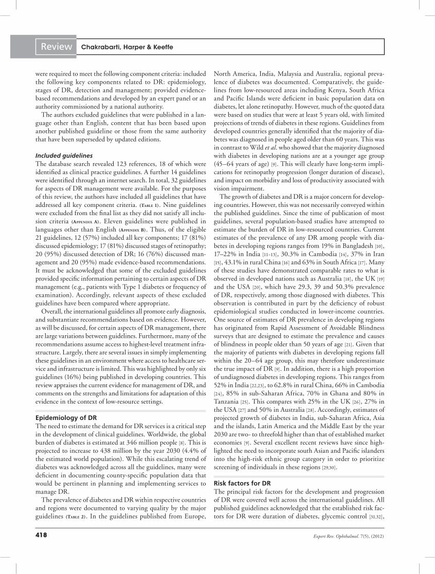
were required to meet the following component criteria: included the following key components related to DR: epidemiology, stages of DR, detection and management; provided evidence-based recommendations and developed by an expert panel or an authority commissioned by a national authority.
The authors excluded guidelines that were published in a lan-guage other than English, content that has been based upon another published guideline or those from the same authority that have been superseded by updated editions.
Included guidelinesThe database search revealed 123 references, 18 of which were identified as clinical practice guidelines. A further 14 guidelines were identified through an internet search. In total, 32 guidelines for aspects of DR management were available. For the purposes of this review, the authors have included all guidelines that have addressed all key component criteria. (Table 1). Nine guidelines were excluded from the final list as they did not satisfy all inclu-sion criteria (appendix a). Eleven guidelines were published in languages other than English (appendix b). Thus, of the eligible 21 guidelines, 12 (57%) included all key components; 17 (81%) discussed epidemiology; 17 (81%) discussed stages of retinopathy; 20 (95%) discussed detection of DR; 16 (76%) discussed man-agement and 20 (95%) made evidence-based recommendations. It must be acknowledged that some of the excluded guidelines provided specific information pertaining to certain aspects of DR management (e.g., patients with Type 1 diabetes or frequency of examination). Accordingly, relevant aspects of these excluded guidelines have been compared where appropriate.
Overall, the international guidelines all promote early diagnosis, and substantiate recommendations based on evidence. However, as will be discussed, for certain aspects of DR management, there are large variations between guidelines. Furthermore, many of the recommendations assume access to highest-level treatment infra-structure. Largely, there are several issues in simply implementing these guidelines in an environment where access to healthcare ser-vice and infrastructure is limited. This was highlighted by only six guidelines (16%) being published in developing countries. This review appraises the current evidence for management of DR, and comments on the strengths and limitations for adaptation of this evidence in the context of low-resource settings.
Epidemiology of DRThe need to estimate the demand for DR services is a critical step in the development of clinical guidelines. Worldwide, the global burden of diabetes is estimated at 346 million people [8]. This is projected to increase to 438 million by the year 2030 (4.4% of the estimated world population). While this escalating trend of diabetes was acknowledged across all the guidelines, many were deficient in documenting county-specific population data that would be pertinent in planning and implementing services to manage DR.
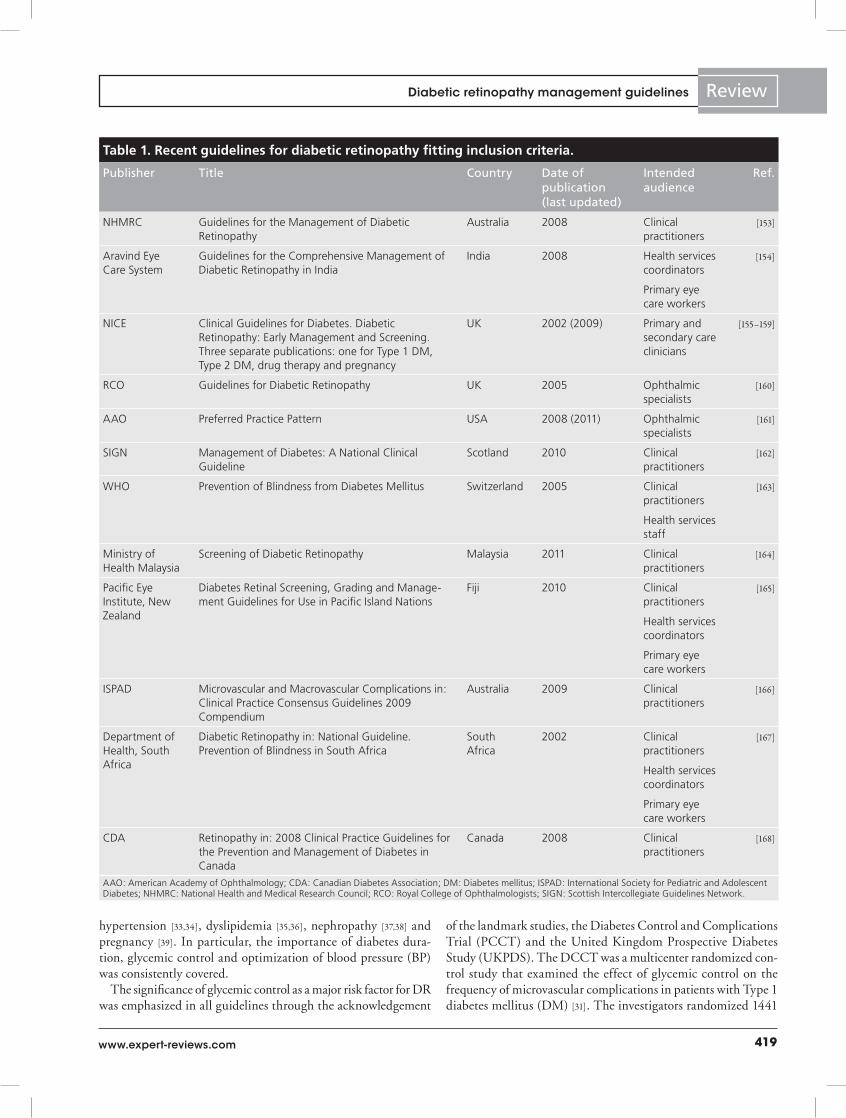
The prevalence of diabetes and DR within respective countries and regions were documented to varying quality by the major guidelines (Table 2). In the guidelines published from Europe,
North America, India, Malaysia and Australia, regional preva-lence of diabetes was documented. Comparatively, the guide-lines from low-resourced areas including Kenya, South Africa and Pacific Islands were deficient in basic population data on diabetes, let alone retinopathy. However, much of the quoted data were based on studies that were at least 5 years old, with limited projections of trends of diabetes in these regions. Guidelines from developed countries generally identified that the majority of dia-betes was diagnosed in people aged older than 60 years. This was in contrast to Wild et al. who showed that the majority diagnosed with diabetes in developing nations are at a younger age group (45–64 years of age) [9]. This will clearly have long-term impli-cations for retinopathy progression (longer duration of disease), and impact on morbidity and loss of productivity associated with vision impairment.
The growth of diabetes and DR is a major concern for develop-ing countries. However, this was not necessarily conveyed within the published guidelines. Since the time of publication of most guidelines, several population-based studies have attempted to estimate the burden of DR in low-resourced countries. Current estimates of the prevalence of any DR among people with dia-betes in developing regions ranges from 19% in Bangladesh [10], 17–22% in India [11–13], 30.3% in Cambodia [14], 37% in Iran [15], 43.1% in rural China [16] and 63% in South Africa [17]. Many of these studies have demonstrated comparable rates to what is observed in developed nations such as Australia [18], the UK [19] and the USA [20], which have 29.3, 39 and 50.3% prevalence of DR, respectively, among those diagnosed with diabetes. This observation is contributed in part by the deficiency of robust epidemiological studies conducted in lower-income countries. One source of estimates of DR prevalence in developing regions has originated from Rapid Assessment of Avoidable Blindness surveys that are designed to estimate the prevalence and causes of blindness in people older than 50 years of age [21]. Given that the majority of patients with diabetes in developing regions fall within the 20–64 age group, this may therefore underestimate the true impact of DR [9]. In addition, there is a high proportion of undiagnosed diabetes in developing regions. This ranges from 52% in India [22,23], to 62.8% in rural China, 66% in Cambodia [24], 85% in sub-Saharan Africa, 70% in Ghana and 80% in Tanzania [25]. This compares with 25% in the UK [26], 27% in the USA [27] and 50% in Australia [28]. Accordingly, estimates of projected growth of diabetes in India, sub-Saharan Africa, Asia and the islands, Latin America and the Middle East by the year 2030 are two- to threefold higher than that of established market economies [9]. Several excellent recent reviews have since high-lighted the need to incorporate south Asian and Pacific islanders into the high-risk ethnic group category in order to prioritize screening of individuals in these regions [29,30].
Risk factors for DRThe principal risk factors for the development and progression of DR were covered well across the international guidelines. All published guidelines acknowledged that the established risk fac-tors for DR were duration of diabetes, glycemic control [31,32],
Chakrabarti, Harper & Keeffe
419www.expert-reviews.com
Review
hypertension [33,34], dyslipidemia [35,36], nephropathy [37,38] and pregnancy [39]. In particular, the importance of diabetes dura-tion, glycemic control and optimization of blood pressure (BP) was consistently covered.
The significance of glycemic control as a major risk factor for DR was emphasized in all guidelines through the acknowledgement
of the landmark studies, the Diabetes Control and Complications Trial (PCCT) and the United Kingdom Prospective Diabetes Study (UKPDS). The DCCT was a multicenter randomized con-trol study that examined the effect of glycemic control on the frequency of microvascular complications in patients with Type 1 diabetes mellitus (DM) [31]. The investigators randomized 1441
Table 1. Recent guidelines for diabetic retinopathy fitting inclusion criteria.
Publisher Title Country Date of publication (last updated)
Intended audience
Ref.
NHMRC Guidelines for the Management of Diabetic Retinopathy
Australia 2008 Clinical practitioners
[153]
Aravind Eye Care System
Guidelines for the Comprehensive Management of Diabetic Retinopathy in India
India 2008 Health services coordinators
[154]
Primary eye care workers
NICE Clinical Guidelines for Diabetes. Diabetic Retinopathy: Early Management and Screening. Three separate publications: one for Type 1 DM, Type 2 DM, drug therapy and pregnancy
UK 2002 (2009) Primary and secondary care clinicians
[155–159]
RCO Guidelines for Diabetic Retinopathy UK 2005 Ophthalmic specialists
[160]
AAO Preferred Practice Pattern USA 2008 (2011) Ophthalmic specialists
[161]
SIGN Management of Diabetes: A National Clinical Guideline
Scotland 2010 Clinical practitioners
[162]
WHO Prevention of Blindness from Diabetes Mellitus Switzerland 2005 Clinical practitioners
[163]
Health services staff
Ministry of Health Malaysia
Screening of Diabetic Retinopathy Malaysia 2011 Clinical practitioners
[164]
Pacific Eye Institute, New Zealand
Diabetes Retinal Screening, Grading and Manage-ment Guidelines for Use in Pacific Island Nations
Fiji 2010 Clinical practitioners
[165]
Health services coordinators
Primary eye care workers
ISPAD Microvascular and Macrovascular Complications in: Clinical Practice Consensus Guidelines 2009 Compendium
Australia 2009 Clinical practitioners
[166]
Department of Health, South Africa
Diabetic Retinopathy in: National Guideline. Prevention of Blindness in South Africa
South Africa
2002 Clinical practitioners
[167]
Health services coordinators
Primary eye care workers
CDA Retinopathy in: 2008 Clinical Practice Guidelines for the Prevention and Management of Diabetes in Canada
Canada 2008 Clinical practitioners
[168]
AAO: American Academy of Ophthalmology; CDA: Canadian Diabetes Association; DM: Diabetes mellitus; ISPAD: International Society for Pediatric and Adolescent Diabetes; NHMRC: National Health and Medical Research Council; RCO: Royal College of Ophthalmologists; SIGN: Scottish Intercollegiate Guidelines Network.
Diabetic retinopathy management guidelines
Expert Rev. Ophthalmol. 7(5), (2012)420
Review
patients with Type 1 DM to receive intensive glycemic control (median HbA1c: 7.3%) compared with conventional levels of control (median HbA1c: 9.1%). The results demonstrated that over a 6.5-year follow-up, intensive glycemic control compared with conventional treatment was associated with reduction in any DR by 76% (95% CI: 62–85) [40], and progression of DR by 54% (95% CI: 39–66) [41]. Similarly, in the UKPDS, the investigators randomized 3867 patients with newly diagnosed Type 2 DM to receive intensive treatment (oral hypoglycemic medication or insulin) or conventional glycemic control (diet control) over a period of 10 years [32]. The results demonstrated that intensive treatment reduced the development of any DR by 25% (95% CI: 7–40). Furthermore, this was associated with a 29% reduction (relative risk: 0.71; 95% CI: 0.53–0.96; p = 0.003) in progression to requirement of laser photocoagulation in the intensive group compared with conventional treatment.
The rationale for tight BP control has similarly been explored in landmark studies. In the UKPDS, 1048 patients with Type 2 DM were randomized to receive intensive BP control of patients with Type 2 DM (target BP; <150/<85 mmHg) versus <180/<105 mmHg, with observation over a period of 9 years. The study demonstrated that intensive control of hypertension was associ-ated with reduction in progression of DR (34 vs 51%; p < 0.05), reduction in moderate vision loss (10 vs 19%; p < 0.05), and reduction in need for photocoagulation (relative risk: 0.65, 95%
CI: 0.39–1.06; p = 0.023) [33]. However, there is conflicting evidence regarding whether aggressive treatment of normoten-sive patients is beneficial in DR. The EUCLID demonstrated over a 2-year follow-up that lisinopril (anti hypertensive agent) reduced the progression of any DR by 50% (95% CI: 0.28–0.89; p = 0.02), and progression to proliferative DR (PDR) by 80% (95% CI: 0.04–0.91; p = 0.04) in patients with Type 1 DM with normal BP and renal function [36]. Recently, a 4-year follow-up from the ACCORD Eye study ‘blood pressure’ trial demonstrated that there was no significant difference in the progression of DR with intensive (target systolic BP: <120 mmHg) versus standard BP control (target systolic BP: <140 mmHg) in patients with Type 2 DM [42]. The authors reported no statistically significant difference in any progression of DR (odds ratio: 1.23; 95% CI: 0.84–1.79); or rate of moderate vision loss (hazard ratio: 1.17; 95% CI: 0.96–1.42). Nevertheless, it is clear that optimization of BP in patients with hypertension is a major factor in attenuating the progression of DR.
Guidelines also consistently emphazised the role of early exami-nation in reducing the risk of DR progression. Guidelines from developing regions used data from WESDR and the ETDRS, which demonstrated that after 15 years, retinopathy is noted in almost all people with Type 1 DM and 75% of people with Type 2 DM; 2% become blind and 10% develop severe vision impair-ment [8,43,44]. The role of puberty as a risk factor for DR among
Table 2. Country/region-specific epidemiology of diabetes and diabetic retinopathy as stated in the major guidelines.
Guideline Country Prevalence of diabetes (%) (year of reference statistics)
Prevalence of any DR among patients with diabetes (%) (year of reference statistics)
NHMRC Australia 8 in adult males and 6.9 adult females (2002) Overall: 25.4 (2002)
Indigenous: 20–50% of adults (1991) Indigenous: 31 (1985)
AAO USA 5.2 European–Americans; 11.0 African–Americans and 10.4 Mexican descent (2006)
80 (Type 1 patients [after 15 years] and 84 (Type 2 patients on insulin [after 19 years]) (WESDR, 1984)
SIGN Scotland Incidence Type 1 DM: 35 per 100,000 (2003)
Pacific Eye Institute Pacific Islands >50 in certain Pacific countries (unreferenced)
New Zealand New Zealand 30 (2006)
Ministry of Health Malaysia
Malaysia 11.6 (aged >18 years) and 14.9 (aged >30 years) (2006) 36.8 (2007)
Aravind Eye Care India 3.2 (2000) 15–25 (2000)
CDA Canada 5.5 adult population (2005) and 26 in aboriginal people of Canada (1997)
40.3 (2004)
NICE UK 4.3 (males, Type 2 DM, aged >55) and 3.4 (females, Type 2 DM, aged >55)
60 after 20 years (2002)
50–200,000 Type 1 DM (2001) Type 1 DM: 9 (aged <11 years) and 29 (aged >11 years; 1999)
Department of Health
South Africa 5 of Africans
11 of Indians in South Africa (2002)
AAO: American Academy of Ophthalmology; CDA: Canadian Diabetic Association; DM: Diabetes mellitus; DR: Diabetic retinopathy; NHMRC: National Health and Medical Research Council; SIGN: Scottish Intercollegiate Guidelines Network.
Chakrabarti, Harper & Keeffe
421www.expert-reviews.com
Review
patients with Type 1 DM was acknowledged by all guidelines except South Africa and Aravind. This was consistent with find-ings from several studies that demonstrated that physiological changes post puberty accelerated the development of microvas-cular complications including DR [45,46]. Consequently, puberty is now accepted as a risk factor for onset of DR.
The significance of ethnic background upon risk of DR is well established. Many of the guidelines identified high-risk ethnic groups within their population. The American Academy of Ophthalmology (AAO) identified African–Americans and Mexicans as having a greater risk of developing any DR com-pared with Americans of European descent [47]. The NHMRC in Australia estimated that 31% of indigenous people with DM had evidence of retinopathy, compared with 20% in the nonindig-enous population. Recent meta-analysis of population-based stud-ies from around the world demonstrated that the age-standard-ized prevalence of any DR at 49.6% among African–Americans, 34.6% among Hispanic populations, 19.9% in Asians, compared with 45.8% in Caucasians [30]. While this was the first meta-analysis to incorporate risk factor data from Asia, the authors acknowledged the deficiency of good quality population studies from the Middle East, Africa and South America. The guide-lines for the Pacific Islands, although based on the New Zealand Ministry of Health recommendation, stated that the proportion of any DR among the diabetic population exceeded 50% in some Pacific countries. This is consistent with trends from a recent systematic review that demonstrated that Oceania had the largest DM prevalence (15%) and highest average fasting plasma glu-cose level of any region in the world [48]. The South African and Malaysian guidelines each acknowledged that people of Indian background were at elevated risk of DR compared with the locals (African and Malay, respectively). This was supported by findings from several cross-sectional studies in India that demonstrated up to 25% prevalence of DR among patients with DM [11,22]. More recently, the UK Asian Diabetes Study also identified peo-ple with south Asian ethnicity as possessing an elevated risk of DR after controlling for other risk factors [49]. However, none of the published guidelines mentioned Indians or south Asians as a high-risk ethnic group. The Australian guideline was the only one to offer insight into factors contributing to ethnic differences. They acknowledged the role of westernization and change from traditional diets and lifestyle of indigenous people as a significant contributor to the higher prevalence rate of DM.
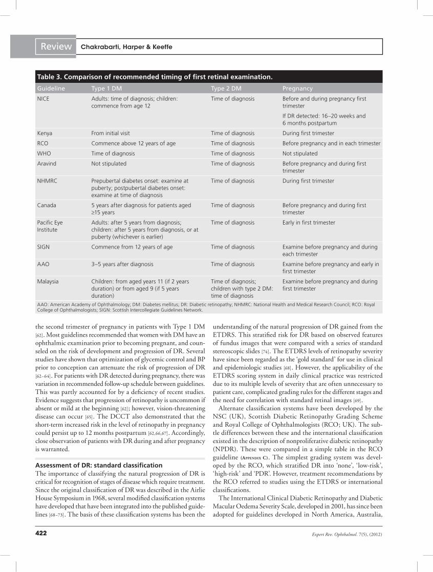
Disease onset & progression & implications for timing of first examinationThe necessity to examine all patients with DM for retinopathy at least every 2 years is uniformly accepted by all international guidelines. The recommended timing of first examination is largely consistent between publications and is supported by the published literature (Table 3). In the context of Type 2 DM, there was unanimous concordance among the major international guidelines is that all people should be examined using a minimum of dilated fundoscopy and visual acuity measurement by an oph-thalmologist, optometrist or suitably trained professional at the
time of diagnosis. For patients with Type 2 DM, consensus in the guidelines recommended ophthalmic examination (comprising of fundoscopy and repeated visual acuity measurement) at the time of diagnosis. The rationale for this was supported by the observation that time of onset of Type 2 DM is often difficult to determine [50], and that a third of Type 2 DM patients will have some evidence of retinopathy at diagnosis [44,51].
For children with Type 1 DM, the majority of guidelines rec-ommend first examination to commence at or soon after puberty (aged 11–12 years). The rationale for delayed screening in chil-dren was based primarily from the WESDR, which demonstrated that DR rarely developed in children with Type 1 DM younger than 10 years of age [43]. Several follow-up studies concluded that sight-threatening retinopathy (proliferative retinopathy or macular edema) was rare before puberty [52,53]. In postpubertal patients with Type 1 DM, guidelines from New Zealand, the Pacific Islands and North America recommended first retinal examination commence after 5 years from the time of diagnosis. This is supported by evidence that showed that the prevalence of DR rapidly increased after 5 years duration of DM [52]. More recent prospective studies have demonstrated that after at least 25 years with DM, almost all patients with Type 1 DM developed DR, and between 44 and 50% developed advanced retinopathy [54,55]. For patients with Type 1 DM for more than 20 years, this conferred a 15-times greater risk of proliferative DR, and five-times greater risk of diabetic macular edema (DME), compared with those with Type 2 DM for <10 years [30]. It is important to note that guidelines were based on studies conducted in Western populations where retinopathy occurred in 8–9% in patients younger than 13 years of age, and 28–34% in those older than 13 years [56,57]. Comparatively, recent observations from Tanzania showed 22% of 5–18 year olds with Type 1 DM had evidence of retinopathy at diagnosis. These suggest that emphasis on facili-tating examination earlier than 5 years from time of diagnosis for people with Type 1 DM may be required in low-resourced countries [58].
It is established that physiologic and metabolic changes associ-ated with pregnancy can accelerate DR [39,59]. The recommended timing of examination during pregnancy was stratified into patients with existing DM and those who developed gestational DM (GDM). In the context of patients who develop glucose intol-erance during pregnancy (GDM), the AAO and NHMRC stated that DR screening was not routinely required as there was mini-mal risk for DR in such individuals during pregnancy. Presently, only a single case report has identified vision-threatening retin-opathy arising in a patient with GDM [60]. While there is insuf-ficient evidence assessing the temporal progression of retinopathy in this group, GDM is associated with elevated risk of long-term DM [61]. Accordingly, ophthalmic review at time of diagnosis of GDM may be justified as per the Malaysian guidelines.
For patients with DM diagnosed before pregnancy, the con-sensus among guidelines recommended a comprehensive eye examination for all pregnant women with DM during the first trimester of pregnancy (Table 4). This was supported by findings from the DCCT that showed greatest risk of DR progression in
Diabetic retinopathy management guidelines
Expert Rev. Ophthalmol. 7(5), (2012)422
Review
the second trimester of pregnancy in patients with Type 1 DM [62]. Most guidelines recommended that women with DM have an ophthalmic examination prior to becoming pregnant, and coun-seled on the risk of development and progression of DR. Several studies have shown that optimization of glycemic control and BP prior to conception can attenuate the risk of progression of DR [62–64]. For patients with DR detected during pregnancy, there was variation in recommended follow-up schedule between guidelines. This was partly accounted for by a deficiency of recent studies. Evidence suggests that progression of retinopathy is uncommon if absent or mild at the beginning [62]; however, vision-threatening disease can occur [65]. The DCCT also demonstrated that the short-term increased risk in the level of retinopathy in pregnancy could persist up to 12 months postpartum [62,66,67]. Accordingly, close observation of patients with DR during and after pregnancy is warranted.
Assessment of DR: standard classificationThe importance of classifying the natural progression of DR is critical for recognition of stages of disease which require treatment. Since the original classification of DR was described in the Airlie House Symposium in 1968, several modified classification systems have developed that have been integrated into the published guide-lines [68–73]. The basis of these classification systems has been the
understanding of the natural progression of DR gained from the ETDRS. This stratified risk for DR based on observed features of fundus images that were compared with a series of standard stereoscopic slides [74]. The ETDRS levels of retinopathy severity have since been regarded as the ‘gold standard’ for use in clinical and epidemiologic studies [68]. However, the applicability of the ETDRS scoring system in daily clinical practice was restricted due to its multiple levels of severity that are often unnecessary to patient care, complicated grading rules for the different stages and the need for correlation with standard retinal images [69].
Alternate classification systems have been developed by the NSC (UK), Scottish Diabetic Retinopathy Grading Scheme and Royal College of Ophthalmologists (RCO; UK). The sub-tle differences between these and the international classification existed in the description of nonproliferative diabetic retinopathy (NPDR). These were compared in a simple table in the RCO guideline (appendix C). The simplest grading system was devel-oped by the RCO, which stratified DR into ‘none’, ‘low-risk’, ‘high-risk’ and ‘PDR’. However, treatment recommendations by the RCO referred to studies using the ETDRS or international classifications.
The International Clinical Diabetic Retinopathy and Diabetic Macular Oedema Severity Scale, developed in 2001, has since been adopted for guidelines developed in North America, Australia,
Table 3. Comparison of recommended timing of first retinal examination.
Guideline Type 1 DM Type 2 DM Pregnancy
NICE Adults: time of diagnosis; children: commence from age 12
Time of diagnosis Before and during pregnancy first trimester
If DR detected: 16–20 weeks and 6 months postpartum
Kenya From initial visit Time of diagnosis During first trimester
RCO Commence above 12 years of age Time of diagnosis Before pregnancy and in each trimester
WHO Time of diagnosis Time of diagnosis Not stipulated
Aravind Not stipulated Time of diagnosis Before pregnancy and during first trimester
NHMRC Prepubertal diabetes onset: examine at puberty; postpubertal diabetes onset: examine at time of diagnosis
Time of diagnosis During first trimester
Canada 5 years after diagnosis for patients aged ≥15 years
Time of diagnosis Before pregnancy and during first trimester
Pacific Eye Institute
Adults: after 5 years from diagnosis; children: after 5 years from diagnosis, or at puberty (whichever is earlier)
Time of diagnosis Early in first trimester
SIGN Commence from 12 years of age Time of diagnosis Examine before pregnancy and during each trimester
AAO 3–5 years after diagnosis Time of diagnosis Examine before pregnancy and early in first trimester
Malaysia Children: from aged years 11 (if 2 years duration) or from aged 9 (if 5 years duration)
Time of diagnosis; children with type 2 DM: time of diagnosis
Examine before pregnancy and during first trimester
AAO: American Academy of Ophthalmology; DM: Diabetes mellitus; DR: Diabetic retinopathy; NHMRC: National Health and Medical Research Council; RCO: Royal College of Ophthalmologists; SIGN: Scottish Intercollegiate Guidelines Network.
Chakrabarti, Harper & Keeffe
423www.expert-reviews.com
Review
Asia, Africa and Asia–Pacific region. The new classification incor-porated evidence on disease progression from the ETDRS, and stratified DR into five levels of severity based on observed retinal changes [69]. The main distinctions offered in the international severity scale are that the levels of severity are each relevant to the clinical management decisions for the patient. This offered a simpler method to assess risk of progression of DR, and facilitated
communication between ophthalmologists and primary health-care providers. Accordingly, the international classification system has been endorsed by most international authorities including the WHO as a standard system for guiding evidence-based practice. While the international classification system has not replaced the ETDRS, it has been demonstrated as a useful guide for population screening, and facilitating timely treatment [15,75].
Table 4. Frequency of examination and referral to ophthalmologist.
Guideline No retinopathy
Mild NPDR Moderate NPDR
Severe NPDR
PDR CSME Pregnancy
NICE Annual review
Annual review 3–6 months Within 4 weeks
Within 1 week
Within 4 weeks
16–20 weeks and 6 months postpartum
South Africa
Annual review
12 months 6 months Urgently for pan-retinal photo coagulation
Urgently for pan-retinal photocoagula-tion
Urgent referral to ophthalmologist
Not stipulated
RCO Annual review
Annual review Within 4 months
Within 4 months
Within 2 weeks
Within 2 weeks
In each trimester, and 3–9 months postpartum
WHO Annual review
6–12 months 6–12 months 2–4 months 2–4 months 2–4 months Not stipulated
NHMRC 2 yearly annual review for high-risk patients
Annual review 3–6 months Within 3–6 months
Within 4 weeks
Within 4 weeks
If DR found, need close follow-up throughout pregnancy
Pacific Eye Institute
12 months 6 months Within 6 weeks
Within 4 weeks
Within 1 week
Stable: 12 months
If DR detected at least 2- monthly intervals
Severe: within 1 week
Moderate: 1 month
Mild: 2 months
Minimal: 6 months
SIGN 2 yearly Annual review Within 18 weeks
Within 12 weeks
Urgently for laser treatment
During each trimester
AAO Annual review
6–12 months 6–12 months 2–4 months 2–4 months Presence of CSME requires minimum 2–4 monthly review
If nil or minimal DR: 3–12-monthly follow-up
Severe NPDR or worse: 1–3-monthly
Malaysia 12–24 months
9–12 months 6 months Within 4 weeks
Within 1 week Any maculopathy: within 4 weeks
Nil to moderate DR: 3-monthly
Moderate or worse: urgent referral
AAO: American Academy of Ophthalmology; CSME: Clinically significant macular edema; DR: Diabetic retinopathy; NHMRC: National Health and Medical Research Council; NPDR: Nonproliferative DR; PDR: Proliferative DR; RCO: Royal College of Ophthalmologists.
Diabetic retinopathy management guidelines
Expert Rev. Ophthalmol. 7(5), (2012)424
Review
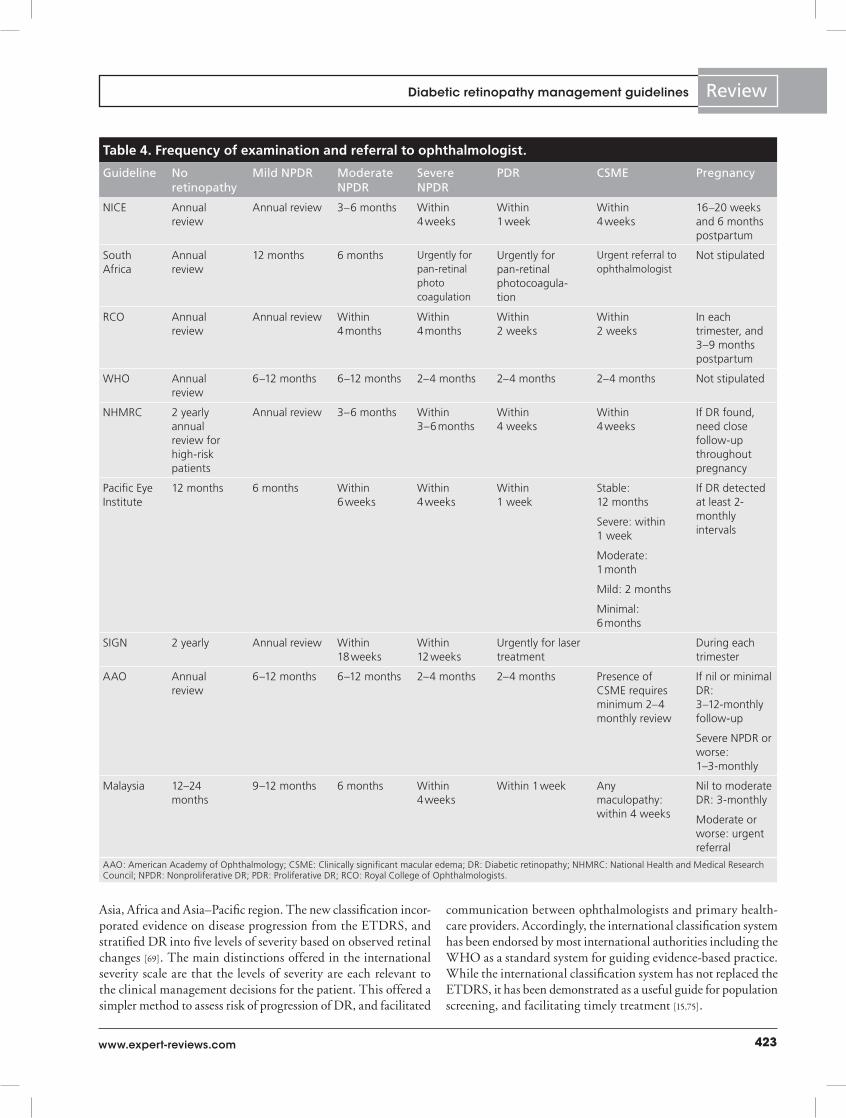
Assessment of DR: frequency of examinationWhile there was general consensus among published guide-lines regarding the timing of the initial examination, there were differences between follow-up examination schedules (Table 4).
No retinopathyFor patients without evidence of retinopathy, guidelines from South Africa, NICE, RCO, AAO and the Pacific Islands recom-mended annual follow-up. The AAO referenced the WESDR that demonstrated that at 1 year, 5–10% of patients with a normal retinal examination at baseline had progressed to some evidence of DR. The 4-year incidence of any DR was 59% in patients with Type 1 DM and 34% in Type 2 patients [43].The NHMRC was the only publication that identified and recommended that patients at elevated risk (longer duration, poor glycemic control, hyper tension, hyperlipidemia or from an indigenous background) required at minimum annual review. The necessity for greater vigilance particularly for the indigenous population is supported by recent evidence which demonstrated that indigenous people in Australia developed vision-threatening disease (particularly clini-cally significant macular edema [CSME]) from a normal base-line at an earlier stage than nonindigenous populations [76,77]. Extension of the examination schedule to 2-yearly intervals for most patients was recommended by the NHMRC, Scottish Intercollegiate Guidelines Network (SIGN), New Zealand and Malaysia. Evidence-based justification for the timing between examinations were based on findings from studies subsequent to WESDR, including the UKPDS, showed that 22% with a normal baseline examination developed DR after 6 years [78]. Comparable data showing ≤half the incidence in the WESDR was also demonstrated in the Blue Mountains Eye Study [79], Melbourne Visual Impairment Project [80] and UKPDS [78]. In the context of these findings, The Liverpool Diabetic Eye Study concluded that the rate of progression to sight-threatening DR among people with normal baseline was so low that a conserva-tive screening period of 2–3 years could be reliably adopted [81]. A more recent meta-analysis by Wong et al. demonstrated that for patients with nil retinopathy at baseline, the progression to PDR after 4 years was 2.6% in studies published between 1986 and 2008, compared with 6.3% in prior studies [82]. The authors concluded that this difference may be accounted for by optimiza-tion of risk-factor control among patients with DM. This was supported by a meta-analysis of health economic evidence that demonstrated that for patients with good glycemic control and no background retinopathy, biennial or triennial screening was more cost effective than annual examination [83]. Despite this evidence, guidelines developed for low-resource areas (South Africa, Kenya and the Pacific Islands) all recommend annual screening intervals. The rationale for annual screening in these areas can perhaps be contextualized by the differences in dis-ease prevalence and ‘high-risk’ ethnic groups. Furthermore, it must be considered that findings from the Liverpool Diabetic Eye Study related to the end point of sight-threatening disease. The extension of the screening interval beyond 2 years failed to consider the effect that lower levels of DR severity impart on
patient visual morbidity, and the additional benefits associated with clinician–patient continuity of care to opportunistically detect other associated eye conditions more frequent in DM (e.g., cataract and glaucoma) [84,85]. Thus, at present, current evidence indicates that patients without an elevated risk of DR can safely be reviewed at 2-yearly intervals.
Mild-to-moderate NPDR without macular edemaThe recommended frequency of examination for patients with mild-to-moderate nonproliferative DR without macular edema varied between published guidelines. While all guidelines recom-mended annual examination, the AAO, WHO, Pacific Islands and Malaysia suggested patients could be reviewed more fre-quently, at 6–12 monthly intervals. The AAO referenced the WESDR that demonstrated that 16% of Type 1 DM patients with mild retinopathy (hard exudates and microaneurysms only at baseline examination) progressed to proliferative disease after 4 years [52]. For Type 2 DM, 34–47% experienced worsening of retinopathy over a similar period [86]. More conservative esti-mates of progression were demonstrated in the 6-year follow-up data from the UKPDS. This showed that 29% of patients with retinopathy at baseline progressed by at least two ETDRS lev-els of severity. Eighteen percent with mild-to-moderate NPDR at baseline progressed to need photocoagulation at 6 years [78]. Recently, a 4-year follow-up of patients with Type 2 DM dem-onstrated an escalation in incidence of DR from 5.8 to 20.3% between 1- and 2-year follow-ups [87]. This strongly supports the current recommendations for at least annual review in these patients.
Severe NPDRIn the context of patients with severe nonproliferative DR, all guidelines addressed the necessity for more frequent review of patients. Prompt referral within 4 weeks was advocated by NICE, New Zealand/Pacific Islands, Malaysian and South African guide-lines. Comparatively, the WHO, AAO, NHMRC, RCO and SIGN while acknowledging the importance of early examination by an ophthalmologist, offered a range between 2 and 6 months. The rationale for at least four monthly examinations was derived from the ETDRS protocol, which reviewed patients with mild-to-severe NPDR. This demonstrated that 45% of patients with severe NPDR developed PDR within 1 year, increasing to 71% after 5 years [3]. Subsequent analyses from the ETDRS demonstrated that early referral for retinal photocoagulation for patients with severe NPDR reduced the risk of severe vision loss or need for vitrectomy by 50%, compared with deferring until high-risk PDR developed [88]. The difficulty in determining an ‘optimal’ period of review for severe NPDR rests with limitations in the literature. As highlighted by Wong et al., many of the ‘landmark’ studies had larger proportions of more advanced DR at baseline [82]. In addition, the advances and access to modern treatment modalities would therefore pose challenges to designing a new prospective study. Thus, given the evidence that suggests the propensity for severe NPDR to progress rapidly, current evidence supports a maximum of 4-monthly intervals.
Chakrabarti, Harper & Keeffe

425www.expert-reviews.com
Review
Criteria for urgent referral to an ophthalmologistThe necessity to expedite ophthalmic review for patients with vision-threatening retinopathy was consistently established across the guidelines reviewed. Fundamentally, ‘vision-threatening’ retin-opathy was uniformly accepted and defined as encompassing the presence of severe retinopathy (severe NPDR and proliferative DR) and DME. Further consensus was achieved across guidelines that any sudden severe vision loss, or symptoms of retinal detachment, required same-day referral to an ophthalmologist. Overall, all guidelines based their recommendations based on observations from the sentinel studies, the DRS [89] and ETDRS [90] in which photocoagulation was referred as soon as high-risk PDR was detected. These studies demonstrated significant reduction in the risk of severe vision loss among patients with advanced retinopathy with timely retinal photocoagulation. The NICE defined three lev-els of ‘urgency’: emergency (same day); within 1 week and within 4 weeks. Patients with any form of maculopathy or severe NPDR required review within 4 weeks. The presence of proliferative retin-opathy (anywhere), preretinal or vitreous hemorrhage required review within 1 week. This model was incorporated into both the Australian and Malaysian guidelines. Similarly, the AAO, SIGN, WHO and South African guidelines all recommended ophthalmic review and treatment to be performed expeditiously for patients with PDR and macular edema. However, they failed to clearly define an ‘urgent’ time frame.
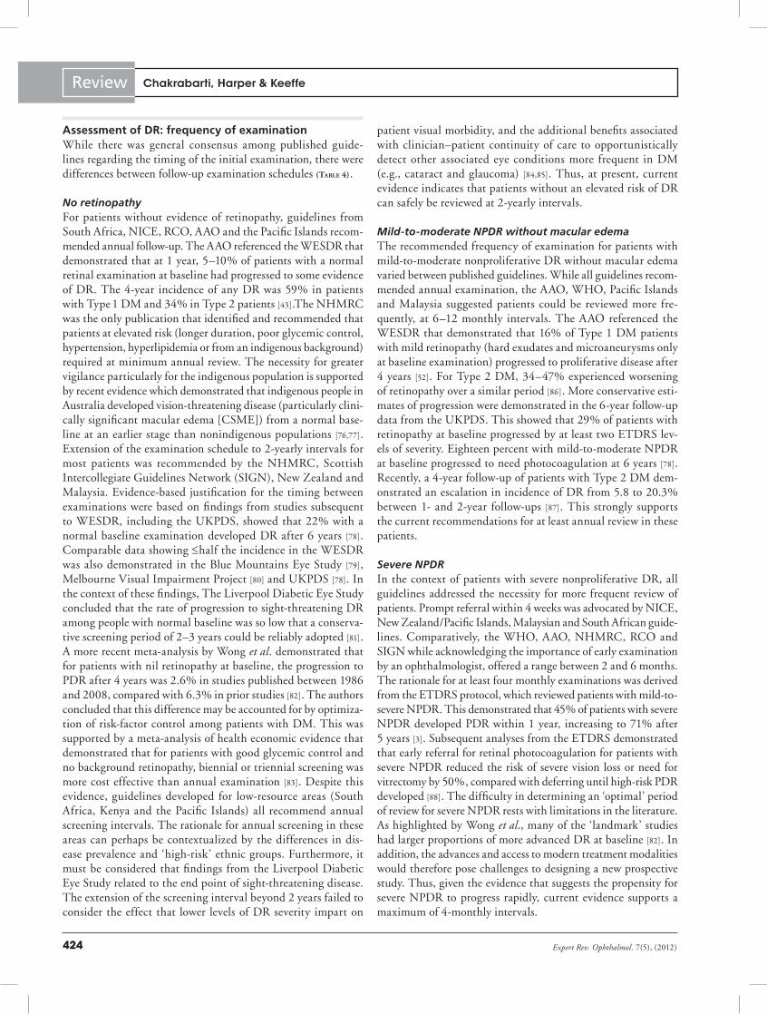
Assessment of DR: detection of DRThe recommended modality for screening for DR varied consid-erably across the published guidelines (Table 5). While there is no doubt that the advent of digital retinal photography has facilitated greater coverage of retinal photography for the purposes of popu-lation screening, there was considerable variation regarding the accepted criteria for the use of digital imaging in DR screening. While different studies have had variations in criteria for reference standards, NICE stipulated that an acceptable DR screening tool with a minimum of 80% sensitivity, 95% specificity and technical failure rate of 5% [91]. This contrasted with the NHMRC that set the minimum sensitivity for a screening test as 60%, with the requisite that repeated examinations would detect any retinopathy missed at earlier examination [92]. The current ‘gold standard’ ETDRS protocol of seven standard (30°) field 35-mm stereoscopic mydriatic color fundus photographs was recommended by the SIGN and Canadian guidelines. Alternative digital fundus imag-ing protocols capturing two or three fields were recommended by NICE, New Zealand and the WHO. Systematic review of evidence suggests that mydriatic photography is the most effective screening strategy, with high sensitivity (87–97%) and specificity (83–92%) for detection of sight-threatening DR [93]. Joannou et al. showed that single-field 60° mydriatic photography had a sensitivity and specificity of 93 and 89% for detection of any retinopathy, and 100 and 75%, respectively for severe DR [94]. Importantly, Maberley et al. showed that mydriatic 45° fundus images were shown to have a high sensitivity (93.3%), specific-ity (96.8%) and positive-predictive value (67.8%) for detecting PDR or CSME [95]. While multiple-field mydriatic photography
demonstrated greater sensitivity compared with single-field pho-tography, several limitations were noted in the guidelines [96–98]. These included the time taken to obtain and interpret the photo-graphs, constraints upon availability and training for ophthalmic workforce, the need for dilating drops and its associated issues related to patient compliance [99].
The limitations of mydriatic photography prompted the NHMRC, Pacific Islands and Malaysian guidelines to propose the use of non-mydriatic retinal cameras as a suitable alterna-tive for DR screening. Their recommendations were supported by evidence that showed that high-quality single-field, 45° non-mydriatic photography demonstrated sensitivity (71–84%) and specificity (93–98%) for the detection of referrable retinopathy, including proliferative DR and maculopathy [100–103]. A grow-ing body of evidence has endorsed non-mydriatic photography as a practical method for population screening, particularly in rural and remote areas where fundamentally, the decision to refer or not is required [104]. Harper et al. showed that single 45° field non-mydriatic images using the Canon CR45 camera could be reliably performed with a 5% technical failure rate, in rural areas by trained, nonophthalmic technicians, with off-site central grading of images [101]. Diamond et al. using the same camera further demonstrated in a rural setting, that single-field non-mydriatic imaging for DR screening was able to capture equivalent ‘adequate’ quality images compared with mydri-atic photographs in order to establish presence of retinopathy and need for referral [105]. Importantly, in a systematic review, Jones and Edwards concluded that the use of digital photogra-phy, with the use of tele medicine for off-site grading, achieved greater cost–effectiveness than conventional ophthalmoscopy by a traveling ophthalmologist [83].
The limitations of non-mydriatic photography are noted in the literature. In a sample of 3611 patients, Scanlon et al. iden-tified a technical failure rate of satisfactory images using non-mydriatic photography of 19.7%, with full assessment of both eyes achieved in only 48% of patients [106]. However, their study acknowledged that the Sony digital camera used for the study achieved a resolution well below the minimum recommended threshold by the UK NSC. Significant advances in camera reso-lution have occurred since Pugh et al. showed lower sensitivity of non-mydriatic photography using a Canon CR3 camera com-pared with mydriatic images in detecting more severe DR [107]. Comparative studies of non-mydriatic to mydriatic retinal pho-tography have widely reported that single-field, nonstereoscopic retinal photographs taken with mydriasis provides superior qual-ity images for the diagnosis of DR [105,106], and reduces the number of patients referred due to ungradable photographs [98]. Furthermore, Bursell et al. noted that in the presence of only a few exudates, distinguishing CSME was limited with nonstereo-scopic views through undilated pupils [108]. The image quality was further reduced by the presence of other ocular pathology such as lens opacity [109]. While Aptel et al. demonstrated that sensitivity of non-mydriatic photography was improved to 90% by capturing three 45° retinal fields [100], the use of multiple fields had a small improvement in the positive-predictive value
Diabetic retinopathy management guidelines
Expert Rev. Ophthalmol. 7(5), (2012)426
Review
of the test [106]. Thus, consistent themes that emerged from such studies were the limitations on available technology at the time, and the effect of pupil size.
Specifications of available technology must therefore be consid-ered in evaluating the quality of retinal imaging reported in stud-ies. The popularity of digital photography emerged through the observation of costs and time required for processing, developing and shipping original 35-mm film images for interpretation [110]. In order to comply with ‘gold standard’ ETDRS protocol of seven-field stereoscopic, 35-mm color film protocol, achieving a 2400 × 2000 pixel resolution equated to almost half a gigabyte per patient per visit. With the evolution of high-resolution digital cameras, there is a clear necessity for images to be compressed in order to facilitate archiving and transmission across computer networks [111]. Presently, the United Kingdom NSC recommends high-quality image compression (1:12 rather than 1:20), and a
minimum resolution of 20 pixels per degree of retinal imag-ing [112]. Basu et al. concluded from their evaluation of 290 single-field images taken through a non-mydriatic Canon CR6, 45° field fundus camera that image compression ratios between 1:20 and 1:12 (equating to a file size of 66–107 kilobytes) was the thresh-old for gradable image quality [113]. Advances in non-mydriatic technology have clearly improved the diagnostic validity of this modality. In 2009, Vujosevic et al. conducted a well-designed masked prospective case series comparing single- and three-field non-mydriatic photography to ETDRS protocol using the Nidek 45° non-mydriatic camera (1392 × 1040 resolution). The authors demonstrated that sensitivity and specificity for referable retinopa-thy was 71 and 96%, respectively [114]. The sensitivity improved to 82% with three-field non-mydriatic 45° images. Importantly, the study concluded that the resolution offered by a single 45° central field was adequate to determine presence of DR and DME.
Table 5. Detection of diabetic retinopathy.
Guideline Recommended screening modality Personnel performing visual acuity and retinal examination
NICE Retinal photography (mydriatic, 45°) or slit-lamp indirect ophthalmoscopy
Mydriatic photograph evaluated by trained personnel
Slit-lamp examination by ophthalmologist or optometrist
South Africa Dilated ophthalmoscopy Ophthalmic medical officer, trained ophthalmic nurse or optometrist
Kenya Dilated fundoscopy Did not stipulate
RCO Dilated digital retinal photography Primary care physicians, optometrists or ophthalmologists
WHO Dilated retinal photography (three-field images at a reading centre or two-field images against a photographic standard) or slit-lamp biomicroscopy (dilated) with a lens or dilated fundoscopy including stereoscopic examination of the posterior pole
Trained photographer, optometrists and ophthalmologists
Aravind Wide-angle fundus photography Trained ophthalmic technician (for fundus photography), physicians, diabetologists and ophthalmologists
NHMRC Dilated ophthalmoscopy or slit-lamp biomicroscopy (dilated) with a lens or photography (non-mydriatic adequate) if dilated exam not possible
Ophthalmologists, optometrists and other trained medical examiners
Canada Seven-standard field stereoscopic colour fundus photography or dilated direct ophthalmoscopy or dilated indirect slit-lamp fundoscopy
Fundus photography interpreted by trained reader
New Zealand
Dilated retinal photography (2 × 45° fields-macular and nasal) or slit-lamp biomicroscopy
Trained screener and grader (nurses, allied health, mid-level health professionals)
Secondary grader: ophthalmologists and optometrists
Pacific Eye Institute
Non-mydriatic digital retinal photography (single 45° field) or slit-lamp or indirect ophthalmoscopy through dilated pupils
Trained screener and grader (nurses, allied health, mid-level health professionals)
SIGN Retinal photography (seven-field stereoscopic) or slit-lamp biomicroscopy (dilated) one-field 45–50° can be used for screening
Ophthalmologists
AAO Slit-lamp biomicroscopy (dilated) with a lens or dilated fundoscopy including stereoscopic examination of the posterior pole
Ophthalmologists
Trained individuals under ophthalmologist supervision
Malaysia Non-mydriatic retinal imaging (angle not specified) or dilated ophthalmoscopy
Screening and grading by trained doctors, optometrists, assistant medical officers and nurses
AAO: American Academy of Ophthalmology; NHMRC: National Health and Medical Research Council; RCO: Royal College of Ophthalmologists.
Chakrabarti, Harper & Keeffe
427www.expert-reviews.com
Review
Dilated ophthalmoscopy or dilated fundus examination using an indirect lens on a slit-lamp is the preferred practice in the NHMRC, South Africa and Kenyan guidelines. The remain-ing guidelines recommended that clinical examination was an appropriate method for screening in the absence of photographic facilities or poor quality images. However, evidence suggests sig-nificant variability of direct and indirect ophthalmoscopy, even among experienced hands in the ability to detect retinopathy [115]. The Liverpool Diabetic Eye Study demonstrated that direct oph-thalmoscopy with mydriasis, even if performed by an experienced ophthalmologist, had inferior sensitivity (65 vs 89%), and speci-ficity for detection of sight-threatening eye disease (86 vs 97%) compared with three-field 45° nonstereoscopic mydriatic pho-tography [116]. The systematic review conducted by Hutchinson et al. demonstrated that the use of direct ophthalmoscopy through dilated pupils in screening for DR was associated with variations in sensitivity (45–98%) and specificity (62–100%) [93]. The valid-ity of direct ophthalmoscopy was further reduced in the hands of less-experienced medical officers [93].
Given this evidence, slit-lamp biomicroscopy performed using an appropriate lens (78 or 90 dioptre) remained an important modality in screening for DR. The necessity for slit-lamp exami-nation has been partly attributed to the influence of ungradable photography. The utility of slit-lamp biomicroscopy was demon-strated by Scanlon et al. who showed that in comparison with seven-field stereoscopic retinal photography, dilated slit-lamp biomicroscopy performed by an ophthalmologist had a sensi-tivity of 87.4% (95% CI: 83.5–91.5), and specificity of 94.9% (95% CI: 91.5–98.3%) in identifying referable DR (k = 0.80) [106]. The use of slit-lamp biomicroscopy has since been adopted as the ‘reference’ standard in several recent studies comparing modalities of retinal photography for DR as it is much less sus-ceptible to media-opacity related failure [117,118]. While the slit-lamp has advantages of availability and affordability compared with photography, the disadvantages of its routine use in a low-resourced setting included availability of trained ophthalmic staff and need for pupil dilatation.
Current evidence suggests single-field non-mydriatic photo-graphy using trained readers is an adequate modality for detecting referable DR, but not a substitute for comprehensive ophthalmic examination when needed. Compared with ophthalmoscopy, single-field photography can offer screening to a greater popula-tion. While mydriasis improves the sensitivity, it is restricted by practical limitations. As such, current evidence concurs with the recommendation for the Health Technology Board of Scotland that non-mydriatic one-field photography be used as a first stage, with mydriatic photography used for failures of non-mydriatic photography and examination [71].
Assessment of DR: Who can examine?The availability of sufficient numbers of ophthalmologists to meet the growing demands around the world, particularly in developing regions, has emerged as a major barrier to delivery of timely ophthalmic care. Recent evidence suggests the problem is masked by inequities in distribution of the health workforce,
whereby poor working and living conditions and greater income-earning capacity in urban areas means that medical staff are often reluctant to relocate to work in remote areas, and less willing to work in the government health system [119,120]. In order to implement sustainable guidelines, an approach that has been pro-posed was to ‘task-shift’ to increase reliance on community level and non ophthalmic workers in the process of DR screening [121]. Consensus was achieved among the guidelines that in addition to ophthalmologists, screening could be reliably performed by adequately trained doctors, retinal photographers and optom-etrists (Table 5). This was particularly emphasized in guidelines from developing regions including India and South Africa where the issue of adequate healthcare personnel has demanded innova-tive methods of delivering care. The SIGN and AAO guidelines, while recommending that ophthalmologists perform most of the examinations and surgery, acknowledged that ‘trained individu-als’ could be involved in the screening process in order to improve access to care. Several studies comparing accuracy of other health professionals in detection and grading DR have been covered well in the literature.
The sensitivity of detecting vision-threatening retinopathy using direct ophthalmoscopy ranged from 41 to 87% among general practitioners [122,123]; 74–100% by optometrists/ opticians [124,125] and 14–55% by nurses [107,126]. Buxton et al. compared the sensitivity of detecting vision-threatening retinopathy by hospital physicians and general practitioners in 3318 patients with DR using direct ophthalmoscopy in the UK [122]. General practitioners demonstrated a sensitivity and specificity of 41 and 89%; compared with 67 and 96%, respectively, for hospital phy-sicians. Recently, Gill et al. evaluated the ability of 11 general practitioners to assess for referrable DR in 28 patients using a non-mydriatic panoptic ophthalmoscope [123]. The authors com-pared findings with a series of reference standard retinal diagrams. The results demonstrated a sensitivity of 87% with specificity of 57% for detecting referable DR. Despite these findings, a survey of DR screening practices by Australian family physicians found only 26% routinely examined their patients with DM for DR. The low rate for ophthalmoscopy was largely accounted for by the deficiency in confidence in detecting changes as reported by 84% of doctors surveyed [127]. Importantly, in the study by Gill et al., prior to examination general practitioners were required to participate in a 4-h tutorial program conducted by a retinal specialist. These findings were consistent with further studies that have demonstrated that the level of knowledge, and clinical skills for detection of DR increased after appropriate and standardized training [128,129].
Several studies demonstrated that optometrists had a high sen-sitivity for the detection of retinopathy. Kleinstein et al., assessing the accuracy of optometrists using direct ophthalmoscopy in the UK, showed a sensitivity of 74% and specificity of 84% for the presence of DR [124]. Furthermore, the accuracy for diagnosis of retinopathy severity was comparable with general ophthalmolo-gists. Importantly, Burnett et al., in a sample of patients referred from general practices in north London (UK), demonstrated that optometrists were able to assess referrable DR with 100%
Diabetic retinopathy management guidelines
Expert Rev. Ophthalmol. 7(5), (2012)428
Review
sensitivity and 94% specificity [125]. In addition, Schmid et al. conducted a comprehensive study of optometrist DR screening practices in northern Australia [130]. They demonstrated a com-bined approach integrating education of optometrists yielded an agreement of 79% with retinal specialists for appropriately identifying patients requiring specialist-level care.
The utilization of nurses and physician assistants for DR screening has also been explored with varying results. Pugh et al. demonstrated that dilated ophthalmoscopy conducted by trained physician assistants yielded a sensitivity of only 14%, with a speci-ficity of 99%, for assessment of different severity levels of DR in 250 patients compared with the ‘gold standard’ ETDRS [107]. Furthermore, in a community-based setting, Forrest et al. showed that while the accuracy of nurses (sensitivity 50% and specificity 99%) was comparable with diabetologists for the detection of DR using dilated ophthalmoscopy, the ability to detect serious retinopathy was lower by nurses [126].
Due to the variable accuracy of non-ophthalmic personnel to detect DR using ophthalmoscopy, evaluation of clinicians to read retinal images has also been evaluated. Farley et al. evaluated and assessed the accuracy of general practitioners (family physicians) compared with ophthalmologists to grade and appropriately refer retinal images taken using a single-field 45° non-mydriatic retinal camera, of 1040 predominantly Hispanic-background patients attending a general medical clinic. The authors concluded as a primary end point that general practitioners failed to refer only 10.2% of cases which ophthalmologists would have considered necessary [131]. Furthermore, the use of trained graders examining non-mydriatic images for detecting sight-threatening and refer-able DR has demonstrated a sensitivity of 85–97% and speci-ficity of 80–96% [107,132]. In Spain, Andonegui et al. compared the accuracy of primary care physicians to ophthalmologist in reviewing five-field non-mydriatic photographs in a randomized sample of 200 patients, half with DR [133]. Primary care physi-cians received online clinical education prior to reviewing the images. The study showed agreement between physicians and ophthalmologists of between 80 and 95%. However, the study failed to assess accuracy in DR severity, which would be important for guiding referral. Nevertheless, a growing body of evidence suggests that dilated examination and reliable interpretation of non-mydriatic retinal photo graphy can be performed by trained personnel to meet screening sensitivity criteria.
Treatment of DR: laser photocoagulation & vitrectomyThe indications and timing for photocoagulation and vitrectomy achieved consensus across all guidelines. Laser photo coagulation was consistently observed as the standard practice for treating DR. The NHMRC, AAO, WHO, SIGN, International Society for Pediatric and Adolescent Diabetes and RCO all specified the tim-ing and type of photocoagulation in accordance with the strength of evidence from ETDRS [134] and DRS [2]. Laser photocoagulation was indicated in patients with Type 1 and Type 2 DM with new vessels elsewhere in the presence of vitreous hemorrhage, or with new vessels on the optic disc with or without vitreous hemorrhage. Patients with severe or very-severe NPDR were to be considered for
pan-retinal photo coagulation. Furthermore, all guidelines recom-mended modified ETDRS grid laser photocoagulation in the setting of clinically significant macular edema when macular ischemia is absent. Guidelines also acknowledged the possible adverse effects of laser by suggesting that evaluation of risk and benefits was required when considering photocoagulation for less severe retinopathy. The RCO, Pacific Island, South African, Kenyan and Aravind guide-lines did not specify the type of laser used for clinical severity of retinopathy. However, these guidelines were designed principally to guide screening and referral practice to an ophthalmologist for patients with any vision-threatening retinopathy.
Similarly, there was consensus regarding the timing of vit-rectomy. The indications and rationale for vitrectomy were derived from the sentinel findings from the Diabetic Retinopathy Vitrectomy Study that demonstrated statistically significant recovery of visual acuity in patients with Type 1 DM [135]. Vitrectomy was indicated across all guidelines recommending vitrectomy in the setting of advanced DR including severe PDR with nonresolving vitreous hemorrhage or fibrosis, retinal detach-ment or areas of retinal traction that threatened the macula. While the rationale for vitrectomy has changed little since the Diabetic Retinopathy Vitrectomy Study, thresholds for per-forming surgery have lowered due to the advances in surgical methods and instrumentation [136,137]. The NHMRC was the only guideline to incorporate more recent evidence supporting the consideration of vitrectomy in the management of persistent diffuse macular edema [138,139].
Emerging ophthalmic treatmentsThe recommendations made in the majority of the guidelines were designed to facilitate timely diagnosis and treatment. The content of the guidelines were generally tailored to planning ser-vices in their respective regions with prioritization given to meet-ing the demands in the context of available resources. As such, only the AAO, NHMRC, RCO, SIGN and Malaysian guidelines discussed the role of emerging ophthalmic treatments. While several excellent reviews have discussed the role of medical and ancillary therapies for DR [4,29,140], intraocular steroids and anti-VEGF agents have consistently generated interest as having the greatest potential in treatment of diabetic macular edema and proliferative disease.
VEGF has long been considered an important mediator of neo-vascularization, and retinal vascular permeability, and therefore a likely therapeutic target for the treatment of proliferative DR and macular edema. Randomized clinical trials have demon-strated that the suppression of VEGF is particularly beneficial in the context of vision-threatening macular edema. Presently, three anti-VEGF medications are available for use: pegaptanib, ranibizumab and bevacizumab.
Bevacizumab is a full length humanized anti-VEGF antibody that inhibits all forms of VEGF-A. Two-year results from the prospective BOLT study suggests that intravitreal bevacizumab is beneficial in reducing DME. The study has demonstrated that among 80 patients with DME, intra vitreal bevacizumab was associated with a significant gain of visual acuity, and greater
Chakrabarti, Harper & Keeffe
429www.expert-reviews.com
Review
improvement reduction of central macular thickness letters com-pared with patients assigned to macular laser treatment alone [141].
Similarly, ranibizumab is a recombinant antibody fragment derived from humanized anti-VEGF antibody that inhibits all isoforms of VEGF-A. The preliminary RESOLVE study dem-onstrated that intravitreal ranibizumab monotherapy delivered as three consecutive monthly injections (plus ‘as necessary’ injec-tions thereafter) compared with placebo improved visual acuity by an average of ten letters on a Snellen chart at 12 months in 151 patients with diabetic macular edema. This corresponded with a significant reduction in central retinal thickness [142].
Several subsequent randomized control studies have explored the clinical effect of ranibizumab in combination with laser treatment. In the READ-2 study, 126 patients with DME were randomized to receive ranibizumab monotherapy (at baseline, 1, 3 and 5 months), or laser monotherapy (at baseline and 3 months), or combination (at baseline and 3 months). While all treatment groups recorded mean improvement in visual acuity, the greatest gain was recorded in the ranibizumab monotherapy group at 6 months. Importantly, this was preserved at 2-year follow-up [143].
The diabetic retinopathy clinical research network (DRCR.net) randomized 854 eyes with DME to receive either ranibizumab with prompt laser (within 3–10 days of injection), ranibizumab with deferred laser (greater than 24 weeks after injection), tri-amcinolone plus prompt laser or sham injection plus prompt laser. At 2-year follow-up, greatest improvement in visual acuity from baseline was observed in patients who received intravitreal ranibi-zumab and deferred laser. When compared with laser plus sham injection, the ranibizumab group were less likely to have marked vision loss compared with laser alone, and sustained an average gain of one-line vision at 2-year follow-up [144,145]. Furthermore, patients in the triamcinolone group were noted to have a mean decrease in visual acuity and significantly greater central retinal thickness.
Studies of pegaptanib, a pegylated aptamer against the VEGF-A 165 isomer, have similarly demonstrated promising results for patients with diabetic macular edema. Sultan et al. randomized 260 patients with macular edema to receive pegaptanib or sham injections every 6 weeks, with photocoagulation delivered as required after 18 weeks [146]. Over the 2-year follow-up, patients treated with pegaptanib recorded an average of one-line gain in visual acuity compared with controls, with significantly fewer laser treatments required.
Despite the promising clinical benefits of anti-VEGF agents, uncertainty into long-term potential side effects including infec-tion, retinal detachment, vitreous hemorrhage and systemic ischemic events remains. Therefore, given the absence of longer-term safety data in patients with DM, evaluating the risks and benefits for the individual patient is advised.
The role of anti-VEGF medications was discussed by the NHMRC, AAO, Malaysian and SIGN with varying detail on indications and timing. Given the emerging evidence at the time of their publications, the SIGN and AAO guidelines simply acknowledged that anti-VEGF medications were useful as an adjunct to laser for the treatment of PDR and macular edema. The
Malaysian guideline indicated that intraocular anti-VEGF agents were to be considered in addition to intraocular steroids and vit-rectomy in the management of advanced retinopathy. The most comprehensive recommendation was from the NHMRC that recommended anti-VEGF for consideration in use as an adjunct to laser treatment and prior to vitrectomy. The authors also acknowl-edged the accumulating evidence for its role in reducing macular thickness and for consideration in diabetic macular edema.
Recommendations for the use of intraocular corticosteroids in treatment of DME achieved consensus across the guidelines. In general, the NHMRC, AAO, SIGN and Malaysian guide-lines all acknowledged that intravitreal corticosteroids includ-ing triamcinolone (IVTA) was widely used in managing DME that was refractory to focal/grid laser. The NHMRC further recommended that IVTA could also be considered as adjunct to PRP for proliferative DR, or for treating large hard exudates. However, guidelines were also reserved in their recommendations, by acknowledging potential adverse effects and unresolved issues such as optimal dosage, timing and duration of therapy. The recommendations were consistent with the current literature. The multicenter randomized control trial conducted by the DRCR.net failed to demonstrate benefit in visual acuity at 3 years in eyes with DME that were treated with IVTA compared with focal/grid photocoagulation [147]. Comparatively, Gillies et al., in a smaller placebo-controlled trial of patients with macular edema refractory to prior laser, demonstrated that IVTA alone improved visual acuity in 56% of patients compared with 26% (placebo injection) [148]. This effect persisted after 2 years. However, in both studies, IVTA was associated with adverse events including ocular hypertension and early cataract formation. Thus, current evidence supports the use of IVTA in patients with refractory DME. However, the individualized treatment considering the risk of adverse outcomes is imperative.
Expert commentaryThe preparation of evidence-based guidelines is highlighted by the WHO as an important component in the concerted effort to elimi-nate avoidable blindness [149]. This review has highlighted consid-erable regional variation in recommendations between guidelines, despite the availability of common medical evidence. Consensus among guidelines was achieved overall regarding the need for opti-mization of the established risk factors, timing of initial screening and indications for laser photocoagulation and vitrectomy.
Differences between guidelines have been addressed by a growing body of evidence. Ethnic background is emerging as an important risk factor. As such, south Asian and Pacific Island populations should now be considered ‘high-risk’ populations. Current evidence supports all patients with Type 2 DM com-mence screening at the time of diagnosis. Patients with Type 1 DM require examination at puberty. Women with DM should be examined before pregnancy, and during their first trimester. Patients with DM, regardless of the severity of DR, should be examined at least every 2 years. The detection of referrable retin-opathy in accordance with the international scoring system can be reliably made with a single, 45°, non-mydriatic camera, using
Diabetic retinopathy management guidelines
Expert Rev. Ophthalmol. 7(5), (2012)430
Review
a trained operator, with off-site grading by an ophthalmologist. In areas where this is not possible, an ophthalmologist or trained ophthalmic medical officer or optometrist can be used to perform retinal examination through a dilated pupil.
Worldwide, DM and DR is escalating, particularly in low and low-middle income countries [30]. However, this review dem-onstrated that of the comprehensive DR guidelines available, a minority were developed from low-resource regions. This is per-tinent, given that none of the guidelines reviewed addressed the feasibility of implementing recommendations. In order to plan DR services in these ‘high-risk’ regions, key themes have emerged from Latin America, south India and rural China. These have prioritized the need to obtain accurate epidemiologic data, patient identification, retinal examination methods that take into account available resources, establishing centers for photocoagulation, edu-cation for the whole population and need for integration into a public health system [150–152].
Five-year viewThe prevalence of blindness caused by DR will escalate in developing nations over the next 5 years. Governments of low-resourced countries who have prioritized DR in their national blindness prevention plans will implement national DR screen-ing programs of varying descriptions. Survey methodology such as the adopted Rapid Assessment of Avoidable Blindness will provide estimates of DR prevalence and guide the distribution
of resources to manage DR. However, given that DM has a younger age of onset in developing countries, higher quality epidemiologic studies will be required to capture an accurate representation of disease distribution. Successful DR manage-ment programs will need to integrate evidence-based planning in order to maximize efficiency of healthcare resources. The development of lower-cost retinal cameras and lasers will make it more accessible for patients to receive treatment. Educating and empowering primary eye care workers with basic skills has been demonstrated as a feasible intervention in low-resource environments and will ensure that timely diagnosis and highest quality of care is instituted to the greatest number and at all levels of society. The greatest challenge to the sustainability of DR management programs will be to ensure that patients are adequately followed-up and are compliant with treatment. This will require emphasis on public education by community lead-ers and healthcare workers in order to improve knowledge and awareness of DM and its consequences.
Financial & competing interests disclosureThe authors have no relevant affiliations or financial involvement with any organization or entity with a financial interest in or financial conflict with the subject matter or materials discussed in the manuscript. This includes employment, consultancies, honoraria, stock ownership or options, expert testimony, grants or patents received or pending, or royalties.
No writing assistance was utilized in the production of this manuscript.
Key issues
• Diabetic retinopathy (DR) is an important contributor to the burden of vision impairment.
• DR management needs to be guided by evidence-based recommendations on who should be examined, methods for retinal examination, frequency of review, where to refer and interventions for treatment.
• A simple classification system with clear referral criteria must be disseminated at all levels of the health system in order to minimize inappropriate referrals.
• Non-mydriatic single 45° field retinal photography has adequate sensitivity, specificity and low technical failure rate to detect DR. It is a cost-effective option.
• Adults with diabetes need to have visual acuity assessment with either dilated retinal examination or retinal imaging at time of diagnosis of diabetes.
• Patients with diabetes without evidence of DR can be safely examined every 2 years. Patients at high risk (long duration of diabetes, poor glycemic and lipid control and hypertension) require annual examination.
• In addition to ophthalmologists, low-resourced countries must train and employ healthcare workers to conduct DR screening in order to bridge the gap between growing demand and supply of competent workforce.
• DR guidelines must be integrated into the existing public health system to achieve sustainability.
ReferencesPapers of special note have been highlighted as:• of interest•• of considerable interest
1 Field M, Lohr K. Clinical Practice Guidelines: Directions for a New Program. National Academy Press, Washington, DC, USA (1992).
2 The Diabetic Retinopathy Study Research Group. Photocoagulation treatment of proliferative diabetic retinopathy. Clinical application of Diabetic Retinopathy Study
(DRS) findings, DRS Report Number 8. Ophthalmology 88(7), 583–600 (1981).
3 Early Treatment Diabetic Retinopathy Study Research Group. Early photocoagulation for diabetic retinopathy. ETDRS report number 9. Ophthalmology 98(Suppl. 5), 766–785 (1991).
4 Mohamed Q, Gillies MC, Wong TY. Management of diabetic retinopathy: a systematic review. JAMA 298(8), 902–916 (2007).
5 Friedman DS, Ali F, Kourgialis N. Diabetic retinopathy in the developing world: how to approach identifying and treating underserved populations. Am. J. Ophthalmol. 151(2), 192–194.e1 (2011).
6 Shiffman RN, Shekelle P, Overhage JM, Slutsky J, Grimshaw J, Deshpande AM. Standardized reporting of clinical practice guidelines: a proposal from the Conference on Guideline Standardization. Ann. Intern. Med. 139(6), 493–498 (2003).
Chakrabarti, Harper & Keeffe
431www.expert-reviews.com
Review
7 AGREE Collaboration. Development and validation of an international appraisal instrument for assessing the quality of clinical practice guidelines: the AGREE project. Qual. Saf. Health Care, 12(1), 18–23 (2003).
8 WHO. Diabetes fact sheet. WHO, Geneva, Switzerland (2011).
9 Wild S, Roglic G, Green A, Sicree R, King H. Global prevalence of diabetes: estimates for the year 2000 and projections for 2030. Diabetes Care 27(5), 1047–1053 (2004).
• Originalpaperthatcomparedtheprevalenceofdiabetesfromaroundtheworldaccordingtosocioeconomicstatus.Offeredinsightintothedifferenceinthedemographyofpatientswithdiabetesindevelopingversusdevelopedcountries,andprojectionsintothefuture.
10 Ahmed K. Incidence of diabetic retinopathy: a 15 year follow-up in a hospital population (Bangladesh). In: Institute of General practice and Community Medicine. University of Oslo, Oslo, Norway (2009).
11 Rema M, Premkumar S, Anitha B, Deepa R, Pradeepa R, Mohan V. Prevalence of diabetic retinopathy in urban India: the Chennai Urban Rural Epidemiology Study (CURES) eye study, I. Invest. Ophthalmol. Vis. Sci. 46(7), 2328–2333 (2005).
12 Ramachandran A, Snehalatha C, Vijay V, King H. Impact of poverty on the prevalence of diabetes and its complications in urban southern India. Diabet. Med. 19(2), 130–135 (2002).
13 Dandona L, Dandona R, Naduvilath TJ, McCarty CA, Rao GN. Population based assessment of diabetic retinopathy in an urban population in southern India. Br. J. Ophthalmol. 83(8), 937–940 (1999).
14 Ruamviboonsuk P, Wongcumchang N, Surawongsin P, Panyawatananukul E, Tiensuwan M. Screening for diabetic retinopathy in rural area using single-field, digital fundus images. J. Med. Assoc. Thai. 88(2), 176–180 (2005).
15 Javadi MA, Katibeh M, Rafati N et al. Prevalence of diabetic retinopathy in Tehran province: a population-based study. BMC Ophthalmol. 9, 12 (2009).
16 Wang FH, Liang YB, Zhang F et al. Prevalence of diabetic retinopathy in rural China: the Handan Eye Study. Ophthalmology 116(3), 461–467 (2009).
17 Mash B, Powell D, du Plessis F, van Vuuren U, Michalowska M, Levitt N. Screening for diabetic retinopathy in primary care with a mobile fundal
camera – evaluation of a South African pilot project. S. Afr. Med. J. 97(12), 1284–1288 (2007).
18 Cugati S, Kifley A, Mitchell P, Wang JJ. Temporal trends in the age-specific prevalence of diabetes and diabetic retinopathy in older persons: population-based survey findings. Diabetes Res. Clin. Pract. 74(3), 301–308 (2006).
19 Kohner EM, Aldington SJ, Stratton IM et al. United Kingdom Prospective Diabetes Study, 30: diabetic retinopathy at diagnosis of non-insulin-dependent diabetes mellitus and associated risk factors. Arch. Ophthalmol. 116(3), 297–303 (1998).
20 Klein R, Klein BE, Moss SE. The Wisconsin epidemiological study of diabetic retinopathy: a review. Diabetes. Metab. Rev. 5(7), 559–570 (1989).
21 Kuper H, Polack S, Limburg H. Rapid assessment of avoidable blindness. Community Eye Health 19(60), 68–69 (2006).
22 Raman R, Rani PK, Reddi Rachepalle S et al. Prevalence of diabetic retinopathy in India: Sankara Nethralaya Diabetic Retinopathy Epidemiology and Molecular Genetics Study report 2. Ophthalmology 116(2), 311–318 (2009).
23 Namperumalsamy P, Kim R, Vignesh TP et al. Prevalence and risk factors for diabetic retinopathy: a population-based assessment from Theni District, south India. Br. J. Ophthalmol. 93(4), 429–434 (2009).
24 King H, Keuky L, Seng S, Khun T, Roglic G, Pinget M. Diabetes and associated disorders in Cambodia: two epidemiological surveys. Lancet 366(9497), 1633–1639 (2005).
25 International Diabetes Federation. IDF Diabetes Atlas, 5th Edition. International Diabetes Federation, Brussels, Belgium (2011).
26 Diabetes in the UK. Key Statistics on Diabetes. Diabetes UK, London, UK (2010).
27 ADA. Diabetes Statistics. American Diabetes Association, Alexandria, VA, USA (2011).
28 Tapp RJ, Shaw JE, Harper CA et al. AusDiab Study Group. The prevalence of and factors associated with diabetic retinopathy in the Australian population. Diabetes Care 26(6), 1731–1737 (2003).
29 Cheung N, Mitchell P, Wong TY. Diabetic retinopathy. Lancet 376(9735), 124–136 (2010).
30 Yau JW, Rogers SL, Kawasaki R et al. Meta-Analysis for Eye Disease (META-EYE) Study Group. Global prevalence and major risk factors of diabetic retinopathy. Diabetes Care 35(3), 556–564 (2012).
•• Excellentrecentpooledanalysisofepidemiologicalstudiesdocumentingthelongitudinalprofileofriskfactorsfordiabeticretinopathy.IncorporatesrecentdatafromSoutheastAsiaandthePacificIslands.
31 Diabetes Control and Complications Trial Research Group. Progression of retinopathy with intensive versus conventional treatment in the Diabetes Control and Complications Trial. Ophthalmology 102(4), 647–661 (1995).
32 UK Prospective Diabetes Study (UKPDS) Group. Intensive blood–glucose control with sulphonylureas or insulin compared with conventional treatment and risk of complications in patients with Type 2 diabetes (UKPDS 33). Lancet 352(9131), 837–853 (1998).
33 UK Prospective Diabetes Study Group. Tight blood pressure control and risk of macrovascular and microvascular complications in Type 2 diabetes: UKPDS 38. BMJ 317(7160), 703–713 (1998).
34 Schrier RW, Estacio RO, Esler A, Mehler P. Effects of aggressive blood pressure control in normotensive Type 2 diabetic patients on albuminuria, retinopathy and strokes. Kidney Int. 61(3), 1086–1097 (2002).
35 van Leiden HA, Dekker JM, Moll AC et al. Blood pressure, lipids, and obesity are associated with retinopathy: the Hoorn Study. Diabetes Care 25(8), 1320–1325 (2002).
36 Chaturvedi N, Sjolie AK, Stephenson JM et al. Effect of lisinopril on progression of retinopathy in normotensive people with Type 1 diabetes. The EUCLID Study Group. EURODIAB Controlled Trial of Lisinopril in Insulin-Dependent Diabetes Mellitus. Lancet 351(9095), 28–31 (1998).
37 Klein R, Moss SE, Klein BE. Is gross proteinuria a risk factor for the incidence of proliferative diabetic retinopathy? Ophthalmology 100(8), 1140–1146 (1993).
38 Cruickshanks KJ, Ritter LL, Klein R, Moss SE. The association of microalbuminuria with diabetic retinopathy. The Wisconsin Epidemiologic Study of Diabetic Retinopathy. Ophthalmology 100(6), 862–867 (1993).
39 Klein BE, Moss SE, Klein R. Effect of pregnancy on progression of diabetic
Diabetic retinopathy management guidelines
Expert Rev. Ophthalmol. 7(5), (2012)432
Review
retinopathy. Diabetes Care 13(1), 34–40 (1990).
40 Zhang L, Krzentowski G, Albert A, Lefebvre PJ. Risk of developing retinopathy in Diabetes Control and Complications Trial Type 1 diabetic patients with good or poor metabolic control. Diabetes Care 24(7), 1275–1279 (2001).
41 Writing Team for the Diabetes Control and Complications Trial/Epidemiology of Diabetes Interventions and Complications Research Group. Effect of intensive therapy on the microvascular complications of Type 1 diabetes mellitus. JAMA 287(19), 2563–2569 (2002).
42 Chew EY, Ambrosius WT, Davis MD et al. ACCORD Study Group; ACCORD Eye Study Group. Effects of medical therapies on retinopathy progression in Type 2 diabetes. N. Engl. J. Med. 363(3), 233–244 (2010).
43 Klein R, Klein BE, Moss SE, Davis MD, DeMets DL. The Wisconsin epidemiologic study of diabetic retinopathy. II. Prevalence and risk of diabetic retinopathy when age at diagnosis is less than 30 years. Arch. Ophthalmol. 102(4), 520–526 (1984).
44 Klein R, Klein BE, Moss SE, Davis MD, DeMets DL. The Wisconsin epidemiologic study of diabetic retinopathy. III. Prevalence and risk of diabetic retinopathy when age at diagnosis is 30 or more years. Arch. Ophthalmol. 102(4), 527–532 (1984).
45 Klein BE, Moss SE, Klein R. Is menarche associated with diabetic retinopathy? Diabetes Care 13(10), 1034–1038 (1990).
46 Harvey JN. The influence of sex and puberty on the progression of diabetic nephropathy and retinopathy. Diabetologia 54(8), 1943–1945 (2011).
47 Cowie CC, Rust KF, Byrd-Holt DD et al. Prevalence of diabetes and impaired fasting glucose in adults in the U.S. population: National Health And Nutrition Examination Survey 1999–2002. Diabetes Care 29(6), 1263–1268 (2006).
48 Danaei G, Finucane MM, Lu Y et al. Global Burden of Metabolic Risk Factors of Chronic Diseases Collaborating Group (Blood Glucose). National, regional, and global trends in fasting plasma glucose and diabetes prevalence since 1980: systematic analysis of health examination surveys and epidemiological studies with 370 country-years and 2.7 million participants. Lancet 378(9785), 31–40 (2011).
49 Raymond NT, Varadhan L, Reynold DR et al. UK Asian Diabetes Study Retinopathy Study Group. Higher prevalence of retinopathy in diabetic
patients of South Asian ethnicity compared with white Europeans in the community: a cross-sectional study. Diabetes Care 32(3), 410–415 (2009).
50 Klein R, Klein BE, Moss SE. Epidemiology of proliferative diabetic retinopathy. Diabetes Care 15(12), 1875–1891 (1992).
51 Diabetes Prevention Program Research Group. The prevalence of retinopathy in impaired glucose tolerance and recent-onset diabetes in the Diabetes Prevention Program. Diabet. Med. 24(2), 137–144 (2007).
52 Klein R, Klein BE, Moss SE, Davis MD, DeMets DL. The Wisconsin Epidemiologic Study of Diabetic Retinopathy. IX. Four-year incidence and progression of diabetic retinopathy when age at diagnosis is less than 30 years. Arch. Ophthalmol. 107(2), 237–243 (1989).
53 Raman V, Campbell F, Holland P et al. Retinopathy screening in children and adolescents with diabetes. Ann. N. Y. Acad. Sci. 958, 387–389 (2002).
54 Klein R, Knudtson MD, Lee KE, Gangnon R, Klein BE. The Wisconsin Epidemiologic Study of Diabetic Retinopathy XXIII: the twenty-five-year incidence of macular edema in persons with Type 1 diabetes. Ophthalmology 116(3), 497–503 (2009).
55 Hammes HP, Kerner W, Hofer S, Kordonouri O, Raile K, Holl RW; DPV-Wiss Study Group. Diabetic retinopathy in Type 1 diabetes – a contemporary analysis of 8,784 patients. Diabetologia 54(8), 1977–1984 (2011).
56 Klein R, Klein BE, Moss SE, Davis MD, DeMets DL. Retinopathy in young-onset diabetic patients. Diabetes Care 8(4), 311–315 (1985).
57 Gallego PH, Wiltshire E, Donaghue KC. Identifying children at particular risk of long-term diabetes complications. Pediatr. Diabetes 8(Suppl. 6), 40–48 (2007).
58 Majaliwa ES, Munubhi E, Ramaiya K et al. Survey on acute and chronic complications in children and adolescents with Type 1 diabetes at Muhimbili National Hospital in Dar es Salaam, Tanzania. Diabetes Care 30(9), 2187–2192 (2007).
59 Loukovaara S, Immonen IR, Loukovaara MJ, Koistinen R, Kaaja RJ. Glycodelin: a novel serum anti-inflammatory marker in Type 1 diabetic retinopathy during pregnancy. Acta Ophthalmol. Scand. 85(1), 46–49 (2007).
60 Khaldi N, Essid M, Malek I et al. [Proliferative diabetic retinopathy
inauguring gestational diabetes]. Ann. Endocrinol. (Paris) 69(5), 449–452 (2008).
61 Kim C, Newton KM, Knopp RH. Gestational diabetes and the incidence of Type 2 diabetes: a systematic review. Diabetes Care 25(10), 1862–1868 (2002).
62 The Diabetes Control and Complications Trial Research Group. Effect of pregnancy on microvascular complications in the diabetes control and complications trial. Diabetes Care, 23(8), 1084–1091 (2000).
63 Rosenn B, Miodovnik M, Kranias G et al. Progression of diabetic retinopathy in pregnancy: association with hypertension in pregnancy. Am. J. Obstet. Gynecol. 166(4), 1214–1218 (1992).
64 Chew EY, Mills JL, Metzger BE et al. Metabolic control and progression of retinopathy. The Diabetes in Early Pregnancy Study. National Institute of Child Health and Human Development Diabetes in Early Pregnancy Study. Diabetes Care 18(5), 631–637 (1995).
65 Rasmussen KL, Laugesen CS, Ringholm L, Vestgaard M, Damm P, Mathiesen ER. Progression of diabetic retinopathy during pregnancy in women with Type 2 diabetes. Diabetologia 53(6), 1076–1083 (2010).
66 Vestgaard M, Ringholm L, Laugesen CS, Rasmussen KL, Damm P, Mathiesen ER. Pregnancy-induced sight-threatening diabetic retinopathy in women with Type 1 diabetes. Diabet. Med. 27(4), 431–435 (2010).
67 Stalnikiewicz L, Floriot M, Guerci B, Angioi K. [Progression of diabetic retinopathy during pregnancy: a retrospective analysis of a series of 77 consecutive patients]. J. Fr. Ophtalmol. 33(7), 481–486 (2010).
68 Early Treatment Diabetic Retinopathy Study Research Group. Grading diabetic retinopathy from stereoscopic color fundus photographs – an extension of the modified Airlie House classification. ETDRS report number 10. Ophthalmology 98(Suppl. 5), 786–806 (1991).
69 Wilkinson CP, Ferris FL 3rd, Klein RE et al. Global Diabetic Retinopathy Project Group. Proposed international clinical diabetic retinopathy and diabetic macular edema disease severity scales. Ophthalmology 110(9), 1677–1682 (2003).
70 Harding S, Greenwood R, Aldington S et al. Diabetic Retinopathy Grading and Disease Management Working Party. Grading and disease management in national screening for diabetic retinopathy in England and Wales. Diabet. Med. 20(12), 965–971 (2003).
Chakrabarti, Harper & Keeffe
433www.expert-reviews.com
Review
71 Leese GP, Morris AD, Olson J. A national retinal screening programme for diabetes in Scotland. Diabet. Med. 20(12), 962–964 (2003).
72 RCO. Guidelines for diabetic retinopathy 2005. RCO, London, UK, 2–72 (2005).
73 Goldberg M, Fine S. Symposium on the Treatment of Diabetic Retinopathy, Airlie House. Department of Health, Education and Welfare, Warrenton, VA, USA, 1–913 (1968).
74 Davis MD, Fisher MR, Gangnon RE et al. Risk factors for high-risk proliferative diabetic retinopathy and severe visual loss: Early Treatment Diabetic Retinopathy Study Report #18. Invest. Ophthalmol. Vis. Sci. 39(2), 233–252 (1998).
75 Krishnaiah S, Das T, Nirmalan PK et al. Risk factors for diabetic retinopathy: findings from the Andhra Pradesh Eye Disease Study. Clin. Ophthalmol. 1(4), 475–482 (2007).
76 Jaross N, Ryan P, Newland H. Incidence and progression of diabetic retinopathy in an Aboriginal Australian population: results from the Katherine Region Diabetic Retinopathy Study (KRDRS). Report no. 2. Clin. Experiment. Ophthalmol. 33(1), 26–33 (2005).
77 Xie J, Arnold AL, Keeffe J et al. Prevalence of self-reported diabetes and diabetic retinopathy in indigenous Australians: the National Indigenous Eye Health Survey. Clin. Experiment. Ophthalmol. 39(6), 487–493 (2011).
78 Stratton IM, Kohner EM, Aldington SJ et al. UKPDS 50: risk factors for incidence and progression of retinopathy in Type II diabetes over 6 years from diagnosis. Diabetologia 44(2), 156–163 (2001).
79 Cikamatana L, Mitchell P, Rochtchina E, Foran S, Wang JJ. Five-year incidence and progression of diabetic retinopathy in a defined older population: the Blue Mountains Eye Study. Eye (Lond.) 21(4), 465–471 (2007).
80 McCarty DJ, Fu CL, Harper CA, Taylor HR, McCarty CA. Five-year incidence of diabetic retinopathy in the Melbourne Visual Impairment Project. Clin. Experiment. Ophthalmol. 31(5), 397–402 (2003).
81 Younis N, Broadbent DM, Vora JP, Harding SP; Liverpool Diabetic Eye Study. Incidence of sight-threatening retinopathy in patients with Type 2 diabetes in the Liverpool Diabetic Eye Study: a cohort study. Lancet 361(9353), 195–200 (2003).
• TheLiverpoolDiabeticEyeStudyprovidedinsightintomodalitiesandoptimalscreeningintervalforpatientswithdiabetesthroughawell-conductedlongitudinalcohortstudydesign.Theresultsgaveinsightintothenaturalprogressionofdiabeticretinopathyandimplicationsforscreeningintervals.
82 Wong TY, Mwamburi M, Klein R et al. Rates of progression in diabetic retinopathy during different time periods: a systematic review and meta-analysis. Diabetes Care 32(12), 2307–2313 (2009).
83 Jones S, Edwards RT. Diabetic retinopathy screening: a systematic review of the economic evidence. Diabet. Med. 27(3), 249–256 (2010).
• Insightfulsystematicreviewofhealth-economicevidenceforscreeningfordiabeticretinopathyandalternatemodalities.Thispaperishighlyrelevantforplanningdiabeticretinopathyservicesinlow-resourcedenvironments.
84 Fong DS, Gottlieb J, Ferris FL 3rd, Klein R. Understanding the value of diabetic retinopathy screening. Arch. Ophthalmol. 119(5), 758–760 (2001).
85 Klein R. Screening interval for retinopathy in Type 2 diabetes. Lancet 361(9353), 190–191 (2003).
86 Klein R, Klein BE, Moss SE, Davis MD, DeMets DL. The Wisconsin Epidemiologic Study of Diabetic Retinopathy. X. Four-year incidence and progression of diabetic retinopathy when age at diagnosis is 30 years or more. Arch. Ophthalmol. 107(2), 244–249 (1989).
87 Manaviat MR, Rashidi M, Afkhami-Ardekani M. Four years incidence of diabetic retinopathy and effective factors on its progression in Type II diabetes. Eur. J. Ophthalmol. 18(4), 572–577 (2008).
88 Ferris F. Early photocoagulation in patients with either Type I or Type II diabetes. Trans. Am. Ophthalmol. Soc. 94, 505–537 (1996).
89 The Diabetic Retinopathy Study Research Group. Indications for photocoagulation treatment of diabetic retinopathy: Diabetic Retinopathy Study Report no. 14. Int. Ophthalmol. Clin. 27(4), 239–253 (1987).
90 Early Treatment Diabetic Retinopathy Study Research Group. Photocoagulation for diabetic macular edema. Early Treatment Diabetic Retinopathy Study report number 1. Arch. Ophthalmol. 103(12), 1796–1806 (1985).
91 NICE. Retinopathy – screening and early management. In: Management of Type 2 Diabetes. NICE, London, UK (2002).
92 Javitt JC, Aiello LP, Bassi LJ, Chiang YP, Canner JK. Detecting and treating retinopathy in patients with Type I diabetes mellitus. Savings associated with improved implementation of current guidelines. American Academy of Ophthalmology. Ophthalmology 98(10), 1565–1573; discussion 1574 (1991).
93 Hutchinson A, McIntosh A, Peters J et al. Effectiveness of screening and monitoring tests for diabetic retinopathy – a systematic review. Diabet. Med. 17(7), 495–506 (2000).
94 Joannou J, Kalk WJ, Mahomed I et al. Screening for diabetic retinopathy in South Africa with 60 degrees retinal colour photography. J. Intern. Med. 239(1), 43–47 (1996).
95 Maberley D, Cruess AF, Barile G, Slakter J. Digital photographic screening for diabetic retinopathy in the James Bay Cree. Ophthalmic Epidemiol. 9(3), 169–178 (2002).
96 Liesenfeld B, Kohner E, Piehlmeier W et al. A telemedical approach to the screening of diabetic retinopathy: digital fundus photography. Diabetes Care 23(3), 345–348 (2000).
97 Fransen SR, Leonard-Martin TC, Feuer WJ, Hildebrand PL; Inoveon Health Research Group. Clinical evaluation of patients with diabetic retinopathy: accuracy of the Inoveon diabetic retinopathy-3DT system. Ophthalmology 109(3), 595–601 (2002).
98 Baeza M, Orozco-Beltrán D, Gil-Guillen VF et al. Screening for sight threatening diabetic retinopathy using non-mydriatic retinal camera in a primary care setting: to dilate or not to dilate? Int. J. Clin. Pract. 63(3), 433–438 (2009).
99 Razvi S, Myers L, Patton K, McCulloch AJ. Screening for diabetic retinopathy: a cause for concern in people who drive. Diabet. Med. 20(10), 812–815 (2003).
100 Aptel F, Denis P, Rouberol F, Thivolet C. Screening of diabetic retinopathy: effect of field number and mydriasis on sensitivity and specificity of digital fundus photography. Diabetes Metab. 34(3), 290–293 (2008).
101 Harper CA, Livingston PM, Wood C et al. Screening for diabetic retinopathy using a non-mydriatic retinal camera in rural Victoria. Aust. N. Z. J. Ophthalmol. 26(2), 117–121 (1998).
Diabetic retinopathy management guidelines
Expert Rev. Ophthalmol. 7(5), (2012)434
Review
102 Murgatroyd H, Ellingford A, Cox A et al. Effect of mydriasis and different field strategies on digital image screening of diabetic eye disease. Br. J. Ophthalmol. 88(7), 920–924 (2004).
103 Williams R, Nussey S, Humphry R, Thompson G. Assessment of non-mydriatic fundus photography in detection of diabetic retinopathy. Br. Med. J. (Clin. Res. Ed.) 293(6555), 1140–1142 (1986).
104 Williams GA, Scott IU, Haller JA, Maguire AM, Marcus D, McDonald HR. Single-field fundus photography for diabetic retinopathy screening: a report by the American Academy of Ophthalmology. Ophthalmology 111(5), 1055–1062 (2004).
105 Diamond JP, McKinnon M, Barry C et al. Non-mydriatic fundus photography: a viable alternative to fundoscopy for identification of diabetic retinopathy in an Aboriginal population in rural Western Australia? Aust. N. Z. J. Ophthalmol. 26(2), 109–115 (1998).
106 Scanlon PH, Malhotra R, Greenwood RH et al. Comparison of two reference standards in validating two field mydriatic digital photography as a method of screening for diabetic retinopathy. Br. J. Ophthalmol. 87(10), 1258–1263 (2003).
107 Pugh JA, Jacobson JM, Van Heuven WA et al. Screening for diabetic retinopathy. The wide-angle retinal camera. Diabetes Care 16(6), 889–895 (1993).
108 Bursell SE, Cavallerano JD, Cavallerano AA et al. Joslin Vision Network Research Team. Stereo nonmydriatic digital-video color retinal imaging compared with Early Treatment Diabetic Retinopathy Study seven standard field 35-mm stereo color photos for determining level of diabetic retinopathy. Ophthalmology 108(3), 572–585 (2001).
109 Gómez-Ulla F, Fernandez MI, Gonzalez F et al. Digital retinal images and teleophthalmology for detecting and grading diabetic retinopathy. Diabetes Care 25(8), 1384–1389 (2002).
110 WHO. Prevention of blindness from diabetes mellitus. WHO, Geneva, Switzerland, 3–24 (2006).
111 Basu A. Digital image compression should be limited in diabetic retinopathy screening. J. Telemed. Telecare 12(4), 163–165 (2006).
112 Atan D, Foy C, Scanlon PH. Reply to ‘Evaluation of the effect of JPEG and JPEG2000 image compression on the detection of diabetic retinopathy’. Eye (Lond.) 22(3), 471; author reply 473 (2008).
113 Basu A, Kamal AD, Illahi W, Khan M, Stavrou P, Ryder RE. Is digital image compression acceptable within diabetic retinopathy screening? Diabet. Med. 20(9), 766–771 (2003).
114 Vujosevic S, Benetti E, Massignan F et al. Screening for diabetic retinopathy: 1 and 3 nonmydriatic 45-degree digital fundus photographs vs 7 standard early treatment diabetic retinopathy study fields. Am. J. Ophthalmol. 148(1), 111–118 (2009).
115 Lienert RT. Inter-observer comparisons of ophthalmoscopic assessment of diabetic retinopathy. Aust. N. Z. J. Ophthalmol. 17(4), 363–368 (1989).
116 Harding SP, Broadbent DM, Neoh C, White MC, Vora J. Sensitivity and specificity of photography and direct ophthalmoscopy in screening for sight threatening eye disease: the Liverpool Diabetic Eye Study. BMJ 311(7013), 1131–1135 (1995).
117 Lopez-Bastida J, Cabrera-Lopez F, Serrano-Aguilar P. Sensitivity and specificity of digital retinal imaging for screening diabetic retinopathy. Diabet. Med. 24(4), 403–407 (2007).
118 Wilson PJ, Ellis JD, MacEwen CJ, Ellingford A, Talbot J, Leese GP. Screening for diabetic retinopathy: a comparative trial of photography and scanning laser ophthalmoscopy. Ophthalmologica. 224(4), 251–257 (2010).
119 Heywood PF, Harahap NP. Human resources for health at the district level in Indonesia: the smoke and mirrors of decentralization. Hum. Resour. Health 7, 6 (2009).
120 Hon NH. Vietnam National Prevention of Blindness Programme and Vision 2020. Vietnam National Institute of Ophthalmology, Ha Noi, Vietnam (2005).
121 Kanchanachitra C, Lindelow M, Johnston T et al. Human resources for health in southeast Asia: shortages, distributional challenges, and international trade in health services. Lancet 377(9767), 769–781 (2011).
•• ExcellentpublicationthatoutlinestheimpactofmaldistributionandshortageofhealthcareworkersinsoutheastAsiauponhealthoutcomes.Thearticlealsooffersinsightintomethodstoovercomethesechallenges.
122 Buxton MJ, Sculpher MJ, Ferguson BA et al. Screening for treatable diabetic retinopathy: a comparison of different methods. Diabet. Med. 8(4), 371–377 (1991).
123 Gill JM, Cole DM, Lebowitz HM, Diamond JJ. Accuracy of screening for diabetic retinopathy by family physicians. Ann. Fam. Med. 2(3), 218–220 (2004).
124 Kleinstein RN, Roseman JM, Herman WH, Holcombe J, Louv WC. Detection of diabetic retinopathy by optometrists. J. Am. Optom. Assoc. 58(11), 879–882 (1987).
125 Burnett S, Hurwitz B, Davey C et al. The implementation of prompted retinal screening for diabetic eye disease by accredited optometrists in an inner-city district of North London: a quality of care study. Diabet. Med. 15(Suppl. 3), S38–S43 (1998).
126 Forrest RD, Jackson CA, Yudkin JS. Screening for diabetic retinopathy – comparison of a nurse and a doctor with retinal photography. Diabetes Res. 5(1), 39–42 (1987).
127 Ting D, Ng J, Morlet N et al. Diabetic retinopathy – screening and management by Australian GPs. Aust. Fam. Physician 40(4), 233–238 (2011).
128 Jackson CL, Hirst L, de Jong IC, Smith N. Can Australian general practitioners effectively screen for diabetic retinopathy? A pilot study. BMC Fam. Pract. 3, 4 (2002).
129 Confos N, Frith J, Mitchell P. Training GPs to screen for diabetic retinopathy. The impact of short term intensive education. Aust. Fam. Physician 32(5), 381–382, 384 (2003).
130 Schmid KL, Swann PG, Pedersen C, Schmid LM. The detection of diabetic retinopathy by Australian optometrists. Clin. Exp. Optom. 85(4), 221–228 (2002).
131 Farley TF, Mandava N, Prall FR, Carsky C. Accuracy of primary care clinicians in screening for diabetic retinopathy using single-image retinal photography. Ann. Fam. Med. 6(5), 428–434 (2008).
132 Klein R, Klein BE, Neider MW, Hubbard LD, Meuer SM, Brothers RJ. Diabetic retinopathy as detected using ophthalmoscopy, a nonmydriatic camera and a standard fundus camera. Ophthalmology 92(4), 485–491 (1985).
133 Andonegui J, Berástegui L, Serrano L, Eguzkiza A, Gaminde I, Aliseda D. [Agreement among ophthalmologists and primary care physicians in the evaluation of retinographies of diabetic patients]. Arch. Soc. Esp. Oftalmol. 83(9), 527–531 (2008).
134 ETDRS Report Number 7. Early Treatment Diabetic Retinopathy Study design and baseline patient characteristics.
Chakrabarti, Harper & Keeffe
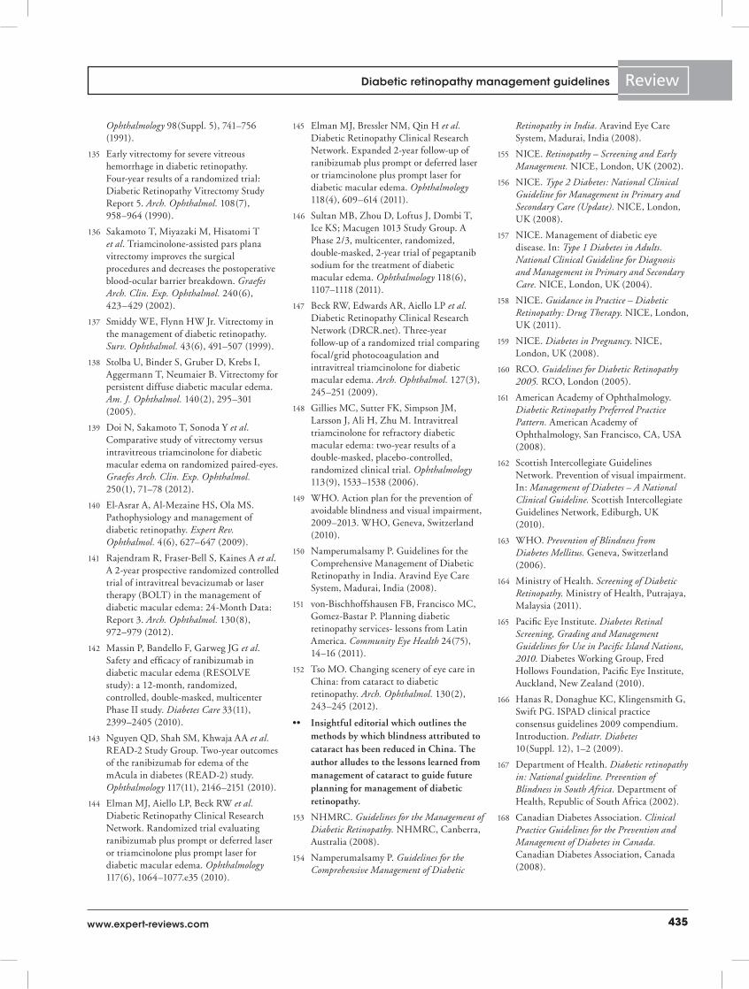
435www.expert-reviews.com
Review
Ophthalmology 98(Suppl. 5), 741–756 (1991).
135 Early vitrectomy for severe vitreous hemorrhage in diabetic retinopathy. Four-year results of a randomized trial: Diabetic Retinopathy Vitrectomy Study Report 5. Arch. Ophthalmol. 108(7), 958–964 (1990).
136 Sakamoto T, Miyazaki M, Hisatomi T et al. Triamcinolone-assisted pars plana vitrectomy improves the surgical procedures and decreases the postoperative blood-ocular barrier breakdown. Graefes Arch. Clin. Exp. Ophthalmol. 240(6), 423–429 (2002).
137 Smiddy WE, Flynn HW Jr. Vitrectomy in the management of diabetic retinopathy. Surv. Ophthalmol. 43(6), 491–507 (1999).
138 Stolba U, Binder S, Gruber D, Krebs I, Aggermann T, Neumaier B. Vitrectomy for persistent diffuse diabetic macular edema. Am. J. Ophthalmol. 140(2), 295–301 (2005).
139 Doi N, Sakamoto T, Sonoda Y et al. Comparative study of vitrectomy versus intravitreous triamcinolone for diabetic macular edema on randomized paired-eyes. Graefes Arch. Clin. Exp. Ophthalmol. 250(1), 71–78 (2012).
140 El-Asrar A, Al-Mezaine HS, Ola MS. Pathophysiology and management of diabetic retinopathy. Expert Rev. Ophthalmol. 4(6), 627–647 (2009).
141 Rajendram R, Fraser-Bell S, Kaines A et al. A 2-year prospective randomized controlled trial of intravitreal bevacizumab or laser therapy (BOLT) in the management of diabetic macular edema: 24-Month Data: Report 3. Arch. Ophthalmol. 130(8), 972–979 (2012).
142 Massin P, Bandello F, Garweg JG et al. Safety and efficacy of ranibizumab in diabetic macular edema (RESOLVE study): a 12-month, randomized, controlled, double-masked, multicenter Phase II study. Diabetes Care 33(11), 2399–2405 (2010).
143 Nguyen QD, Shah SM, Khwaja AA et al. READ-2 Study Group. Two-year outcomes of the ranibizumab for edema of the mAcula in diabetes (READ-2) study. Ophthalmology 117(11), 2146–2151 (2010).
144 Elman MJ, Aiello LP, Beck RW et al. Diabetic Retinopathy Clinical Research Network. Randomized trial evaluating ranibizumab plus prompt or deferred laser or triamcinolone plus prompt laser for diabetic macular edema. Ophthalmology 117(6), 1064–1077.e35 (2010).
145 Elman MJ, Bressler NM, Qin H et al. Diabetic Retinopathy Clinical Research Network. Expanded 2-year follow-up of ranibizumab plus prompt or deferred laser or triamcinolone plus prompt laser for diabetic macular edema. Ophthalmology 118(4), 609–614 (2011).
146 Sultan MB, Zhou D, Loftus J, Dombi T, Ice KS; Macugen 1013 Study Group. A Phase 2/3, multicenter, randomized, double-masked, 2-year trial of pegaptanib sodium for the treatment of diabetic macular edema. Ophthalmology 118(6), 1107–1118 (2011).
147 Beck RW, Edwards AR, Aiello LP et al. Diabetic Retinopathy Clinical Research Network (DRCR.net). Three-year follow-up of a randomized trial comparing focal/grid photocoagulation and intravitreal triamcinolone for diabetic macular edema. Arch. Ophthalmol. 127(3), 245–251 (2009).
148 Gillies MC, Sutter FK, Simpson JM, Larsson J, Ali H, Zhu M. Intravitreal triamcinolone for refractory diabetic macular edema: two-year results of a double-masked, placebo-controlled, randomized clinical trial. Ophthalmology 113(9), 1533–1538 (2006).
149 WHO. Action plan for the prevention of avoidable blindness and visual impairment, 2009–2013. WHO, Geneva, Switzerland (2010).
150 Namperumalsamy P. Guidelines for the Comprehensive Management of Diabetic Retinopathy in India. Aravind Eye Care System, Madurai, India (2008).
151 von-Bischhoffshausen FB, Francisco MC, Gomez-Bastar P. Planning diabetic retinopathy services- lessons from Latin America. Community Eye Health 24(75), 14–16 (2011).
152 Tso MO. Changing scenery of eye care in China: from cataract to diabetic retinopathy. Arch. Ophthalmol. 130(2), 243–245 (2012).
•• InsightfuleditorialwhichoutlinesthemethodsbywhichblindnessattributedtocataracthasbeenreducedinChina.Theauthoralludestothelessonslearnedfrommanagementofcataracttoguidefutureplanningformanagementofdiabeticretinopathy.
153 NHMRC. Guidelines for the Management of Diabetic Retinopathy. NHMRC, Canberra, Australia (2008).
154 Namperumalsamy P. Guidelines for the Comprehensive Management of Diabetic
Retinopathy in India. Aravind Eye Care System, Madurai, India (2008).
155 NICE. Retinopathy – Screening and Early Management. NICE, London, UK (2002).
156 NICE. Type 2 Diabetes: National Clinical Guideline for Management in Primary and Secondary Care (Update). NICE, London, UK (2008).
157 NICE. Management of diabetic eye disease. In: Type 1 Diabetes in Adults. National Clinical Guideline for Diagnosis and Management in Primary and Secondary Care. NICE, London, UK (2004).
158 NICE. Guidance in Practice – Diabetic Retinopathy: Drug Therapy. NICE, London, UK (2011).
159 NICE. Diabetes in Pregnancy. NICE, London, UK (2008).
160 RCO. Guidelines for Diabetic Retinopathy 2005. RCO, London (2005).
161 American Academy of Ophthalmology. Diabetic Retinopathy Preferred Practice Pattern. American Academy of Ophthalmology, San Francisco, CA, USA (2008).
162 Scottish Intercollegiate Guidelines Network. Prevention of visual impairment. In: Management of Diabetes – A National Clinical Guideline. Scottish Intercollegiate Guidelines Network, Ediburgh, UK (2010).
163 WHO. Prevention of Blindness from Diabetes Mellitus. Geneva, Switzerland (2006).
164 Ministry of Health. Screening of Diabetic Retinopathy. Ministry of Health, Putrajaya, Malaysia (2011).
165 Pacific Eye Institute. Diabetes Retinal Screening, Grading and Management Guidelines for Use in Pacific Island Nations, 2010. Diabetes Working Group, Fred Hollows Foundation, Pacific Eye Institute, Auckland, New Zealand (2010).
166 Hanas R, Donaghue KC, Klingensmith G, Swift PG. ISPAD clinical practice consensus guidelines 2009 compendium. Introduction. Pediatr. Diabetes 10(Suppl. 12), 1–2 (2009).
167 Department of Health. Diabetic retinopathy in: National guideline. Prevention of Blindness in South Africa. Department of Health, Republic of South Africa (2002).
168 Canadian Diabetes Association. Clinical Practice Guidelines for the Prevention and Management of Diabetes in Canada. Canadian Diabetes Association, Canada (2008).
Diabetic retinopathy management guidelines
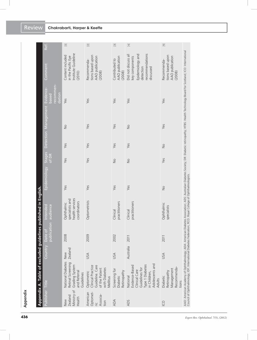
Expert Rev. Ophthalmol. 7(5), (2012)436
Review
Ap
pen
dix
A. T
able
of
excl
ud
ed g
uid
elin
es p
ub
lish
ed in
En
glis
h.
Pub
lish
erTi
tle
Co
un
try
Dat
e o
f p
ub
licat
ion
Inte
nd
ed
aud
ien
ceEp
idem
iolo
gy
Stag
es
of
DR
Det
ecti
on
Man
agem
ent
Evid
ence
-b
ased
re
com
men
-d
atio
n
Co
mm
ent
Ref
.
New
Ze
alan
d M
inis
try
of
Hea
lth
Nat
iona
l Dia
bete
s Re
tinal
Scr
eeni
ng
Gra
ding
Sys
tem
an
d Re
ferr
al
Gui
delin
es
New
Ze
alan
d20
08
Oph
thal
mic
sp
ecia
lists
and
he
alth
ser
vice
s co
ordi
nato
rs
Yes
Yes
Yes
No
Yes
Con
tent
incl
uded
in
the
Pac
ific
Eye
Inst
itute
Gui
delin
e (2
010
)
[1]
Am
eric
an
Opt
omet
-ric
A
ssoc
ia-
tion
Opt
omet
ric
Clin
ical
Pra
ctic
e G
uide
line.
Car
e of
the
Pat
ient
w
ith D
iabe
tes
Mel
litus
USA
2009
Opt
omet
rists
Yes
Yes
Yes
Yes
Yes
Reco
mm
enda
-tio
ns b
ased
upo
n A
AO
pub
licat
ion
(20
08
)
[2]
AD
ASc
reen
ing
for
Dia
betic
Re
tinop
athy
USA
2002
Clin
ical
pr
actit
ione
rsYe
sN
oYe
sYe
sYe
sC
ontr
ibut
ed t
o A
AO
pub
licat
ion
(20
08
)
[3]
AD
SN
atio
nal
Evid
ence
-Bas
ed
Clin
ical
Car
e G
uide
lines
for
Ty
pe 1
Dia
bete
s in
Chi
ldre
n,
Ado
lesc
ents
and
A
dult
s
Aus
tral
ia20
11C
linic
al
prac
titio
ners
Yes
No
Yes
No
Yes
Did
not
dis
cuss
all
key
com
pone
nts
[4]
Epid
emio
logy
and
de
tect
ion
reco
mm
enda
tions
di
scus
sed
ICO
Dia
betic
Re
tinop
athy
M
anag
emen
t Re
com
men
da-
tions
USA
2011
Oph
thal
mic
sp
ecia
lists
No
Yes
No
Yes
Yes
Reco
mm
enda
-tio
ns b
ased
upo
n A
AO
pub
licat
ion
(20
08
)
[5]
AA
O: A
mer
ican
Aca
dem
y of
Oph
thal
mol
ogy;
AD
A: A
mer
ican
Dia
bete
s A
ssoc
iati
on; A
DS:
Aus
tral
ian
Dia
bete
s So
ciet
y; D
R: D
iabe
tic r
etin
opat
hy; H
TBS:
Hea
lth T
echn
olog
y Bo
ard
for
Scot
land
; IC
O: I
nter
nati
onal
C
ounc
il of
Oph
thal
mol
ogy;
IDF:
Inte
rnat
iona
l Dia
bete
s Fe
dera
tion
; RC
O: R
oyal
Col
lege
of
Oph
thal
mol
ogis
ts.
Ap
pen
dixChakrabarti, Harper & Keeffe
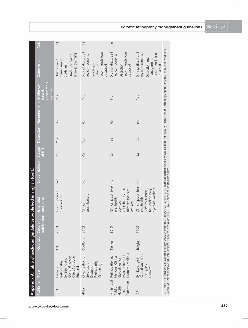
437www.expert-reviews.com
ReviewA
pp
end
ix A
. Tab
le o
f ex
clu
ded
gu
idel
ines
pu
blis
hed
in E
ng
lish
(co
nt.
).
Pub
lish
erTi
tle
Co
un
try
Dat
e o
f p
ub
licat
ion
Inte
nd
ed
aud
ien
ceEp
idem
iolo
gy
Stag
es
of
DR
Det
ecti
on
Man
agem
ent
Evid
ence
-b
ased
re
com
men
-d
atio
n
Co
mm
ent
Ref
.
RCO
Dia
betic
Re
tinop
athy
Sc
reen
ing
and
Oph
thal
mol
ogy
Clin
ic S
et U
p in
En
glan
d
UK
2010
Hea
lth s
ervi
ces
coor
dina
tors
Yes
Yes
Yes
No
Yes
Not
a c
linic
al
man
agem
ent
guid
elin
e
[6]
Use
ful f
or h
ealth
se
rvic
es p
lann
ing
HTB
SO
rgan
isat
ion
of
Serv
ices
for
D
iabe
tic
Retin
opat
hy
Scre
enin
g
Scot
land
2002
Clin
ical
pr
actit
ione
rsN
oYe
sYe
sN
oYe
sD
id n
ot d
iscu
ss a
ll ke
y co
mpo
nent
s[7]
Gra
ding
and
de
tect
ion
reco
mm
enda
tions
di
scus
sed
Min
istr
y of
Pu
blic
H
ealth
an
d Sa
nita
tion
Retin
opat
hy in
: N
atio
nal C
linic
al
Gui
delin
es f
or
Man
agem
ent
of
Dia
bete
s M
ellit
us
Ken
ya20
10C
linic
al p
ract
ition
-er
s, h
ealth
se
rvic
es
coor
dina
tors
and
pr
imar
y ey
e ca
re
wor
kers
No
No
Yes
No
No
Did
not
dis
cuss
all
key
com
pone
nts
[8]
Det
ectio
n re
com
men
datio
ns
disc
usse
d
IDF
Eye
Dam
age
in:
Glo
bal G
uide
line
for
Type
2
Dia
bete
s
Belg
ium
2005
Clin
ical
pra
ctiti
on-
ers,
hea
lth
serv
ices
coo
rdin
a-to
rs a
nd p
rimar
y ey
e ca
re w
orke
rs
No
No
Yes
Yes
Yes
Did
not
dis
cuss
all
key
com
pone
nts
Det
ectio
n an
d m
anag
emen
t re
com
men
datio
ns
disc
usse
d
AA
O: A
mer
ican
Aca
dem
y of
Oph
thal
mol
ogy;
AD
A: A
mer
ican
Dia
bete
s A
ssoc
iati
on; A
DS:
Aus
tral
ian
Dia
bete
s So
ciet
y; D
R: D
iabe
tic r
etin
opat
hy; H
TBS:
Hea
lth T
echn
olog
y Bo
ard
for
Scot
land
; IC
O: I
nter
nati
onal
C
ounc
il of
Oph
thal
mol
ogy;
IDF:
Inte
rnat
iona
l Dia
bete
s Fe
dera
tion
; RC
O: R
oyal
Col
lege
of
Oph
thal
mol
ogis
ts.
Diabetic retinopathy management guidelines
Expert Rev. Ophthalmol. 7(5), (2012)438
Review
Appendix B. Table of published guidelines not in English.
Publisher Title Country Date of publication Language of publication
Ref.
SERV Guidelines of Clinical Practice of the SERV: Management of Ocular Complications of Diabetes. Diabetic Retinopathy and Macular Oedema
Spain 2009 Spanish [9]
Norway College of General Practitioners
Diabetic Retinopathy Screening in: Guidelines for Diabetes in General Practice
Norway 2000 Norwegian [10]
Ophthalmological Society of Finland
Diabetic Retinopathy. Current Care Summary
Finland 2006 Finnish [11]
Medica del Insituto Mexicano del Seguro
Diagnosis and Treatment of Diabetic Retinopathy
Mexico 2011 Spanish [12]
Slovene Medical Society
Guidelines for Screening and Treatment for Diabetic Retinopathy
Slovenia 2010 Slovene [13]
Tagaki H Guideline-Based Planning for the Treatment of Diabetic Retinopathy
Japan 2010 Japanese [14]
Professional Association of Ophthalmologists in Germany
Treatment of Diabetic Maculopathy Germany 2011 German [15]
Polak et al. Revised Guideline for Diabetic Retinopathy: Screening, Diagnosis and Treatment
The Netherlands 2008 Dutch [16]
Deb et al. Screening for Diabetic Retinopathy in France
France 2004 French [17]
IAPB Latin America Clinical Practice Guidelines. Diabetic Retinopathy – Latin America
Ecuador 2011 Spanish [18]
Kalvodová Screening for Diabetic Retinopathy in the Czech Republic Guideline
Czech Republic 2002 Czech [19]
IAPB: International Agency for Prevention of Blindness; SERV: Spanish Retina and Vitreous Society.
Chakrabarti, Harper & Keeffe
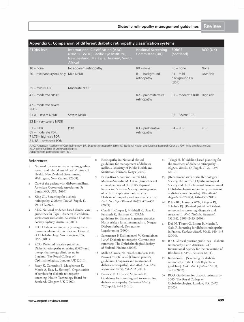
439www.expert-reviews.com
Review
References1 National diabetes retinal screening grading
system and referral guidelines. Ministry of Health, New Zealand Government, Wellington, New Zealand (2008).
2 Care of the patient with diabetes mellitus. American Optometric Association, St Louis, MO, USA (2009).
3. King GL. Screening for diabetic retinopathy. Diabetes Care 25(Suppl. 1), 90–93 (2002).
4 ADS. National evidence-based clinical care guidelines for Type 1 diabetes in children, adolescents and adults. Australian Diabetes Society, Sydney, Australia (2011).
5 ICO. Diabetic retinopathy (management recommendations). International Council of Ophthalmology, San Francisco, CA, USA (2011).
6 RCO. Preferred practice guideline. Diabetic retinopathy screening (DRS) and the ophthalmology clinic set up in England. The Royal College of Ophthalmologists, London, UK (2010).
7 Facey K, Cummins E, Macpherson K, Morris A, Reay L, Slattery J. Organisation of services for diabetic retinopathy screening. Health Technology Board for Scotland, Glasgow, UK (2002).
8 Retinopathy in: National clinical guidelines for management of diabetes mellitus. Ministry of Public Health and Sanitation, Nairobi, Kenya (2010).
9 Pareja-Ríos A, Serrano-García MA, Marrero-Saavedra MD et al. [Guidelines of clinical practice of the SERV (Spanish Retina and Vitreous Society): management of ocular complications of diabetes. Diabetic retinopathy and macular oedema]. Arch. Soc. Esp. Oftalmol. 84(9), 429–450 (2009).
10 Claudi T, Cooper J, Midthjell K, Daae C, Furuseth K, Hanssen K. NSAMs guidelines for diabetes in general practice. Norsk selskap for allmennmedisin, Norges Diabetesforbund, Den norske Lægeforening (2000).
11 Summanen P, Kallionienmi V, Komulainen J et al. Diabetic retinopathy. Current care summary. The Ophthalmological Society of Finland, Finland (2006).
12 Millán-Gámez YK, Wacher-Rodarte NH, Bravo-Ortiz JC et al. [Clinical practice guidelines. Diagnosis and treatment of diabetic retinopathy]. Rev. Med. Inst. Mex. Seguro Soc. 49(5), 551–562 (2011).
13 Petrovic M, Urbancic M, Sevsek D. Guidelines for screening and treatment for diabetic retinopathy. Slovenian Med. J. 79(Suppl.), 7–18 (2010).
14 Takagi H. [Guideline-based planning for the treatment of diabetic retinopathy]. Nippon. Rinsho. 68(Suppl. 9), 291–297 (2010).
15 [Recommendation of the Retinological Society, the German Ophthalmological Society and the Professional Association of Ophthalmologists in Germany: treatment of diabetic maculopathy]. Klin Monbl Augenheilkd 228(5), 446–459 (2011).
16 Polak BC, Hartstra WW, Ringens PJ, Scholten RJ. [Revised guideline ‘Diabetic retinopathy: screening, diagnosis and treatment’]. Ned. Tijdschr. Geneeskd. 152(44), 2406–2413 (2008).
17 Deb N, Thuret G, Estour B, Massin P, Gain P. Screening for diabetic retinopathy in France. Diabetes Metab. 30(2), 140–145 (2004).
18 ICO. Clinical practice guidelines – diabetic retinopathy, Latin America. ICO International Agency for the Prevention of Blindness (IAPB), Ecuador (2011).
19 Kalvodová B. [Screening for diabetic retinopathy in the Czech Republic – guideline]. Cesk. Slov. Oftalmol. 58(1), 3–10 (2002).
20 RCO. Guidelines for diabetic retinopathy 2005. The Royal College of Ophthalmologists, London, UK, 2–72 (2005).
Appendix C. Comparison of different diabetic retinopathy classification systems.
ETDRS level International Classification (AAO, NHMRC, WHO, Pacific Eye Institute, New Zealand, Malaysia, Aravind, South Africa)
National Screening Committee (UK)
SDRGS (Scotland)
RCO (UK)
10 – none No apparent retinopathy R0 – none R0 – none None
20 – microaneurysms only Mild NPDR R1 – background retinopathy
R1 – mild background DR (BDR)
Low Risk
35 – mild NPDR Moderate NPDR
43 – moderate NPDR R2 – preproliferative retinopathy
R2 – moderate BDR High risk
47 – moderate severe NPDR
53 A – severe NPDR Severe NPDR R3 – Severe BDR
53 E – very severe NPDR
61 – PDR65 – moderate PDR71,75 – high-risk PDR81, 85 – advanced PDR
PDR R3 – proliferative retinopathy
R4 – PDR PDR
AAO: American Academy of Ophthalmology; DR: Diabetic retinopathy; NHMRC: National Health and Medical Research Council; PDR: Mild proliferative DR; RCO: Royal College of Ophthalmologists. Adapted with permission from [20].
Diabetic retinopathy management guidelines


![The Guide - Diabetic Retinopathy - Vision Lossvisionloss.org.au/wp-content/uploads/2016/05/The... · the guide [diabetic retinopathy] What is Diabetic Retinopathy? Diabetic Retinopathy](https://img.pdfslide.us/doc/110x75/5e3ed00bf9c32e41ea6578a8/the-guide-diabetic-retinopathy-vision-the-guide-diabetic-retinopathy-what.jpg)