Embed Size (px)
Citation preview

2/1/2017
1
Diabetic Retinopathy Update 2017
Sundeep Dev, MDVitreoRetinal Surgery, PA
Basis for DR management standards for past 40 years
Diabetic Retinopathy Study (DRS) 1971-1989 and Early Treatment of Diabetic Retinopathy Study (ETDRS) 1979-1990
- Defined – high risk proliferative DR- Staging system for DR – modern wide field
peripheral imaging not available- Established clear benefit for laser treatment for
CSDME and HRPDR – no pharmacologic options then
ETDRS: CSDME
Clinically Significant Macular Edema –Focal/Grid Laser
Retinal thickening <500 μm from fovea Hard exudates <500 μm from fovea with edemaRetinal thickening ≥1 disc area within 1 disc diameter of fovea
ETDRS/DRS: HRPDR
High Risk PDR – PRP laserNVD > 1/3 Disc areaAny NVD with Vitreous hemorrhageNVE > 1/2 Disc area with VH
Laser Photocoagulation: Efficacy
1. ETDRS. Arch Ophthalmol. 1995;113:1144-1145. 2. DRS. Ophthalmology. 1978;85:82-106.
Outcomes in CSDME:
50% reduction in moderate visual loss
15% Increased chance of mod visual gain
Reduced retinal thickening
In CSDME and Less Severe DR1
% of Patients with Moderate Visual Loss
Years after randomization
Focal + FU Scatter (N=299)
Deferral (N=611)
*3-step loss on ETDRS chart
1 2 3 4
33%
13%
60
50
40
30
20
10
0
P<0.01
P<0.001
Outcomes in PDR:
50% reduction in severe visual loss
Indicated only for high risk PDR
*
*
In PDR2
% of Patients with Severe Visual Loss
Years after randomization
*P<0.001
*VA<5/200At 2 consecutive visits
60
50
40
30
20
10
0
Photocoagulation
N=1681
N=1782
1 2 3 4 5
Untreated
ETDRS
Clinically Significant Edema (CSDME) –– need to redefine with presence of OCT imaging
and intravitreal pharmacologic therapiesNewer terminology:• Center involving vs non-center involving• Focal vs Diffuse – definitions vary, but based on:
• Source of fluorescein leakage• Extent and location of macular thickening on
OCT• Pattern of lipid exudates

2/1/2017
2
Diffuse Central Macular edema with heavy lipid
Are prior clinical trials still applicable today - PDR?
ETDRS/DRS studies• HR PDR – required significant NV or NV with
presence of Vitreous blood• Widefield FA to assess ischemia unavailable• PRP was only Rx and given discomfort and
side effects, higher risk threshold to use• Better laser technology now• Pharmacologic therapy did not exist
Macular Edema Current Pharmacologic options
• FDA Approved therapy– Ranibizumab (Lucentis 0.3 mg) – RIDE/RISE
($1150)
– Aflibercept (Eylea) – VIVID/VISTA ($2000)
– Dexamaethasone implant (Ozurdex) – MEAD ($1400)
– Fluocinolone implant (Iluvien) –FAME($9300)
• Off label therapy– Bevacizumab (Avastin) – DRCR.net ($70)
– Intravitreal Triamcinolone (Triessence) - $150
Ranibizumab (Lucentis) for DME
• 1st drug to be approved for DME • RIDE/RISE trials: 759 patients• Evaluated 0.3 mg vs. 0.5 mg (AMD dose)
vs. sham/laser• FDA approved 0.3 mg dose for DME 8/2012• Results equivalent to 0.5 mg and so 0.3 mg
felt to be a safer dose in higher cardiovascular risk diabetic patients
15
20
10
5
0
-5
0 12 24 36
Sham Sham /0.5 mg LUCENTIS 0.3 mg
BC
VA
ch
ange
fro
m b
asel
ine,
ETD
RS
lett
ers
12.4
4.511.7
2.5
Month
Day 7+4.4+1.3
10.6
1.0
Summary of RIDE/RISE Study

2/1/2017
3
Secondary Endpoint: Mean Change in OCT CFT Over Time
Last observation carried forward imputation method was used. Vertical bars are ±1 SE of the mean. CFT is defined as center point thickness. Independent review of OCT performed at the University of Wisconsin Fundus Photograph Reading Center.Brown DM, et al. Ophthalmology. 2013;120:2013-2022
-300
-250
-200
-150
-100
-50
0
0 6 12 18 24 30 36
Mea
n C
han
ge
in C
FT
(µ
m)
−255.2
−262.0
−129.5
1 2 3Day 7
Sham (n=257) RBZ 0.3 mg (n=250) RBZ 0.5 mg (n=252)
Month
−261.5−267.9
−206.7
RIDERISEPooled Ranibizumab for DME
• Patients in the RIDE/RISE trials that switched from sham to Lucentis after 2 years never achieved same vision benefit
• Benefit seems to be best within 12 months of diagnosis
Proportion of Subjects Losing ≥15 ETDRS Letters ( 3 lines) From Baseline at Month 24
(Secondary Endpoint)
RIDE/RISE
The last observation carried forward imputation method was used. Reported percentages and differences vs sham are unadjusted, test and p-value are adjusted for baseline visual acuity (≤55, >55 letters), baseline HbA1c (≤8%, >8%) and prior treatment for DME (yes, no). Boyer, AAO, 2011. ETDRS = Early Treatment Diabetic Retinopathy Study.
Only 2% of LUCENTIS treated patients lost 3 or more lines in vision
Sham(n=257)
0.3 mg(n=250)
Prop
ortio
n of
sub
ject
s lo
sing
≥
15 le
tter
s
Pooled RIDE and RISE
Systemic Serious Adverse Events Potentially Related to VEGF Inhibition
aIncludes Sham and No crossover to 0.5 mg, and Sham and Crossover to 0.5 mg. Some patients may have experienced multiple events.LUCENTIS FDA Briefing Book.
RIDE/RISE
24-Month Pooled RIDEand RISE
36-Month Pooled RIDEand RISE
SAE Group Term, n (%)Sham
(n=250)LUCENTIS
0.3 mg (n=250)Sham/0.5 mg
(n=251)aLUCENTIS
0.3 mg (n=250)
Any systemic SAE 29 (11.6%) 27 (10.8%) 33 (13.1%) 42 (16.8%)
Any bleeding/hemorrhage adverse event 7 (2.8%) 7 (2.8%) 8 (3.2%) 11 (4.4%)
Bleeding/hemorrhage (CNS and cerebrovascular hemorrhage)
3 (1.2%) 3 (1.2%) 3 (1.2%) 5 (2.0%)
Bleeding/hemorrhage (non-CNS hemorrhage)
4 (1.6%) 4 (1.6%) 5 (2.0%) 6 (2.4%)
Congestive heart failure 11 (4.4%) 7 (2.8%) 13 (5.2%) 10 (4.0%)
Fistulae (other) 0 1 (0.4%) 0 2 (0.8%)
Gastrointestinal perforation 0 0 0 0
Hypertension 1 (0.4%) 3 (1.2%) 1 (0.4%) 4 (1.6%)
Proteinuria 0 0 0 0
Thromboembolic event, arterial 17 (6.8%) 14 (5.6%) 21 (8.4%) 26 (10.4%)
Thromboembolic event, venous 1 (0.4%) 4 (1.6%) 1 (0.4%) 4 (1.6%)
Wound-healing complications 0 0 0 0
Ranibizumab for DME – longer term
• RISE/RIDE – extension beyond month 36– patients maintained vision and anatomic benefit with PRN dosing and fewer injections (mean 4.5 injections over next 14.1 months)1
- 25% didn’t require further injections
1 Ophthalmology 2015;122:2504-13
Mean BCVA Maintained During OLE*
55
60
65
70
75
80
85
0 6 12 18 24 30 36 42 48 54
*Data become unstable after Month 54 due to the low number of patients at that point. †Treatment during core study. Observed data. OLE = open-label extension; RBZ = ranibizumab.Boyer DS, et al. Ophthalmology. 2015;122:2504-2513.e1.
Sham ControlSham/
RBZ 0.5 mg Crossover
OLE*
163 161 162 163 163 164 124 82 35168 168 167 170 168 172 126 101 39158 158 155 159 152 161 115 95 47
Patients, n†
Sham/crossoverRBZ 0.3 mgRBZ 0.5 mg
PRN RBZ 0.5 mg
Mea
n B
CV
A (
ETD
RS
Lett
ers)
Month
Sham/crossover†
RBZ 0.3 mg†
RBZ 0.5 mg†
RIDERISEPooled
2/1/2017
4
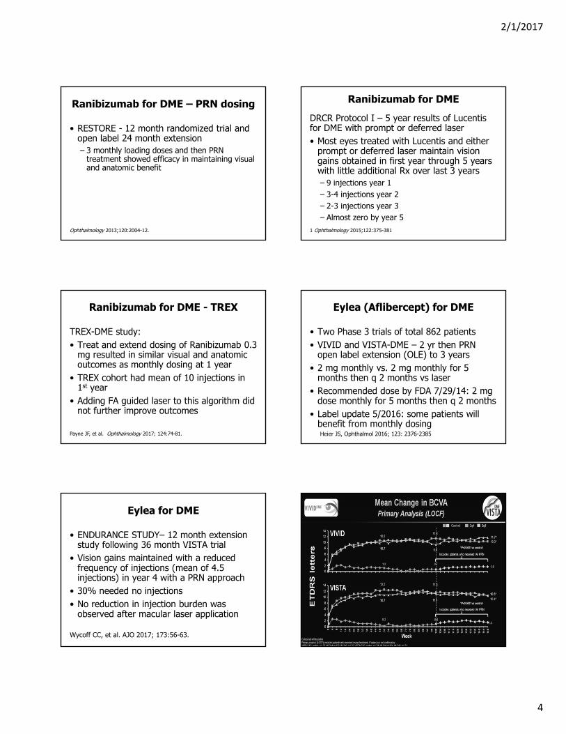
Ranibizumab for DME – PRN dosing
• RESTORE - 12 month randomized trial and open label 24 month extension– 3 monthly loading doses and then PRN
treatment showed efficacy in maintaining visual and anatomic benefit
Ophthalmology 2013;120:2004-12.
Ranibizumab for DME
DRCR Protocol I – 5 year results of Lucentisfor DME with prompt or deferred laser• Most eyes treated with Lucentis and either
prompt or deferred laser maintain vision gains obtained in first year through 5 years with little additional Rx over last 3 years– 9 injections year 1– 3-4 injections year 2– 2-3 injections year 3– Almost zero by year 5
1 Ophthalmology 2015;122:375-381
Ranibizumab for DME - TREX
TREX-DME study:• Treat and extend dosing of Ranibizumab 0.3
mg resulted in similar visual and anatomic outcomes as monthly dosing at 1 year
• TREX cohort had mean of 10 injections in 1st year
• Adding FA guided laser to this algorithm did not further improve outcomes
Payne JF, et al. Ophthalmology 2017; 124:74-81.
Eylea (Aflibercept) for DME
• Two Phase 3 trials of total 862 patients• VIVID and VISTA-DME – 2 yr then PRN
open label extension (OLE) to 3 years• 2 mg monthly vs. 2 mg monthly for 5
months then q 2 months vs laser• Recommended dose by FDA 7/29/14: 2 mg
dose monthly for 5 months then q 2 months• Label update 5/2016: some patients will
benefit from monthly dosingHeier JS, Ophthalmol 2016; 123: 2376-2385
Eylea for DME
• ENDURANCE STUDY– 12 month extension study following 36 month VISTA trial
• Vision gains maintained with a reduced frequency of injections (mean of 4.5 injections) in year 4 with a PRN approach
• 30% needed no injections• No reduction in injection burden was
observed after macular laser application
Wycoff CC, et al. AJO 2017; 173:56-63.

2/1/2017
5
Diabetic Retinopathy
• 2/2015 – Lucentis approved to treat DR in patients with DME
• 3/25/15 - Eylea approved to treat DR in patients with DME
• Both shown to reduce progression and improve grade of retinopathy
≥ 2-Step Improvements in DR Seen With Ranibizumab as Early as Month 3
3 3 35
24
18
27
3438 39
16
2731
3639
0
10
20
30
40
50
3 6 12 24 36
Sham/ Crossover (n = 239) RBZ 0.3 mg (n = 234) RBZ 0.5 mg (n = 234)
Pat
ient
s W
ith ≥
2-S
tep
DR
Im
prov
emen
t, %
P < .0001
Month• After 12 months of 0.5 mg RBZ, sham/crossover patients experienced less DR
improvement than patients who initially received 12 months of RBZ
RIDE/RISE Pooled. DR = diabetic retinopathy; RBZ = ranibizumab. Wykoff C, et al. Presented at: Ret Soc 2015.
Most DR Improvements Maintained During OLE
29
21
6 5
46
27
18
11
44
36
19
8
0
10
20
30
40
50
60
Month 36Monthly Tx
Month 48 (OLE)PRN Tx
Month 36Monthly Tx
Month 48 (OLE)PRN Tx
Prior Sham/Crossover (n = 124) RBZ 0.3 mg/ Prior RBZ 0.3 mg (n = 125)
RBZ 0.5 mg/ Prior RBZ 0.5 mg (n = 118)
Pa
tien
ts,
%
DRSS Change From Baseline
Patients enrolled in the open-label extension with diabetic retinopathy outcomes at baseline and month 48.DR = diabetic retinopathy; DRSS = Diabetic Retinopathy Severity Score; OLE = open-label extension; PRN = as needed; RBZ = ranibizumab; Tx = treatment. Sun J, et al. Presented at: ARVO 2015.
≥ 2-Step Improvement ≥ 3-Step Improvement

2/1/2017
6
ANDROID STUDY• Evaluate change in peripheral perfusion
using UWFA after treatment with Aflibercept(Eylea) in patients with PDR and CRVO
• Eylea q mo for 12 months vs. q mo for 6 months then q 2 mo for 6 mo
• Both groups followed to month 18 with PRN• UWFA - 55% reduction in area of peripheral
non-perfusion by 12 months in both groups• At Month 18 with PRN, 50% maintained
benefit without injections, 50% regressedHeier J, presented at AAO Retina 10/2016
What about Bevacizumab?
• Bevacizumab (Avastin) - off label, compounded at pharmacy – commonly used due to huge cost savings.
DRCR.net Protocol T
• Comparative trial of Avastin, Lucentis and Eylea for center involved DME (n=660)
• Year 1: q 4 week exam and retreatment if improvement or worsened by >5 letters or OCT CST by > 10%.
• PRN once acheived 20/20 and CST normal (250 um or less)
• Laser at 6 mo if vision/OCT stable for 2 consecutive injections and persistent edema
• Year 2: visits could be extended to 16 wks. ReRx if vision or OCT worsened
Mean Change in Visual Acuity Over 2 Years
Full Cohort
Week 52 Week 104
Bevacizumab +9.7 +10.0
Ranibizumab +11.2 +12.3
Aflibercept +13.3 +12.8
104-Week Treatment Group Comparison• Aflibercept vs. Bevacizumab P = 0.02• Aflibercept vs. Ranibizumab P = 0.47• Ranibizumab vs. Bevacizumab P = 0.11
Mean Change in Visual AcuityBaseline Visual Acuity 20/32 to 20/40
Week 52 Week 104
Bevacizumab +7.5 +6.8
Ranibizumab +8.3 +8.6
Aflibercept +8.0 +7.8
~50% of Cohort
104-Week Treatment Group Comparison• Aflibercept vs. Bevacizumab P = 0.51• Aflibercept vs. Ranibizumab P = 0.51• Ranibizumab vs. Bevacizumab P = 0.31
Mean Change in OCT CST Over 2 YearsBaseline Visual Acuity 20/32 to 20/40
Week 52 Week 104
Bevacizumab -67 -68
Ranibizumab -110 -126
Aflibercept -129 -133
Ranibizumab/Aflibercept resulted in singificant OCT CST improvement vs. Bevacizumab

2/1/2017
7
Mean Change in Visual AcuityBaseline Visual Acuity 20/50 or Worse
Week 52 Week 104
Bevacizumab +11.8 +13.3
Ranibizumab +14.2 +16.1
Aflibercept +18.9 +18.3
~50% of Cohort
104-Week Treatment Group Comparison• Aflibercept vs. Bevacizumab P = 0.02• Aflibercept vs. Ranibizumab P = 0.18• Ranibizumab vs. Bevacizumab P = 0.18
Mean Change in OCT CST Over 2 YearsBaseline Visual Acuity 20/50 or Worse
Week 52 Week 104
Bevacizumab -135 -185
Ranibizumab -176 -174
Aflibercept -210 -211
2-Year Treatment Group Comparison:• Aflibercept vs. Bevacizumab P = 0.01• Aflibercept vs. Ranibizumab P = 0.18• Ranibizumab vs. Bevacizumab P = 0.18
Results: DME Treatment: # anti-VEGF injections
Aflibercept Bevacizumab Ranibizumab Global P-value
# of injections: Median (25th, 75th percentile)
Year 1 9 (8, 11) 10 (8, 12) 10 (8, 11) 0.045
Year 2 5 (2, 7) 6 (2, 9) 6 (2, 9) 0.32
Over 2 Years 15 (11, 17) 16 (12, 20) 15 (11, 19) 0.08
Note: 98% of protocol required re-injections were given over 2 years.
DME Treatment: Adjunctive Laser
Aflibercept Bevacizumab Ranibizumab Global P-value
At least one focal/grid laser
Year 1 37% 56% 46% <0.001
Year 2 20% 31% 27% 0.046
Over 2 Years 41% 64% 52% <0.001
Pre-Specified Ocular Adverse Events through 2 Years (Study Eyes)
% of pts with atleast one event
Aflibercept(N = 224)
Bevacizumab(N = 218)
Ranibizumab(N = 218) Global P-value
No. of injections 2998 3115 3066
Endophthalmitis* 0 <1% 0 0.68
Inflammation 3% 1% 2% 0.69
Retinal detachment/tear 1% 1% 1% 1.0
Vitreoushemorrhage 7% 8% 5% 0.37
Injection-related cataract 1% <1% 0 0.38
IOP elevation 17% 12% 16% 0.31
Pre-specified APTC* Adverse Eventsthrough 2 years
% of pts with atleast one event
Aflibercept(N = 224)
Bevacizumab(N = 218)
Ranibizumab(N = 218) Global P-value
Non-fatal MI 3% 1% 3%
Non-fatal stroke <1% 3% 5%
Vascular Death 1% 4% 4%
Any APTC Event 5% 8% 12% 0.047

2/1/2017
8
Conclusions
• Vision gains (from baseline) at 2 years were seen in all 3 groups ~half the number of injections, slightly decreased frequency of visits, and decreased amounts of laser in the 2nd year
• Among eyes with better VA, no difference in 2-year vision outcomes were identified
• Among eyes with worse baseline VA:– Aflibercept, on average, had superior 2-year VA
outcomes compared with bevacizumab, although the difference was diminished
– The VA difference between aflibercept and ranibizumabthat was noted at 1 year had decreased at 2 years and was no longer statistically significant.
Conclusions
• All 3 agents reduce OCT thickness but bevacizumab appears to be less effective than aflibercept and ranibizumab
• The implication of the increased rate of APTC events with ranibizumab found in the current study is uncertain due to inconsistency with prior trials, warranting continued evaluation
Jampol LM, et al. JAMA Ophthalmol 2016; 134(12): 1429-1434(The source of the data is from the DRCR.net, but the analyses, content and conclusions presented have not been reviewed or approved by DRCR.net.)
DRCR.net Protocol T: My take
• Anti-VEGF clearly best treatment for center involved and diffuse edema
• All agents are effective• Lucentis/Eylea give better results especially in
more severe cases with worse vision ( <20/50)• Eylea results in more rapid improvement in severe
cases in year 1, no significant difference by year 2• Lucentis is cheaper than Eylea ($1150 vs. $2000)• If quiet, PRN ok - recurrence less risky as in AMD• Case by case basis – cost and insurance a factor• Avastin may be “good enough”
Ranibizumab for DME: Early and Long Term Responses
• Protocol I: EARLY Analylsis• ~ 40% of patients had suboptimal early
responses ( < 5 letter) and 40% had pronounced early responses (>10 letter) at 12 weeks with ranibizumab +/- laser
• Eyes with suboptimal early visual response by 12 weeks showed poorer long term visual outcomes than eyes with pronounced early response (mean 3.0 vs. 13.8 letters at 156 weeks)Gonzalez VH, et al. AJO 2016; 172: 72-79.
Ranibizumab for DME: Early and Long Term Responses
• Of the 40% with poor responses early, 28% did get some additional improvement over time
• Need to personalize Rx• Consider Steroid use in poor responders• Still those in Ranibizumab group with
deferred laser had best results compared to Ranibizumab plus prompt laser and Triamcinolone plus laser with deferred Ranibizumab – Protocol I
OCT - Predicting Visual Acuity
• Disruption of ellipsoid zone (IS/OS junction) correlates with poorer visual outcomes
• Increased Disorganization of retinal inner layers length (DRIL) - poorer visual outcomes
• DRIL - the horizontal extent for which boundaries between the ganglion cell –inner plexiform, inner nuclear and outer nuclear layer could not be identified within the central 3 mm
Balaratnasingam C et al; Ophthalmol 2016; 123:2352-2367. Lee; same; 2368-2375

2/1/2017
9
OCT
20/70 rapidly improved to 20/30 in 2 injections
20/125 rapildy improved to 20/40 in 3 injections
• Ellipsoid layer disruption
20/100 to 20/70 with 5 injections
• Ellipsoid layer disruption and DRIL
20/400 to 20/160 with 8 injections
• Ellipsoid layer disruption and DRIL
20/400 to 20/150 with 5 injections

2/1/2017
10
Summary: When to use anti-VEGF
Anti-VEGF therapy for Diffuse DME/DR:• largest chance of vision improvement• Prevents worsening DR and actually improves
grade of retinopathy and reverses ischemia while treating DME
• Low risk of vision loss from DR while on therapy• Highly effective for NV – compliance more critical• Long term sustained benefit – injection burden
seems to decrease over time• Very low risk of complications (ATE,
endophthalmitis)
Other treatments for Macular Edema Vitrectomy
• Diffuse macular edema caused by posterior hyaloid thickening
• Leakage results from direct vitreoretinaltraction
• OCT demonstrates elevation of the fovea
• DRCR Protocol D – after vitrectomy for DME/VMT – edema reduced in most eyes. 28-49% Va improvement,13-31% worse Va
• Several smaller studies have shown variable visual improvement
Ophthalmology 2010;117:1087-1093
Vitreomacular Traction and Macular Edema When to use steroids
1. Diffuse DME with no anatomic response to anti-VEGF – suspect inflammatory role
2. Vitrectomized eye requiring very frequent anti-VEGF therapy – implants work best
3. Prior to vitrectomy for ERM/VMT with diffuse DME
4. Post cataract surgery CME/DME – usually cystoid in pattern to suggest inflammatory cause
Post-Cataract surgery Diabetic CME Intravitreal Triamcinolone for DME
• DRCR Protocol B1– focal/grid more effective for DME with fewer side effects than 1 mg or 4 mg IVTA
• Short term effect, but can give an inexpensive trial to see if inflammatory mediated DME would improve – would use lower dose 1 mg
1. Ophthalmology 2008; 115:1447-1459

2/1/2017
11
Ozurdex for DME
• Dexamethasone implant – office injection• 3 year study for DME (MEAD study)• Evaluated q 3 months but could have
injections 6 months apart• Mean 4 treatments in 3 years• Study 1: 21% vs sham 12% gain > 3 lines• Study 2: 18% vs sham 10% gain > 3 lines
Ozurdex for DME• Approved for DME 6/30/2014• Main risk is 28% incidence of IOP increase
> 10 mm Hg and 68% cataract formation• 42% needed IOP lowering therapy• Contraindicated in patients with torn or
ruptured posterior capsule – anterior chamber migration and corneal edema
• Contraindicated in patients with significant glaucoma (c/d >0.8)
• Glaucoma surgery usually not needed. IOP normalizes by 6 months
Iluvien for DME- approved 2/2015
• 3 year study for DME• 1 injection of implant lasting 3 years• Steroid challenge prior recommended• Combined results of 2 trials: 28.7% treated
vs. 16.2% control had a > 3 line improvement 36 mo.
• Improvement seen at 3 weeks and sustained through month 36
• 18.4% sustained IOP rise, 5% needed glaucoma surgery
Is focal laser still of value?Considerations
• Bottom line - It works!
• Non central edema, focal edema• MA’s surrounded by circinate lipid• Central edema with leaking MA’s peripheral• Good vision and good perfusion, mild-mod
NPDR• Use modified focal/grid treatment• Micropulse laser studies ongoing - unclear

2/1/2017
12
Future options
• Refillable implantable ocular drug pump (Replenish, Inc, Pasadena, CA)
• Refillable drug port delivery (Genentech and ForSight Vision4)
Future options: Target DR
• Aerpio Therapeutics, AKB-9778 - activates TIE-2 transmembrane tyrosine kinase receptor on endothelial cells
• Inhibits permeability, blood retinal barrier breakdown and inflammation
• Subcutaneous self administered injection, high ocular bioavailability, rapid clearance systemically
Future options: AKB-9778
• Phase 2 TIME-2 study: 3 month safety/efficacy study of daily subcuinjections with and without monthly IntravitRanibizumab
• % achieving a > 2 step reduction in DRSS– 8.8% Ranibizumab alone– 10% AKB-9778 alone– 11.4% Ranibizumab/AKB-9778 group– AKB-9778 had an effect on fellow eye
Future options
• Angiopoietin-1 (Ang-1) – endogenous protein that can activate TIE-2 to stabilize vascular permeability
• Ang-2 competes with Ang-1 for binding and is high in patients with DME
• ROCHE-GENENTECH – testing an Ang-2 antibody: BOULEVARD study at VRS
• REGENERON trial forthcoming to compare Eylea alone with Eylea plus an antibody to Ang-2

2/1/2017
13
DRCR.net Protocol SPRP vs. Ranibizumab for PDR
• Summary of Ranibizumab results vs. PRP:Ranibizumab had:• Change in VA from baseline no worse than
PRP (non-inferiority trial)• Superior mean VA over the course of 2 yrs• Better mean VF outcomes• Decreased need for vitrectomies• Decreased development of central DME• PRP rarely given for failure or futility of
ranibizumab
Potential Impact of DME presence when treating PDR
• Presence of DME may influence the benefit of Ranibizumab over PRP
• When DME is present and treatment with anti-VEGF agent is planned, PRP may be unnecessary in most cases, if patient can be compliant with follow-up
• 6 monthly initial injections, mean number of injections was about 50% less in year 2
• Long term impact: unknown - A hybrid approach is likely the safest
DRCR.net Protocol SConclusions
• PRP has been effective for 40 years and remains an effective therapy today
• Ranibizumab is an effective alternative to PRP for atleast 2 years
• Ranibizumab may be first choice when DME is present
• No substantial safety concerns for atleast 2 years – need longer term data
• No reason to believe other anti-VEGF drugs wouldn’t also be effective
DRCR.net Protocol SPatient centered Oucomes
• Work productivity loss (WPAIQ) was 15.6% less (p=.005) with Ranibizumab at 1 year and 2.9% less (p=.54) at 2 years
• 97% Ranibizumab patients were 20/40 or better in 1 eye at 2 years compared to 87% PRP group (p=.005)
• No other differences found using NEI- VFQ-25
Beaulieu WT, Bressler NM,et al. AJO 2016; 170:206-213
Cost
• First year drug cost alone:• Ranibizumab ~ $13,800 1st year • Aflibercept ~ $18000-$24000• Bevacizumab ~ $900• Frequent examsBenefit:• Excellent drying of macula• VA improvement 7-11 letters
DRCR.net Protocol S: My take
• Individualize approach: what the patient wants, systemic status, compliance ability, other eye, presence of DME, degree of retinopthy, vision
• Anti-VEGF for DME/PDR• Also prefer anti-VEGF for aggressive PDR without
edema to quiet down first• Gradually integrate PRP and may require less
intense PRP• Often use atleast one injection prior to PRP to try
to prevent DME progression even in cases where long term treatment not possible

2/1/2017
14
21 y.o. female IDDM x 16 yrs.
1 year later after Avastin x6 and PRPx2 - quiet
Other helpful uses of anti-VEGF therapy in DR
Leaking MA’s without edema with need for cataract surgery – often can prevent worsening –( increased risk of central DME after CE shown in DRCR Protocol Q)
Pre-Vitrectomy surgery for PDR– reduces intraoperative and postoperative bleeding, shortens surgery
NVI/NVG – highly effective to calm eye down for glaucoma surgery and easier PRP
When is anti-VEGF not appropriate
Poor complianceSick patientsActive Traction
Consequences of failing to return for anti-VEGF treatment for PDR can be disastrous –perform PRP soon
Traction – use Anti-VEGF immediately prior to surgery to
avoid contraction
What is the role of Vitrectomy for DR
• Dense or recurring Vitreous hemorrhage –– hemorrhage often due to ongoing vitreous
traction– Anti-VEGF of limited benefit in dense VH, but
useful as an adjunct preoperatively– More immediate vision needs
• Macular subhyaloid hemorrhage• TRD• ERM/VMT and DME
2/1/2017
15
Avastin (1 day prior) followed with vitrectomy/PRP Summary
• Management of diabetic retinopathy as it develops– Careful screening of patients at risk – systemic
control
– Anti-VEGF therapy first line for central/diffiseedema. Focal laser for focal non-central DME
– Try steroid implants for refractory cases
– Anti-VEGF (often followed with gradual PRP for proliferative retinopathy
– Vitrectomy (often with preoperative anti-VEGF) for advanced complications
Thanks for attending!!!
![The Guide - Diabetic Retinopathy - Vision Lossvisionloss.org.au/wp-content/uploads/2016/05/The... · the guide [diabetic retinopathy] What is Diabetic Retinopathy? Diabetic Retinopathy](https://img.pdfslide.us/doc/110x75/5e3ed00bf9c32e41ea6578a8/the-guide-diabetic-retinopathy-vision-the-guide-diabetic-retinopathy-what.jpg)