Embed Size (px)
Citation preview

DIABETIC RETINOPATHY 2019
WHAT EVERY PROVIDER NEEDS TO KNOW ABOUT DIABETIC RETINAL EXAM
1
Mohan Iyer, MD
Athens Retina Center

Financial Disclosure: None

Learning Objectives
Basic anatomy of the eye
Recognize the importance of diabetic retinopathy as a public health problem
Identify the risk factors for diabetic retinopathy
Describe the stages of diabetic retinopathy
Understand the role of risk factor control and annual dilated eye exams in the prevention of vision loss

• What is the most common cause of vision loss among working age adults in the United States?
• 1. Glaucoma
• 2. Cataract
• 3. Diabetic Retinopathy
• 4. Retinal Detachment

• The most common cause of moderate vision loss in diabetic retinopathy is:
• 1. Refractive Change
• 2. Cataract
• 3. Diabetic Macular Edema
• 4. Proliferative Diabetic Retinopathy

A patient with Type II diabetes should get their first dilated eye exam:
1. Only when the vision is affected
2. In 3-5 years after initial diagnosis of diabetes
3. At the time of diagnosis of diabetes
4. 1 year after the diagnosis of diabetes

Anatomy of the Eye
National Eye Institute

Anatomy of the Eye

RETINA

Healthy Retina Diabetic Retinopathy

Diabetes Epidemiology


Age-adjusted Prevalence of Obesity and Diagnosed Diabetes
Among US Adults
Obesity (BMI ≥30 kg/m2)
Diabetes
1994
1994
2000
2000
No Data <14.0% 14.0%–17.9% 18.0%–21.9% 22.0%–25.9% > 26.0%
No Data <4.5% 4.5%–5.9% 6.0%–7.4% 7.5%–8.9% >9.0%
CDC’s Division of Diabetes Translation. United States Surveillance System available at
http://www.cdc.gov/diabetes/data
2015
2015

my

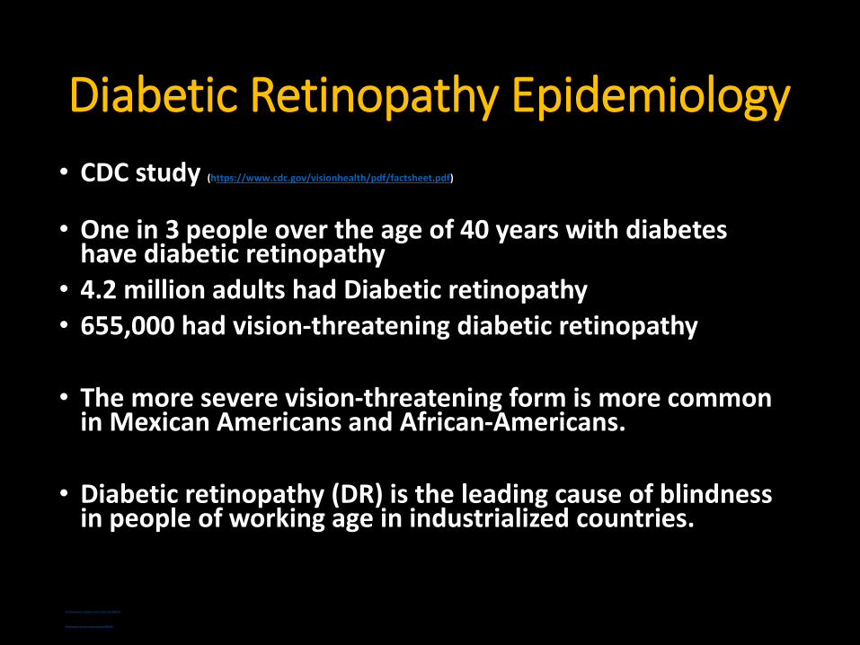
Diabetic Retinopathy Epidemiology
• CDC study (https://www.cdc.gov/visionhealth/pdf/factsheet.pdf)
• One in 3 people over the age of 40 years with diabetes have diabetic retinopathy
• 4.2 million adults had Diabetic retinopathy
• 655,000 had vision-threatening diabetic retinopathy
• The more severe vision-threatening form is more common in Mexican Americans and African-Americans.
• Diabetic retinopathy (DR) is the leading cause of blindness in people of working age in industrialized countries.
http://www.who.int/bulletin/volumes/82/11/en/844.pdf
http://www.ncbi.nlm.nih.gov/pubmed/19896746
http://www.who.int/bulletin/volumes/82/11/en/844.pdf

Risk factors for DR
• Male sex
• Higher A1C
• Longer duration of diabetes
• Insulin use
• Higher systolic blood pressure
• Barriers to care
http://jama.ama-assn.org/content/304/6/649.short?rss=1

Diabetic RetinopathyEpidemiology
• The best predictor of diabetic retinopathy is the duration of the disease
• After 20 years of diabetes, nearly 99% of patients with type 1 diabetes and 60% with type 2 have some degree on diabetic retinopathy
• 33% of patients with diabetes have signs of diabetic retinopathy
• People with diabetes are 25 times more likely to become blind than the general population.
Ophthalmology Myron Yanoff MD and Jay S. Duker
Basic and Clinical Science Course, Section 12: Retina and Vitreous AAO
http://www.aao.org/eyecare/news/upload/Eye-Health-Fact-Sheet.pdf -

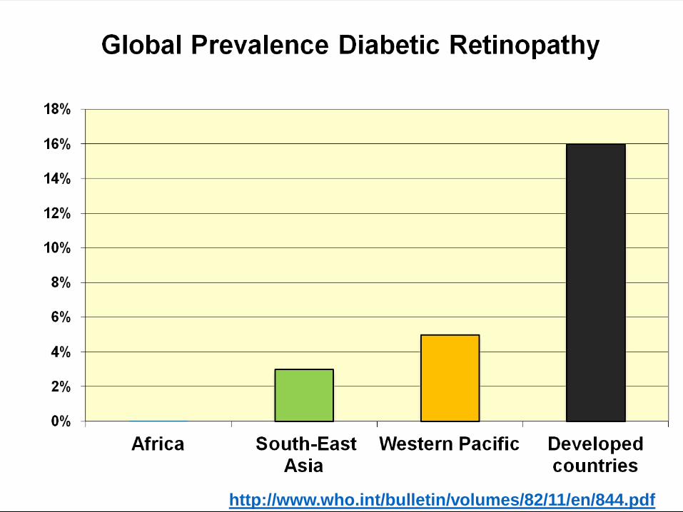
Prevalence of diabetic retinopathy after 20 years of diagnosis

Diabetic Retinopathy
Pathophysiology
• Elevated blood glucose results in physiologic changes that cause vascular endothelial damage.• Loss of pericytes• Basement membrane thickening
• Pathologic processes associated with diabetic retinopathy• Formation of microaneurysms• Closure of retinal capillaries and arterioles• Increased vascular permeability of retinal capillaries• Proliferation of new vessels and fibrous tissue• Contraction of vitreous and fibrous proliferation leading to tractional
retinal detachment

Diabetic Retinopathy
• Risk Factors associated with progression of diabetic retinopathy :• Hypertension• Elevated triglycerides• Elevated lipids,• Gross proteinuria
• Patients with Proliferative Diabetic Retinopathy are at increased risk of myocardial infarction, stroke, diabetic nephropathy, amputation, and death
• NOTE: No ocular contraindications to aspirin when required for cardiovascular disease or other medical conditions.

Diabetic Retinopathy
Causes of vision loss
• Macular edema (thickening of central retina)
• Macular ischemia
• Macular/foveal hemorrhage
• Vitreous or preretinal hemorrhage
• Retinal traction and detachment

Diabetic retinopathy symptoms
Diabetic retinopathy is asymptomatic in early stages of the disease
As the disease progresses symptoms may include
• Blurred vision
• Floaters
• Fluctuating vision
• Distorted vision
• Dark areas in the vision
• Poor night vision
• Impaired color vision
• Partial or total loss of vision

4 Stages of Diabetic Retinopathy:
1. Mild Nonproliferative Retinopathy (NPDR)
2. Moderate Nonproliferative Retinopathy
3. Severe Nonproliferative Retinopathy
4. Proliferative Retinopathy (PDR)
Goal is to diagnose as early as
possible!
National Eye Institute

Risk of Progression from NPDR to PDR
1 year 5yrs
Mild NPDR 5% 15%
Moderate
NPDR
27% 33%
Severe
NPDR
52% 60%
Very Severe
NPDR
75%

No retinopathy

MILD NONPROLIFERATIVE DIABETIC RETINOPATHYCharacteristics
• Microaneurysms only

MILD NONPROLIFERATIVE DIABETIC RETINOPATHY
Microaneurysms

Moderate Nonproliferative Diabetic Retinopathy (NPDR)
Characteristics
• More than just microaneurysms but less than severe NPDR but less than severe NPD
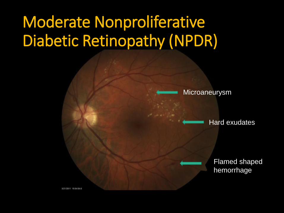
Moderate Nonproliferative Diabetic Retinopathy (NPDR)
Hard exudates
Flamed shaped
hemorrhage
Microaneurysm

Severe Nonproliferative Diabetic Retinopathy (NPDR)
Any of the following:
• More than 20 intraretinal hemorrhages in each of four quadrants
• Venous beading in two or more quadrants
• Prominent Intraretinal Microvascular Abnormalities (IRMA) in one or more quadrants
• And no signs of proliferative retinopathy

Severe Nonproliferative Diabetic Retinopathy (NPDR)
Venous beading

PROLIFERATIVE DIABETIC RETINOPATHY Neovascularization

Diabetic macular edema
• Diabetic macular edema is the leading cause of legal blindness in diabetics.
• Diabetic macular edema can be present at any stage of the disease, but is more common in patients with proliferative diabetic retinopathy.

Normal Macular Edema

Imaging of macular edema with optical
coherence tomography

Meta analysis and review on the effect on bevacizumab on diabetic macular edema
Graefes Arch Clin Exp Ophthalmol(2011) 249:15-27

Why is Diabetic macular edema so important?
• The macula is responsible for central vision.
• Diabetic macular edema may be asymptomatic at first. As the edema moves in to the fovea (the center of the macula) the patient will notice blurry central vision. The ability to read and recognize faces will be compromised.
Macula
Fovea

Normal Vision
Vision with Diabetic
Retinopathy
National Eye Institute

Macular Ischemia can lead to profound vision loss
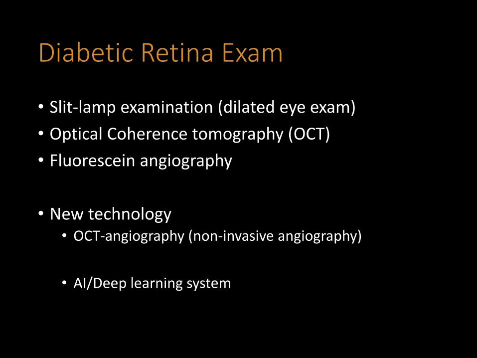
Diabetic Retina Exam
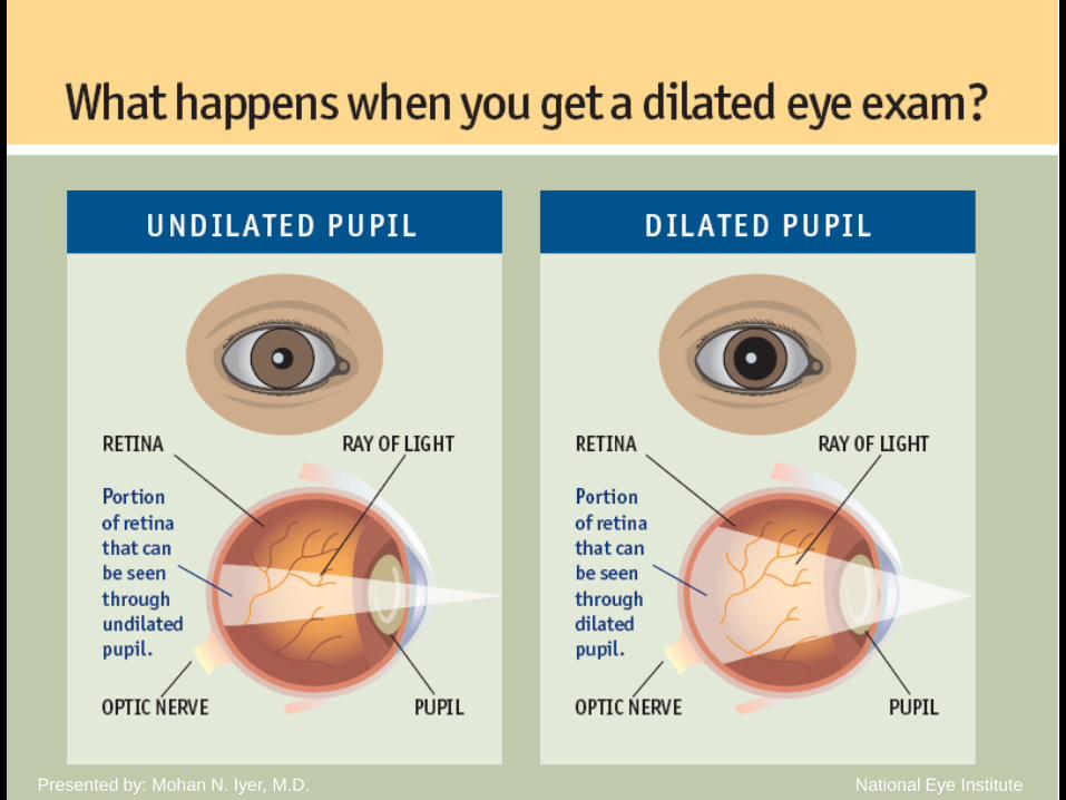
• Slit-lamp examination (dilated eye exam)
• Optical Coherence tomography (OCT)
• Fluorescein angiography
• New technology• OCT-angiography (non-invasive angiography)
• AI/Deep learning system

Association Between Vessel Density and Visual Acuity in Patients With Diabetic Retinopathy and Poorly Controlled Type 1 Diabetes.Bénédicte Dupas, MD; Wilfried Minvielle, MD; Sophie Bonnin, MD; Aude Couturier, MD; Ali Erginay, MD; Pascale Massin,
MD, PhD; Alain Gaudric, MD; Ramin Tadayoni, MD, PhD
JAMA Ophthalmol. 2018;136(7):721-728. doi:10.1001/jamaophthalmol.2018.1319

JAMA 2017
71 896 images; 14 880 patients. DLS had
90.5% sensitivity and 91.6% specificity for detecting referable diabetic
retinopathy;
100% sensitivity and 91.1% specificity for vision-threatening diabetic
retinopathy;
96.4%sensitivity and 87.2%specificity for possible glaucoma;
93.2%sensitivity and 88.7% specificity for age-related macular degeneration,
compared with professional graders.Sensitivity – true positive rate (high sens = few false negatives)
Specificity – true negative rate (high spec = few false positives)

DIABETIC RETINOPATHY TREATMENT
The best measure for prevention of loss of vision from diabetic retinopathy is strict glycemic control

The Effect of Intensive Diabetes Treatment
On the Progression of Diabetic Retinopathy
In Insulin-Dependent Diabetes Mellitus
The Diabetes Control and Complications Trial
The Diabetes Control and Complications Trial Research Group
Intensive control reduced the risk of developing retinopathy by 76%
and slowed progression of retinopathy by 54%; intensive control
also reduced the risk of clinical neuropathy by 60% and albuminuria
by 54%.
Arch Ophthalmol. 1995; 113:36-51

Primary prevention
Strict glycemic control
Blood pressure control
Secondary prevention
Annual eye exams
Tertiary prevention
Retinal Laser photocoagulationAnti-VEGF injectionsVitrectomy

1985
ETDRSDRCR.netProtocol B
2008
laserIntravitreal
triamcinolone
Treatment Options


Protocol T DRCR.Net 2 year resultsAflibercept, Bevacizumab, or Ranibizumab for Diabetic Macular Edema Two-Year Results from a Comparative Effectiveness Randomized Clinical TrialWells JA, et al for the DRCR network Ophthalmology 2016

Protocol T DRCR.Net2 year results
Baseline VA: 20/50 or worse Baseline VA: 20/32 to 20/40

1 month after antiVEGF treatment


28 y.o WM with blurry vision right eye for 6 months, left eye for 1 week Diagnosed with DM 2 weeks agoVision 20/400 OD; 20/200 OS
Plan: PRP Left eye same day
Vitrectomy, membrane peel, laser, gas Right eye in 10 days


Follow-up Guidelines
Age of Onset First Examination Follow-up
0 to 30 years (Type 1) Within 5 years Yearly
31 years and older (Type 2) Upon diagnosis Yearly
Prior to pregnancy (Type 1 or 2) Prior to conception or early 1st
trimester
No retinopathy to mild-moderate
NPDR: 3-12 months
Severe NPDR or worse: 1-3
months
Severity of Retinopathy
Diabetes only Yearly
Mild-moderate NPDR Every 6 months
Severe NPDR Early 3-4 months
PDR Every 3 months

CONCLUSIONS
Diabetic Retinopathy is preventable through strict glycemic control and annual dilated eye exams by an ophthalmologist.

National Eye Institute
National Eye Institute

National Eye InstitutePresented by: Mohan N. Iyer, M.D.

National Eye Institute

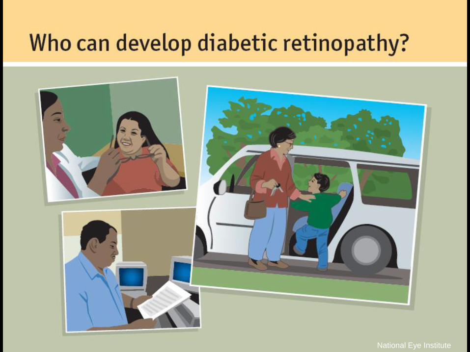
Who is at risk for diabetic retinopathy?• All people with diabetes
• Type 1
• AND Type 2
• During pregnancy, diabetic retinopathy may be a problem for women with diabetes.
Between 40 to 45 percent of Americans
diagnosed with diabetes have some
stage of diabetic retinopathy.
National Eye Institute

! Important !• It is important to diagnose or catch diabetic
retinopathy before symptoms occur!
• You may see great – and still have the early stages of diabetic retinopathy.
Key is to catch and manage the
disease early in its stages to
preserve vision.National Eye Institute

National Eye InstitutePresented by: Mohan N. Iyer, M.D.
National Eye InstitutePresented by: Mohan N. Iyer, M.D.

Risk factors Diabetic Retinopathy
Duration of diabetes is a major risk factor associated with the development of diabetic retinopathy
The severity of hyperglycemia is the key alterable risk factor associated with the development of diabetic retinopathy
http://one.aao.org/CE/PracticeGuidelines/PPP_Content.aspx?cid=d0c853d3-219f-487b-a524-326ab3cecd9a

Diabetic Retinopathy
• Diabetes is the leading cause of blindness in patients aged 20-64 years.
• Patients can have severe diabetic retinopathy and still be asymptomatic. Early detection and treatment can help prevent vision loss.
• Regular exams, treatment guidelines for medical and surgical management of diabetic eye disease are capable of reducing the risk of severe vision loss and blindness by 90%
• Treatment options for diabetic macular edema and proliferative diabetic retinopathy include laser photocoagulation, intravitreal injection of steroid or anti-VEGF agents, and vitrectomy surgery.

• What is the most common cause of vision loss among working age adults in the United States?
• 1. Glaucoma
• 2. Cataract
• 3. Diabetic Retinopathy
• 4. Retinal Detachment

• The most common cause of moderate vision loss in diabetic retinopathy is:
• 1. Refractive Change
• 2. Cataract
• 3. Diabetic Macular Edema
• 4. Proliferative Diabetic Retinopathy

A patient with Type II diabetes should get their first dilated eye exam:
1. Only when the vision is affected
2. In 3-5 years after initial diagnosis of diabetes
3. At the time of diagnosis of diabetes
4. 1 year after the diagnosis of diabetes

Thank you!


![The Guide - Diabetic Retinopathy - Vision Lossvisionloss.org.au/wp-content/uploads/2016/05/The... · the guide [diabetic retinopathy] What is Diabetic Retinopathy? Diabetic Retinopathy](https://img.pdfslide.us/doc/110x75/5e3ed00bf9c32e41ea6578a8/the-guide-diabetic-retinopathy-vision-the-guide-diabetic-retinopathy-what.jpg)