Embed Size (px)
Citation preview
Gene Expression Patterns 7 (2007) 491–500
www.elsevier.com/locate/modgep
Developmental expression of mouse Follistatin-like 1 (Fstl1): Dynamic regulation during organogenesis of the kidney and lung
Derek Adams, Barry Larman, Leif Oxburgh ¤
Maine Medical Center Research Institute, 81 Research Drive, Scarborough, 04074, ME USA
Received 27 July 2006; received in revised form 17 October 2006; accepted 19 October 2006Available online 26 October 2006
Abstract
Follistatin-like 1 (Fstl1) is a distantly related homolog of the Activin and Bone Morphogenetic Protein antagonist Follistatin. Interest-ingly, this molecule also has homology with the extracellular matrix modifying protein BM-40/SPARC/osteonectin. Previous studies inchick have identiWed Fstl1 as a regulator of early mesoderm patterning, somitogenesis, myogenesis and neural development. In this study,we determine the developmental expression pattern of Fstl1 in the mouse. We Wnd that Fstl1 is ubiquitously expressed in the earlyembryo, and that expression becomes regionalized later during development. In the majority of tissues, Fstl1 is strongly expressed in themesenchymal component and excluded from the epithelium. Notable exceptions include the central nervous system, in which Fstl1expression is entirely absent with the exception of the choroid plexi and Xoor plate, the lung, in which Fstl1 expression can be seen in air-way epithelia and the kidney, in which collecting ducts and nascent nephron epithelia express the highest levels of Fstl1.© 2006 Elsevier B.V. All rights reserved.
Keywords: Follistatin; Activin antagonist; BMP antagonist; Bone development; Gonad development; Gut development; Heart development; Kidneydevelopment; Limb development; Lung development; Neural development; Skin development; Tooth development
1. Results and discussion
The Activin and Bone Morphogenetic Protein (BMP)antagonist Follistatin plays an essential role during mouseembryogenesis, regulating development of musculature,skin, palate and teeth (Matzuk et al., 1995). Furthermore,Follistatin plays a key role in regulating folliculogenesis inthe adult female (Jorgez et al., 2004). A number of homo-logs of this developmentally important gene have beenidentiWed since completion of the mouse genome sequence(Fig. 1A). Although distantly related, they are classiWed asbelonging to the Follistatin family based on the presence ofa conserved Follistatin (FS) domain (Fig. 1B). Biochemicalanalyses of the Follistatin-like 3 (Fstl3) member show thatthis protein acts as an antagonist of Activin signaling, dem-onstrating functional homology with Follistatin (Sidis
* Corresponding author. Tel.: +1 207 885 8115; fax: +1 207 885 8110.E-mail address: [email protected] (L. Oxburgh).
1567-133X/$ - see front matter © 2006 Elsevier B.V. All rights reserved.doi:10.1016/j.modgep.2006.10.009
et al., 2006). RNA knock-down experiments in chick haveidentiWed developmental roles for Follistatin-like 1 (Fstl1,syn TSC-36, Flik), diVering signiWcantly from those of Fol-listatin. Fstl1 is involved in early mesoderm patterning,somitogenesis, myogenesis and neural development(Amthor et al., 1996; Patel et al., 1996; Towers et al., 1999).In zebraWsh, morpholino knock-down studies show that thezFstl2 homolog of mouse Fstl1 acts together with Chordinand Noggin to establish the dorso–ventral body axis(Dal-Pra et al., 2006), arguing that Fstl1 acts as a BMPantagonist. However, in addition to homology with Follist-atin (Fig. 1B), Fstl1 contains regions of homology with theBM-40/SPARC/osteonectin protein (Hambrock et al.,2004). BM-40 is an extracellular matrix modifying proteinthat binds collagen I–IV, nidogen and thrombospondin,and also antagonizes the activities of PDGF and VEGFgrowth factors (Brekken and Sage, 2000). Functional con-servation of these properties are unclear however as Fstl1lacks key BM-40 family characteristics such as calciumbinding (Hambrock et al., 2004). Fstl1 is inducible by
492 D. Adams et al. / Gene Expression Patterns 7 (2007) 491–500
TGF� (Shibanuma et al., 1993) and estrogen (Ohashi et al.,1997), and has been identiWed as a potential tumor suppres-sor (Hodgson et al., 2001). Expression of Fstl1 is reduced inhuman cancer cell lines (Hambrock et al., 2004; Mashimoet al., 1997; Sumitomo et al., 2000) and experimentallytransformed cells (Johnston et al., 2000). Reintroduction ofFstl1 to transformed cells reduces proliferation (Sumitomoet al., 2000) and invasion (Johnston et al., 2000), suggestingthat mechanisms of Fstl1 action during embryonic develop-ment may be to inhibit cell growth and migration. Theseactions could be due either to antagonism of Activin/BMPsignals which are proliferative in some developmental con-texts, or to modiWcation of the extracellular matrix environ-ment. To deWne the expression of this potentially importantdevelopmental regulator during mouse embryogenesis, weperformed in situ hybridization using a Fstl-1 speciWcriboprobe on a time-course of embryonic tissues from E7 toE17.5, and adult ovary.
1.1. Early embryonic expression
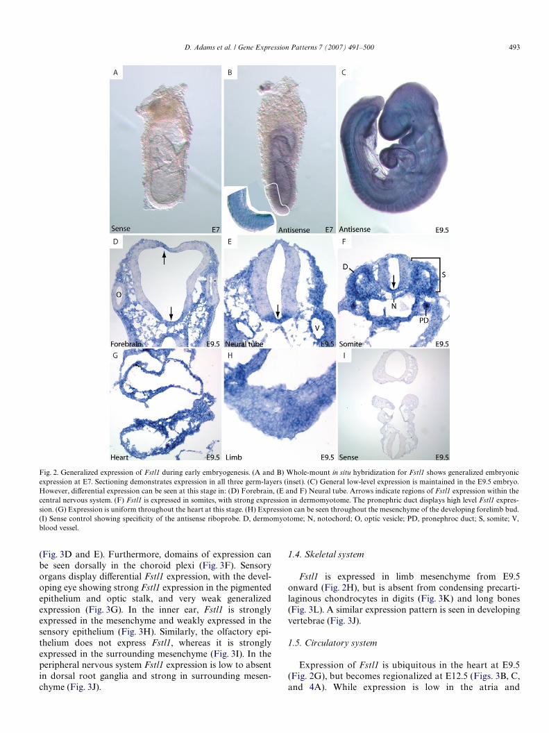
Fstl1 is expressed throughout the E7 embryo, andexcluded from extra-embryonic tissues (Fig. 2B). At E9.5,
ubiquitous expression is largely maintained, with the excep-tion of the developing nervous system. Fstl1 is speciWcallyexpressed in the Xoorplate throughout the neural tube(Fig. 2D and E) with dorsal aspects lacking expression.Expression diVers slightly in the forebrain (Fig. 2D), inwhich Fstl1 can be seen in sections of the roof-plate. At thisstage, the optic vesicles also completely lack Fstl1. Thedermomyotome of somites and pronephric ducts displaynotably high levels of Fstl1 expression at E9.5 (Fig. 2F).
1.2. Expression at mid-gestation
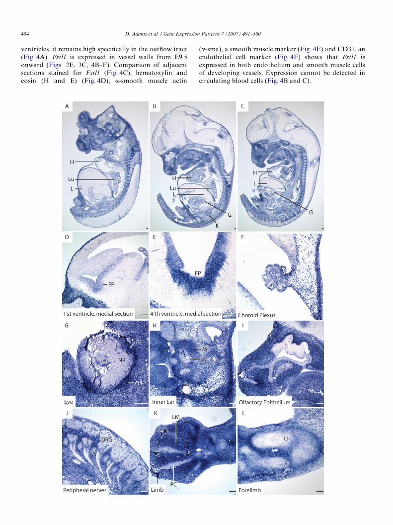
The ubiquitous early developmental expression of Fstl1resolves during mid-gestation (Fig. 3A–C). Expression isentirely absent in the E12.5 embryonic liver (Fig. 3A) andregions of the central nervous system (Fig. 3B). In otherorgan systems, Fstl1 expression is expressed diVerentially ina variety of cell types (Fig. 3D–L).
1.3. Central and peripheral nervous systems
The ventral localization of Fstl1 is maintained in E12.5neural tissue, with extensive expression in the Xoor-plate
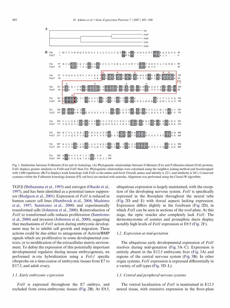
Fig. 1. Similarities between Follistatin (Fst) and its homologs. (A) Phylogenetic relationships between Follistatin (Fst) and Follistatin-related (Fstl) proteins.Fstl1 displays greater similarity to Fstl4 and Fstl5 than Fst. Phylogenetic relationships were calculated using the neighbor joining method and bootstrappedwith 1,000 repetitions. (B) Fst displays weak homology with Fstl1 at the amino acid level. Overall, amino acid identity is 22%, and similarity is 36%. Conservedcysteines within the Follistatin homology domain (FS, red box) are marked with asterisks. Alignment was performed using the Clustal W algorithm.
D. Adams et al. / Gene Expression Patterns 7 (2007) 491–500 493
(Fig. 3D and E). Furthermore, domains of expression canbe seen dorsally in the choroid plexi (Fig. 3F). Sensoryorgans display diVerential Fstl1 expression, with the devel-oping eye showing strong Fstl1 expression in the pigmentedepithelium and optic stalk, and very weak generalizedexpression (Fig. 3G). In the inner ear, Fstl1 is stronglyexpressed in the mesenchyme and weakly expressed in thesensory epithelium (Fig. 3H). Similarly, the olfactory epi-thelium does not express Fstl1, whereas it is stronglyexpressed in the surrounding mesenchyme (Fig. 3I). In theperipheral nervous system Fstl1 expression is low to absentin dorsal root ganglia and strong in surrounding mesen-chyme (Fig. 3J).
1.4. Skeletal system
Fstl1 is expressed in limb mesenchyme from E9.5onward (Fig. 2H), but is absent from condensing precarti-laginous chondrocytes in digits (Fig. 3K) and long bones(Fig. 3L). A similar expression pattern is seen in developingvertebrae (Fig. 3J).
1.5. Circulatory system
Expression of Fstl1 is ubiquitous in the heart at E9.5(Fig. 2G), but becomes regionalized at E12.5 (Figs. 3B, C,and 4A). While expression is low in the atria and
Fig. 2. Generalized expression of Fstl1 during early embryogenesis. (A and B) Whole-mount in situ hybridization for Fstl1 shows generalized embryonicexpression at E7. Sectioning demonstrates expression in all three germ-layers (inset). (C) General low-level expression is maintained in the E9.5 embryo.However, diVerential expression can be seen at this stage in: (D) Forebrain, (E and F) Neural tube. Arrows indicate regions of Fstl1 expression within thecentral nervous system. (F) Fstl1 is expressed in somites, with strong expression in dermomyotome. The pronephric duct displays high level Fstl1 expres-sion. (G) Expression is uniform throughout the heart at this stage. (H) Expression can be seen throughout the mesenchyme of the developing forelimb bud.(I) Sense control showing speciWcity of the antisense riboprobe. D, dermomyotome; N, notochord; O, optic vesicle; PD, pronephroc duct; S, somite; V,blood vessel.
494 D. Adams et al. / Gene Expression Patterns 7 (2007) 491–500
ventricles, it remains high speciWcally in the outXow tract (�-sma), a smooth muscle marker (Fig. 4E) and CD31, an
(Fig. 4A). Fstl1 is expressed in vessel walls from E9.5onward (Figs. 2E, 3C, 4B–F). Comparison of adjacentsections stained for Fstl1 (Fig. 4C), hematoxylin andeosin (H and E) (Fig. 4D), �-smooth muscle actinendothelial cell marker (Fig. 4F) shows that Fstl1 isexpressed in both endothelium and smooth muscle cellsof developing vessels. Expression cannot be detected incirculating blood cells (Fig. 4B and C).
D. Adams et al. / Gene Expression Patterns 7 (2007) 491–500 495
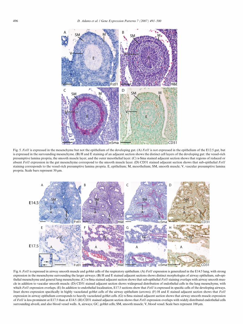
1.6. Gut
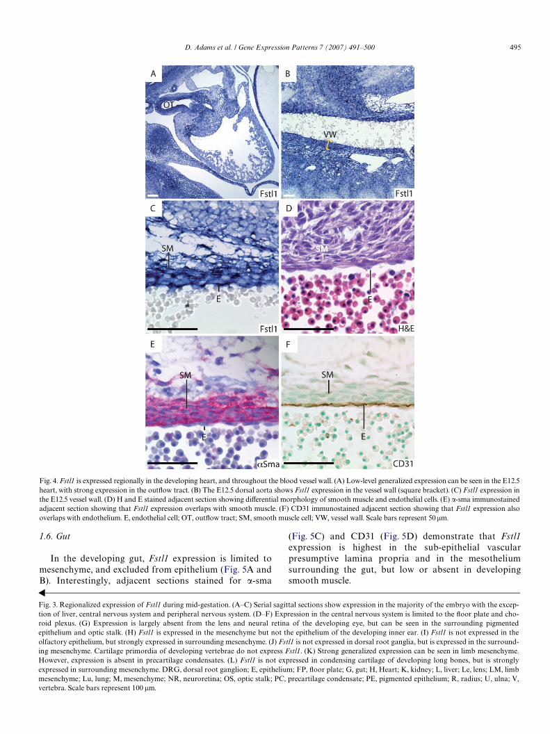
In the developing gut, Fstl1 expression is limited tomesenchyme, and excluded from epithelium (Fig. 5A andB). Interestingly, adjacent sections stained for �-sma
Fig. 3. Regionalized expression of Fstl1 during mid-gestation. (A–C) Serial sagtion of liver, central nervous system and peripheral nervous system. (D–F) Exroid plexus. (G) Expression is largely absent from the lens and neural retiepithelium and optic stalk. (H) Fstl1 is expressed in the mesenchyme but notolfactory epithelium, but strongly expressed in surrounding mesenchyme. (J) Fing mesenchyme. Cartilage primordia of developing vertebrae do not expressHowever, expression is absent in precartilage condensates. (L) Fstl1 is not eexpressed in surrounding mesenchyme. DRG, dorsal root ganglion; E, epithelmesenchyme; Lu, lung; M, mesenchyme; NR, neuroretina; OS, optic stalk; PCvertebra. Scale bars represent 100 �m.
(Fig. 5C) and CD31 (Fig. 5D) demonstrate that Fstl1expression is highest in the sub-epithelial vascularpresumptive lamina propria and in the mesotheliumsurrounding the gut, but low or absent in developingsmooth muscle.
ittal sections show expression in the majority of the embryo with the excep-pression in the central nervous system is limited to the Xoor plate and cho-na of the developing eye, but can be seen in the surrounding pigmented the epithelium of the developing inner ear. (I) Fstl1 is not expressed in thestl1 is not expressed in dorsal root ganglia, but is expressed in the surround- Fstl1. (K) Strong generalized expression can be seen in limb mesenchyme.xpressed in condensing cartilage of developing long bones, but is stronglyium; FP, Xoor plate; G, gut; H, Heart; K, kidney; L, liver; Le, lens; LM, limb, precartilage condensate; PE, pigmented epithelium; R, radius; U, ulna; V,
Fig. 4. Fstl1 is expressed regionally in the developing heart, and throughout the blood vessel wall. (A) Low-level generalized expression can be seen in the E12.5heart, with strong expression in the outXow tract. (B) The E12.5 dorsal aorta shows Fstl1 expression in the vessel wall (square bracket). (C) Fstl1 expression inthe E12.5 vessel wall. (D) H and E stained adjacent section showing diVerential morphology of smooth muscle and endothelial cells. (E) �-sma immunostainedadjacent section showing that Fstl1 expression overlaps with smooth muscle. (F) CD31 immunostained adjacent section showing that Fstl1 expression alsooverlaps with endothelium. E, endothelial cell; OT, outXow tract; SM, smooth muscle cell; VW, vessel wall. Scale bars represent 50 �m.
496 D. Adams et al. / Gene Expression Patterns 7 (2007) 491–500
surrounding alveoli, and also blood vessel walls. A, airways; GC, goblet cells; SM
, smooth muscle; V, blood vessel. Scale bars represent 100 �m.Fig. 5. Fstl1 is expressed in the mesenchyme but not the epithelium of the developing gut. (A) Fstl1 is not expressed in the epithelium of the E12.5 gut, butis expressed in the surrounding mesenchyme. (B) H and E staining of an adjacent section shows the distinct cell layers of the developing gut: the vessel-richpresumptive lamina propria, the smooth muscle layer, and the outer mesothelial layer. (C) �-Sma stained adjacent section shows that regions of reduced orabsent Fstl1 expression in the gut mesenchyme correspond to the smooth muscle layer. (D) CD31 stained adjacent section shows that sub-epithelial Fstl1staining corresponds to the vessel-rich presumptive lamina propria. E, epithelium; M, mesothelium; SM, smooth muscle; V, vascular presumptive laminapropria. Scale bars represent 50 �m.
Fig. 6. Fstl1 is expressed in airway smooth muscle and goblet cells of the respiratory epithelium. (A) Fstl1 expression is generalized in the E14.5 lung, with strongexpression in the mesenchyme surrounding the larger airways. (B) H and E stained adjacent sections shows distinct morphologies of airway epithelium, sub-epi-thelial mesenchyme and general lung mesenchyme. (C) �-Sma stained adjacent section shows that sub-epithelial Fstl1 staining overlaps with airway smooth mus-cle in addition to vascular smooth muscle. (D) CD31 stained adjacent section shows widespread distribution of endothelial cells in the lung mesenchyme, withwhich Fstl1 expression overlaps. (E) In addition to endothelial localization, E17.5 sections show that Fstl1 is expressed in speciWc cells of the developing airways.Inset shows expression speciWcally in highly vacuolated goblet cells of the airway epithelium (arrows). (F) H and E stained adjacent section shows that Fstl1expression in airway epithelium corresponds to heavily vacuolated goblet cells. (G) �-Sma stained adjacent section shows that airway smooth muscle expressionof Fstl1 is less prominent at E17.5 than at E14.5. (H) CD31 stained adjacent section shows that Fstl1 expression overlaps with widely distributed endothelial cells
D. Adams et al. / Gene Expression Patterns 7 (2007) 491–500 497
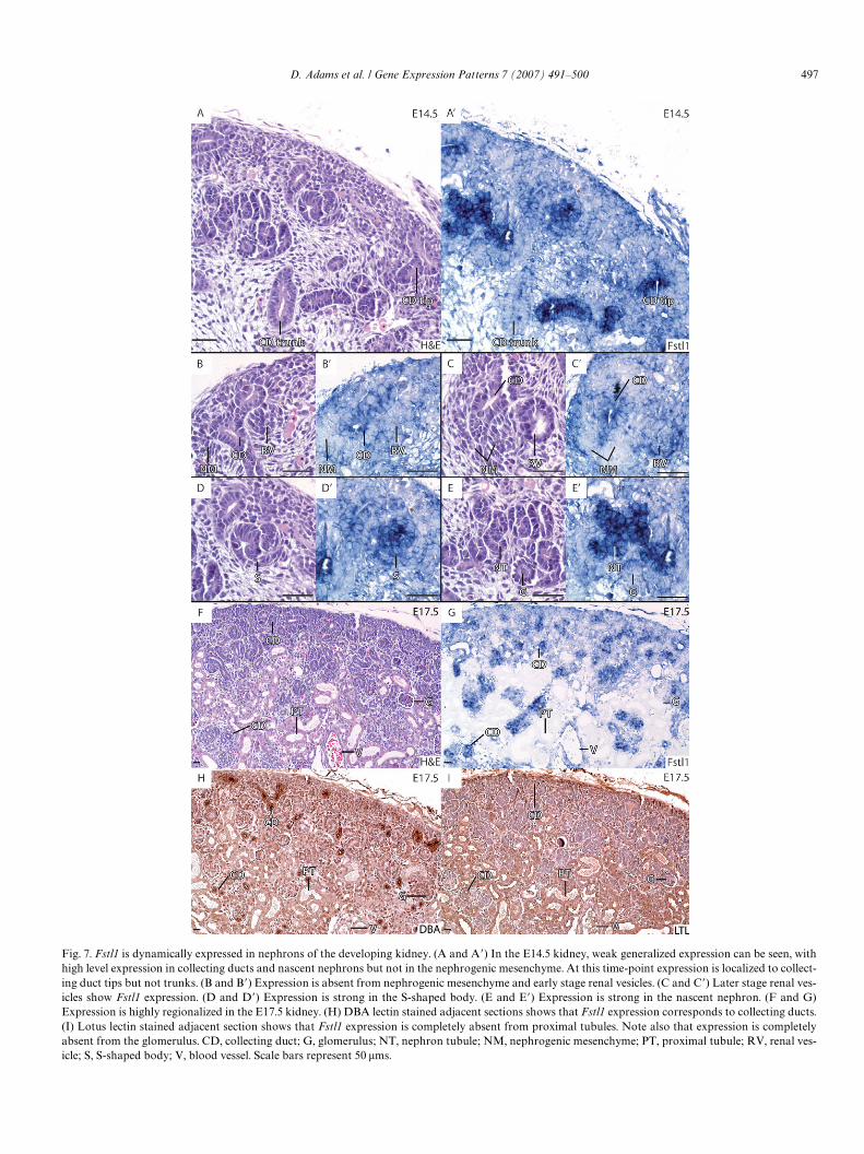
Fig. 7. Fstl1 is dynamically expressed in nephrons of the developing kidney. (A and A�) In the E14.5 kidney, weak generalized expression can be seen, withhigh level expression in collecting ducts and nascent nephrons but not in the nephrogenic mesenchyme. At this time-point expression is localized to collect-ing duct tips but not trunks. (B and B�) Expression is absent from nephrogenic mesenchyme and early stage renal vesicles. (C and C�) Later stage renal ves-icles show Fstl1 expression. (D and D�) Expression is strong in the S-shaped body. (E and E�) Expression is strong in the nascent nephron. (F and G)Expression is highly regionalized in the E17.5 kidney. (H) DBA lectin stained adjacent sections shows that Fstl1 expression corresponds to collecting ducts.(I) Lotus lectin stained adjacent section shows that Fstl1 expression is completely absent from proximal tubules. Note also that expression is completelyabsent from the glomerulus. CD, collecting duct; G, glomerulus; NT, nephron tubule; NM, nephrogenic mesenchyme; PT, proximal tubule; RV, renal ves-icle; S, S-shaped body; V, blood vessel. Scale bars represent 50 �ms.
498 D. Adams et al. / Gene Expression Patterns 7 (2007) 491–500
1.7. Lung
The developing lung shows a generalized expression ofFstl1 at E12.5 (Fig. 3A and B) and E14.5 (Fig. 6A and B),with high levels in lung mesenchyme surrounding airways(Fig. 6A). �-sma and CD31 staining of adjacent sectionsshows that this high level expression is within the smoothmuscle layer surrounding airways, as well as in walls ofblood vessels. Furthermore, CD31 staining shows a distri-bution of endothelial cells throughout the mesenchymethat closely correlates with the distribution of Fstl1-stained cells, indicating that mesenchymal Fstl1 expres-sion is localized to vasculature developing around alveoli(Fig. 6A and D). At E17.5, our expression analysis indi-cates that Fstl1 expression is weak in airway smooth mus-cle, remains in endothelial cells surrounding alveoli and islocalized to goblet cells of the airway epithelium andblood vessel walls (Fig. 6E–H).
1.8. Kidney
In the kidney, expression is generalized at E12.5 andE14.5 (Figs. 3B, 7A, A�), with regions of intense expres-sion. Collecting ducts express strongly, with tips showingmore intense staining than trunks (Figs. 7A, A�, B, and
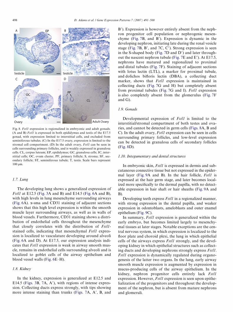
Fig. 8. Fstl1 expression is regionalized in embryonic and adult gonads.(A and B) Fstl1 is expressed in both epididymus and testis of the E17.5gonad, with expression limited to interstitial cells, and excluded fromseminiferous tubules. (C) In the E17.5 ovary, expression is limited to thestromal cell compartment. (D) In the adult ovary, Fstl1 can be seen incells surrounding primary follicles, and is weakly expressed in granulosacells. CL, corpus luteum; EP, epididymus; GC, granulosa cells; IC, inter-stitial cells; OC, ovum cluster, PF, primary follicle; S, stroma; SF, sec-ondary follicle; ST, seminiferous tubule; T, testis. Scale bars represent100 �m.
B�). Expression is however entirely absent from the neph-ron progenitor cell population or nephrogenic mesen-chyme (Fig. 7B, and B�). Expression is dynamic in thedeveloping nephron, initiating late during the renal vesiclestage (Fig. 7B, B�, and 7C, C�). Strong expression is seenin the S-shaped body (Fig. 7D and D�) and later through-out the nascent nephron tubule (Fig. 7E and E�). At E17.5,nephrons have matured and regionalized to proximaland distal tubules (Fig. 7F). Staining of adjacent sectionswith lotus lectin (LTL), a marker for proximal tubule,and dolichos biXoris lectin (DBA), a collecting ductmarker, shows that Fstl1 expression is maintained incollecting ducts (Fig. 7G and H) but completely absentfrom proximal tubules (Fig. 7G and I). Fstl1 expressionis also completely absent from the glomerulus (Fig. 7Fand G).
1.9. Gonads
Developmental expression of Fstl1 is limited to theinterstitial/stromal compartment of both testes and ova-ries, and cannot be detected in germ cells (Figs. 8A, B andC). In the adult ovary, Fstl1 expression can be seen in cellssurrounding primary follicles, and low-level expressioncan be detected in granulosa cells of secondary follicles(Fig. 8D).
1.10. Integumentary and dental structures
In embryonic skin, Fstl1 is expressed in dermis and sub-cutaneous connective tissue but not expressed in the epider-mal layer (Fig. 9A and B). In the hair follicle, Fstl1 isexpressed at the hair germ stage, and later becomes local-ized more speciWcally to the dermal papilla, with no detect-able expression in hair shaft or hair sheaths (Fig. 9A andB).
Developing teeth express Fstl1 in a regionalized manner,with strong expression in the dental papilla, and weakerexpression in odontoblasts, ameloblasts and outer enamelepithelium (Fig. 9C).
In summary, Fstl1 expression is generalized within theearly embryo, but becomes limited largely to mesenchy-mal tissues at later stages. Notable exceptions are the cen-tral nervous system, in which expression is localized to theXoor plate and choroid plexi, the lung in which epithelialcells of the airways express Fstl1 strongly, and the devel-oping kidney in which epithelial structures such as collect-ing ducts and developing nephrons strongly express Fstl1.Fstl1 expression is dynamically regulated during organo-genesis of the latter two organs. In the lung, early airwaysmooth muscle expression is augmented by expression inmucus-producing cells of the airway epithelium. In thekidney, nephron progenitor cells entirely lack Fstl1expression. However, Fstl1 expression is seen upon epithe-lialization of the progenitors and throughout the develop-ment of the nephron, but is absent from mature nephronsand glomeruli.
D. Adams et al. / Gene Expression Patterns 7 (2007) 491–500 499
2. Experimental procedures
2.1. Mouse embryos
Mouse embryos of the ICR strain were used for all experiments andthe day of vaginal plug was recorded as embryonic day 0.5.
2.2. in situ hybridization probe
PCR derived DNA template was made using the following primerscontaining the promoter sequences for the T7 and T3 RNA polymerase (inbold) and were designed to amplify a segment of exons 6–9 from embry-onic cDNA: Fstl1T7: 5�-CAGAGATGCA TAATACGACT CACTATAGGG AGAGCTTCCA AGAGTTCCTC AAGTGC-3�, Fstl1T3:5�-CCAAGCCTTC ATTAACCCTC ACTAAAGGGA GACGATGCTGTTTCCTTAGT TGCC-3�. Sense (T7) and antisense (T3) probes weregenerated using the Roche DIG RNA Labeling Mix according to themanufacturer’s instructions.
2.3. in situ hybridization
Whole mount and non-radioactive section in situ hybridizations wereperformed as previously described (Oxburgh et al., 2004).
2.4. Immunostaining and lectin staining
Immunostaining and lectin staining were performed on sections adja-cent to those used for in situ hybridization as previously described(Oxburgh and Robertson, 2002).
Acknowledgements
Many thanks to the histopathology core at Maine Medi-cal Center Research Institute for tissue processing services.The project described was supported by Grant No. P20RR18789 from the National Center for ResearchResources (NCRR), a component of the National Insti-tutes of Health (NIH). Its contents are solely the responsi-bility of the authors and do not necessarily represent theoYcial views of NCRR or NIH.
References
Amthor, H., Connolly, D., Patel, K., Brand-Saberi, B., Wilkinson, D.G.,Cooke, J., Christ, B., 1996. The expression and regulation of follistatin
and a follistatin-like gene during avian somite compartmentalizationand myogenesis. Dev. Biol. 178, 343–362.
Brekken, R.A., Sage, E.H., 2000. SPARC, a matricellular protein: at thecrossroads of cell-matrix. Matrix Biol. 19, 569–580.
Dal-Pra, S., Furthauer, M., Van-Celst, J., Thisse, B., Thisse, C., 2006. Nog-gin1 and Follistatin-like2 function redundantly to Chordin to antago-nize BMP activity. Dev. Biol..
Hambrock, H.O., Kaufmann, B., Muller, S., Hanisch, F.G., Nose, K., Paulsson,M., Maurer, P., Hartmann, U., 2004. Structural characterization of TSC-36/Flik: analysis of two charge isoforms. J. Biol. Chem. 279, 11727–11735.
Hodgson, G., Hager, J.H., Volik, S., Hariono, S., Wernick, M., Moore, D.,Nowak, N., Albertson, D.G., Pinkel, D., Collins, C., Hanahan, D.,Gray, J.W., 2001. Genome scanning with array CGH delineatesregional alterations in mouse islet carcinomas. Nat. Genet. 29, 459–464.
Johnston, I.M., Spence, H.J., Winnie, J.N., McGarry, L., Vass, J.K., Meagher,L., Stapleton, G., Ozanne, B.W., 2000. Regulation of a multigenic invasionprogramme by the transcription factor, AP-1: re-expression of a down-regulated gene, TSC-36, inhibits invasion. Oncogene 19, 5348–5358.
Jorgez, C.J., Klysik, M., Jamin, S.P., Behringer, R.R., Matzuk, M.M., 2004.Granulosa cell-speciWc inactivation of follistatin causes female fertilitydefects. Mol. Endocrinol. 18, 953–967.
Mashimo, J., Maniwa, R., Sugino, H., Nose, K., 1997. Decrease in theexpression of a novel TGF beta1-inducible and ras-recision gene, TSC-36, in human cancer cells. Cancer Lett. 113, 213–219.
Matzuk, M.M., Lu, N., Vogel, H., Sellheyer, K., Roop, D.R., Bradley, A.,1995. Multiple defects and perinatal death in mice deWcient in follista-tin. Nature 374, 360–363.
Ohashi, T., Sato, S., Yoshiki, A., Kusakabe, M., 1997. TSC-36 (follistatin-related polypeptide) gene expression in estrogen receptor positiveosteoblastic cell line, CDO7F. Calcif. Tissue Int. 61, 400–403.
Oxburgh, L., Robertson, E.J., 2002. Dynamic regulation of Smad expres-sion during mesenchyme to epithelium transition in the metanephrickidney. Mech. Dev. 112, 207–211.
Oxburgh, L., Chu, G.C., Michael, S.K., Robertson, E.J., 2004. TGFb super-family signals are required for morphogenesis of the kidney mesen-chyme progenitor population. Development 131, 4593–4605.
Patel, K., Connolly, D.J., Amthor, H., Nose, K., Cooke, J., 1996. Cloningand early dorsal axial expression of Flik, a chick follistatin-relatedgene: evidence for involvement in dorsalization/neural induction. Dev.Biol. 178, 327–342.
Shibanuma, M., Mashimo, J., Mita, A., Kuroki, T., Nose, K., 1993. Cloningfrom a mouse osteoblastic cell line of a set of transforming-growth-fac-tor-beta 1-regulated genes, one of which seems to encode a follistatin-related polypeptide. Eur. J. Biochem. 217, 13–19.
Sidis, Y., Mukherjee, A., Keutmann, H., Delbaere, A., Sadatsuki, M., Sch-neyer, A., 2006. Biological activity of follistatin isoforms and follista-tin-like-3 is dependent on diVerential cell surface binding andspeciWcity for activin, myostatin, and bone morphogenetic proteins.Endocrinology 147, 3586–3597.
Fig. 9. Fstl1 is expression in developing skin and teeth. (A) Fstl1 is expressed in connective tissue and dermis, and not in the epidermal layer of the skin.Expression can also be seen in hair germs. (B) In the hair follicle, Fstl1 expression is limited to the dermal papilla, and cannot be seen in hair sheath, inneror outer root sheaths. (C) In developing teeth, Fstl1 is strongly expressed in the dental papilla and moderately expressed in odontoblasts, ameloblasts andouter enamel epithelium. AB, ameloblast; CT, connective tissue; D, dermis; DeP, dental papilla; DP, dermal papilla; ED, epidermis; HG, hair germ; HS,hair shaft; IRS, inner root sheath; OB, odontoblast; OEE, outer enamel epithelium; ORS, outer root sheath. Scale bars represent 100 �m.

500 D. Adams et al. / Gene Expression Patterns 7 (2007) 491–500
Sumitomo, K., Kurisaki, A., Yamakawa, N., Tsuchida, K., Shimizu, E.,Sone, S., Sugino, H., 2000. Expression of a TGF-beta1 inducible gene,TSC-36, causes growth inhibition in human lung cancer cell lines. Can-cer Lett. 155, 37–46.
Towers, P., Patel, K., Withington, S., Isaac, A., Cooke, J., 1999. Flik, achick follistatin-related gene, functions in gastrular dorsalisation/neu-ral induction and in subsequent maintenance of midline Sonic hedge-hog signalling. Dev. Biol. 214, 298–317.


![Direct Organogenesis from Cotyledonary Node Explants of ... · shoot organogenesis in C. peporeported [19] direct organogenesis in Cucumis sativus [20] and reported L. cy-lindrica](https://img.pdfslide.us/doc/110x75/5fac27dc76c37d66627b9b5d/direct-organogenesis-from-cotyledonary-node-explants-of-shoot-organogenesis.jpg)