Embed Size (px)
Citation preview

AD ------------------
USAARL REPORT NO. 73-3
DEVELOPMENT OF A BIO-PAC FOR CARDIAC EVALUATION OF PORCINE RESEARCH ANIMALS
BY
LCDR Thomas L. Wachtel, M.D. CPT G. R. McCahan, Jr., DVM
Mr. Lynn A. Alford
August 1972
U. S. ARMY AEROMEDICAL RESEARCH LABORATORY
Fort Rucker, Alabama 36360

Unclassified Security Classification
ADA750100 Technical Report
DOCUMENT CONTROL OAT A - R & D (Security cl.essillc•tion ol title, body of •batract end lndeKinll annot•tion mual be entered when the overall report I• cl•••ll~•d)
1 ORIGINATING ACTIVITY (Corpor•te autiJorJ a.. REPORT SECURITY CLASSIFICATION
u. s. Army Aeromedical Research Laboratory Unclassified
2b. GROUP
Fort Rucker, Alabama 36360 3. REPORT TITLE
Development of a Bio-Pac for Cardiac Evaluation of Porcine Research Animals 4. DESCRIPTIVE NOTES (Type of report and lnclulllVe date•)
Paper for publication a. AUTHOR(SJ (Fir•t rMme, middle lnltl•l, Ia at name)
Thomas L. Wachtel, M.D. G. R. McCahan, Jr.' DVM Mr. Lynn A. A 1 ford
e;. REPORT DATE 7a. TOTAL NO. OF PACiiES l'b. NO. OF
1;KFS
Auaust 1972 14 ... CONTRACT OR <;RANT NO. ~M. OAICINATOA1 S REPORT NUI\t'BIER(S)
b. PRO.JECT NO. 3AO 6211 OA 819 73-3
"· eb. 0 TH ER REPORT NOCSI (Any other n-bere that ,..T be aeef.,.ed lhla report)
d.
10. DISTRIBUTION ~TATEMENT
This document has been approved for public release and sale; its distribution is unlimited.
II· SUPPLEMENTARY NOTES 12. SPONSORING MILITAFIY ACTIVITY
u. S. Army Medical R&D Command Washington, D.C. 20314
,3. ABSTRACT
This report describes a technique for implanting central venous and aortic catheters via the jugular veins and carotid arteries in miniature swine and the device designed and utilized to protect these catheters. Such indwelling catheters were easily main-tained for fourteen (14) days in unrestrained, free-roaming pigs while serial blood sampies, pressure recording, electrocardiographic monitoring, and cardiac output mea-suring were conducted and infusion of precise amounts of fluids or drugs administered.
DD ,':: •• 1473 Unclassified security Cl•••lRc•tion

Unclassified Security Classirlcation
14. LINK A LINK 8 LINt< C KEY WORDS
AO!....E WT ROLE WT ROLE WT
l Miniature Swine
I
I
Catheterization Aortic Arch Central Venous External Catheter Protection Device l Cardiovascular Studies
I I I
I I I I I I I
I
I I
' I
I !
I !
I !
i I
I I
I I !
I I
I
Unc 1 ass ifi ed FT RliCK FR 10200' Secutity Claaalflcatlon

NOTICE
Qualified requesters may obtain copies from the Defense Documentation Center (DOC), Cameron Station, Alexandria, Virginia. Orders will be expedited if placed through the librarian or other person designated to request documents from DDC (formerly ASTIA).
Change of Address
Organizations receiving reports from the US Army Aeromedical Research Laboratory on automatic mailing lists should confirm correct address when corresponding about laboratory reports.
Disposition
This document has been approved for public release and sale; its distribution is unlimited.
Disclaimer
The findings in this report are not to be construed as an official Department of the Army position unless so designated by other authorized documents.
The products referred to in this report are not considered as an endorsement by the authors or the Department of the Army.
~ -

AD -------------------
USAARL REPORT NO. 73-3
DEVELOPMENT OF A BIO-PAC FOR CARDIAC EVALUATION OF PORCINE RESEARCH ANIMALS
BY
LCDR Thomas L. Wachtel, M.D. CPT G. R. McCahan, Jr., DVM
Mr. Lynn A. Alford
August 1972
U. S. ARMY AEROMEDICAL RESEARCH LABORATORY
Fort Rucker, Alabama 36360
U. S. Army Medical Research and Development Command
Distribution Statement: This document has been approved for public release and sale; its distribution is unlimited.

The Vivarium of the United States Army Aeromed~cal Research Laboratory is fully accredited by the American Associatiot~ for 1\ccrPditation of Laboratory Animal Care.
In conducting this research, the investit)ators adhered to the "Guide for Laboratory Animals Facilities and Care" preparcc' by the committee on the Guide for Laboratory Animals Facilities and Care, National Academy of Sciences, National Research Counci I. Hu~ane procedure were utilized throughout, and a graduate veterinarian was 10 constan attendance to perform all surgical procedures and to ensure that all an mals were fully anesthetized and insensitive to pain.

ACKNOWLEDGMENTS
The authors are indebted to Janice Speigner, Diana Patrick, Tom Downs, Richard Chapman, Malcolm Kirk, John J. Barbaccia, C. D. Williams, and Patricia Wagner for their generous assistance, without which this project could not have been completed.
ABSTRACT
This report describes a technique for implanting central venous and aortic catheters via the jugular veins and carotid arteries in miniature swine and the device designed and utilized to protect these catheters. Such indwelling catheters were easily maintained for fourteen (14) days in unrestrained, free-roaming pigs while serial blood sampling, pressure recording, electrocardiographic monitoring, and cardiac output measuring were conducted and infusion of precise amounts of fluids or drugs administered.·
~· APPROVED: RO~~
Colonel, MSC Corrmanding

TABLE OF CONTENTS
Page
INTRODUCTION . . . . .
METHODS AND MATERIALS
RESULTS 8
DISCUSSION 10
CONCLUSIONS 12
REFERENCES . 13

FIGURE 1
FIGURE 2
FIGURE 3
FIGURE 3A
FIGURE 4
FIGURE 5
,, '
LIST OF FIGURES
Bio-Pac on Research Animal
Detailed Drawing of Bio-Pac
Schematic Drawing of Catheterization Procedure
Detailed Drawing of Incision
Modified Metabolic Cage
Typical Data Recordings
3
4
5
6
7
9
INTRODUCTION
DEVELOPMENT OF A BIO-PAC FOR CARDIAC EVALUATION OF PORCINE RESEARCH ANIMALS
Swine are of particular interest for cardiovascular research because they have many physiologic characteristics that are similar to man, 1 2 and are subject to a number of naturally occurring cardiovascular diseases. 3
For many of these experiments it is necessary to evaluate cardiac function in conscious, unrestrained, free-roaming pigs, and this presents several problems. It is usually necessary to insert catheters into centrally located blood vessels to take serial blood samples, record pressures and assay cardiac output. It is also important that these catheters be protected to prevent damage due to the rubbing or scratching of the animal and to minimize the distortion from the normal motion of the animal. Such requirements have fostered several techniques for implanting arterial and venous catheters. 4 -
8 Each has some limiting factor. This is a description of a method for implanting and protecting catheters (Bio-Pac) that we have designed and utilized which has eliminated most of the problems and is simple, effective, and economical.
METHODS AND MATERIALS
Thirty-five (35) male and female adult (20 to 26 months old and weighing 60.7 kg+ 12.7) and young (5 months old and weighing 21.7 kg+ 3.3) Minipigs* were used for this experiment. They were housed in 4 x-16 foot concrete floor-wire enclosed runs (one or two per run) that were equipped with self-watering devices. The pigs were fed twice daily with commercial pellet pig feed. They were quarantined and verified to be healthy and free of internal parasites prior to- use in this study. The animals were handled frequently by the investigators and vivarium personnel.
After an overnight fast each animal was premedicated with atropine (0.8-1 .5 mg) and Innovar-Vet** (lee per 20 pounds), entubated, and anesthetized with Halothane U.S.P. The appropriate area of the neck of the
*Modified Pitman-Moore Strain of Miniature Swine, Vita-Vet Laboratories, Marion, IN 46452
**McNeil Laboratories, Ft. Washington, PA 19304

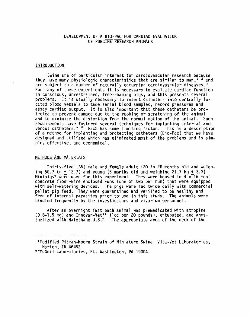
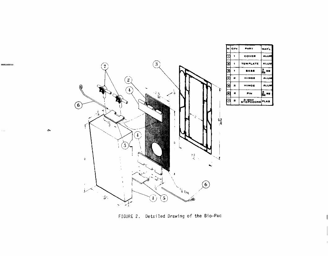
animal was clipped with a #40 clipper head, and the exact location of catheter site and our protective device (Bio-Pac - See Figures 1 & 2) was marked. Care was given to planning the placement of the Bio-Pac high enough on the lateral neck to allow a normal range of head and neck motion while permitting the investigator free access to the Bio-Pac while the animal was standing or lying on either side.
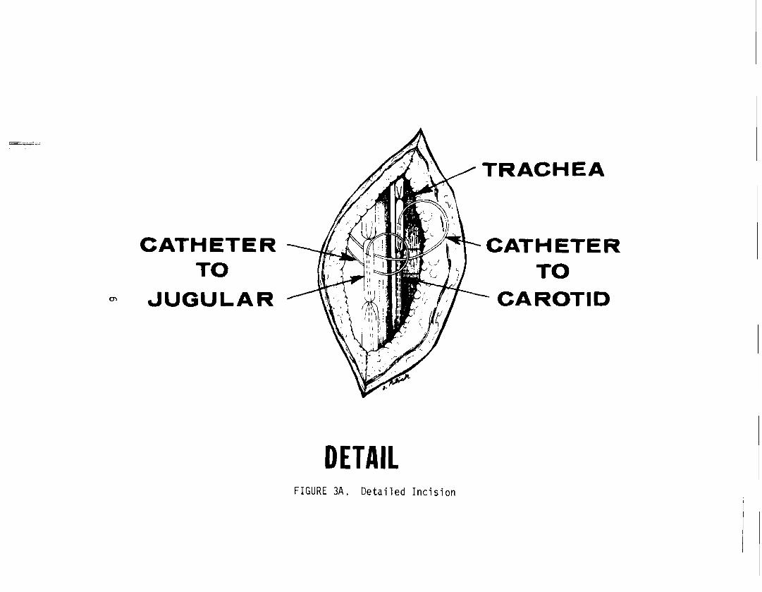
The operative area and the area of attachment of the Bio-Pac were prepared with a surgical detergent scrub and an application of an iodine solution. Either side could be utilized. With the animal in a semidorsal recumbent position, a 10 em incision was made in the middle one-third of the anterior neck parallel to and slightly lateral of the ventral midline (See Figure 3). Using blunt dissection, the carotid artery and the internal or external jugular vein were identified and isolated with umbilical tape. Two 24-inch 16 gauge Intracath* catheters (filled with dilute haparinized saline and secured with stopcocks) were introduced through a separate stab wound in the upper lateral neck and passed through a subcutaneous tunnel to the operative area. The catheters then were implanted into the appropriate vessels (via a small transverse incision using a catheter introducer**) and passed until the tip (factory shaped end) was approximately in the superior vena cava (venous) or the arch of the aorta (arterial) and secured with heavy silk sutures (See Figure 3). More accurate placement was made using radiography, ECG monitoring, or wave forms from pressure transducers. The superior aspect of each vessel was ligated in place. The wounds were closed with interrupted vertical mattress wire sutures.
The Bio-Pac was sutured in its predesignated place using six #24 gauge wire sutures. The catheters and three-way stopcocks were affixed to the template with pre-placed heavy sutures (See Figure 1). The catheters were flushed with dilute heparinized saline and the protective cover applied.
The animals were permitted to recover and stabilize for 24 to 48 hours in their runs before any studies were performed. The Minipigs then were transferred to a modified metabolic cage (See Figure 4). Serial blood samples, cardiac output determinations (dye dilution method) and blood pressure and pulse recordings were obtained. The notched opening permitted intravenous fluids to be administered continuously with the protective cover
*Deseret Pharmaceutical Co., Catalog No. 3182, Sandy, UT 84070 **Becton-Dickinson, Rutherford, NJ 07070
2
Figure 1. Open Bio-Pac showing the exact location of catheter site and protective device, the method of affixing the catheters and stopcocks, and the simplicity and versatility of using the Bio-Pac. The sterile caps on the three-way stopcocks have been removed for this photograph.
3
N. QTV. PART MAT'L
"' '.::J 1 COVER ALUM
~.;;.~ "i' = 1 TEMPLATE ALUM
"i' 1 BABE 3 '-=' i"z •• f.i' '--'
2 HINGE ALUM
."'--~ '\.I 11 a R111'111 '&--
'i' 2 HINGE ALUM I '.:>
'8' 2 PIN 3
= i"z-1
I
9 2 3-WAV PLA8 "-" STOPCOCKS
I 10~ !~
' . ----- ----- ~. . . . ... --+::>
53
" ·1 •,'l
--J " I
i...~'-1 ) l,.-· \
'/ \ 0 \.__ (6)
0)~? FIGURE 2. Detailed Drawing of the Bio-Pac

A B
INCISION SHOWN
ON DETAIL
FIGURE 3. Schematic drawing of catheterization procedure showing relative position of the incision, the location of the catheter tips within the vessels, and the ingress of these catheters to the Bio-Pac (A= Venous; B =Arterial).
5
~;.,;
CATHETER TO
~UGULAR
DETAIL FIGURE 3A. Detailed Incision
TRACHEA
CATHETER TO
CAROTID

FIGURE 4. Modified Metabolic Cage. The door was designed to provide easy access to both upper and lower areas of the animal. The cage is also equipped with an adjustable wooden squeeze baffle.
7

in place (See Figures 1 & 2). ECG monitoring via permanently implanted stainless steel wire electrodes and physiological monitoring was conducted by means of a commercial system.*
The majority of these animals were prepared for use in other studies which required central venous and arterial catheters. Their catheters were maintained patent and functional by flushing with dilute heparinized saline at least twice daily. Several animals had more than one set of catheters implanted. In these select animals, the original neck wound was reopened to afford direct visual ligation of the vessel from whence the catheter was removed. Following wound closure the entire procedure was repeated on the opposite side utilizing another set of catheters and Bio-Pac. Following the studies, a necropsy was performed on all animals.
RESULTS
There was no adverse reaction to the procedure or anesthesia. The Bio-Pac shown in Figures l and 2 was used to protect the catheters. It proved to be sturdy and withstood the repeated contacts with the expanded metal metabolic cage doors (See Figure 4), the wire enclosed runs, automatic waterers, and other animals. The double pinned protective cover permitted easy access either by removing one pin and flipping the BioPac open (See Figure 1), or by withdrawing both pins and removing the entire protective cover. The cost was under fifty dollars, including the material and the five hours of labor required to fabricate the BioPac.
The Minipigs adapted easily to the Bio-Pac and to the metabolic cages with minimal training. The heart rate monitored during the manipulation of the Bio-Pac generally showed no change indicative of excitement after the initial handling.
The wire sutures used to attach the Bio-Pac were strong and evoked little or no inflammatory reaction from the animal. Occasionally, with large and very active animals, a suture would break or pull through. These were easily replaced under local anesthesia, leaving the Bio-Pac undisturbed.
The plastic three-way stopcocks permitted easy access and control of both arterial and venous catheters. Various sized disposable syringes, the
*Model 410, Tektronix, Inc., S.W. Millikan Way, P. 0. Box 500, Beaverton, OR 97005
8

blood pressure transducers and standard intravenous tubing could be connected to these stopcocks. Patency and function were maintained by periodic flushing of each catheter with dilute heparinized saline.
Aortic pressures, central venous pressures and cardiac outputs were recorded. Typical pressure readings and cardiac output wave forms are shown in Figure 5. Serial arterial and venous blood samples for blood gases, chemistries, hematology, etc., were satisfactorily obtained from these catheters. Intravenous fluids were given in varying amounts without complications.
+20
0
-20
125
100
75
'
,.,.
J
-" _],.-
'
s:. Fi~,...---.-.--....,---~~
C) 12'1 . .. . •. .· ~ :·.· .r :::
10.['·. . !\··· z :. '" 0 8 -~~~ .... l ' ~ .. c:t: 6.. \ 01!: 41 ' . . . . ·+--:...J-'-'~.....)...__ z 2 1 .. 1'-k;. ~ 0 L :: .:..:..:.: :.:i. :~_ _ . , • z 0 5 10 15 20 25 seconds 0 CARDIAC OUTPUT u
'- I
r---------------·· --
50 t-- ARTERIAL PRESSURE 1---
25 PULSE: 114/MINUTE 1---1---I--
0 f-- ECG INTRAVENOUS
FIGURE 5. Typical Data Recordings
9

Animals necropsied before seven days showed minimal reaction to the catheters. The majority of the Minipigs were necropsied 10 to 14 days after implant and generally had a fibrous reaction around the catheters which prevented easy dislocation of the catheters.
At necropsy the position of the catheter tips was observed. The majority of the venous catheters were in the superior vena cava 1 to 3 em from the right atrium .. Occasionally, the catheter tip was found in the inferior vena cava, right atrium, right ventricle, ':Jr pulmonary artery. The arterial catheter tip usually was in the ar·ch uf ~he aorta, but could occasionally be found in the ascending or descPnding thcracic aorta or in the left subclavian artery.
DISCUSSION
The increased popularity of miniature swine 1n biomedical research has required experimental techniques that arP slightly different from the ones used for other research animals. The evJ1uation of cardiac function is an important aspect in many experiments, but the relatively unaccessible peripheral vessels are a definite disadvantage for studying systemic venous and arterial pressures, 9 cardiac output, 1nd serial blood sampling as well as for administering intravenous fluids and drugs. For this reason it is necessary to implJnt arterial a~d venous ~atneters.
The procedure fur implanting central venous .Jnd Jortic catheters has been dec.cribed,''-'' altfiOuSJh it i:; not v1ithc·~1! :;t~:~ ,;o,;~:1erns. Linzell '~tresses the tendency of the o.rteries uf ~he ._,i'; ~L :,f-Jasr,,, and the layers of the wall to strip, as the catheter is passei dn~n.- ~or this reason ive used as little 111anipulation as possible 'n ltiSf'rt '''c' ::he catheter and always used the factory prepared catheter tip. This ~lso avoided the problem experienced by other of loss of patency pruduLed ~Y a valve-like closure of the vessel wall over the precut ac~te a0glPd ratheter tip. 4
Unilateral occulsion of one carotid or jugular vein seldom leads to illeffects.7 10 However, the carotid reflexes dre ~ ~ctcntial problem, at least in theory. 11 \~e did have some transient ata;ia ;r: a few of the animals with bilateral occulsion for catheterization.
The motion of the catheters through the wound was minimized by allowing small loops of catheters to remain within the neck incision and by placing the ingress of our Bio-Pac directly over the site of exteriorization of the catheters. The use of the punch biopsy and cannula described by Christison and Curtin 4 greatly simplifies the subcutaneous passage of the catheters.
10

The major problem then is protecting the external connections of the catheters. 7 Adhesive tape and stockinet has been used to protect catheters and flow prebe leads. These catheters were exteriorized either on the back of the neck or on the lateral side. 9 This would seem to be a vulnerable system even though their experiments were Qf long duration. Christison and Curtin 4 used tag cement to attach the catheter to the side of the neck and covered it with adhesive tape. This required reclipping and cement attachment renewal. In addition, the animals required individual smooth-sided pins to prevent dislodgment. Mount 7 has described a harness and small box carried on the animal •s back in which connections can be made to indwelling catheters or to a radio transmitter for telemetry. There was some movement of the catheters which prevented complete wound healing. Harnesses have been unsatisfactory in our experience because of extensive motion in normal fitting apparatus or respiratory restriction in snug fitting harnesses. Linzell 6 uses small metal plates fixed to the skin, one inside and the other outside, and a metal box fixed to the skin, which can be closed over catheter holes. Our metal protective box (Bio-Pac) utilizes the best features of these methods to give a sturdy, simple, and economical, yet extremely versatile system of protecting the implanted catheters. When properly located the Bio-Pac caused no impairment to the experimental animal. It was not necessary to keep the animal wearing a Bio-Pac in an individual or especially prepared pin. Automatic waterers, feeding pans and pallets could be left in place. Other animals did not chew on the Bio-Pac as they did on harnesses, plaster, and tapes. Likewise, there was little or no movement of the catheters through the wound to carry infection into the body. The design of the Bio-Pac permitted easy accessibility to the catheters both in the runs and in the metabolic cages, and was a major factor in simplifying cardiac evaluation. Although not performed in this experiment, the Bio-Pac was designed to permit placement of telemetry equipment within by attaching it to the template or the cover.
The catheters were connected to blood pressure transducers, cardiac output apparatus, syringes, and intravenous tubing. It was not the objective of this paper to report a statistical analysis of these results, but rather to emphasize the ease with which all these procedures could be accomplished under sterile conditions using our method. The cardiac output, heart rate, and systemic pressures, however, did compare to the 1 iter a ture. 8 12
At necropsy the location of the catheter tips was of primary importance for comparison with the method of placement and accuracy of data. With experience the catheter tip location could be estimated, but fluoroscopy or radiographs gave the most accurate placement and are to be recommended. Wave forms from the pressure transducers at the time of catheter
11

implantation could also ,be used with some degree of accuracy as could an intravenous ECG. The fibrous reaction present around the catheters and compensatory enlargement of the major blood vessels were expected findings.4 7
CONCLUSIONS
A simple sturdy metal box (Bio-Pac) was designed and utilized to protect the indwelling central venous and arterial catheters in miniature swine.
These indwelling catheters were easily maintained for 14 days in unrestrained, free-roaming pigs.
Serial blood sampling, pressure recording, ECG monitoring, and cardiac output measuring were conducted, and the infusion of precise amounts of fluids and/or drugs could be administered.
12
i .

REFERENCES
1. Bustad, L. K.: "Pigs in the Laboratory," Sci. Am., 214(6):94-100, 1966.
2. McClellan, R. 0.: "Applications of Swine in Biomedical Research," Lab. Animal Care, ]~:120-126, 1968.
3. Detweiler, D. K.: "Swine in Comparative Cardiovascular Research. 11
In Swine in Biomedical Research, Seattle, Frayne Printing Company, 1966, pp. 301-306.
4. Christison, G. I. and Curtin, T. M.: "A Simple Venous Catheter for Sequential Blood Sampling from Unrestrained Pigs," Lab. Animal Care, J.i: 259-262. 1969.
5. L i nze 11 , J . L . , Mepham, T. B. , Anni son, E. F. , et. a 1 . : "Mammary Metabolism in the Lactating Sow," Biochem. J., 103:42P, 1967.
6. Linzell, J. L., Mepham, T. B., Annison, E. F., et. al.: "Mammary Metabolism in Lactating Sows: Arteriovenous Differences of Milk Precursors and the Mammary Metabolism of [ 14C] Glucose and [ 14C] Acetate," Brit. J. Nutr., 23:319-332, 1969.
7. Mount, L. E. and Ingram, D. L.: "Vascular Surgery: Insertion of Catheters and Wires," The Pig as a Laboratory Animal, London and New York, Academic Press, 1971, pp. 99-101.
8. Stone, H. L. and Sawyer, D. C.: "Cardiac Output and Related Measurements in Unanesthetized Miniature Swine." In Swine in Biomedical Research, Seattle, Frayne Printing Company, 1966, pp. 411-418.
9. Sawyer, D. C. and Stone, H. L.: "Thoracic Surgery for Electromagnetic Flow Sensor Implantation on Miniature Swine." In Swine in Biomedical Research, Seattle, Frayne Printing Company, 1966, pp. 405-409.
10. Wachtel, W.: "Zur Frage der reflektorischen Kreislaufregulierung beim Schwein, 11 Arch. Exp. Veterinaer. Med., 20:1101-1113, 1966.
11 . Booth, N. H., Bredeck, H. E. and Herin, R. A. : "Baroceptor and Chemoceptor Reflex Mechanisms in Swine." In Swine in Biomedical Research Seattle, Frayne Printing Company, 1966, pp. 331-346.
13

12. Engelhardt, W. v.: "Swine Cardiovascular' Physiology--A Review." In Swine in Biomedical Research, Seattle, Frayne Printing Company, 1966, pp. 307-329.
14