Embed Size (px)
Citation preview
Dermatology and
Infectious Diseases
Disorders of Keratinisation
Dermatitis
Blistering Disorders
Immunology
Disorders of Keratinisation
Overview Psoriasis Icthyosis

Objectives
Disorders of Keratinisation
Disorders of Keratinization
KeratinizationTerminal differentiation of epithelia
– epithelial proteins (Keratin)– Glycoproteins (Cell Envelope)– intercellular lipids
Disorders of Keratinization
Cause changes in the skin– Dry, Scaly, Thickened, Flaky– Blistering
Cause changes in Mucous membranes, Nails and Hair
Disorders of Keratinization
Change in Type of Keratin Made
Disorders of Keratinisation
Disorders of Keratinisation
Disorders of Keratinization
Psoriasis Icthyosis
Disorders of KeratinizationPsoriasis Chronic , relapsing and remitting skin
disease. May appear at any age may affect any part of the skin Common Locations:
– Extensor surfaces Knees and Elbows
Disorders of KeratinizationPsoriasis Characterised by hyperproliferation of skin
and inflammation
Etiology– Inherited
• Abnormality of Ca++ metabolism• Genetic Predisposition
– HLA Cw6
– HLA DR7
– HLA B27 (Pustular)
Disorders of KeratinizationPsoriasis
Disorders of KeratinizationPsoriasis Etiology
– Environmental Factors• Stress, Smoking and Alcohol
• Systemic Drugs
• Infection
– Immunological Factors
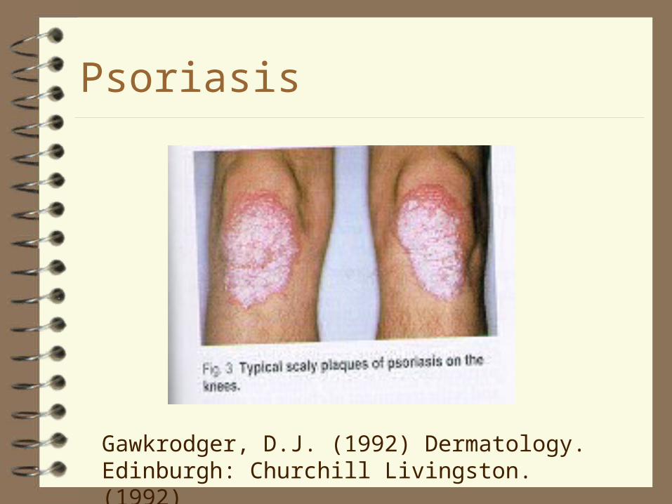
Psoriasis
Gawkrodger, D.J. (1992) Dermatology. Edinburgh: Churchill Livingston. (1992)
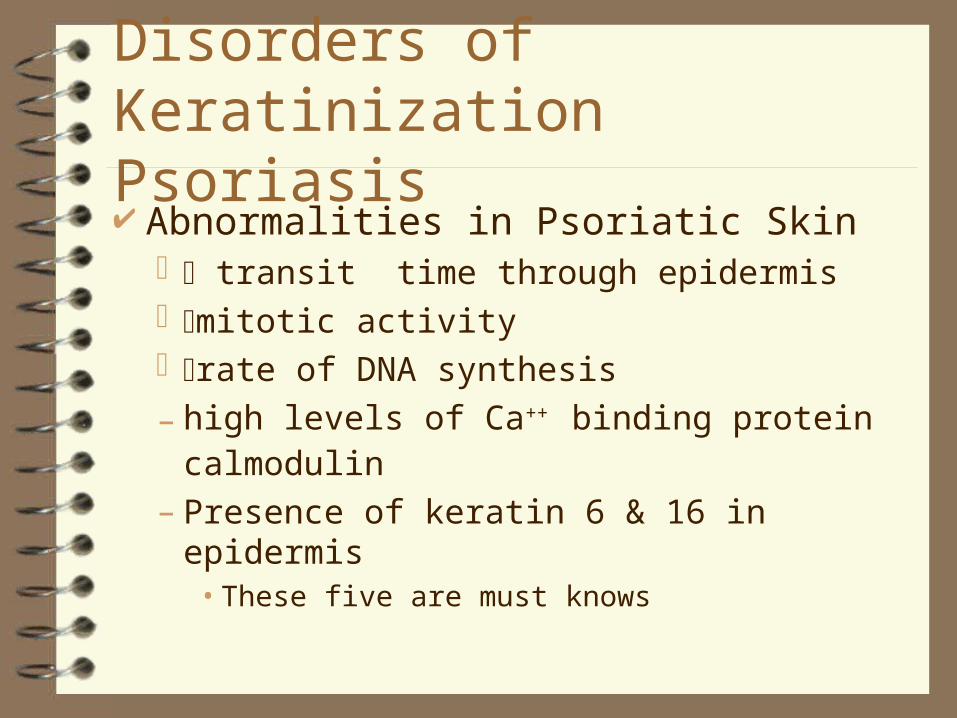
Disorders of KeratinizationPsoriasis Abnormalities in Psoriatic Skin
transit time through epidermis mitotic activity rate of DNA synthesis
– high levels of Ca++ binding protein calmodulin
– Presence of keratin 6 & 16 in epidermis• These five are must knows
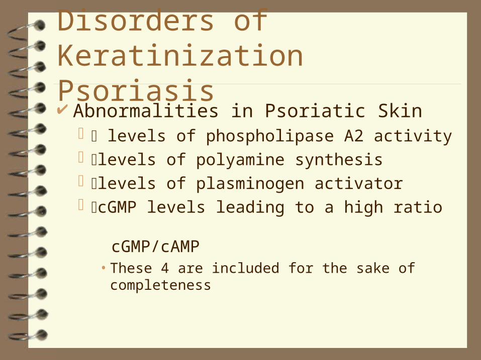
Disorders of KeratinizationPsoriasis Abnormalities in Psoriatic Skin
levels of phospholipase A2 activity levels of polyamine synthesis levels of plasminogen activator cGMP levels leading to a high ratio
cGMP/cAMP• These 4 are included for the sake of completeness
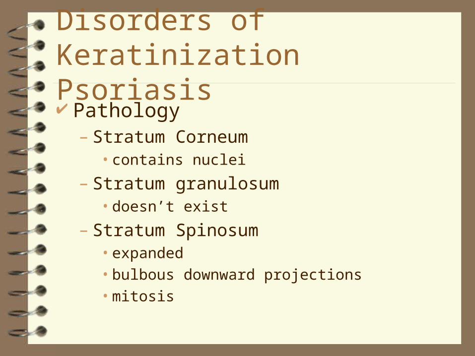
Disorders of KeratinizationPsoriasis Pathology
– Stratum Corneum• contains nuclei
– Stratum granulosum• doesn’t exist
– Stratum Spinosum• expanded• bulbous downward projections• mitosis
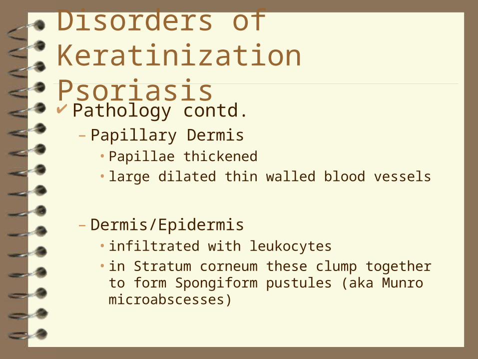
Disorders of KeratinizationPsoriasis Pathology contd.
– Papillary Dermis• Papillae thickened
• large dilated thin walled blood vessels
– Dermis/Epidermis• infiltrated with leukocytes
• in Stratum corneum these clump together to form Spongiform pustules (aka Munro microabscesses)
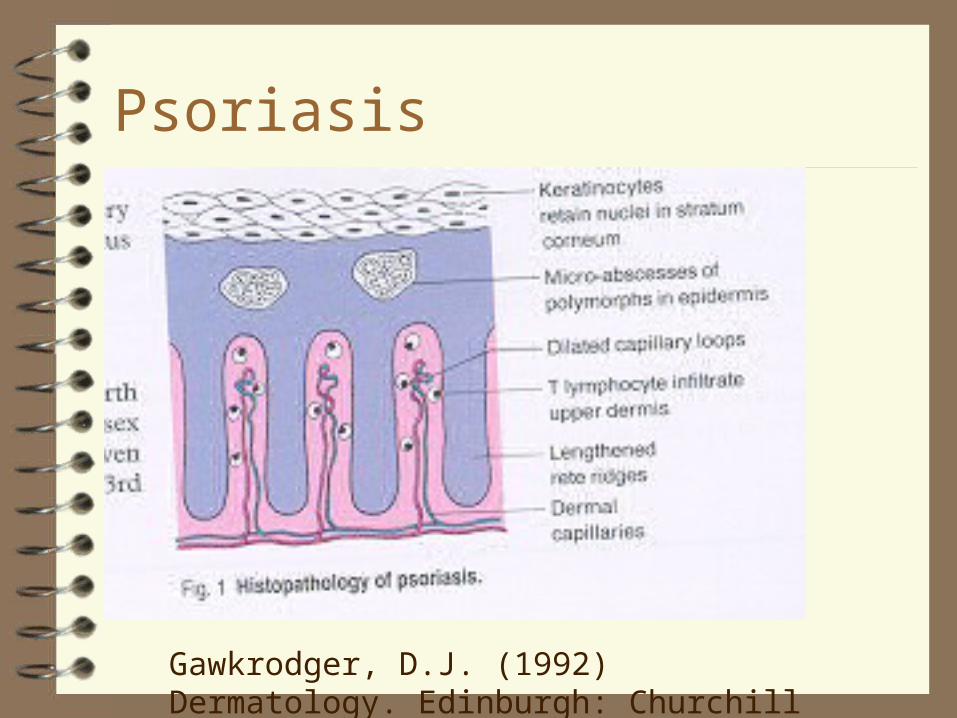
Psoriasis
Gawkrodger, D.J. (1992) Dermatology. Edinburgh: Churchill Livingston. (1992)
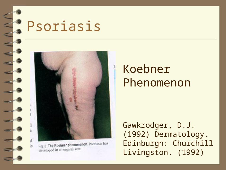
Psoriasis
Gawkrodger, D.J. (1992) Dermatology. Edinburgh: Churchill Livingston. (1992)
Koebner Phenomenon
Required Reading
Gawkrodger, D.J. (1992) Dermatology. Edinburgh: Churchill Livingston. (1992)
Psoriasis
Disorders of KeratinisationIcthyoses A variety of hereditary keratinisation
disorders visible scales on the skin Forms include
– autosomal dominant– x-linked– associated with multisystem changes
Disorders of KeratinisationIcthyoses May vary from very mild to very severe
The keratinisation process which is changed varies from condition to condition
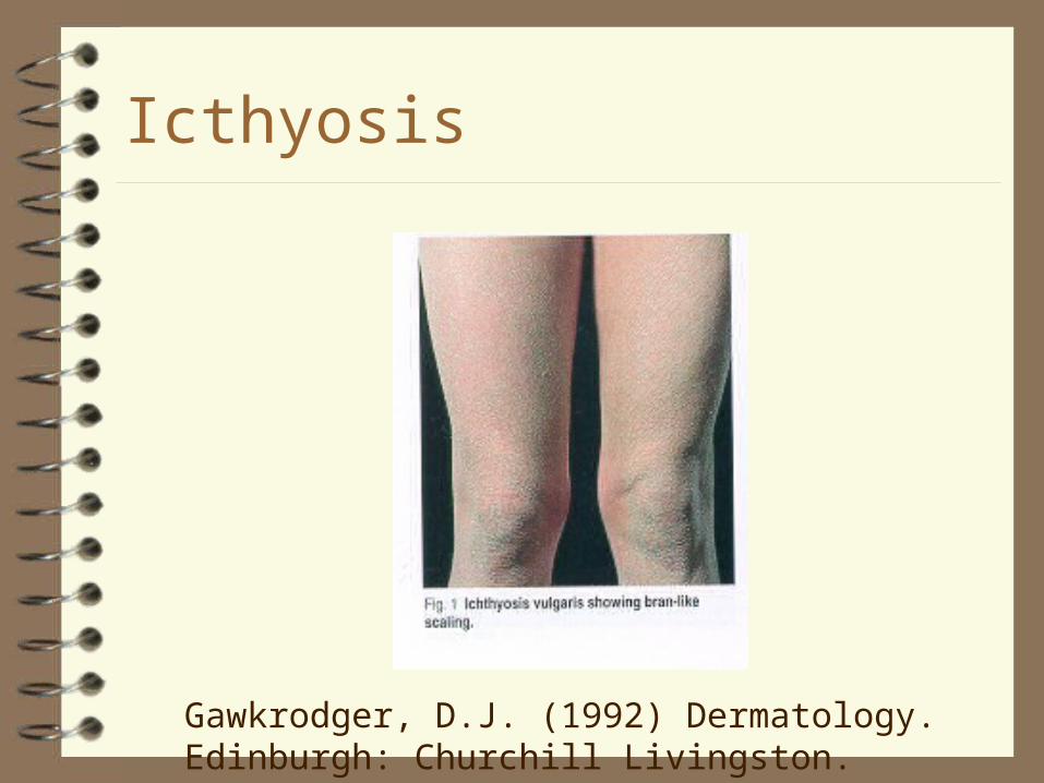
Icthyosis
Gawkrodger, D.J. (1992) Dermatology. Edinburgh: Churchill Livingston. (1992)
Required Reading
Gawkrodger, D.J. (1992) Dermatology. Edinburgh: Churchill Livingston. on Keratinization and Blistering Syndromes
Dermatitis/eczema dermatitis = eczema non-infective inflammation of the skin Greek for ‘to boil over’ reaction to various stimuli
– some known, some unknown
DermatitisClassification current classification
unsatisfactory/inconsistent distinctions are often difficult to determine endogenous (internal factors) exogenous (external factors) acute chronic
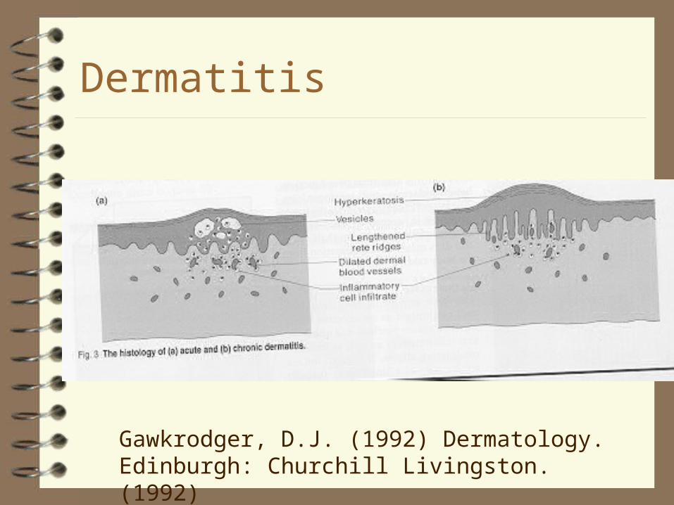
DermatitisAcute acute eczema leads to epidermal
oedema (spongiosis), with separation of keratinocytes
leads to epidermal vesicles dermal vessels become dilated inflammatory cells invade the dermis
and epidermis
DermatitisChronic
chronic eczema leads to a thickening of the stratum spinosum (acanthosis) & stratum corneum (hyperkeratosis)
also get retention of nuclei by some corneocytes
rete ridges are lengthened dermal vessels are dilated inflammatory mononuclear cells infiltrate the
skin
Dermatitis
Gawkrodger, D.J. (1992) Dermatology. Edinburgh: Churchill Livingston. (1992)
DermatitisTypes of dermatitis/eczema
contact dermatitis/eczema– contact with an irritant
atopic dermatitis/eczema– associated with a history of asthma, allergic
rhinitis, conjunctivitis
seborrhoeic dermatitis/eczema– commonly affects the scalp and face
DermatitisTypes of dermatitis/eczema cont.
discoid (nummular) dermatitis/eczema– often presents as coin-shaped lesions on
the limbs of middle aged or older people
venous stasis dermatitis/eczema– associated with venous disease– commonly involves the medial aspect of
the ankle
Required Reading
Gawkrodger, D.J. (1992) Dermatology. Edinburgh: Churchill Livingston., D.J. (1992) Dermatology. Edinburgh: Churchill Livingston.
Chapters on Eczema
Bullous Disorders
blistering (bullous) disorders are often seen with skin disease
found with common skin conditions like acute contact dermatitis
Etiology-autoimmune mechanisms, inheredited errors in metabolism and mechanical trauma
Types of Bullous Disorders
Pemphigus Pemphigoid Epidermolysis bullosa dermatitis
herpetiformis linear lgA disease Fungi Friction
Systemic lupus erythematosis (SLE)
Erythema multiforme Stevens-Johnson
syndrome Bullous impetigo Bullous diabeticorum
Bullous disorders
Blisters are classified according to their position in the epidermis.
SubCorneal :Stratum Corneum Intraepidermal: Lower levels of the
epidermis Sub Epidermal: At the dermo-epidermal
junction
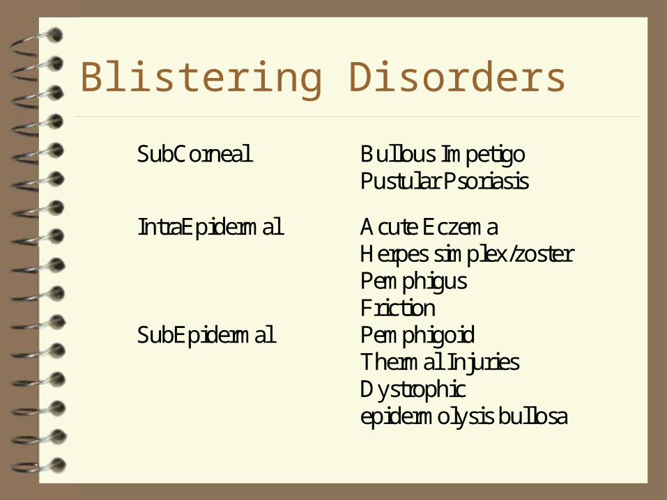
Blistering Disorders
SubCorneal Bullous ImpetigoPustular Psoriasis
IntraEpidermal Acute EczemaHerpes simplex/zosterPemphigusFriction
SubEpidermal PemphigoidThermal InjuriesDystrophicepidermolysis bullosa
Friction Blisters
direct mechanical trauma Treatment: avoidance-look at footwear,
protective taping and padding, 2 pairs of socks, lubrication.
sock design and reduction of blistering Ref.-Herring and Ritchie in JAPMA 1990
and 1993.
Required Reading
Gawkrodger, D.J. (1992) Dermatology. Edinburgh: Churchill Livingston.
Read Chapter on Blistering Disorders and chapter on Keratinization and Blistering Syndromes
Hypersensitivity Reactions and the Skin inappropriate or
exaggerated response to the degree that tissue damage occurs.
4 Types Type l -immediate Type ll -antibody
dependant cytotoxicity Type lll-immune
complex disease Type lV-cell mediated
or delayed