Embed Size (px)
Citation preview

Neonatal blistering -
a butterfly child
SWISS SOCIETY OF NEONATOLOGY
August 2010

2
Kaelin S, Weibel L, Arlettaz Mieth R, Neonatal Intensive
Care Unit (KS, AMR), Department of Dermatology (WL),
University Hospital Zurich, Switzerland and Division of
Dermatology, University Children’s Hospital Zurich,
Switzerland
© Swiss Society of Neonatology, Thomas M Berger, Webmaster
3
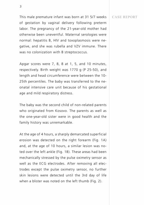
This male premature infant was born at 31 5/7 weeks
of gestation by vaginal delivery following preterm
labor. The pregnancy of the 21-year-old mother had
otherwise been uneventful. Maternal serologies were
normal: hepatitis B, HIV and toxoplasmosis were ne-
gative, and she was rubella and VZV immune. There
was no colonization with B streptococcus.
Apgar scores were 7, 8, 8 at 1, 5, and 10 minutes,
respectively. Birth weight was 1770 g (P 25-50), and
length and head circumference were between the 10-
25th percentiles. The baby was transferred to the ne-
onatal intensive care unit because of his gestational
age and mild respiratory distress.
The baby was the second child of non-related parents
who originated from Kosovo. The parents as well as
the one-year-old sister were in good health and the
family history was unremarkable.
At the age of 4 hours, a sharply demarcated superficial
erosion was detected on the right forearm (Fig. 1A)
and, at the age of 10 hours, a similar lesion was no-
ted over the left ankle (Fig. 1B). These areas had been
mechanically stressed by the pulse oximetry sensor as
well as the ECG electrodes. After removing all elec-
trodes except the pulse oximetry sensor, no further
skin lesions were detected until the 3rd day of life
when a blister was noted on the left thumb (Fig. 2).
CASE REPORT
4
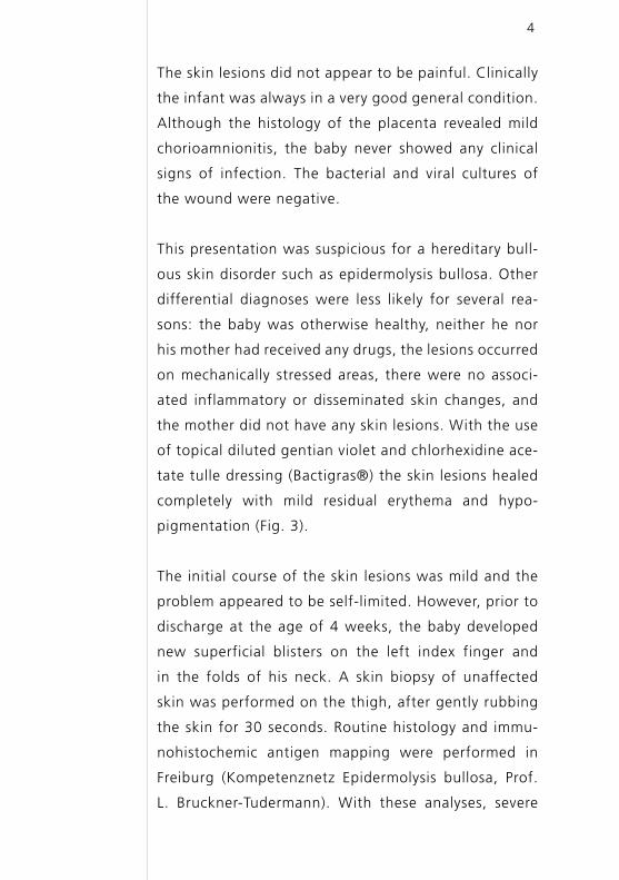
The skin lesions did not appear to be painful. Clinically
the infant was always in a very good general condition.
Although the histology of the placenta revealed mild
chorioamnionitis, the baby never showed any clinical
signs of infection. The bacterial and viral cultures of
the wound were negative.
This presentation was suspicious for a hereditary bull-
ous skin disorder such as epidermolysis bullosa. Other
differential diagnoses were less likely for several rea-
sons: the baby was otherwise healthy, neither he nor
his mother had received any drugs, the lesions occurred
on mechanically stressed areas, there were no associ-
ated inflammatory or disseminated skin changes, and
the mother did not have any skin lesions. With the use
of topical diluted gentian violet and chlorhexidine ace-
tate tulle dressing (Bactigras®) the skin lesions healed
completely with mild residual erythema and hypo-
pigmentation (Fig. 3).
The initial course of the skin lesions was mild and the
problem appeared to be self-limited. However, prior to
discharge at the age of 4 weeks, the baby developed
new superficial blisters on the left index finger and
in the folds of his neck. A skin biopsy of unaffected
skin was performed on the thigh, after gently rubbing
the skin for 30 seconds. Routine histology and immu-
nohistochemic antigen mapping were performed in
Freiburg (Kompetenznetz Epidermolysis bullosa, Prof.
L. Bruckner-Tudermann). With these analyses, severe
5
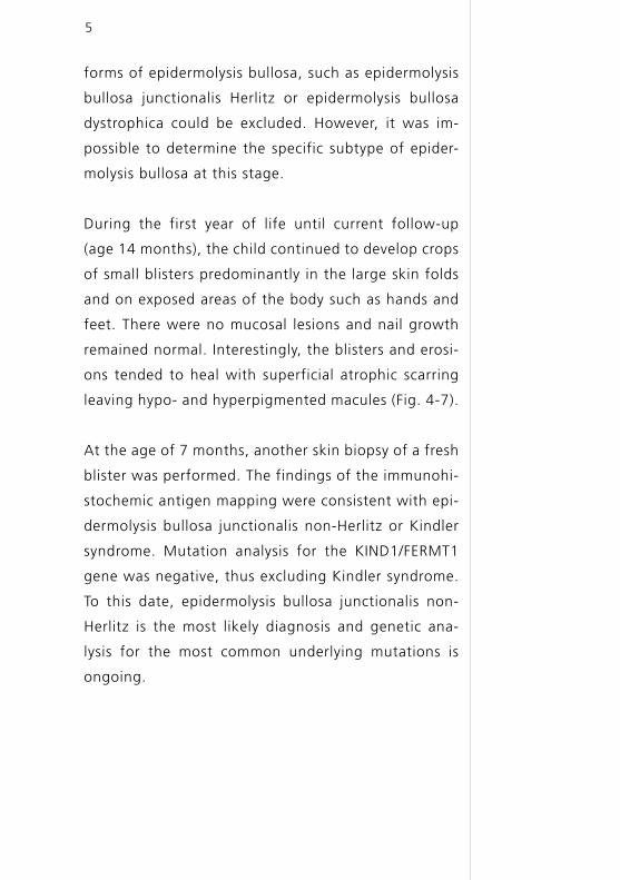
forms of epidermolysis bullosa, such as epidermolysis
bullosa junctionalis Herlitz or epidermolysis bullosa
dystrophica could be excluded. However, it was im-
possible to determine the specific subtype of epider-
molysis bullosa at this stage.
During the first year of life until current follow-up
(age 14 months), the child continued to develop crops
of small blisters predominantly in the large skin folds
and on exposed areas of the body such as hands and
feet. There were no mucosal lesions and nail growth
remained normal. Interestingly, the blisters and erosi-
ons tended to heal with superficial atrophic scarring
leaving hypo- and hyperpigmented macules (Fig. 4-7).
At the age of 7 months, another skin biopsy of a fresh
blister was performed. The findings of the immunohi-
stochemic antigen mapping were consistent with epi-
dermolysis bullosa junctionalis non-Herlitz or Kindler
syndrome. Mutation analysis for the KIND1/FERMT1
gene was negative, thus excluding Kindler syndrome.
To this date, epidermolysis bullosa junctionalis non-
Herlitz is the most likely diagnosis and genetic ana-
lysis for the most common underlying mutations is
ongoing.

6
Fig. 1
A B
Blisters on day of life 1. A) Sharply demarcated
superficial erosion on right forearm in the area of
earlier ECG electrode placement, B) Eroded superficial
blister on left ankle.
A B

7
Fig. 2
A B
Newly developed tense blister on left thumb on day
of life 3.
A B
8
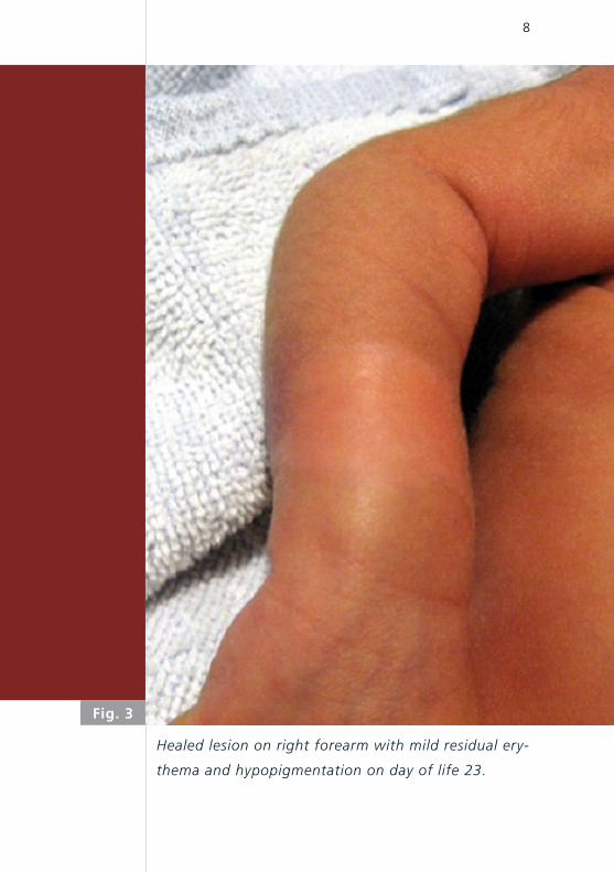
Fig. 3
Healed lesion on right forearm with mild residual ery-
thema and hypopigmentation on day of life 23.
9
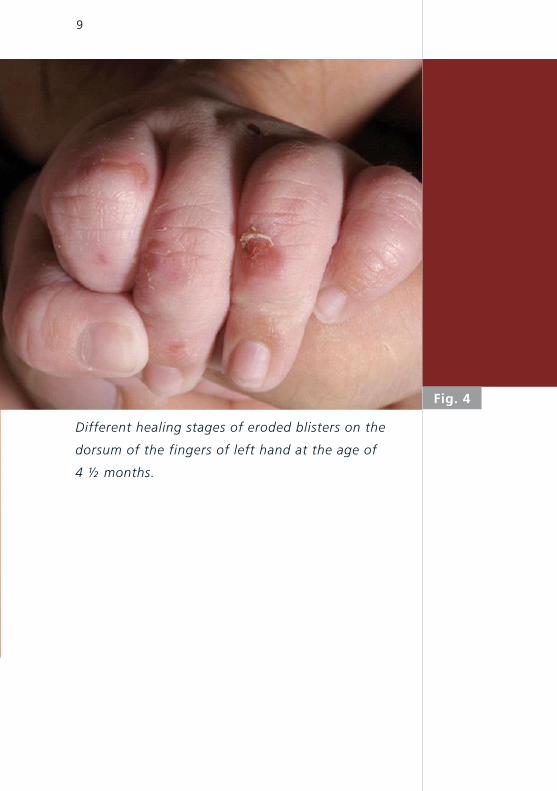
Fig. 4
Different healing stages of eroded blisters on the
dorsum of the fingers of left hand at the age of
4 ½ months.

10
Fig. 5
Annular erythematous macules, papules, small
blisters and crusts as well as hyper- and hypopigmen-
ted superficial scarring on right axilla and upper arm
at the age of 7 months.
11
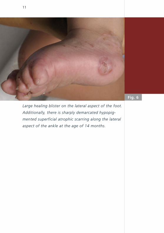
Large healing blister on the lateral aspect of the foot.
Additionally, there is sharply demarcated hypopig-
mented superficial atrophic scarring along the lateral
aspect of the ankle at the age of 14 months.
Fig. 6
12
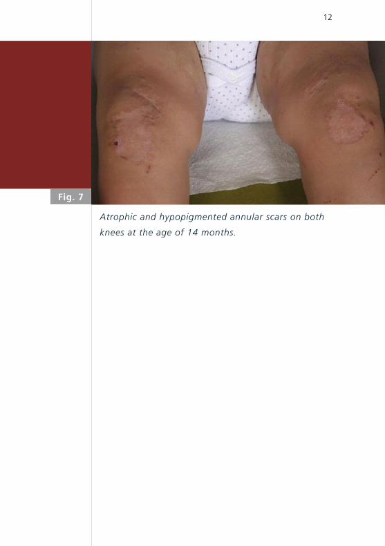
Fig. 7
Atrophic and hypopigmented annular scars on both
knees at the age of 14 months.
13
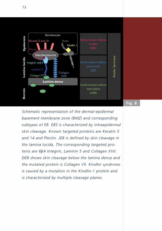
Schematic representation of the dermal-epidermal
basement membrane zone (BMZ) and corresponding
subtypes of EB. EBS is characterized by intraepidermal
skin cleavage. Known targeted proteins are Keratin 5
and 14 and Plectin. JEB is defined by skin cleavage in
the lamina lucida. The corresponding targeted pro-
teins are 6b4 Integrin, Laminin 5 and Collagen XVII.
DEB shows skin cleavage below the lamina densa and
the mutated protein is Collagen VII. Kindler syndrome
is caused by a mutation in the Kindlin-1 protein and
is characterized by multiple cleavage planes.
Fig. 8

14
Fig. 9
Differential diagnoses of blistering diseases. The red
colored diagnoses are described in the supplement.

15
Evaluation of suspected EB.
Fig. 10
16
DISCUSSION Epidermolysis bullosa (EB) comprises a large heteroge-
neous group of hereditary dermatoses characterized by
fragility of the skin and mucous membranes, leading
to blistering and erosions as a result minor trauma.
“Butterfly Children” is a term often used to describe
younger patients because the skin is said to be as fra-
gile as a butterfly’s wings.
The fragility is caused by gene mutations encoding a
range of epidermal basement membrane molecules
(1). The inheritance can be either autosomal recessive
or dominant. Currently, over 1000 mutations in 13 dis-
tinct genes are known (2). The clinical picture ranges
from mild subtypes with minor skin reactions to severe
generalized forms with lethal outcome within the first
months of life. The incidence is 19-26 to 1‘000‘000
live-births (3).
On the basis of the level of the cleavage plane within
the dermal-epidermal basement membrane zone, EB
can be categorized into 4 major groups: EB simplex,
EB junctionalis, EB dystrophica and, according to the
latest classification of 2008 (2), the Kindler syndrome
(Fig. 8). All together, 33 subtypes can be distinguis-
hed, based on the new EB classification system (2) and
the molecular background.
Blistering skin diseases are rare but the differential
diagnosis, especially in the neonatal period, is wide.
As some of these disorders can be life-threatening or
17
result in long-term impairment one should be aware
of the most common ones (Fig. 9). Clinical course and
presentation of the skin lesions, family history and
gender help in establishing the diagnosis. Essentially,
there are no conditions that can mimic EB in every
respect. In neonates, the clinical presentation of EB
(e.g., involved areas, extension of skin lesions) does
not allow to differentiate one form of EB from another
and the main challenge usually is to determine the
specific subtype of EB (5).
Diagnosis in blistering diseases and EB in particular is
sequential (Fig. 10). With routine histology, electron
microscopy and immuno-histochemic antigen map-
ping an initial classification of EB can be made. Allow-
ing a semi-quantitative evaluation of the expression
of structure proteins (normal, reduced or missing),
immuno-histochemic antigen mapping should precede
the far more complex mutation analyses. For the de-
finite diagnosis, prognosis and genetic counselling in-
cluding prenatal testing, however, the identification
of the underlying mutation is essential (6, 7).
Treatment of EB is symptomatic. Attempts at gene the-
rapy (cell therapy, vector therapy and protein therapy)
in animal models are in progress (8-10) and there are a
few reports on promising approaches to gene therapy
in humans (11).

18
Please, also note the attached picture book of neo-
natal blistering skin disorders (Supplement). See also:
COTM 08/2005: Epidermolysis bullosa.

19
1. Has C, Bruckner-Tuderman L. Molecular and diagnostic
aspects of genetic skin fragility. J Dermatol Sci 2006
Dec;44:129-144
2. Fine JD, Eady RA, Bauer EA et al. The classification of
inherited epidermolysis bullosa (EB): Report of the Third
International Consensus Meeting on Diagnosis and
Classification of EB. J Am Acad Dermatol. 2008;58:931-950
3. Fine JD et al (1999). The epidemiology of inherited
epidermolysis bullosa: findings within American, Canadian
and European study population. In: Fine JD, Bauer EA, Mc
Guire J, Moshell A (eds) Epidermolysis bullosa: clinical,
epidemiologic and laboratory advances and findings of the
national epidermolysis bullosa registry. Johns Hopkins
University Press, Baltimore, 1999, pp 101-113
4. Lai-Cheong JE, Tanaka A, Hawche G et al. Kindler syndrome:
a focal adhesion genodermatosis. Br J Dermatol. 2009;
160:223-242
5. Lin AN, Carter DM (1992). Differential Diagnosis in
Epidermolysis bullosa. In: Epidermolysis bullosa Basic and
Clinical Aspects, Springer Verlag, pp 8-11
6. Laimer M, Landschützer CM, Nischler E et al. Hereditary
blistering diseases. Symptoms, diagnosis and treatment of
epidermolysis bullosa. Hautarzt 2009;605:378-388
7. Shimizu H. Prenatal diagnosis of epidermolysis bullosa. Prenat
Diagn 2006;2613:1260-1261
8. Tamai K, Kaneda Y, Uitto J. Molecular therapies for heritable
blistering diseases. Trends Mol Med 2009;157:285-292
9. Remington J, Wang X, Hou Y et al. Injection of recombinant
human type VII collagen corrects the disease phenotype
in a murine model of dystrophic epidermolysis bullosa.
Mol Ther 2009;171:26-33
REFERENCES

20
10. De Luca M, Pellegrini G, Mavilio F. Gene therapy of inherited
skin adhesion disorders: a critical overview. Br J Dermatol
2009;1611:19-24
11. Mavilio F, Pellegrini G, Ferrari S et al. Correction of junctional
epidermolysis bullosa by transplantation of genetically
modified epidermal stem cells. Nat Med 2006;12:1397-1402

21
Neonatal blistering diseases
Supplement
The authors confirm that they are the copyright
owners of these images, or, alternatively,
that they have obtained a reprint permission
from the copyright owners.

22
Fig. 1
Epidermolysis bullosa (EB): a group of inherited
bullous disorders characterized by blister formation in
response to mechanical trauma.

23
Aplasia cutis congenita is characterized by the
absence of a portion of skin in a localized or wide-
spread area at birth. Most commonly it is localized
on the scalp. Defects in the skin that form early in
gestation may heal before delivery and appear as an
atrophic, membranous or bullous scar with associated
alopecia, whereas less mature defects present as ul-
cerations. It may be an isolated finding or associated
with other congenital malformation syndromes (i.e.
Adams-Oliver Syndrome (see also COTM 12/2001) or
Trisomy 13). Bart-Syndrome is an overlap of Aplasia
cutis congenita and epidermolysis bullosa (see also
COTM 05/2003).
Fig. 2
24
Fig. 3
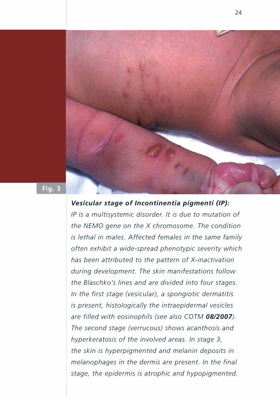
Vesicular stage of Incontinentia pigmenti (IP):
IP is a multisystemic disorder. It is due to mutation of
the NEMO gene on the X chromosome. The condition
is lethal in males. Affected females in the same family
often exhibit a wide-spread phenotypic severity which
has been attributed to the pattern of X-inactivation
during development. The skin manifestations follow
the Blaschko’s lines and are divided into four stages.
In the first stage (vesicular), a spongiotic dermatitis
is present, histologically the intraepidermal vesicles
are filled with eosinophils (see also COTM 08/2007).
The second stage (verrucous) shows acanthosis and
hyper keratosis of the involved areas. In stage 3,
the skin is hyperpigmented and melanin deposits in
melanophages in the dermis are present. In the final
stage, the epidermis is atrophic and hypopigmented.
25
Fig. 4
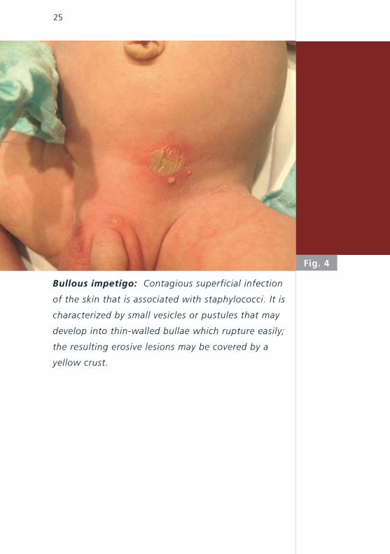
Bullous impetigo: Contagious superficial infection
of the skin that is associated with staphylococci. It is
characterized by small vesicles or pustules that may
develop into thin-walled bullae which rupture easily;
the resulting erosive lesions may be covered by a
yellow crust.
26
Fig. 5
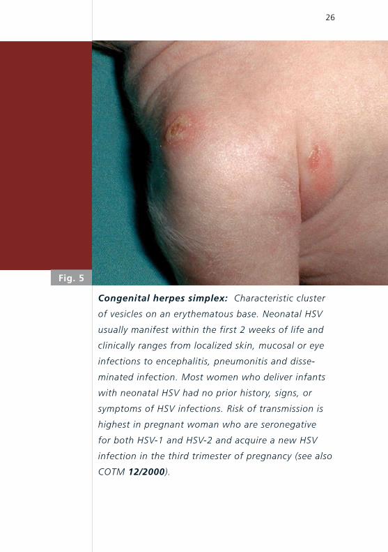
Congenital herpes simplex: Characteristic cluster
of vesicles on an erythematous base. Neonatal HSV
usually manifest within the first 2 weeks of life and
clinically ranges from localized skin, mucosal or eye
infections to encephalitis, pneumonitis and disse-
minated infection. Most women who deliver infants
with neonatal HSV had no prior history, signs, or
symptoms of HSV infections. Risk of transmission is
highest in pregnant woman who are seronegative
for both HSV-1 and HSV-2 and acquire a new HSV
infection in the third trimester of pregnancy (see also
COTM 12/2000).
27
Fig. 6
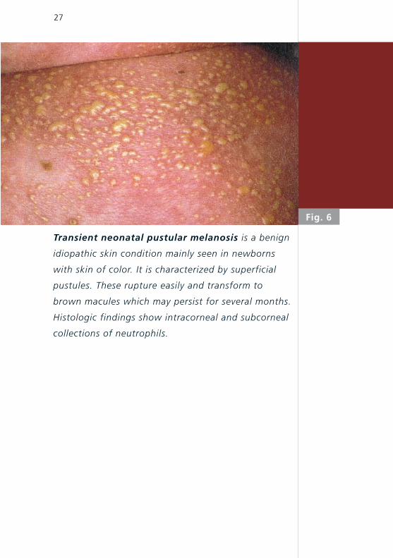
Transient neonatal pustular melanosis is a benign
idiopathic skin condition mainly seen in newborns
with skin of color. It is characterized by superficial
pustules. These rupture easily and transform to
brown macules which may persist for several months.
Histologic findings show intracorneal and subcorneal
collections of neutrophils.
28
Fig. 7
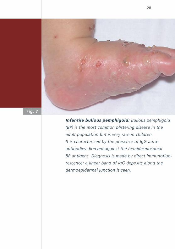
Infantile bullous pemphigoid: Bullous pemphigoid
(BP) is the most common blistering disease in the
adult population but is very rare in children.
It is characterized by the presence of IgG auto-
antibodies directed against the hemidesmosomal
BP antigens. Diagnosis is made by direct immunofluo-
rescence: a linear band of IgG deposits along the
dermoepidermal junction is seen.
29
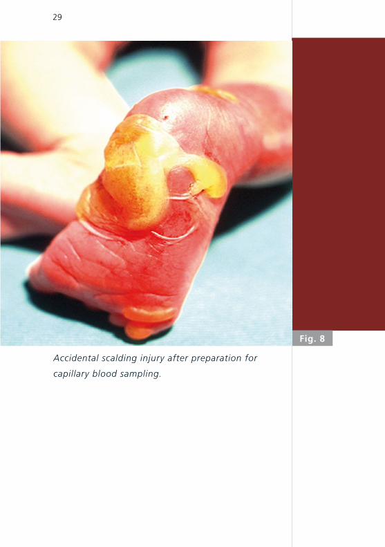
Fig. 8
Accidental scalding injury after preparation for
capillary blood sampling.

30
Fig. 9
Sucking blister: These lesions are present at birth,
most often over the dorsal and lateral aspect of the
wrist. They are due to sucking of the affected areas
in utero. They appear as well demarcated bruises and
may be vesicular. Commonly, the infant shows exces-
sive sucking activity.

31
Staphylococcal scaled skin syndrome: In newborns
this condition is also known as Ritter von Rittershain
disease. It is a superficial blistering skin disorder
caused by the exfoliative toxin of some strains of
Staphylococcus aureus. The toxins likely act as
proteases that target the protein desmoglein-1, an
important cell-to-cell attachment protein found only
in the superficial epidermis. In contrast to bullous
impetigo, the exfoliative toxins are not restricted to
the area of infection but can spread hematogenously
from a localized source.
Fig. 10
32
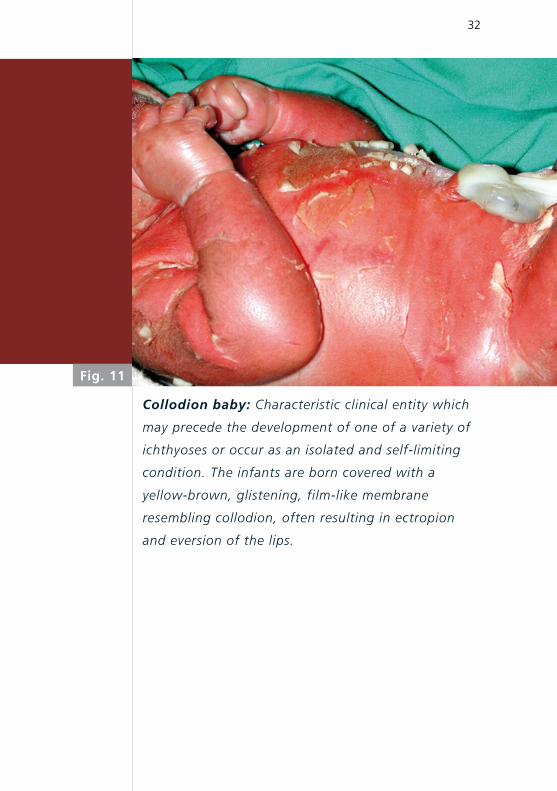
Fig. 11
Collodion baby: Characteristic clinical entity which
may precede the development of one of a variety of
ichthyoses or occur as an isolated and self-limiting
condition. The infants are born covered with a
yellow-brown, glistening, film-like membrane
resembling collodion, often resulting in ectropion
and eversion of the lips.

Swiss Society of Neonatology
www.neonet.ch
CONTACT
SUPPORTED BY
con
cep
t &
des
ign
by
mes
ch.c
h