Embed Size (px)
Citation preview
DEPOSITION OF THE HYDROXYAPATITE PARTICLES ON THE
POLYETHER ETHER KETONE SURFACE FOR ORTHOPAEDIC
APPLICATIONS
DAVOOD ALMASI
A thesis submitted in fulfilment of the
requirements for the award of the degree of
Doctor of Philosophy (Mechanical Engineering)
Faculty of Mechanical Engineering
Universiti Teknologi Malaysia
AUGUST 2015

iii
I would like to dedicate this thesis to
My Parents

iv
AKNOWLEDGEMENT
At first, thanks to ALLAH, the most gracious and the most merciful, for
providing me the opportunity of doctoral study and ability to accomplish this
research.
I would like to express my deepest gratitude towards Assoc. Prof. Dr. Izman
Sudin who nurtured me to become a researcher. Without his kind guidance,
encouragement and valuable advices during the research and writing, this thesis
would not be come to the light. I wish also to express my sincere appreciation to my
co-supervisor Prof. Dr. Mohammed Rafiq Bin Dato’ Abdul Kadir for his generous
time and patience to teach me becoming a researcher.
Special gratitude to Dr. Maliheh Sadeghi, Dr. Naznin Sultana, Dr. Nida Iqbal
and Dr. Ali Asadi for their kind helps during the research.
v
ABSTRACT
Polyether-ether-ketone (PEEK) has a similar elastic modulus close to bone and
as a result it can be a suitable alternative material to metallic implants which
generally have higher moduli. However, the bio-inertness of PEEK prevents it from
integrating well with the surrounding tissues. Many efforts have been made to
overcome this problem including the deposition of hydroxyapatite (HA) on PEEK
via plasma spraying. Current issues which have arisen with this method are low
bonding strength between the substrate and the coating layer, as well as producing a
non-uniform density of the coating. In this study, chemical deposition method was
used to deposit HA crystalline particles on a treated PEEK substrate without any
subsequent sintering process. The surface of PEEK was first sulphonated to create –
SO3H functional group. It was then immersed in suspension of HA in water where
the HA particles were chemically connected to the –SO3H functional group. The
treated layer has a 3D porous property but exhibits very low mechanical properties.
A compress force was applied on the coated layer for improving its mechanical
properties. EDX and XRD results confirmed the existence of crystalline HA on the
treated layer. FT-IR analysis was used to confirm the chemical bonding between HA
and the substrate. The bioactivity of the HA treated layer was evaluated in terms of
wettability. The water contact angle test showed 50% increase in wettability of the
treated samples as compared to PEEK. The scratch and nano-indentation tests were
carried out on the treated layer to assess its adhesion strength to the substrate and its
mechanical strength respectively. The results showed that the compression caused
142% and 36.9% increment of the elasticity modulus and scratch hardness of the
treated layer respectively. However, it caused 59% increase in the water contact
angle. These findings indicate that the proposed new method is easy to process and
does not require special equipment as compared to the Plasma Spray technique. The
proposed method can be an alternative technique to change the bio-inertness of
PEEK to become more bioactive PEEK which is good for polymeric implant
material.
vi
ABSTRAK
Polieter - eter – keton (PEEK) mempunyai modulus kekenyalan yang hampir
sama dengan tulang dan oleh yang demikian, ia sesuai digunakan sebagai satu bahan
alternatif kepada implan logam sedia ada yang secara umumnya mempunyai
modulus kekenyalan yang lebih tinggi. Namun, sifat bio-lengai PEEK
menghalangnya daripada menyatu dengan baik dengan tisu di sekelilingnya. Banyak
usaha telah dilakukan untuk mengatasi masalah ini termasuklah melalui pemendapan
hidroksiapatit (HA) ke atas PEEK dengan kaedah penyemburan plasma. Isu-isu
terkini yang timbul daripada kaedah ini adalah daya ikatan yang lemah di antara
substrat dan lapisan salutan serta penghasilan ketumpatan salutan yang tidak
seragam. Dalam kajian ini, kaedah mendapan kimia telah digunakan untuk
memendapkan zarah hablur HA ke atas substrat PEEK terawat tanpa memerlukan
proses pengsinteran selepasnya. Mula-mula sekali permukaan PEEK menjalani
proses pengsulfonan untuk membentuk kumpulan berfungsi –SO3H. Kemudian, ia
direndamkan dalam larutan HA di dalam air yang mana zarah-zarah HA tergabung
secara kimia dengan kumpulan berfungsi –SO3H. Lapisan yang dirawat mempunyai
sifat keliangan 3 dimensi tetapi mempamerkan sifat kekuatan mekanikal yang sangat
rendah. Satu daya mampatan telah dikenakan ke atas lapisan salutan tersebut untuk
memperbaiki sifat mekanikalnya. Keputusan EDX dan XRD mengesahkan
kewujudan HA berhablur pada lapisan salutan yang terawat. Analisa FT-IR telah
digunakan untuk mengesahkan ikatan kimia di antara HA dan substrat. Keaktifan
bio lapisan terawat HA dinilai dari segi sifat kebolehbasahannya. Ujian sudut
sentuhan air menunjukkan peningkatan sebanyak 50% sifat kebolehbasahan bagi
sampel terawat berbanding dengan sampel PEEK. Ujian calar dan ujian lekukan nano
masing-masing telah dijalankan ke atas lapisan terawat untuk menilai kekuatan
lekatannya kepada substrat dan juga menilai kekuatan mekanikalnya. Keputusan
menunjukkan bahawa mampatan telah menyebabkan peningkatan sebanyak 142%
dan 36.9% kepada modulus kekenyalan dan kekerasan calar masing-masing ke atas
lapisan terawat. Walau bagaimana pun, mampatan ini juga menyebabkan
peningkatan sebanyak 59% kepada sudut sentuhan air. Penemuan-penemuan ini
menunjukkan kaedah baru yang dicadangkan adalah mudah untuk diproses dan tidak
memerlukan peralatan khas jika dibandingkan dengan teknik semburan plasma.
Kaedah yang dicadangkan ini boleh menjadi satu kaedah alternatif bagi mengubah
sifat bio-lengai PEEK kepada sifat yang lebih bioaktif yang mana ia baik bagi bahan
implan berasaskan polimer.
vii
TABLE OF CONTENTS
CHAPTER TITLE PAGE
DECLARATION ii
DEDICATION iii
AKNOWLEDGEMENT iv
ABSTRACT v
ABSTRAK vi
TABLE OF CONTENTS vii
LIST OF TABLES xii
LIST OF FIGURES xiii
LIST OF ABBREVIATIONS xvi
LIST OF APPENDICES xix
1 INTRODUCTION 1
1.1 Background of the Research 1
1.2 Problem Statement 3
1.3 Objectives of the Research 4
1.4 Scopes of the Research 4
1.5 Significance of the Research 5
1.6 Organization of the Thesis 6
2 LITERATURE REVIEW 7

viii
2.1 Introduction 7
2.2 Overview of Biomaterials and their Applications 7
2.2.1 PEEK’s Biomedical Applications 10
2.3 Brief Overview of PEEK 12
2.3.1 Physical and Chemical Properties of
PEEK 13
2.3.2 Mechanical Properties of PEEK 16
2.3.3 Sterilization of PEEK 20
2.3.4 Imaging Properties of PEEK 21
2.3.5 Biocompatibility of PEEK 22
2.3.6 PEEK Wettability 23
2.4 Bioactive PEEK Implant 25
2.4.1 Surface Modification of PEEK 25
2.4.1.1 Direct Surface Modification 26
2.4.1.2 Surface Coating 29
2.4.2 Bioactive PEEK Composites 33
2.5 Principle of Nanoindentation and Scratch Tests 38
2.5.1 Nanoindentation Test 38
2.5.2 Scratch Test 40
2.6 Summary 43
3 RESEARCH METHODOLOGY 45
3.1 Introduction 45
3.2 Research Design 45
3.3 Material 46
3.4 Sample Preparation 48
3.4.1 Preparation of PEEK Substrates 48
3.4.2 Sulphonation of PEEK Surface 49
3.4.3 Deposition HA Particles on the SPEEK
Treated Layer 50
3.4.4 Compression Test on Treated Layer 51
3.5 Characterization 52
3.5.1 Fourier Transform Infrared Spectroscopy
(FTIR) Study 52
3.5.2 X-ray Diffraction Study 52
3.5.3 Surface Morphology Study 52
ix
3.5.4 Surface Roughness Study 53
3.5.5 Water Contact Angle Study 53
3.5.6 In vitro Bioactivity Tests Procedure 54
3.5.6.1 Appatite Formation Test 54
3.5.6.2 Cell Attachment Analysis 55
3.6 Mechanical Properties Analysis 57
3.6.1 Nanoindentation Test 57
3.6.2 Scratch Test 58
3.7 Summary 60
4 RESULTS AND DISCUSSION 61
4.1 Introduction 61
4.2 Preliminary Results and Discussion 61
4.2.1 Effect of Sulphonation Time on the
Treated Layer Thickness 62
4.2.2 Effect of Immersion Time on the
Deposition of HA Particles 62
4.2.3 Summary 64
4.3 Detailed Experiment Results and Discussions 64
4.3.1 Fine Tuning of the Sulphonation Time 64
4.3.2 Results and Analysis of HA Deposited
Layer on the Treated PEEK 65
4.3.2.1 FT-IR Analysis for HA
Chemical Bonding 66
4.3.2.2 XRD Analysis for HA
Crysalinity 68
4.3.2.3 Morphology of the Treated
Layer 70
4.3.2.4 Effect of Sulphonation Time on
the Treated Layer Properties 77
4.3.2.5 Surface Roughness Study 77
4.3.2.6 Water Contact Angle 79
4.3.2.7 Apatite Formation (Bioactivity)
Study 81
4.3.2.8 In-vitro Cell Responses 83
4.3.3 Effect of the Compression Load on the
Treated Layer Properties 84
x
4.3.3.1 Comparison of Surface
Morphology of Treated Layer
Before and After Compression 85
4.3.3.2 Scratch Test Results and
Discussion 86
4.3.3.3 Nanoindentation Results and
Discussion 97
4.3.3.4 Surface Roughness 101
4.3.3.5 Water Contact Angle Analysis 103
4.4 Summary of the Findings 104
5 CONCLUSIONS AND RECOMMENDATONS FOR
FUTURE WORK 107
5.1 Introduction 107
5.2 Conclusions 107
5.3 Recommendations for Future Work 108
REFERENCES 110
Appendices A-C 129-131
xi
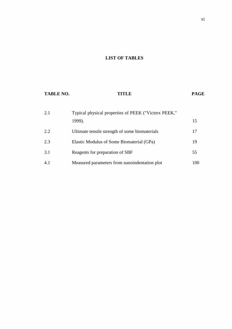
LIST OF TABLES
TABLE NO. TITLE PAGE
2.1 Typical physical properties of PEEK ("Victrex PEEK,"
1999). 15
2.2 Ultimate tensile strength of some biomaterials 17
2.3 Elastic Modulus of Some Biomaterial (GPa) 19
3.1 Reagents for preparation of SBF 55
4.1 Measured parameters from nanoindentation plot 100

xii
LIST OF FIGURES
FIGURE NO. TITLE PAGE
2.1 Some of the polymer applications in orthopedic area
(Ramakrishna et al., 2001) 9
2.2 Cross section of human vertebra (Sandukas, 2012). 11
2.3 Chemical structure of PEEK (Kurtz, 2012). 14
2.4 Hydrophilic surface (left), normal hydrophilic surface
(middle) and hydrophobic surface (right) (Yuan and
Lee, 2013). 24
2.5 (a) surface profile during loading with an indenter (b)
schematic of indentation force curve (Oliver and Pharr,
1992). 39
2.6 Schematic of scratch test process 41
3.1 Flow chart of the overall research design 47
3.2 Preparation of the PEEK disc samples involves cutting
the PEEK rod, grinding, and ultrasonically cleaning via
acetone 48
3.3 Sulphonation of the PEEK disc via immersion in
sulphoric acid and followed by immersion in water. 49
3.4 Deposition steps of HA particles on SPEEK via
immersion in HA suspension followed by ultrasonic
cleaning. 50
3.5 Applying of compressive load on the treated layer. 51
xiii
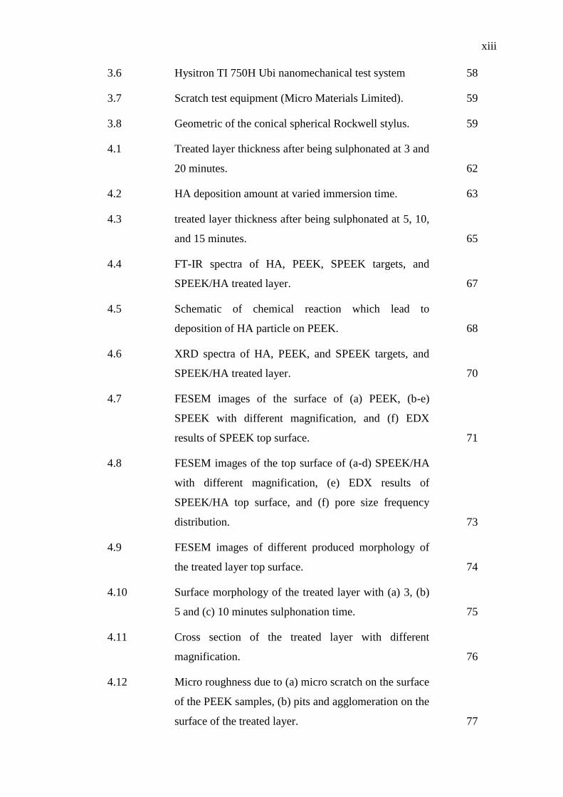
3.6 Hysitron TI 750H Ubi nanomechanical test system 58
3.7 Scratch test equipment (Micro Materials Limited). 59
3.8 Geometric of the conical spherical Rockwell stylus. 59
4.1 Treated layer thickness after being sulphonated at 3 and
20 minutes. 62
4.2 HA deposition amount at varied immersion time. 63
4.3 treated layer thickness after being sulphonated at 5, 10,
and 15 minutes. 65
4.4 FT-IR spectra of HA, PEEK, SPEEK targets, and
SPEEK/HA treated layer. 67
4.5 Schematic of chemical reaction which lead to
deposition of HA particle on PEEK. 68
4.6 XRD spectra of HA, PEEK, and SPEEK targets, and
SPEEK/HA treated layer. 70
4.7 FESEM images of the surface of (a) PEEK, (b-e)
SPEEK with different magnification, and (f) EDX
results of SPEEK top surface. 71
4.8 FESEM images of the top surface of (a-d) SPEEK/HA
with different magnification, (e) EDX results of
SPEEK/HA top surface, and (f) pore size frequency
distribution. 73
4.9 FESEM images of different produced morphology of
the treated layer top surface. 74
4.10 Surface morphology of the treated layer with (a) 3, (b)
5 and (c) 10 minutes sulphonation time. 75
4.11 Cross section of the treated layer with different
magnification. 76
4.12 Micro roughness due to (a) micro scratch on the surface
of the PEEK samples, (b) pits and agglomeration on the
surface of the treated layer. 77
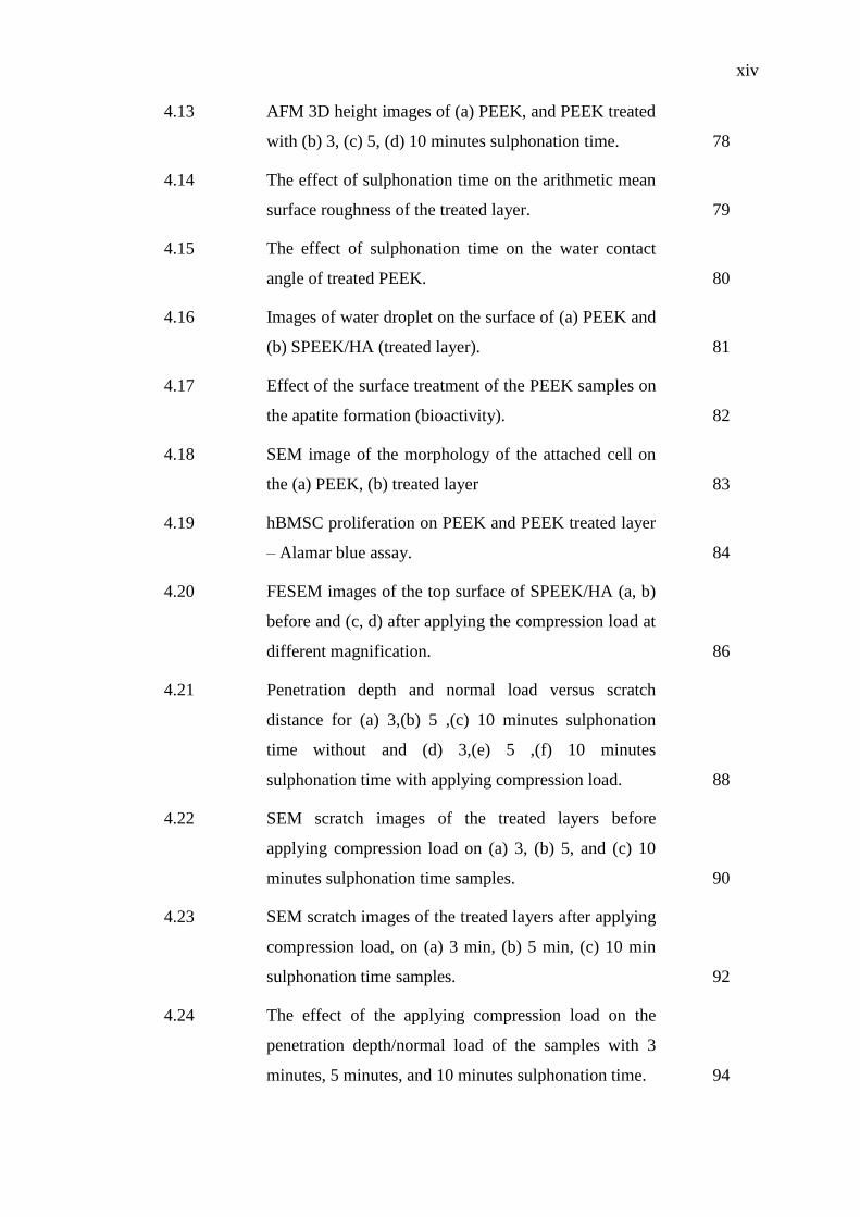
xiv
4.13 AFM 3D height images of (a) PEEK, and PEEK treated
with (b) 3, (c) 5, (d) 10 minutes sulphonation time. 78
4.14 The effect of sulphonation time on the arithmetic mean
surface roughness of the treated layer. 79
4.15 The effect of sulphonation time on the water contact
angle of treated PEEK. 80
4.16 Images of water droplet on the surface of (a) PEEK and
(b) SPEEK/HA (treated layer). 81
4.17 Effect of the surface treatment of the PEEK samples on
the apatite formation (bioactivity). 82
4.18 SEM image of the morphology of the attached cell on
the (a) PEEK, (b) treated layer 83
4.19 hBMSC proliferation on PEEK and PEEK treated layer
– Alamar blue assay. 84
4.20 FESEM images of the top surface of SPEEK/HA (a, b)
before and (c, d) after applying the compression load at
different magnification. 86
4.21 Penetration depth and normal load versus scratch
distance for (a) 3,(b) 5 ,(c) 10 minutes sulphonation
time without and (d) 3,(e) 5 ,(f) 10 minutes
sulphonation time with applying compression load. 88
4.22 SEM scratch images of the treated layers before
applying compression load on (a) 3, (b) 5, and (c) 10
minutes sulphonation time samples. 90
4.23 SEM scratch images of the treated layers after applying
compression load, on (a) 3 min, (b) 5 min, (c) 10 min
sulphonation time samples. 92
4.24 The effect of the applying compression load on the
penetration depth/normal load of the samples with 3
minutes, 5 minutes, and 10 minutes sulphonation time. 94
xv
4.25 Scratch hardness of the treated layer for different
sulphonation time, without and with applying
compression load. 95
4.26 The effect of compression on horizontal load/scratch
distance with (a) 3 , (b) 5, and (c) 10 minutes
sulphonation time. 97
4.27 Indentation load versus penetration depth curves 99
4.28 The effect of compression on the Elastic modulus of
treated layer with different sulphonation time. 100
4.29 AFM 3D height images of the treated PEEK with (a) 3,
(b) 5, and (c) 10 minutes sulphonation time after
applying the compression load 102
4.30 The effect of compression load on surface roughness of
the treated layer at different sulphonation time. 103
4.31 Water contact angle of the samples (untreated and
treated PEEK) at different sulphonation times before
and after being compressed. 104
xvi
LIST OF ABBREVIATIONS
Ac - projected contact area between the sample and indenter
ALP - Alkaline phosphatase
ANAB - Accelerated Neutral Atom Beam
ASTM - American Society for Testing and Materials
A-TiO2 Anatase-rich titanium dioxide
BCP - Biphasic Calcium Phosphates
BIC - Bone - in- contact
BMP-2 - Bone morphogenetic protein-2
CFR-PEEK - Carbon-fiber-reinforced PEEK
CS - Calcium oxide and silicon dioxide
CT - Computed tomography
CVD - Chemical Vapour Deposition
DLC - Diamond-like carbon
DSC - Differential scanning calorimetry
E - Modulus of elasticity
ECM - Extracellular matrixes
EDX - Energy Dispersive X-ray Spectroscopy
F - Load
FDA - Food and Drug Administration of united state
FN - Fibronectin
GPa - Giga Pascal
gr - Gram
Gy - Gray (SI unit of absorbed radiation)
H - Hardness
HA - Hydroxyapatite
HBMSC - Human Bone Mesenchymal Stem Cells

xvii
hr/hrs - Hour/Hours
Hs - Scratch hardness
ISO - International Organization for Standardization
LC - Critical load
LC1 - First cohesive failure in coating layer
LC2 - Adhesive failure
min - Minute
MPa - Mega Pascal
MRI - Magnetic resonance imaging
MTS - Methoxyphenyl tetrazolium salt
NMR - Infrared spectroscopy nuclear magnetic resonance
ºC - Degree of Celsius
PAEK - Polyaryl ether ketones
PEEK - Polyether ether ketone
PEKEKK - Poly-ether-ketone-ether-ketone-ketone
PLIF - Posterior lumbar inter-body fusion cage
PLLA - Poly-L-lactic acid
PMMA - Poly (methyl methacrylate)
ppb - Part per billion
PS - Polystyrene
PVD - Plasma vapor deposition
R-TiO2 Rutile-rich titanium dioxide
S - Stiffness
SBF - Simulated Body Fluid
SPEEK - Sulphonated Polyether ether ketone
Tg - Glass transition temperature
Ti - Titanium
TiO2 - Titanium dioxide
UV - Ultraviolet
VPS - Vacuum plasma spraying
W - Width of the scratch
XRD - X-ray diffraction
XRD - X-ray Diffraction Spectroscopy
YSZ - Yttria-stabilized zirconia
xviii
α - Indenter face angle
βTCP - β-tricalciumphosphate
δ - Indentation depth
δc - Contact depth
ε - Geometric constant of indenter
μm - Micro meter
υ - Poisson’s ratio

xix
LIST OF APPENDICES
APPENDIX TITLE PAGE
A List of Publications 134
B Calculation Details of the HA Crystallinity 135
C Table of the Reading Data of the Ra in Nanometer 136
CHAPTER 1
1 INTRODUCTION
1.1 Background of the Research
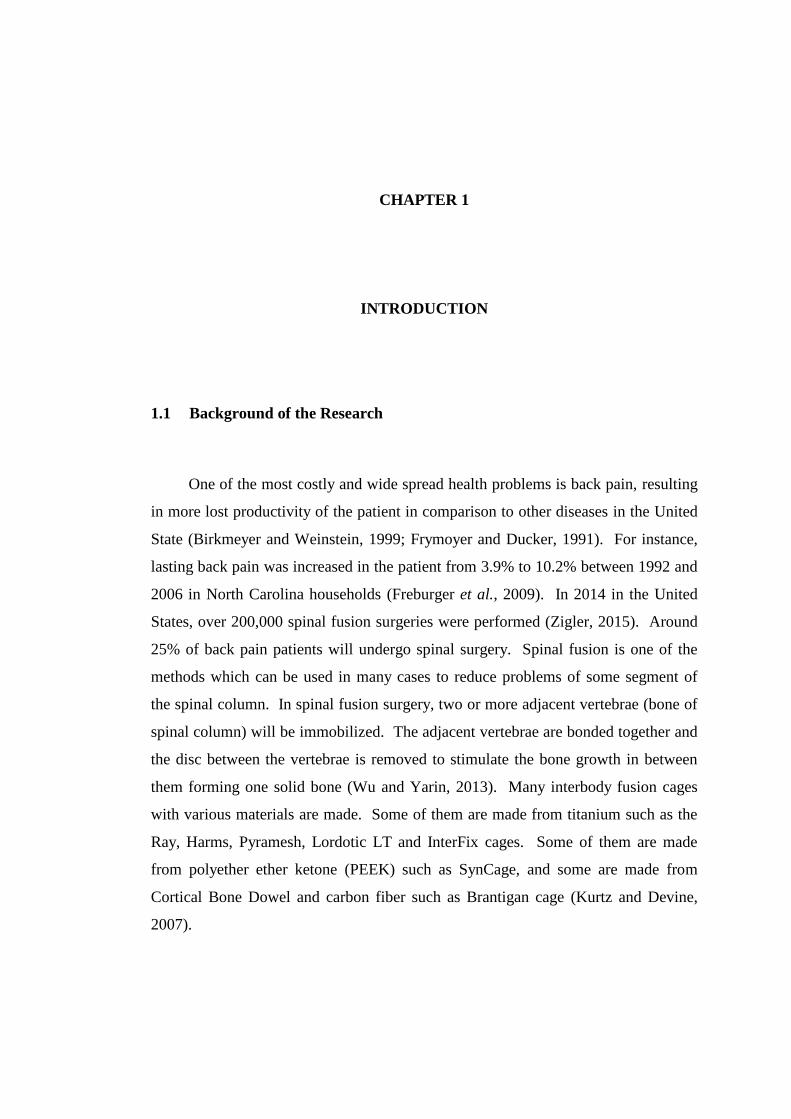
One of the most costly and wide spread health problems is back pain, resulting
in more lost productivity of the patient in comparison to other diseases in the United
State (Birkmeyer and Weinstein, 1999; Frymoyer and Ducker, 1991). For instance,
lasting back pain was increased in the patient from 3.9% to 10.2% between 1992 and
2006 in North Carolina households (Freburger et al., 2009). In 2014 in the United
States, over 200,000 spinal fusion surgeries were performed (Zigler, 2015). Around
25% of back pain patients will undergo spinal surgery. Spinal fusion is one of the
methods which can be used in many cases to reduce problems of some segment of
the spinal column. In spinal fusion surgery, two or more adjacent vertebrae (bone of
spinal column) will be immobilized. The adjacent vertebrae are bonded together and
the disc between the vertebrae is removed to stimulate the bone growth in between
them forming one solid bone (Wu and Yarin, 2013). Many interbody fusion cages
with various materials are made. Some of them are made from titanium such as the
Ray, Harms, Pyramesh, Lordotic LT and InterFix cages. Some of them are made
from polyether ether ketone (PEEK) such as SynCage, and some are made from
Cortical Bone Dowel and carbon fiber such as Brantigan cage (Kurtz and Devine,
2007).
2
The high elastic modulus of titanium (~100GPa) as compared to the adjacent
bone, which is implanted in, for instance cortical bone (~15GPa) can result in stress
shielding which cause decline of the bone that is in contact with the titanium implant.
In the case of spinal implants, in which a large percentage volume of the vertebrae
consists of cancellous bone (the modulus of elasticity ~0.3GPa) the shielding
problem becomes one of the main issues. In the design of spinal cage, choosing
material with low modulus of elasticity is desired. Cancellous bone is a soft,
cellular-structured bone, which absorbs shock in the spinal column. Metal ions
release is another issue in metallic implants which can cause failure (Ramakrishna et
al. 2001; Stadelmann et al. 2008). These phenomena increase motivation of research
towards polymeric implants. In order to reduce the problem of titanium spinal fusion
cage, polymer materials with closer modulus elasticity to the bone and high strength
have been utilized (Kurtz and Devine, 2007).
Low modulus of elasticity, excellent chemical stability, resistance to radiation
used in sterilization procedures, transparency to radio waves, compatibility with
reinforcing agent (such as carbon fiber) which can get wide variety of mechanical
strength (4~20GPa, depending on fiber volume fraction), etc. have made PEEK as an
ideal choice for load bearing implants (Han et al., 2010; Kurtz and Devine, 2007;
Sobieraj et al. 2009; Toth et al., 2006; Williams, 2008; Xing et al., 2004). PEEK has
been used for load bearing orthopaedic applications such as screws, spinal cage, and
dental implant (Schwitalla and Muller, 2011; Williams et al. 1987).
Food and Drug Administration of the United States (FDA) have accepted
carbon-fiber-reinforced PEEK (CFR-PEEK) for spinal fusion cages for human use
since the 1990s. Biomaterial PEEK has twenty years of successful clinical history in
spinal fusion cage (Kurtz and Devine, 2007). While titanium is still being used for
manufacturing the spinal fusion cages, most of the companies have switched their
focus to PEEK instead of titanium. For instance, the designers of the Wallis
posterior dynamic stabilization system have changed their titanium inter-spinous
component to PEEK (Senegas, 2002).

3
Despite these excellent properties, PEEK is still categorized as bioinert due to
its very low reaction with the surrounding bone tissue, which limits its potential
applications (Kurtz and Devine, 2007; Rabiei and Sandukas, 2013). To overcome
this problem several methods have been proposed, in which the deposition of the
hydroxyapatite is one the most attractive methods for researchers. This coating has
attracted the attention of many researchers and is the only commercial method for
improving the bioactivity of the PEEK implants. Crystalline HA coating is required
to stabilize the layer especially for long term orthopaedic and dental implants. In the
present methods, the coated HA on the implants require sintering process in order to
transform amorphous structure to crystalline (Rabiei and Sandukas, 2013).
Sulphonation of PEEK is one of the activation method which deposit the SO3H
functional group via immersion in sulphuric acid (Zhao et al., 2013). This active
surface of sulphonated PEEK (SPEEK) can be use for binding with particles.
1.2 Problem Statement
Traditional heat treatment for crystallizing of amorphous HA coating layer has
been done via annealing at 600˚C or higher. This temperature is above the melting
point of PEEK (340˚C). To overcome this problem, three new methods of
microwave sintering (Adams et al., 2006), laser-induced crystallization (P. M. Smith,
Carey, and Sigmon, 1997), and hydrothermal annealing (Ozeki et al. 2003; Tong et
al., 1997; Yang et al. 2007) have been investigated and reported with some success.
PEEK is transparent to microwave and it allows heating on HA layer. Laser method
is able to provide localized heating and saturated steam is utilized in hydrothermal
annealing. Although these methods can crystallize the HA coating layer without
spoiling the bulk of the PEEK substrate, at the interface layer between the HA
coating and the substrate is impaired (Rabiei and Sandukas, 2013). This research
was guided by the following questions:
1. Can sulphonated PEEK be linked to crystalline HA?

4
2. What is the effect of the sulphonation time on the wettability (bioactivity) of the
treated PEEK?
3. What is the effect of the compression load on the properties of the treated layer?
1.3 Objectives of the Research
The principal objective of this research was to establish a new method for
depositing crystalline HA on PEEK with the intention to eliminate subsequent
sintering of the HA coating. The specific objectives for this research were as
follows:
1. To evaluate the linking process via sulphonation for depositing the HA particles
with high percentage of crystallinity.
2. To evaluate the effect of different sulphonation process parameters on the surface
wettability of the treated PEEK.
3. To compare the mechanical properties and surface morphology of the treated
layer before and after applying compression load.
1.4 Scopes of the Research
The research was conducted within the following limits:
1. Polyether ether ketone (PEEK) was used as the substrate material.
2. Sulphonation process was conducted to activate the surface of the PEEK by
depositing –SO3H polar functional group on the molecular chain of the PEEK.
The sulphonation process time was varied between 3 to 20 minutes.

5
3. Fourier Transfer Infrared Spectroscopy (FTIR) was used for evaluating the
chemical bonding; X-Ray Diffraction (XRD) for evaluating the crystallinity of
the treated layer; Field Emission Scanning Electron Microscope (FESEM) for
observing surface morphology; Energy Dispersive X-Ray Spectroscopy (EDX)
for determining elements; and Atomic Force Microscope (AFM) for evaluating
surface roughness.
4. Mechanical properties of the treated layer were investigated using
nanoindentation and micro scratch test.
5. Wettability property of the treated layer was evaluated using water contact angle
analysis.
6. Hydraulic press was employed to apply compression load of 15MPa on the
treated layer.
1.5 Significance of the Research
Plasma spray is the most popular method used to deposit HA on the PEEK
biomedical implant. It was followed by microwave sintering process treatment for
increasing the HA crystallinity. Due to high temperature processing, this method has
been reported posing problems at the interface HA - substrate layer though the
process is capable to provide the required crystallinity level. The equipment used for
depositing HA via plasma spray technique is also expensive and demands a
secondary microwave process which adds cost and time for manufacturing an
implant. In contrast, the proposed method uses a chemical deposition of crystalline
HA particles on sulphonated PEEK that could eliminate these costly equipment and
secondary processes to manufacture an implant. It is expected the cost will become
cheaper with the new method. This encouraging technique also has a great potential
to eliminate problems occurred at the interface layer due to high temperature. It is
also expected that the manufactured implant via the proposed method becomes more
sustainable in the long run.

6
1.6 Organization of the Thesis
This thesis consists of five chapters: Chapter 1 briefly discusses the
background of problem, problem statement, objectives of the research, scope of the
research, contribution and organization of the thesis. Chapter 2 discusses the
background of biomaterials, and recent strategies for improving the bioactivity of the
PEEK’s implant. Chapter 3 describes the research methodology used in conducting
this research. Chapter 4 presents the experimental results and discusses the findings
from experimental trials. Chapter 5 provides the conclusions of the research.

110
REFERENCES
Abu Bakar, M. S., Cheang, P., and Khor, K. A. (1999). Thermal processing of
hydroxyapatite reinforced polyetheretherketone composites. Journal of
Materials Processing Technology, 89–90(0), 462-466.
Abu Bakar, M. S., Cheang, P., and Khor, K. A. (2003). Tensile properties and
microstructural analysis of spheroidized hydroxyapatite–poly
(etheretherketone) biocomposites. Materials Science and Engineering: A,
345(1–2), 55-63.
Abu Bakar, M. S., Cheng, M. H., Tang, S. M., Yu, S. C., Liao, K., Tan, C. T.,
Cheang, P. (2003). Tensile properties, tension-tension fatigue and biological
response of polyetheretherketone-hydroxyapatite composites for load-bearing
orthopedic implants. Biomaterials, 24(13), 2245-2250.
Adams, D., Malgas, G., Smith, R. D., Massia, S. P., Alford, T. L., and Mayer, J. W.
(2006). Microwave annealing for preparation of crystalline hydroxyapatite
thin films. Journal of Materials Science, 41(21), 7150-7158.
ASTM, and C1624-05. (2010). Standard Test Method for Adhesion Strength and
Mechanical Failure Modes of Ceramic Coatings by Quantitative Single Point
Scratch Testing.
ASTM, and D7027-13. (2013). Standard Test Method for Evaluation of Scratch
Resistance of Polymeric Coatings and Plastics Using an Instrumented Scratch
Machine. ASTM standard.
Awaja, F., Bax, D. V., Zhang, S., James, N., and McKenzie, D. R. (2012). Cell
Adhesion to PEEK Treated by Plasma Immersion Ion Implantation and
Deposition for Active Medical Implants. Plasma Processes and Polymers,
9(4), 355-362.
Baier, R. E., Shafrin, E. G., and Zisman, W. A. (1968). Adhesion: mechanisms that
assist or impede it. Science, 162(3860), 1360-1368.

111
Barkarmo, S., Wennerberg, A., Hoffman, M., Kjellin, P., Breding, K., Handa, P., and
Stenport, V. (2013). Nano-hydroxyapatite-coated PEEK implants: a pilot
study in rabbit bone. J Biomed Mater Res A, 101(2), 465-471.
Barletta, M., Gisario, A., and Rubino, G. (2011). Scratch response of high-
performance thermoset and thermoplastic powders deposited by the
electrostatic spray and ‘hot dipping’fluidised bed coating methods: The role
of the contact condition. Surface and Coatings Technology, 205(21), 5186-
5198.
Batchelor, A. W., and Chandrasekaran, M. (2004). Service Characteristics of
Biomedical Materials and Implants: Imperial College Press.
Beauvais, S., and Decaux, O. (2007). Plasma Sprayed Biocompatible Coatings on
PEEK Implants. Thermal Spray 2007: Global Coating Solutions: Proceedings
of the 2007 International Thermal Spray Conference: ASM International.
Birkmeyer, N. J., and Weinstein, J. N. (1999). Medical versus surgical treatment for
low back pain: evidence and clinical practice. Eff Clin Pract, 2(5), 218-227.
Blackwood, D. J. (2003). Biomaterials: Past successes and future problems.
Corrosion Reviews, 21(2-3), 97-124.
Blundell, D. J., and Osborn, B. N. (1983). The morphology of poly(aryl-ether-ether-
ketone). Polymer, 24(8), 953-958.
Bombac, D. (2007). Review of materials in medical applications. Pregled materialov
v medicinskih aplikacijah. RMZ - Materials and geoenvironment, 54(4), 471-
499.
Boyan, B. D., Hummert, T. W., Dean, D. D., and Schwartz, Z. (1996). Role of
material surfaces in regulating bone and cartilage cell response. Biomaterials,
17(2), 137-146.
Brantigan, J. W., and Steffee, A. D. (1993). A carbon fiber implant to aid interbody
lumbar fusion. Two-year clinical results in the first 26 patients. Spine (Phila
Pa 1976), 18(14), 2106-2107.
Brantigan, J. W., Steffee, A. D., and Geiger, J. M. (1991). A carbon fiber implant to
aid interbody lumbar fusion. Mechanical testing. Spine (Phila Pa 1976), 16(6
Suppl), S277-282.
Briem, D., Strametz, S., Schröoder, K., Meenen, N. M., Lehmann, W., Linhart, W.,
Rueger, J. M. (2005). Response of primary fibroblasts and osteoblasts to

112
plasma treated polyetheretherketone (PEEK) surfaces. Journal of Materials
Science: Materials in Medicine, 16(7), 671-677.
Chen, Q., and Thouas, G. A. (2015). Metallic implant biomaterials. Materials
Science and Engineering: R: Reports, 87(0), 1-57.
Chi, M.H., Tsou, H.K., Chung, C.J., and He, J.-L. (2013). Biomimetic
hydroxyapatite grown on biomedical polymer coated with titanium dioxide
interlayer to assist osteocompatible performance. Thin Solid Films, 549(0),
98-102.
Cho, D. Y., Liau, W. R., Lee, W. Y., Liu, J. T., Chiu, C. L., and Sheu, P. C. (2002).
Preliminary experience using a polyetheretherketone (PEEK) cage in the
treatment of cervical disc disease. Neurosurgery, 51(6), 1343-1349;
discussion 1349-1350.
Chou, L., Marek, B., and Wagner, W. R. (1999). Effects of hydroxylapatite coating
crystallinity on biosolubility, cell attachment efficiency and proliferation in
vitro. Biomaterials, 20(10), 977-985.
Cizek, G. R., and Boyd, L. M. (2000). Imaging Pitfalls of Interbody Spinal Implants.
Spine, 25(20), 2633-2636.
Clifford, C. A., and Seah, M. P. (2005). Quantification issues in the identification of
nanoscale regions of homopolymers using modulus measurement via AFM
nanoindentation. Applied Surface Science, 252(5), 1915-1933.
Comesaña, R., Quintero, F., Lusquiños, F., Pascual, M. J., Boutinguiza, M., Durán,
A., and Pou, J. (2010). Laser cladding of bioactive glass coatings. Acta
Biomaterialia, 6(3), 953-961.
Comyn, J., Mascia, L., Xiao, G., and Parker, B. M. (1996). Plasma-treatment of
polyetheretherketone (PEEK) for adhesive bonding. International Journal of
Adhesion and Adhesives, 16(2), 97-104.
Converse, G. L., Conrad, T. L., Merrill, C. H., and Roeder, R. K. (2010).
Hydroxyapatite whisker-reinforced polyetherketoneketone bone ingrowth
scaffolds. Acta Biomaterialia, 6(3), 856-863.
Converse, G. L., Conrad, T. L., and Roeder, R. K. (2009). Fatigue life of
hydroxyapatite whisker reinforced polyetherketoneketone. Trans. Soc.
Biomaterials, 32, 584.
Converse, G. L., Conrad, T. L., and Roeder, R. K. (2009). Mechanical properties of
hydroxyapatite whisker reinforced polyetherketoneketone composite
113
scaffolds. Journal of the Mechanical Behavior of Biomedical Materials, 2(6),
627-635.
Converse, G. L., Yue, W., and Roeder, R. K. (2007). Processing and tensile
properties of hydroxyapatite-whisker-reinforced polyetheretherketone.
Biomaterials, 28(6), 927-935.
Cook, S. D., and Rust-Dawicki, A. M. (1995). Preliminary evaluation of titanium-
coated PEEK dental implants. J Oral Implantol, 21(3), 176-181.
Corvelli, A. A., Roberts, J. C., Biermann, P. J., and Cranmer, J. H. (1999).
Characterization of a peek composite segmental bone replacement implant.
Journal of Materials Science, 34(10), 2421-2431.
D907-12a, A. (2012). Standard Terminology of Adhesives. www.astm.org.
D7334-08, A. (2013). Standard Practice for Surface Wettability of Coatings,
Substrates and Pigments by Advancing Contact Angle Measurement.
Dai, Y., Xu, M., Wei, J., Zhang, H., and Chen, Y. (2012). Surface modification of
hydroxyapatite nanoparticles by poly(l-phenylalanine) via ROP of l-
phenylalanine N-carboxyanhydride (Pha-NCA). Applied Surface Science,
258(7), 2850-2855.
Davis, J. (2003). Overview of biomaterials and their use in medical devices.
Handbook of materials for medical devices. Illustrated edition, Ohio: ASM
International, 1-11.
Dennes, T. J., and Schwartz, J. (2009). A Nanoscale Adhesion Layer to Promote Cell
Attachment on PEEK. Journal of the American Chemical Society, 131(10),
3456-3457.
Devine, D. M., Hahn, J., Richards, R. G., Gruner, H., Wieling, R., and Pearce, S. G.
(2013). Coating of carbon fiber-reinforced polyetheretherketone implants
with titanium to improve bone apposition. J Biomed Mater Res B Appl
Biomater, 101(4), 591-598.
Edited by Basil R. Marple, M. (2007). Thermal Spray 2007: Global Coating
Solutions: Proceedings of the 2007 International Thermal Spray Conference:
ASM International.
Effect of Silk in Silk/PLGA Hybrid Films on Attachment and Proliferation of Human
Aortic Endothelial Cells. (2013). Polymer Korea, 37(2), 127.
114
Fan, J. P., Tsui, C. P., Tang, C. Y., and Chow, C. L. (2004). Influence of interphase
layer on the overall elasto-plastic behaviors of HA/PEEK biocomposite.
Biomaterials, 25(23), 5363-5373.
Freburger, J. K., Holmes, G. M., Agans, R. P., Jackman, A. M., Darter, J. D.,
Wallace, A. S., Carey, T. S. (2009). The rising prevalence of chronic low
back pain. Arch Intern Med, 169(3), 251-258.
Frymoyer, J. W., and Ducker, T. B. (1991). The Adult Spine: Principles and
Practice: Raven Press.
Gao, C., Gao, Q., Li, Y., Rahaman, M. N., Teramoto, A., and Abe, K. (2012).
Preparation and in vitro characterization of electrospun PVA scaffolds coated
with bioactive glass for bone regeneration. J Biomed Mater Res A, 100(5),
1324-1334.
García, A. J., Vega, M. D., and Boettiger, D. (1999). Modulation of cell proliferation
and differentiation through substrate- dependent changes in fibronectin
conformation. Molecular Biology of the Cell, 10(3), 785-798.
Geetha, M., Singh, A. K., Asokamani, R., and Gogia, A. K. (2009). Ti based
biomaterials, the ultimate choice for orthopaedic implants – A review.
Progress in Materials Science, 54(3), 397-425.
Green, S., and Devine, J. (2004). PEEK-OPTIMA Polymer: An Alternative Material
for the Development of Long-Term Medical Implant Applications.
BONEZone, 3-5.
Ha, S. W., Eckert, K. L., Wintermantel, E., Gruner, H., Guecheva, M., and Vonmont,
H. (1997). NaOH treatment of vacuum-plasma-sprayed titanium on carbon
fibre-reinforced poly(etheretherketone). J Mater Sci Mater Med, 8(12), 881-
886.
Ha, S. W., Gisep, A., Mayer, J., Wintermantel, E., Gruner, H., and Wieland, M.
(1997). Topographical characterization and microstructural interface analysis
of vacuum-plasma-sprayed titanium and hydroxyapatite coatings on carbon
fibre-reinforced poly(etheretherketone). J Mater Sci Mater Med, 8(12), 891-
896.
Ha, S. W., Kirch, M., Birchler, F., Eckert, K. L., Mayer, J., Wintermantel, E.,
Vonmont, H. (1997). Surface activation of polyetheretherketone (PEEK) and
formation of calcium phosphate coatings by precipitation. J Mater Sci Mater
Med, 8(11), 683-690.
115
Ha, S. W., Mayer, J., Koch, B., and Wintermantel, E. (1994). Plasma-sprayed
hydroxylapatite coating on carbon fibre reinforced thermoplastic composite
materials. Journal of Materials Science: Materials in Medicine, 5(6-7), 481-
484.
Hahn, B. D., Park, D. S., Choi, J. J., Ryu, J., Yoon, W. H., Choi, J. H., Jung, I. K.
(2013). Osteoconductive hydroxyapatite coated PEEK for spinal fusion
surgery. Applied Surface Science, 283(0), 6-11.
Han, C. M., Lee, E. J., Kim, H. E., Koh, Y. H., Kim, K. N., Ha, Y., and Kuh, S. U.
(2010). The electron beam deposition of titanium on polyetheretherketone
(PEEK) and the resulting enhanced biological properties. Biomaterials,
31(13), 3465-3470.
Han, C. M., Jang, T. S., Kim, H. E., and Koh, Y. H. (2014). Creation of nanoporous
TiO2 surface onto polyetheretherketone for effective immobilization and
delivery of bone morphogenetic protein. J Biomed Mater Res A, 102(3), 793-
800.
Han, C. M., Lee, E. J., Kim, H. E., Koh, Y. H., Kim, K. N., Ha, Y., and Kuh, S. U.
(2010). The electron beam deposition of titanium on polyetheretherketone
(PEEK) and the resulting enhanced biological properties. Biomaterials,
31(13), 3465-3470.
Hansen, D. C. (2008). Metal corrosion in the human body: the ultimate bio-corrosion
scenario. The Electrochemical Society Interface, 17(2), 31.
Heiland, K., Hill, D. J. T., O'Donnell, J. H., and Pomery, P. J. (1994). Radiation
degradation of poly(arylene ether ketone)s. Polymers for Advanced
Technologies, 5(2), 116-121.
Helsen, J. A., and Breme, H. J. (1998). Metals as biomaterials: Wiley.
Hemlata Garg, Gaurav Bedi, and Garg, A. (2012). Implant Surface Modifications: A
Review. Journal of Clinical and Diagnostic Reseach, 6(2), 319-324.
Hench, L. L., and Paschall, H. A. (1973). Direct chemical bond of bioactive glass-
ceramic materials to bone and muscle. Journal of Biomedical Materials
Research, 7(3), 25-42.
Hengky, C., Kelsen, B., and Saraswati, P. (2009). Mechanical and biological
characterization of pressureless sintered hydroxapatite-polyetheretherketone
biocomposite. International Conference on Biomedical Engineering (ICBME)
Proceedings, 23, 261-264.

116
Heo, Y., Im, H., and Kim, J. (2013). The effect of sulfonated graphene oxide on
Sulfonated Poly (Ether Ether Ketone) membrane for direct methanol fuel
cells. Journal of Membrane Science, 425–426(0), 11-22.
Ilze, M., and Janis, M. (2008). Effect of substrate hardness and film structure on
indentation depth criteria for film hardness testing. Journal of Physics D:
Applied Physics, 41(7), 074010.
Jaafar, J., Ismail, A. F., and Mustafa, A. (2007). Physicochemical study of poly(ether
ether ketone) electrolyte membranes sulfonated with mixtures of fuming
sulfuric acid and sulfuric acid for direct methanol fuel cell application.
Materials Science and Engineering: A, 460–461(0), 475-484.
Jahan, M. S., Wang, C., Schwartz, G., and Davidson, J. A. (1991). Combined
chemical and mechanical effects on free radicals in UHMWPE joints during
implantation. J Biomed Mater Res, 25(8), 1005-1017.
Janaki, K., Elamathi, S., and Sangeetha, D. (2008). Development and
Characterization of Polymer Ceramic Composites for Orthopedic
Applications. Trends Biomater. Artif. Organs, 22(3), 169-178.
Jarcho, M., Kay, J. F., Gumaer, K. I., Doremus, R. H., and Drobeck, H. P. (1977).
Tissue, cellular and subcellular events at a bone-ceramic hydroxylapatite
interface. J Bioeng, 1(2), 79-92.
Jian, S. R., Chen, G. J., and Lin, T. C. (2010). Berkovich nanoindentation on AlN
thin films. Nanoscale Research Letters, 5(6), 935-940.
Jiya, T., Smit, T., Deddens, J., and Mullender, M. (2009). Posterior lumbar interbody
fusion using nonresorbable poly-ether-ether-ketone versus resorbable poly-L-
lactide-co-D,L-lactide fusion devices: a prospective, randomized study to
assess fusion and clinical outcome. Spine (Phila Pa 1976), 34(3), 233-237.
Jockisch, K. A., Brown, S. A., Bauer, T. W., and Merritt, K. (1992). Biological
response to chopped-carbon-fiber-reinforced peek. J Biomed Mater Res,
26(2), 133-146.
Jung, H. D., Sun Park, H., Kang, M. H., Lee, S. M., Kim, H. E., Estrin, Y., and Koh,
Y.H. (2014). Polyetheretherketone/magnesium composite selectively coated
with hydroxyapatite for enhanced in vitro bio-corrosion resistance and
biocompatibility. Materials Letters, 116(0), 20-22.

117
Kasemo, B., and Lausmaa, J. (1988). Biomaterial and implant surfaces: on the role of
cleanliness, contamination, and preparation procedures. J Biomed Mater Res,
22(A2 Suppl), 145-158.
Katzer, A., Marquardt, H., Westendorf, J., Wening, J. V., and von Foerster, G.
(2002). Polyetheretherketone—cytotoxicity and mutagenicity in vitro.
Biomaterials, 23(8), 1749-1759.
Keselowsky, B. G., Collard, D. M., and García, A. J. (2003). Surface chemistry
modulates fibronectin conformation and directs integrin binding and
specificity to control cell adhesion. Journal of Biomedical Materials
Research - Part A, 66(2), 247-259.
Khoury, J., Kirkpatrick, S. R., Maxwell, M., Cherian, R. E., Kirkpatrick, A., and
Svrluga, R. C. (2013). Neutral atom beam technique enhances bioactivity of
PEEK. Nuclear Instruments and Methods in Physics Research Section B:
Beam Interactions with Materials and Atoms, 307(0), 630-634.
Kim, I. Y., Sugino, A., Kikuta, K., Ohtsuki, C., and Cho, S. B. (2009). Bioactive
composites consisting of PEEK and calcium silicate powders. Journal of
Biomaterials Applications, 24(2), 105-118.
Kirkpatrick, A., Kirkpatrick, S., Walsh, M., Chau, S., Mack, M., Harrison, S.,
Khoury, J. (2013). Investigation of accelerated neutral atom beams created
from gas cluster ion beams. Nuclear Instruments and Methods in Physics
Research Section B: Beam Interactions with Materials and Atoms, 307(0),
281-289.
Koenig, A. L., Gambillara, V., and Grainger, D. W. (2003). Correlating fibronectin
adsorption with endothelial cell adhesion and signaling on polymer
substrates. Journal of Biomedical Materials Research - Part A, 64(1), 20-37.
Kokubo, T., and Takadama, H. (2006). How useful is SBF in predicting in vivo bone
bioactivity? Biomaterials, 27(15), 2907-2915.
Krishnamurithy, G., Murali, M. R., Hamdi, M., Abbas, A. A., Raghavendran, H. B.,
and Kamarul, T. (2014a). Characterization of bovine-derived porous
hydroxyapatite scaffold and its potential to support osteogenic differentiation
of human bone marrow derived mesenchymal stem cells. Ceramics
International, 40(1), 771-777.
Krishnamurithy, G., Murali, M. R., Hamdi, M., Abbas, A. A., Raghavendran, H. B.,
and Kamarul, T. (2014b). Characterization of bovine-derived porous
118
hydroxyapatite scaffold and its potential to support osteogenic differentiation
of human bone marrow derived mesenchymal stem cells. Ceramics
International, 40(1, Part A), 771-777.
Kumar, S., Anderson, D. P., and Adams, W. W. (1986). Crystallization and
morphology of poly(aryl-ether-ether-ketone). Polymer, 27(3), 329-336.
Kurtz, S. M. (2012). Chapter 1 - An Overview of PEEK Biomaterials PEEK
Biomaterials Handbook (pp. 1-7). Oxford: William Andrew Publishing.
Kurtz, S. M., and Devine, J. N. (2007). PEEK biomaterials in trauma, orthopedic,
and spinal implants. Biomaterials, 28(32), 4845-4869.
Landi, E., Tampieri, A., Celotti, G., and Sprio, S. (2000). Densification behaviour
and mechanisms of synthetic hydroxyapatites. Journal of the European
Ceramic Society, 20(14), 2377-2387.
Laurens, P., Ould Bouali, M., Meducin, F., and Sadras, B. (2000). Characterization
of modifications of polymer surfaces after excimer laser treatments below the
ablation threshold. Applied Surface Science, 154–155(0), 211-216.
Laurens, P., Sadras, B., Decobert, F., Arefi-Khonsari, F., and Amouroux, J. (1998).
Enhancement of the adhesive bonding properties of PEEK by excimer laser
treatment. International Journal of Adhesion and Adhesives, 18(1), 19-27.
Laurens, P., Sadras, B., Décobert, F., Aréfi-Khonsari, F., and Amouroux, J. (1999).
Laser-induced surface modifications of poly(ether ether ketone): Influence of
the excimer laser wavelength. Journal of Adhesion Science and Technology,
13(9), 983-997.
Le Guéhennec, L., Soueidan, A., Layrolle, P., and Amouriq, Y. (2007). Surface
treatments of titanium dental implants for rapid osseointegration. Dental
Materials, 23(7), 844-854.
Lee, J., Lee, S., Kim, S., Kim, K., Kim, Y., Song, J., Khang, G. (2013). Effect of Silk
in Silk/PLGA Hybrid Films on Attachment and Proliferation of Human
Aortic Endothelial Cells. POLYMER-KOREA, 37(2), 127-134.
Lee, J. H., Jang, H. L., Lee, K. M., Baek, H.-R., Jin, K., Hong, K. S., Lee, H. K.
(2013). In vitro and in vivo evaluation of the bioactivity of hydroxyapatite-
coated polyetheretherketone biocomposites created by cold spray technology.
Acta Biomaterialia, 9(4), 6177-6187.
Lehnert, D., Wehrle-Haller, B., David, C., Weiland, U., Ballestrem, C., Imhof, B. A.,
and Bastmeyer, M. (2004). Cell behaviour on micropatterned substrata:
119
Limits of extracellular matrix geometry for spreading and adhesion. Journal
of Cell Science, 117(1), 41-52.
Li, H. M., Fouracre, R. A., Given, M. J., Banford, H. M., Wysocki, S., and
Karolczak, S. (1999). The effects on polyetheretherketone and
polyethersulfone of electron and andgamma; irradiation. Dielectrics and
Electrical Insulation, IEEE Transactions on, 6(3), 295-303.
Li, K., Yeung, C. Y., Yeung, K. W. K., and Tjong, S. C. (2012). Sintered
Hydroxyapatite/Polyetheretherketone Nanocomposites: Mechanical Behavior
and Biocompatibility. Advanced Engineering Materials, 14(4), B155-B165.
Lin, T. W., Corvelli, A. A., Frondoza, C. G., Roberts, J. C., and Hungerford, D. S.
(1997). Glass peek composite promotes proliferation and osteocalcin
production of human osteoblastic cells. J Biomed Mater Res, 36(2), 137-144.
Liu, F., Yang, F., Gao, Y., Jiang, W., Guan, Y., Rack, P., Liaw, P. K. (2009). Micro-
scratch study of a magnetron-sputtered Zr-based metallic-glass film. Surface
and Coatings Technology, 203(22), 3480-3484.
Liu, X., Chu, P. K., and Ding, C. (2004). Surface modification of titanium, titanium
alloys, and related materials for biomedical applications. Materials Science
and Engineering: R: Reports, 47(3–4), 49-121.
Ma, R., Weng, L., Bao, X., Ni, Z., Song, S., and Cai, W. (2012). Characterization of
in situ synthesized hydroxyapatite/polyetheretherketone composite materials.
Materials Letters, 71(0), 117-119.
Ma, R., Weng, L., Bao, X., Song, S., and Zhang, Y. (2013). In vivo biocompatibility
and bioactivity of in situ synthesized hydroxyapatite/polyetheretherketone
composite materials. Journal of Applied Polymer Science, 127(4), 2581-2587.
Ma, R., Weng, L., Fang, L., Luo, Z., and Song, S. (2012). Structure and mechanical
performance of in situ synthesized hydroxyapatite/polyetheretherketone
nanocomposite materials. Journal of Sol-Gel Science and Technology, 62(1),
52-56.
Marchand-Brynaert, J., Pantano, G., and Noiset, O. (1997). Surface fluorination of
PEEK film by selective wet-chemistry. Polymer, 38(6), 1387-1394.
Mathieson, I., and Bradley, R. H. (1996). Improved adhesion to polymers by
UV/ozone surface oxidation. International Journal of Adhesion and
Adhesives, 16(1), 29-31.
120
McAfee, P. C. (1999). Interbody fusion cages in reconstructive operations on the
spine. J Bone Joint Surg Am, 81(6), 859-880.
McMillin, C. (1993). Evaluation of PEKEKK composites for spine implants. Paper
presented at the 38th international SAMPE symposium.
Melnyk, A. D., Chak, J. D., Cripton, P. A., Dvorak, M. F., and Oxland, T. R. (2012).
Shear force measurements on low- and high-stiffness posterior fusion
devices. Med Eng Phys, 34(9), 1260-1267.
Michael, K. E., Vernekar, V. N., Keselowsky, B. G., Meredith, J. C., Latour, R. A.,
and García, A. J. (2003). Adsorption-induced conformational changes in
fibronectin due to interactions with well-defined surface chemistries.
Langmuir, 19(19), 8033-8040.
Mittal, K. L. (1995). Adhesion Measurement of Films and Coatings: Taylor and
Francis.
Modic, M. T., Steinberg, P. M., Ross, J. S., Masaryk, T. J., and Carter, J. R. (1988).
Degenerative disk disease: assessment of changes in vertebral body marrow
with MR imaging. Radiology, 166(1 Pt 1), 193-199.
Morrison, C., Macnair, R., MacDonald, C., Wykman, A., Goldie, I., and Grant, M.
H. (1995). In vitro biocompatibility testing of polymers for orthopaedic
implants using cultured fibroblasts and osteoblasts. Biomaterials, 16(13),
987-992.
Nieminen, T., Kallela, I., Wuolijoki, E., Kainulainen, H., Hiidenheimo, I., and
Rantala, I. (2008). Amorphous and crystalline polyetheretherketone:
Mechanical properties and tissue reactions during a 3-year follow-up. J
Biomed Mater Res A, 84(2), 377-383.
Noiset, O., Schneider, Y.-J., and Marchand–Brynaert, J. (1997). Surface modification
of poly(aryl ether ether ketone) (PEEK) film by covalent coupling of amines
and amino acids through a spacer arm. Journal of Polymer Science Part A:
Polymer Chemistry, 35(17), 3779-3790.
Noiset, O., Schneider, Y. J., and Marchand-Brynaert, J. (1997). Surface modification
of poly(aryl ether ether ketone) (PEEK) film by covalent coupling of amines
and amino acids through a spacer arm. Journal of Polymer Science, Part A:
Polymer Chemistry, 35(17), 3779-3790.

121
Noiset, O., Schneider, Y. J., and Marchand-Brynaert, J. (1999). Fibronectin
adsorption or/and covalent grafting on chemically modified PEEK film
surfaces. J Biomater Sci Polym Ed, 10(6), 657-677.
Noiset, O., Schneider, Y. J., and Marchand-Brynaert, J. (2000). Adhesion and growth
of CaCo2 cells on surface-modified PEEK substrata. J Biomater Sci Polym
Ed, 11(7), 767-786.
Occhiello, E., Morra, M., Guerrini, G. L., and Garbassi, F. (1992). Adhesion
properties of plasma-treated carbon/PEEK composites. Composites, 23(3),
193-200.
Oliver, W. C., and Pharr, G. M. (1992). An improved technique for determining
hardness and elastic modulus using load and displacement sensing
indentation experiments. Journal of Materials Research, 7, 1564-1583.
Ozeki, K., Mishima, A., Yuhta, T., Fukui, Y., and Aoki, H. (2003). Bone bonding
strength of sputtered hydroxyapatite films subjected to a low temperature
hydrothermal treatment. Biomed Mater Eng, 13(4), 451-463.
Parsons, J. R., Bhayani, S., Alexander, H., and Weiss, A. B. (1985). Carbon fiber
debris within the synovial joint. A time-dependent mechanical and histologic
study. Clin Orthop Relat Res(196), 69-76.
Petrovic, L., Pohle, D., Münstedt, H., Rechtenwald, T., Schlegel, K. A., and
Rupprecht, S. (2006). Effect of βTCP filled polyetheretherketone on
osteoblast cell proliferation in vitro. Journal of Biomedical Science, 13(1),
41-46.
Pharr, G. M., Oliver, W. C., Brotzen, F. R., and Others. (1992a). On the generality of
the relationship among contact stiffness, contact area, and elastic modulus
during indentation. Journal of Materials Research, 7(3), 612-617.
Pharr, G. M., Oliver, W. C., Brotzen, F. R., and Others. (1992b). On the generality of
the relationship among contact stiffness, contact area, and elastic modulus
during indentation. Journal of Materials Research, 7(3), -617.
Pino, M., Stingelin, N., and Tanner, K. E. (2008). Nucleation and growth of apatite
on NaOH-treated PEEK, HDPE and UHMWPE for artificial cornea
materials. Acta Biomaterialia, 4(6), 1827-1836.
Pohle, D., Ponader, S., Rechtenwald, T., Schmidt, M., Schlegel, K. A., Münstedt, H.,
Von Wilmowsky, C. (2007). Processing of three-dimensional laser sintered

122
polyetheretherketone composites and testing of osteoblast proliferation in
vitro. Macromolecular Symposia, 253, 65-70.
Poulsson, A. H., Eglin, D., Zeiter, S., Camenisch, K., Sprecher, C., Agarwal, Y.,
Richards, R. G. (2014). Osseointegration of machined, injection moulded and
oxygen plasma modified PEEK implants in a sheep model. Biomaterials,
35(12), 3717-3728. doi: 10.1016/j.biomaterials.2013.12.056
R Wieling, A. G. (2009). Osteointegrative surfaces for CF/PEEK implants. European
Cells and Materials, 17(1), 10.
Rabiei, A., and Sandukas, S. (2013). Processing and evaluation of bioactive coatings
on polymeric implants. Journal of Biomedical Materials Research Part A,
101A(9), 2621-2629.
Ramakrishna, S., Mayer, J., Wintermantel, E., and Leong, K. W. (2001). Biomedical
applications of polymer-composite materials: a review. Composites Science
and Technology, 61(9), 1189-1224.
Recent progress in interfacial toughening and damage self-healing of polymer
composites based on electrospun and solution-blown nanofibers: An
overview. (2013). Journal of Applied Polymer Science.
Reisch, M. S. (2007). Medical polymers renaissance. Chemical and Engineering
News, 85(45), 14-17.
Rickerby, D. S. (1988). A review of the methods for the measurement of coating-
substrate adhesion. Surface and Coatings Technology, 36(1–2), 541-557.
Rivard, C. H., Rhalmi, S., and Coillard, C. (2002). In vivo biocompatibility testing of
peek polymer for a spinal implant system: A study in rabbits. Journal of
Biomedical Materials Research, 62(4), 488-498.
Riveiro, A., Soto, R., Comesaña, R., Boutinguiza, M., del Val, J., Quintero, F., Pou,
J. (2012). Laser surface modification of PEEK. Applied Surface Science,
258(23), 9437-9442.
Roach, P., Farrar, D., and Perry, C. C. (2005). Interpretation of protein adsorption:
Surface-induced conformational changes. Journal of the American Chemical
Society, 127(22), 8168-8173.
Roeder, R. K., Smith, S. M., Conrad, T. L., Yanchak, N. J., Merrill, C. H., and
Converse, G. L. (2009). Porous and bioactive PEEK implants for interbody
spinal fusion. Adv Mater Process, 167(10), 46-48.

123
Sáenz, A., Rivera, E., Brostow, W., and Castano, V. M. (1999). Ceramic
biomaterials: an introductory overview. Journal of Materials Education,
21(5/6), 267-276.
Sagomonyants, K. B., Jarman-Smith, M. L., Devine, J. N., Aronow, M. S., and
Gronowicz, G. A. (2008). The in vitro response of human osteoblasts to
polyetheretherketone (PEEK) substrates compared to commercially pure
titanium. Biomaterials, 29(11), 1563-1572.
Sandukas, S. (2012). Development and Analysis of Bioactive CaP Coatings for
Biomedical Implants. (Doctor of Philosophy), North Carolina State
University.
Scholes, S. C., and Unsworth, A. (2009). Wear studies on the likely performance of
CFR-PEEK/CoCrMo for use as artificial joint bearing materials. J Mater Sci
Mater Med, 20(1), 163-170.
Schröder, K., Meyer-Plath, A., Keller, D., and Ohl, A. (2002). On the Applicability
of Plasma Assisted Chemical Micropatterning to Different Polymeric
Biomaterials. Plasmas and Polymers, 7(2), 103-125.
Schwitalla, A. D., and Muller, W. D. (2011). PEEK dental implants: A Review of the
Literature. J Oral Implantol.
Scotchford, C. A., Ball, M., Winkelmann, M., Vörös, J., Csucs, C., Brunette, D. M.,
Textor, M. (2003). Chemically patterned, metal-oxide-based surfaces
produced by photolithographic techniques for studying protein- and cell-
interactions. II: Protein adsorption and early cell interactions. Biomaterials,
24(7), 1147-1158.
Senegas, J. (2002). Mechanical supplementation by non-rigid fixation in
degenerative intervertebral lumbar segments: the Wallis system. Eur Spine J,
11 Suppl 2, S164-169.
Smith, D. C. (1993). Dental implants: materials and design considerations. Int J
Prosthodont, 6(2), 106-117.
Smith, D. C., Pilliar, R. M., and Chernecky, R. (1991). Dental implant materials. I.
Some effects of preparative procedures on surface topography. J Biomed
Mater Res, 25(9), 1045-1068.
Smith, P. M., Carey, P. G., and Sigmon, T. W. (1997). Excimer laser crystallization
and doping of silicon films on plastic substrates. Applied Physics Letters,
70(3), 342-344.

124
Sobieraj, M. C., Kurtz, S. M., and Rimnac, C. M. (2009). Notch sensitivity of PEEK
in monotonic tension. Biomaterials, 30(33), 6485-6494.
Spruit, M., Falk, R. G., Beckmann, L., Steffen, T., and Castelein, R. M. (2005). The
in vitro stabilising effect of polyetheretherketone cages versus a titanium cage
of similar design for anterior lumbar interbody fusion. European Spine
Journal, 14(8), 752-758.
Stadelmann, V. A., Terrier, A., and Pioletti, D. P. (2008). Microstimulation at the
bone-implant interface upregulates osteoclast activation pathways. Bone,
42(2), 358-364.
Staff, P. D. L. (1994). Effect of Sterilization Methods on Plastics and Elastomers (pp.
173): William Andrew Publishing/Plastics Design Library.
Steinmann, P. A., and Hintermann, H. E. (1989). A review of the mechanical tests
for assessment of thin‐film adhesion. Journal of Vacuum Science andamp;
Technology A, 7(3), 2267-2272.
Strnad, Z., Strnad, J., Povysil, C., and Urban, K. (2000). Effect of plasma-sprayed
hydroxyapatite coating on the osteoconductivity of commercially pure
titanium implants. Int J Oral Maxillofac Implants, 15(4), 483-490.
Suture-reinforced electrospun polydioxanone–elastin small-diameter tubes for use in
vascular tissue engineering: A feasibility study. (2008). Acta Biomaterialia,
4(1), 58.
Talbott, M. F., Springer, G. S., and Berglund, L. A. (1987). The Effects of
Crystallinity on the Mechanical Properties of PEEK Polymer and Graphite
Fiber Reinforced PEEK. Journal of Composite Materials, 21(11), 1056-1081.
Tan, K. H., Chua, C. K., Leong, K. F., Cheah, C. M., Cheang, P., Abu Bakar, M. S.,
and Cha, S. W. (2003). Scaffold development using selective laser sintering
of polyetheretherketone-hydroxyapatite biocomposite blends. Biomaterials,
24(18), 3115-3123.
Tan, K. H., Chua, C. K., Leong, K. F., Cheah, C. M., Gui, W. S., Tan, W. S., and
Wiria, F. E. (2005). Selective laser sintering of biocompatible polymers for
applications in tissue engineering. Bio-Medical Materials and Engineering,
15(1-2), 113-124.
Tan, K. H., Chua, C. K., Leong, K. F., Naing, M. W., and Cheah, C. M. (2005).
Fabrication and characterization of three-dimensional poly(ether-ether-
ketone)/-hydroxyapatite biocomposite scaffolds using laser sintering.
125
Proceedings of the Institution of Mechanical Engineers, Part H: Journal of
Engineering in Medicine, 219(3), 183-194.
Tanaka-Kamioka, K., Kamioka, H., Ris, H., and Lim, S.-S. (1998). Osteocyte Shape
Is Dependent on Actin Filaments and Osteocyte Processes Are Unique Actin-
Rich Projections. Journal of Bone and Mineral Research, 13(10), 1555-1568.
Tang, S. M., Cheang, P., AbuBakar, M. S., Khor, K. A., and Liao, K. (2004).
Tension–tension fatigue behavior of hydroxyapatite reinforced
polyetheretherketone composites. International Journal of Fatigue, 26(1), 49-
57.
Tong, W., Chen, J., Cao, Y., Lu, L., Feng, J., and Zhang, X. (1997). Effect of water
vapor pressure and temperature on the amorphous-to-crystalline HA
conversion during heat treatment of HA coatings. J Biomed Mater Res, 36(2),
242-245.
Toth, J. M., Wang, M., Estes, B. T., Scifert, J. L., Seim Iii, H. B., and Turner, A. S.
(2006). Polyetheretherketone as a biomaterial for spinal applications.
Biomaterials, 27(3), 324-334.
Tsou, H.K., Hsieh, P.Y., Chung, C.J., Tang, C.H., Shyr, T.W., and He, J.L. (2009).
Low-temperature deposition of anatase TiO2 on medical grade
polyetheretherketone to assist osseous integration. Surface and Coatings
Technology, 204(6–7), 1121-1125.
Underwood, P. A., Steele, J. G., and Dalton, B. A. (1993). Effects of polystyrene
surface chemistry on the biological activity of solid phase fibronectin and
vitronectin, analysed with monoclonal antibodies. Journal of Cell Science,
104(3), 793-803.
Valli, J. (1986). A review of adhesion test methods for thin hard coatings. Journal of
Vacuum Science andamp; Technology A, 4(6), 3007-3014.
van Dijk, M., Tunc, D. C., Smit, T. H., Higham, P., Burger, E. H., and Wuisman, P.
I. (2002). In vitro and in vivo degradation of bioabsorbable PLLA spinal
fusion cages. J Biomed Mater Res, 63(6), 752-759. doi: 10.1002/jbm.10466
van Kooten, T. G., Spijker, H. T., and Busscher, H. J. (2004). Plasma-treated
polystyrene surfaces: model surfaces for studying cell–biomaterial
interactions. Biomaterials, 25(10), 1735-1747.
. Victrex PEEK. (1999). Product Guide Medical.
126
Von Wilmonsky, C., Lutz, R., Meisel, U., Srour, S., Rupprecht, S., Toyoshima, T.
Schmidt, M. (2009). In Vivo Evaluation of ß-TCP Containing 3D Laser
Sintered Poly(ether ether ketone) Composites in Pigs. Journal of Bioactive
and Compatible Polymers, 24(2), 169-184.
Von Wilmowsky, C., Vairaktaris, E., Pohle, D., Rechtenwald, T., Lutz, R., Münstedt,
H., Nkenke, E. (2008). Effects of bioactive glass and β-TCP containing three-
dimensional laser sintered polyetheretherketone composites on osteoblasts in
vitro. Journal of Biomedical Materials Research - Part A, 87(4), 896-902.
Wang, H., Eliaz, N., Xiang, Z., Hsu, H. P., Spector, M., and Hobbs, L. W. (2006).
Early bone apposition in vivo on plasma-sprayed and electrochemically
deposited hydroxyapatite coatings on titanium alloy. Biomaterials, 27(23),
4192-4203.
Wang, H., Lu, T., Meng, F., Zhu, H., and Liu, X. (2014). Enhanced osteoblast
responses to poly ether ether ketone surface modified by water plasma
immersion ion implantation. Colloids Surf B Biointerfaces, 117C, 89-97.
Wang, H., Xu, M., Zhang, W., Kwok, D. T., Jiang, J., Wu, Z., and Chu, P. K. (2010).
Mechanical and biological characteristics of diamond-like carbon coated poly
aryl-ether-ether-ketone. Biomaterials, 31(32), 8181-8187.
Wang, L., Weng, L., Song, S., and Sun, Q. (2010). Mechanical properties and
microstructure of polyetheretherketone–hydroxyapatite nanocomposite
materials. Materials Letters, 64(20), 2201-2204.
Wang, L., Weng, L., Song, S., Zhang, Z., Tian, S., and Ma, R. (2011).
Characterization of polyetheretherketone–hydroxyapatite nanocomposite
materials. Materials Science and Engineering: A, 528(10–11), 3689-3696.
Waser-Althaus, J., Salamon, A., Waser, M., Padeste, C., Kreutzer, M., Pieles, U.,
Peters, K. (2014). Differentiation of human mesenchymal stem cells on
plasma-treated polyetheretherketone. Journal of Materials Science: Materials
in Medicine, 25(2), 515-525.
Wenz, L. M., Merritt, K., Brown, S. A., Moet, A., and Steffee, A. D. (1990). In vitro
biocompatibility of polyetheretherketone and polysulfone composites.
Journal of Biomedical Materials Research, 24(2), 207-215.
Williams, D. (2008). Polyetheretherketone for long-term implantable devices.
Medical device technology, 19(1), 8, 10-11.

127
Williams, D. F., McNamara, A., and Turner, R. M. (1987). Potential of
polyetheretherketone (PEEK) and carbon-fibre-reinforced PEEK in medical
applications. Journal of Materials Science Letters, 6(2), 188-190.
Wilson, C. J., Clegg, R. E., Leavesley, D. I., and Pearcy, M. J. (2005). Mediation of
biomaterial-cell interactions by adsorbed proteins: A review. Tissue
Engineering, 11(1-2), 1-18.
Wong, K. L., Wong, C. T., Liu, W. C., Pan, H. B., Fong, M. K., Lam, W. M., Lu, W.
W. (2009). Mechanical properties and in vitro response of strontium-
containing hydroxyapatite/polyetheretherketone composites. Biomaterials,
30(23–24), 3810-3817.
Wu, X.-F., and Yarin, A. L. (2013). Recent progress in interfacial toughening and
damage self-healing of polymer composites based on electrospun and
solution-blown nanofibers: An overview. Journal of Applied Polymer
Science, 130(4), 2225-2237.
Wu, X., Liu, X., Wei, J., Ma, J., Deng, F., and Wei, S. (2012). Nano-TiO2/PEEK
bioactive composite as a bone substitute material: in vitro and in vivo studies.
Int J Nanomedicine, 7, 1215-1225.
Xing, P., Robertson, G. P., Guiver, M. D., Mikhailenko, S. D., Wang, K., and
Kaliaguine, S. (2004). Synthesis and characterization of sulfonated poly(ether
ether ketone) for proton exchange membranes. Journal of Membrane Science,
229(1–2), 95-106.
Xu, S., Ma, X., Wen, H., Tang, G., and Li, C. (2014). Effect of annealing on the
mechanical and scratch properties of BCN films obtained by magnetron
sputtering deposition. Applied Surface Science.
Xue, W., Tao, S., Liu, X., Zheng, X., and Ding, C. (2004). In vivo evaluation of
plasma sprayed hydroxyapatite coatings having different crystallinity.
Biomaterials, 25(3), 415-421.
Yang, C. W., Lui, T. S., and Chang, E. (2007). Low temperature crystallization and
structural modification of plasma-sprayed hydroxyapatite coating with
hydrothermal treatment. Advanced Materials Research, 15, 147-152.
Yu, S., Hariram, K. P., Kumar, R., Cheang, P., and Aik, K. K. (2005). In vitro apatite
formation and its growth kinetics on hydroxyapatite/polyetheretherketone
biocomposites. Biomaterials, 26(15), 2343-2352.
128
Yuan, Y., and Lee, T. R. (2013). Contact Angle and Wetting Properties. In G. Bracco
and B. Holst (Eds.), Surface Science Techniques (Vol. 51, pp. 3-34): Springer
Berlin Heidelberg.
Zhang, G., Leparoux, S., Liao, H., and Coddet, C. (2006). Microwave sintering of
poly-ether-ether-ketone (PEEK) based coatings deposited on metallic
substrate. Scripta Materialia, 55(7), 621-624.
Zhang, Y., Hao, L., Savalani, M. M., Harris, R. A., Di Silvio, L., and Tanner, K. E.
(2009). In vitro biocompatibility of hydroxyapatite-reinforced polymeric
composites manufactured by selective laser sintering. J Biomed Mater Res A,
91(4), 1018-1027.
Zhao, Y., Wong, H. M., Wang, W., Li, P., Xu, Z., Chong, E. Y., Chu, P. K. (2013).
Cytocompatibility, osseointegration, and bioactivity of three-dimensional
porous and nanostructured network on polyetheretherketone. Biomaterials,
34(37), 9264-9277.
Ziats, N. P., Miller, K. M., and Anderson, J. M. (1988). In vitro and in vivo
interactions of cells with biomaterials. Biomaterials, 9(1), 5-13.
Zigler, J. (2015). http://www.spine-health.com/treatment/artificial-disc-
replacement/lumbar-artificial-disc-surgery-chronic-back-pain.