Embed Size (px)
Citation preview
Title Delayed diagnosis of 22q11.2 deletion syndrome in an adultChinese lady
Author(s) Shea, Yat-Fung; Lee, Chi-ho; Gill, Harinder; Chow, Wing-sun;Lam, Yui-Ming; Luk, Ho-ming; Lam, Stephen; Chu, Leung-wing
Citation Chinese Medical Journal, 2012, v. 125, n. 16, p. 2945-2947
Issued Date 2012
URL http://hdl.handle.net/10722/200106
Rights This work is licensed under a Creative Commons Attribution-NonCommercial-NoDerivatives 4.0 International License.
Chinese Medical Journal 2012;125(16):2945-2947 2945
Case report
Delayed diagnosis of 22q11.2 deletion syndrome in an adult Chinese lady SHEA Yat-fung, LEE Chi-ho, Harinder Gill, CHOW Wing-sun, LAM Yui-ming, LUK Ho-ming, LAM Stephen Tak-sum and CHU Leung-wing Keywords: hypocalcemia; DiGeorge syndrome; adult
We report a 32 year-old Chinese lady with history of tetralogy of Fallot, presented to us with chest pain due to hypocalcemia secondary to hypoparathyroidism. With her dysmorphic facial features and intellectual disability 22q11.2 deletion was suspected and confirmed by genetic study. Clinicians should consider the diagnosis of DiGeorge syndrome in adult patient with past medical history of congenital heart disease, facial dysmorphism, intellectual disability and primary hypoparathyroidism.
Chin Med J 2012;125(16):2945-2947
ypocalcemia due to hypoparathyroidism usually manifests with tetany including carpopedal spasm,
circumoral or distal paresthesiae and in more severe depletion with seizures and confusion. Rarely hypocalcemia could present as chest pain.1,2 Hypoparathyroidism in adult is most commonly due to acquired condition like autoimmune processes or injury of parathyroid glands due to surgery. Very rarely will an adult patient with hypoparathyroidism be due to genetic disorder, such as 22q11.2 deletion syndrome (22q11.2 DS). 22q11.2 DS is the most common human deletion syndrome occurring in 1 in 4000 live births.3 The incidence is 1 in 10 989 in Chinese.4 It encompasses DiGeroge syndrome, velocardiofacial syndrome, cardiofacial syndrome and conotruncal anomaly face syndrome. More than 180 features have been associated with the deletion. Common clinical features include mental retardation, facial dysmorphism, cleft palate, velopharyngeal insufficiency with hypernasal speech, congenital heart diseases and hypocalcemia.3 We report a Chinese lady with underlying history of corrected tetralogy of Fallot (TOF) presented with chest pain and subsequently noted to have primary hypoparathyroidism and 22q11.2 DS.
CASE REPORT
A 32 year-old Chinese lady who was married and planning to have children was known to have TOF. She received transannular pericardial patch repair when she was three years old. She had residual left pulmonary artery hypoplasia and stenosis and received balloon angioplasty when she was 22 years old. Because of persistent right ventricular dysfunction, she received pulmonary valve replacement with tissue graft and repair of left pulmonary artery stenosis uneventfully one month before the current admission. Post-operatively, the ionized calcium was noted to be low varying between 0.66–1.05 (normal range: 1.12–1.23) mmol/L, but this
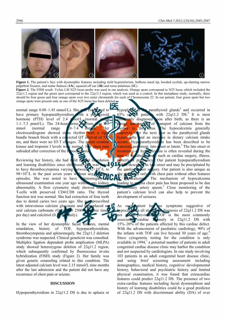
was overlooked previously. She was admitted into the General Medical Ward of Queen Mary Hospital, complaining of persistent retrosternal chest pain which was unrelated to exertion. She did not have any heart failure symptoms, carpopedal spasm or seizures. She had no history of recurrent infection or neck surgery. Her regular medications were warfarin and furosemide. On physical examination, the blood pressure was 98/57 mm Hg, pulse 86 beats per minute, and oxygen saturation in room air was 99%. She was well oriented. She spoke with a hypernasal speech, and displayed dysmorphic facial features with mild hypertelorism, bulbous nasal tip, hooded eyelids, up-slanting narrow palpebral fissures, malar flatness and squared off ears (Figure 1 A and B). In the oral examination, we detected torus palatinus (Figure 1C). In the cardiovascular examination, she had a central median sternotomy scar, and a right parasternal heave. The apex beat was located 1 cm lateral to the mid clavicular line and in the 5th intercostal space. A grade 2/6 ejection systolic murmur was heard over the left upper sternal border. Respiratory, cardiovascular and neurological systems’ examination was normal. Chovostek’s and Trousseau’s sign were negative. Laboratory investigations showed hypocalcemia with an adjusted calcium level of 1.78 (normal range 2.11–2.55) mmol/L, and ionized calcium level of 0.88 (normal range 1.12–1.23) mmol/L and hyperphosphatemia (1.73 mmol/L;
H
DOI: 10.3760/cma.j.issn.0366-6999.2012.16.027 Department of Medicine, Queen Mary Hospital, the University ofHong Kong, Hong Kong, China (Shea YF, Lee CH, Gill H, ChowWS, Lam YM and Chu LW) Clinical Genetic Service, Department of Health, TheGovernment of Hong Kong Special Administrative Region, HongKong, China (Luk HM and Lam STS) Correspondence to: Dr. SHEA Yat-fung, Department of Medicine,Queen Mary Hospital, the University of Hong Kong, 102 Pok FuLam Road, Hong Kong (Tel: 852-94159313. Email:[email protected])
Chin Med J 2012;125(16):2945-2947 2946
Figure 1. The patient’s face with dysmorphic features including mild hypertelorism, bulbous nasal tip, hooded eyelids, up-slanting narrow palpebral fissures, and malar flatness (1A), squared off ear (1B) and torus palatinus (1C). Figure 2. The FISH result. Vylsis LSI N25 locus probe was used in our analysis. Orange spots correspond to N25 locus which included the 22q11.2 region and the green spot correspond to the 22q13.3 region, which was used as a control. In this metaphase study, normally, there should be four green and four orange spots over two sister chromatids for each of Chromosome 22. In our patient, four green spots but two orange spots were present only as one of the N25 locus has been deleted. normal range 0.88–1.45 mmol/L). She was confirmed to have primary hypoparathyroidism with a parathyroid hormone (PTH) level of 2.4 pmol/L (normal range 1.1–7.3 pmol/L). The 24-hour urine calcium was 0.7 mmol (normal range 2.0–7.4 mmol). Her electrocardiogram showed sinus rhythm with a right bundle branch block with a corrected QT interval of 512 ms, and there were no ST-T changes. The serum creatine kinase and troponin I levels were normal. Her chest pain subsided after correction of the hypocalcemia. Reviewing her history, she had mild mental retardation and learning disabilities since childhood. She was noted to have thrombocytopenia varying between 72×109/L to 98×109/L in the past seven years without any bleeding episodes. She was noted to have splenomegaly on ultrasound examination one year ago without any kidney abnormality. A flow cytometry study showed reduced T-cells with preserved CD4:CD8 ratio. The thyroid function test was normal. She had extraction of four teeth due to dental caries two years ago. She was prescribed with intravenous calcium gluconate and then placed on oral calcium carbonate (Oscal-500 2500 mg three times per day) and calcitriol (0.25 mg daily). In the view of her dysmorphic facial features, mental retardation, history of TOF, hypoparathyroidism, thrombocytopenia and splenomegaly, the 22q11.2 deletion syndrome was suspected. Clinical geneticist was consulted. Multiplex ligation dependent probe amplication (MLPA) study showed heterozygous deletion of 22q11.2 region, which subsequently confirmed by fluorescence in-situ hybridization (FISH) study (Figure 2). Her family was given genetic counseling related to this condition. The latest adjusted calcium level was 2.15 mmol/L nine months after the last admission and the patient did not have any recurrence of chest pain or seizure.
DISCUSSION
Hypoparathyroidism in 22q11.2 DS is due to aplasia or
hypoplasia of the parathyroid glands3 and occurred in 65% of the patients with 22q11.2 DS.5 It is most commonly diagnosed soon after birth, as there is an abrupt interruption of transport of calcium from the mother to the fetus. The hypocalcemia generally improves over the next year as the parathyroid glands hypertrophy and an increase in dietary calcium intake occurs. Hypoparathyroidism has been described to be transient, persistent, late-onset or latent.3 The late onset or recurrence of hypocalcemia is often revealed during the time of metabolic stress such as cardiac surgery, illness, puberty or pregnancy.5 Our patient hypoparathyroidism can be described as late onset and may be precipitated by the cardiac repair surgery. Our patient is also unique as she presented only with chest pain without other features of hypocalcemia. The mechanism of hypocalcemia leading to angina chest pain has been proposed to be due to coronary artery spasm.2 Close monitoring of the patient’s calcium level can also help to prevent the development of seizures. As our patient has no symptoms suggestive of hypocalcemia all along, the diagnosis of 22q11.2 DS was not made previously. TOF is the most commonly associated cardiac anomaly in 22q11.2 DS with 15%–20% of the patients affected by this cardiac defect. With the advancement of paediatric cardiology, 90% of the infants with TOF can live beyond 30 years of age.6 Since cytogenetic testing for the condition is only available in 1994,7 a potential number of patients in adult congenital cardiac disease clinic may harbor the condition and not suspected by cardiologists. In one study involving 103 patients in an adult congenital heart disease clinic, and using brief screening assessment including demographics, medical history, cognitive/ developmental history, behavioral and psychiatric history and limited physical examination, it was found that extracardiac features could predict 22q11.2 DS. The presence of two extra-cardiac features including facial dysmorphism and history of learning disabilities could be a good predictor of 22q11.2 DS with discriminant ability (DA) of over
Chinese Medical Journal 2012;125(16):2945-2947 2947
80%, while a combination of any three features including facial dysmorphism, hypernasal speech, history of learning disabilities and age <30 years old yielded a DA of over 85% and a sensitivity of 100%. In fact, our patient has three of the extra-cardiac features! This case fully illustrates that careful observation of these extra-cardiac clinical features can help physicians, cardiologists as well as cardiothoracic surgeons to recognize and diagnose 22q11.2 DS clinically,6 In most patients, this condition is usually diagnosed shortly after birth.8 The condition often remains under-recognized in the adults as a result of its diverse clinical variability with multiple systems involvement, subtle clinical features, and lack of clinical genetics services and non-familiarity of clinicians to this condition.7 There are only a few studies on Chinese patients with 22q11.2 DS.9,10 Early diagnosis allows anticipatory clinical care. Most importantly the condition is autosomal dominant with 50% chance of having affected children.7 Because of poor cognitive function, 22q11.2 DS patients are prone to dental problems because of poor dental hygiene, resulting in dental caries which may increase the risk of infective endocarditis.11 The deletion happened in 22q11.2 region is because of its architecture with low copy number repeats (LCRs) with high homology contributing to the mispairing of LCR, unequal meiotic crossovers and aberrant inter-chromosomal exchanges.5,12 Most patients (70%–80%) have 3 Mb deletion encompassing 35–45 genes while 15%–30% slightly smaller 1.5 Mb deletion.5,12 Over 90% of the deletions occur de novo without affected parents while 10% of patients will have deletion identified in the parents.12 Our patient has lost contact with her parents. The deletion results in abnormal development of facial structures, parathyroid gland, thyroid, thymus and the heart (especially the cardiac outflow tract and right ventricle). Deleted genes well characterized to be related to clinical phenotype include TBX which is important for cardiac, thymus & parathyroid development; catecho-O-methyltransferase (COMT) with behavioral & psychiatric problems and glycoprotein Ibβ with thrombocytopenia.12 The typical deletion is submicroscopic that needs FISH for identification and the turnaround time is around 3 to 14 days. Alternatively we can detect the microdeletion by polymerase chain reaction based technology like MLPA study which is faster or single nucleotide polymorphism (SNP) arrays for atypical microdeletion.3 All patients with the 22q11.2 deletion should refer to clinical geneticist for genetic counseling as the risk of recurrence for their offsprings will increase up to 50%. If the female patient is found to be pregnant, they should manage by high risk obstetric team. Detailed fetal echocardiography by experienced paediatric cardiologist is warranted as there is high chance of conotruncal anomalies among those 22q11.2 deletion patients. Prenatal diagnosis of fetus by
chorionic villous sampling as early as 11 weeks gestation and amniocentesis as early as 15 weeks of gestation is indicated. Fetus with pre-natal diagnosis of 22q11.2 deletion syndrome with cardiac involvement should be delivered in tertiary center with neonatal intensive care unit and cardiothoracic surgery support. Prostaglandin E2 infusion is needed for ductal dependent circulation or even cardiac surgery for critical left or right outflow tract obstructive lesions soon after birth. Calcium level should also need to be monitor closely during the neonatal period. Advice from immunologist may also indicated before live vaccine immunization.
REFERENCES
1. Turk C, Stollberger C, Huber J, Sehnal E, Finsterer J.
Priapism as a manifestation of tetania. Scand J Urol Nephrol 2009; 43: 94-95.
2. Lehmann G, Deisenhofer I, Ndrepepa G, Schmitt C. ECG changes in a 25-year-old woman with hypocalcemia due to hypoparathyroidism. Hypocalcemia mimicking acute myocardial infarction. Chest 2000; 118: 260-262.
3. Tonelli AR, Kosuri K, Wei S, Chick D. Seizures as the first manifestation of chromosome 22q11.2 deletion syndrome in a 40-year old man: a case report. J Med Case Reports 2007; 3: 167.
4. Tan KB, Chew SK, Yeo GS. 22q11.2 deletion syndrome in Singapore (2000-2003): a case for active ascertainment. Singapore Med J 2008; 49: 286-289.
5. McDonald-McGinn DM, Sullivan KE. Chromosome 22q11.2 deletion syndrome (DiGeorge syndrome/ velocardiofacial syndrome). Medicine (Baltimore) 2011; 90: 1-18.
6. Fung WL, Chow EW, Webb GD, Gatzoulis MA, Bassett AS. Extracardiac features predicting 22q11.2 deletion syndrome in adult congenital heart disease. Int J Cardiol 2008; 131: 51-58.
7. Kapadia RK, Bassett AS. Recognizing a common genetic syndrome: 22q11.2 deletion syndrome. CMAJ 2008; 178: 391-393.
8. McCusker LA, Jenkins NP, Hancock JE. Hypocalcaemia in a patient with congenital heart disease. J R Soc Med 2007; 100: 51-53.
9. Yi L, Xu ZF, Mo XM, Hu YL, Wang DJ, Han B, et al. New tetranucleotide STRP markers for detecting the 22q11.2 deletion. Mol Cell Probes 2006; 20: 359-365.
10. Lee ML, Chen HN, Chen M, Tsao LY, Wang BT, Lee MH, et al. Persistent fifth aortic arch associated with 22q11.2 deletion syndrome; J Formos Med Assoc 2006; 105: 284-289.
11. Oberoi S, Huynh L, Vargervik K. Velopharyngeal, speech and dental characteristics as diagnostic aids in 22q11.2 deletion syndrome. J Calif Dent Assoc 2011; 39: 327-332.
12. Bassett AS, McDonald-McGinn DM, Devriendt K, Digilio MC, Goldenberg P, Habel A, et al. Practical Guidelines for Managing Patients with 22q11.2 Deletion Syndrome. J Pediatr 2011; 159: 332-339.
(Received November 15, 2011) Edited by GUO Li-shao