Embed Size (px)
Citation preview

The 2010-1st International OCARINA Symposium
OCU Media Center 10F
December 8th, 2010
The OCU Advanced Research Institute
for Natural Science and Technology
http://www.ocarina-ocu.jp/
3
The 2010-1st- International OCARINA Symposium
Venue: OCU Media Center 10F, December 8th, 2010
Program
13:00-13:05 Opening Remark
13:05-13:50 “Solar to Fuels; Natural Tactics of Solar Energy Conversion by
Photosynthesis”
Hideki Hashimoto (OCARINA A-group Leader)
13:55-14:15 “Organo-copper complex as an interface for photosynthetic system”
Isamu Kinoshita (OCARINA A-group)
14:20-14:40 “Key Residues and their Protonation States in the Reaction Pathway
of TtADPRase”
Yoshihiko Furuike (OCARINA A-group)
14:45-15:05 “Atomic Force Microscopy Observation of Reconstituted
Photosynthetic Membranes”
Yuko Sugai (OCARINA A-Group)
15:05-15:30 Break
15:30-16:30 “EXCITED STATE LEVEL STRUCTURE AND ULTRAFAST
RELAXATION DYNAMICS IN CAROTENOIDS”
Alfred R. Holzwarth (Max-Planck-Institut für Bioanorganische
Chemie, Germany)
16:30-17:30 “Contrasting One-Photon and Two-Photon Initiated Dynamics of
Carotenoids in Solution and Light Harvesting Proteins”
Delmar S. Larsen (Department of Chemistry, University of
California Davis, USA)
Informal banquet will be opened at AOGAKI
Solar to Fuels; Natural Tactics of Solar Energy Conversion by Photosynthesis
Hideki Hashimoto
CREST-JST and The OCU Advanced Research Institute for Natural Science and Technology (OCARINA), Osaka City University, Sugimoto, Sumiyoshi-ku, Osaka 558-8585, Japan
70% of mankind’s current energy needs are met by burning fossil fuels. This is already
problematic since oil and gas supplies are limited and because of the adverse environmental effects of rising levels of carbon dioxide in the atmosphere. Moreover this situation is set to get worse as current predictions estimate that our energy needs will double by 2050. Mankind is, therefore, facing a major challenge to find new ways of creating clean, renewable fuels. One potentially abundant source of energy is solar radiation. More energy strikes the earth surface every hour than mankind uses each year. The problem is how to harness such an abundant yet diffuse source of energy. Photosynthesis has evolved mechanisms to achieve this. Conceptually photosynthesis can be divided into the following partial reactions, light-harvesting (concentration), charge-separation (conversion of solar energy into chemical energy) and finally multi-electron catalysis that takes electrons from water and uses them to reduce carbon dioxide to carbohydrates (fuel).
Any proposed strategies that set out to mimic this process in order to make solar fuels must begin with a light-harvesting or light-concentration step. We know a great deal about the structure and function of photosynthetic light-harvesting complexes as individual molecules, but rather little is known about how their supra-molecular organization within their photosynthetic membranes affects their overall function in vivo. We wish to understand how the supra-molecular arrangement of the light-harvesting apparatus relates to overall light-harvesting efficiency and to be able to translate this information to inform the design of robust artificial light-harvesting arrays that can, in the long term, be used in devices for producing solar fuels. Photosynthetic antenna complexes are organised on the nanoscale (see Figure 1) and a major question is how to translate this information into the design of meso- to macroscale light-harvesting devices.
This lecture will outline how photosynthesis achieves ‘Solar to Fuels’ conversion concentrating on the general principles involved. Recent progress on understating the molecular details of the key reactions in the photosynthetic process has been remarkable. We are now at the stage where it is realistic to start to use this ‘biological blueprint’ to begin to construct devices that have the capability to mimic the key steps in the natural process. This is one of the grand scientific challenges of our time.
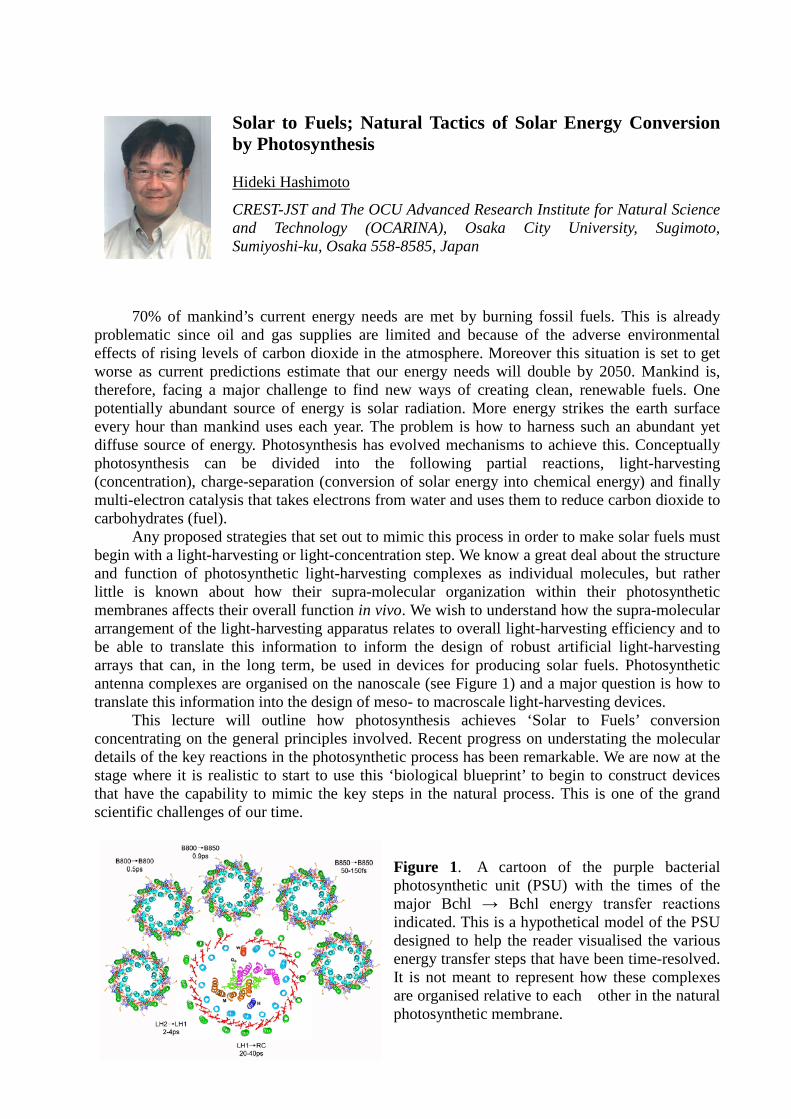
Figure 1. A cartoon of the purple bacterial photosynthetic unit (PSU) with the times of the major Bchl → Bchl energy transfer reactions indicated. This is a hypothetical model of the PSU designed to help the reader visualised the various energy transfer steps that have been time-resolved. It is not meant to represent how these complexes are organised relative to each other in the natural photosynthetic membrane.
Atomic Force Microscopy Observation of Reconstituted Photosynthetic Membranes Yuko Sugai1, Ayumi Sumino2,3, Chiasa Uragami1, Ritsuko Fujii1,4, Takanori Nishioka1, Takehisa Dewa2, Isamu Kinoshita1,4, Mamoru Nango1, Hideki Hashimoto1,4 1CREST/JST and Graduate School of Science, Osaka City University, Osaka, Japan, 2Department of Life and Materials Engineering, Nagoya Institute of Technology, Nagoya, Japan, 3Institute of Advanced Energy, Kyoto University, Kyoto, Japan, 4The OCU Advanced Research Institute for Natural Science and Technology (OCARINA), Osaka City University, Osaka, Japan,
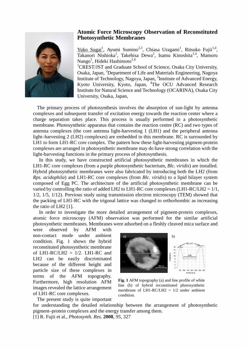
The primary process of photosynthesis involves the absorption of sun-light by antenna complexes and subsequent transfer of excitation energy towards the reaction center where a charge separation takes place. This process is usually performed in a photosynthetic membrane. Photosynthetic apparatus that contains the reaction centre (RC) and two types of antenna complexes (the core antenna light-harvesting 1 (LH1) and the peripheral antenna light–harvesting 2 (LH2) complexes) are embedded in this membrane. RC is surrounded by LH1 to form LH1-RC core complex. The pattern how these light-harvesting pigment-protein complexes are arranged in photosynthetic membrane may do have strong correlation with the light-harvesting functions in the primary process of photosynthesis. In this study, we have constructed artificial photosynthetic membranes in which the LH1-RC core complexes (from a purple photosynthetic bacterium, Blc. viridis) are installed. Hybrid photosynthetic membranes were also fabricated by introducing both the LH2 (from Rps. acidophila) and LH1-RC core complexes (from Blc. viridis) to a lipid bilayer system composed of Egg PC. The architecture of the artificial photosynthetic membrane can be varied by controlling the ratio of added LH2 to LH1-RC core complexes (LH1-RC/LH2 = 1/1, 1/2, 1/5, 1/12). Previous study using transmission electron microscopy (TEM) showed that the packing of LH1-RC with the trigonal lattice was changed to orthorhombic as increasing the ratio of LH2 [1]. In order to investigate the more detailed arrangement of pigment-protein complexes, atomic force microscopy (AFM) observation was performed for the similar artificial photosynthetic membranes. Membranes were adsorbed on a fleshly cleaved mica surface and were observed by AFM with non-contact mode under ambient condition. Fig. 1 shows the hybrid reconstituted photosynthetic membrane of LH1-RC/LH2 = 1/2. LH1-RC and LH2 can be easily discriminated because of the different height and particle size of these complexes in terms of the AFM topography. Furthermore, high resolution AFM images revealed the lattice arrangement of LH1-RC core complexes. The present study is quite important for understanding the detailed relationship between the arrangement of photosynthetic pigment–protein complexes and the energy transfer among them. [1] R. Fujii et al., Photosynth. Res. 2008, 95, 327
Fig. 1 AFM topography (a) and line profile of white line (b) of hybrid reconstituted photosynthetic membrane of LH1-RC/LH2 = 1/2 under ambient condition.
a)
b)
EXCITED STATE LEVEL STRUCTURE AND ULTRAFAST RELAXATION DYNAMICS IN
CAROTENOIDS
Alfred R. Holzwarth
Max-Planck-Institut für Bioanorganische Chemie, Stiftstrasse 34-36, 45470 Mülheim a.d. Ruhr, Germany, e-mail: [email protected]
There exists general agreement about the location and dynamics of two excited states in “normal” carotenoids not possessing charge transfer states. These are the low-lying first excited 2Ag
- state, which is a dark state relative to the ground state, and the 1Bu+ state
(generally called the S2 state) which gives the colour to carotenoids due to its very strong transition moment from the ground state (for a review see 1). However a long-standing debate has been taking place in the literature about the nature and energetic location of additional so-called “dark states” between the S1 and the S2 states. The most prominent one that also has a good theoretical foundation is the 1Bu
- state, generally considered to be a “dark state”, i.e. not carrying oscillator strength from the ground state. We have shown recently that in carotenoids of conjugation length n=10-11 the 1Bu
- state actually is located slightly below the 1Bu+ state
and that the two potential curves cross each other, giving rise to electronic coherence udner certain conditions2. Besides some vibrationally unrelaxed S2 states, some publications reported additional states with fairly unusual spectroscopic properties which were proposed to be located between the S1 and the S2 states (4-6). These states were termed e.g. Sx, S
* etc. and their molecular and electronic assignment is at best highly controversial if not totally unclear. For example, we recently showed that the so-called S* state in β-carotene is not a separate electronic state but a vibrationally excited (hot) 2Ag
- state3. In this talk an effort will be made to sort out some of the controversies and confusion
about this “zoo” of excited states in “normal” (i.e. ignoring those situations where CT states are involved) longer chain carotenoids of conjugation length n=9-13. A new electronic level scheme will be proposed explaining the puzzles of the ultrafast relaxation of the S2 state and the related electronic coherences.
References
1. Polivka, T.; Sundström, V. Dark excited states of carotenoids: Consensus and
controversy. Chem. Phys. Lett. 2009, 477 (1-3), 1-11.
2. Ostroumov, E.; Müller, M. G.; Marian, C. M.; Kleinschmidt, M.; Holzwarth, A. R. Electronic coherence provides a direct proof for energy-level crossing in photoexcited lutein and β-carotene. Phys. Rev. Lett. 2009, 103, 108302-1-108302-4.
3. Ostroumov, E. E.; Müller, M.; Reus, M.; Holzwarth, A. R. On the nature of the "dark S*" excited state of β-carotene. J. Phys. Chem. B 2010, in print.

Contrasting One-Photon and Two-Photon Initiated Dynamics of Carotenoids in Solution and Light Harvesting Proteins Delmar S. Larsen
Department of Chemistry, University of California, Davis
One Shields Avenue Davis, CA 95616 USA
Although observed in multiple carotenoid samples, the exact nature of the S* state is
still debated, including the electronic structure and ensuring formation and decay
dynamics. Consequently, several models have been proposed regarding the ultrafast
excited state kinetics in carotenoids; unfortunately, many studies are hindered by the
inability to cleanly resolve the S* state from the overlapping S1 excited state absorption
and bleach contributions to the transient signals. This is further compounded by the
similarity in S* lifetime to S1. We explore the nature of S* with femtosecond
one-photon and two-photon initiated dynamics of the all trans β-carotene carotenoid,
which is further characterized and dissected with excitation-wavelength and
temperature-dependent ultrafast dispersed transient absorption signals. The S*
population kinetics prompted the development of a modified multi-compartment model.
The interconnectivity of the S* and S1 populations is explored and discussed and no
observed population flow is resolved between S* and S1. Comparison of samples
obtained from different laboratories with different purity levels demonstrates that
sample contamination is not the primary origin of the S* state. Comparisons to the
dynamics in the rhodopsin glucoside carotenoid bound to Rps. Acidophila will be made.