Embed Size (px)
Citation preview

DARK-FIELD SMARTPHONE MICROSCOPE WITH NANOSCALE RESOLUTION FOR MOLECULAR DIAGNOSTICS
Brian N. Kim1, Julian A. Diaz1, Soon Gweon Hong1, Sang Hun Lee1, and Luke P. Lee1 1Departments of Bioengineering,
Berkeley Sensor and Actuator Center, University of California, Berkeley, CA 94720, USA
ABSTRACT Dark-field microscopy allows highly sensitive detection of metal nanoparticles that exhibit surface
plasmon resonance (SPR) scattering by exclusively collecting scattered light. Here, we report a dark-field smartphone microscope with single nanoparticle resolution for point-of-care testing (POCT). A simple flat-lens LED is coupled to transparent substrate to generate total internal reflection (TIR) and evanescent field for surface-confined illumination, which scatters at nanoparticles. The scattering light is detected using smartphone camera through an external lens attachment. Dark-field smartphone microscope will facilitate the rapid translation of sensitive nanoparticle-based bioassays to point-of-care testing (POCT). KEYWORDS: Dark-field microscopy, nanoparticle, total internal reflection (TIR), smartphone microscopy, surface plasmon resonance (SPR), point-of-care testing (POCT)
INTRODUCTION
Smartphone-based diagnostic platform takes advantage of biotechnology (BT) and information technology (IT) [1,2]. Recently, fluorescence-based nanoparticle detection was reported using smartphone [2], adding nanotechnology (NT) to smartphone-based imaging methods. With dark-field microscopy, SPR scattering at the surface of metal nanoparticles can be imaged. Since gold or silver nanoparticles are versatile substrate for various surface conjugations of biomolecules and offers enhanced signal due to plasmon-resonance, bioassays based on metal nanoparticles for medical diagnosis are being developed to produce highly sensitive assays [3]. As the type of bioassays grow that rely on plasmon-resonance particles, dark-field smartphone microscope will allow the use of existing bioassays in remote sites.
EXPERIMENTAL
Smartphone is converted to a dark-field microscope by an attachment that consists of dark-field illumination and external lens for magnification, shown in Figure 1a. An external lens (f = 3 mm) is attached and aligned to the smartphone camera to magnify and capture scattering light from nanoparticles.
Figure 1: Dark-field smartphone microscope. (a) Dark-field illumination is based on evanescent field generated by LED total internal reflection (TIR). The scattered light at the surface of nanoparticle is collected without any background due to short penetration depth of evanescent field. (b) Photograph of smartphone inserted into dark-field microscope attachment. Unlike oblique dark-field illumination that requires condenser and spider light stop, we use evanescent field to avoid complexity of the system and create highly localized illumination (fixed z) at the surface of specimen slide (Figure 1a). And also, TIR illumination yields higher signal-to-noise ratio (SNR =
978-0-9798064-7-6/µTAS 2014/$20©14CBMS-0001 2247 18th International Conference on MiniaturizedSystems for Chemistry and Life Sciences
October 26-30, 2014, San Antonio, Texas, USA
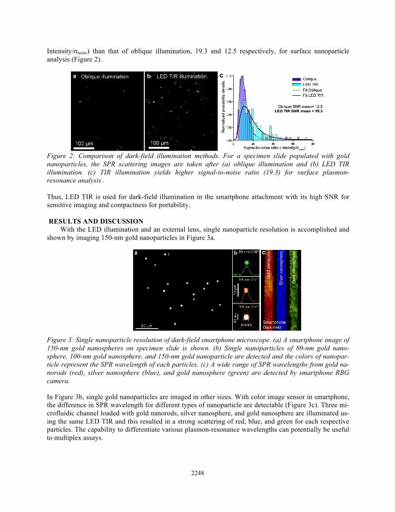
Intensity/σnoise) than that of oblique illumination, 19.3 and 12.5 respectively, for surface nanoparticle analysis (Figure 2).
Figure 2: Comparison of dark-field illumination methods. For a specimen slide populated with gold nanoparticles, the SPR scattering images are taken after (a) oblique illumination and (b) LED TIR illumination. (c) TIR illumination yields higher signal-to-noise ratio (19.3) for surface plasmon-resonance analysis . Thus, LED TIR is used for dark-field illumination in the smartphone attachment with its high SNR for sensitive imaging and compactness for portability. RESULTS AND DISCUSSION
With the LED illumination and an external lens, single nanoparticle resolution is accomplished and shown by imaging 150-nm gold nanoparticles in Figure 3a.
Figure 3: Single nanoparticle resolution of dark-field smartphone microscope. (a) A smartphone image of 150-nm gold nanospheres on specimen slide is shown. (b) Single nanoparticles of 80-nm gold nano-sphere, 100-nm gold nanosphere, and 150-nm gold nanoparticle are detected and the colors of nanopar-ticle represent the SPR wavelength of each particles. (c) A wide range of SPR wavelengths from gold na-norods (red), silver nanosphere (blue), and gold nanosphere (green) are detected by smartphone RBG camera.
In Figure 3b, single gold nanoparticles are imaged in other sizes. With color image sensor in smartphone, the difference in SPR wavelength for different types of nanoparticle are detectable (Figure 3c). Three mi-crofluidic channel loaded with gold nanorods, silver nanosphere, and gold nanosphere are illuminated us-ing the same LED TIR and this resulted in a strong scattering of red, blue, and green for each respective particles. The capability to differentiate various plasmon-resonance wavelengths can potentially be useful to multiplex assays.
c
c
2248

Figure 4: SPR wavelength shift by gold nanoparticle aggregation is detected using dark-field
smartphone microscope. (a) Gold nanoparticles (80-nm) coated with biotin exhibit SPR wavelength in green. And (b) when gold nanoparticles (40-nm) coated with streptavidin is introduced, gold nanoparti-cles start to aggregate due to strong interaction of biotin-streptavidin and SPR wavelength shifts from (c) green to (d) orange color.
For initial proof-of-concept, a bioassay of gold aggregation from biotin-streptavidin interaction is performed under the dark-field smartphone microscope, shown in Figure 4. Gold nanoparticles that are coated with biotin exhibited a SPR wavelength near green and once 40-nm gold nanoparticles coated with streptavidin is introduced, SPR wavelength shifted to orange indicating the aggregation of gold nanoparticles by biotin-streptavidin interaction. CONCLUSION
We report dark-field smartphone microscope that consists of TIR illumination and simple external lens. This device will allow gold or silver nanoparticle-based bioassays to be easily converted into POCT systems. Due to the single nanoparticle resolution, extreme detection limits (single molecule) accom-plished by existing bioassays may be brought to resource-limited settings. ACKNOWLEDGEMENTS
The authors thank Bill & Melinda Gates Foundation for the grant support.
REFERENCES [1] N. Chronis and L. P. Lee, “Total internal reflection-based biochip utilizing a polymer-filled cavity
with a micromirror sidewall,” Lab Chip, 4, 125-130, 2004. [2] Q. Wei, H. Qi, W. Luo, D. Tseng, S. J. Ki, Z. Wan, Z. Göröcs, L. A. Bentolila, T. Wu, R. Sun, and
A. Ozcan, “Fluorescent Imaging of Single Nanoparticles and Viruses on a Smart Phone,” ACS Nano, 7, (10), pp 9147-9155, 2013.
[3] A. Crut, P. Maioli, N. D. Fatti and F. Vallée, “Optical absorption and scattering spectroscopies of single nano-objects,” Chem. Soc. Rev., 43, 3921-3956, 2014.
CONTACT * B. N. Kim; phone: +1-607-280-7925; [email protected]
2249

![Nanoscale electrical property evaluation using Scanning ...kuclf.kyushu-u.ac.jp/2010.3.15/h21_0315_abstract/Aisoabstract-11.pdf · [3] Agilent Tehnologies, Inc. 5600 SPM/AFM Microscope](https://img.pdfslide.us/doc/110x75/5e65624f61491155dd455ff8/nanoscale-electrical-property-evaluation-using-scanning-kuclfkyushu-uacjp2010315h210315abstractaisoabstract-11pdf.jpg)