Embed Size (px)
Citation preview
D-Amino Acids Indirectly Inhibit Biofilm Formation in Bacillus subtilisby Interfering with Protein Synthesis
Sara A. Leiman,a Janine M. May,b Matthew D. Lebar,b Daniel Kahne,b Roberto Kolter,c Richard Losicka
Department of Molecular and Cellular Biology, Harvard University, Cambridge, Massachusetts, USAa; Department of Chemistry and Chemical Biology, Harvard University,Cambridge, Massachusetts, USAb; Department of Microbiology and Immunobiology, Harvard Medical School, Boston, Massachusetts, USAc
The soil bacterium Bacillus subtilis forms biofilms on surfaces and at air-liquid interfaces. It was previously reported that thesebiofilms disassemble late in their life cycle and that conditioned medium from late-stage biofilms inhibits biofilm formation.Such medium contained a mixture of D-leucine, D-methionine, D-tryptophan, and D-tyrosine and was reported to inhibit biofilmformation via the incorporation of these D-amino acids into the cell wall. Here, we show that L-amino acids were able to specifi-cally reverse the inhibitory effects of their cognate D-amino acids. We also show that D-amino acids inhibited growth and theexpression of biofilm matrix genes at concentrations that inhibit biofilm formation. Finally, we report that the strain routinelyused to study biofilm formation has a mutation in the gene (dtd) encoding D-tyrosyl-tRNA deacylase, an enzyme that preventsthe misincorporation of D-amino acids into protein in B. subtilis. When we repaired the dtd gene, B. subtilis became resistant tothe biofilm-inhibitory effects of D-amino acids without losing the ability to incorporate at least one noncanonical D-amino acid,D-tryptophan, into the peptidoglycan peptide side chain. We conclude that the susceptibility of B. subtilis to the biofilm-inhibi-tory effects of D-amino acids is largely, if not entirely, due to their toxic effects on protein synthesis.
The soil bacterium Bacillus subtilis forms architecturally com-plex communities of cells encased in a self-produced matrix
consisting of exopolysaccharide and the amyloid-like proteinTasA (1, 2). These biofilms form on solid surfaces and at air-liquidinterfaces (floating biofilms are known as pellicles). Whereasmuch is known about the mechanisms underlying biofilm forma-tion, the mechanisms that cause cells to disperse from the biofilmand resume a planktonic existence are less well understood (3).Recently, interest has focused on the production of small mole-cules that inhibit biofilm formation and could therefore be in-volved in biofilm disassembly. Of particular interest are four D-amino acids (D-Leu, D-Met, D-Trp, and D-Tyr) that were found tobe produced at late stages of biofilm development and were re-ported to have biofilm-inhibitory activity. Previous work fromtwo of our laboratories suggested that the incorporation of thesefour D-amino acids into the peptidoglycan peptide side chain inplace of D-Ala triggers the release of TasA fibers from the cellwall (4).
Here, we present evidence that indicates that the D-amino acidsused (D-Leu, D-Met, D-Trp, and D-Tyr) can be growth inhibitoryand that the biofilm inhibition observed when the culture me-dium contains these D-amino acids is largely, if not entirely, aconsequence of their misincorporation into protein. This findingis in keeping with the report that D-Tyr, the most potent of thefour D-amino acids, is a metabolic inhibitor of B. subtilis (5).
MATERIALS AND METHODSStrains and growth conditions. Bacillus subtilis NCIB3610 (hereinafter,3610) or 168 and Escherichia coli (New England BioLabs, USA) weregrown in Luria-Bertani (LB) broth (10 g tryptone per liter, 5 g yeast extractper liter, 5 g NaCl per liter) or on LB agar plates containing 1.5% Bactoagar at 37°C. When appropriate, 1 �g/ml erythromycin and 25 �g/mllincomycin were added to liquid or solid medium. The strains used in thisstudy are listed in Table S1 in the supplemental material.
Strain construction. The markerless repair of dtd in B. subtilis wasaccomplished using the pMiniMAD protocol as described by Patrick andKearns (6) and using primers 51 to 54, given in Table S1 in the supple-
mental material. Successful double-crossover events leading to the dtd�
strain were confirmed by restreaking individual clones on 1% agar platescontaining LB, LB plus erythromycin and lincomycin, or LB plus 400 �MD-LMWY. Isolates which grew on LB and on LB plus D-LMWY but not onLB plus erythromycin and lincomycin were submitted for sequencingusing primers 7 and 8 (see Table S1).
Luciferase reporters for epsA and tapA operon expression were con-structed as described previously (7) and transferred by phage transduc-tion into dtd� cells (8).
Growth measurements. Unless otherwise indicated, cells were grownto mid-exponential phase and diluted to a final optical density at 600 nm(OD600) of �0.015 into MSgg medium (8) to which amino acid or theequivalent volume of distilled water (dH2O) was added to a final volumeof 20 ml. Cells were grown in shaking culture at 200 rpm at 37°C, and theOD600 was measured every hour for 7 h. Alternatively, cells grown tomid-exponential phase were diluted 1:1,000 into MSgg plus amino acidsor the equivalent volume of dH2O, and 250-�l aliquots were transferredto a Costar polystyrene 96-well plate with a low-evaporation lid (FisherScientific, USA). The OD600 was measured every 10 min for 24 h in aBioTek Synergy 2 luminometer (BioTek, USA) with continuous slowshaking at 30°C.
Pellicle assays. Cultures of B. subtilis 3610 or dtd� cells were grown toearly stationary phase and then diluted 1:1,000 into MSgg (9) or modifiedMSgg. Amino acids were stored as stock solutions of 40 mM or 60 mM indH2O, except for D-Tyr and L-Tyr, which were stored as 10 mM stocksolutions in 0.1 M HCl. Treatment solutions were diluted from stocksolutions to achieve the final concentrations, and volumes were normal-ized using dH2O. All experiments were conducted in BD Falcon 24-well
Received 20 August 2013 Accepted 26 September 2013
Published ahead of print 4 October 2013
Address correspondence to Richard Losick, [email protected].
Supplemental material for this article may be found at http://dx.doi.org/10.1128/JB.00975-13.
Copyright © 2013, American Society for Microbiology. All Rights Reserved.
doi:10.1128/JB.00975-13
December 2013 Volume 195 Number 23 Journal of Bacteriology p. 5391–5395 jb.asm.org 5391
on June 14, 2019 by guesthttp://jb.asm
.org/D
ownloaded from
plates with low-evaporation lids (BD Biosciences, USA) in a final volumeof 2 ml. Plates were incubated at 30°C or 25°C, and photographs weretaken every 24 h.
Kinetic luciferase assay. Cultures of B. subtilis 3610 or dtd� cells weregrown in LB to mid-exponential phase and diluted 1:1,000 in fresh MSggwith amino acid treatments or an equivalent volume of dH2O. Dilutionswere plated in triplicate (250 �l each) in a 96-well polystyrene Costar plate(white with a clear bottom; Fisher Scientific, USA). Luciferase activity wasmeasured on a BioTek Synergy 2 luminometer (BioTek, USA) with con-tinuous slow shaking at 30°C. Luciferase luminescence was measured at asensitivity setting of 200, and the culture optical density was measured at600 nm every 10 min for 24 h. The final luciferase activity values werecalculated by normalizing luciferase luminescence to culture density.
Evaluation of D-amino acid incorporation into peptidoglycan. Anal-ysis of B. subtilis peptidoglycan fragments by liquid chromatography-mass spectrometry (LC-MS) was performed as described by Lebar et al.(10) with the modifications detailed in the supplemental material. Theextracted [M�2H]/2 ions are provided in Fig. S4 in the supplementalmaterial.
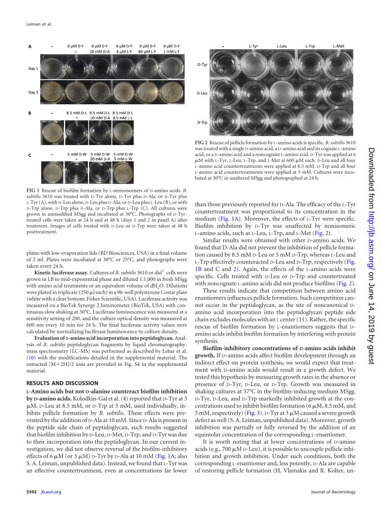
RESULTS AND DISCUSSIONL-Amino acids but not D-alanine counteract biofilm inhibitionby D-amino acids. Kolodkin-Gal et al. (4) reported that D-Tyr at 3�M, D-Leu at 8.5 mM, or D-Trp at 5 mM, used individually, in-hibits pellicle formation by B. subtilis. These effects were pre-vented by the addition of D-Ala at 10 mM. Since D-Ala is present inthe peptide side chain of peptidoglycan, such results suggestedthat biofilm inhibition by D-Leu, D-Met, D-Trp, and D-Tyr was dueto their incorporation into the peptidoglycan. In our current in-vestigation, we did not observe reversal of the biofilm-inhibitoryeffects of 6 �M (or 3 �M) D-Tyr by D-Ala at 10 mM (Fig. 1A; alsoS. A. Leiman, unpublished data). Instead, we found that L-Tyr wasan effective countertreatment, even at concentrations far lower
than those previously reported for D-Ala. The efficacy of the L-Tyrcountertreatment was proportional to its concentration in themedium (Fig. 1A). Moreover, the effects of L-Tyr were specific.Biofilm inhibition by D-Tyr was unaffected by nonisomericL-amino acids, such as L-Leu, L-Trp, and L-Met (Fig. 2).
Similar results were obtained with other D-amino acids. Wefound that D-Ala did not prevent the inhibition of pellicle forma-tion caused by 8.5 mM D-Leu or 5 mM D-Trp, whereas L-Leu andL-Trp effectively counteracted D-Leu and D-Trp, respectively (Fig.1B and C and 2). Again, the effects of the L-amino acids werespecific. Cells treated with D-Leu or D-Trp and countertreatedwith noncognate L-amino acids did not produce biofilms (Fig. 2).
These results indicate that competition between amino acidenantiomers influences pellicle formation. Such competition can-not occur in the peptidoglycan, as the site of noncanonical D-amino acid incorporation into the peptidoglycan peptide sidechain excludes molecules with an L center (11). Rather, the specificrescue of biofilm formation by L-enantiomers suggests that D-amino acids inhibit biofilm formation by interfering with proteinsynthesis.
Biofilm-inhibitory concentrations of D-amino acids inhibitgrowth. If D-amino acids affect biofilm development through anindirect effect on protein synthesis, we would expect that treat-ment with D-amino acids would result in a growth defect. Wetested this hypothesis by measuring growth rates in the absence orpresence of D-Tyr, D-Leu, or D-Trp. Growth was measured inshaking cultures at 37°C in the biofilm-inducing medium MSgg.D-Tyr, D-Leu, and D-Trp markedly inhibited growth at the con-centrations used to inhibit biofilm formation (6 �M, 8.5 mM, and5 mM, respectively) (Fig. 3). D-Tyr at 3 �M caused a severe growthdefect as well (S. A. Leiman, unpublished data). Moreover, growthinhibition was partially or fully reversed by the addition of anequimolar concentration of the corresponding L-enantiomer.
It is worth noting that at lower concentrations of D-aminoacids (e.g., 700 �M D-Leu), it is possible to uncouple pellicle inhi-bition and growth inhibition. Under such conditions, both thecorresponding L-enantiomer and, less potently, D-Ala are capableof restoring pellicle formation (H. Vlamakis and R. Kolter, un-
FIG 1 Rescue of biofilm formation by L-stereoisomers of D-amino acids. B.subtilis 3610 was treated with D-Tyr alone, D-Tyr plus D-Ala, or D-Tyr plusL-Tyr (A), with D-Leu alone, D-Leu plus D-Ala, or D-Leu plus L-Leu (B), or withD-Trp alone, D-Trp plus D-Ala, or D-Trp plus L-Trp (C). All cultures weregrown in unmodified MSgg and incubated at 30°C. Photographs of D-Tyr-treated cells were taken at 24 h and at 48 h (days 1 and 2 in panel A) aftertreatment. Images of cells treated with D-Leu or D-Trp were taken at 48 hposttreatment.
FIG 2 Rescue of pellicle formation by L-amino acids is specific. B. subtilis 3610was treated with a single D-amino acid, a D-amino acid and its cognate L-aminoacid, or a D-amino acid and a noncognate L-amino acid. D-Tyr was applied at 6�M with L-Tyr, L-Leu, L-Trp, and L-Met at 600 �M each. D-Leu and all fourL-amino acid countertreatments were applied at 8.5 mM. D-Trp and all fourL-amino acid countertreatments were applied at 5 mM. Cultures were incu-bated at 30°C in unaltered MSgg and photographed at 24 h.
Leiman et al.
5392 jb.asm.org Journal of Bacteriology
on June 14, 2019 by guesthttp://jb.asm
.org/D
ownloaded from
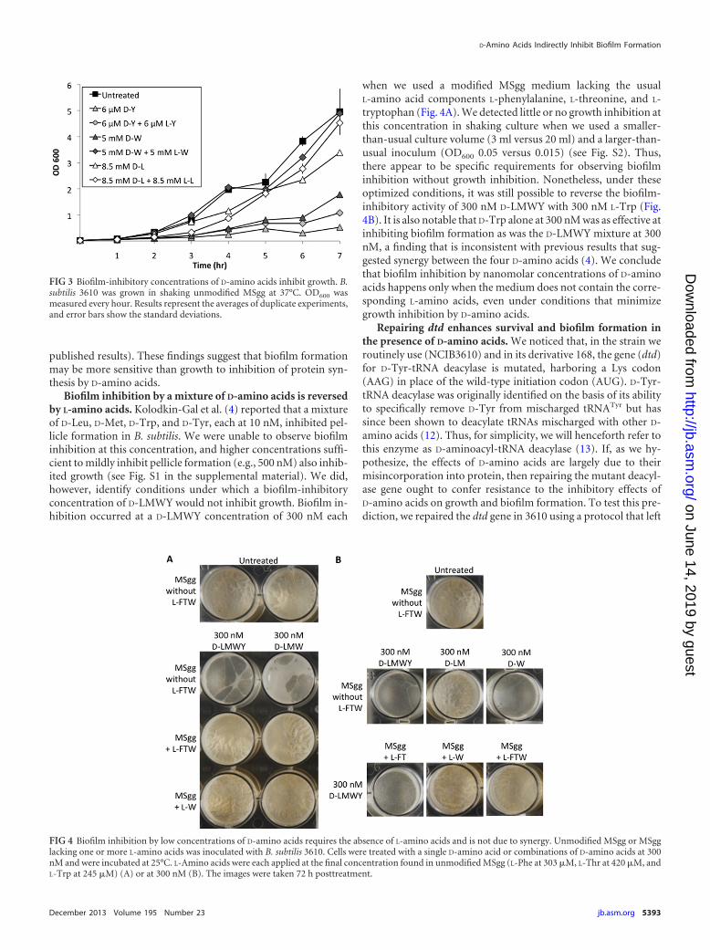
published results). These findings suggest that biofilm formationmay be more sensitive than growth to inhibition of protein syn-thesis by D-amino acids.
Biofilm inhibition by a mixture of D-amino acids is reversedby L-amino acids. Kolodkin-Gal et al. (4) reported that a mixtureof D-Leu, D-Met, D-Trp, and D-Tyr, each at 10 nM, inhibited pel-licle formation in B. subtilis. We were unable to observe biofilminhibition at this concentration, and higher concentrations suffi-cient to mildly inhibit pellicle formation (e.g., 500 nM) also inhib-ited growth (see Fig. S1 in the supplemental material). We did,however, identify conditions under which a biofilm-inhibitoryconcentration of D-LMWY would not inhibit growth. Biofilm in-hibition occurred at a D-LMWY concentration of 300 nM each
when we used a modified MSgg medium lacking the usualL-amino acid components L-phenylalanine, L-threonine, and L-tryptophan (Fig. 4A). We detected little or no growth inhibition atthis concentration in shaking culture when we used a smaller-than-usual culture volume (3 ml versus 20 ml) and a larger-than-usual inoculum (OD600 0.05 versus 0.015) (see Fig. S2). Thus,there appear to be specific requirements for observing biofilminhibition without growth inhibition. Nonetheless, under theseoptimized conditions, it was still possible to reverse the biofilm-inhibitory activity of 300 nM D-LMWY with 300 nM L-Trp (Fig.4B). It is also notable that D-Trp alone at 300 nM was as effective atinhibiting biofilm formation as was the D-LMWY mixture at 300nM, a finding that is inconsistent with previous results that sug-gested synergy between the four D-amino acids (4). We concludethat biofilm inhibition by nanomolar concentrations of D-aminoacids happens only when the medium does not contain the corre-sponding L-amino acids, even under conditions that minimizegrowth inhibition by D-amino acids.
Repairing dtd enhances survival and biofilm formation inthe presence of D-amino acids. We noticed that, in the strain weroutinely use (NCIB3610) and in its derivative 168, the gene (dtd)for D-Tyr-tRNA deacylase is mutated, harboring a Lys codon(AAG) in place of the wild-type initiation codon (AUG). D-Tyr-tRNA deacylase was originally identified on the basis of its abilityto specifically remove D-Tyr from mischarged tRNATyr but hassince been shown to deacylate tRNAs mischarged with other D-amino acids (12). Thus, for simplicity, we will henceforth refer tothis enzyme as D-aminoacyl-tRNA deacylase (13). If, as we hy-pothesize, the effects of D-amino acids are largely due to theirmisincorporation into protein, then repairing the mutant deacyl-ase gene ought to confer resistance to the inhibitory effects ofD-amino acids on growth and biofilm formation. To test this pre-diction, we repaired the dtd gene in 3610 using a protocol that left
FIG 3 Biofilm-inhibitory concentrations of D-amino acids inhibit growth. B.subtilis 3610 was grown in shaking unmodified MSgg at 37°C. OD600 wasmeasured every hour. Results represent the averages of duplicate experiments,and error bars show the standard deviations.
FIG 4 Biofilm inhibition by low concentrations of D-amino acids requires the absence of L-amino acids and is not due to synergy. Unmodified MSgg or MSgglacking one or more L-amino acids was inoculated with B. subtilis 3610. Cells were treated with a single D-amino acid or combinations of D-amino acids at 300nM and were incubated at 25°C. L-Amino acids were each applied at the final concentration found in unmodified MSgg (L-Phe at 303 �M, L-Thr at 420 �M, andL-Trp at 245 �M) (A) or at 300 nM (B). The images were taken 72 h posttreatment.
D-Amino Acids Indirectly Inhibit Biofilm Formation
December 2013 Volume 195 Number 23 jb.asm.org 5393
on June 14, 2019 by guesthttp://jb.asm
.org/D
ownloaded from
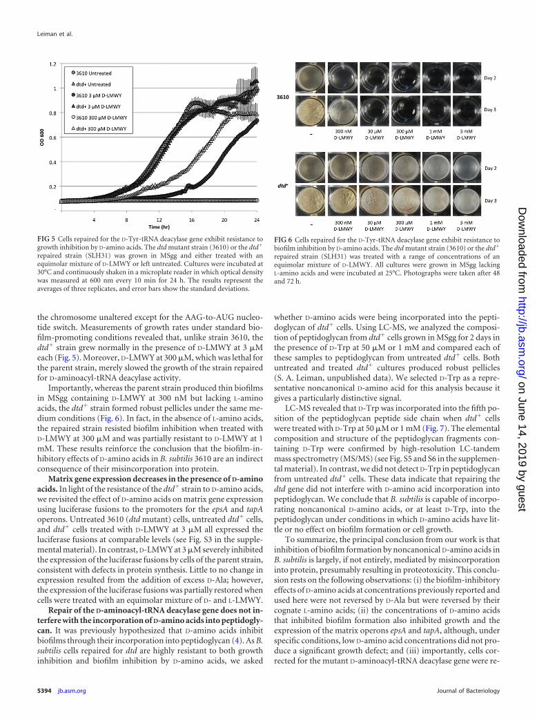
the chromosome unaltered except for the AAG-to-AUG nucleo-tide switch. Measurements of growth rates under standard bio-film-promoting conditions revealed that, unlike strain 3610, thedtd� strain grew normally in the presence of D-LMWY at 3 �Meach (Fig. 5). Moreover, D-LMWY at 300 �M, which was lethal forthe parent strain, merely slowed the growth of the strain repairedfor D-aminoacyl-tRNA deacylase activity.
Importantly, whereas the parent strain produced thin biofilmsin MSgg containing D-LMWY at 300 nM but lacking L-aminoacids, the dtd� strain formed robust pellicles under the same me-dium conditions (Fig. 6). In fact, in the absence of L-amino acids,the repaired strain resisted biofilm inhibition when treated withD-LMWY at 300 �M and was partially resistant to D-LMWY at 1mM. These results reinforce the conclusion that the biofilm-in-hibitory effects of D-amino acids in B. subtilis 3610 are an indirectconsequence of their misincorporation into protein.
Matrix gene expression decreases in the presence of D-aminoacids. In light of the resistance of the dtd� strain to D-amino acids,we revisited the effect of D-amino acids on matrix gene expressionusing luciferase fusions to the promoters for the epsA and tapAoperons. Untreated 3610 (dtd mutant) cells, untreated dtd� cells,and dtd� cells treated with D-LMWY at 3 �M all expressed theluciferase fusions at comparable levels (see Fig. S3 in the supple-mental material). In contrast, D-LMWY at 3 �M severely inhibitedthe expression of the luciferase fusions by cells of the parent strain,consistent with defects in protein synthesis. Little to no change inexpression resulted from the addition of excess D-Ala; however,the expression of the luciferase fusions was partially restored whencells were treated with an equimolar mixture of D- and L-LMWY.
Repair of the D-aminoacyl-tRNA deacylase gene does not in-terfere with the incorporation of D-amino acids into peptidogly-can. It was previously hypothesized that D-amino acids inhibitbiofilms through their incorporation into peptidoglycan (4). As B.subtilis cells repaired for dtd are highly resistant to both growthinhibition and biofilm inhibition by D-amino acids, we asked
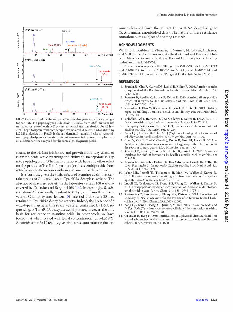
whether D-amino acids were being incorporated into the pepti-doglycan of dtd� cells. Using LC-MS, we analyzed the composi-tion of peptidoglycan from dtd� cells grown in MSgg for 2 days inthe presence of D-Trp at 50 �M or 1 mM and compared each ofthese samples to peptidoglycan from untreated dtd� cells. Bothuntreated and treated dtd� cultures produced robust pellicles(S. A. Leiman, unpublished data). We selected D-Trp as a repre-sentative noncanonical D-amino acid for this analysis because itgives a particularly distinctive signal.
LC-MS revealed that D-Trp was incorporated into the fifth po-sition of the peptidoglycan peptide side chain when dtd� cellswere treated with D-Trp at 50 �M or 1 mM (Fig. 7). The elementalcomposition and structure of the peptidoglycan fragments con-taining D-Trp were confirmed by high-resolution LC-tandemmass spectrometry (MS/MS) (see Fig. S5 and S6 in the supplemen-tal material). In contrast, we did not detect D-Trp in peptidoglycanfrom untreated dtd� cells. These data indicate that repairing thedtd gene did not interfere with D-amino acid incorporation intopeptidoglycan. We conclude that B. subtilis is capable of incorpo-rating noncanonical D-amino acids, or at least D-Trp, into thepeptidoglycan under conditions in which D-amino acids have lit-tle or no effect on biofilm formation or cell growth.
To summarize, the principal conclusion from our work is thatinhibition of biofilm formation by noncanonical D-amino acids inB. subtilis is largely, if not entirely, mediated by misincorporationinto protein, presumably resulting in proteotoxicity. This conclu-sion rests on the following observations: (i) the biofilm-inhibitoryeffects of D-amino acids at concentrations previously reported andused here were not reversed by D-Ala but were reversed by theircognate L-amino acids; (ii) the concentrations of D-amino acidsthat inhibited biofilm formation also inhibited growth and theexpression of the matrix operons epsA and tapA, although, underspecific conditions, low D-amino acid concentrations did not pro-duce a significant growth defect; and (iii) importantly, cells cor-rected for the mutant D-aminoacyl-tRNA deacylase gene were re-
FIG 5 Cells repaired for the D-Tyr-tRNA deacylase gene exhibit resistance togrowth inhibition by D-amino acids. The dtd mutant strain (3610) or the dtd�
repaired strain (SLH31) was grown in MSgg and either treated with anequimolar mixture of D-LMWY or left untreated. Cultures were incubated at30°C and continuously shaken in a microplate reader in which optical densitywas measured at 600 nm every 10 min for 24 h. The results represent theaverages of three replicates, and error bars show the standard deviations.
FIG 6 Cells repaired for the D-Tyr-tRNA deacylase gene exhibit resistance tobiofilm inhibition by D-amino acids. The dtd mutant strain (3610) or the dtd�
repaired strain (SLH31) was treated with a range of concentrations of anequimolar mixture of D-LMWY. All cultures were grown in MSgg lackingL-amino acids and were incubated at 25°C. Photographs were taken after 48and 72 h.
Leiman et al.
5394 jb.asm.org Journal of Bacteriology
on June 14, 2019 by guesthttp://jb.asm
.org/D
ownloaded from
sistant to the biofilm-inhibitory and growth-inhibitory effects ofD-amino acids while retaining the ability to incorporate D-Trpinto peptidoglycan. Whether D-amino acids have any other effecton the process of biofilm formation (or disassembly) aside frominterference with protein synthesis remains to be determined.
It is curious, given the toxic effects of D-amino acids, that cer-tain strains of B. subtilis lack D-Tyr-tRNA deacylase activity. Theabsence of deacylase activity in the laboratory strain 168 was dis-covered by Calendar and Berg in 1966 (14). Interestingly, B. sub-tilis strain 23 is naturally resistant to D-Tyr, and from this obser-vation, Champney and Jenson (5) inferred that strain 23 hadretained D-Tyr-tRNA deacylase activity. Indeed, the presence of awild-type dtd gene in this strain was later confirmed by DNA se-quencing. D-Tyr-tRNA deacylase activity is not, however, the onlybasis for resistance to D-amino acids. In other work, we havefound that when treated with lethal concentrations of D-LMWY,B. subtilis strain 3610 readily gives rise to resistant mutants that are
nonetheless still have the mutant D-Tyr-tRNA deacylase gene(S. A. Leiman, unpublished data). The nature of these resistancemutations is the subject of ongoing research.
ACKNOWLEDGMENTS
We thank L. Foulston, H. Vlamakis, T. Norman, M. Cabeen, A. Elsholz,and N. Bradshaw for discussions. We thank G. Byrd and The Small Mol-ecule Mass Spectrometry Facility at Harvard University for performinghigh-resolution LC-MS/MS.
This work was supported by NIH grants GM18568 to R.L., GM58213and GM82137 to R.K., GM103056 to M.D.L., and GM066174 andGM076710 to D.K., as well as by NSF grant DGE-1144152 to J.M.M.
REFERENCES1. Branda SS, Chu F, Kearns DB, Losick R, Kolter R. 2006. A major protein
component of the Bacillus subtilis biofilm matrix. Mol. Microbiol. 59:1229 –1238.
2. Romero D, Aguilar C, Losick R, Kolter R. 2010. Amyloid fibers providestructural integrity to Bacillus subtilis biofilms. Proc. Natl. Acad. Sci.U. S. A. 107:2230 –2234.
3. Vlamakis H, Chai Y, Beauregard P, Losick R, Kolter R. 2013. Stickingtogether: building a biofilm the Bacillus subtilis way. Nat. Rev. Microbiol.11:157–168.
4. Kolodkin-Gal I, Romero D, Cao S, Clardy J, Kolter R, Losick R. 2010.D-Amino acids trigger biofilm disassembly. Science 328:627– 629.
5. Champney WS, Jensen RA. 1969. D-Tyrosine as a metabolic inhibitor ofBacillus subtilis. J. Bacteriol. 98:205–214.
6. Patrick JE, Kearns DB. 2008. MinJ (YvjD) is a topological determinant ofcell division in Bacillus subtilis. Mol. Microbiol. 70:1166 –1179.
7. Chen Y, Cao S, Chai Y, Clardy J, Kolter R, Guo JH, Losick R. 2012. ABacillus subtilis sensor kinase involved in triggering biofilm formation onthe roots of tomato plants. Mol. Microbiol. 85:418 – 430.
8. Kearns DB, Chu F, Branda SS, Kolter R, Losick R. 2005. A masterregulator for biofilm formation by Bacillus subtilis. Mol. Microbiol. 55:739 –749.
9. Branda SS, Gonzalez-Pastor JE, Ben-Yehuda S, Losick R, Kolter R.2001. Fruiting body formation by Bacillus subtilis. Proc. Natl. Acad. Sci.U. S. A. 98:11621–11626.
10. Lebar MD, Lupoli TJ, Tsukamoto H, May JM, Walker S, Kahne D.2013. Forming cross-linked peptidoglycan from synthetic gram-negativelipid II. J. Am. Chem. Soc. 135:4632– 4635.
11. Lupoli TJ, Tsukamoto H, Doud EH, Wang TS, Walker S, Kahne D.2011. Transpeptidase-mediated incorporation of D-amino acids into bac-terial peptidoglycan. J. Am. Chem. Soc. 133:10748 –10751.
12. Soutourina O, Soutourina J, Blanquet S, Plateau P. 2004. Formation ofD-tyrosyl-tRNATyr accounts for the toxicity of D-tyrosine toward Esch-erichia coli. J. Biol. Chem. 279:42560 – 42565.
13. Yang H, Zheng G, Peng X, Qiang B, Yuan J. 2003. D-Amino acids andD-Tyr-tRNA(Tyr) deacylase: stereospecificity of the translation machinerevisited. FEBS Lett. 552:95–98.
14. Calendar R, Berg P. 1966. Purification and physical characterization oftyrosyl ribonucleic acid synthetases from Escherichia coli and Bacillussubtilis. Biochemistry 5:1681–1690.
FIG 7 Cells repaired for the D-Tyr-tRNA deacylase gene incorporate D-tryp-tophan into the peptidoglycan side chain. Pellicles from dtd� cultures leftuntreated or treated with D-Trp were harvested after incubation for 48 h at25°C. Peptidoglycan from each sample was isolated, digested, and analyzed byLC-MS as depicted in Fig. S4 in the supplemental material. Peaks correspond-ing to peptidoglycan fragments of interest were selected by mass. Samples fromall conditions were analyzed for the same eight fragment peaks.
D-Amino Acids Indirectly Inhibit Biofilm Formation
December 2013 Volume 195 Number 23 jb.asm.org 5395
on June 14, 2019 by guesthttp://jb.asm
.org/D
ownloaded from