Embed Size (px)
Citation preview

Wayne State University
Wayne State University Dissertations
1-1-2014
Cxcr2 Macromolecular Complex In PancreaticCancer: A Potential Therapeutic Target In TumorGrowthShuo WangWayne State University,
Follow this and additional works at: http://digitalcommons.wayne.edu/oa_dissertations
Part of the Cell Biology Commons
This Open Access Dissertation is brought to you for free and open access by DigitalCommons@WayneState. It has been accepted for inclusion inWayne State University Dissertations by an authorized administrator of DigitalCommons@WayneState.
Recommended CitationWang, Shuo, "Cxcr2 Macromolecular Complex In Pancreatic Cancer: A Potential Therapeutic Target In Tumor Growth" (2014).Wayne State University Dissertations. Paper 934.

CXCR2 MACROMOLECULAR COMPLEX IN PANCREATIC CANCER: A POTENTIAL THERAPEUTIC TARGET IN TUMOR GROWTH
by
SHUO WANG
DISSERTATION
Submitted to the Graduate School
of Wayne State University,
Detroit, Michigan
in partial fulfillment of the requirements
for the degree of
DOCTOR OF PHILOSOPHY
2014
MAJOR: BIOCHEMISTRY AND MOLECULAR BIOLOGY
Approved by:
Advisor
Date

© COPYRIGHT BY
SHUO WANG
2014
All Rights Reserved

ii
DEDICATION
to my parents
for their unconditional love and support.

iii
ACKNOWLEDGEMENTS
I would like sincerely and grateful thank my dissertation advisor, Dr. Chunying Li,
for his guidance, training, understanding and patience during my graduate studies at
Wayne State University. His mentorship was superb by providing a well-rounded
training plan allowing me to obtain adequate experimental experience and also
encouraging me to develop my own individuality and independence. I would also like to
thank all the group members I have been working with: Yanning Wu, Annette Stewart,
Yuning Hou, Marcello P. Castelvetere, and Xiaoqing Guan.
I would also like to thank Dr. Timothy Stemmler, Dr. Jianjun Wang, and Dr.
Xuequn Chen for their valuable suggestions, accessibilities and all kinds of help on my
graduate studies.
I would also like to thank everyone in the department with whom I have the most
meaningful years at Wayne State.

iv
TABLE OF CONTENTS
Dedication ………………………………………………………………………………….........ii
Acknowledgements …………………………………………………………………………….iii
List of Abbreviations ………………………………………………………………………….viii
List of Tables …………………………………………………………………………………..xii
List of Figures………………………………………………………………………………….xiii
Chapter I – Introduction ……………………………………………………………………..1
1.1 Overview of CXCR2 Chemokine Receptor……………………………………………...1
1.1.1 Chemokines and Chemokine Receptors ……………………………………………..1
1.1.2 CXCR2 Structure ………………………………………………………………………..2
1.1.3 CXCR2 Ligands and Signaling ………………………………………………………..3
1.1.4 Roles of CXCR2 Biological Axis …………………………………………………..…..5
1.2 CXCR2 in Diseases and Its Clinical Significance ……………………………………..5
1.2.1 CXCR2 in inflammatory diseases ……………………………………………………..5
1.2.2 CXCR2 in Cancer ……………………………………………………………………….8
1.3 CXCR2 Antagonists and Therapeutic Implications ……………………………………9
1.4 CXCR2 Macromolecular Complex – Identification and Therapeutic Potential ……14
Chapter II – CXCR2 Macromolecular Complex in Pancre atic Cancer ……………..20
2.1 Introduction ………………………………………………………………………………..20
2.1.1 Overview of Pancreatic Cancer ………………………………………………………20
2.1.2 Biological Axis of CXCR2 in Pancreatic Cancer ……………………………………20
2.1.3 PDZ proteins and PDZ domain-mediated protein-protein interaction ……………21
2.2 Materials and Methods …………………………………………………………………..23

v
2.2.1 Antibodies and Reagents ……………………………………………………………..23
2.2.2 Bacterial Strains, plasmid constructions, and mutagenesis ………………………25
2.2.3 Protein Expression and Purification …………………………………………………27
2.2.4 Cell Culture ……………………………………………………………………………..31
2.2.5 DNA Transfection ……………………………………………………………………...32
2.2.6 Western Blot Analysis …………………………………………………………………32
2.2.7 GST Pull-down Assay …………………………………………………………………33
2.2.8 Pair-wise Binding Assay ………………………………………………………………33
2.2.9 Macromolecular Complex Assembly Assay ………………………………………...34
2.2.10 Co-Immunoprecipitation Assay ……………………………………………………..35
2.2.11 Cell Proliferation Assay ………………………………………………………………35
2.2.12 Cell Invasion Assay …………………………………………………………………..36
2.2.13 Pancreatic Cancer-induced angiogenesis of endothelial cells …………………..37
2.2.14 Xenografts of Human Pancreatic Cancer cells in Immunodeficient Mice ………37
2.2.15 Immunohistochemistry and Quantification of Proliferation Index ……………….38
2.2.16 Statistical Analysis ……………………………………………………………………38
2.3 Results ……………………………………………………………………………………..39
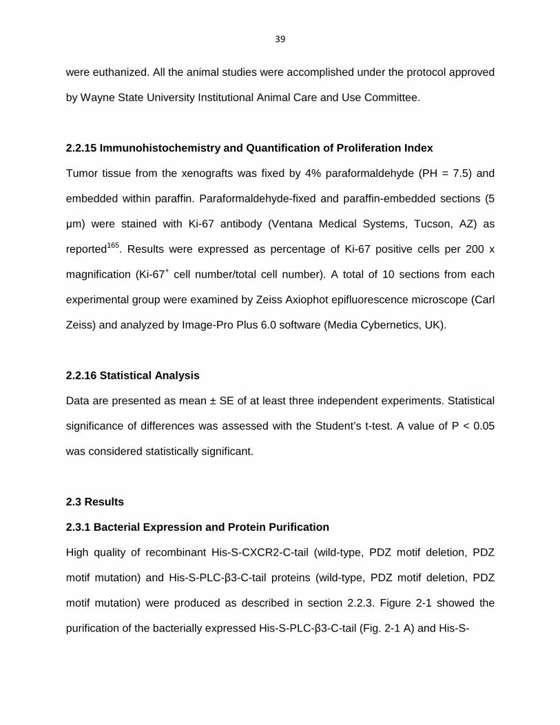
2.3.1 Bacterial Expression and Protein Purification ………………………………………39
2.3.2 Overexpression of CXCR2 in human pancreatic cancer cells …………………….42
2.3.3 Endogenous CXCR2 and PLC-β3 in human pancreatic cancer cells preferentially Interacts with NHERF1…………………………………………………………………42 2.3.4 CXCR2 and PLC-β3 Interact with NHERF1 in a Direct and PDZ Motif-Dependent Manner …………………………………………………………………………………..44
2.3.5 CXCR2 interacts with both PDZ domains of NHERF1 (PDZ1 and PDZ2) ………46

vi
2.3.6 Endogenous PLC-β3 in human pancreatic cancer cells preferentially interacts with NHERF1-PDZ2 ………………………………………………………………….49
2.3.7 NHERF1 Clusters CXCR2 and PLC-β3 into a Macromolecular Complex both In Vitro and in human pancreatic cancer cells ……………………………………….52
2.3.8 CXC Chemokine/CXCR2 Biological Axis Promotes Pancreatic Cancer Cell Proliferation ……………………………………………………………………..……54
2.3.9 Disrupting the CXCR2 Macromolecular Complex Inhibits Pancreatic Cancer Proliferation …………………………………………………………………………..56
2.3.10 Inhibitory effect of EF1060 on MIA PaCa-2 proliferation ………………………59
2.3.11 Disrupting the CXCR2 Macromolecular Complex Blocks Pancreatic Cancer Cell Invasion ………………………………………………………………………...62
2.3.12 Disrupting the CXCR2 Macromolecular Complex Blocks Pancreatic Cancer- induced angiogenesis ……………………………………………………………...62
2.3.13 Disrupting the CXCR2 Macromolecular Complex Inhibits Pancreatic Tumor Growth In Vivo ………………………………………………………………………66
2.4 Discussion ………………………………………………………………………………68
Chapter III – Crystallographic Analysis of NHERF1-P LCβ3 Interaction Provides Structural Basis for CXCR2 Signaling in Pa ncreatic Cancer …..……..71
3.1 Introduction ……………………………………………………………………………..71
3.2 Materials and Methods ………………………………………………………………..79
3.2.1 Protein Expression and Purification ………………………………………….…...79
3.2.2 Crystallization, Data Collection and Structure Determination ………………….80
3.2.3 Protein Data Bank Accession Number ……………………………………………82
3.3 Results …………………………………………………………………..……………..82
3.4 Discussion……………….……………………………………………………………..86
Chapter IV – Conclusion and Future Directions ……………………….……………88
4.1 Conclusion …………………….……………………………………………………….88

vii
4.2 Future Directions ……………………………………………………………………...89
4.2.1 Investigation of the effect of the CXCR2 macromolecular complex in CXCR2 Signaling ……………………………………………………………………………………89
4.2.2 Further Characterization of Interactions between NHERF1 and CXCR2 by Flourescence Recovery After Photobleaching (FRAP) ……………………………….91
Appendix: Correspondance ………………………………………………………………96
References …………………………………………………………………………………98
Abstract ……………………………………………………………………………………111
Autobiographical Statement ……………………………………………………………..113

viii
LIST OF ABBREVIATIONS
AAA PDZ motif mutants (last three amino acids of PDZ motifs of
CXCR2/PLC-β3 were replaced by amino acids AAA using
mutagenesis)
AAV2 Adeno-Associated Virus, Serotype 2
ALI Acute Lung Injury
ARDS Acute Respiratory Distress Syndrome
BALF Bronchoalveolar Lavage Fluid
BK Bradykinin
BSA Bovine Serum Albumin
cAMP Cyclic Adenosine Monophosphate
CCK Cholecystokinin
CCR2 Chemokine (C-C Motif) Receptor 2
CF Cystic Fibrosis
CFTR Cystic Fibrosis Transmembrane Conductance Regulator
co-IP Co-Immunoprecipitation
COPD Chronic Obstructive Pulmonary Disease
C-tail Last 45 amino acids (or 100 amino acids) of the C-terminus of
CXCR2 (or PLC-β3)
CXCL1 Chemokine (C-X-C Motif) Ligand 1
CXCL5 Chemokine (C-X-C Motif) Ligand 5
CXCL8 Chemokine (C-X-C Motif) Ligand 8
CXCR2 Chemokine (C-X-C Motif) Receptor 2

ix
DAG 1, 2-Diacylglycerol
DMSO Dimethyl Sulfoxide
E. coli Escherichia Coli
ENA-78 Epithelial-Derived Neutrophil-Activating Peptide 78
EPC Endothelial Progenitor Cell
ERM Ezrin-Radixin-Moesin
EYFP Enhanced Yellow Fluorescent Protein
FBS Fetal Bovine Serum
FL Full-length
FLAG Flag-tag (or Flag octapeptide) with the protein sequence of
DYKDDDDK
FRAP Flourescence Recovery After Photobleaching
GAIP Guanosine Triphosphatase-Activating Protein
GAPDH Glyceraldehyde 3-Phosphate Dehydrogenase
GFP Green Fluorescent Protein
GIPC GAIP-Interacting Protein, C Terminus
GPCR G-Protein Coupled Receptor
GROα Growth-Related Oncogene α
GST Glutathione S-Transferase
GTPase Hydrolase Enzymes That Can Bind And Hydrolyze Guanosine
Triphosphate (GTP)
HA Hemagglutinin
HEK293 Human Embryonic Kidney 293 Cells

x
HUVEC Human Umbilical Vein Endothelial Cells
I/R-I Ischemia Reperfusion Injury
IBD Inflammatory Bowel Diseases
IL-8 Interkeukin-8 (as known as CXCL8)
IL-8RB (= CXCR2) Interkeukin-8 Receptor Type B
IP3 Inositol-1,4,5-Trisphosphate
IPTG Isopropyl β-D-1-Thiogalactopyranoside
IQGAP1 Ras GTPase-activating-like protein
kDa Kilo Dalton
LASP-1 LIM and SH3 protein-1
LB Broth medium Luria-Bertani Broth medium
LIC Ligation Independent Cloning
LPAR2 Lysophosphatidic Acid Receptor 2
MAPK Mitogen Activated Protein Kinase
MPO Myeloperoxidase
MTT 3-(4,5-Dimethylthiazol-2-yl)-2,5-Diphenyltetrazolium Bromide
NHERF1 Sodium-Hydrogen Antiporter 3 Regulator 1
OD600 Optical density of a sample measured at wavelength of 600nm
PAGE Polyacrylamide Gel Electrophoresis
PDAC Pancreatic Ductal Adenocarcinoma
PDGFR Platelet-Derived Growth Factor Receptor
PDZ Post Synaptic Density Protein (PSD95), Drosophila Disc Large
Tumor Suppressor (Dlg1), and Zonula Occludens-1 Protein (Zo-1);
xi
PSD-95/Dlga/ZO-1
PGP tri-peptide Proline-Glycine-Proline
PI3K Phosphoinositide 3-Kinase
PIP2 Phosphatidylinositol 4, 5- Bisphosphate
PLC Phospholipase C
PLC-β Phospholipase C, β Isozyme
PMN Polymorphonuclear Neutrophil
PMSF Phenylmethylsulfonyl Fluoride
PVDF Polyvinylidene Fluoride
Rab Ras-related protein that in humans is encoded by the RAB8A gene
Rab11-FIP2 Rab11-family interacting protein 2
S.O.C. medium Super Optimal Broth medium with Catabolite repression
SCID Severe Combined Immunodeficiency
SDS Sodium Dodecyl Sulfate
TIRFM Total Internal Reflection Fluorescence Microscope
VASP Vasodilator-Stimulated Phosphoprotein
β2AR β2-Adrenergic Receptor
∆TTL/∆TQL PDZ Motif Deletion Of CXCR2/PLC-β3

xii
LIST OF TABLES
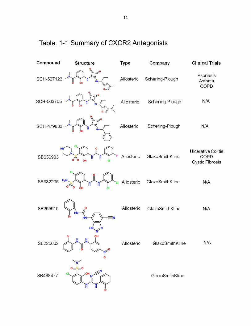
Table 1-1 – CXCR2 Antagonists ……………………………………………………………11
Table 2-1 – Primes used in the current study ……………………………………………..28
Table 3-1 – Table 2-1 – Primes used in the current study ……………………………….81

xiii
LIST OF FIGURES
Figure 1-1 CXCR2 structure and receptor-ligand binding …………………………..4
Figure 2-1 Purification of the bacterially expressed GST-His-S-CXCR2-C- tail and His-S-PLC-β3-C-tail ………………………………………………40
Figure 2-2 Overexpression of CXCR2 in human pancreatic cancer cells ………..43
Figure 2-3 CXCR2 and PLC-β3 in PDAC cells preferentially interact with NHERF1……………………………………………………………….45
Figure 2-4 CXCR2 and PLC-β3 interact with NHERF1 in a direct and PDZ motif-dependent manner …...……………………………………………..47
Figure 2-5 CXCR2 interacts with both PDZ1 and PDZ2 of NHERF1 …………….48
Figure 2-6 Endogenous PLC-β3 in human pancreatic cancer cells preferentially interacts with NHERF1-PDZ2……………………………50
Figure 2-7 NHERF1 clusters CXCR2 and PLC-β3 into a macromolecular complex in vitro and in PDAC cells …………………….…………………………53
Figure 2-8 CXC Chemokine/CXCR2 Biological Axis Promotes Pancreatic Cancer Cell Proliferation …………………………………………………55
Figure 2-9 Disrupting the CXCR2 Macromolecular Complex Inhibits Pancreatic Cancer Proliferation ……………………………..………….57
Figure 2-10 Inhibitory effect of EF1060 on MIA PaCa-2 proliferation……..………60
Figure 2-11 Disrupting the CXCR2 Macromolecular Complex Blocks Pancreatic Cancer Cell Invasion ……………………………………..…63
Figure 2-12 Disrupting the CXCR2 Macromolecular Complex Blocks Pancreatic Cancer-induced angiogenesis ………………………..……64
Figure 2-13 Transduction efficiency of AAV2/2CMV constructs ……………..…….67
Figure 2-14 Disrupting the CXCR2 Macromolecular Complex Inhibits Pancreatic Tumor Growth In Vivo ………………………………...……..69
Figure 2-15 Proposed mechanism of the NHERF1-mediated coupling of CXCR2

xiv
to PLC- β3 signaling in PDAC cells ……………………………………74
Figure 3-1 Structure of NHERF1 PDZ1 in complex with the PLCβ3 C-terminal sequence ENTQL ……………………………………...…….83
Figure 3-2 Structural comparison of NHERF1 PDZ1 and PDZ2….. ……………..85

1
Chapter 1
Introduction
1.1 Overview of CXCR2 Chemokine Receptor
1.1.1 Chemokines and Chemokine Receptors
Chemokines are a family of small cytokines that are responsible for directed cellular
chemotaxis. Chemokines can be divided into two categories: inflammatory chemokines
that recruit leukocytes in response to physiological stimulus and homeostatic
chemokines that are constitutively expressed, and responsible for the continuous basal
level of cell migration and the architecture of secondary lymphoid organs1. Inflammatory
chemokines and their receptors have pivotal roles in both innate and adaptive immunity
in response to infection, tissue damage, and other physiological abnormalities.
Homeostatic chemokines involve in the migration of B and T cells through specialized
areas of secondary lymphoid organs, and migration of lymphocytes involved in immune
surveillance2. In spite of the critical roles of chemokines and their receptors in the
immune system, they have been documented to be actively involved in enormous
pathologies1,3,4, including inflammatory diseases (inflammatory bowel disease,
atherosclerosis), pulmonary diseases (chronic obstructive pulmonary disease (COPD),
asthma), autoimmune diseases (psoriasis, rheumatoid arthritis, and multiple sclerosis),
and cancers.
To date, approximately 50 chemokines and 20 corresponding chemokine receptors
have been identified. Based on the conserved pattern of cysteine residues at N-

2
terminus, chemokines have been divided into four different subfamilies: C, CC, CXC,
and CX3C, where C represents cysteine residue, and X represents any non-cysteine
residue. In human, there are 7 CXC subfamily chemokine receptors (CXCR1 ~ CXCR7),
and 15 cognate CXC chemokines (or CXCR ligands). CXCR2, a prototypical member of
the CXC chemokine receptors, was first discovered as a neutrophil receptor5. Years of
intensive research has also delineated its roles in a variety of diseases including cancer
and inflammatory diseases. Furthermore, several pharmaceutical companies have
identified a number of potent CXCR2 antagonists that have been evaluated in the
clinical trials. However, many clinical trials of CXCR2 antagonists/inhibitors have not
been successful due to the suboptimal clinical endpoints. Recent studies targeting
compartmentalized interactions of CXCR2 complexes inside the cells open the door for
development of a new class of CXCR2 inhibitors, which target the specific interactions
between CXCR2 and its binding partners. This chapter focuses on the most recent
progress about the interactions between CXCR2 and its interacting partners, and the
fine-tuned regulation of CXCR2 compartmentalized signalings in inflammation, cancer,
and angiogenesis.
1.1.2 CXCR2 Structure
CXCR2 was first designated as IL-8 receptor type B (IL-8RB) by Murphy and his
colleagues5, sharing 77% identity with another closely-related receptor IL-8RA (also
known as CXCR1) at the amino acid sequence level. CXCR2 is a G protein-coupled
receptor which is composed of seven transmembrane domains, an extracellular N-
terminal domain, three extracellular loops, three intracellular loops, and a cytoplasmic

3
C-terminus (Fig. 1 - 1). The extracellular N-terminal domain is involved in ligand binding,
and the DRY (Asp-Arg-Tyr) motif in the second intracellular loop is the G protein
docking site, and is responsible for intracellular downstream signaling upon ligand
binding6. Also, the Asp 199 in the second extracellular loop7 and an LLKIL motif in the
C-terminus8 are both required for receptor internalization.
1.1.3 CXCR2 Ligands and Signaling
CXCR2 is the cognate receptor for the CXC chemokines CXCL1 ~ 3 and CXCL5 ~ 8
(also referred to as CXCR2 ligands)9. The characteristic ELR (Glu-Leu-Arg) motif
located in the N terminus of the CXCR2 ligands are responsible for the ligand-receptor
binding10, and is strongly associated with the leukocyte attraction11. Although further
structural studies regarding the ligand-receptor interaction of CXCR2 are still in need,
the binding mechanism has been proposed based on modeling and structural studies of
CXCR4 and CCR2 binding modes12,13. Conceptually, the binding involves the binding
between N-terminal residues from both the receptor and its ligands (preceding the 1st
cysteine), followed by the interaction of the ELR motif of the ligands with the 2nd and 3rd
extracellular loops of the receptor. N-terminus of the receptor is responsible for the
binding selectivity and affinity14, whereas the ELR motif from the ligands enables the
stabilization of the binding interaction and leads to the activation of the downstream
signaling cascade15. Upon ligand-receptor binding, the heterotrimeric G protein is
activated, whereby the α-subunit and βγ-complex dissociate, leading to the activation or
inhibition of a wide variety of downstream targets (phospholipase C, adenylyl cyclase,
cAMP-dependent protein kinase, GTPases, PI3K, etc.). CXCR2 has been implicated in

4
5
activation of several signaling transduction pathways, including phospholipase C (PLC)
pathway, PI3K pathway, and Rho, Rac and mitogen activated protein kinase (MAPK)
pathways16.
1.1.4 Roles of CXCR2 Biological Axis
Signaling pathways evoked by the activation of CXCR2 have been borne out to play
important roles in a variety of cellular responses, including cell migration, chemotaxis,
cell adhesion, cellular morphological change, cytoskeletal rearrangement, etc17. The
expression of CXCR2 on leukocytes, especially neutrophils, has been well-established
for its roles in leukocyte homeostasis as well as the recruitment of leukocytes from bone
marrow in respond to inflammation and injury18-20. CXCR2 had also been reported in
preservation of oligodendrocyte function and myelinization of neural tissues21.
Furthermore, CXCR2 and its ligands have also been demonstrated to play essential
roles in cutaneous wound healing22.
1.2 CXCR2 in Diseases and Its Clinical Significance
1.2.1 CXCR2 in inflammatory diseases
Although the recruitment of neutrophils, which is regulated primarily by CXCR2, is
important in response to acute inflammation and injury19,20, persistent or over-exuberant
expression of CXCR2 and its ligands could subvert the protective effects of neutrophils
into a manner that cause diseases, since excessive neutrophil influx and accumulation
at the inflammation sites is well-believed to play crucial roles in pathogenic progression
of clinical and experimental inflammatory diseases. Therefore, CXCR2 has been widely

6
demonstrated to play important role in various inflammatory diseases, including chronic
obstructive pulmonary disease (COPD), acute lung injury (ALI), cystic fibrosis (CF),
inflammatory bowel diseases (IBD), and ischemia-reperfusion injury. Increased
expression of CXCR2 and its cognate CXC chemokines have been reported in recent
clinical studies in various inflammatory disease systems, suggesting the clinical
significance of CXC chemokine/CXCR2 biological axis.
It has been documented that CXCR223,24 and its cognate ligands (CXCL125, CXCL523,
CXCL723,24, CXCL826, and PGP27) are significantly up-regulated in the patients with
severe COPD compared with healthy smokers and patients with mild/moderate COPD.
It has been reported that the patients with acute respiratory distress syndrome (ARDS)
has a dramatically higher concentration CXCL128, CXCL528 and CXCL828-30 in
bronchoalveolar lavage fluid (BALF), and the level of the CXCL8: anti-CXCL8 antibody
complexes profoundly correlated with the clinical disease activity31 and mortality29. CF
patients also showed significantly elevated levels of CXCL8 in sputum, BALF, and sera,
and the high level of CXCL8 significantly correlated with clinical status32. Expression
levels of CXCR233,34 and its cognate ligands, CXCL133,34, CXCL233, CXCL333,34,
CXCL535, and CXCL833,34,36-38, in the patients with IBD were substantially higher than
the control individuals. Clinically, neutrophil-mediated reperfusion injury significantly
increased mortality and morbidity when patients underwent ischemia -reperfusion
injury (I/R-I) such as organ transplantation39,40 and cardiopulmonary bypass41. CXCR2
cognate ligands, CXCL342, CXCL742 and CXCL842-44 were enormously increased in the
BALF from patients underwent lung transplantation compared to the control subjects,

7
and that high level of CXCL8 in the donor BALF profoundly correlated with the
development of severe early graft dysfunction and with early recipient mortality44.
Significantly increased CXCL8 was also detected in patients underwent
cardiopulmonary bypass45.
Laboratory investigations also showed that CXCR2 deficiency or CXCR2 blockage
prevented the Polymorphonuclear neutrophil (PMN) recruitment/accumulation in various
experimental disease models. In COPD model, Weathington et al. reported that a
CXCR2 specific peptide (PGP) failed to induce neutrophil accumulation in CXCR2-/-
mice27. Miller et al. also reported that CXCR2 deficiency substantially decreased mucus
secretion in the BALF, which is associated with chronic bronchitis. In ALI , animal
studies have reported that PMN recruitment in the lung was significantly decreased in
CXCR2-/- mice46-49. Kordonowy et al. also reported that ALI was attenuated in the obese
mice, in which neutrophil CXCR2 expression was dramatically decreased50.
Furthermore, blockade of CXCR2 by neutralizing antibody also markedly reduced
neutrophil accumulation in ALI models51,52. In IBD model, Buanne et al. reported that
PMN infiltration into mucosa was prominently reduced in CXCR2-/- mice with limited
degree of mucosal damage and reduced clinical symptoms53. Elevated CXCR2
expression has been reported during post-lung transplantation I/R-I in rats42, and
CXCR2 deficiency has demonstrated to reduce the impairment caused by I/R injury in
different models54,55. Also, blockade of CXCR2 markedly prevented graft malfunction
and inhibited neutrophil recruitment and accumulation in transplantation models42,56.

8
1.2.2 CXCR2 in Cancers
Overexpression of CXCR2 has been detected in patients with various cancers including
breast cancer57, laryngeal squamous cell carcinoma58, colon cancer59, prostate
cancer60,61, pancreatic cancer62,63, lung adenocarcinoma64, ovarian cancer (associated
with patient survival)65, nasopharyngeal carcinoma66 and brain tumor67. Besides,
CXCR2 cognate ligands (CXCL1, CXCL5, and/or CXCL8) also overexpressed in
patients with prostate cancer61, colon cancer59,68-70, breast cancer71,72, and pancreatic
cancer73-75. Furthermore, overexpression of CXCR2 and/or its ligands prominently
correlated with tumor stage, disease severity and patient survival in most cases.
Recent studies using CXCR2-/- mice have reported that CXCR2 deficiency profoundly
prevented primary tumor growth and spontaneous metastases in lung cancer76,
inhibited human melanoma tumor growth and experimental lung metastasis77, and
suppressed human prostate tumor growth in vivo78. Blockade of CXCR2, by either
neutralizing antibodies or short hairpin RNA (shRNAs), also significantly suppressed the
proliferation, invasion and tumor growth in vitro and in vivo in many cancer models67,79-82.
Recent advances also suggest that CXCR2 not only contributed to the cancer cell
biology, but also played important roles in the interplay between tumor cell and tumor
microenvironment. For example, Jamieson et al. reported that inflammation-driven and
spontaneous tumorigenesis in skin and intestine has been distinctively subsided in
CXCR2-/- mice, in which tumor-associated leukocyte recruitment has also been
attenuated83.

9
1.3 CXCR2 Antagonists and Therapeutic Implications
Given the essential role of CXCR2 biological axis in inflammatory diseases and cancers,
pharmacological studies targeting CXCR2 has dramatically increased over the last
decade. Several pharmaceutical companies have developed potent and selective
CXCR2 antagonists, and laboratory investigations have achieved significant progress
on CXCR2 antagonists in various disease models. Chapman et al. reported that SCH-
527123, one of the CXCR2 allosteric inhibitors, significantly inhibited the neutrophil
recruitment, mucus production, and goblet cell hyperplasia in a murine COPD model84.
SCH-563705, another CXCR2 antagonist with similar structure with SCH-527123, has
been demonstrated to reduce the arthritis disease severity in vivo85, and block the
mammosphere formation ex vivo71. Singh et al. demonstrated the inhibitory effect of
SCH-479833, with similar structure as SCH-527123, on tumor growth and angiogenesis
in melanoma86 and colon cancer87 animal models. SB225002, a well-established
competitive CXCR2 inhibitor, has also been reported to exert the inhibitory effect in
inflammatory disease and cancer models. Braber et al. reported that SB225002
significantly inhibited CXCL1-induced increase of myeloperoxidase (MPO) levels in
pulmonary tissue88. SB-225002 has also been demonstrated to significantly reduce
MPO activity, BALF neutrophil accumulation, and edma formation in the injured lung89.
Herbold et al. demonstrated the inhibitory effect of SB225002 in alveolar neutrophil and
exudate macrophage recruitment in mice in a lung injury model49. In breast cancer
models, SB-225002 has also been reported to inhibit mammary epithelial cell
migration90 and breast cancer cell invasion91. Two groups also reported SB656933,
10
another allosteric CXCR2 inhibitor, and its predecessor SB332235, could block the
neutrophil activation, recruitment, and accumulation in COPD models88,92,93. SB265610,
another allosteric CXCR2 inhibitor, has also been reported to suppress mammary tumor
cell migration, myeloid cell recruitment and lung metastasis in breast cancer
models80,94,95. Another CXCR2 inhibitor, SB-468477 developed by Glaxo-Smith-Kline,
has been shown to inhibit monocyte migration in response to selective CXCR2 ligands,
CXCL1, CXCL5 and CXCL796. Repertaxin , a CXCR2 inhibitor also known as Reparixin,
was reported to block the neutrophil accumulation in a lung injury model, and abate the
pancreatic cancer tumor growth in vivo97. DF2162, another allosteric CXCR2 inhibitor
developed by Dompé S.P.A., has been reported to inhibit the arthritis in vivo with
diminished neutrophil infiltration, oedema formation, and hypernociception98-100. Besides
the effect on arthritis, DF2162 was also demonstrated to reduce the airway neutrophil
transmigration, and improve lung pathology in lung fibrosis model101. G31P, as known
as CXCL8(3–72)K11R, is an orthosteric inhibitor of CXCR2, which has been reported to
fully abolish Benzo(a)pyrene triggered neutrophil recruitment in BALFs102. Liu et al.
reported that G31P significantly suppressed prostate cancer cell proliferation both in
vitro and in vivo103. AZ10397767, developed by AstraZeneca as a thiazolopyrimidine-
based CXCR2 antagonist, has been shown to suppress lung cancer growth with
decreased neutrophil infiltration104, and sensitize prostate cancer cells to
chemotherapy105. Furthermore, some of the CXCR2 antagonists have been progressed
into clinical trials106,107. Some of the well-tested CXCR2 antagonists have been
summarized in Table 1-1.
11

12

13
SCH-527123, developed by Schering-Plough, has been evaluated in several neutrophil
dominant diseases including psoriasis (NCT00684593, phase II), asthma
(NCT00688467, Phase II; NCT00632502, Phase II) and COPD (NCT01068145, Phase I;
NCT00441701, Phase II). Holz et al. has reported that SCH-527123 caused significant
attenuation of ozone-induced neutrophilia in healthy subjects108. Reparixin , developed
by Dompé S.P.A., has also been tested in phase II clinical trials of I/R-I after lung and
kidney transplantation, and in phase III clinical in pancreatic islet transplantation. SB-
656933, the derivative of SB-332235109 developed by GlaxoSmithKline, has also been
assessed in several phase I/II COPD clinical trials. Moss et al. reported that SB656933
was well-tolerated in adult CF patients, and significantly suppressed sputum neutrophil
and elatase110. AZD-5069, developed by AstraZeneca, has also been evaluated in
phase II clinical trials in COPD and bronchiectasis patients111.
In spite of the availability of several databases of clinical trials, scant results of
completed clinical trials have been publicly released. Termination or discontinuation of
the trials might result from the difficulty in patient recruitment, financial issues, or failure
to reach clinical end points107. Furthermore, small molecule drugs/antagonists targeting
at chemokine receptors still face several challenges that have been well-discussed in
the excellent review paper by Proudfoot et al, including the interspecies difference in
antagonist potency, redundancy in receptor-ligand pairing and subsequent biological
functions, suboptimal pharmacokinetic and toxicity profiles, and the inadequate ultimate
curative effects107.

14
1.4 CXCR2 Macromolecular Complex – Identification a nd Therapeutic Potential
Besides the challenges mentioned above, emphasis is also placed on identifying novel
CXCR2 inhibitors, which can be more tissue-specific and/or disease-specific112,
because CXCR2 functions in normal physiology (including early tumor surveillance and
immune system physiology) have to be protected in any antagonist that will be
advanced to clinical trials. Recent studies aiming at protein-protein interactions involving
CXCR2 have explored some novel avenues for innovative drug discovery. It has been
documented that chemokine receptors couple not only to G proteins but also to
additional non-G proteins, especially the scaffolding proteins, which could provide
binding sites for downstream effector proteins113, and direct CXCR2 to operate in
specific signaling networks. With no doubt it would be beneficial if we could identify
novel CXCR2-interacting proteins or tissue/disease specific interacting partners of
CXCR2, which could enhance the CXCR2 signaling in certain disease conditions. By
disrupting these protein-protein interactions of CXCR2, we may develop more effective
treatment options.
Recent advances have already revealed the functional importance of the CXCR2-
interacting partners in CXCR2 trafficking, recycling, signal transduction, and it has also
been suggested that different repertoires of adaptor/scaffolding proteins binding to
CXCR2 and other chemokine receptors at varying spatiotemporal points are, at least
partially, responsible for the versatile biological/cellular responses in different disease
conditions114. Richmond group has identified several CXCR2-interacting proteins, which
associates with CXCR2 and modulate CXCR2 trafficking, signaling, and CXCR2-

15
mediated cellular functions. Hsc/Hsp70 interacting protein (Hip) interacts with a C-
terminal domain of CXCR2 (KILAIHGLI; i.e. a.a 327 ~ a.a 335), and affects CXCR2-
mediated chemotaxis115. Several Ras-related proteins (Rab), including Rab5, Rab11a,
and Rab11-family interacting protein 2 (Rab11-FIP2), and Rho GTP-binding protein
(RhoB) were also shown to bind CXCR2, and play important roles in CXCR2 trafficking,
recycling and CXCR2-mediated chemotaxis116-118. β-arrestin 2, a member of arrestin
family of adaptor proteins, has been demonstrated to bind phosphorylated carboxyl-
regions of CXCR1 and CXCR2, and modulate receptor internalization and activation of
signal transduction pathways119,120. Besides β-arrestin 2, other scaffolding proteins,
such as vasodilator-stimulated phosphoprotein (VASP)121, LIM and SH3 protein-1
(LASP-1)122, and Ras GTPase-activating-like protein (IQGAP1)114, were also identified
by proteomics approaches to associate with CXCR2, and regulate CXCR2-mediated
signaling and chemotaxis. Furthermore, it has been unveiled that the C-terminal
cytoplasmic domain of CXCR2, which contains several specific motifs (amino acid
sequences) such as LLKIL motif8, PDZ motif (-STTL)123 and phosphorylation sites, is
responsible for CXCR2 signaling, post-endocytic sorting, and/or CXCR2-mediated
cellular chemotaxis. These studies not only led to identification of novel CXCR2-
interacting partners and the critical motifs in CXCR2 responsible for the binding, but
also revealed the functional significance of the coordinated regulation of CXCR2
signaling by these CXCR2-interacting partners, suggesting the therapeutic potential for
targeting CXCR2-specific protein interactions.

16
Recently, we have also identified a PDZ domain-containing protein, Na+/H+ exchange
regulatory cofactor (NHERF1), as a novel CXCR2-interacting protein. NHERF1
associates with CXCR2 by PDZ interaction, and clusters the downstream effector, PLC-
β2, into a CXCR2 macromolecular complex (CXCR2•NHERF1•PLC-β2) in neutrophils124.
The formation of this CXCR2 complex is PDZ motif-dependent (i.e. –STTL-COOH for
CXCR2; -ESRL-COOH for PLC-β2). An exogenous CXCR2 C-terminal domain containing
PDZ motif (last 45 amino acids or last 13 amino acids) was demonstrated to compete
and disrupt the physical interaction between CXCR2 and NHERF1, and also were
shown to impair the CXCR2-mediated intracellular calcium mobilization, chemotaxis,
and transepithelial migration in neutrophils. We also identified another CXCR2
macromolecular complex (CXCR2•NHERF1•PLC-β3) in human pancreatic ductal
adenocarcinoma cells (PDACs), gene delivery or peptide delivery of exogenous CXCR2
C-terminal domain (last 45 or 13 amino acids) impaired cancer cell invasion and cell
proliferation in vitro and in vivo 125.
Most recently, we also demonstrated the importance of CXCR2 PDZ-mediated protein
interactions in the function of endothelial progenitor cells (EPCs)126. By using an
exogenous CXCR2 C-tail peptide or DNA construct (containing CXCR2 PDZ motif),
CXCR2-mediated intracellular calcium mobilization in EPC, EPC in vitro migratory
responses and neovessel formation, and EPC in vivo angiogenesis were significantly
impaired. Our findings not only identified the existence and biological functions of the
CXCR2 complex, but also revealed the therapeutic potential of targeting PDZ-mediated
CXCR2 macromolecular complex in inflammation, cancer, and angiogenesis.

17
Although the scaffolding proteins have been demonstrated to regulate the CXCR2
signalings in diseases; however, it has been proved that eradication of the scaffolding
proteins would not simply achieve the desired inhibitory effect in disease conditions. For
example, although the β-arrestin 2 plays crucial roles in mediating CXCR2-
signaling119,120, eradication of β-arrestin 2, unexpectedly, caused enhanced CXCR2
signaling and functional endpoints in vitro and in vivo127. This warrants the necessity of
unveiling more structural details of the interaction between CXCR2 and its interacting
partners to develop new therapeutic strategies. Therefore, we further investigated the
structural basis of the NHERF1 PDZ1 domain in complex with the C-terminal sequence
of CXCR2 at 1.1 Å resolution128. We not only determined that the positions 0 and -2 of
the PDZ motif (position 0 referring to the extreme C-terminal residue) possess the ability
to form networks of hydrogen bonds and hydrophobic interactions with NHERF1-PDZ1
domain, which are responsible for the stabilization and specificity of PDZ1-CXCR2
interaction, but also observed that the interaction between NHERF1-PDZ1 and CXCR2
at position -1 and -3 of the PDZ motif are very different from other PDZ1-PDZ motif
interactions (i.e. PDZ1-CFTR, PDZ1- β2AR, and PDZ1-PDGFR). Specifically, the
orientation, the rotameric states, and binding preference of His29 and Arg40 in the
NHERF1 PDZ1 domain is very different in PDZ1-CXCR2 interaction than in PDZ1-
CFTR, PDZ1-β2AR, and PDZ1-PDGFR interactions. The unveiled structural basis
suggests that even though both -1 and -3 residues in PDZ motif were less stringently
specified than 0 and -2 residues in the PDZ domains, they could still interact with a few
key residues in the PDZ motif-binding pocket specifically, which endow -1 and -3

18
residues with critical roles in PDZ motif recognition and selection within a network of
NHERF-scaffolded interactions. Also, the conformation and rotameric states of the
residues in the PDZ motif-binding pocket are also important in different side chain
recognition of the PDZ motif and specific interaction. Our most recent study has also
demonstrated the presence of the substantial structural flexibility in the PDZ1 peptide-
binding pocket, which provides potential strategies for drug design against the NHERF1
PDZ1 domain129.
Our findings of the functional significance and therapeutic potential of the interaction
between CXCR2 and NHERF1 in inflammation, tumorigenesis, and angiogenesis may
be valuable in the development of innovative strategies for targeted drug discovery.
However, the complexity of the NHERF1 interaction network and its versatile roles in
regulation of many cellular processes essential to normal physiology130,131 poses a
challenge for designing CXCR2 inhibitors that are specific to the CXCR2-NHERF1
interaction without cross-inhibiting any of the other NHERF1-coordinated signaling
events. Therefore, the specificity and selectivity of the novel CXCR2 inhibitors targeting
on the CXCR2-NHERF1 interaction is dependent upon the understanding of the
structural features that how the PDZ motif and critical residues in PDZ binding pocket
work in coordination to determine the PDZ motif recognition and specificity of the
interaction, and upon the exploitation of these features to differentiate CXCR2 from
other NHERF1-interacting proteins. Further structural studies regarding identification of
critical residues, from both PDZ motif and PDZ binding pocket, which are responsible
for CXCR2 recognition, would be beneficial to design novel inhibitors that disrupt

19
CXCR2-NHERF1 interaction specifically without affecting other NHERF1 interaction and
NHERF1-scaffolded signaling complex.

20
Chapter II
CXCR2 Macromolecular Complex in Pancreatic Cancer
© 2013 Neoplasia Press (Translational Oncology)
2.1 Introduction
2.1.1 Overview of Pancreatic Cancer
Pancreatic cancer, the most lethal malignancy of the gastrointestinal tract with 5-year
survival rates of less than 5%, is the fourth leading cause of cancer-related deaths in
both men and women in the United States132. The most common type of pancreatic
cancer is pancreatic ductal adenocarcinoma (PDAC). Chemoresistance, early
metastases and late clinical presentation in this incurable human malignancy result in
no effective methods for early prognosis as well as a lack of effective systemic therapies
with reduced side effects133,134. Therefore, a more comprehensive understanding of the
biology of PDAC and the mechanisms/factors that promote invasion and tumor growth
may help identify new molecular targets for the development of diagnostics and/or
therapeutics of pancreatic cancer.
2.1.2 Biological Axis of CXCR2 in Pancreatic Cancer
CXC-chemokine receptor 2 (CXCR2) is the cognate receptor for the CXC-chemokines,
CXCL1 ~ 3 and CXCL5 ~ 89. The CXC-chemokine/CXCR2 signaling has been reported
to promote malignant cancer progression in many cancer types including pancreatic
cancer135-139. It has been documented that the elevated expression of CXCL5 and

21
CXCL8 is correlated with poor differentiation, histopathologic grade and advanced
clinical grade pancreatic adenocarcinomas in patients74,140. Recent studies also suggest
that CXCR2 is expressed in various PDAC cell lines141-144 and is primarily involved in
enhancing the proliferation and survival of cancer cells via the autocrine and/or
paracrine effect74,141,144. More importantly, increased expression of CXCR2 and its
ligands has been shown in higher grades and stages of pancreatic adenocarcinomas in
patients 63,140, indicating that CXCR2 is involved in the exacerbation of tumors and could
be a promising target for developing selective and effective treatments for pancreatic
cancer. As a G-protein coupled receptor (GPCR), CXCR2 couples to the pertussis
toxin-sensitive Gi proteins to stimulate phosphatidylinositide-specific phospholipase C
(PLC) activities145. Agonist-induced activation of PLC-β, one of the six families of PLC
isozymes, catalyzes the hydrolysis of phosphatidylinositol 4, 5-bisphosphate (PIP2),
generating 1, 2-diacylglycerol (DAG) and inositol-1, 4, 5-trisphosphate (IP3) which
activates PKC isoforms and triggers the release of Ca2+ from internal sources,
respectively.
2.1.3 PDZ proteins and PDZ domain-mediated protein- protein interaction
PDZ (PSD-95/DlgA/ZO-1) domains are ubiquitous protein-protein recognition modules
that form peptide-binding pockets and generally mediate physical interaction with the
carboxyl termini of a wide variety of proteins (such as membrane receptors, ion channel,
etc.) that terminate in consensus binding motifs (referred to as PDZ motif)146,147. PDZ
domains are important to nucleate the formation of compartmentalized multi-protein
complexes that are critical for efficient and specific cell signaling148. These domains are

22
able to specifically recognize and bind short carboxyl-terminal peptides of a variety of
proteins (membrane receptors, ion channel families, etc.). The specific C-terminal
sequence motifs are usually 4 ~ 5 residues in length, and are referred to as PDZ motifs.
The nomenclature for the PDZ motif is described as follows: the extreme C-terminal
residue is referred to as the P0 residue; subsequent residues towards the N-terminal are
termed P-1, P-2, P-3, etc. Comprehensive peptide library screens suggest that P0 and P-2
residues at the C-terminus of the membrane receptors are most critical for recognition
and physical interaction between membrane receptors and PDZ domain-containing
proteins (also referred to as PDZ scaffold proteins or PDZ proteins)149. In general,
particular hydrophobic P0 residues, such as Valine (V) or Leucine (L), are preferred,
although sequence variations of PDZ domains could change the size and shape
preference150,151. Also, variations in the P-2 binding pocket result in distinct preferences
for hydroxylated, charged or hydrophobic amino acids. In class I PDZ domains,
hydroxylated side chain of either a Serine (S) or Threonine (T) is favored at P-2 due to a
specific hydrogen bond that is formed from a histidine residue152.
A variety of PDZ domain-containing proteins (also referred to as PDZ scaffold/adaptor
proteins) have been reported to nucleate the formation of compartmentalized multi-
protein complexes that are critical for efficient and specific cell signaling153-157. Some
PDZ scaffold proteins, such as Na+/H+ exchange regulatory factors (NHERF1 &
NHERF2) and PDZ domain containing 1 (PDZK1), preferentially associate with the
surface membrane of epithelial cells and interact with membrane receptors and their
downstream effectors. PLC-β is one of the downstream effectors for GPCR signaling,

23
and it has been reported to specifically bind with the PDZ scaffold proteins via PDZ-
based interaction since all PLC-β isoforms possess consensus PDZ motifs, -X-S/T-X-
L/V-COOH (X represents any amino acid), at their carboxyl termini158-161. Therefore, the
specificity of agonist-induced PLC-β activation and subsequent intracellular signaling
might be dependent upon the specific interactions of PLC-β with particular PDZ scaffold
proteins162. Similar to PLC-β isoforms, CXCR2 also possesses a consensus PDZ motif
(-S-T-T-L-COOH) at its carboxyl termini. Previous studies by us and others have
demonstrated that the PDZ motif of CXCR2 is involved in the regulation of intracellular
signaling and cell functions in neutrophils163 as well as post-endocytic sorting and
cellular chemotaxis in CXCR2-overexpressing HEK293 cells123. Hence, the PDZ motif of
CXCR2 can, theoretically, mediate potential interaction with certain PDZ scaffold
proteins, which subsequently binds relevant downstream effectors, forming multi-protein
macromolecular complexes. However, the molecular mechanism(s) as to how this
potential CXCR2 macromolecular complex are formed and/or regulated, as well as what
role the CXCR2 complex might play in PDAC growth and progression have not been
determined.
2.2 Materials and Methods
2.2.1 Antibodies and Reagents
Anti-human CXCR2, NHERF1, PLC-β3, and GAPDH antibodies were purchased from
Santa Cruz Biotechnology (Santa Cruz, CA). Recombinant human chemokines,
CXCL8/IL-8, CXCL5/ENA-78, and CXCL1/GROα, were obtained from ProSpec-Tany
Technogene (East Brunswick, NJ). Growth factor reduced Matrigel matrix, glutathione

24
agarose beads and Transwell inserts were purchased from BD Bioscience (Franklin
Lakes, NJ). MTT (3-(4,5-Dimethylthiazol-2-yl)-2,5-diphenyltetrazolium bromide) and
protease inhibitors (Aprotinin, Leupeptin, Pepstatin A, and phenylmethylsulfonyl fluoride)
were purchased from Sigma-Aldrich (St. Louis, MO). Human CXCR2 C-tail peptides
(biotin-conjugated at N-terminus): wild-type “WT” (biotin-FVGSSSGHTSTTL), PDZ motif
deletion mutant “∆TTL” (biotin-FVGSSSGHTS) and PDZ motif mutant “AAA” (biotin-
FVGSSSGHTSAAA) were synthesized by Genemed Synthesis Inc. (San Antonio, TX)
and used as reported before163. ChariotTM peptide/protein delivery reagent was
purchased from Active Motif (Carlsbad, CA). S-protein agarose and streptavidin beads
were purchased from Novagen/EMD Millipore (Billerica, MA). Coomassie Brilliant Blue,
bradford protein assay kit and Western Blot apparatus were obtained from Bio-Rad
Laboratories, Inc. (Hercules, CA). Polyacrylamide gel for Western Blot analysis was
purchased from Genscript Corp. (Piscataway Township, NJ). Fetal bovine serum (FBS)
and antibiotics, Ampicillin sodium salt, Kanamycin sulfate, and Puromycin
dihydrochloride, were purchased from Fisher Scientific (Pittsburgh, PA). Penicillin and
Streptomycin were purchased from Invitrogen/Life Technologies (Carlsbad, CA). The
plasmid pcDNA3.1(+)-FLAG-PLC-β3 was obtained from the laboratory of Dr. Theresa M.
Filtz (Oregon State University), and used for construct generation was described
before164. Bacterial stock of plamid DNA of pGEX-4T-1-NHREF1, pGEX-4T-1-NHERF2,
pGEX-4T-1-PDZK1, pGEX-4T-1-NHERF1-PDZ1, pGEX-4T-1-NHERF1-PDZ2, and
pGEX-4T-1-PDZ1&2 were obtained from Dr. Anjaparavanda P. Naren’s laboratory at
The University of Tennessee Health Science Center (current institution: Cincinnati

25
Children’s Hospital Medical Center). All the pGEX-4T-1 plasmids were used to generate
purified proteins with GST tag (see section 2.2.3).
2.2.2 Bacterial Strains, plasmid constructions, and mutagenesis
E. coli strain NovaBlue GigaSinglesTM Competent cells (genotype: endA1 hsdR17(rk12-
mk12+)supE44thi-1 recA1 gyrA96relA1 lacF’[proA+B+lacqZ∆M15::Tn10(TcR)], Novagene/
EMD Millipore, Billerica, MA) was used for molecular cloning. Strain DH5αTM (genotype:
F– Φ80lacZ∆M15 ∆(lacZYA-argF) U169 recA1 endA1 hsdR17 (rK–, mK+) phoA
supE44 λ– thi-1 gyrA96 relA1) was obtained from Dr. Anjaparavanda P. Naren’s
laboratory at The University of Tennessee Health Science Center, and was used for
molecular cloning. E.coli strain BL21 (DE3) (genotype: [F- ompT hsdSB (rB-mB-) gal dcm
(DE3 [lacl lacUV5-T7 gene1 ind1 Sam7 nin5; New England Biolabs] was used for
protein expression and purification.
Plasmid vector, pGEX-4T-1, which contains a Glutathione S-transferase tag (GST-tag)
in frame and upstream of the multiple cloning site, was purchased from GE Healthcare
(formerly Amersham Biosciences). Plasmid vector, pET-41, which contains a GST-His-
S tag in frame and upstream of the multiple cloning site, was purchased from Novagen
/EMD Millipore (Billerica, MA). Plasmid vectors, pET-30 and pTriEx-4, which both
contain His-S tag in frame and upstream of the multiple cloning site, were purchased
from Novagen/EMD Millipore (Billerica, MA). Plasmid vectors, pGEX-4T-1, pET-30, and
pET-41, were used to generate purified proteins fused with GST tag, His-S tag, and
GST-His-S tag, respectively.

26
The construct, pcDNA3.1(+)-3HA-CXCR2 (full length), was purchased from Missouri
University of Science and Technology cDNA Resource Center (Prod# CXCR20TN00,
Rolla, MO), and used as a template to create the following constructs by ligation
independent cloning (LIC) kit from Novagen/EMD Millipore (Billerica, MA): pET-30-
hCXCR2-C-tail (Wild-type), pET30-hCXCR2-C-tail (PDZ motif deletion), pET-30-
hCXCR2-C-tail (PDZ motif mutation), pET-41-hCXCR2-C-tail (Wild-type), pET-41-
hCXCR2-C-tail (PDZ motif deletion), pET41-hCXCR2-C-tail (PDZ motif deletion),
pTriEx4-hCXCR2-C-tail (Wild-type), pTriEx4-hCXCR2-C-tail (PDZ motif deletion),
pTriEx4-hCXCR2-C-tail (PDZ motif mutation). The primers used for plasmid
constructions were summarized in Table 2.1. The PCR conditions used were as follows:
1 minute denaturation at 95°C, 35 amplification cyc les with an annealing temperature of
55°C, and final extension of 72°C for 5 minutes. PC R products were run on a 1.5% DNA
agarose gel. PCR products of the correct size were excised from the gel, extracted and
purified using Wizard® SV Gel and PCR Clean-Up System (Promega, Madison, WI).
The plasmids were purified by the PureYieldTM DNA preparation system (Promega).
The construct pcDNA3.1(+)-FLAG-PLC-β3-FL that encodes the full length of human
PLC-β3, was used as a template to create the following PLC-β3 constructs by ligation
independent cloning (LIC) kit from Novagen/EMD Millipore (Billerica, MA): pET-30-
hPLC-β3-C-tail (Wild-type), pET30-hPLC-β3-C-tail (PDZ motif deletion), pET-30-hPLC-
β3-C-tail (PDZ motif mutation), pET-41-hPLC-β3-C-tail (Wild-type), pET-41-hPLC-β3-C-
tail (PDZ motif deletion), pET41-hPLC-β3-C-tail (PDZ motif deletion), pTriEx4-hPLC-β3-

27
C-tail (Wild-type), pTriEx4-hPLC-β3-C-tail (PDZ motif deletion), pTriEx4-hPLC-β3-C-tail
(PDZ motif mutation). The primers used for plasmid constructions were also
summarized in Table 2.1. All the plasmids were sent for DNA sequencing (Genewiz,
South Plainfield, NJ) to ensure the accurate DNA sequence.
2.2.3 Protein Expression and Purification
Aforementioned pET41 constructs (GST-His-S-tagged) and pET30 constructs (His-S-
tagged) were transformed into E. Coli BL21 (DE3) cells for protein expression. A heat-
shock method was used in bacterial transformation. Briefly, a sudden increase of
temperature (42oC for 30s) creates pores in the plasma membrane of the bacterial
competent cells, allowing the plasmid DNA entering the competent cells. A rich medium,
S.O.C. medium, was used for the recovery of the E. Coli competent cells, maximizing
the transformation efficiency of competent cells. SOC medium contains 0.5% Yeast
Extract, 2% Tryptone, 10mM NaCl, 2.5mM KCl, 10mM MgCl2, 10mM MgSO4, and
20mM glucose.
GST-tagged proteins were purified from transformed E. Coli BL21 (DE3) bacteria. A 5ml
overnight culture in Luria-Bertani broth medium (Fisher Scientific, Pittsburgh, PA) was
made from a glycerol stock. The overnight culture was diluted into the large scale LB
Broth medium (500ml), and culture was grown in LB Broth medium with 15µg/ml
kanamycin at 37oC with constant shaking until it reached an OD600 of 0.6~ 1.0. The
protein expression was induced using isopropyl β-D-1-thiogalactopyranoside (IPTG)
with 200µM final concentration at 37 oC with constant shaking for 3hrs. Bacteria were
pelleted by centrifugation at 5,000g for 15min. Bacterial pellets were frozen at -80 oC

28
Table 2.1 Primers used in this study
Plasmid Sequence
pET30/41/pTriEx4-CXCR2-C-tail (WT)
Forward (5’) GACGACGACAAGATGTTCATTGGCCAGAAG
Reverse (5’) GAGGAGAAGCCCGGTTTAGAGAGTAGTGGAAGT
pET30/41/pTriEx4-CXCR2-C-tail (∆TTL)
Forward (5’) GACGACGACAAGATGTTCATTGGCCAGAAG
Reverse (5’) GACGGATCCTTAGAGAGTAGTGGA
pET30/41/pTriEx4-CXCR2-C-tail (AAA)
Forward (5’) CAGGGCACACTTCCGCTGCTGCCTAAACCGGGCTTCTCC
Reverse (5’) GGAGAAGCCCGGTTTAGGCAGCAGCGGAAGTGTGCCCTG
pET30/41/pTriEx4-CXCR2-C-tail (ATA)
Forward (5’) CAGGGCACACTTCCGCTACTGCCTAAACCGGGCTCTCC
Reverse (5’) GGAGAAGCCCGGTTTAGGCAGTAGCGGAAGTGTGCCCTG
pET30/41/pTriEx4-hPLC-β3-full-length (WT)
Forward (5’) GACGACGACAAGATGGCGGGCGCCCAG
Reverse (5’) GAGGAGAAGCCCGGTTAGAGCTGCGTGTTCTC
pET30/41/pTriEx4-hPLC-β3-full-length (∆TTL)
Forward (5’) GACGACGACAAGATGGCGGGCGCCCAG
Reverse (5’) GAGGAGAAGCCCGGTTAGTTCTCCTCCTGGCTC
pET30/41/pTriEx4-hPLC-β3-C-tail (WT)
Forward (5’) GACGACGACAAGATGGTCAACTCCATCCGT
Reverse (5’) GAGGAGAAGCCCGGTTAGAG
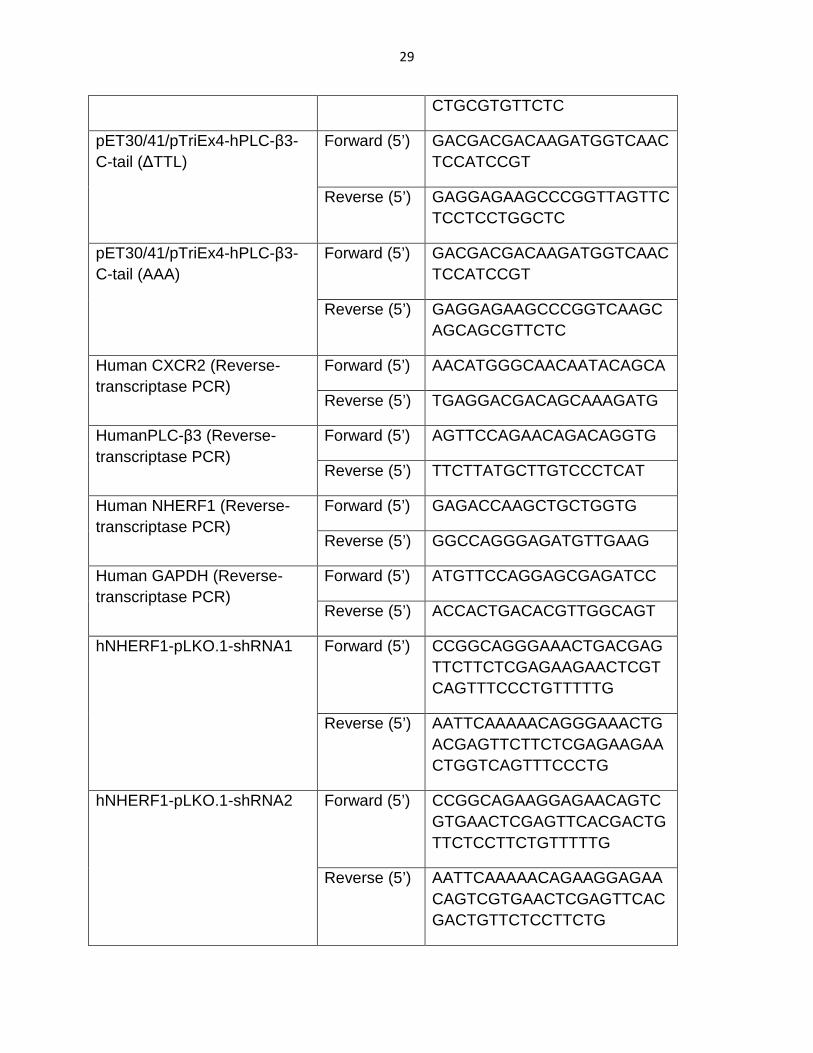
29
CTGCGTGTTCTC
pET30/41/pTriEx4-hPLC-β3-C-tail (∆TTL)
Forward (5’) GACGACGACAAGATGGTCAACTCCATCCGT
Reverse (5’) GAGGAGAAGCCCGGTTAGTTCTCCTCCTGGCTC
pET30/41/pTriEx4-hPLC-β3-C-tail (AAA)
Forward (5’) GACGACGACAAGATGGTCAACTCCATCCGT
Reverse (5’) GAGGAGAAGCCCGGTCAAGCAGCAGCGTTCTC
Human CXCR2 (Reverse-transcriptase PCR)
Forward (5’) AACATGGGCAACAATACAGCA
Reverse (5’) TGAGGACGACAGCAAAGATG
HumanPLC-β3 (Reverse-transcriptase PCR)
Forward (5’) AGTTCCAGAACAGACAGGTG
Reverse (5’) TTCTTATGCTTGTCCCTCAT
Human NHERF1 (Reverse-transcriptase PCR)
Forward (5’) GAGACCAAGCTGCTGGTG
Reverse (5’) GGCCAGGGAGATGTTGAAG
Human GAPDH (Reverse-transcriptase PCR)
Forward (5’) ATGTTCCAGGAGCGAGATCC
Reverse (5’) ACCACTGACACGTTGGCAGT
hNHERF1-pLKO.1-shRNA1 Forward (5’) CCGGCAGGGAAACTGACGAGTTCTTCTCGAGAAGAACTCGTCAGTTTCCCTGTTTTTG
Reverse (5’) AATTCAAAAACAGGGAAACTGACGAGTTCTTCTCGAGAAGAACTGGTCAGTTTCCCTG
hNHERF1-pLKO.1-shRNA2 Forward (5’) CCGGCAGAAGGAGAACAGTCGTGAACTCGAGTTCACGACTGTTCTCCTTCTGTTTTTG
Reverse (5’) AATTCAAAAACAGAAGGAGAACAGTCGTGAACTCGAGTTCACGACTGTTCTCCTTCTG

30
overnight, and resuspended in lysis buffer (Tris base, NaCl) supplemented with
protease inhibitors (aprotinin [1µg/ml], leupeptin [1µg/ml], pepstatin [1µg/ml],
phenylmethylsulfonyl fluoride (PMSF) [500µM], and Lysozyme [100µg/ml]). The
bacterial pellets were sonicated on ice, and allowed to mix at 4oC for 30 minutes.
Subsequently, 10% Triton-X was added to the suspension and LYallowed to mix at 4oC
for 30 minutes. The bacterial debris was then pelleted down by centrifugation at
27,000g for 20 minutes at 4oC. Glutathione agarose beads (50% slurry) were then
added to the cleared supernatant and allowed to mix for 2 hour at 4oC. Glutathione
agarose beads were pelleted down by centrifugation at 800g and washed by TBS
(25mM Tris, 137mM NaCl, 2.7mM KCl, PH = 7.4) for 5 times. Proteins were eluted from
the glutathione agarose beads using 50mM glutathione (PH 7.5), and the excessive
glutathione was subsequently dialyzed away by centrifugation in the Amicon®
Centrifugal Filters (Molecular Weight Cut-Off: 3kDa, EMD/Millipore, Billerica, MA). The
concentration of purified proteins was estimated using the Braford protein estimation
assay (Bio-Rad). A 20µl sample of the protein was eluted using Laemmli sample buffer
and run on a polyacrylamide gel to visualize the quantity and quality of the purified
protein. Laemmili sample buffer contains 60mM Tris-base, 10% (v/v) glycerol, and 2%
SDS (electrophoresis grade).
His-S-tagged proteins were purified using a similar protocol with a few changes. Cobalt
beads (50% slurry) were added to the cleared supernatant and allowed to mix at 4oC for
30 minutes instead of glutathione agarose beads. Proteins were eluted from the cobalt

31
beads using His binding buffer (500mM NaCl, 80mM Tris-base, 25mM Imidazole,
PH=7.4)
2.2.4 Cell Culture
Human pancreatic ductal adenocarcinoma cell lines (PANC-1, MIA PaCa-2, AsPC-1,
BxPC-3 and HPAC) and Human Embryonic Kidney 293 cells (HEK293) were obtained
from American Type Culture Collection (Manassas, VA). Normal human pancreatic duct
epithelial (HPDE) cells, pancreatic cancer cell lines, Colo357 and L3.6pl, were obtained
from Dr. Paul J Chiao at the University of Texas MD Anderson Cancer Center (Houston,
TX). PDAC cells (PANC-1, MIA PaCa-2, HPAC, AsPC-1, BxPC-3, Colo357, and L3.6pl)
and HEK293 cells were cultured in Dulbecco’s modified Eagle’s medium (Thermo
Scientific, Rockford, IL) containing 4.5g/l D-glucose and L-glutamine supplemented with
10% fetal bovine serum (FBS), 100 units/ml penicillin, ad 100µg/ml streptomycin at
37oC in humidified air with 5% CO2. HPDE cells were cultured in keratinocyte serum-
free medium (Invitrogen/Life Technologies, Carlsbad, CA) supplemented with 5ng/ml
epidermal growth factor, 50µg/ml bovine pituitary extract, 100 units/ml penicillin, ad
100µg/ml streptomycin. PANC-1, MIA PaCa-2, AsPC-1, BxPC-3 and HPAC were
derived from different pancreatic cancer patients with adenocarcinoma of the pancreas.
All the PDAC cells were subcultured as a ratio of 1:6 ~ 1:8, and cryopreserved by the
complete medium with 5% (v/v) DMSO in liquid nitrogen. Human Umbilical Vein
Endothelial cells (HUVEC) were obtained from American Type Culture Collection
(Manassas, VA). HUVEC cells were cultured in EBM-2 medium supplemented with 5%
FBS, human recombinant Epidermal Growth Factor, hydrocortisone, human Fibroblast
32
Growth Factor Basic with heparin, Vascular Endothelial Growth Factor, human
recombinant Insulin-like Growth Factor, Ascorbic Acid, and Gentamicin (Amphotericin-B)
(Fisher Scientific, Pittsburgh, PA). The HUVEC cells were cryopreserved by the 70%
(v/v) complete medium, 20% (v/v) FBS, and 10% (v/v) DMSO.
2.2.5 DNA Transfection
LipofectamineTM 2000 transfection reagent (Invitrogen/Life Technologies, Carlsbad, CA)
was used for the DNA transfection. Briefly, cells were seeded on tissue culture dishes
until 70% ~ 80% confluency. On the transfection day, the cells were washed and
cultured with basal medium (without serum and antibiotics). The transfection complex
was set up in two tubes. The first tube contained the Opti-MEM medium (Invitrogen/Life
Technologies, Carlsbad, CA) and DNA, and the second tube contained Opti-MEM
medium with LipofectaminTM 2000 transfection reagent based on the optimum condition
provided by the manufacturer. The transfection complex was then added to the cell
culture dishes, and incubated at 37oC for 6 hours. Then, the medium was cultured in the
complete growth medium for 24 ~ 48 hours for the studies.
2.2.6 Western Blot Analysis
Cells were lysed in lysis buffer (50 mM Tris - pH 8.0, 150 mM NaCl, 1% Nonidet P-40,
0.5% sodium deoxycholate, 0.1% SDS) supplemented with a mixture of protease
inhibitors (containing 1 mM phenylmethylsulfonyl fluoride, 1 µg/ml aprotinin, 1 µg/ml
pepstatin and 1 µg/ml leupeptin). Protein concentration of the cleared supernatant
(17,000 x g, 15 min) was estimated by Bradford protein assay (Bio-Rad). Proteins were

33
eluted in Laemmli sample buffer containing β-mercaptoethonal, separated by SDS-
PAGE (7.5% or 4–15%), and immunoblotted using indicated antibodies. The signal was
detected by SuperSignal® West Pico (or Femto) substrate (Thermo Scientific, Rockford,
IL). The blots were visualized and recorded using a BioSpectrum 500 Imaging system
(UVP, Upland, CA). The images were analyzed using ImageJ software (National
Institutes of Health, Bethesda, MD).
2.2.7 GST Pull-down Assay
GST pull-down assay was performed as previously described163. Briefly, fresh PDAC
cells were lysed in binding buffer (PBS + 0.2% Triton X-100, supplemented with a
mixture of protease inhibitors (aprotinin [1µg/ml], leupeptin [1µg/ml], pepstatin [1µg/ml],
phenylmethylsulfonyl fluoride (PMSF) [500µM]). Cell lysate was mixed at 4oC for 15
minutes and centrifuged at 17,000g for 15 minutes to retrieve the cleared supernatant.
The cleared supernatant was equally mixed with GST alone or various GST-PDZ fusion
proteins (GST-NHERF1, GST-NHERF2, or GST-PDZK1) at 4ºC for 2 hrs. The mixture
was pulled down by glutathione agarose beads (BD Biosciences) at 4ºC overnight. The
glutathione agarose beads were centrifuged at 700g for 1 minute and washed three
times with binding buffer. The proteins were eluted in Laemmli sample buffer containing
β-mercaptoethonal (5%). The eluents were separated by SDS-PAGE, transferred to a
PVDF membrane by Western Blot apparatus (Bio-Rad) and immunoblotted with anti-
CXCR2 or anti-PLC-β3 antibodies (Santa Cruz Biotechnology, Santa Cruz, CA).
2.2.8 Pair-wise Binding Assay

34
Purified GST-NHERF1 was mixed with various purified His-S-PLC-β3 C-tail fragments
(WT, or PDZ motif mutants, ∆TQL, AAA), or CXCR2 C-tail peptides (biotin-conjugate at
N-terminus; WT, or PDZ motif mutants, ∆TTL, AAA) in binding buffer (PBS + 0.2%
Triton X-100 + protease inhibitors (aprotinin [1µg/ml], leupeptin [1µg/ml], pepstatin
[1µg/ml], phenylmethylsulfonyl fluoride (PMSF) [500µM])) at 22-24ºC for 1 hr. The
mixtures were incubated with S-protein agarose (for His-S-tagged fusion proteins), or
streptavidin beads (for biotin-conjugated peptides) for 2 hrs. The beads were
centrifuged at 700g for 1 minute and washed three times with binding buffer. The bound
proteins were eluted with Laemmli sample buffer containing β-mercaptoethonal (5%).
The eluents were resolved and separated by SDS-PAGE, transferred to a PVDF
membrane by Western Blot apparatus (Bio-Rad) and immunblotted with anti-NHERF1
antibody (Santa Cruz Biotechnology).
2.2.9 Macromolecular Complex Assembly Assay
Purified His-S-tagged CXCR2 C-tail fragments (WT, PDZ motif mutants ∆TTL or AAA)
or His-S-tagged PLC-β3 C-tail fragments (WT, PDZ motif mutants ∆TQL or AAA) were
mixed with GST-NHERF1 (or GST alone) in 200 µl of binding buffer (PBS + 0.2% Triton
X-100 + protease inhibitors (aprotinin [1µg/ml], leupeptin [1µg/ml], pepstatin [1µg/ml],
phenylmethylsulfonyl fluoride (PMSF) [500µM])), and the complex was pulled down with
S-protein agarose. This step is also referred to as pair-wise binding as described above.
The dimeric complex was then mixed with the lysates of PDAC cells expressing
endogenous full-length PLC-β3 and CXCR2 for 3 hrs at 4°C. The S-protein agarose
were centrifuged at 700g for 1 minute and washed extensively with binding buffer. The

35
bound proteins were then eluted by Laemmli sample buffer containing β-
mercaptoethonal (5%), resolved and separated by SDS-PAGE, transferred to a PVDF
membrane by Western Blot apparatus (Bio-Rad) and immunoblotted using anti-PLC-β3
or anti-CXCR2 antibodies (Santa Cruz Biotechnology, Santa Cruz, CA).
2.2.10 Co-Immunoprecipitation Assay
A co-immunoprecipitation kit (Thermo Scientific/Pierce, Rockford, IL) was used to
immobilize the normal IgG control and anti-CXCR2 IgG to the resin according to
manufacturer’s instruction. PDAC cells were lysed in binding buffer (PBS + 0.2% Triton
X-100 + protease inhibitors (aprotinin [1µg/ml], leupeptin [1µg/ml], pepstatin [1µg/ml],
phenylmethylsulfonyl fluoride (PMSF) [500µM])), and cleared cell lysates (17,000 × g,
15 min) were processed for co-immunoprecipitation (co-IP) as reported before155,163.
The co-precipitated protein complex was resolved and separated by SDS-PAGE, and
probed for NHERF1 and PLC-β3. For the reverse co-IP, anti-PLC-β3 IgG was used to
immunoprecipitate the complex. The co-precipitated protein complex was separated by
SDS-PAGE and probed for NHERF1 and CXCR2. The signal was detected by
SuperSignal® West Pico (or Femto) substrate (Thermo Scientific, Rockford, IL).
2.2.11 Cell Proliferation Assay
Cell proliferation was assessed by MTT assay as reported before74. In brief, PDAC cells
were seeded in 96-well plates (7 × 10³ cells per well) and allowed to adhere overnight.
Then, the cells were fed with serum-free fresh media with or without 100 ng/mL of
CXCR2 ligands (CXCL1, CXCL5 or CXCL8). After indicated growth periods, cells were
36
incubated with 20µl of MTT solution (1 mg/ml) at 37ºC for 3.5 hrs and then incubated
with MTT solvent (4 mM HCl, 0.1% Nonidet P-40 in isopropanol) under constant mixing
protected from light for 15 min at 22-24ºC. Spectrophotometric absorbance of the
samples at 590 nm was determined by a microplate reader (Bio-Rad). In parallel,
Colo357 and HPAC cells were transfected with various pTriEx4 plasmids (vector alone,
CXCR2 C-tail ∆TTL, or CXCR2 C-tail WT) or delivered with CXCR2 C-tail peptides (WT
or ∆TTL) for 24-48 hours prior to the MTT assay as indicated.
Another peptide inhibitor targeting the interaction between CXCR2 and NHERF1,
EF1060, was synthesized in Dr. Spaller’s laboratory (Dartmouth College, NH). EF1060
is myristoylated at N-terminus and bears the last eight amino acids of the C-terminal
sequence of CXCR2 (N-myristoyl-SGHTSTTL), as shown in Figure 2-10 A. MTT (3-(4,5-
Dimethylthiazol-2-yl)-2,5-diphenyltetrazolium bromide) was purchased from Sigma-
Aldrich (St Louis, MO). EF1060 was dissolved in DMSO. Cell proliferation was
assessed by MTT assay as reported before74. In brief, MIA PaCa-2 cells were seeded in
96-well plates (7 × 10³ cells per well) and allowed to adhere overnight. The cells were
treated with EF1060 with control groups (DMSO and non-treatment). After indicated
growth periods, cells were incubated with 20 µl of MTT solution (1 mg/ml) at 37ºC for
3.5 hrs and then incubated with MTT solvent (4 mM HCl, 0.1% Nonidet P-40 in
isopropanol) under constant mixing protected from light for 15 min at 22 ~ 24ºC.
Spectrophotometric absorbance of the samples at 590 nm was determined by a
microplate reader (Bio-Rad).

37
2.2.12 Cell Invasion Assay
PDAC cells were plated at a density of 1.5 × 10⁵ cells per well in 24-well plate. Cells
were transfected with various pTriEx4 plasmids (vector alone, CXCR2 C-tail ∆TTL, or
CXCR2 C-tail WT). After 48 hrs’ incubation, in vitro invasion assay was performed using
Transwell inserts (BD Biosciences) with 8.0 µm pore size. Briefly, 1 × 10⁵ transfected
PDAC cells were suspended in serum-free medium and seeded onto the Transwell
inserts pre-coated with diluted (1:3) Matrigel. The Transwell inserts were then placed
into 24-well plates filled with the same medium containing 100 ng/ml CXCL8. After 16
hrs’ incubation, the upper surface of the Transwell inserts were wiped with a cotton
swab and the invaded cells were fixed and stained with Diff-Quick stain (IMEB Inc., San
Marcos, CA). The number of invading cells was counted under an inverted microscope
(× 50) in 3 randomly selected fields per well. The data were analyzed by ImageJ
software (National Institutes of Health, Bethesda, MD) and Microsoft Excel software. In
separate experiments, PDAC cells were delivered with CXCR2 C-tail peptides (WT or
∆TTL) for 24 hrs, and the cell invasion was assessed as described above.
2.2.13 Pancreatic Cancer-induced angiogenesis of en dothelial cells
The PANC-1 cells were transfected with various pTriEx4 plasmids (vector alone,
CXCR2 C-tail ∆TTL, or CXCR2 C-tail WT) or delivered with CXCR2 C-tail peptides (WT
or ∆TTL) for 24-48 hrs prior to the angiogenesis assay. The transfected PANC-1 cells
were seeded onto the Transwell inserts with 0.4µm pore size, and allowed to adhere for
3hrs. Growth Factor Reduced Matrigel (Fisher Scientific, Pittsburgh, PA) was used to
mimic the extracellular matrix, and was diluted with the basal EBM-2 medium, and

38
allowed to form the gel in the 24-well plates for 2 hours at 37oC. HUVEC cells were
treated with basal EBM-2 medium (serum- and growth factors-free) for 3 hours prior to
the angiogenesis assay, and were seeded onto the diluted Matrigel. Then, the
transfected PANC-1 cells on the Transwell inserts were placed onto the 24-well plates
containing the HUVEC cells. After indicated periods, tube formation was examined
under the microscope. The lengths of the tube formed by HUVEC cells were measured
by ImageJ software (National Institutes of Health, Bethesda, MD), and analyzed by
Microsoft Excel software.
2.2.14 Xenografts of Human Pancreatic Cancer cells in Immunodeficient Mice
Three GFP-tagged AAV2 constructs, AAV2/2CMV-GFP, AAV2/2CMV-GFP-CXCR2 C-
tail ∆TTL and AAV2/2CMV-GFP-CXCR2 C-tail WT (customized by the Gene Transfer
Vector Core, University of Iowa) were used to transduce HPAC cells. CB17 severe
combined immunodeficient (CB17-SCID) mice (female, 4 ~ 6 weeks old) were randomly
divided into four groups (n = 10-12), and each mouse received 200 µl serum-free
DMEM containing 3 × 10⁶ HPAC cells (transduced or non-transduced) subcutaneously
in the unilateral flank area. The mice were subjected to measurement of subcutaneous
tumors every other day and monitored for changes in body weight and other side
effects. Tumor volume was calculated by the formula (L × W2)/2, where L and W are the
tumor length and width (in mm), respectively. To avoid severe discomfort in the control
group, animals were euthanized after 4 weeks. Tumor tissues were harvested for
histological analysis and immunohistochemical staining. Tumor volume in SCID mice
was plotted against time, and the final tumor weights were measured after the mice
39
were euthanized. All the animal studies were accomplished under the protocol approved
by Wayne State University Institutional Animal Care and Use Committee.
2.2.15 Immunohistochemistry and Quantification of P roliferation Index
Tumor tissue from the xenografts was fixed by 4% paraformaldehyde (PH = 7.5) and
embedded within paraffin. Paraformaldehyde-fixed and paraffin-embedded sections (5
µm) were stained with Ki-67 antibody (Ventana Medical Systems, Tucson, AZ) as
reported165. Results were expressed as percentage of Ki-67 positive cells per 200 x
magnification (Ki-67+ cell number/total cell number). A total of 10 sections from each
experimental group were examined by Zeiss Axiophot epifluorescence microscope (Carl
Zeiss) and analyzed by Image-Pro Plus 6.0 software (Media Cybernetics, UK).
2.2.16 Statistical Analysis
Data are presented as mean ± SE of at least three independent experiments. Statistical
significance of differences was assessed with the Student’s t-test. A value of P < 0.05
was considered statistically significant.
2.3 Results
2.3.1 Bacterial Expression and Protein Purification
High quality of recombinant His-S-CXCR2-C-tail (wild-type, PDZ motif deletion, PDZ
motif mutation) and His-S-PLC-β3-C-tail proteins (wild-type, PDZ motif deletion, PDZ
motif mutation) were produced as described in section 2.2.3. Figure 2-1 showed the
purification of the bacterially expressed His-S-PLC-β3-C-tail (Fig. 2-1 A) and His-S-

40
41
42
CXCR2-C-tail (Fig. 2-2 B). All the fractions were stained with Coomassie Brilliant Blue
(Bio-Rad).
2.3.2 Overexpression of CXCR2 in human pancreatic c ancer cells
The expression of CXCR2 in normal human pancreatic duct epithelial (HPDE) cells and
several PDAC cell lines (HPAC, Colo357, PANC-1, and MIA PaCa-2) were examined
and compared by Western blotting as described in section 2.2.6. All the five PDAC cell
lines tested in our study showed significantly increased CXCR2 expression (both protein
and mRNA) as compared to HPDE cells (Fig. 2-2), which is in agreement with the
previous clinical study that reported an up-regulation of IL-8/CXCL8 and its receptors in
both pancreatic adenocarcinomas and neuroendocrine tumors140.
2.3.3 Endogenous CXCR2 and PLC- β3 in human pancreatic cancer cells
preferentially interacts with NHERF1
In a recent study, we demonstrated that the consensus PDZ motif at the carboxyl
terminus of CXCR2 mediates PDZ-based interactions with certain PDZ scaffold proteins
(such as NHERF1, NHERF2) in neutrophils163. In order to investigate if endogenous
CXCR2 in PDAC cells binds to any PDZ scaffold proteins, we performed a pull-down
assay 163 as described in Materials and Methods. As shown in Fig. 2-3 A, we observed
interactions between CXCR2 and the membrane-associated PDZ proteins NHERF1 and
NHERF2 in Colo357, L3.6pl, and HPAC cells, among which NHERF1 has a higher
binding affinity for CXCR2 as compared with NHERF2. However, neither GST (the
negative control) nor PDZK1 was found to bind to endogenous CXCR2 in these PDAC
cell lines (Fig. 2-3 A).

43

44
CXCR2 couples to the pertussis toxin-sensitive Gi to stimulate phosphatidylinositide-
specific phospholipase C (PLC) activities145. Similar to CXCR2, all human PLC-β
isoforms possess consensus class I PDZ motifs at their carboxyl termini162. Our
previous study has demonstrated that PLC-β3, containing a PDZ motif (TQL-COOH),
overexpressed in HEK293 cells interact with NHERF1 and NHERF2163. Here, we
explored the potential interactions between PDZ scaffold proteins (NHERF1, NHERF2
and PDZK1) and endogenous PLC-β3 in PDAC cell lines. By similar GST pull-down
experiments, we observed that endogenous PLC-β3 in PDAC cells bind to both
NHERF1 and NHERF2; however, it did not bind to PDZK1 or to GST alone (Fig. 2 -3 B).
Moreover, in comparison to NHERF2, NHERF1 appears to interact with PLC-β3 with a
higher affinity in most of the PDAC cell lines we tested (Fig. 2-3 B). In addition, the
binding between endogenous PLC-β3 in PDAC cells and NHERF1 increased with
increasing amounts of NHERF1 in a dose-dependent manner (Fig. 2-3 C).
2.3.4 CXCR2 and PLC-β3 Interact with NHERF1 in a Direct and PDZ Motif-
Dependent Manner
The data resulting from the GST pull-down studies presented above (Fig. 2-3) did not
provide information whether the interactions between CXCR2 or PLC-β3 and NHERF1
are direct, as cell lysates contain large numbers of other proteins as well. In order to test
if CXCR2 or PLC-β3 binds NHERF1 directly or by other intermediary proteins, and to
test the PDZ motif dependence, we performed a pair-wise binding assay that detects a
direct interaction between purified proteins in vitro163. Purified His-S fusion proteins
containing the C-tail fragments (last 45 amino acids) of CXCR2 WT, C-tail ∆TTL (with

45

46
PDZ motif TTL deleted), or C-tail AAA or ATA (with PDZ motif TTL mutated to AAA or
ATA) were mixed with GST-NHERF1, and the mixtures were pulled down by S-protein-
agarose (the S-protein can specifically bind to the S-tag within the fusion proteins). The
protein complex was immunoblotted with anti-NHERF1 antibody. We observed that the
CXCR2 C-tail directly interacts with NHERF1 in a PDZ motif-dependent manner, as the
interaction between NHERF1 and CXCR2 C-tail lacking the PDZ motif (∆TTL) or
CXCR2 C-tail with PDZ motif mutations (AAA or ATA) was remarkably reduced (Fig. 2-
4A). We also observed that PLC-β3 C-tail (containing the PDZ motif) directly binds to
NHERF1 in a PDZ motif-dependent manner, as the interaction between NHERF1 and
PLC-β3 C-tail lacking the PDZ motif (∆TQL) or PLC-β3 C-tail with PDZ motif mutation
(AAA) was almost completely abolished compared to wild-type (WT) PLC-β3 C-tail (Fig.
2-4 B).
2.3.5 CXCR2 interacts with both PDZ domains of NHER F1 (PDZ1 and PDZ2)
We also showed that CXCR2 interacts with both PDZ domains of NHERF1 (PDZ1 and
PDZ2) in the GST pull-down assays, with PDZ2 exhibiting higher binding affinities (Fig.
2-5)128. Specifically, CXCR2 overexpressed in HEK293 cells were pulled down by using
various purified GST-NHERF1 fractions (GST-PDZ1, GST-PDZ2, and GST-PDZ1-2).
Whereas no CXCR2 was detected in the negative control lane containing GST alone,
significant amounts of CXCR2 were found in the lanes containing PDZ1 domain (GST-
PDZ1), PDZ2 domain (GST-PDZ2), and both PDZ domains together (GST-PDZ1-PDZ2)
(Fig. 2-5). To test whether the PDZ-CXCR2 interactions are direct, we performed an in
vitro pair-wise binding assay with a biotinylated peptide containing the last 13 amino

47

48

49
acids of CXCR2 (Biotin-FVGSSSGHTSTTL-COOH). Similar binding results were
observed in the experiments where CXCR2 interacts with both PDZ domains of
NHERF1 (Fig. 2-5).
2.3.6 Endogenous PLC- β3 in human pancreatic cancer cells preferentially
interacts with NHERF1-PDZ2
We also demonstrated that endogenous PLC-β3 in various PDAC cells (PANC-1, MIA
PaCa-2, AsPC-1, BxPC-3, and HPAC) preferentially interacts with PDZ2 domain of
NHERF1. Specifically, the total cell lysates of PDAC cells (PANC-1, MIA PaCa-2,
AsPC-1, BxPC-3, and HPAC) were mixed with various purified GST-NHERF1 fractions
(GST-PDZ1, GST-PDZ2, and GST-PDZ1-2), and GST-NHERF1 full length. By the GST
pull-down assays, we observed that the PLC-β3 endogenously expressed in all the
PDAC cell lines preferentially interacts with PDZ2 domain with higher binding affinity
(Fig. 2-6 A). It’s also worth mentioning that the binding affinity between PLC-β3 and
PDZ2 domain is even stronger than the binding between PLC-β3 and full-length
NHERF1. Morales and colleagues have demonstrated a head-to-tail intramolecular
interaction between the PDZ2 domain and the C-terminal ezrin-radixin-moesin (ERM)-
binding region that could masks the association between NHERF1 and other PDZ
domain ligands166,167. The higher binding affinity of PDZ2 domain compared to the full-
length NHERF1 protein is in agreement with the findings by Georgescu group. We also
demonstrated the PLC-β3 interacts with both PDZ1 and PDZ2 domains of NHERF1
directly by a pair-wise binding study (Fig. 2-6 B). Also, the PDZ2 domain has a slight
higher binding affinity.

50

51

52
2.3.7 NHERF1 Clusters CXCR2 and PLC- β3 into a Macromolecular Complex both
In Vitro and in human pancreatic cancer cells
Results from the above GST pull-down experiments demonstrated that both
endogenous CXCR2 and PLC-β3 in PDAC cells preferentially interact with NHERF1
(Fig. 2-3 A and 2-3 B), and PLC-β3 binds to NHERF1 in a direct and PDZ motif-
dependent manner (Fig. 2-4 B), similar to the interaction between CXCR2 and NHERF1
as we previously reported163. Hence, we hypothesized that NHERF1 might nucleate
CXCR2 and PLC-β3 forming a macromolecular complex in a PDZ motif-dependent
manner and this complex might be critical for efficient and specific signaling mediated
by CXC-chemokine/CXCR2 biological axis in PDAC cells. Towards this end, we sought
to determine if we could detect a macromolecular complex containing CXCR2, NHERF1,
and PLC-β3 in vitro. Using an in vitro macromolecular complex assembly assay (Fig. 2-
7A)163, we observed the existence of a complex composed of CXCR2 C-tail, NHERF1
and endogenous PLC-β3 in PDAC cells (Fig. 2-7 B). Furthermore, we demonstrated
that CXCR2 C-tail did not bind to PLC-β3 directly, and the macromolecular complex was
not formed by CXCR2 C-tail PDZ motif mutants (∆TTL or AAA), indicating that the
formation of the CXCR2 macromolecular complex is PDZ motif-dependent (Fig. 2-7 B).
In addition, we detected a similar complex consisting of PLC-β3 C-tail (containing the
PDZ motif TQL), NHERF1, and endogenous CXCR2 from PDAC cells (data not shown).
Results from the above demonstrated that a CXCR2 macromolecular complex exists in
vitro; however, it did not provide the evidence whether this complex exists in native

53

54
membranes of PDAC cells that endogenously express all the relevant interacting
proteins. To address this issue, co-immunoprecipitation was performed using either
anti-CXCR2 or anti-PLC-β3 antibodies as described in section 2.2.10. We observed that
NHERF1 and PLC-β3 in PDAC cells were co-immunoprecipitated with CXCR2 (Fig. 2-7
C). Similarly, CXCR2 and NHERF1 were also co-precipitated with PLC-β3 from PDAC
cells (Fig. 2-7 D), indicating that there is likely to be a macromolecular complex
composed of endogenous CXCR2, NHERF1 and PLC-β3 on the surface membranes of
Colo357 and HPAC cells.
2.3.8 CXC Chemokine/CXCR2 Biological Axis Promotes Pancreatic Cancer Cell
Proliferation
To examine the effect of CXCR2 signaling on PDAC cell proliferation, PDAC cells were
treated with CXCR2 ligands followed by the MTT cell proliferation assay as described in
section 2.2.11. As illustrated in Fig. 2-8 A, HPAC cells showed significantly elevated cell
proliferative activities in response to both CXCL8 and CXCL1 (P < 0.05), and CXCL5
also promoted HPAC proliferation, though without statistical significance (P = 0.07).
Proliferation of Colo357 cells was significantly augmented by CXCL5 (P < 0.05), and
also by CXCL8 and CXCL1 though without statistical significance (P = 0.09 and 0.2,
respectively) (Fig. 2-8 B). It has been reported that Colo357 cells showed a significant
high level of CXCL5 secretion73, and it has also been documented that CXCL5 is
overexpressed in the pancreatic cancer patients compared to the normal individuals,
which is associated with the poor survival in patients74. Our results underpinned the
autocrine effects of CXCL5 on Colo357 cell proliferation. However, the normal
pancreatic duct HPDE cells did not demonstrate significantly increased growth

55

56
stimulated by the treatment of either of these CXCR2 ligands (CXCL1, CXCL5, or
CXCL8) (Fig. 2-8 C), indicating that CXCR2 biological axis significantly promoted
pancreatic cancer cell proliferation.
2.3.9 Disrupting the CXCR2 Macromolecular Complex I nhibits Pancreatic Cancer
Proliferation
Given that the CXC-chemokine/CXCR2 biological axis plays an important role in PDAC
cell proliferation (Fig. 2-8 A and 2-8 B), and here we also demonstrated that the PDZ
motif of CXCR2 is essential for the physical coupling of CXCR2 to PLC-β3 mediated by
NHERF1 into a macromolecular signaling complex (Fig. 2-7 B); therefore, it is possible
that perturbation of the CXCR2 macromolecular complex might affect CXCR2 ligand-
induced PDAC cell proliferation. In a recent study, we utilized a CXCR2 C-tail peptide
(containing the PDZ motif) to disrupt the CXCR2 PDZ motif-mediated interaction with
NHERF1 and observed a functional inhibition of CXCR2 ligand-induced cell migration in
neutrophils163. We went on to evaluate the functional significance of this CXCR2
macromolecular complex in the CXCR2 ligand-induced PDAC cell proliferation. We
transfected HPAC and Colo357 cells with plasmids encoding CXCR2 C-tail (WT or PDZ
deletion ∆TTL), and evaluated cell proliferative activities. As shown in Fig. 2-9 A, in
response to CXCL5, Colo357 transfected with plasmid containing CXCR2 C-tail WT
showed significantly reduced proliferative activities as compared to cells transfected
with the vector alone or CXCR2 C-tail ∆TTL, suggesting that disrupting CXCR2
macromolecular complex inhibits CXCR2 ligand-induced Colo357 growth, and PDZ
motif on the C-terminus of CXCR2 is important for PDAC cell proliferation. HPAC

57

58

59
transfected with CXCR2 C-tail WT showed significantly decreased cell proliferation in
response to CXCL8 (Fig. 2-9 B). In addition, we delivered the CXCR2 C-tail peptides
(WT and ∆TTL) used in our previous studies163 to Colo357 and HPAC cells, and we
also observed significantly reduced cell proliferation stimulated by CXCL5 (Fig. 2-9 C)
and CXCL8 (Fig. 2-9 D). Furthermore, using the pair-wise binding assay, we also
demonstrated that the CXCR2 C-tail peptide interacts with NHERF1 in a direct and PDZ
motif-dependent manner, because the CXCR2 WT peptide interacts with NHERF1,
while the PDZ deletion peptide (∆TTL) or mutation peptide (AAA) failed to bind to
NHERF1 (Fig. 2-9 E).
2.3.10 Inhibitory effect of EF1060 on MIA PaCa-2 pr oliferation
The peptide inhibitor, EF1060, was dissolved in DMSO as 200mM stock with good
solubility. Initially, concentration (500 ng/ml) of EF1060 was used to compare with the
peptide synthesized by Genemed Synthesis (Biotin-CXCR2 C-tail; 13 a.a.). Significant
change in cell proliferation was not detected at the peptide concentration of 500ng/ml
(of 96-well plate) (Fig. 2-10 B).
However, by using similar peptide concentration (200µg/ml) that was previously
reported by Dr. Spaller’s group, significant reduction in cell proliferation by EF1060 has
been observed compared with DMSO control (Fig. 2-10 C).
It is noteworthy that, EF1060, when used at low concentration (in Fig. 2-10 B) without
Chariot, seemed to have inhibitory effect on cell proliferation, although had not achieved
statistical significance given the small sample number. Meanwhile, EF1060 significantly

60

61

62
inhibited cell proliferation when used at high concentration without Chariot (as in Fig.2-
10 C). However, when used at low concentration (in Fig. 2-10 B), EF1060 (+ ChariotTM)
had no effect on cell proliferation, which suggests that EF1060 with myristoylation at N-
terminus might not require the commercial peptide delivery system.
2.3.11 Disrupting the CXCR2 Macromolecular Complex Blocks Pancreatic Cancer
Cell Invasion
We then examined the effect of disrupting CXCR2 signaling complex on the invasive
capability of PDAC cells. HPAC cells were transfected with plasmids encoding CXCR2
C-tail (WT or ∆TTL) and used to evaluate the invasive potency by an in vitro invasion
assay as reported before73. As illustrated in Fig. 2-11, gene delivery of CXCR2 C-tail
WT sequence, but not the ∆TTL PDZ deletion sequence, significantly inhibited invasion
of HPAC cells through Matrigel induced by CXCL8, implicating that the PDZ motif of
CXCR2 is important for PDAC cell invasion.
2.3.12 Disrupting the CXCR2 Macromolecular Complex Blocks Pancreatic Cancer-
induced angiogenesis
We next investigated the effect of disrupting CXCR2 macromolecular complex on the
capability of PDAC cell-induced angiogenesis. PANC-1 cells were transfected with
plasmids encoding CXCR2 C-tail (WT or ∆TTL) and used to evaluate the tumor-induced
angiogenesis potential by an in vitro tube formation assay as described in section 2.2.13.
Figure 2-12 showed that gene delivery of CXCR2 C-tail WT sequence, but not the ∆TTL

63

64

65

66
PDZ deletion sequence or pTriEx4 vector alone, significantly suppressed the tube
formation of HUVEC cells induced by the PANC-1 cells, indicating the CXCR2
macromolecular complex through PDZ binding is important for PDAC-induced
angiogenesis.
2.3.13 Disrupting the CXCR2 Macromolecular Complex Inhibits Pancreatic Tumor
Growth In Vivo
To analyze the functional significance of the CXCR2 macromolecular complex in PDAC
growth in vivo, a subcutaneous xenograft induced by HPAC cells in CB17-SCID mice
was developed to determine whether disrupting the CXCR2 signaling complex could
lead to inhibition of tumor growth in vivo. HPAC cells that were transduced with Adeno-
Associated Virus (Serotype 2) expressing GFP - human CXCR2 C-tails (WT or ∆TTL) or
GFP alone, or non-transduced cells, were injected subcutaneously into CB17-SCID
mice. The transduction efficiency was inspected visually under the fluorescence
microscope (Fig. 2-13). Tumor volume was measured every other day starting at day 12
(when the tumor became palpable). At day 28, all mice were euthanized, and tumors
were excised and final tumor weights were determined by the scale. We found that
HPAC cells expressing GFP alone or GFP-CXCR2 C-tail ∆TTL grew into sizable tumors
underneath the skin that were comparable to non-transduced cells (Fig 2-14 A).
However, HPAC cells expressing GFP-CXCR2 C-tail WT grew into significantly smaller
tumors compared with the cancer cells expressing GFP vector alone or GFP-CXCR2 C-
tail ∆TTL, or non-transduced cells (Fig 2-14 A). Furthermore, the HPAC cells,
transduced with GFP-CXCR2 C-tail WT, showed significantly reduced final tumor weight

67

68
compared to the 3 other groups (Fig. 2-14 B). This result suggests an inhibitory effect of
exogenous CXCR2 C-tail (containing the PDZ motif) on human pancreatic tumor
development and progression in vivo. We then demonstrated that the proliferation index,
determined by Ki-67 expression, in the group of mice injected with HPAC cells
expressing GFP–CXCR2 C-tail WT was significantly lower (p < 0.01) compared to the 3
other groups (non-transduction group, AAV2-GFP group, and AAV2-GFP-CXCR2 C-tail
∆TTL group) (Fig. 2-14 C and D). Collectively, our date indicates the potential
therapeutic effect of disrupting the CXCR2 macromolecular complex on the primary
tumor of PDAC in a subcutaneous mouse model. It is noteworthy that in the group of
mice injected with HPAC cells expressing GFP–CXCR2 C-tail WT did not show
significant reduced in vivo tumor-induced angiogenesis compared to other groups.
2.4 Discussion
CXC-chemokines (such as CXCL1/GRO-α, CXCL8/IL-8, CXCL5/ENA-78) and their
cognate receptor CXCR2 have been reported to play a critical role in tumor growth and
tumor invasion and tumor-induced angiogenesis, as blockade of the CXC-
chemokines/CXCR2 biological axis reduced tumorigenesis and angiogenesis in many
human cancers including pancreatic cancer63,73,75,168. However, most of the
interventional approaches have been conducted by systemic blockade or depletion of
CXCR2 and/or its ligands, which might cause global undesired effects on other vital
functions, as CXCR2 had also been reported in many essential normal cellular functions,
such as in preservation of oligodendrocyte function and myelinization of neural tissues21.
This issue warrants the necessity of a more comprehensive understanding of the

69

70

71
molecular mechanisms of CXCR2 and its signaling, on the basis of which, selective
and cell-specific therapeutic targets could be identified.
CXCR2 possesses a consensus PDZ motif at their carboxyl termini, and the PDZ motif
has been reported to modulate cellular chemotaxis123. Recently, we demonstrated that
the PDZ motif of CXCR2 plays an important role in regulating neutrophil functions as
disrupting the interaction mediated by PDZ motif via using an exogenous peptide mimic
(mapping CXCR2 PDZ motif) significantly inhibited CXCR2-mediated calcium
mobilization and neutrophilic transepithelial migration163. In the present study, we
identified the PDZ scaffold protein NHERF1 as a previously unrecognized interacting
partner for CXCR2 in PDAC cells, and we also demonstrated the existence of a PDZ-
based CXCR2 macromolecular signaling complex containing endogenous CXCR2,
NHERF1 and PLC-β3 in PDAC cells. Furthermore, we provided functional evidence
showing that disrupting the CXCR2 complex significantly inhibited the malignant cellular
functions (i.e. proliferation and invasion) in vitro and pancreatic tumor growth in vivo.
Controversy exists regarding the expression of CXCR2 in human pancreatic cancer cell
lines. Despite some studies reported that CXCR2 was not detected in some human
PDAC cell lines (such as PANC-1, MIA PaCa-2, AsPC-1, BxPC-3 and HPAF-II)73,165,169,
other groups reported the detection of CXCR2 and/or the autocrine effect of CXCR2
ligands in various human PDAC cells74,141-144,170 (such as PANC-1, MIA PaCa-2, Capan-
1, Capan-2, SUIT-2, HuP-T4, Bx-PC-3, Panc03.27), and pancreatic tumors specimens
from patients63,140. In the present study, we also detected the expression of CXCR2 in

72
PANC-1, MIA PaCa-2, HPAC, Colo357, and L3.6pl cells in Western blotting (Fig. 2-2 A)
by using the same antibody Frick et al used to detect the CXCR2 expression in patient
specimens63. RT-PCR results from our study also confirmed the expression of CXCR2
in these cell lines (Fig. 2-2 B). One possible reason for the failure of some groups to
detect CXCR2 in PDAC cell lines might be that expression level of CXCR2 varies in
different conditions. Yamamoto et al reported that CXCR2 is up-regulated in an
orthotopic colon cancer model compared to the subcutaneous model171. Hussain et al
also reported that the expression level of CXCR2 correlates with tumor grade and stage
in pancreatic adenocarcinomas140.
PLC-β is the major isozyme that has been well-studied to participate in GPCR-mediated
signaling and modulate the physiological responses, such as promoting cell growth in
many cancer types172-174. Each PLC-β subtype has its distinct expression pattern and
physiological relevance172. Among the four subtypes of PLC-β, PLC-β3 is expressed in
a wide range of cells and tissues174, and exhibits the highest affinity to Gβγ subunits and
subsequent activation by Gβγ subunits175. A growing body of evidence suggests that
PDZ scaffold proteins are involved in the modulation of PLC-β isoforms in the PDZ
motif-dependent manner. PLC-β3, with its PDZ motif (-STQL-COOH), was reported to
bind to PDZ domains of NHERF2 and Shank2 via its PDZ motif at its carboxyl termini, in
mouse small intestine161 and in the postsynaptic density region in neuronal cells158,
respectively. It has also been shown that PLC-β3 was down-regulated in jejuna villus
cells in NHERF1- knockout mice160. Results from our present study revealed that PLC-
β3 preferentially binds to NHERF1 in PDAC cells in a direct and PDZ motif-dependent
73
pattern. Therefore, the specificity and diversity of agonist-induced PLC-β activity and its
downstream signaling may be regulated by the specific interactions of PLC-β isoforms
and certain PDZ scaffold proteins.
There is accumulating evidence suggesting that the formation of spatial compact
macromolecular signaling complexes beneath the plasma membrane enables the
membrane receptors to transduce signals into cell interior and thereafter influence cell
behavior with higher specificity and efficiency163,176-179. These findings have therapeutic
implications in various diseases including inflammatory diseases, genetic diseases and
cancer153-159. Our present study revealed a PDZ motif-dependent CXCR2
macromolecular signaling complex, in which CXCR2 and PLC-β3 were bridged by
NHERF1, in the PDAC cells (Fig. 2-15). Results from our study also demonstrated the
functional importance of the CXCR2 complex in pancreatic cancer cell functions in vitro
and in vivo, as disturbing the CXCR2 complex by using an exogenous CXCR2 C-tail
sequence (containing the PDZ motif) significantly attenuated malignant cell proliferation
and cell invasion of PDAC cells and tumor growth in vivo. Our present study introduced
a new concept of the CXCR2 macromolecular complex mediated via PDZ-based
interactions, and we elucidated the molecular evidence and functional importance of this
CXCR2 complex in pancreatic cancer progression. Moreover, by investigating a network
of protein complexes rather than CXCR2 alone in pancreatic cancer, our results reveal
a novel molecular target for the development of specific therapeutic strategies and
agents that could combat pancreatic cancer.

74

75
Recent advances regarding PDZ proteins have suggested that the specific inhibition of
the protein-protein interaction mediated by PDZ domains, with subsequent intervention
of the signaling pathways mediated by the PDZ proteins, appears to be a promising
strategy for developing novel peptide inhibitors in cancer180. Furthermore, our studies
have shown that our CXCR2 specific peptide (bearing the last 13 residues of the C-
terminal sequence of CXCR2) exhibit significant inhibitory effect on the malignant
cellular functions of pancreatic cancer125. However, before the clinical trials commence,
our strategy still needs to be further investigated by addressing several challenges. The
membrane permeability of the small peptide is one of the frequently questioned
problems in novel inhibitor development180. Also, additional effort is also required on
maximizing the binding affinity and specificity of the peptide inhibitors. Recent studies
have reported that myristate, a saturated 14-carbon alkyl acid, would function as a
promising delivery system for membrane transport181-183. Furthermore, it has been
suggested that organic halogen modifications appear to be promising strategies to
enhance the binding affinity and specificity of the novel peptide inhibitors, as the
halogen atoms have been reported to enhance the stability of protein-ligand
interaction184,185, increase the binding contacts186, and also improve the membrane
permeability186. Dr. Mark Spaller’s group from Dartmouth College recently reported a
novel peptide targeting the PDZ domain of GAIP-interacting protein, C terminus (GIPC)
bearing the last 8 residues of the C-terminal sequence of GAIP (N-myristoyl-
PSQSKSKA). The peptide was modified by chemical substitution of two bromobenzoate
moieties at the “-1” and “-3” positions180. The halogenated benzoyl-group has introduced

76
the new potential contacts between the “-1” and “-3” residues and the PDZ domain,
resulting in the significantly increased inhibitory effect of this peptide compared to the
non-modified peptide. Similar enhanced inhibitory effects of the halogenated
modification have been observed in other peptide, such as flavopiridol187. Further
studies should involve control peptides to test the specificity of the peptide inhibitor
(peptide with scrambled sequences, or CXCR2 PDZ mutation peptide such as Myr-
SGHTSAAA or Myr-SGHTS).

77
Chapter III
Crystallographic Analysis of NHERF1-PLC β3 Interaction Provides Structural
Basis for CXCR2 Signaling in Pancreatic Cancer
© 2014 Elsevier 3.1 Introduction :
CXC chemokine receptor 2 (CXCR2) was initially identified as the neutrophil receptor
that is activated by binding to interleukin-8188. Besides the roles in mediating
neutrophilic migration and positioning of oligodentrocyte precursors in developing spinal
cord 163,189,190, a growing body of evidence has suggested that the enhanced expression
of CXCR2 correlates with aggressive stages and poor survival in pancreatic cancer
patients 62,191. More recent studies have also revealed that CXCR2 and its ligands
(CXCL1, CXCL5, and CXCL8) are overexpressed in pancreatic ductal adenocarcinoma
cell lines and enhance cell proliferation141,144,192. The evidence has made CXCR2 an
attractive drug target for developing highly-specific inhibitors for pancreatic cancer.
Recent studies regarding protein-protein interactions suggested that CXCR2 interacts
with other proteins, including ion channels, transporters, scaffolding proteins, and
cytoskeletal elements to form macromolecular complexes at specific subcellular
domains within cytoplasma163,192. These dynamic protein-protein interactions regulate
CXCR2 signaling function as well as its localization and processing within cells 193,194. In
chapter 2, I have detected a macromolecular complex containing CXCR2, Na+/H+
exchanger regulatory factor-1 (NHERF1), and phosphoipase C-β3 (PLC-β3), at the
plasma membrane of pancreatic cancer cells, which functionally initiated the CXCR2

78
signaling cascade through PLC-β3192. PLC-β3, a membrane bound enzyme, catalyzes
the formation of inositol 1,4,5-trisphosphate and diacylglycerol from phosphatidylinositol
4,5-bisphosphate. NHERF1 is a PDZ domain-containing protein that typically functions
as a scaffold to cluster transporters, receptors, and signaling molecules into
macromolecular complexes 195. In chapter 2, I have already demonstrated that the
formation of the CXCR2 macromolecular complex is mediated by PDZ-binding through
the PDZ domains in NHERF1 and the PDZ binding motifs from CXCR2 (-STTL-COOH)
and PLC-β3 (-STQL-COOH). I also showed that disruption of this PDZ-mediated
interaction attenuated the cellular functions (i.e. proliferation, invasion, and tumor
growth) in pancreatic cancer, implicating that targeting the PDZ binding-mediated
CXCR2-PLC-β3 interaction could provide new strategies for therapeutic interventions.
In general, PDZ domains recognize the C-terminal sequences of the target proteins (i.e.
membrane receptors, ion channels, etc.) and bind to the targets via a canonically and
structurally conserved PDZ peptide-binding pocket196, thereby mediating the protein-
protein interactions and the formation of macromolecular complex. The specificity of the
interactions is primarily determined by the amino acids at positions 0 and -2 of the C-
terminal sequences of the target proteins, whereas other residues do not significantly
contribute to the interaction (position 0 refers to as the extreme residue at the C-
terminus) 196. The characteristics of the side chains of the residues at 0 and -2 positions
have led to the classification of the PDZ domains into two major classes: I, S/T)X(V/I/L)
(X denoting any amino acid); class II, (F/Y)X(F/V/A) 197-199. However, more recent
studies have suggested that the specificity of the PDZ binding is unexpectedly

79
complicated, with the PDZ domain family recognizing up to 7 C-terminal residues and
forming multiple PDZ binding classes200. The promiscuity of the binding between
different PDZ binding motifs and distinct PDZ domains raises a challenging problem of
how PDZ domains, which are structurally simple protein interaction modules, achieve
the broad substrate specificity. In this chapter, crystal structure of the complex,
NHERF1-PDZ1 with the PLC-β3 C-terminal peptide ENTQL, reveals that the PLC-β3
peptide binds to PDZ1 domain in an extended conformation with the last four residues
making specific side chain contacts. The results that PLC-β3 can bind both NHERF1
PDZ1 and PDZ2 in PDAC cells (chapter 2), are consistent with the observation that the
two PDZ domains, PDZ1 and PDZ2, share the peptide-binding pockets which are highly
structurally conserved. The study in this chapter provides the structural basis of the PDZ
interaction-mediated NHERF1-PLCβ3 interaction, and provides the novel insights in
development of novel therapeutic strategies against lethal pancreatic cancer.
3.2 Materials and Methods
3.2.1 Protein Expression and Purification
For X-ray crystallography, a DNA fragment encoding the human NHERF1 PDZ1
(residues 11–94) was amplified by PCR using the full-length human NHERF1 cDNA as
a template. The C-terminal extension ENTQL that corresponds to residues 1230–1234
of human PLCβ3 was created by inclusion of 15 extra bases in the reverse primer. The
PCR products were cloned in the pSUMO vector containing an N-terminal His6-SUMO
tag. The resulting clone was transformed into Escherichia coli BL21 Condon Plus (DE3)
cells for protein expression. The transformants were grown to an OD600 (optical density
80
at 600 nm) of 0.4 at 37 °C in LB medium, and then i nduced with 0.1 mM isopropylthio-β-
D-galactoside at 15 °C overnight. The cells were ha rvested by centrifugation and lysed
by French Press. The soluble fraction was then subjected to Ni2+ affinity
chromatography purification, followed by the cleavage of the His6-SUMO tag with yeast
SUMO Protease 1. PDZ1 proteins were separated from the cleaved tag by a second
Ni2+ affinity chromatography and further purified by size-exclusion chromatography.
Finally, the proteins were concentrated to 30–40 mg/ml in a buffer containing 20 mM
Tris–HCl (pH 8.0), 150 mM NaCl, 1 mM β-mercaptoethanol, and 5% glycerol. For
pulldown experiments, glutathione S-transferase (GST) fusion proteins were generated
by cloning NHERF1 PDZ1, PDZ2, or PDZ1-PDZ2 into the pGEX4T-1 vector 163. His-S-
tagged proteins were generated by cloning PLCβ3 C-terminal fragment (residues 1135–
1234) into the pET30 vector 192. GST-PDZ proteins were purified using glutathione
agarose beads (BD Biosciences) and eluted with 50 mM glutathione. His-S-PLCβ3 was
purified using Cobalt resins (Thermo Scientific) and eluted with 200 mM imidazole.
3.2.2 Crystallization, Data Collection and Structur e Determination
Crystals were grown by the hanging-drop vapor-diffusion method by mixing the protein
(~25 mg/ml) with an equal volume of a reservoir solution containing 100 mM sodium
acetate, pH 4.6, 2.5 M sodium chloride at 20 °C. Cr ystals typically appeared overnight
and continued to grow to their full size in 3–4 days. Prior to X-ray diffraction data
collection, crystals were cryoprotected in a solution containing the mother liquor and
25% glycerol and flash cooled in liquid nitrogen. The data were collected at 100 K at
beamline 21-ID-F at the Advanced Photon Source (Argonne, IL) and processed and
scaled using the program XDS 201. Crystals belong to the space group P3121 with unit

81
Table 3-1. Crystallographic data and refinement statistics
Data Space group P21 Cell parameters (Å)
a 26.4 b 40.3 c 37.1
Wavelength (Å) 1.2719 Resolution (Å) 24.2-1.34 (1.37-Rmerge
a 0.039 (0.250)b Redundancy 4.1 (4.0) Unique reflections 17966 Completeness (%) 99.8 (99.6) ⟨I/σ⟩ 15.3 (3.0) Refinement Resolution (Å) 24.2-1.34 (1.37-Molecules/AU 1 Rwork
c 0.145 (0.268) Rfree
d 0.177 (0.275) Ramachandran plot Residues in favored 97.9% Residues in allowed 2.1% RMSD Bond lengths (Å) 0.007 Bond angels (°) 1.2 No. of atoms Protein 1347 Peptide 73 Water 143 Chloride 2 B-factor (Å2) Protein 17.2 Peptide 17.7 Water 28.5 Chloride 20.2 SCN 12.4 aRmerge= Σ|I-⟨I⟩| / ΣI, where I is the observed intensity and ⟨I⟩ is the averaged intensity of multiple observations of symmetry-related reflections.
bNumbers in parentheses refer to the highest resolution shell.
cRwork= Σ|Fo-Fc| / Σ|Fo|, where Fo is the observed structure factor, Fc is the calculated structure factor.
dRfree was calculated using a subset (5%) of the reflection not used in the refinement.

82
cell dimensions a = b = 50.7 Å, c = 66.7 Å, and one molecule in the asymmetric unit
(Table 1). The structure was solved by the molecular replacement method with the
program PHASER 202 using the PDZ1-CXCR2 structure (PDB code: 4JL7) as a search
model. Structure modeling was carried out in COOT203, and refinement was performed
with PHENIX 204. To reduce the effects of model bias, iterative-build OMIT maps were
used during model building and structure refinement. The final models were analyzed
and validated with Molprobity205. All figures of 3D representations of the PDZ1-PLCβ3
structure were made with PyMOL (www.pymol.org).
3.2.3 Protein Data Bank Accession Number
Coordinates and structure factors have been deposited in the Protein Data Bank with
accession number 4PQW.
3.3 Results
Binding specificity of NHERF1-PLC- β3 Interaction
The overall structure of NHERF1-PDZ1 is similar to other PDZ domains199,206, consisting
of six β strands (β1–β6) and two α-helices (αA and αB) (Fig. 3-1 A and Fig. 3-1 B). The
PLC-β3 peptide binds in the cleft between β2 strand and αB helix, burying a total
solvent-accessible surface area of 600 Å2. The binding specificity of the PDZ1-PLC-β3
interaction is achieved through networks of hydrogen bonds and hydrophobic
interactions (Fig. 3-1 C). At the ligand position 0, the side chain of Leu0 is nestled in a
deep hydrophobic pocket formed by invariant residues Tyr24, Phe26, and Leu28 from
β2 and Val76 and Ile79 from αB (Fig. 3-1 D). In the binding pocket, the position of Leu0

83

84
is further secured by both a hydrogen bond from its amide nitrogen to the Phe26
carbonyl oxygen and triplet hydrogen bonding between the Leu0 carboxylate and the
amides of Tyr24, Gly25, and Phe26. Similar interactions have been observed in several
other PDZ-mediated complexes199,206, which represent the most-conserved binding
mode for terminal Leu recognition.
Residues at other positions also contribute to the PDZ1-PLC-β3 complex formation (Fig.
3-1 C). At the position -1, the aliphatic portion of the Gln-1 side chain makes Van der
Waals interaction with the imidazole ring of His27. At position -2, Thr-2 makes one
hydrogen bond to the His72 imidazole group and two hydrogen bonds to the highly
conserved residue Leu28. At the position -3, the interactions with Asn-3 include a direct
hydrogen bond from its side chain oxygen to the Nη1 atom of Arg40 and a water-
mediated hydrogen bond to the Nε atom of Arg40. The latter two interactions represent
ligand specific interactions, as the small side chain of Ser-3 is recognized by His29 in
PDZ1-CXCR2 complex129,207. Finally, the peptide residue Glu-4 engages in a main
chain contact with Gly30, but does not participate in any specific side chain interactions.
These observations indicate that the last four residues of PLC-β3 contribute to the
binding specificity in the PDZ1-PLC-β3 complex formation.
To understand the structural basis of the bivalent binding, we performed a structural
alignment between the structure of NHERF1 PDZ1 and the structure of NHERF1 PDZ2.
The alignment reveals that NHERF1 PDZ1 and PDZ2 share highly similar overall
structures and highly conserved ligand-binding pockets (Fig. 3-2). The root mean

85

86
square (rms) difference is 0.63 Å for the overall structure (92 Cα atoms), and for the
ligand-interacting residues, 0.27 Å. The only distinct difference in the ligand-binding
sites is residue 27, which is His in PDZ1 and Asn (residue 164) in PDZ2. It is
noteworthy that this conserved substitution maintains the amino functionality of the side
chain, which is not expected to disrupt the observed Van der Waals contact between
PDZ1 and PLC-β3 (Fig. 3-1 C). Therefore, the comparison of PDZ1 and PDZ2 provides
a structural explanation for the ability of PLC-β3 to bind to both PDZ domains.
3.4 Discussion
Our previous study has suggested that targeting the PDZ-mediated CXCR2
macromolecular complex have a therapeutic potential in PDAC treatment, as disruption
of this interaction has been found to inhibit cellular functions both in vitro and tumor
growth in vivo192. These findings highlight the significance of our present structure
studies and also indicate that the structural details of the NHERF1-PLC-β3 interaction
could open the new avenues in developing novel methods and strategies for selective
drug design (i.e. design new NHERF1 inhibitors that could block the NHERF1-PLC-β3
interaction with high specificity). Such inhibitors have the potential to inhibit pancreatic
tumor growth by suppressing CXCR2 signaling and preventing tumor cell proliferation
and invasion. In addition, the fact that PLC-β3 binds to both PDZ domains of NHERF1
(Fig. 2-6), together with structural similarity of PDZ1 domain and PDZ2 domain (Fig. 3-
2), suggests NHERF1 inhibitors may be capable of targeting PDZ1 and PDZ2
simultaneously. Such inhibitors might be advantageous in cancer treatment, as PDZ1
and PDZ2 have been shown to have differential roles during metastasis. NHERF1

87
PDZ2 promotes visceral metastasis via invadopodia-dependent invasion and
anchorage-independent growth, as well as by inhibition of apoptosis, while PDZ1
promotes bone metastasis by stimulating podosome nucleation, motility, angiogenesis,
and osteoclastogenesis in the absence of increased growth or invasion208. It is
conceivable that simultaneous targeting of the PDZ domains could lead to a
combinatorially synergetic effect that would prevent cellular functions in pancreatic
cancer. The biological impact of the bivalent NHERF1-PLC-β3 interaction remains
mysterious, which directs future studies toward the evaluation of its effect on CXCR2-
mediated cellular functions in pancreatic cancer. Especially, it is worthy determining
whether different PDZ domains could mediate the assembly of distinct CXCR2 signal
transduction complexes.

88
Chapter IV
Conclusion and Future Directions
4.1 Conclusion
Pancreatic cancer is a disease with poor prognosis, high level of mortality, significant
high death rates, and accounting for 6 ~ 7% of all cancer-related deaths for both men
and women in the United States. Thus, a more comprehensive understanding of the
biology of PDAC and the mechanisms/factors that promote invasion and tumor growth
may help identify new molecular targets for the development of diagnostics and/or
therapeutics of pancreatic cancer.
My dissertation research study has identified a CXCR2 macromolecular complex
(CXCR2•NHERF1•PLC-β3) in human pancreatic cancer cells, the scaffolding protein
NHERF1 clusters CXCR2 and PLC-β3 in close proximity via a specific PDZ domain-
based interaction, thereby forming spatially compact signaling complexes beneath the
plasma membrane. Consequently, the CXCR2 macromolecular complex enables
CXCR2 to transduce its signal to PLC-β3 with efficiency and specificity. Using various
cellular functional assays and an in vivo model, we also demonstrated this CXCR2
macromolecular complex is critical for the malignant cellular functions of pancreatic
cancer including cell proliferation, cell invasion, tumor-induced angiogenesis, and tumor
growth in vitro and in vivo. We further tested a peptide inhibitor specifically designed
based on CXCR2 PDZ-mediated protein-protein interaction, and showed that this

89
peptide inhibitor significantly inhibited the malignant cellular functions of pancreatic
cancer cells, suggesting that it could be a promising strategy for further drug design.
Our findings of the functional significance and therapeutic potential of the interaction
between CXCR2 and NHERF1 in pancreatic cancer may be valuable in the
development of innovative strategies for targeted drug discovery. Systemic blockade of
CXCR2 is the primary approach being used currently. However, the important roles of
CXCR2 in regulation of normal physiology (including early tumor surveillance and
immune system physiology) warrants the necessity to identify novel CXCR2 inhibitors,
which can be more tissue-specific and/or disease-specific112. The results of this
dissertation project have proved that the PDZ-mediated interaction between CXCR2
and NHERF1 is important in pancreatic cancer, and targeting this interaction could
represent novel approach to tackle this lethal disease. Also, our subsequent structural
analysis have revealed the structural specificity between NHERF1 and CXCR2/PLC-β3,
and provided insights that would be valuable for designing CXCR2 inhibitors that are
specific to the CXCR2-NHERF1 interaction without cross-inhibiting any of the other
NHERF1-coordinated signaling events, considering the complexity of the NHERF1
interaction network and its versatile roles in regulation of many cellular processes
essential to normal physiology130,131.
4.2 Future Directions
4.2.1 Investigation of the effect of the CXCR2 macr omolecular complex in CXCR2 signaling

90
CXCR2 couples to the pertussis toxin-sensitive Gi proteins to stimulate
phosphatidylinositide-specific phospholipase C (PLC) activities209. PLC molecules can
be divided into six families of isozymes: β, γ, δ, ε, η, and ζ210. The β family consists of
four isoforms, PLC-β1 to PLC-β4. PLC-β2 has been detected primarily in hematopoietic
cells, whereas PLC-β1 and PLC-β3 are found in a wide range of cells and tissue174.
PLC-β4 is predominantly expressed in certain neuronal cells211,212. Activation of PLC-β
results in hydrolysis of the lipid phosphatidylinositol 4, 5-bisphosphate, generating
diacylglycerol (DAG), which activates PKC isoforms, and inositol 1,3,4-triphosphate
(IP3), which releases calcium from intracellular stores. Stimulation of PDAC cells with
CXCR2 ligands (such as CXCL1, CXCL5, and CXCL8) results in increases in cytosolic
calcium due to a combination of intracellular calcium release mediated by inositol 1,3,4-
triphosphate (IP3) and an influx of extracellular calcium. Furthermore, we have already
demonstrated that PLC-β3 and CXCR2 is nucleated by NHERF1 into a macromolecular
complex (see section 2.3.7). The specific interactions of different PLC-β3 with NHERF1
may be responsible for the specific intracellular cell signaling (e.g. intracellular calcium
mobilization) induced by CXCR2 biological axis. Our previous study also demonstrated
that CXCR2 peptide (WT) disrupted the CXCR2 macromolecular complex and inhibited
intracellular Ca2+ increase in neutrophils124. Therefore, this peptide may also have
similar inhibitory effect on the CXCR2 ligands elicited calcium signals in pancreatic
cancer cells. In order to test the effect of the CXCR2 peptide (WT) on CXCL5/8 induced
intracellular calcium mobilization, I will deliver this peptide into pancreatic cancer cells,
and then measure the intracellular calcium moblization induced by CXCL5/8 as
described above124. My prediction is that the CXCR2 peptide (WT), when delivered into

91
PDAC cells, will disrupt the macromolecular complex (CXCR2•NHERF1•PLC-β3) by
competing with the endogenous CXCR2 for binding to NHERF1; and PDAC cells
delivered with this peptide will show markedly reduced calcium signals compared to the
control. The CXCR2 peptide (∆TTL) will be used in parallel to demonstrate whether the
PDZ motif of CXCR2 is critical for the CXCL5/8-induced calcium mobilization.
Also, to confirm the specificity of the CXCR2 peptide in the CXCL5/8-induced calcium
signal, other peptides (e.g. C-terminal peptides of CFTR, or LPAR2 receptor
synthesized by Genemed Synthesis. Please note: all of these peptides contain the PDZ
motif consensus at their C-termini154) will also be delivered into PDAC cells in parallel as
controls, followed by measurement of the intracellular calcium mobilization induced by
CXCL5/8. In addition, the effect of CXCR2 peptide will also be assessed in the other
ligands-induced calcium mobilization in PDAC cells, such as bradykinin (BK),
cholecystokinin (CCK)165. My prediction is that the CXCR2 peptide (both WT and ∆TTL)
will not affect the intracellular calcium signals induced by BK or CCK, but will only inhibit
the CXCL5/8 induced calcium signals.
4.2.2 Further Characterization of Interactions betw een NHERF1 and CXCR2 by Flourescence Recovery After Photobleaching (FRAP) Flourescence Recovery After Photobleaching (FRAP) is an optical technique for
measuring membrane transport or two dimensional lateral diffusion of fluorescent
molecules on the cell membrane213-215. The FRAP technique was initially developed
back in the 70’s, and was primarily used to investigate the lateral diffusion of the
membrane proteins in single cells216-218. Recently, with the rapid development of the

92
fluorescent-fused protein technology, FRAP technique has been increasingly used in
investigating the protein-protein interactions and macromolecular dynamics in the living
cells. Qualitative assessment of the recovery rate (slower or faster) has been used to
characterize specific perturbations of the protein-protein interaction219.
Briefly, all the fluorescent probes or fluorescent-fused proteins could emit light of one
wave length (e.g. green) after the light of another wavelength (e.g. blue) was absorbed.
However, the fluorescent probes or fluorescent proteins will be photo-bleached if a high
density light is delivered. The bilayer membrane or a particular membrane protein is
uniformly labeled with a fluorescent tag, or the membrane protein is fused with a
fluorescent protein, such as green fluorescent protein (GFP) or yellow fluorescent
protein (YFP). The fluorescence is initially evenly distributed on the cell membrane. A
particular area on the cell membrane is selectively photo-bleached by a small fast light
pulse. After the photo-bleach, due to the diffusion or active movements of the molecules
on the cell membrane, the fluorescence intensity in the photo-bleached area will
increase as the unbleached molecules continuously diffuse into this area, until the
fluorescence intensity become stabilized over time. By monitoring the recovery of the
fluorescence intensity after the photo-bleach, the lateral diffusion of the membrane
proteins could be characterized.
I will make constructs of CXCR2-FL (WT, ∆TTL, and AAA) with YFP tagged at N-
terminus, and co-transfect PDAC cells with pEYFP-CXCR2 (WT, ∆TTL, or AAA) and
pcDNA3-NHERF1, and perform the FRAP assay to monitor CXCR2 motility. I anticipate

93
that PDAC cells co-overexpressing YFP-CXCR2 (WT) and NHERF1 will show the
greatest T1/2 (lowest motility), because forming the spatially compact macromolecular
complex will retard the motility (lateral diffusion) of CXCR2. Also, I will deliver CXCR2
peptides (WT or ∆TTL) into PDAC cells co-overexpressing EYFP-CXCR2 (WT) and
NHERF1, and perform the FRAP experiments. I anticipate that CXCR2 peptide (WT)
will enhance the recovery, whereas CXCR2 peptide (∆TTL) will not.
Besides, I will transfect PDAC cells with pEYFP-CXCR2 (WT), and transduced with α-
NHERF1 lenti-shRNAs or control. Gene sequence-specific shRNA clones anti-NHERF1
and anti-NHERF2 (containing CTCGAG as the loop sequence)220 have been
constructed within the lentivirus plasmid vector pLKO.1-puromycin (obtained from Dr.
Fei Sun at Physiology dept, and Dr. Fei Sun has the expertise with Lentivirus
construction). The knockdown effect of anti-NHERF1 shRNA constructs have been
confirmed by transient transfection into a pancreatic cancer cell L3.6pl (data not shown).
All cells will be subjected to the FRAP experiments. My prediction is that the cells with
NHERF1 knockdown will show the greatest recovery after photo-bleach, because the
binding between NHERF1 and CXCR2 could stabilize CXCR2 in the membrane.
I have also planned an alternative approach due to the possibility that I might not detect
the anticipated difference in my proposed FRAP assays, because FRAP only measures
the average of the fluorescence recovery (lateral diffusion) in the photo-bleached region,
not the individual behavior of the single receptor molecule (such as CXCR2). Therefore,
I will also use single molecule imaging via Total Internal Reflection Fluorescence

94
Microscope (TIRFM) to study the molecular dynamics of individual CXCR2 in the
plasma membrane. Briefly, PDAC cells will be transduced by Lenti-virus encoding 3HA-
CXCR2-FL. PDAC cells stably expressing 3HA-CXCR2 will be overexpressed with
NHERF1, and grown on 35-mm glass-bottom dishes. Then, the cells will be incubated
with biotin α-HA antibody (Sigma-Aldrich, St. Louis, MO), followed by a 2nd incubation
with streptavidin-conjugated Qdot-655, which specifically binds to biotin, before
mounted on an inverted fluorescence microscope. The images will be captured at 1 ~ 3
frames per second for 1 ~ 3min with 50-ms exposure time, 100x oil-immersion objective
(NA 1.40). Data will be analyzed by SlideBook 4.2 software, which generates the
diffusion coefficient (D) and mean squared displacement (MSD). To monitor the
changes in lateral diffusion of CXCR2 with complex disruption, cells will be pre-
delivered with CXCR2 peptide (WT or ∆TTL).

95
APPENDIX: CORRESPONDANCE
Re: Copyright Permission Request on Translational Oncology Subject Re: Copyright Permission Request on Translational Oncology
From TransOnc <[email protected]>
To Wang, Shuo <[email protected]>
Sent Monday, March 17, 2014 8:27 AM
Dear Shuo,
Translational Oncology is happy to give permission for the use of figures and text, from your recent
publication in the journal in your thesis submission.
Editorial Office
On Sat, Mar 15, 2014 at 11:19 AM, Wang, Shuo <[email protected]> wrote:
Dear Dr. Lawrence, My name is Shuo Wang. I am a Ph.D. student at Wayne State University. I am writing for your permission to include the results of my recently-published paper in Translational Oncology in my dissertation (Transl Oncol. 6(2):216-25 (2013), http://www.transonc.com/pdf/manuscript/v06i02/neo13133.pdf) The dissertation will be made available to the public through the graduate school of Wayne State University, and will be available on ProQuest (formerly UMI). Please let me know if further information needed. I really appreciate your time and help on this matter. Thank you, Shuo Wang Ph.D. Candidate Biochemistry & Molecular Biology Wayne State University School of Medicine

96
© Elsevier : Authors can use either their accepted author manuscript or final published
article for inclusion in a thesis or dissertation.
http://www.elsevier.com/journal-authors/author-rights-and-responsibilities
© American Society for Biochemistry and Molecular B iology : Authors need NOT
contact the journal to obtain rights to reuse their own material. They are automatically
granted permission to Use an article in a thesis and/or dissertation.
http://www.jbc.org/site/misc/Copyright_Permission.xhtml
© Plos One : Open Access.

97
REFERENCES
1 Gerard, C. & Rollins, B. J. Chemokines and disease. Nature immunology 2, 108-115,
doi:Doi 10.1038/84209 (2001).
2 Moser, B. & Willimann, K. Chemokines: role in inflammation and immune surveillance.
Annals of the rheumatic diseases 63 Suppl 2 , ii84-ii89, doi:10.1136/ard.2004.028316
(2004).
3 Baggiolini, M. Chemokines in pathology and medicine. J Intern Med 250, 91-104,
doi:DOI 10.1046/j.1365-2796.2001.00867.x (2001).
4 Proudfoot, A. E. I. Chemokine receptors: Multifaceted therapeutic targets. Nat Rev
Immunol 2, 106-115, doi:Doi 10.1038/Nri722 (2002).
5 Murphy, P. M. & Tiffany, H. L. Cloning of Complementary-DNA Encoding a Functional
Human Interleukin-8 Receptor. Science 253, 1280-1283, doi:DOI
10.1126/science.1891716 (1991).
6 Allen, S. J., Crown, S. E. & Handel, T. M. Chemokine: Receptor structure, interactions,
and antagonism. Annu Rev Immunol 25, 787-820, doi:DOI
10.1146/annurev.immunol.24.021605.090529 (2007).
7 Nasser, M. W. et al. CXCR1 and CXCR2 activation and regulation - Role of aspartate
199 of the second extracellular loop of CXCR2 in CXCL8-mediated rapid receptor
internalization. Journal of Biological Chemistry 282, 6906-6915, doi:DOI
10.1074/jbc.M610289200 (2007).
8 Sai, J. Q., Walker, G., Wikswo, J. & Richmond, A. The IL sequence in the LLKIL motif in
CXCR2 is required for full ligand-induced activation of Erk, Akt, and chemotaxis in HL60
cells. Journal of Biological Chemistry 281, 35931-35941, doi:DOI
10.1074/jbc.M605883200 (2006).

98
9 Olson, T. S. & Ley, K. Chemokines and chemokine receptors in leukocyte trafficking.
American journal of physiology. Regulatory, integrative and comparative physiology 283,
R7-28, doi:10.1152/ajpregu.00738.2001 (2002).
10 Murdoch, C. & Finn, A. Chemokine receptors and their role in inflammation and
infectious diseases. Blood 95, 3032-3043 (2000).
11 Charo, I. F. & Ransohoff, R. M. Mechanisms of disease - The many roles of chemokines
and chemokine receptors in inflammation. New Engl J Med 354, 610-621, doi:Doi
10.1056/Nejmra052723 (2006).
12 Monteclaro, F. S. & Charo, I. F. The amino-terminal extracellular domain of the MCP-1
receptor, but not the RANTES/MIP-1 alpha receptor, confers chemokine selectivity -
Evidence for a two-step mechanism for MCP-1 receptor activation. Journal of Biological
Chemistry 271, 19084-19092 (1996).
13 Crump, M. P. et al. Solution structure and basis for functional activity of stromal cell-
derived factor-1; dissociation of CXCR4 activation from binding and inhibition of HIV-1.
Embo J 16, 6996-7007, doi:DOI 10.1093/emboj/16.23.6996 (1997).
14 Rajagopalan, L. & Rajarathnam, K. Ligand selectivity and affinity of chemokine receptor
CXCR1. Role of N-terminal domain. The Journal of biological chemistry 279, 30000-
30008, doi:10.1074/jbc.M313883200 (2004).
15 Kraemer, S. et al. MIF-chemokine receptor interactions in atherogenesis are dependent
on an N-loop-based 2-site binding mechanism. FASEB journal : official publication of the
Federation of American Societies for Experimental Biology 25, 894-906,
doi:10.1096/fj.10-168559 (2011).
16 Waugh, D. J. J. & Wilson, C. The Interleukin-8 Pathway in Cancer. Clinical Cancer
Research 14, 6735-6741, doi:Doi 10.1158/1078-0432.Ccr-07-4843 (2008).
17 Baggiolini, M. Chemokines and leukocyte traffic. Nature 392, 565-568,
doi:10.1038/33340 (1998).

99
18 Mei, J. J. et al. Cxcr2 and Cxcl5 regulate the IL-17/G-CSF axis and neutrophil
homeostasis in mice. J Clin Invest 122, 974-986, doi:Doi 10.1172/Jci60588 (2012).
19 Eash, K. J., Greenbaum, A. M., Gopalan, P. K. & Link, D. C. CXCR2 and CXCR4
antagonistically regulate neutrophil trafficking from murine bone marrow. J Clin Invest
120, 2423-2431, doi:Doi 10.1172/Jci41649 (2010).
20 von Vietinghoff, S., Asagiri, M., Azar, D., Hoffmann, A. & Ley, K. Defective Regulation of
CXCR2 Facilitates Neutrophil Release from Bone Marrow Causing Spontaneous
Inflammation in Severely NF-kappa B-Deficient Mice. J Immunol 185, 670-678, doi:DOI
10.4049/jimmunol.1000339 (2010).
21 Hosking, M. P., Tirotta, E., Ransohoff, R. M. & Lane, T. E. CXCR2 signaling protects
oligodendrocytes and restricts demyelination in a mouse model of viral-induced
demyelination. PLoS One 5, e11340, doi:10.1371/journal.pone.0011340 (2010).
22 Zaja-Milatovic, S. & Richmond, A. CXC chemokines and their receptors: a case for a
significant biological role in cutaneous wound healing. Histology and histopathology 23,
1399-1407 (2008).
23 Di Stefano, A. et al. Association of increased CCL5 and CXCL7 chemokine expression
with neutrophil activation in severe stable COPD. Thorax 64, 968-975,
doi:10.1136/thx.2009.113647 (2009).
24 Qiu, Y. S. et al. Biopsy neutrophilia, neutrophil chemokine and receptor gene expression
in severe exacerbations of chronic obstructive pulmonary disease. Am J Resp Crit Care
168, 968-975, doi:DOI 10.1164/rccm.200208-794OC (2003).
25 Traves, S. L., Culpitt, S. V., Russell, R. E., Barnes, P. J. & Donnelly, L. E. Increased
levels of the chemokines GROalpha and MCP-1 in sputum samples from patients with
COPD. Thorax 57, 590-595 (2002).

100
26 de Boer, W. I. et al. Monocyte chemoattractant protein 1, interleukin 8, and chronic
airways inflammation in COPD. J Pathol 190, 619-626, doi:Doi 10.1002/(Sici)1096-
9896(200004)190:5<619::Aid-Path555>3.0.Co;2-6 (2000).
27 Weathington, N. M. et al. A novel peptide CXCR ligand derived from extracellular matrix
degradation during airway inflammation. Nature medicine 12, 317-323,
doi:10.1038/nm1361 (2006).
28 Keane, M. P. et al. Imbalance in the expression of CXC chemokines correlates with
bronchoalveolar lavage fluid angiogenic activity and procollagen levels in acute
respiratory distress syndrome. J Immunol 169, 6515-6521 (2002).
29 Miller, E. J. et al. Elevated Levels of Nap-1/Interleukin-8 Are Present in the Airspaces of
Patients with the Adult Respiratory-Distress Syndrome and Are Associated with
Increased Mortality. Am Rev Respir Dis 146, 427-432 (1992).
30 Kurdowska, A. et al. Anti-interleukin-8 autoantibodies in patients at risk for acute
respiratory distress syndrome. Crit Care Med 30, 2335-2337, doi:Doi
10.1097/01.Ccm.0000030442.30078.C7 (2002).
31 Kurdowska, A. et al. Anti-interleukin 8 autoantibody : interleukin 8 complexes in the
acute respiratory distress syndrome - Relationship between the complexes and clinical
disease activity. Am J Resp Crit Care 163, 463-468 (2001).
32 Dean, T. P., Dai, Y., Shute, J. K., Church, M. K. & Warner, J. O. Interleukin-8
Concentrations Are Elevated in Bronchoalveolar Lavage, Sputum, and Sera of Children
with Cystic-Fibrosis. Pediatr Res 34, 159-161 (1993).
33 Puleston, J. et al. A distinct subset of chemokines dominates the mucosal chemokine
response in inflammatory bowel disease. Alimentary pharmacology & therapeutics 21,
109-120, doi:10.1111/j.1365-2036.2004.02262.x (2005).

101
34 Willson, T. A. et al. STAT3 Genotypic Variation and Cellular STAT3 Activation and Colon
Leukocyte Recruitment in Pediatric Crohn Disease. J Pediatr Gastr Nutr 55, 32-43,
doi:Doi 10.1097/Mpg.0b013e318246be78 (2012).
35 MohanKumar, K. et al. Gut mucosal injury in neonates is marked by macrophage
infiltration in contrast to pleomorphic infiltrates in adult: evidence from an animal model.
Am J Physiol-Gastr L 303, G93-G102, doi:DOI 10.1152/ajpgi.00016.2012 (2012).
36 Izzo, R. S. et al. Interleukin-8 and Neutrophil Markers in Colonic Mucosa from Patients
with Ulcerative-Colitis. Am J Gastroenterol 87, 1447-1452 (1992).
37 Daig, R. et al. Increased interleukin 8 expression in the colon mucosa of patients with
inflammatory bowel disease. Gut 38, 216-222 (1996).
38 Keshavarzian, A. et al. Increased interleukin-8 (IL-8) in rectal dialysate from patients with
ulcerative colitis: Evidence for a biological role for IL-8 in inflammation of the colon. Am J
Gastroenterol 94, 704-712, doi:DOI 10.1111/j.1572-0241.1999.00940.x (1999).
39 King, R. C. et al. Reperfusion injury significantly impacts clinical outcome after
pulmonary transplantation. Ann Thorac Surg 69, 1681-1685, doi:Doi 10.1016/S0003-
4975(00)01425-9 (2000).
40 Turunen, A. J. et al. Association of graft neutrophil sequestration with delayed graft
function in clinical renal transplantation. Transplantation 77, 1821-1826, doi:DOI
10.1097/01.tp.0000122231.43653.cc (2004).
41 Palatianos, G. M. et al. Neutrophil depletion reduces myocardial reperfusion morbidity.
Ann Thorac Surg 77, 956-961, doi:DOI 10.1016/j.athoracsur.2003.10.004 (2004).
42 Belperio, J. A. et al. CXCR2/CXCR2 ligand biology during lung transplant ischemia-
reperfusion injury. J Immunol 175, 6931-6939 (2005).
43 De Perrot, M. et al. Interleukin-8 release during early reperfusion predicts graft function
in human lung transplantation. Am J Respir Crit Care Med 165, 211-215 (2002).

102
44 Fisher, A. J. et al. Elevated levels of interleukin-8 in donor lungs is associated with early
graft failure after lung transplantation. Am J Respir Crit Care Med 163, 259-265,
doi:10.1164/ajrccm.163.1.2005093 (2001).
45 Kalfin, R. E. et al. Induction of Interleukin-8 Expression during Cardiopulmonary Bypass.
Circulation 88, 401-406 (1993).
46 Reutershan, J. et al. Critical role of endothelial CXCR2 in LPS-induced neutrophil
migration into the lung. J Clin Invest 116, 695-702, doi:Doi 10.1172/Jci27009 (2006).
47 Johnston, R. A., Mizgerd, J. P. & Shore, S. A. CXCR2 is essential for maximal neutrophil
recruitment and methacholine responsiveness after ozone exposure. Am J Physiol-Lung
C 288, L61-L67, doi:DOI 10.1152/ajplung.00101.2004 (2005).
48 Ahuja, N. et al. Circulating IL-6 mediates lung injury via CXCL1 production after acute
kidney injury in mice. American journal of physiology. Renal physiology 303, F864-872,
doi:10.1152/ajprenal.00025.2012 (2012).
49 Herbold, W. et al. Importance of CXC Chemokine Receptor 2 in Alveolar Neutrophil and
Exudate Macrophage Recruitment in Response to Pneumococcal Lung Infection. Infect
Immun 78, 2620-2630, doi:Doi 10.1128/Iai.01169-09 (2010).
50 Kordonowy, L. L. et al. Obesity Is Associated with Neutrophil Dysfunction and
Attenuation of Murine Acute Lung Injury. Am J Resp Cell Mol 47, 120-127, doi:DOI
10.1165/rcmb.2011-0334OC (2012).
51 Seki, M. et al. Critical Role of IL-1 Receptor-Associated Kinase-M in Regulating
Chemokine-Dependent Deleterious Inflammation in Murine Influenza Pneumonia. J
Immunol 184, 1410-1418, doi:DOI 10.4049/jimmunol.0901709 (2010).
52 Londhe, V. A. et al. CXCR2/CXCR2 ligand biological axis impairs alveologenesis during
dsRNA-induced lung inflammation in mice. Pediatr Res 58, 919-926, doi:Doi
10.1203/01.Pdr.0000181377.78061.3e (2005).

103
53 Buanne, P. et al. Crucial pathophysiological role of CXCR2 in experimental ulcerative
colitis in mice. J Leukocyte Biol 82, 1239-1246, doi:Doi 10.1189/Jlb.0207118 (2007).
54 Kuboki, S. et al. Hepatocyte signaling through CXC chemokine receptor-2 is detrimental
to liver recovery after ischemia/reperfusion in mice. Hepatology 48, 1213-1223, doi:Doi
10.1002/Hep.22471 (2008).
55 Tarzami, S. T. et al. Opposing effects mediated by the chemokine receptor CXCR2 on
myocardial ischemia-reperfusion injury - Recruitment of potentially damaging neutrophils
and direct myocardial protection. Circulation 108, 2387-2392, doi:Doi
10.1161/01.Cir.0000093192.72099.9a (2003).
56 Cugini, D. et al. Inhibition of the chemokine receptor CXCR2 prevents kidney graft
function deterioration due to ischemia/reperfusion. Kidney Int 67, 1753-1761, doi:DOI
10.1111/j.1523-1755.2005.00272.x (2005).
57 Ruan, J. W. et al. Human pituitary tumor-transforming gene 1 overexpression reinforces
oncogene-induced senescence through CXCR2/p21 signaling in breast cancer cells.
Breast Cancer Res 14, doi:Artn R106
Doi 10.1186/Bcr3226 (2012).
58 Han, L. et al. High expression of CXCR2 is associated with tumorigenesis, progression,
and prognosis of laryngeal squamous cell carcinoma. Med Oncol 29, 2466-2472,
doi:DOI 10.1007/s12032-011-0152-1 (2012).
59 Rubie, C. et al. ELR plus CXC chemokine expression in benign and malignant colorectal
conditions. Bmc Cancer 8, doi:Artn 178
Doi 10.1186/1471-2407-8-178 (2008).
60 Huang, J. T. et al. Differential expression of interleukin-8 and its receptors in the
neuroendocrine and non-neuroendocrine compartments of prostate cancer. Am J Pathol
166, 1807-1815, doi:Doi 10.1016/S0002-9440(10)62490-X (2005).

104
61 Murphy, C. et al. Nonapical and cytoplasmic expression of interleukin-8, CXCR1, and
CXCR2 correlates with cell proliferation and microvessel density in prostate cancer. Clin
Cancer Res 11, 4117-4127, doi:Doi 10.1158/1078-0432.Ccr-04-1518 (2005).
62 Hussain, F. et al. The expression of IL-8 and IL-8 receptors in pancreatic
adenocarcinomas and pancreatic neuroendocrine tumours. Cytokine 49, 134-140 (2010).
63 Frick, V. O. et al. Enhanced ENA-78 and IL-8 expression in patients with malignant
pancreatic diseases. Pancreatology 8, 488-497, doi:Doi 10.1159/000151776 (2008).
64 Saintigny, P. et al. CXCR2 Expression in Tumor Cells Is a Poor Prognostic Factor and
Promotes Invasion and Metastasis in Lung Adenocarcinoma. Cancer Res 73, 571-582,
doi:Doi 10.1158/0008-5472.Can-12-0263 (2013).
65 Yang, G. et al. CXCR2 Promotes Ovarian Cancer Growth through Dysregulated Cell
Cycle, Diminished Apoptosis, and Enhanced Angiogenesis. Clin Cancer Res 16, 3875-
3886, doi:Doi 10.1158/1078-0432.Ccr-10-0483 (2010).
66 Horikawa, T., Kaizaki, Y., Kato, H., Furukawa, M. & Yoshizaki, T. Expression of
interleukin-8 receptor A predicts poor outcome in patients with nasopharyngeal
carcinoma. Laryngoscope 115, 62-67, doi:DOI 10.1097/01.mlg.0000150675.37860.f7
(2005).
67 Korkolopoulou, P. et al. Expression of Interleukin-8 Receptor CXCR2 and Suppressor of
Cytokine Signaling-3 in Astrocytic Tumors. Mol Med 18, 379-388, doi:DOI
10.2119/molmed.2011.00449 (2012).
68 Ning, Y. et al. Interleukin-8 is associated with proliferation, migration, angiogenesis and
chemosensitivity in vitro and in vivo in colon cancer cell line models. Int J Cancer 128,
2038-2049, doi:Doi 10.1002/Ijc.25562 (2011).
69 Asfaha, S. et al. Mice That Express Human Interleukin-8 Have Increased Mobilization of
Immature Myeloid Cells, Which Exacerbates Inflammation and Accelerates Colon

105
Carcinogenesis. Gastroenterology 144, 155-166, doi:DOI 10.1053/j.gastro.2012.09.057
(2013).
70 Ogatai, H. et al. GRO alpha promotes invasion of colorectal cancer cells. Oncol Rep 24,
1479-1486, doi:Doi 10.3892/or_00001008 (2010).
71 Singh, J. K. et al. Targeting CXCR1/2 Significantly Reduces Breast Cancer Stem Cell
Activity and Increases the Efficacy of Inhibiting HER2 via HER2-Dependent and -
Independent Mechanisms. Clin Cancer Res 19, 643-656, doi:Doi 10.1158/1078-
0432.Ccr-12-1063 (2013).
72 Acharyya, S. et al. A CXCL1 Paracrine Network Links Cancer Chemoresistance and
Metastasis. Cell 150, 165-178, doi:DOI 10.1016/j.cell.2012.04.042 (2012).
73 Matsuo, Y. et al. CXC-chemokine/CXCR2 biological axis promotes angiogenesis in vitro
and in vivo in pancreatic cancer. Int J Cancer 125, 1027-1037, doi:Doi 10.1002/Ijc.24383
(2009).
74 Li, A. H. et al. Overexpression of CXCL5 Is Associated With Poor Survival in Patients
With Pancreatic Cancer. Am J Pathol 178, 1340-1349, doi:DOI
10.1016/j.ajpath.2010.11.058 (2011).
75 Wente, M. N. et al. Blockade of the chemokine receptor CXCR2 inhibits pancreatic
cancer cell-induced angiogenesis. Cancer letters 241, 221-227, doi:DOI
10.1016/j.canlet.2005.10.041 (2006).
76 Keane, M. P., Belperio, J. A., Xue, Y. Y., Burdick, M. D. & Strieter, R. M. Depletion of
CXCR2 inhibits tumor growth and angiogenesis in a murine model of lung cancer. J
Immunol 172, 2853-2860 (2004).
77 Singh, S., Varney, M. & Singh, R. K. Host CXCR2-Dependent Regulation of Melanoma
Growth, Angiogenesis, and Experimental Lung Metastasis. Cancer Res 69, 411-415,
doi:Doi 10.1158/0008-5472.Can-08-3378 (2009).
106
78 Shen, H., Schuster, R., Lu, B., Waltz, S. E. & Lentsch, A. B. Critical and opposing roles
of the chemokine receptors CXCR2 and CXCR3 in prostate tumor growth. The Prostate
66, 1721-1728, doi:10.1002/pros.20476 (2006).
79 Saijo, Y. et al. Proinflammatory cytokine IL-1 beta promotes tumor growth of Lewis lung
carcinoma by induction of angiogenic factors: In vivo analysis of tumor-stromal
interaction. J Immunol 169, 469-475 (2002).
80 Halpern, J. L., Kilbarger, A. & Lynch, C. C. Mesenchymal stem cells promote mammary
cancer cell migration in vitro via the CXCR2 receptor. Cancer Lett 308, 91-99, doi:DOI
10.1016/j.canlet.2011.04.018 (2011).
81 Gabellini, C. et al. Functional activity of CXCL8 receptors, CXCR1 and CXCR2, on
human malignant melanoma progression. Eur J Cancer 45, 2618-2627, doi:DOI
10.1016/j.ejca.2009.07.007 (2009).
82 Li, A. H., Varney, M. L. & Singh, R. K. Constitutive expression of growth regulated
oncogene (gro) in human colon carcinoma cells with different metastatic potential and its
role in regulating their metastatic phenotype. Clin Exp Metastas 21, 571-579 (2004).
83 Jamieson, T. et al. Inhibition of CXCR2 profoundly suppresses inflammation-driven and
spontaneous tumorigenesis. J Clin Invest 122, 3127-3144, doi:Doi 10.1172/Jci61067
(2012).
84 Chapman, R. W. et al. A novel, orally active CXCR1/2 receptor antagonist, sch527123,
inhibits neutrophil recruitment, mucus production, and goblet cell hyperplasia in animal
models of pulmonary inflammation. J Pharmacol Exp Ther 322, 486-493, doi:DOI
10.1124/jpet.106.119040 (2007).
85 Min, S. H. et al. Pharmacological targeting reveals distinct roles for CXCR2/CXCR1 and
CCR2 in a mouse model of arthritis. Biochem Bioph Res Co 391, 1080-1086, doi:DOI
10.1016/j.bbrc.2009.12.025 (2010).

107
86 Singh, S. et al. Small-molecule antagonists for CXCR2 and CXCR1 inhibit human
melanoma growth by decreasing tumor cell proliferation, survival, and angiogenesis. Clin
Cancer Res 15, 2380-2386, doi:10.1158/1078-0432.CCR-08-2387 (2009).
87 Varney, M. L. et al. Small molecule antagonists for CXCR2 and CXCR1 inhibit human
colon cancer liver metastases. Cancer Lett 300, 180-188, doi:DOI
10.1016/j.canlet.2010.10.004 (2011).
88 Braber, S. et al. CXCR2 antagonists block the N-Ac-PGP-induced neutrophil influx in the
airways of mice, but not the production of the chemokine CXCL1. Eur J Pharmacol 668,
443-449, doi:DOI 10.1016/j.ejphar.2011.03.025 (2011).
89 Zhang, S. G. et al. Simvastatin regulates CXC chemokine formation in streptococcal M1
protein-induced neutrophil infiltration in the lung. Am J Physiol-Lung C 300, L930-L939,
doi:DOI 10.1152/ajplung.00422.2010 (2011).
90 Bohrer, L. R. & Schwertfeger, K. L. Macrophages Promote Fibroblast Growth Factor
Receptor-Driven Tumor Cell Migration and Invasion in a Cxcr2-Dependent Manner. Mol
Cancer Res 10, 1294-1305, doi:Doi 10.1158/1541-7786.Mcr-12-0275 (2012).
91 Yu, M., Berk, R. & Kosir, M. A. CXCL7-Mediated Stimulation of Lymphangiogenic
Factors VEGF-C, VEGF-D in Human Breast Cancer Cells. Journal of oncology 2010,
939407, doi:10.1155/2010/939407 (2010).
92 Stevenson, C. S. et al. Characterization of cigarette smoke-induced inflammatory and
mucus hypersecretory changes in rat lung and the role of CXCR2 ligands in mediating
this effect. Am J Physiol-Lung C 288, L514-L522, doi:DOI 10.1152/ajplung.00317.2004
(2005).
93 Lazaar, A. L. et al. SB-656933, a novel CXCR2 selective antagonist, inhibits ex vivo
neutrophil activation and ozone-induced airway inflammation in humans. British journal
of clinical pharmacology 72, 282-293, doi:10.1111/j.1365-2125.2011.03968.x (2011).

108
94 Yang, L. et al. Abrogation of TGF beta signaling in mammary carcinomas recruits Gr-
1+CD11b+ myeloid cells that promote metastasis. Cancer Cell 13, 23-35, doi:DOI
10.1016/j.ccr.2007.12.004 (2008).
95 Acharyya, S. et al. A CXCL1 paracrine network links cancer chemoresistance and
metastasis. Cell 150, 165-178, doi:10.1016/j.cell.2012.04.042 (2012).
96 Traves, S. L., Smith, S. J., Barnes, P. J. & Donnelly, L. E. Specific CXC but not CC
chemokines cause elevated monocyte migration in COPD: a role for CXCR2. J Leukoc
Biol 76, 441-450, doi:10.1189/jlb.1003495 (2004).
97 Ijichi, H. et al. Inhibiting Cxcr2 disrupts tumor-stromal interactions and improves survival
in a mouse model of pancreatic ductal adenocarcinoma. J Clin Invest 121, 4106-4117,
doi:Doi 10.1172/Jci42754 (2011).
98 Cunha, T. M. et al. Treatment with DF 2162, a non-competitive allosteric inhibitor of
CXCR1/2, diminishes neutrophil influx and inflammatory hypernociception in mice. Brit J
Pharmacol 154, 460-470, doi:Doi 10.1038/Bjp.2008.94 (2008).
99 Coelho, F. M. et al. The chemokine receptors CXCR1/CXCR2 modulate antigen-induced
arthritis by regulating adhesion of neutrophils to the synovial microvasculature. Arthritis
Rheum 58, 2329-2337, doi:Doi 10.1002/Art.23622 (2008).
100 Barsante, M. M. et al. Blockade of the chemokine receptor CXCR2 ameliorates adjuvant-
induced arthritis in rats. Br J Pharmacol 153, 992-1002, doi:10.1038/sj.bjp.0707462
(2008).
101 Russo, R. C. et al. Role of the Chemokine Receptor CXCR2 in Bleomycin-Induced
Pulmonary Inflammation and Fibrosis. Am J Resp Cell Mol 40, 410-421, doi:DOI
10.1165/rcmb.2007-0364OC (2009).
102 Podechard, N. et al. Interleukin-8 induction by the environmental contaminant
benzo(a)pyrene is aryl hydrocarbon receptor-dependent and leads to lung inflammation.
Toxicol Lett 177, 130-137, doi:DOI 10.1016/j.toxlet.2008.01.006 (2008).

109
103 Liu, X. et al. G31P, an Antagonist against CXC Chemokine Receptors 1 and 2, Inhibits
Growth of Human Prostate Cancer Cells in Nude Mice. Tohoku J Exp Med 228, 147-156,
doi:Doi 10.1620/Tjem.228.147 (2012).
104 Tazzyman, S. et al. Inhibition of neutrophil infiltration into A549 lung tumors in vitro and
in vivo using a CXCR2-specific antagonist is associated with reduced tumor growth. Int J
Cancer 129, 847-858, doi:Doi 10.1002/Ijc.25987 (2011).
105 Wilson, C. et al. Constitutive and Treatment-Induced CXCL8-Signalling Selectively
Modulates the Efficacy of Anti-Metabolite Therapeutics in Metastatic Prostate Cancer.
PloS one 7, doi:ARTN e36545
DOI 10.1371/journal.pone.0036545 (2012).
106 Allegretti, M. et al. Allosteric inhibitors of chemoattractant receptors: opportunities and
pitfalls. Trends Pharmacol Sci 29, 280-286, doi:DOI 10.1016/j.tips.2008.03.005 (2008).
107 Proudfoot, A. E. I., Power, C. A. & Schwarz, M. K. Anti-chemokine small molecule drugs:
a promising future? Expert Opin Inv Drug 19, 345-355, doi:Doi
10.1517/13543780903535867 (2010).
108 Holz, O. et al. SCH527123, a novel CXCR2 antagonist, inhibits ozone-induced
neutrophilia in healthy subjects. Eur Respir J 35, 564-570, doi:Doi
10.1183/09031936.00048509 (2010).
109 Chapman, R. W. et al. CXCR2 antagonists for the treatment of pulmonary disease.
Pharmacol Therapeut 121, 55-68, doi:DOI 10.1016/j.pharmthera.2008.10.005 (2009).
110 Moss, R. B. et al. Safety and early treatment effects of the CXCR2 antagonist SB-
656933 in patients with cystic fibrosis. Journal of cystic fibrosis : official journal of the
European Cystic Fibrosis Society 12, 241-248, doi:10.1016/j.jcf.2012.08.016 (2013).
111 Norman, P. Evidence on the identity of the CXCR2 antagonist AZD-5069. Expert Opin
Ther Pat 23, 113-117, doi:Doi 10.1517/13543776.2012.725724 (2013).

110
112 Hertzer, K. M., Donald, G. W. & Hines, O. J. CXCR2: a target for pancreatic cancer
treatment? Expert opinion on therapeutic targets 17, 667-680,
doi:10.1517/14728222.2013.772137 (2013).
113 Thelen, M. & Stein, J. V. How chemokines invite leukocytes to dance. Nature
immunology 9, 953-959, doi:Doi 10.1038/Ni.F.207 (2008).
114 Neel, N. F. et al. IQGAP1 Is a Novel CXCR2-Interacting Protein and Essential
Component of the "Chemosynapse". PloS one 6, doi:ARTN e23813
115 Fan, G. H., Yang, W., Sai, J. Q. & Richmond, A. Hsc/Hsp70 interacting protein (Hip)
associates with CXCR2 and regulates the receptor signaling and trafficking. Journal of
Biological Chemistry 277, 6590-6597, doi:DOI 10.1074/jbc.M110588200 (2002).
116 Fan, G. H., Lapierre, L. A., Goldenring, J. R. & Richmond, A. Differential regulation of
CXCR2 trafficking by Rab GTPases. Blood 101, 2115-2124, doi:DOI 10.1182/blood-
2002-07-1965 (2003).
117 Neel, N. F., Lapierre, L. A., Goldenring, J. R. & Richmond, A. RhoB plays an essential
role in CXCR2 sorting decisions. J Cell Sci 120, 1559-1571, doi:Doi 10.1242/Jcs.03437
(2007).
118 Fan, G. H., Lapierre, L. A., Goldenring, J. R., Sai, J. Q. & Richmond, A. Rab11-family
interacting protein 2 and myosin Vb are required for CXCR2 recycling and receptor-
mediated chemotaxis. Mol Biol Cell 15, 2456-2469, doi:DOI 10.1091/mbc.E03-09-0706
(2004).
119 Richardson, R. M., Marjoram, R. J., Barak, L. S. & Snyderman, R. Role of the
cytoplasmic tails of CXCR1 and CXCR2 in mediating leukocyte migration, activation, and
regulation. J Immunol 170, 2904-2911 (2003).
120 Barlic, J. et al. Regulation of tyrosine kinase activation and granule release through beta-
arrestin by CXCRI. Nature immunology 1, 227-233, doi:Doi 10.1038/79767 (2000).

111
121 Neel, N. F. et al. VASP is a CXCR2-interacting protein that regulates CXCR2-mediated
polarization and chemotaxis. J Cell Sci 122, 1882-1894, doi:Doi 10.1242/Jcs.039057
(2009).
122 Raman, D., Sai, J. Q., Neel, N. F., Chew, C. S. & Richmond, A. LIM and SH3 Protein-1
Modulates CXCR2-Mediated Cell Migration. PloS one 5, doi:ARTN e10050
123 Baugher, P. J. & Richmond, A. The Carboxyl-terminal PDZ Ligand Motif of Chemokine
Receptor CXCR2 Modulates Post-endocytic Sorting and Cellular Chemotaxis. Journal of
Biological Chemistry 283, 30868-30878, doi:DOI 10.1074/jbc.M804054200 (2008).
124 Wu, Y. N. et al. A Chemokine Receptor CXCR2 Macromolecular Complex Regulates
Neutrophil Functions in Inflammatory Diseases. Journal of Biological Chemistry 287,
5744-5755, doi:DOI 10.1074/jbc.M111.315762 (2012).
125 Wang, S. et al. CXCR2 Macromolecular Complex in Pancreatic Cancer: A Potential
Therapeutic Target in Tumor Growth. Translational oncology 6, 216-225, doi:Doi
10.1593/Tlo.13133 (2013).
126 Yanning Wu, S. M. F., Shuo Wang, Xiaoqing Guan, Yanxia Liu, Yuning Hou, Robert M.
Strieter, Ali S. Arbab, Chunying Li. A critical role of CXCR2 PDZ-mediated interactions in
endothelial progenitor cell neovascularization. Angiogenesis 2013, 253,
doi:10.1007/s10456-012-9326-5 (2013).
127 Su, Y. J. et al. Altered CXCR2 signaling in beta-arrestin-2-deficient mouse models. J
Immunol 175, 5396-5402 (2005).
128 Lu, G. R. et al. Structural Insights into Neutrophilic Migration Revealed by the Crystal
Structure of the Chemokine Receptor CXCR2 in Complex with the First PDZ Domain of
NHERF1. Plos One 8, doi:ARTN e76219

112
129 Jiang, Y. et al. New Conformational State of NHERF1-CXCR2 Signaling Complex
Captured by Crystal Lattice Trapping. PloS one 8, e81904,
doi:10.1371/journal.pone.0081904 (2013).
130 Weinman, E. J., Minkoff, C. & Shenolikar, S. Signal complex regulation of renal transport
proteins: NHERF and regulation of NHE3 by PKA. Am J Physiol-Renal 279, F393-F399
(2000).
131 Wang, B., Yang, Y. M., Abou-Samra, A. B. & Friedman, P. A. NHERF1 Regulates
Parathyroid Hormone Receptor Desensitization: Interference with beta-Arrestin Binding.
Mol Pharmacol 75, 1189-1197, doi:DOI 10.1124/mol.108.054486 (2009).
132 Siegel, R., Naishadham, D. & Jemal, A. Cancer statistics, 2012. CA Cancer J Clin 62,
10-29, doi:10.3322/caac.20138 (2012).
133 Huang, Z. Q., Saluja, A. K., Dudeja, V., Vickers, S. M. & Buchsbaum, D. J. Molecular
targeted approaches for treatment of pancreatic cancer. Current pharmaceutical design
17, 2221-2238 (2011).
134 Wang, S. & Li, C. Tumor Microenvironment and Pancreatic Cancer. Journal of Molecular
Biology 1, e104, doi:10.4172/2168-9547.1000e104 (2012).
135 Xie, K., Wei, D. & Huang, S. Transcriptional anti-angiogenesis therapy of human
pancreatic cancer. Cytokine & growth factor reviews 17, 147-156,
doi:10.1016/j.cytogfr.2006.01.002 (2006).
136 Xiong, H. Q. et al. NF-kappaB activity blockade impairs the angiogenic potential of
human pancreatic cancer cells. International journal of cancer. Journal international du
cancer 108, 181-188, doi:10.1002/ijc.11562 (2004).
137 Shi, Q. et al. Constitutive and inducible interleukin 8 expression by hypoxia and acidosis
renders human pancreatic cancer cells more tumorigenic and metastatic. Clinical cancer
research : an official journal of the American Association for Cancer Research 5, 3711-
3721 (1999).

113
138 Zhu, V. F., Yang, J., Lebrun, D. G. & Li, M. Understanding the role of cytokines in
Glioblastoma Multiforme pathogenesis. Cancer Lett 316, 139-150,
doi:10.1016/j.canlet.2011.11.001 (2012).
139 Waugh, D. J. & Wilson, C. The interleukin-8 pathway in cancer. Clinical cancer research :
an official journal of the American Association for Cancer Research 14, 6735-6741,
doi:10.1158/1078-0432.CCR-07-4843 (2008).
140 Hussain, F. et al. The expression of IL-8 and IL-8 receptors in pancreatic
adenocarcinomas and pancreatic neuroendocrine tumours. Cytokine 49, 134-140,
doi:DOI 10.1016/j.cyto.2009.11.010 (2010).
141 Takamori, H., Oades, Z. G., Hoch, O. C., Burger, M. & Schraufstatter, I. U. Autocrine
growth effect of IL-8 and GROalpha on a human pancreatic cancer cell line, Capan-1.
Pancreas 21, 52-56 (2000).
142 Hidaka, H. et al. Curcumin inhibits interleukin 8 production and enhances interleukin 8
receptor expression on the cell surface:impact on human pancreatic carcinoma cell
growth by autocrine regulation. Cancer 95, 1206-1214, doi:10.1002/cncr.10812 (2002).
143 Kuwada, Y. et al. Potential involvement of IL-8 and its receptors in the invasiveness of
pancreatic cancer cells. Int J Oncol 22, 765-771 (2003).
144 Miyamoto, M. et al. Effect of interleukin-8 on production of tumor-associated substances
and autocrine growth of human liver and pancreatic cancer cells. Cancer immunology,
immunotherapy : CII 47, 47-57 (1998).
145 Wu, D., Jiang, H., Katz, A. & Simon, M. I. Identification of critical regions on
phospholipase C-beta 1 required for activation by G-proteins. J Biol Chem 268, 3704-
3709 (1993).
146 Fanning, A. S. & Anderson, J. M. PDZ domains: fundamental building blocks in the
organization of protein complexes at the plasma membrane. Journal of Clinical
Investigation 103, 767-772 (1999).

114
147 Li, C. Y. & Naren, A. P. CFTR chloride channel in the apical compartments:
spatiotemporal coupling to its interacting partners. Integr Biol-Uk 2, 161-177, doi:Doi
10.1039/B924455g (2010).
148 Jelen, F., Oleksy, A., Smietana, K. & Otlewski, J. PDZ domains - common players in the
cell signaling. Acta Biochim Pol 50, 985-1017, doi:035004985 (2003).
149 Songyang, Z. et al. Recognition of unique carboxyl-terminal motifs by distinct PDZ
domains. Science 275, 73-77 (1997).
150 Doyle, D. A. et al. Crystal structures of a complexed and peptide-free membrane protein-
binding domain: Molecular basis of peptide recognition by PDZ. Cell 85, 1067-1076
(1996).
151 Karthikeyan, S., Leung, T., Birrane, G., Webster, G. & Ladias, J. A. A. Crystal structure
of the PDZ1 domain of human Na+/H+ exchanger regulatory factor provides insights into
the mechanism of carboxyl-terminal leucine recognition by class IPDZ domains. Journal
of Molecular Biology 308, 963-973 (2001).
152 Daniels, D. L., Cohen, A. R., Anderson, J. M. & Brunger, A. T. Crystal structure of the
hCASK PDZ domain reveals the structural basis of class II PDZ domain target
recognition. Nature Structural Biology 5, 317-325 (1998).
153 Naren, A. P. et al. A macromolecular complex of beta(2) adrenergic receptor, CFTR, and
ezrin/radixin/moesin-binding phosphoprotein 50 is regulated by PKA. Proceedings of the
National Academy of Sciences of the United States of America 100, 342-346, doi:DOI
10.1073/pnas.0135434100 (2003).
154 Li, C. Y. et al. Lysophosphatidic acid inhibits cholera toxin-induced secretory diarrhea
through CFTR-dependent protein interactions. Journal of Experimental Medicine 202,
975-986, doi:Doi 10.1084/Jem.20050421 (2005).

115
155 Li, C. Y. et al. Spatiotemporal coupling of cAMP transporter to CFTR chloride channel
function in the gut epithelia. Cell 131, 940-951, doi:DOI 10.1016/j.cell.2007.09.037
(2007).
156 Li, C., Schuetz, J. D. & Naren, A. P. Tobacco carcinogen NNK transporter MRP2
regulates CFTR function in lung epithelia: implications for lung cancer. Cancer Lett 292,
246-253, doi:10.1016/j.canlet.2009.12.009 (2010).
157 Wu, Y., Wang, S. & Li, C. In vitro analysis of PDZ-dependent CFTR macromolecular
signaling complexes. Journal of visualized experiments : JoVE, doi:10.3791/4091 (2012).
158 Hwang, J. I. et al. The interaction of phospholipase C-beta 3 with Shank2 regulates
mGluR-mediated calcium signal. Journal of Biological Chemistry 280, 12467-12473,
doi:DOI 10.1074/jbc.M410740200 (2005).
159 Choi, J. W. et al. Subtype-specific role of phospholipase C-beta in bradykinin and LPA
signaling through differential binding of different PDZ scaffold proteins. Cell Signal 22,
1153-1161, doi:DOI 10.1016/j.cellsig.2010.03.010 (2010).
160 Donowitz, M. et al. Alterations in the proteome of the NHERF1 knockout mouse jejunal
brush border membrane vesicles. Physiol Genomics 42A, 200-210,
doi:10.1152/physiolgenomics.00001.2010 (2010).
161 Hwang, J. I. et al. Regulation of phospholipase C-beta 3 activity by Na+/H+ exchanger
regulatory factor 2. Journal of Biological Chemistry 275, 16632-16637 (2000).
162 Suh, P. G., Hwang, J. I., Ryu, S. H., Donowitz, M. & Kim, J. H. Breakthroughs and views
- The roles of PDZ-containing proteins in PLC-beta-mediated signaling. Biochemical and
Biophysical Research Communications 288, 1-7 (2001).
163 Wu, Y. et al. A chemokine receptor CXCR2 macromolecular complex regulates
neutrophil functions in inflammatory diseases. The Journal of biological chemistry 287,
5744-5755, doi:10.1074/jbc.M111.315762 (2012).

116
164 Zhang, Y., Vogel, W. K., McCullar, J. S., Greenwood, J. A. & Filtz, T. M. Phospholipase
C-beta3 and -beta1 form homodimers, but not heterodimers, through catalytic and
carboxyl-terminal domains. Molecular pharmacology 70, 860-868,
doi:10.1124/mol.105.021923 (2006).
165 Guha, S. et al. Broad-spectrum G protein-coupled receptor antagonist, [D-Arg1,D-
Trp5,7,9,Leu11]SP: a dual inhibitor of growth and angiogenesis in pancreatic cancer.
Cancer Res 65, 2738-2745, doi:10.1158/0008-5472.CAN-04-3197 (2005).
166 Morales, F. C. et al. NHERF1/EBP50 head-to-tail intramolecular interaction masks
association with PDZ domain ligands. Molecular and cellular biology 27, 2527-2537,
doi:10.1128/MCB.01372-06 (2007).
167 Georgescu, M. M., Morales, F. C., Molina, J. R. & Hayashi, Y. Roles of NHERF1/EBP50
in cancer. Current molecular medicine 8, 459-468 (2008).
168 Matsuo, Y. et al. CXCL8/IL-8 and CXCL12/SDF-1alpha co-operatively promote
invasiveness and angiogenesis in pancreatic cancer. International journal of cancer.
Journal international du cancer 124, 853-861, doi:10.1002/ijc.24040 (2009).
169 Hill, K. S. et al. Met Receptor Tyrosine Kinase Signaling Induces Secretion of the
Angiogenic Chemokine Interleukin-8/CXCL8 in Pancreatic Cancer. PLoS One 7,
doi:ARTN e40420
170 Li, M. et al. Interleukin-8 increases vascular endothelial growth factor and neuropilin
expression and stimulates ERK activation in human pancreatic cancer. Cancer science
99, 733-737, doi:10.1111/j.1349-7006.2008.00740.x (2008).
171 Yamamoto, M. et al. TSU68 prevents liver metastasis of colon cancer xenografts by
modulating the premetastatic niche. Cancer Res 68, 9754-9762, doi:10.1158/0008-
5472.CAN-08-1748 (2008).

117
172 Kim, J. K. et al. Subtype-specific roles of phospholipase C-beta via differential
interactions with PDZ domain proteins. Advances in enzyme regulation 51, 138-151,
doi:10.1016/j.advenzreg.2010.10.004 (2011).
173 Kim, C. G., Park, D. & Rhee, S. G. The role of carboxyl-terminal basic amino acids in
Gqalpha-dependent activation, particulate association, and nuclear localization of
phospholipase C-beta1. Journal of Biological Chemistry 271, 21187-21192 (1996).
174 Rhee, S. G. Regulation of phosphoinositide-specific phospholipase C. Annu Rev
Biochem 70, 281-312 (2001).
175 Lee, C. W., Lee, K. H., Lee, S. B., Park, D. & Rhee, S. G. Regulation of phospholipase
C-beta 4 by ribonucleotides and the alpha subunit of Gq. Journal of Biological Chemistry
269, 25335-25338 (1994).
176 Tang, Y. et al. Association of mammalian trp4 and phospholipase C isozymes with a
PDZ domain-containing protein, NHERF. J Biol Chem 275, 37559-37564,
doi:10.1074/jbc.M006635200
177 E, S. Y. et al. Lysophosphatidic Acid 2 Receptor-mediated Supramolecular Complex
Formation Regulates Its Antiapoptotic Effect. Journal of Biological Chemistry 284,
14558-14571, doi:DOI 10.1074/jbc.M900185200 (2009).
178 Maudsley, S. et al. Platelet-derived growth factor receptor association with Na+/H+
exchanger regulatory factor potentiates receptor activity. Molecular and Cellular Biology
20, 8352-8363 (2000).
179 Davare, M. A. et al. A beta(2) adrenergic receptor signaling complex assembled with the
Ca2+ channel Ca(v)1.2. Science 293, 98-101, doi:DOI 10.1126/science.293.5527.98
(2001).
118
180 Patra, C. R. et al. Chemically modified peptides targeting the PDZ domain of GIPC as a
therapeutic approach for cancer. ACS chemical biology 7, 770-779,
doi:10.1021/cb200536r (2012).
181 Sankaram, M. B. Membrane Interaction of Small N-Myristoylated Peptides - Implications
for Membrane Anchoring and Protein-Protein Association. Biophysical journal 67, 105-
112 (1994).
182 Peitzsch, R. M. & Mclaughlin, S. Binding of Acylated Peptides and Fatty-Acids to
Phospholipid-Vesicles - Pertinence to Myristoylated Proteins. Biochemistry-Us 32,
10436-10443, doi:Doi 10.1021/Bi00090a020 (1993).
183 Losonczi, J. A., Tian, F. & Prestegard, J. H. Nuclear magnetic resonance studies of the
N-terminal fragment of adenosine diphosphate ribosylation factor 1 in micelles and
bicelles: Influence of N-myristoylation. Biochemistry-Us 39, 3804-3816, doi:Doi
10.1021/Bi9923050 (2000).
184 Lu, Y. X., Wang, Y. & Zhu, W. L. Nonbonding interactions of organic halogens in
biological systems: implications for drug discovery and biomolecular design. Phys Chem
Chem Phys 12, 4543-4551, doi:Doi 10.1039/B926326h (2010).
185 Halogen Bonding: Fundamentals and Applications. Struct Bond 126, 1-221, doi:Doi
10.1007/978-3-540-74330-9 (2008).
186 Greenbaum, D. C. et al. Synthesis and structure-activity relationships of parasiticidal
thiosemicarbazone cysteine protease inhibitors against Plasmodium falciparum,
Trypanosoma brucei, and Trypanosoma cruzi. J Med Chem 47, 3212-3219, doi:Doi
10.1021/Jm030549j (2004).
187 Krystof, V. et al. 4-arylazo-3,5-diamino-1H-pyrazole CDK inhibitors: SAR study, crystal
structure in complex with CDK2, selectivity, and cellular effects. J Med Chem 49, 6500-
6509, doi:Doi 10.1021/Jm0605740 (2006).

119
188 Addison, C. L. et al. The CXC chemokine receptor 2, CXCR2, is the putative receptor for
ELR+ CXC chemokine-induced angiogenic activity. J Immunol 165, 5269-5277 (2000).
189 Chapman, R. W. et al. CXCR2 antagonists for the treatment of pulmonary disease.
Pharmacol Ther 121, 55-68 (2009).
190 Tsai, H. H. et al. The chemokine receptor CXCR2 controls positioning of oligodendrocyte
precursors in developing spinal cord by arresting their migration. Cell 110, 373-383
(2002).
191 Li, A. et al. Overexpression of CXCL5 is associated with poor survival in patients with
pancreatic cancer. Am J Pathol 178, 1340-1349 (2011).
192 Wang, S. et al. CXCR2 macromolecular complex in pancreatic cancer: a potential
therapeutic target in tumor growth. Translational oncology 6, 216-225 (2013).
193 Magalhaes, A. C., Dunn, H. & Ferguson, S. S. Regulation of GPCR activity, trafficking
and localization by GPCR-interacting proteins. Br J Pharmacol 165, 1717-1736 (2012).
194 Neel, N. F. et al. VASP is a CXCR2-interacting protein that regulates CXCR2-mediated
polarization and chemotaxis. J Cell Sci 122, 1882-1894 (2009).
195 Shenolikar, S., Voltz, J. W., Cunningham, R. & Weinman, E. J. Regulation of ion
transport by the NHERF family of PDZ proteins. Physiology (Bethesda) 19, 362-369
(2004).
196 Harris, B. Z. & Lim, W. A. Mechanism and role of PDZ domains in signaling complex
assembly. J Cell Sci 114, 3219-3231 (2001).
197 Sheng, M. & Sala, C. PDZ domains and the organization of supramolecular complexes.
Annu Rev Neurosci 24, 1-29 (2001).
198 Lee, H. J. & Zheng, J. J. PDZ domains and their binding partners: structure, specificity,
and modification. Cell Commun Signal 8, 8 (2010).

120
199 Karthikeyan, S., Leung, T. & Ladias, J. A. Structural basis of the Na+/H+ exchanger
regulatory factor PDZ1 interaction with the carboxyl-terminal region of the cystic fibrosis
transmembrane conductance regulator. J Biol Chem 276, 19683-19686 (2001).
200 Tonikian, R. et al. A specificity map for the PDZ domain family. PLoS Biol 6, e239 (2008).
201 Kabsch, W. Xds. Acta Crystallogr D Biol Crystallogr 66, 125-132 (2010).
202 McCoy, A. J. et al. Phaser crystallographic software. J Appl Crystallogr 40, 658-674
(2007).
203 Emsley, P. & Cowtan, K. Coot: model-building tools for molecular graphics. Acta
Crystallogr D Biol Crystallogr 60, 2126-2132 (2004).
204 Adams, P. D. et al. PHENIX: a comprehensive Python-based system for macromolecular
structure solution. Acta Crystallogr D Biol Crystallogr 66, 213-221 (2010).
205 Chen, V. B. et al. MolProbity: all-atom structure validation for macromolecular
crystallography. Acta Crystallogr D Biol Crystallogr 66, 12-21 (2010).
206 Runyon, S. T. et al. Structural and functional analysis of the PDZ domains of human
HtrA1 and HtrA3. Protein Sci 16, 2454-2471 (2007).
207 Lu, G. et al. Structural insights into neutrophilic migration revealed by the crystal
structure of the chemokine receptor CXCR2 in complex with the first PDZ domain of
NHERF1. PLoS One 8, e76219 (2013).
208 Cardone, R. A. et al. NHERF1 acts as a molecular switch to program metastatic
behavior and organotropism via its PDZ domains. Mol Biol Cell 23, 2028-2040 (2012).
209 Wu, D., LaRosa, G. J. & Simon, M. I. G protein-coupled signal transduction pathways for
interleukin-8. Science 261, 101-103 (1993).
210 Harden, T. K., Hicks, S. N. & Sondek, J. Phospholipase C isozymes as effectors of Ras
superfamily GTPases. J Lipid Res 50, S243-S248, doi:DOI 10.1194/jlr.R800045-JLR200
(2009).

121
211 Jiang, H. P. et al. Phospholipase C beta 4 is involved in modulating the visual response
in mice. Proceedings of the National Academy of Sciences of the United States of
America 93, 14598-14601, doi:DOI 10.1073/pnas.93.25.14598 (1996).
212 Kano, M. et al. Phospholipase C beta 4 is specifically involved in climbing fiber synapse
elimination in the developing cerebellum. Proceedings of the National Academy of
Sciences of the United States of America 95, 15724-15729, doi:DOI
10.1073/pnas.95.26.15724 (1998).
213 Axelrod, D., Koppel, D. E., Schlessinger, J., Elson, E. & Webb, W. W. Mobility
measurement by analysis of fluorescence photobleaching recovery kinetics. Biophysical
journal 16, 1055-1069, doi:10.1016/S0006-3495(76)85755-4 (1976).
214 Sprague, B. L., Pego, R. L., Stavreva, D. A. & McNally, J. G. Analysis of binding
reactions by fluorescence recovery after photobleaching. Biophysical journal 86, 3473-
3495, doi:10.1529/biophysj.103.026765 (2004).
215 Houtsmuller, A. B. & Vermeulen, W. Macromolecular dynamics in living cell nuclei
revealed by fluorescence redistribution after photobleaching. Histochemistry and cell
biology 115, 13-21 (2001).
216 Edidin, M., Zagyansky, Y. & Lardner, T. J. Measurement of membrane protein lateral
diffusion in single cells. Science 191, 466-468 (1976).
217 Schlessinger, J. et al. Lateral transport on cell membranes: mobility of concanavalin A
receptors on myoblasts. Proceedings of the National Academy of Sciences of the United
States of America 73, 2409-2413 (1976).
218 Liebman, P. A. & Entine, G. Lateral Diffusion of Visual Pigment in Photoreceptor Disk
Membranes. Science 185, 457-459, doi:DOI 10.1126/science.185.4149.457 (1974).
219 Dou, Y. L., Bowen, J., Liu, Y. F. & Gorovsky, M. A. Phosphorylation and an ATP-
dependent process increase the dynamic exchange of H1 in chromatin. J Cell Biol 158,
1161-1170, doi:DOI 10.1083/jcb.200202131 (2002).

122
220 Sarker, R. et al. NHERF1 and NHERF2 are necessary for multiple but usually separate
aspects of basal and acute regulation of NHE3 activity. Am J Physiol-Cell Ph 300, C771-
C782, doi:DOI 10.1152/ajpcell.00119.2010 (2011).

123
ABSTRACT
CXCR2 MACROMOLECULAR COMPLEX IN PANCREATIC CANCER: A POTENTIAL THERAPEUTIC TARGET IN TUMOR GROWTH
by
SHUO WANG
May 2014
Advisor : Dr. Chunying Li
Major : Biochemistry & Molecular Biology
Degree : Doctor of Philosophy
The signaling mediated by the chemokine receptor CXC chemokine receptor 2
(CXCR2) plays an important role in promoting the progression of many cancers,
including pancreatic cancer, one of the most lethal human malignancies. CXCR2
possesses a consensus PSD-95/DlgA/ZO-1 (PDZ) motif at its carboxyl termini, which
might interact with potential PDZ scaffold/adaptor proteins. We have previously reported
that CXCR2 PDZ motif-mediated protein interaction is an important regulator for
neutrophil functions. Here, using a series of biochemical assays, we demonstrate that
CXCR2 is physically coupled to its downstream effector phospholipase C-β3 (PLC-β3)
that is mediated by PDZ scaffold protein Na(+)/H(+) exchange regulatory factor 1
(NHERF1) into a macromolecular signaling complex both in vitro and in pancreatic
cancer cells. We also observe that disrupting the CXCR2 complex, by gene delivery or
peptide delivery of exogenous CXCR2 C-tail, significantly inhibits the biologic functions
of pancreatic cancer cells (i.e., proliferation and invasion) in a PDZ motif-dependent
manner. In addition, using a human pancreatic tumor xenograft model, we show that
gene delivery of CXCR2 C-tail sequence (containing the PDZ motif) by adeno-

124
associated virus type 2 viral vector potently suppresses human pancreatic tumor growth
in a subcutaneous xenograft mouse model. In summary, our results suggest the
existence of a physical and functional coupling of CXCR2 and PLC-β3 mediated
through NHERF1, forming a macromolecular complex that is critical for efficient and
specific CXCR2 signaling in pancreatic cancer progression. Disrupting this CXCR2
complex could represent a novel and effective treatment strategy against pancreatic
cancer.
125
AUTOBIOGRAPHICAL STATEMENT
SHUO WANG
Contact Information : Department of Biochemistry & Molecular Biology, Wayne State University School of Medicine. 540 E. Canfield St., Detroit, MI-48201 [email protected] Education : Aug. 2009 ~ May 2014 Ph.D. in Biochemistry & Molecular Biology,
Wayne State University School of Medicine Publications :
1. Yanning Wu, Shuo Wang, Chunying Li. In Vitro Analysis of PDZ-dependent CFTR Macromolecular Signaling Complexes. Journal of Visualized Experiments. 2012 Aug 13;(66). pii: 4091. doi: 10.3791/4091
2. Yanning Wu*, Shuo Wang*, Shukkur Muhammed Farooq* (* Equal contributions), Marcello Pasquale Castelvetere, Yuning Hou, Jiliang Gao, Javier V. Navarro, David Oupicky, Fei, Sun, Chunying Li. A Chemokine Receptor CXCR2 Macromolecular Complex Regulates Neutrophil Functions in Inflammatory Diseases. Journal of Biological Chemistry. 2012 Feb 17; 287(8):5744-55. doi: 10.1074/jbc.M111.315762. Recommended and rated “Top 2%” of published articles in biology and medicine by Faculty of 1000 .
3. Shuo Wang, Chunying Li. Tumor Microenvironment and Pancreatic Cancer. Mol. Biol . doi: 10.4172/2168-9547.1000e104 (2012)
4. Shuo Wang, Yanning Wu, Yuning Hou, Marcello P. Castelvetere, Xiaoqing Guan, Jacob J. Oblak, Sanjeev Banerjee, Theresa M. Filtz, Fazlul H. Sarkar, Xuequn Chen, Bhanu P. Jena, Chunying Li. CXCR2 Macromolecular Complex in Pancreatic Cancer: A Potential Therapeutic Target in Tumor growth. Translational Oncology. 2013 Apr;6(2):216-25. Highlighted by Pancreatic Cell News.
5. Yanning Wu, Shukkur M. Farooq, Shuo Wang , Xiaoqing Guan, Yanxia Liu, Yuning Hou, Robert M. Strieter, Ali S. Arbab, Chunying Li. A critical role of CXCR2 PDZ-mediated interactions in endothelial progenitor cell neovascularization. Angiogenesis (2012). DOI 10.1007/s10456-012-9326-5.
6. Guorong Lu, Yanning Wu, Yuanyuan Jiang, Shuo Wang , Yuning Hou, Xiaoqing Guan, Joseph Brunzelle,Nualpun Sirinupong, Shijie Sheng, Chunying Li, Zhe Yang. Structural Insights into Neutrophilic Migration Revealed by the Crystal Structure of the Chemokine Receptor CXCR2 in Complex with the First PDZ Domain of NHERF1. PLoS ONE 8(10): e76219. doi:10.1371/journal.pone.0076219.
7. Yuanyuan Jiang, Guorong Lu, Laura Trescott, Yuning Hou, Xiaoqing Guan, Shuo Wang , Angelique Stamenkovich, Joseph Brunzelle, Nualpun Sirinupong, Mark Spaller, Chunying Li, Zhe Yang. New Conformational State of NHERF1-CXCR2 Signaling Complex Captured by Crystal Lattice Trapping. PLoS ONE 8(12): e81904. doi:10.1371/journal.pone.0081904.
8. Yuanyuan Jiang#, Shuo Wang #, (# Equal Contributions ) Joshua Holcomb, Laura Trescott, Xiaoqing Guan, Yuning Hou, Joseph Brunzelleb, Nualpun Sirinupongc, Chunying Li, Zhe Yang. Crystallographic Analysis of NHERF1-PLCbeta3 Interaction Provides Structural Basis for CXCR2 Signaling in Pancreatic Cancer. Biochemical and biophysical research communications , doi:10.1016/j.bbrc.2014.03.028 (2014)


![[8] Dipolar Couplings in Macromolecular Structure ... · [8] DIPOLAR COUPLINGS AND MACROMOLECULAR STRUCTURE 127 [8] Dipolar Couplings in Macromolecular Structure Determination By](https://img.pdfslide.us/doc/110x75/605c24b70c5494344557be4f/8-dipolar-couplings-in-macromolecular-structure-8-dipolar-couplings-and.jpg)