Embed Size (px)
Citation preview
1
A Role for a CXCR2/PI3K Signaling Axis 1
in Acute and Chronic Vascular Permeability 2
3
Julie Gavard1,4,5
*, Xu Hou2, Yi Qu
2, Andrius Masedunskas
1,3, Daniel Martin
1, Roberto Weigert
1,3, Xuri 4
Li2, and J. Silvio Gutkind
1,* 5
6
Running Title: IL-8/CXCR2/PI3K in vascular leakage 7
8
1. Oral and Pharyngeal Cancer Branch, National Institute of Dental and Craniofacial Research, National 9
Institutes of Health, 30 Convent Drive, Rm. 211, Bethesda, MD 20892, USA. 10
2. Unit of Vascular Retinal Neurobiology Research, Porter Neuroscience Research Center, National Eye 11
Institute, National Institutes of Health, 35 Lincoln Drive, Rm. 2A-108, MSC 3731, Bethesda, MD 12
20892, USA. 13
3. Intracellular Membrane Trafficking Unit, Oral and Pharyngeal Cancer Branch, National Institute of 14
Dental and Craniofacial Research, National Institutes of Health, 30 Convent Drive, Rm. 303A, 15
Bethesda, MD 20892, USA. 16
4. Institut Cochin, Universite Paris Descartes, CNRS (UMR 8104), Paris, France. 17
5. Inserm, U567, Paris, France. 18
19
20
* Correspondence footnote: 21
J. Silvio Gutkind; e-mail: [email protected]; ph: +1-301-496-6259; fax: +1-301-402-0821 22
Julie Gavard; e-mail: [email protected]; ph: + 33-140-516-24; fax: +33-140-516-30 23
24
Word count 25
Material and methods: 1407 26
Introduction, Results and Discussion: 3428 27
Copyright © 2009, American Society for Microbiology and/or the Listed Authors/Institutions. All Rights Reserved.Mol. Cell. Biol. doi:10.1128/MCB.01304-08 MCB Accepts, published online ahead of print on 2 March 2009
on February 13, 2018 by guest
http://mcb.asm
.org/D
ownloaded from
2
Abstract 28
Most pro-angiogenic polypeptide growth factors and chemokines enhance vascular permeability, 29
including VEGF, the main target for anti-angiogenic-based therapies, and IL-8, a potent pro-30
inflammatory mediator. Here, we show that in endothelial cells IL-8 initiates a signaling route that 31
converges with that deployed by VEGF at the level of the small GTPase Rac1, and that both act through 32
the p21-activated kinase (PAK) to promote the phosphorylation and internalization of VE-cadherin. 33
However, whereas VEGF activates Rac1 through Src-related kinases, IL-8 specifically signals to Rac1 34
through its cognate G protein-linked receptor, CXCR2, and the stimulation of the PI3Ki伊 catalytic 35
isoform, thereby providing a specific molecular targeted intervention in vascular permeability. These 36
results prompted us to investigate the potential role of IL-8 signaling in a mouse model for retinal 37
vascular hyper-permeability. Importantly, we observed that IL-8 is up-regulated upon laser-induced 38
retinal damage, which recapitulates enhanced vascularization, leakage and inflammatory responses. 39
Moreover, blockade of CXCR2 and PI3Ki伊伊 was able to limit neo-vascularization and choroidal edema, as 40
well as macrophage infiltration, therefore contributing to reduce retinal damages. These findings 41
indicate that the CXCR2 and PI3Ki signaling pathway may represent a suitable target for the 42
development of novel therapeutic strategies for human diseases characterized by vascular leakage. 43
44
Keywords: permeability; chemokines; angiogenesis; G Protein Coupled Receptor; VE-cadherin 45
on February 13, 2018 by guest
http://mcb.asm
.org/D
ownloaded from

3
Introduction 46
During embryonic development, blood vessels arise from endothelial precursors which share their 47
origin with hematopoietic precursors (8). These progenitors assemble into a primitive vascular network 48
of small capillaries, through a process called vasculogenesis, and this vascular plexus progressively 49
expands and remodels into a highly organized pattern by the growth of blood vessels from pre-existing 50
ones, a processes referred to as angiogenesis (8). During adulthood, endothelial cells that form the 51
vascular wall retain their plasticity and can be engaged in neo-vascularization in response to 52
physiological stimuli, such as hypoxia, wound healing and tissue repair. In addition, numerous human 53
diseases and pathological conditions are characterized by an excessive, uncontrolled and aberrant 54
angiogenesis (35). This physiological process is often co-opted by tumor cells to build a new vascular 55
network dedicated to supply oxygen and nutrients to the cancerous cells, thereby enabling them to 56
proliferate and metastasize (16). 57
Aberrant angiogenesis occurs in numerous pathological conditions, such as in acute and chronic 58
inflammation, thrombotic reactions, edema, tumor-induced angiogenesis and metastasis (35). For 59
example, this process is central in the progression of many ocular diseases, where blood vessels show a 60
disorganized and anarchic pattern and are frequently leaky (17). Indeed, the breakdown of the 61
endothelial barrier, characterized by an uncontrolled increase in the vascular permeability, contributes to 62
edema formation and inflammation in many pathological conditions (49). The enhanced endothelial 63
permeability can be mediated through transcellular and paracellular mechanisms. Of note, the 64
transcellular passage of plasma molecules and cells requires either cell fenestration or a complex system 65
of vesiculo-vacuolar organelle transport, while the paracellular passage is achieved by the coordinated 66
opening and closure of endothelial cell-cell junctions (49). This barrier function of the endothelium 67
requires the adhesive activity of VE-cadherin and claudin-5, which are key components of the adherens 68
on February 13, 2018 by guest
http://mcb.asm
.org/D
ownloaded from
4
and tight endothelial junctions, respectively (9). The formation, maintenance, and remodeling of the 69
intercellular contacts requires a functional interaction between these two adhesive structures. It has been 70
recently demonstrated that the expression of claudin-5 is directly controlled through VE-cadherin 71
adhesion, placing therefore VE-cadherin upstream in the control of the endothelial barrier integrity (45). 72
Moreover, vascular endothelial growth factor-A (VEGF-A) increases vascular permeability through a 73
signaling pathway involving the sequential activation of Src, Vav2, Rac and PAK, which culminates in 74
the phosphorylation of VE-cadherin, thus provoking endothelial cell-cell junction destabilization (18-20, 75
32, 43, 46, 48). 76
In the past few years, progress has been made in the pharmacological inhibition of pro-angiogenic 77
mechanisms, with a focal effort on VEGF-A, as this cytokine plays a key role in the promotion of 78
neovascularization and vascular leakage (16, 21, 31, 40). However, anti-VEGF therapies are not suitable 79
for all patients, as they may affect the function of the normal vasculature, and in the case of ocular 80
diseases, they may require repeated patient visits and demanding clinical procedures, with some 81
transient but yet serious adverse events (15, 21, 52). Thus, understanding the mechanisms that contribute 82
to pathological vascular leakage may help in identifying novel therapeutic targets. Of interest, IL-8 83
(CXCL-8) plays multiple functions in angiogenesis by stimulating endothelial cell growth, permeability, 84
and migration, and by serving as a potent chemo-attractant factor for lymphocytes, macrophages and 85
neutrophils, thus providing an inflammatory vascular bed (10, 24, 26, 34, 37, 44, 53). While the 86
contribution of IL-8 release to inflammatory processes has been extensively documented (6), its 87
participation in pathologies that involved aberrant angiogenesis and vascular leakage been poorly 88
explored. 89
90
91
on February 13, 2018 by guest
http://mcb.asm
.org/D
ownloaded from
5
Methods 92
Cell culture and transfection 93
Immortalized human vascular endothelial cells were described previously (13). SV-40 immortalized 94
mouse endothelial cells (SVEC) and human embryonic kidney cells (HEK-293T) were from the ATCC 95
(Manassas, VA, and Molsheim, France, respectively). DNA transfections of endothelial cells and HEK-96
293T cells were performed using the Amaxa's electroporation system (Amaxa Biosystems, 97
Gaithersburg, MD) and the TurboFECT reagent (Fermentas, Saint-Remy-Les-Chevreuse, France), 98
respectively. siRNA transfections of non-silencing control (Dharmacon, Chicago, IL) and mouse Rac 99
silencing (Invitrogen, Carlsbad, CA) using the Hiperfect reagent (Qiagen, Valencia, CA). 100
Animals 101
All animal studies were carried out according to NIH-approved protocols, in compliance with the Guide 102
for the Care and Use of Laboratory Animals. Ten weeks-old female mice (C57Blw), female athymic 103
(nu/nu) nude mice, and male Sprague-Dawley rats (Harlan Sprague-Dawley, Frederick, MD) were used. 104
Reagents and antibodies 105
Recombinant human VEGF and IL-8 were purchased from PeproTech (Rocky Hill, NJ). Wortmannin, 106
SU1498
(VEGFR2 inhibitor), SU
6656 (Src Family Kinase Inhibitor), SB
225002 (CXCR2 inhibitor), and 107
AS605240
(PI3Ki inhibitor) were from Calbiochem (San Diego, CA), and LY294002
from Ozyme (Saint-108
Quentin-en-Yvelines, France). The following antibodies were used: anti-Rac (BD Biosciences, San Jose, 109
CA), anti-phospho ERK1/2, phospho p21-activated kinase PAK1/2, PAK1, phospho S473 AKT, AKT 110
and PI3Ki伊 (Cell Signaling, Boston, MA), anti-phospho Y1054 VEGFR2 (Biosource QCB, Camarillo, 111
CA), VEGFR2, VEGF-A, ERK1/2, RhoA/B/C and VE-cadherin (Santa Cruz Biotech, Santa Cruz, CA), 112
anti-KC (BioVision, Mountain View, CA), anti-F4/80 (eBiosciences, San Diego, CA), anti-AU5 tag 113
(Clinisciences, Montrouge, France), CXCR2 (Abcam, Cambridge, MA), and anti-phospho S665 VE-114
on February 13, 2018 by guest
http://mcb.asm
.org/D
ownloaded from
6
cadherin (19). Secondary antibodies were from Jackson ImmunoResearch (West Grove, PA) and 115
Southern Biotechnology (Birmingham, AL). 116
DNA constructs 117
pCEFL-GFP-PAK Inhibitory Domain (PID), pCEFL-human VE-cadherin wild-type (WT), S665V and 118
S665D, pCEFL-AU5 Rac WT, QL and N17 were previously described (19). Human CXCR2 119
(#NM_001557) was cloned in frame into the pCEFL-AU5 plasmid. pLKO.1 containing pre-designed 120
shRNAs against human phosphoinositide 3-kinase g or i (PI3K) subunits were obtained from Open 121
Biosystems (TRCN000000390603, shg#1, TRCN000000390607, shg#2, TRCN00000033279, shi伊伊#1, 122
TRCN00000033282, shi伊伊#2, Huntsville, Al). Efficiency and specificity of each shRNA were evaluated 123
by quantitative RT-PCR. pGIPZ lentiviral pre-designed human CXCR2 shRNAmir-GFP were purchased 124
from Open Biosystems (Huntsville, AL) and VSV pseudotyped lentiviruses were produced using 125
standard protocols (19). One week later, infected cells were selected with Puromycin (1.5 µg/ml) for 5 126
days and further selected by fluorescence-activated cell sorting for GFP expression. 127
Miles assays and in vitro permeability assays 128
In vivo permeability assays were conducted as described previously (20). Briefly, sterile Evans Blue dye 129
(EB, 150 µl, 1% in 0.9% NaCl, Sigma) was injected intravenously through the tail vein. Saline control 130
(PBS), VEGF (50 ng in 250 µl), IL-8 (50 ng in 250 µl) alone or in combination with SB225002
(50 µM) or 131
AS605042
(2 µM) were injected sub-dermally. The injection zone was marked for further analysis. Mice 132
were kept for 1 hour, before sacrificing. Skin samples were dissected, photographed and either place in 133
formamide at 56ºC for 36 hours to extract Evans blue or fixed in ethanol for further frozen sections. 134
Dry-ice fast-frozen samples were also saved for western-blot analysis. Non injected skin samples were 135
used to normalize quantification of EB extravasation, read by spectrophotometry at 620 nm optical 136
density. 137
on February 13, 2018 by guest
http://mcb.asm
.org/D
ownloaded from
7
In vitro permeability assays were conducted as described previously (19). Briefly, endothelial cells were 138
grown as a mature monolayer on collagen-coated 3 µm size pore inserts (PTFE, Corning Costar, Acton, 139
MA). Cells were starved overnight, treated as required and incubated with FITC-dextran (60 kDa, 1 140
mg/ml, Molecular Probes, Invitrogen). Each sample from the bottom chamber was read in triplicate on 141
the Victor 3V1420 multicounter (Perkin-Elmer, Wellesley, PA). Results are shown as a mean of 3 142
independent experiments + S.E.M., as calculated by the Prism 4.2 software (GraphPad). 143
VE-cadherin internalization assay and immunofluorescence 144
The protocol was described previously (20). Briefly, cells were incubated in DMEM with anti-VE-145
cadherin (BV6 clone, Research Diagnostics, Inc., Flanders, NJ, 1 µg/ml) at 4°C for 1 hour. The antibody 146
uptake was induced for 30 min at 37°C in serum-free medium or in the presence of VEGF and IL-8. 147
Cells were either fixed or subjected to a mild acid wash (PBS-glycine 25 mM pH 2.0, 15 min) in order 148
to remove plasma membrane-bound antibodies. Immunofluorescence staining was done as in (19). 149
Cryostat sections were obtained from fixed frozen skin samples, and treated as described in (20). 150
Confocal acquisitions were performed on TCS/SP2 Leica microscope (NIDCR Confocal Facility, NIH, 151
Bethesda, MD and Institut Cochin Confocal Facility, Paris, France). 152
Quantitative RT-PCR analysis 153
Total RNA was isolated with the RNeasy isolation kit (Qiagen). Three micrograms of total RNA were 154
used for each RT reaction using the Superscript II reagent (Invitrogen). Quantitative PCR using the 155
iCycler iQ Real time PCR detection system and iQ SYBR Green supermix (Biorad, Hercules, CA) was 156
performed, using primers for PI3Kg (forward 5ガ-ATGCCTCCACGACCATCATCAGG-3’, reverse 5’-157
AAACATTCTACTAGGATTCTTGG-3’), PI3Kけ (forward 5’-ATGGAGCTGGAGAACTATAAAC-3’, 158
reverse 3’-GCGGCTTCATCCTCCGGCGCCTT-5’), and 18s rRNA (forward 5ガ-159
CGCCGCTAGAGGTGAAATTC-3ガ, reverse 5ガ-TTGGCAAATGCTTTCGCTC-3ガ) as an endogenous 160
on February 13, 2018 by guest
http://mcb.asm
.org/D
ownloaded from
8
control for normalization. The comparative CT (DDCT) analysis method (Genex software, Biorad) was 161
used to assess the relative fold changes in gene expression. The experiments were repeated in triplicate 162
and the mean fold changes + S.E.M. are reported. 163
GST-pull down and western-blot 164
Rac and Rho activation was monitored by GST pull-downs, using the GST-PAK-CRIB (Cdc42/Rac 165
Interacting Binding domain) and GST-Rhotekin recombinant proteins, respectively, bound to glutathion 166
slurry resin (Amersham Biosciences, General Electrics, Piscataway, NJ) as described in (30). Cells were 167
lysed in magnesium buffer 10 mM Tris pH7.5, 100 mM NaCl, 1% Triton X-100, 0.5 mM EDTA, 40 168
mM く-glycerophosphate, 10 mM MgCl2, 1 mM Na3VO4, 10 µg/ml aprotinin, 10 µg/ml leupeptin, and 1 169
mM phenylmethylsulfonyl fluoride. For western-blot analysis, equal amounts of protein were separated 170
onto 4-20% poly-acrylamide SDS Tris-Glycine gels (Invitrogen) and transferred onto PVDF membranes 171
(Millipore, Billerica, MA). Horse-raddish peroxidase activity was revealed by chemo-luminescence 172
reaction (ECL kit, Pierce, Rockford, IL). Alternatively, membranes were scanned using the Odyssey 173
Infra-Red Imaging System (Li-Cor BioSciences, EuroSep, Cergy-Pontoise, France) and dylight680 174
fluorescent dye-conjugated secondary antibodies (Thermo Fischer Scientific, Villebon, France). 175
Laser-induced retinal damages 176
Ten weeks-old female mice (C57Blw, Harlan) were anesthetized and their pupils were dilated (ASP#06-177
553 NIH-approved animal protocol). Mice were positioned on a rack connected to a slit lamp delivery 178
system. Four photocoagulation spots were made (75 µm spot size, 75 ms, 90 mW power, Oculight 179
Infrared Laser System 810 nm, IRIDEX Corporation) in the area surrounding the optic nerve in each 180
eye. The sites were visualized through a handheld contact lens and a viscous surface lubricant. Only 181
laser-induced burns with a bubble formation were included in the study. The mice were given lubricant 182
ophthalmic ointment after laser treatment. For inhibitor treatment, 0.5 µl of SB225002
and AS605402
183
on February 13, 2018 by guest
http://mcb.asm
.org/D
ownloaded from
9
chemicals in 0.01% DMSO solution per eye were injected into the vitreous just before the laser 184
treatment. A second injection was given one week later. The same volume of the vehicle was used for 185
the control group. One or two weeks after laser treatment, eyes were removed, fixed, and their retina 186
dissected. Choroids were isolated and stained with isolectin B4 conjugated with AlexaFluor568 187
according to manufacturer’s protocol (Invitrogen). After staining, the eyecups were flat-mounted in 188
Aquamount with the sclera facing down, and the total neovascular area measured (AxioVision software, 189
Carl Zeiss, Inc.) and the mean value per burn presented for each eye. Alternatively, eyecups were fixed 190
overnight in PBS buffered 4% paraformaldehyde, transferred to 95% ethanol, and embedded in paraffin. 191
Five-micron sections were cut and stained with hematoxylin and eosin (HistoServ Inc., Gaithesburg, 192
MD). 193
Two-photon intravital microscopy 194
Rats were anesthetized and placed on an adjustable stage on the side of an Olympus IX81 microscope. 195
FITC-conjugated 500 kDa-dextran (4 mg/kg in 300 µl, Invitrogen) was injected into the tail vein. IL-8 196
(50 ng/10 µl), alone or with SB225002
and AS605240
compounds were injected subcutaneously in the ears. 197
Blood vessels located 0.2-0.5 mm from the injection site were immediately imaged in time-lapse mode 198
for one hour. FITC was excited with an infrared beam (800 nm, Chameleon Ultra II, Palo Alto, CA), 199
attenuated by 1.0 ND filter and directed through a beam expander into an Olympus Fluoview 1000 200
scanning unit (570 nm dichroic mirror, 500-60 nm barrier filter, Chroma Technology). Time-lapse 201
acquisitions were performed at frame/second and the images were processed using Metamorph 202
(Molecular Devices, CA). 203
Statistical analysis 204
Graphs are shown as a mean value + SEM from at least 3 independent experiments, confocal pictures 205
and western-blot scans are representative of at least 3 independent experiments. Statistical analysis was 206
on February 13, 2018 by guest
http://mcb.asm
.org/D
ownloaded from
10
performed with the Prism software (ANOVA test, GraphPad), T-test were performed for the choroidal 207
neovascularization assays. *** p<0.001, ** p<0.01, * p<0.05. 208
209
Results 210
VE-cadherin internalization is required for IL-8-induced endothelial monolayer permeability. 211
To investigate the ability of the IL-8 chemokine to affect the endothelial barrier plasticity, we first 212
tested the effect of IL-8 stimulation on endothelial cell-cell junction remodeling in human vascular 213
endothelial cells cultured as a monolayer. Interestingly, we recently found that VEGF, a pro-angiogenic 214
and pro-permeability factor triggers VE-cadherin serine-phosphorylation and internalization (19). In line 215
with these findings, we observed that IL-8 induces morphological changes in the architecture of VE-216
cadherin-containing junctions, concomitant with an increased endocytosis of VE-cadherin (Fig. 1A). 217
The extent of IL-8-induced VE-cadherin antibody uptake was comparable to the one provoked by VEGF 218
stimulation (Fig. 1B). To further examine the contribution of VE-cadherin phosphorylation on position 219
665 to junctional remodeling in response to IL-8, we engineered mouse endothelial cells expressing wild 220
type human VE-cadherin, its non-phosphorylable S665V and its phospho-mimetic S665D mutants (19). 221
IL-8 normally induced the internalization of human VE-cadherin when expressed in mouse endothelial 222
cells, while the human VE-cadherin S665V mutant failed to do so (Fig. 1C). In contrast, the human VE-223
cadherin S665D mutant was constitutively internalized even in the absence of IL-8, thereby causing an 224
increased basal permeability (Fig. 1D). Importantly, the non-internalizable VE-cadherin S665V mutant 225
strongly reduced IL-8-induced endothelial permeability. Thus, the IL-8 intracellular signaling pathway 226
appears to converge with that deployed by VEGF on the phosphorylation-dependent endocytosis of VE-227
cadherin, which may in turn cause the destabilization of inter-endothelial junctions and enhanced 228
endothelial permeability. 229
on February 13, 2018 by guest
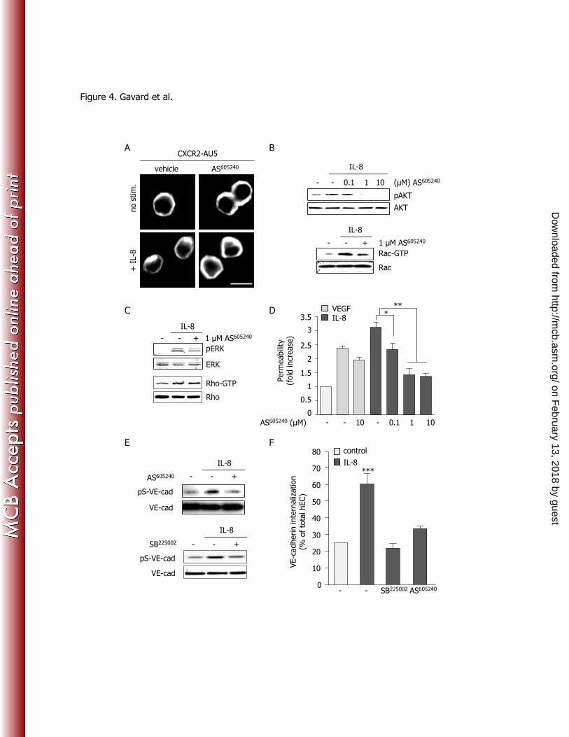
http://mcb.asm
.org/D
ownloaded from
11
IL-8 increases endothelial monolayer permeability through a CXCR2/Rac/PAK signaling axis. 230
We next tested the intracellular signaling events elicited in human endothelial cell monolayers in 231
response to the pro-angiogenic chemokine IL-8 (24). We first observed that IL-8 stimulation induced a 232
remarkable increase in endothelial monolayer permeability, comparable to that provoked by VEGF (Fig. 233
2A), as previously reported (34, 37). Chemical inhibition of VEGF receptor (VEGFR2) failed to block 234
IL-8-induced permeability and ERK activation, while it prevented VEGF-downstream events (Fig. 2A). 235
In contrast, the pharmacological inhibition of the chemokine receptor CXCR2 with SB225002
abolished 236
IL-8-provoked ERK phosphorylation and the increase of endothelial permeability, without interfering 237
with the effects of VEGF. SB225002
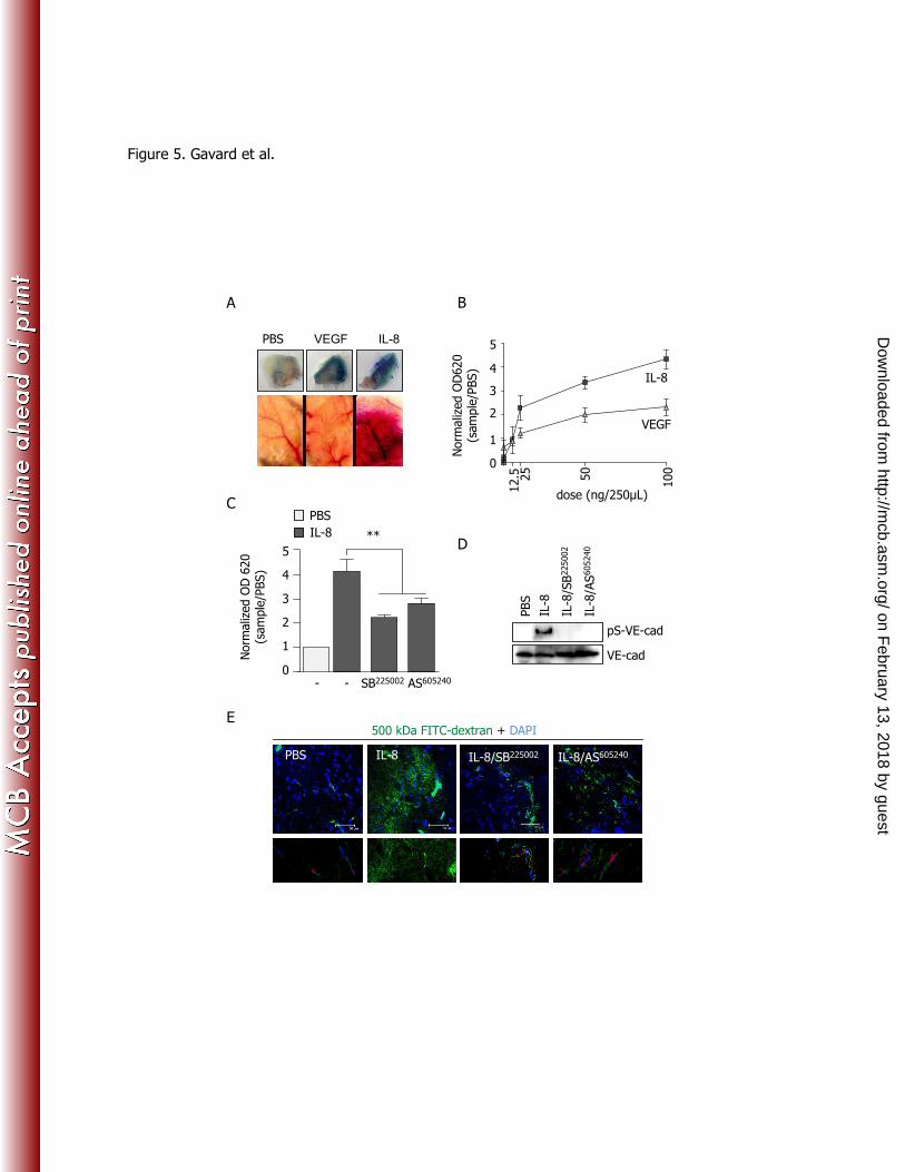
was identified as an antagonist of 125
I-labelled IL-8 binding to 238
CXCR2 with an IC50 about 20 nM, displaying a 150-fold selectivity for CXCR2 over CXCR1 and 239
multiple other G protein coupled receptors (51). The effectiveness of the SB225002
as a CXCR2 inhibitor 240
is further illustrated by the extinction of ERK phosphorylation in response to IL-8 in CXCR2-expressing 241
HEK-293T cells (Fig. S1). Furthermore, we engineered mouse endothelial cells deficient for CXCR2, by 242
lentiviral infections containing CXCR2 shRNA together with GFP (Fig. S2). GFP-sorted cells 243
expressing either a non-silencing shRNA or two independent shRNA sequences for CXCR2 were then 244
analyzed for CXCR2 protein expression. As shown in Supplemental Figure S2, CXCR2 deficient 245
endothelial cells showed a markedly reduced activation of ERK upon IL-8 stimulation, which together 246
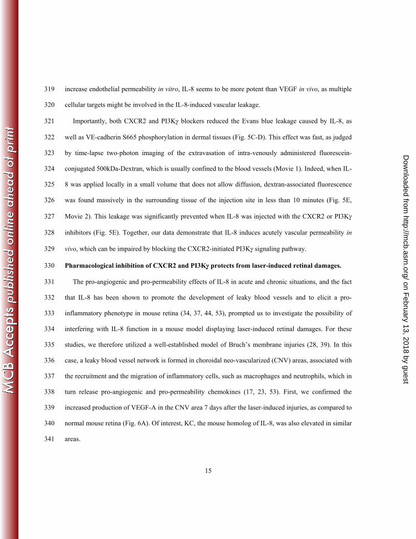
with the effects of the chemical inhibitor SB225002
, provides evidence that IL-8 signals mainly through 247
CXCR2 in mouse and human vascular endothelial cells. 248
Moreover, the stimulation of endothelial cells with ELR+ chemokines, GROg (CXCL1), GROく 249
(CXCL2), and to a lesser extent GROi (CXCL3), ENA-78 (CXCL5) and GCP-2 (CXCL6), which can 250
act on the G-protein coupled receptor (GPCR) CXCR2 (1, 3), could also augment endothelial 251
permeability (Fig. 2B). This increase in FITC-dextran passage could be hampered as well by the 252
on February 13, 2018 by guest
http://mcb.asm
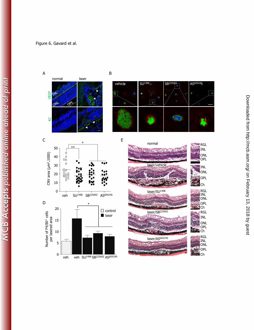
.org/D
ownloaded from
12
CXCR2 antagonist. In addition, IL-8 elicited PAK phosphorylation, as well as tyrosine and serine-253
phosphorylation of the endothelial-specific adhesion molecule, VE-cadherin (Fig. 2C). All these 254
signaling events contribute to trigger cell-cell junction disorganization (19, 43, 48, and Figure 1). 255
However, short-term IL-8 exposure did not provoke full VEGFR2 activation (Fig. 2C), in contrast to 256
that observed under prolonged stimulation with this chemokine in multilayered endothelial culture 257
conditions (34). Thus, our data suggested that IL-8 and VEGF may both modify the endothelial barrier 258
properties independently in an acute response. 259
Because the Rac/PAK signaling axis has emerged as a key regulatory mechanism controlling the 260
endothelial barrier development and function (18-20, 29, 43, 46), we investigated the contribution of this 261
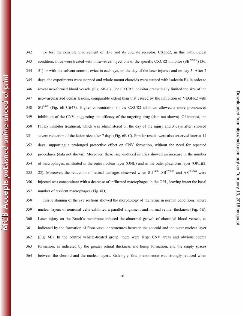
biochemical route to endothelial permeability downstream of IL-8 stimulation. First, we observed that, 262
in addition to ERK, IL-8 exposure induces Rac activation in endothelial cells in a CXCR2-dependent 263
manner (Fig. 2D). We then tested the involvement of Rac activation in endothelial permeability in 264
response to IL-8 by reducing its expression by RNA interference, rather than by the overexpression of 265
mutants forms of Rac, as we noted that overexpression of both active and dominant negative Rac 266
mutants alter the endothelial barrier properties per se (Fig. S3). Importantly, knocking-down Rac 267
resulted in a reduced activation of PAK, its direct downstream target, as well as a diminished 268
phosphorylation of VE-cadherin on its S665 residue (Fig. 2E). This effect correlated with the inability of 269
IL-8 to enhance the passage of fluorescein-conjugated dextran through endothelial monolayers, both 270
when Rac expression was reduced or when the activity of PAK was blocked (Fig. 2F). Thus, PAK may 271
act downstream from Rac to control the IL-8-dependent VE-cadherin phosphorylation and endothelial 272
permeability. 273
PI3K 伊 is involved in IL-8-induced increase of endothelial monolayer permeability. 274
on February 13, 2018 by guest
http://mcb.asm
.org/D
ownloaded from
13
We then explored the nature of the IL-8/CXCR2 initiated pathway leading to the Rac and PAK-275
dependent endothelial permeability. Surprisingly, whereas the blockade of Src kinases abolished the 276
activation of Rac and the enhanced passage of fluorescein-conjugated dextran through endothelial 277
monolayers in response to VEGF (Fig. 3A) (19), Src inhibition did not affect the stimulation of Rac and 278
endothelial permeability when stimulated by IL-8 (Fig. 3A-B). This further suggests that the signaling 279
pathway triggered by IL-8 diverges with the mechanism initiated by VEGF at the level of Src, while 280
both lead to endothelial permeability through Rac. In contrast, we observed a reduction of the VEGF- 281
and IL-8-induced permeability by pre-treating the cells with wortmannin, a general PI3K inhibitor (Fig. 282
3A). In addition, chemical inhibition of PI3K activity by LY294002
prevented IL-8-induced Rac and AKT 283
activation in endothelial cells, as well VE-cadherin phosphorylation, but leaving intact ERK activation 284
(Fig. 3C). 285
As there are multiple isoforms of PI3K catalytic subunits, which might play a distinct role 286
downstream from GPCRs and tyrosine kinase receptors (5, 12, 50), we explored the ability to interfere 287
selectively with the GPCR initiated signaling route. To this aim, we knocked down various PI3K 288
catalytic subunits using specific shRNA (Fig. 3D). The knockdown of PI3Ki伊 catalytic subunit prevented 289
the activation of Akt, a direct PI3K downstream target, as well as Rac activation and S665 VE-cadherin 290
phosphorylation upon IL-8 stimulation (Fig. 3E). Interestingly, PI3Ki伊 knockdown did not alter acute 291
ERK activation by IL-8, suggesting that a subset of CXCR2 signaling pathways are specifically affected 292
by the absence of PI3Ki (Fig. 3E) 伊. Importantly, we found that PI3Ki伊 knockdown strongly diminished 293
the IL-8-induced endothelial permeability, while interfering with the expression of PI3Kg did not alter it 294
(Fig. 3F). Therefore, blocking either CXCR2 or PI3Ki伊 selectively prevents the IL-8-provoked 295
endothelial barrier disruption without affecting the VEGF-initiated response. 296
on February 13, 2018 by guest
http://mcb.asm
.org/D
ownloaded from
14
PI3Ki has been recently shown as an integral signaling molecule involved in inflammation responses, 297
which might in turn provoke vascular activation (4, 7, 24, 37, 42). Interestingly, a PI3Ki伊-specific 298
inhibitor, namely AS605240
, was used throughout these studies and has proven its efficiency in 299
specifically blocking PI3Ki activity in different cell targets (38). We therefore decided to assess whether 300
AS605240
might also interfere with endothelial monolayer remodeling upon IL-8 exposure. First, we 301
checked that AS605240
treatment did not modify CXCR2 expression at the plasma membrane (Fig. 4A). 302
Importantly, the results we obtained by knocking down PI3Ki in endothelial cells were recapitulated 303
upon pharmacological treatment with AS605240
. Indeed, IL-8-dependent Rac activation and Akt 304
phosphorylation were prevented by AS605240
pre-treatment, while ERK and Rho activation were not (Fig. 305
4B-C). In addition, AS605240
interfered with IL-8, but not VEGF-triggered permeability (Fig. 4D). 306
Finally, AS605240
also impeded the IL-8-induced serine-phosphorylation of VE-cadherin, and its 307
endocytosis (Fig. 4E-F). Together, our data suggest that IL-8 utilizes a PI3Ki伊/Rac/PAK pathway to 308
promote VE-cadherin phosphorylation and endocytosis, thereby destabilizing the endothelial junctions. 309
Pharmacological inhibition of PI3K 伊 reduces IL-8/CXCR2-induced acute vascular permeability. 310
These findings prompted us to investigate the involvement of IL-8 in acute vascular permeability in 311
vivo. For this effort, we first used a well-characterized mouse model of acute permeability based on the 312
extravasation of Evans blue dye after sub-cutaneous injections of pro-permeability factors (20, 40, 49). 313
As shown in Fig. 5A, IL-8 induced a robust extravasation of the Evans blue in the surrounding tissue, 314
accompanied with micro-hemorrhages, a phenotype that macroscopically appears more severe than 315
those caused by VEGF. This effect was dose-dependent, and IL-8 enhanced plasma leakage was nearly 316
two fold greater than in response to VEGF after 1 hour exposure (Fig. 5B). Interestingly, while both 317
VEGF and IL-8 converge on Rac to stimulate the PAK-dependent phosphorylation of VE-cadherin to 318
on February 13, 2018 by guest
http://mcb.asm
.org/D
ownloaded from
15
increase endothelial permeability in vitro, IL-8 seems to be more potent than VEGF in vivo, as multiple 319
cellular targets might be involved in the IL-8-induced vascular leakage. 320
Importantly, both CXCR2 and PI3Ki伊 blockers reduced the Evans blue leakage caused by IL-8, as 321
well as VE-cadherin S665 phosphorylation in dermal tissues (Fig. 5C-D). This effect was fast, as judged 322
by time-lapse two-photon imaging of the extravasation of intra-venously administered fluorescein-323
conjugated 500kDa-Dextran, which is usually confined to the blood vessels (Movie 1). Indeed, when IL-324
8 was applied locally in a small volume that does not allow diffusion, dextran-associated fluorescence 325
was found massively in the surrounding tissue of the injection site in less than 10 minutes (Fig. 5E, 326
Movie 2). This leakage was significantly prevented when IL-8 was injected with the CXCR2 or PI3Ki伊 327
inhibitors (Fig. 5E). Together, our data demonstrate that IL-8 induces acutely vascular permeability in 328
vivo, which can be impaired by blocking the CXCR2-initiated PI3Ki伊 signaling pathway. 329
Pharmacological inhibition of CXCR2 and PI3K 伊 protects from laser-induced retinal damages. 330
The pro-angiogenic and pro-permeability effects of IL-8 in acute and chronic situations, and the fact 331
that IL-8 has been shown to promote the development of leaky blood vessels and to elicit a pro-332
inflammatory phenotype in mouse retina (34, 37, 44, 53), prompted us to investigate the possibility of 333
interfering with IL-8 function in a mouse model displaying laser-induced retinal damages. For these 334
studies, we therefore utilized a well-established model of Bruch’s membrane injuries (28, 39). In this 335
case, a leaky blood vessel network is formed in choroidal neo-vascularized (CNV) areas, associated with 336
the recruitment and the migration of inflammatory cells, such as macrophages and neutrophils, which in 337
turn release pro-angiogenic and pro-permeability chemokines (17, 23, 53). First, we confirmed the 338
increased production of VEGF-A in the CNV area 7 days after the laser-induced injuries, as compared to 339
normal mouse retina (Fig. 6A). Of interest, KC, the mouse homolog of IL-8, was also elevated in similar 340
areas. 341
on February 13, 2018 by guest
http://mcb.asm
.org/D
ownloaded from
16
To test the possible involvement of IL-8 and its cognate receptor, CXCR2, in this pathological 342
condition, mice were treated with intra-vitreal injections of the specific CXCR2 inhibitor (SB225002
) (36, 343
51) or with the solvent control, twice in each eye, on the day of the laser injuries and on day 3. After 7 344
days, the experiments were stopped and whole mount choroids were stained with isolectin B4 in order to 345
reveal neo-formed blood vessels (Fig. 6B-C). The CXCR2 inhibitor dramatically limited the size of the 346
neo-vascularized ocular lesions, comparable extent than that caused by the inhibition of VEGFR2 with 347
SU1498
(Fig. 6B-C)(47). Higher concentration of the CXCR2 inhibitor allowed a more pronounced 348
inhibition of the CNV, suggesting the efficacy of the targeting drug (data not shown). Of interest, the 349
PI3Ki伊 inhibitor treatment, which was administered on the day of the injury and 3 days after, showed 350
severe reduction of the lesion size after 7 days (Fig. 6B-C). Similar results were also observed later at 14 351
days, supporting a prolonged protective effect on CNV formation, without the need for repeated 352
procedures (data not shown). Moreover, these laser-induced injuries showed an increase in the number 353
of macrophages, infiltrated in the outer nuclear layer (ONL) and in the outer plexiform layer (OPL)(2, 354
23). Moreover, the reduction of retinal damages observed when SU1498
, SB225002
and AS605240
were 355
injected was concomitant with a decrease of infiltrated macrophages in the OPL, leaving intact the basal 356
number of resident macrophages (Fig. 6D). 357
Tissue staining of the eye sections showed the morphology of the retina in normal conditions, where 358
nuclear layers of neuronal cells exhibited a parallel alignment and normal retinal thickness (Fig. 6E). 359
Laser injury on the Bruch’s membrane induced the abnormal growth of choroidal blood vessels, as 360
indicated by the formation of fibro-vascular structures between the choroid and the outer nuclear layer 361
(Fig. 6E). In the control vehicle-treated group, there were large CNV areas and obvious edema 362
formation, as indicated by the greater retinal thickness and hump formation, and the empty spaces 363
between the choroid and the nuclear layers. Strikingly, this phenomenon was strongly reduced when 364
on February 13, 2018 by guest
http://mcb.asm
.org/D
ownloaded from
17
animals received intra-vitreal injections of CXCR2 or PI3Ki伊 inhibitors, leaving only mild lesions, 365
mainly caused by the direct physical wound provoked by the laser heat in the surrounding tissues (Fig. 366
6E). Similar results were obtained with the VEGFR2 inhibitor, as expected, reflecting the efficiency of 367
anti-VEGF therapies (21). Thus, PI3Ki and CXCR2 pharmacological inhibition can efficiently reduce 368
retinal damages and hyper-permeability-associated ocular lesions, with similar benefits than blocking 369
VEGFR activity. 370
371
Discussion 372
The emerging findings indicate that IL-8 promotes endothelial permeability by initiating the 373
activation of a CXCR2/PI3Ki-regulated signaling pathway, and that the pharmacological blockade of 374
CXCR2 or PI3Ki伊 can efficiently reduce blood vessel leakage and laser-induced ocular lesions in vivo. 375
This effect is likely dependent on the combined direct blockade of vascular permeability and pro-376
angiogenic effects of the mouse homolog of IL8, KC, as well as on the ability to interfere with the acute 377
macrophage activation and recruitment to the damaged retina, which may contribute together to 378
neovascularization by providing growth and survival factors for the vascular bed (2, 5, 6, 23, 44, 53). 379
Blocking CXCR2 and PI3Ki activity in vivo could significantly reduce the laser-induced hyper-380
permeability in the retina, to an extent comparable to that achieved by blocking VEGFR2 activity. It will 381
be important to assess in the future whether combination of therapies designed to target both IL-8 and 382
VEGF might improve available treatment options for retinal hyper-permeability. Thus, the ability to 383
inhibit the CXCR2/PI3Ki伊 signaling network and its multiple cellular targets may provide a novel 384
strategy for pharmacological intervention in many human diseases that involve inflammation and 385
enhanced vascular permeability, such as in neovascular age-related macular degeneration. 386
on February 13, 2018 by guest
http://mcb.asm
.org/D
ownloaded from
18
VEGF is the most described pro-angiogenic and pro-permeability factor, and interfering with VEGF 387
function has elicited considerable interest due to its clinical potential, in particular in the search for 388
molecular approaches to interfere with tumor-induced angiogenesis and ocular diseases (16, 21, 31). 389
However, despite clinical progress, recent reports have raised questions about potential adverse effects 390
of anti-VEGF antibodies or its pharmacological inhibitors, as they may not only affect aberrant vessel 391
outgrowth but also the normal vasculature (14, 15, 22), and may even induce permeability resembling to 392
VEGF stimulation in vivo (25). Other VEGF- and PDGF-related pro-angiogenic ligands have emerged 393
as new targets in blocking blood vessel outgrowth (15, 27, 28, 41), as well as direct intracellular targets 394
of the VEGF pro-permeability pathway, such as Src kinase (39). In this context, we observed that in 395
laser-induced retinal vasculature lesions, IL-8 expression is increased concomitantly with VEGF 396
expression in the damaged and inflamed retinal regions. Our data suggest that blocking CXCR2 might 397
reduce vascular leakage and perivascular inflammation by preventing both endothelial permeability and 398
acute recruitment of immune cells to the retinal wounds, thus halting the development of ocular lesions. 399
Of interest, whereas VEGF and IL-8 converge on Rac to stimulate the PAK-dependent 400
phosphorylation of VE-cadherin to increase endothelial permeability, they both stimulate Rac activation 401
by a divergent mechanism. Indeed, VEGF acts through the Src-family kinases to increase vascular 402
leakage, while blocking Src activity in several mouse models for human diseases has successfully 403
interfered with metastatic cell dissemination, brain stroke neural damages, and neovascular ocular 404
lesions (33, 39, 47). Interestingly, IL-8 does not require Src activity to stimulate Rac, but instead signals 405
through a specific PI3K isoform, PI3Ki, to promote Rac activation, thus resulting in endothelial cell-cell 406
junction dismantlement. This observation provided an opportunity to selectively target the chemokine-407
initiated pathway by blocking PI3Ki. Indeed, we obtained evidence that IL-8 induces a rapid and severe 408
vascular leakage that can be efficiently prevented by the pharmacological inhibition of CXCR2 and 409
on February 13, 2018 by guest
http://mcb.asm
.org/D
ownloaded from
19
PI3Ki in vivo. Strikingly, CXCR2 and PI3Ki inhibition dramatically limited the size of the ocular 410
lesions, edema formation, blood vessel growth, as well as the infiltration of macrophages at the site of 411
injuries induced by laser burns. 412
Taken together, the ability to inhibit the CXCR2/PI3Ki signaling network and its multiple cellular 413
targets may provide a novel strategy to treat pathological neovascularization in damaged retina and in 414
the tumor microenvironment, as many angiogenic mechanisms are similarly deregulated in ocular 415
diseases and tumor vascularization (11). Furthermore, the ability to combine available FDA-approved 416
drugs targeting VEGF-A with new therapeutic pharmacological inhibitors of IL-8 and its downstream 417
signaling molecules may help identify additional treatment options for pathological neovascularization. 418
Indeed, these studies may provide a rationale for the evaluation of anti-IL8 and CXCR2 inhibitors or 419
antibodies, as part of new multi-targeted therapies in ocular diseases and other pathological conditions 420
characterized by hyper-permeability of the vasculature. 421
422
Acknowledgements: 423
This research was supported by the Intramural Research Program of NIH, National Institute of Dental 424
and Craniofacial Research and by funding from the “Centre National de la Recherche Scientifique” 425
(CNRS), and the "Projets Exploratoires/Premier Soutien” (PEPS 2008) program held by CNRS. We 426
thank A. Sodhi (Wilmer Eye Institute, The Johns Hopkins Hospital, Baltimore, MD) for the initial 427
cloning of CXCR2. 428
429
Authorship: JG, RW, XL and JSG planned the experimental design, JG, XH, YQ, DM and AM 430
conducted the experiments, JG, XL and JSG analyzed data, JG and JSG wrote the paper. 431
Conflict-of-interest disclosure: The authors declare no competing financial interests. 432
on February 13, 2018 by guest
http://mcb.asm
.org/D
ownloaded from
20
References: 433
1. Addison, C. L., T. O. Daniel, M. D. Burdick, H. Liu, J. E. Ehlert, Y. Y. Xue, L. Buechi, A. 434
Walz, A. Richmond, and R. M. Strieter. 2000. The CXC chemokine receptor 2, CXCR2, is the 435
putative receptor for ELR+ CXC chemokine-induced angiogenic activity. J Immunol 165:5269-436
77. 437
2. Ambati, J., A. Anand, S. Fernandez, E. Sakurai, B. C. Lynn, W. A. Kuziel, B. J. Rollins, and 438
B. K. Ambati. 2003. An animal model of age-related macular degeneration in senescent Ccl-2- or 439
Ccr-2-deficient mice. Nat Med 9:1390-7. 440
3. Balkwill, F. 2004. Cancer and the chemokine network. Nat Rev Cancer 4:540-50. 441
4. Barber, D. F., A. Bartolome, C. Hernandez, J. M. Flores, C. Redondo, C. Fernandez-Arias, 442
M. Camps, T. Ruckle, M. K. Schwarz, S. Rodriguez, A. C. Martinez, D. Balomenos, C. 443
Rommel, and A. C. Carrera. 2005. PI3Kgamma inhibition blocks glomerulonephritis and 444
extends lifespan in a mouse model of systemic lupus. Nat Med 11:933-5. 445
5. Barberis, L., and E. Hirsch. 2008. Targeting phosphoinositide 3-kinase gamma to fight 446
inflammation and more. Thromb Haemost 99:279-85. 447
6. Belperio, J. A., M. P. Keane, D. A. Arenberg, C. L. Addison, J. E. Ehlert, M. D. Burdick, and 448
R. M. Strieter. 2000. CXC chemokines in angiogenesis. J Leukoc Biol 68:1-8. 449
7. Camps, M., T. Ruckle, H. Ji, V. Ardissone, F. Rintelen, J. Shaw, C. Ferrandi, C. Chabert, C. 450
Gillieron, B. Francon, T. Martin, D. Gretener, D. Perrin, D. Leroy, P. A. Vitte, E. Hirsch, M. 451
P. Wymann, R. Cirillo, M. K. Schwarz, and C. Rommel. 2005. Blockade of PI3Kgamma 452
suppresses joint inflammation and damage in mouse models of rheumatoid arthritis. Nat Med 453
11:936-43. 454
8. Carmeliet, P. 2005. Angiogenesis in life, disease and medicine. Nature 438:932-6. 455
on February 13, 2018 by guest
http://mcb.asm
.org/D
ownloaded from
21
9. Dejana, E. 2004. Endothelial cell-cell junctions: happy together. Nat Rev Mol Cell Biol 5:261-70. 456
10. Dirkx, A. E., M. G. Oude Egbrink, J. Wagstaff, and A. W. Griffioen. 2006. 457
Monocyte/macrophage infiltration in tumors: modulators of angiogenesis. J Leukoc Biol 80:1183-458
96. 459
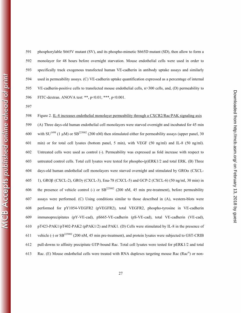
11. Dorrell, M. I., E. Aguilar, L. Scheppke, F. H. Barnett, and M. Friedlander. 2007. 460
Combination angiostatic therapy completely inhibits ocular and tumor angiogenesis. Proc Natl 461
Acad Sci U S A 104:967-72. 462
12. Doukas, J., W. Wrasidlo, G. Noronha, E. Dneprovskaia, R. Fine, S. Weis, J. Hood, A. 463
Demaria, R. Soll, and D. Cheresh. 2006. Phosphoinositide 3-kinase gamma/delta inhibition 464
limits infarct size after myocardial ischemia/reperfusion injury. Proc Natl Acad Sci U S A 465
103:19866-71. 466
13. Edgell, C. J., C. C. McDonald, and J. B. Graham. 1983. Permanent cell line expressing human 467
factor VIII-related antigen established by hybridization. Proc Natl Acad Sci U S A 80:3734-7. 468
14. Eremina, V., J. A. Jefferson, J. Kowalewska, H. Hochster, M. Haas, J. Weisstuch, C. 469
Richardson, J. B. Kopp, M. G. Kabir, P. H. Backx, H. P. Gerber, N. Ferrara, L. Barisoni, C. 470
E. Alpers, and S. E. Quaggin. 2008. VEGF inhibition and renal thrombotic microangiopathy. N 471
Engl J Med 358:1129-36. 472
15. Fischer, C., B. Jonckx, M. Mazzone, S. Zacchigna, S. Loges, L. Pattarini, E. Chorianopoulos, 473
L. Liesenborghs, M. Koch, M. De Mol, M. Autiero, S. Wyns, S. Plaisance, L. Moons, N. van 474
Rooijen, M. Giacca, J. M. Stassen, M. Dewerchin, D. Collen, and P. Carmeliet. 2007. Anti-475
PlGF inhibits growth of VEGF(R)-inhibitor-resistant tumors without affecting healthy vessels. 476
Cell 131:463-75. 477
16. Folkman, J. 2006. Angiogenesis. Annu Rev Med 57:1-18. 478
on February 13, 2018 by guest
http://mcb.asm
.org/D
ownloaded from
22
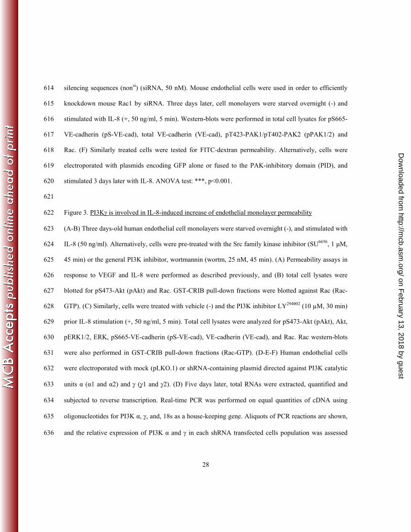
17. Friedlander, M. 2007. Fibrosis and diseases of the eye. J Clin Invest 117:576-86. 479
18. Garrett, T. A., J. D. Van Buul, and K. Burridge. 2007. VEGF-induced Rac1 activation in 480
endothelial cells is regulated by the guanine nucleotide exchange factor Vav2. Exp Cell Res 481
313:3285-97. 482
19. Gavard, J., and J. S. Gutkind. 2006. VEGF controls endothelial-cell permeability by promoting 483
the beta-arrestin-dependent endocytosis of VE-cadherin. Nat Cell Biol 8:1223-34. 484
20. Gavard, J., V. Patel, and J. S. Gutkind. 2008. Angiopoietin-1 prevents VEGF-induced 485
endothelial permeability by sequestering Src through mDia. Dev Cell 14:25-36. 486
21. Gragoudas, E. S., A. P. Adamis, E. T. Cunningham, Jr., M. Feinsod, and D. R. Guyer. 2004. 487
Pegaptanib for neovascular age-related macular degeneration. N Engl J Med 351:2805-16. 488
22. Kamba, T., and D. M. McDonald. 2007. Mechanisms of adverse effects of anti-VEGF therapy 489
for cancer. Br J Cancer 96:1788-95. 490
23. Kelly, J., A. Ali Khan, J. Yin, T. A. Ferguson, and R. S. Apte. 2007. Senescence regulates 491
macrophage activation and angiogenic fate at sites of tissue injury in mice. J Clin Invest 492
117:3421-6. 493
24. Koch, A. E., P. J. Polverini, S. L. Kunkel, L. A. Harlow, L. A. DiPietro, V. M. Elner, S. G. 494
Elner, and R. M. Strieter. 1992. Interleukin-8 as a macrophage-derived mediator of 495
angiogenesis. Science 258:1798-801. 496
25. Lee, S., T. T. Chen, C. L. Barber, M. C. Jordan, J. Murdock, S. Desai, N. Ferrara, A. Nagy, 497
K. P. Roos, and M. L. Iruela-Arispe. 2007. Autocrine VEGF signaling is required for vascular 498
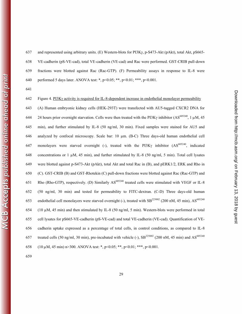
homeostasis. Cell 130:691-703. 499
on February 13, 2018 by guest
http://mcb.asm
.org/D
ownloaded from
23
26. Li, A., S. Dubey, M. L. Varney, B. J. Dave, and R. K. Singh. 2003. IL-8 directly enhanced 500
endothelial cell survival, proliferation, and matrix metalloproteinases production and regulated 501
angiogenesis. J Immunol 170:3369-76. 502
27. Li, X., M. Tjwa, L. Moons, P. Fons, A. Noel, A. Ny, J. M. Zhou, J. Lennartsson, H. Li, A. 503
Luttun, A. Ponten, L. Devy, A. Bouche, H. Oh, A. Manderveld, S. Blacher, D. Communi, P. 504
Savi, F. Bono, M. Dewerchin, J. M. Foidart, M. Autiero, J. M. Herbert, D. Collen, C. H. 505
Heldin, U. Eriksson, and P. Carmeliet. 2005. Revascularization of ischemic tissues by PDGF-506
CC via effects on endothelial cells and their progenitors. J Clin Invest 115:118-27. 507
28. Li, Y., F. Zhang, N. Nagai, Z. Tang, S. Zhang, P. Scotney, J. Lennartsson, C. Zhu, Y. Qu, C. 508
Fang, J. Hua, O. Matsuo, G. H. Fong, H. Ding, Y. Cao, K. G. Becker, A. Nash, C. H. Heldin, 509
and X. Li. 2008. VEGF-B inhibits apoptosis via VEGFR-1-mediated suppression of the 510
expression of BH3-only protein genes in mice and rats. J Clin Invest 118:913-923. 511
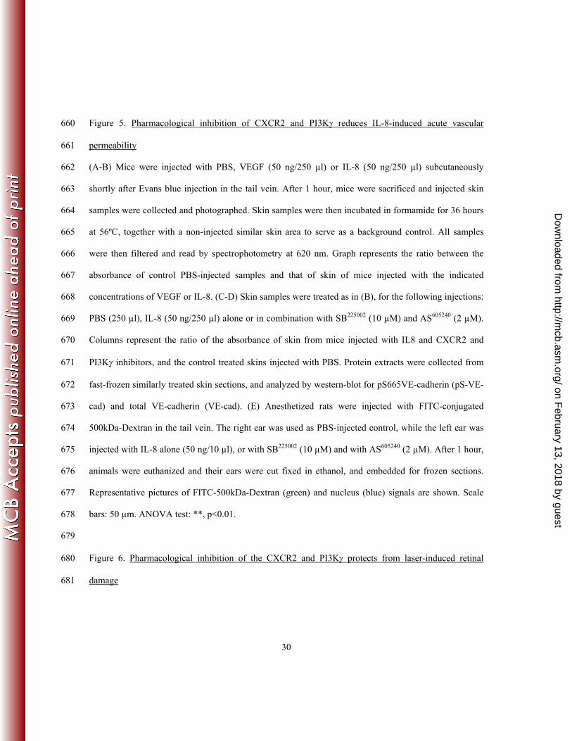
29. Liu, J., S. D. Fraser, P. W. Faloon, E. L. Rollins, J. Vom Berg, O. Starovic-Subota, A. L. 512
Laliberte, J. N. Chen, F. C. Serluca, and S. J. Childs. 2007. A betaPix Pak2a signaling pathway 513
regulates cerebral vascular stability in zebrafish. Proc Natl Acad Sci U S A 104:13990-5. 514
30. Marinissen, M. J., M. Chiariello, T. Tanos, O. Bernard, S. Narumiya, and J. S. Gutkind. 515
2004. The small GTP-binding protein RhoA regulates c-jun by a ROCK-JNK signaling axis. Mol 516
Cell 14:29-41. 517
31. Olsson, A. K., A. Dimberg, J. Kreuger, and L. Claesson-Welsh. 2006. VEGF receptor 518
signalling - in control of vascular function. Nat Rev Mol Cell Biol 7:359-71. 519
32. Orr, A. W., R. Stockton, M. B. Simmers, J. M. Sanders, I. J. Sarembock, B. R. Blackman, 520
and M. A. Schwartz. 2007. Matrix-specific p21-activated kinase activation regulates vascular 521
permeability in atherogenesis. J Cell Biol 176:719-27. 522
on February 13, 2018 by guest
http://mcb.asm
.org/D
ownloaded from
24
33. Paul, R., Z. G. Zhang, B. P. Eliceiri, Q. Jiang, A. D. Boccia, R. L. Zhang, M. Chopp, and D. 523
A. Cheresh. 2001. Src deficiency or blockade of Src activity in mice provides cerebral protection 524
following stroke. Nat Med 7:222-7. 525
34. Petreaca, M. L., M. Yao, Y. Liu, K. Defea, and M. Martins-Green. 2007. Transactivation of 526
vascular endothelial growth factor receptor-2 by interleukin-8 (IL-8/CXCL8) is required for IL-527
8/CXCL8-induced endothelial permeability. Mol Biol Cell 18:5014-23. 528
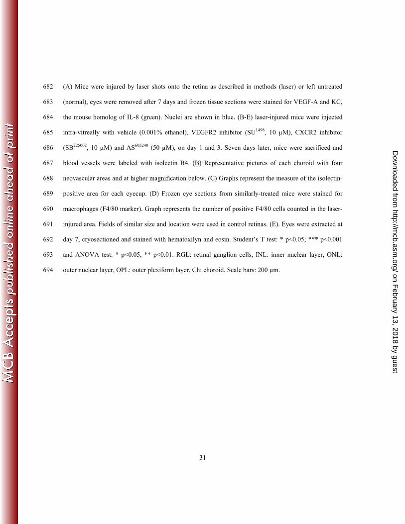
35. Red-Horse, K., Y. Crawford, F. Shojaei, and N. Ferrara. 2007. Endothelium-529
microenvironment interactions in the developing embryo and in the adult. Dev Cell 12:181-94. 530
36. Reutershan, J. 2006. CXCR2--the receptor to hit? Drug News Perspect 19:615-23. 531
37. Reutershan, J., M. A. Morris, T. L. Burcin, D. F. Smith, D. Chang, M. S. Saprito, and K. 532
Ley. 2006. Critical role of endothelial CXCR2 in LPS-induced neutrophil migration into the lung. 533
J Clin Invest 116:695-702. 534
38. Ruckle, T., M. K. Schwarz, and C. Rommel. 2006. PI3K[gamma] inhibition: towards an 'aspirin 535
of the 21st century'? Nat Rev Drug Discov 5:903-918. 536
39. Scheppke, L., E. Aguilar, R. F. Gariano, R. Jacobson, J. Hood, J. Doukas, J. Cao, G. 537
Noronha, S. Yee, S. Weis, M. B. Martin, R. Soll, D. A. Cheresh, and M. Friedlander. 2008. 538
Retinal vascular permeability suppression by topical application of a novel VEGFR2/Src kinase 539
inhibitor in mice and rabbits. J Clin Invest 118:2337-46. 540
40. Senger, D. R., S. J. Galli, A. M. Dvorak, C. A. Perruzzi, V. S. Harvey, and H. F. Dvorak. 541
1983. Tumor cells secrete a vascular permeability factor that promotes accumulation of ascites 542
fluid. Science 219:983-5. 543
41. Sennino, B., B. L. Falcon, D. McCauley, T. Le, T. McCauley, J. C. Kurz, A. Haskell, D. M. 544
Epstein, and D. M. McDonald. 2007. Sequential loss of tumor vessel pericytes and endothelial 545
on February 13, 2018 by guest
http://mcb.asm
.org/D
ownloaded from
25
cells after inhibition of platelet-derived growth factor B by selective aptamer AX102. Cancer Res 546
67:7358-67. 547
42. Smith, D. F., T. L. Deem, A. C. Bruce, J. Reutershan, D. Wu, and K. Ley. 2006. Leukocyte 548
phosphoinositide-3 kinase {gamma} is required for chemokine-induced, sustained adhesion under 549
flow in vivo. J Leukoc Biol 80:1491-9. 550
43. Stockton, R. A., E. Schaefer, and M. A. Schwartz. 2004. p21-activated kinase regulates 551
endothelial permeability through modulation of contractility. J Biol Chem 279:46621-30. 552
44. Strieter, R. M., S. L. Kunkel, V. M. Elner, C. L. Martonyi, A. E. Koch, P. J. Polverini, and S. 553
G. Elner. 1992. Interleukin-8. A corneal factor that induces neovascularization. Am J Pathol 554
141:1279-84. 555
45. Taddei, A., C. Giampietro, A. Conti, F. Orsenigo, F. Breviario, V. Pirazzoli, M. Potente, C. 556
Daly, S. Dimmeler, and E. Dejana. 2008. Endothelial adherens junctions control tight junctions 557
by VE-cadherin-mediated upregulation of claudin-5. Nat Cell Biol 10:923-34. 558
46. Tan, W., T. R. Palmby, J. Gavard, P. Amornphimoltham, Y. Zheng, and J. S. Gutkind. 2008. 559
An essential role for Rac1 in endothelial cell function and vascular development. FASEB J. 560
22:1829-1838. 561
47. Weis, S., J. Cui, L. Barnes, and D. Cheresh. 2004. Endothelial barrier disruption by VEGF-562
mediated Src activity potentiates tumor cell extravasation and metastasis. J Cell Biol 167:223-9. 563
48. Weis, S., S. Shintani, A. Weber, R. Kirchmair, M. Wood, A. Cravens, H. McSharry, A. 564
Iwakura, Y. S. Yoon, N. Himes, D. Burstein, J. Doukas, R. Soll, D. Losordo, and D. Cheresh. 565
2004. Src blockade stabilizes a Flk/cadherin complex, reducing edema and tissue injury following 566
myocardial infarction. J Clin Invest 113:885-94. 567
on February 13, 2018 by guest
http://mcb.asm
.org/D
ownloaded from
26
49. Weis, S. M., and D. A. Cheresh. 2005. Pathophysiological consequences of VEGF-induced 568
vascular permeability. Nature 437:497-504. 569
50. Wetzker, R., and C. Rommel. 2004. Phosphoinositide 3-kinases as targets for therapeutic 570
intervention. Curr Pharm Des 10:1915-22. 571
51. White, J. R., J. M. Lee, P. R. Young, R. P. Hertzberg, A. J. Jurewicz, M. A. Chaikin, K. 572
Widdowson, J. J. Foley, L. D. Martin, D. E. Griswold, and H. M. Sarau. 1998. Identification 573
of a potent, selective non-peptide CXCR2 antagonist that inhibits interleukin-8-induced neutrophil 574
migration. J Biol Chem 273:10095-8. 575
52. Wong, T. Y., G. Liew, and P. Mitchell. 2007. Clinical update: new treatments for age-related 576
macular degeneration. Lancet 370:204-6. 577
53. Zhou, J., L. Pham, N. Zhang, S. He, M. A. Gamulescu, C. Spee, S. J. Ryan, and D. R. Hinton. 578
2005. Neutrophils promote experimental choroidal neovascularization. Mol Vis 11:414-24. 579
580
Figure Legends 581
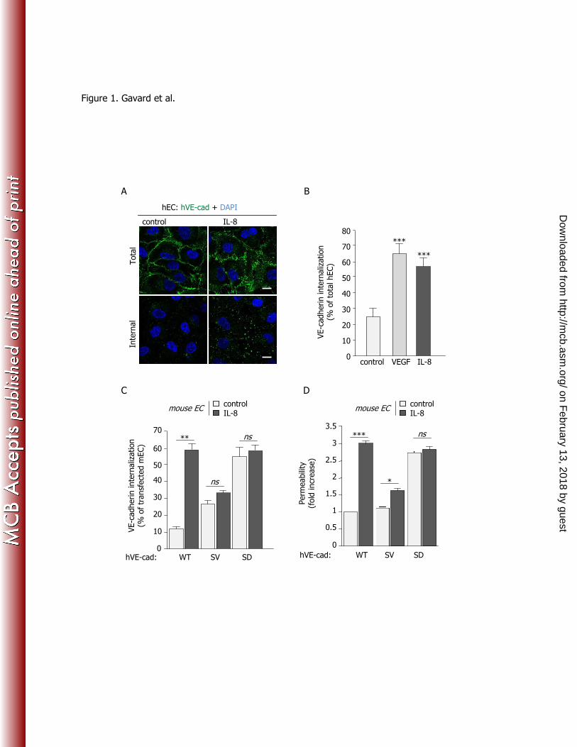
Figure 1. VE-cadherin internalization is required for IL-8-induced endothelial monolayer permeability 582
(A) Three days-old human endothelial monolayers were starved overnight and were subjected to VE-583
cadherin staining (green) in living cells at 4°C, then left unstimulated (control) or exposed to IL-8 (50 584
ng/ml, 30 min) at 37°C. Cells were either fixed (total) or membrane-bound antibodies were stripped 585
away by a mild acid wash before fixation (internal). Representative confocal acquisitions of VE-586
cadherin staining are shown. Nuclei counterstaining is shown in blue (DAPI). Scale bars: 10 µm. (B) 587
Quantification of VE-cadherin uptake expressed as a percentage of internal VE-cadherin-positive cells 588
to total cells, in control conditions, VEGF- or IL-8 treated cells (50 ng/ml, 30 min), n>300. (C-D) Sub-589
confluent mouse endothelial cells were transfected with human VE-cadherin wild-type (WT), its non-590
on February 13, 2018 by guest
http://mcb.asm
.org/D
ownloaded from
27
phosphorylable S665V mutant (SV), and its phospho-mimetic S665D mutant (SD), then allow to form a 591
monolayer for 48 hours before overnight starvation. Mouse endothelial cells were used in order to 592
specifically track exogenous transfected human VE-cadherin in antibody uptake assays and similarly 593
used in permeability assays. (C) VE-cadherin uptake quantification expressed as a percentage of internal 594
VE-cadherin-positive cells to transfected mouse endothelial cells, n>300 cells, and, (D) permeability to 595
FITC-dextran. ANOVA test: **, p<0.01; ***, p<0.001. 596
597
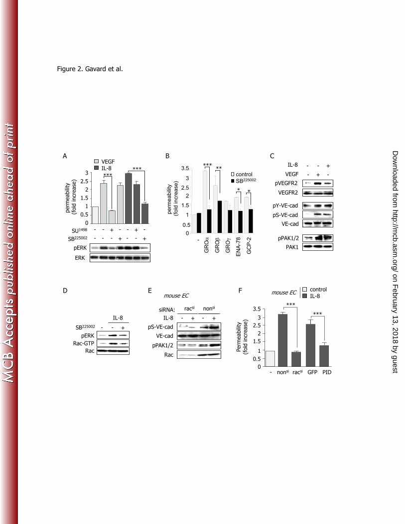
Figure 2. IL-8 increases endothelial monolayer permeability through a CXCR2/Rac/PAK signaling axis 598
(A) Three days-old human endothelial cell monolayers were starved overnight and incubated for 45 min 599
with SU1498
(1 µM) or SB225002
(200 nM) then stimulated either for permeability assays (upper panel, 30 600
min) or for total cell lysates (bottom panel, 5 min), with VEGF (50 ng/ml) and IL-8 (50 ng/ml). 601
Untreated cells were used as control (-). Permeability was expressed as fold increase with respect to 602
untreated control cells. Total cell lysates were tested for phospho-(p)ERK1/2 and total ERK. (B) Three 603
days-old human endothelial cell monolayers were starved overnight and stimulated by GROc (CXCL-604
1), GROd (CXCL-2), GROi (CXCL-3), Ena-78 (CXCL-5) and GCP-2 (CXCL-6) (50 ng/ml, 30 min) in 605
the presence of vehicle control (-) or SB225002
(200 nM, 45 min pre-treatment), before permeability 606
assays were performed. (C) Using conditions similar to those described in (A), western-blots were 607
performed for pY1054-VEGFR2 (pVEGFR2), total VEGFR2, phospho-tyrosine in VE-cadherin 608
immunoprecipitates (pY-VE-cad), pS665-VE-cadherin (pS-VE-cad), total VE-cadherin (VE-cad), 609
pT423-PAK1/pT402-PAK2 (pPAK1/2) and PAK1. (D) Cells were stimulated by IL-8 in the presence of 610
vehicle (-) or SB225002
(200 nM, 45 min pre-treatment), and protein lysates were subjected to GST-CRIB 611
pull-downs to affinity precipitate GTP-bound Rac. Total cell lysates were tested for pERK1/2 and total 612
Rac. (E) Mouse endothelial cells were treated with RNA duplexes targeting mouse Rac (Racsi) or non-613
on February 13, 2018 by guest
http://mcb.asm
.org/D
ownloaded from
28
silencing sequences (nonsi) (siRNA, 50 nM). Mouse endothelial cells were used in order to efficiently 614
knockdown mouse Rac1 by siRNA. Three days later, cell monolayers were starved overnight (-) and 615
stimulated with IL-8 (+, 50 ng/ml, 5 min). Western-blots were performed in total cell lysates for pS665-616
VE-cadherin (pS-VE-cad), total VE-cadherin (VE-cad), pT423-PAK1/pT402-PAK2 (pPAK1/2) and 617
Rac. (F) Similarly treated cells were tested for FITC-dextran permeability. Alternatively, cells were 618
electroporated with plasmids encoding GFP alone or fused to the PAK-inhibitory domain (PID), and 619
stimulated 3 days later with IL-8. ANOVA test: ***, p<0.001. 620
621
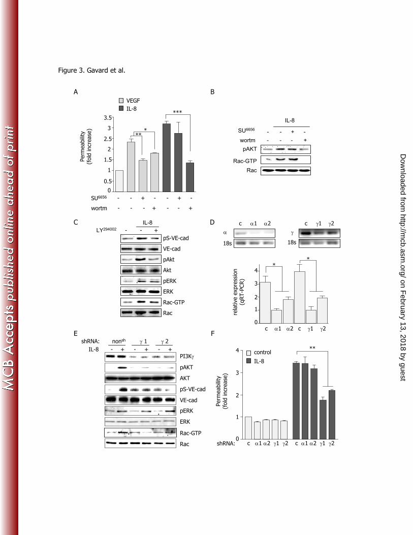
Figure 3. PI3Ki伊 is involved in IL-8-induced increase of endothelial monolayer permeability 622
(A-B) Three days-old human endothelial cell monolayers were starved overnight (-), and stimulated with 623
IL-8 (50 ng/ml). Alternatively, cells were pre-treated with the Src family kinase inhibitor (SU6656
, 1 たM, 624
45 min) or the general PI3K inhibitor, wortmannin (wortm, 25 nM, 45 min). (A) Permeability assays in 625
response to VEGF and IL-8 were performed as described previously, and (B) total cell lysates were 626
blotted for pS473-Akt (pAkt) and Rac. GST-CRIB pull-down fractions were blotted against Rac (Rac-627
GTP). (C) Similarly, cells were treated with vehicle (-) and the PI3K inhibitor LY294002
(10 µM, 30 min) 628
prior IL-8 stimulation (+, 50 ng/ml, 5 min). Total cell lysates were analyzed for pS473-Akt (pAkt), Akt, 629
pERK1/2, ERK, pS665-VE-cadherin (pS-VE-cad), VE-cadherin (VE-cad), and Rac. Rac western-blots 630
were also performed in GST-CRIB pull-down fractions (Rac-GTP). (D-E-F) Human endothelial cells 631
were electroporated with mock (pLKO.1) or shRNA-containing plasmid directed against PI3K catalytic 632
units g (g1 and g2) and i伊 (i伊伊1 and i伊2). (D) Five days later, total RNAs were extracted, quantified and 633
subjected to reverse transcription. Real-time PCR was performed on equal quantities of cDNA using 634
oligonucleotides for PI3K g, i伊, and, 18s as a house-keeping gene伊. Aliquots of PCR reactions are shown, 635
and the relative expression of PI3K g and i in each shRNA transfected cells population was assessed 636
on February 13, 2018 by guest
http://mcb.asm
.org/D
ownloaded from
29
and represented using arbitrary units. (E) Western-blots for PI3Ki伊, p-S473-Akt (pAkt), total Akt, pS665-637
VE-cadherin (pS-VE-cad), total VE-cadherin (VE-cad) and Rac were performed. GST-CRIB pull-down 638
fractions were blotted against Rac (Rac-GTP). (F) Permeability assays in response to IL-8 were 639
performed 5 days later. ANOVA test: *, p<0.05; **, p<0.01; ***, p<0.001. 640
641
Figure 4. PI3Ki activity is required for IL-8-dependent increase in endothelial monolayer permeability 642
(A) Human embryonic kidney cells (HEK-293T) were transfected with AU5-tagged CXCR2 DNA for 643
24 hours prior overnight starvation. Cells were then treated with the PI3Ki inhibitor (AS605240
, 1 oM, 45 644
min), and further stimulated by IL-8 (50 ng/ml, 30 min). Fixed samples were stained for AU5 and 645
analyzed by confocal microscopy. Scale bar: 10 µm. (B-C) Three days-old human endothelial cell 646
monolayers were starved overnight (-), treated with the PI3Ki inhibitor (AS605240
, indicated 647
concentrations or 1 µM, 45 min), and further stimulated by IL-8 (50 ng/ml, 5 min). Total cell lysates 648
were blotted against p-S473-Akt (pAkt), total Akt and total Rac in (B), and pERK1/2, ERK and Rho in 649
(C). GST-CRIB (B) and GST-Rhotekin (C) pull-down fractions were blotted against Rac (Rac-GTP) and 650
Rho (Rho-GTP), respectively. (D) Similarly AS605240
treated cells were stimulated with VEGF or IL-8 651
(50 ng/ml, 30 min) and tested for permeability to FITC-dextran. (C-D) Three days-old human 652
endothelial cell monolayers were starved overnight (-), treated with SB225002
(200 nM, 45 min), AS605240
653
(10 µM, 45 min) and then stimulated by IL-8 (50 ng/ml, 5 min). Western-blots were performed in total 654
cell lysates for pS665-VE-cadherin (pS-VE-cad) and total VE-cadherin (VE-cad). Quantification of VE-655
cadherin uptake expressed as a percentage of total cells, in control conditions, as compared to IL-8 656
treated cells (50 ng/ml, 30 min), pre-incubated with vehicle (-), SB225002
(200 nM, 45 min) and AS605240
657
(10 oM, 45 min) n>300. ANOVA test: *, p<0.05; **, p<0.01; ***, p<0.001. 658
659
on February 13, 2018 by guest
http://mcb.asm
.org/D
ownloaded from
30
Figure 5. Pharmacological inhibition of CXCR2 and PI3Ki伊 reduces IL-8-induced acute vascular 660
permeability 661
(A-B) Mice were injected with PBS, VEGF (50 ng/250 µl) or IL-8 (50 ng/250 µl) subcutaneously 662
shortly after Evans blue injection in the tail vein. After 1 hour, mice were sacrificed and injected skin 663
samples were collected and photographed. Skin samples were then incubated in formamide for 36 hours 664
at 56ºC, together with a non-injected similar skin area to serve as a background control. All samples 665
were then filtered and read by spectrophotometry at 620 nm. Graph represents the ratio between the 666
absorbance of control PBS-injected samples and that of skin of mice injected with the indicated 667
concentrations of VEGF or IL-8. (C-D) Skin samples were treated as in (B), for the following injections: 668
PBS (250 µl), IL-8 (50 ng/250 µl) alone or in combination with SB225002
(10 µM) and AS605240
(2 µM). 669
Columns represent the ratio of the absorbance of skin from mice injected with IL8 and CXCR2 and 670
PI3Ki伊 inhibitors, and the control treated skins injected with PBS. Protein extracts were collected from 671
fast-frozen similarly treated skin sections, and analyzed by western-blot for pS665VE-cadherin (pS-VE-672
cad) and total VE-cadherin (VE-cad). (E) Anesthetized rats were injected with FITC-conjugated 673
500kDa-Dextran in the tail vein. The right ear was used as PBS-injected control, while the left ear was 674
injected with IL-8 alone (50 ng/10 µl), or with SB225002
(10 µM) and with AS605240
(2 µM). After 1 hour, 675
animals were euthanized and their ears were cut fixed in ethanol, and embedded for frozen sections. 676
Representative pictures of FITC-500kDa-Dextran (green) and nucleus (blue) signals are shown. Scale 677
bars: 50 µm. ANOVA test: **, p<0.01. 678
679
Figure 6. Pharmacological inhibition of the CXCR2 and PI3Ki伊 protects from laser-induced retinal 680
damage 681
on February 13, 2018 by guest
http://mcb.asm
.org/D
ownloaded from
31
(A) Mice were injured by laser shots onto the retina as described in methods (laser) or left untreated 682
(normal), eyes were removed after 7 days and frozen tissue sections were stained for VEGF-A and KC, 683
the mouse homolog of IL-8 (green). Nuclei are shown in blue. (B-E) laser-injured mice were injected 684
intra-vitreally with vehicle (0.001% ethanol), VEGFR2 inhibitor (SU1498
, 10 µM), CXCR2 inhibitor 685
(SB225002
, 10 µM) and AS605240
(50 µM), on day 1 and 3. Seven days later, mice were sacrificed and 686
blood vessels were labeled with isolectin B4. (B) Representative pictures of each choroid with four 687
neovascular areas and at higher magnification below. (C) Graphs represent the measure of the isolectin-688
positive area for each eyecup. (D) Frozen eye sections from similarly-treated mice were stained for 689
macrophages (F4/80 marker). Graph represents the number of positive F4/80 cells counted in the laser-690
injured area. Fields of similar size and location were used in control retinas. (E). Eyes were extracted at 691
day 7, cryosectioned and stained with hematoxilyn and eosin. Student’s T test: * p<0.05; *** p<0.001 692
and ANOVA test: * p<0.05, ** p<0.01. RGL: retinal ganglion cells, INL: inner nuclear layer, ONL: 693
outer nuclear layer, OPL: outer plexiform layer, Ch: choroid. Scale bars: 200 µm. 694 on February 13, 2018 by guest
http://mcb.asm
.org/D
ownloaded from
***
A
control IL-8
Tota
lIn
tern
al
hEC: hVE-cad + DAPI
C
hVE-cad: WT SV SD
VE-c
adherin inte
rnaliz
ation
(% o
f tr
ansf
ect
ed m
EC)
D
0
Perm
eabili
ty(f
old
incr
ease
)
hVE-cad: WT SV SD
1
2
3
1.5
2.5
3.5
0.5
0
10
20
30
40
50
60
70
80
control VEGF IL-8
VE-c
adherin inte
rnaliz
ation
(% o
f to
tal hEC)
B
***
***
0
10
20
30
40
50
60
70
control IL-8
mouse ECcontrol IL-8
mouse EC
** ns
ns
ns
*
Figure 1. Gavard et al.
on February 13, 2018 by guest
http://mcb.asm
.org/D
ownloaded from
A B
pVEGFR2
IL-8
VEGF - + -
- - +
VEGFR2
VE-cad
pPAK1/2
pS-VE-cad
PAK1
pY-VE-cad
VEGF IL-8
SU1498
perm
eabili
ty
(fold
incr
ease
)
SB225002
0
0.5
1
1.5
2.5
2
3***
***
pERK
ERK
-
-
-
-
-
+
+
-
-
-
-
+
+
-
D F
Rac
Rac-GTP
SB225002
IL-8
pERK
- - + pS-VE-cad
VE-cad
pPAK1/2
Rac
IL-8
racsi nonsi siRNA:
- + - +
E
- nonsi racsi GFP PID
***
Perm
eabili
ty(f
old
incr
ease
)
***
0
0.5
1
1.5
2
2.5
3
3.5
C
mouse EC
EN
A-7
8
perm
eabili
ty
(fo
ld incre
ase)
0
1
2
3control
SB225002
-
GR
Oc
GR
Od
GR
Oi
GC
P-2
*****
* *
0.5
1.5
2.5
3.5
control IL-8
mouse EC
Figure 2. Gavard et al.
on February 13, 2018 by guest
http://mcb.asm
.org/D
ownloaded from
c c1 c2 i1 i2 c c1 c2 i1 i2 0
1
2
3
4
Perm
eabili
ty(f
old
incr
ease
)
**
shRNA:
control
IL-8
E
pS-VE-cad
PI3Ki
A
- - - + - - +wortm
- - + - - + -SU6656
0
1
2
3
1.5
2.5
3.5
0.5
Perm
eabili
ty(f
old
incr
ease
)
VEGF
***
***
IL-8
B
18s
c
c c1 c2
18s
i
c i1 i2
*
rela
tive e
xpre
ssio
n(q
RT-P
CR)
c c1 c2 c i1 i20
1
2
3
4*
F
SU6656
wortm - - - +
IL-8
Rac
pAKT
- - + -
C D
Rac-GTP
Figure 3. Gavard et al.
pAkt
pS-VE-cad
VE-cad
Akt
Rac-GTP
Rac
IL-8 - + - + - +
pAKT
AKT
shRNA: nonsh i 1 i 2
VE-cad
pERK
ERK
Rac-GTP
Rac
- - +LY294002
IL-8
pERK
ERK
on February 13, 2018 by guest
http://mcb.asm
.org/D
ownloaded from
1.5
1
0
2.5
- - 0.1 1 10
IL-8
(µM) AS605240
pAKT
AKT
- - +
IL-8
1 µM AS605240
Rac-GTP
Rac
VEGF IL-8
AS605240 (µM) - - 10 - 0.1 1 10
2
3
3.5
0.5
Perm
eabili
ty(f
old
incr
ease
)
***
FE
AS605240 - - +
pS-VE-cad
VE-cad
IL-8
A B
0
10
20
30
40
50
60
70
80
AS605240SB225002- -
control
IL-8 ***
VE-c
adherin inte
rnaliz
ation
(% o
f to
tal hEC)
C D
- - +
IL-8
1 µM AS605240
pERK
ERK
+ I
L-8
no s
tim
.
CXCR2-AU5
vehicle AS605240
Rho-GTP
Rho
pS-VE-cad
VE-cad
SB225002 - - +
IL-8
Figure 4. Gavard et al.
on February 13, 2018 by guest
http://mcb.asm
.org/D
ownloaded from
Norm
aliz
ed O
D620
(sam
ple
/PBS)
25
50
100
12.5
VEGF
IL-8
dose (ng/250µL)
0
1
2
3
4
5
C
PBS IL-8
PBS VEGF IL-8
pS-VE-cad
PBS
IL-8
IL-8
/SB
225002
IL-8
/AS
605240
VE-cad
D
E
0SB225002 AS605240
Norm
aliz
ed O
D 6
20
(sam
ple
/PBS)
1
2
3
4
5
**
- -
500 kDa FITC-dextran + DAPI
PBS IL-8 IL-8/SB225002 IL-8/AS605240
A B
PBS
IL-8
Figure 5. Gavard et al.
on February 13, 2018 by guest
http://mcb.asm
.org/D
ownloaded from
veh veh SU1498 SB225002 AS605240
A
normal laser
VEG
F
KC
ONL
INL
OPL
vehicle SU1498 SB225002 AS605240
B
C
CN
V a
rea (
µm
2*1000)
veh SU1498 SB225002 AS605240
*
0
10
20
30
40
**50RGL
INL
ONLOPL
Ch
normal
laser/vehicle
laser/SU1498
laser/SB225002
laser/AS605240
ONL
OPL
Ch
INL
RGLINLONL
OPL
Ch
RGL
INL
OPLONL
Ch
RGLINL
OPL
ONL
E
0
5
10
15
20
Num
ber
of
F4/8
0+
cells
per
lase
red a
rea
*D
control
laser
Figure 6. Gavard et al.
on February 13, 2018 by guest
http://mcb.asm
.org/D
ownloaded from