Embed Size (px)
Citation preview
Case Report
Logan Dance, MD; Patricia Cornejo, MD; Mittun Patel, MD
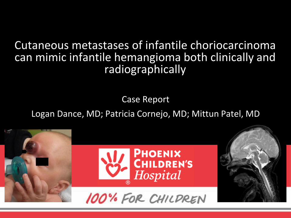
Cutaneous metastases of infantile choriocarcinoma can mimic infantile hemangioma both clinically and
radiographically
Disclosures
• None
Objectives
• Understand the clinical and radiographic challenge of distinguishing between a common infantile hemangioma and a much less common solitary cutaneous metastasis
• Understand the time-critical nature of accurate diagnosis of infantile choriocarcinoma
• Understand the unique predilection of infantile malignancies to metastasize to the skin, most commonly lymphoma/leukemia
• Know when to suggest an alternate diagnosis despite the initial clinical presumption of infantile hemangioma
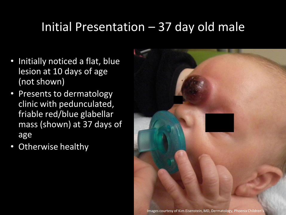
Initial Presentation – 37 day old male
• Initially noticed a flat, blue lesion at 10 days of age (not shown)
• Presents to dermatology clinic with pedunculated, friable red/blue glabellar mass (shown) at 37 days of age
• Otherwise healthy
Images courtesy of Kim Eisenstein, MD, Dermatology, Phoenix Children’s
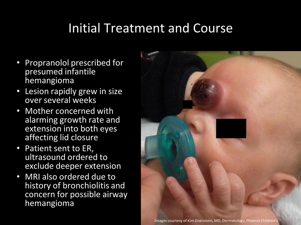
Initial Treatment and Course
• Propranolol prescribed for presumed infantile hemangioma
• Lesion rapidly grew in size over several weeks
• Mother concerned with alarming growth rate and extension into both eyes affecting lid closure
• Patient sent to ER, ultrasound ordered to exclude deeper extension
• MRI also ordered due to history of bronchiolitis and concern for possible airway hemangioma
Images courtesy of Kim Eisenstein, MD, Dermatology, Phoenix Children’s
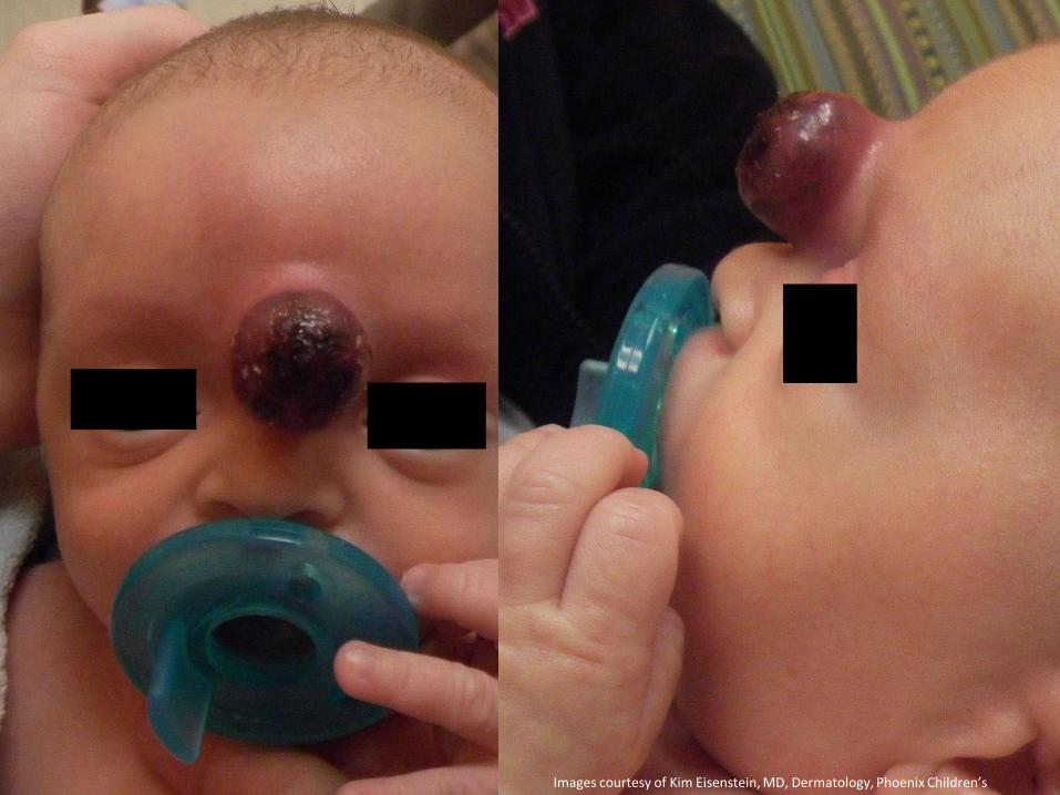
Images courtesy of Kim Eisenstein, MD, Dermatology, Phoenix Children’s
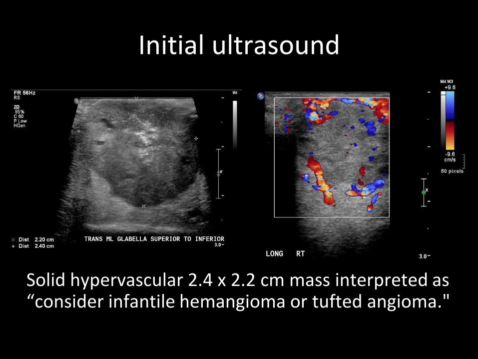
Initial ultrasound
Solid hypervascular 2.4 x 2.2 cm mass interpreted as “consider infantile hemangioma or tufted angioma."
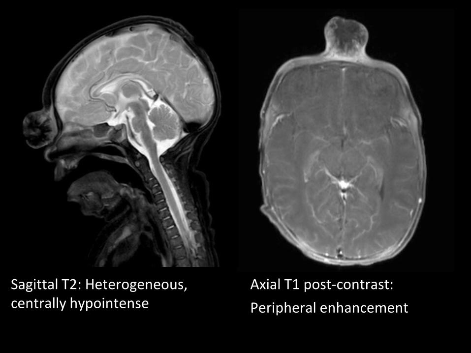
Axial T1 post-contrast:
Peripheral enhancement
Sagittal T2: Heterogeneous, centrally hypointense
MRI results
• No airway or neck hemangioma(s)
• Although some MRI features not characteristic of clinical diagnosis of infantile hemangioma, the exam was interpreted as “vascular mass arising from glabella. Coordinate with upcoming biopsy to confirm diagnosis of hemangioma.”
Initial work-up
• Lab testing: anemia (7.4 mg/dL) and low platelets (95 K/uL)
• Persistent tachycardia, tachypnea • Differential at this time:
– tufted angioma – infantile hemangioma – kaposiform hemangioendothelioma with
concern for possible Kasabach-Merritt syndrome
• Biopsy requested
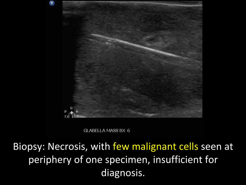
Biopsy: Necrosis, with few malignant cells seen at periphery of one specimen, insufficient for
diagnosis.
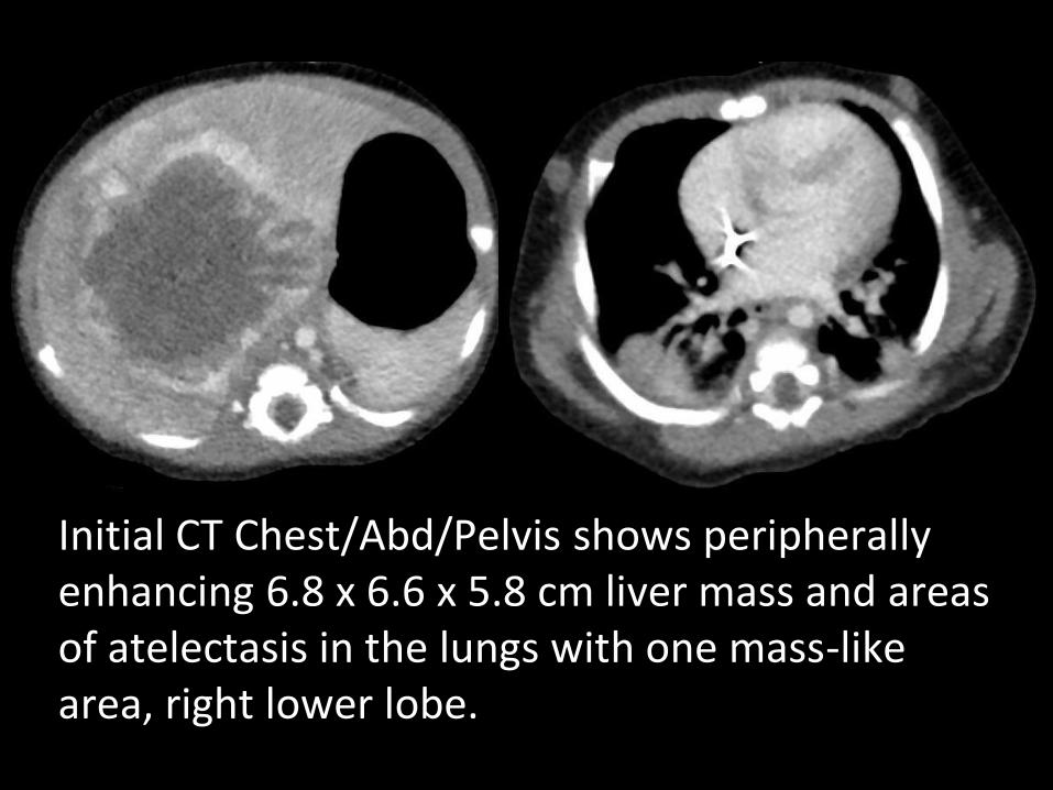
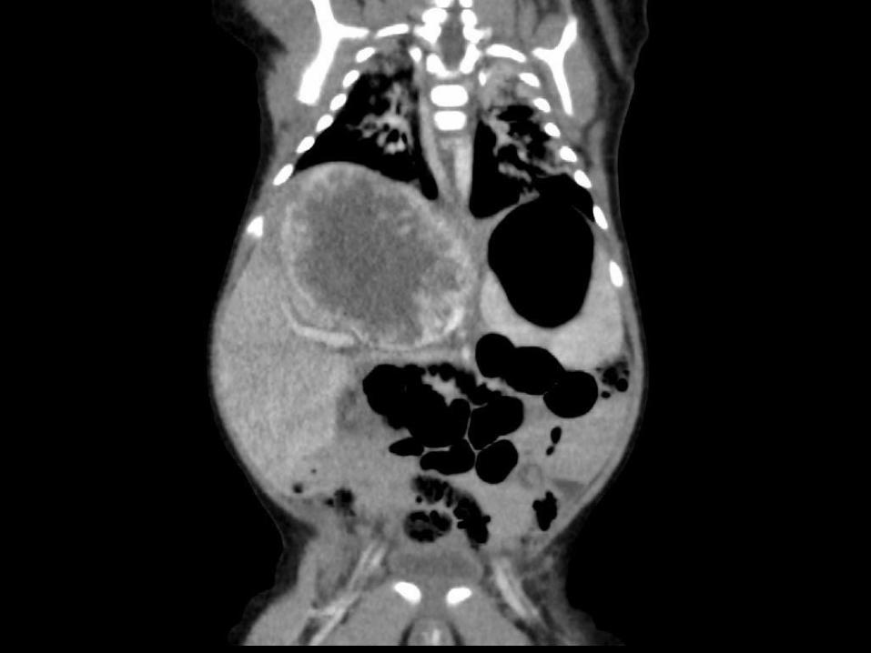
Initial CT Chest/Abd/Pelvis shows peripherally enhancing 6.8 x 6.6 x 5.8 cm liver mass and areas of atelectasis in the lungs with one mass-like area, right lower lobe.
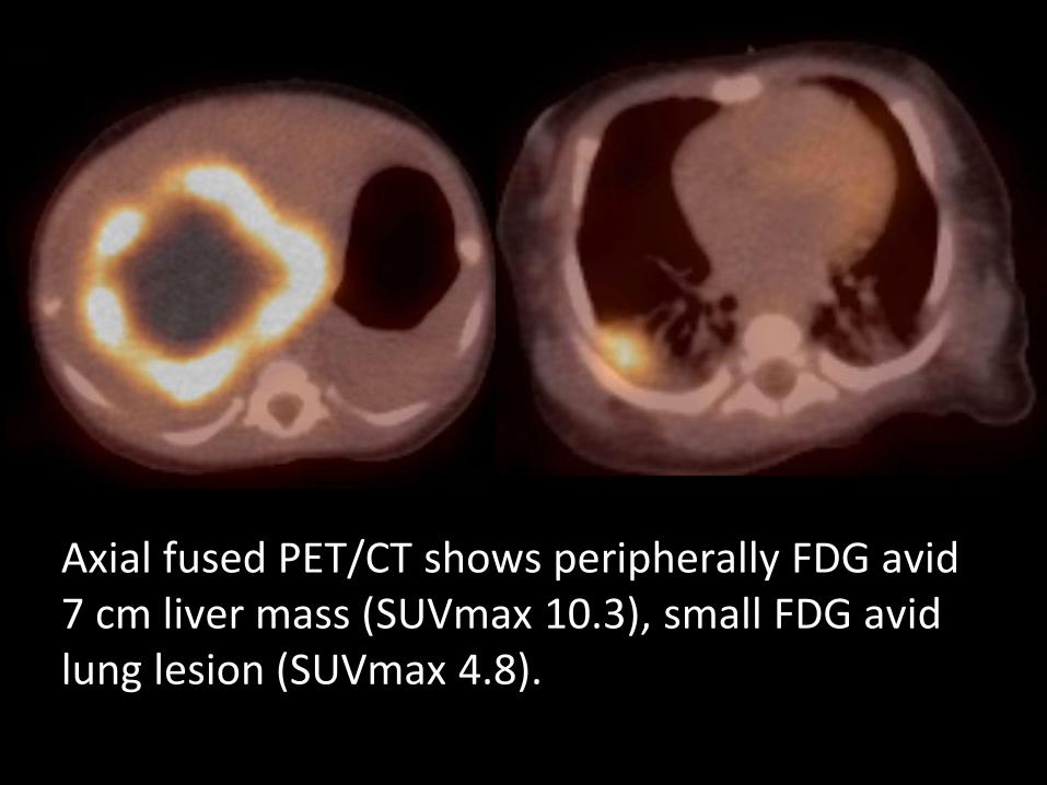
Axial fused PET/CT shows peripherally FDG avid 7 cm liver mass (SUVmax 10.3), small FDG avid lung lesion (SUVmax 4.8).
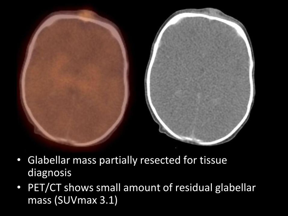
• Glabellar mass partially resected for tissue diagnosis
• PET/CT shows small amount of residual glabellar mass (SUVmax 3.1)
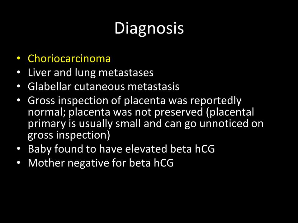
Diagnosis
• Choriocarcinoma • Liver and lung metastases • Glabellar cutaneous metastasis • Gross inspection of placenta was reportedly
normal; placenta was not preserved (placental primary is usually small and can go unnoticed on gross inspection)
• Baby found to have elevated beta hCG • Mother negative for beta hCG
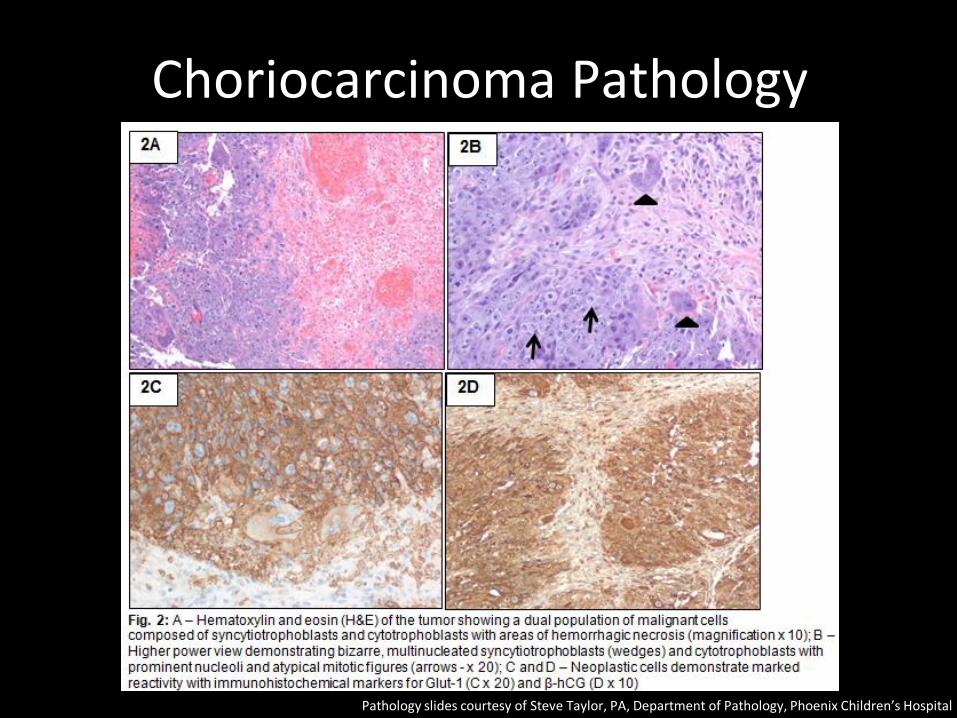
Choriocarcinoma Pathology
Pathology slides courtesy of Steve Taylor, PA, Department of Pathology, Phoenix Children’s Hospital
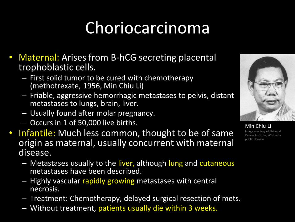
Choriocarcinoma
• Maternal: Arises from B-hCG secreting placental trophoblastic cells. – First solid tumor to be cured with chemotherapy
(methotrexate, 1956, Min Chiu Li) – Friable, aggressive hemorrhagic metastases to pelvis, distant
metastases to lungs, brain, liver. – Usually found after molar pregnancy. – Occurs in 1 of 50,000 live births.
• Infantile: Much less common, thought to be of same origin as maternal, usually concurrent with maternal disease. – Metastases usually to the liver, although lung and cutaneous
metastases have been described. – Highly vascular rapidly growing metastases with central
necrosis. – Treatment: Chemotherapy, delayed surgical resection of mets. – Without treatment, patients usually die within 3 weeks.
Min Chiu Li Image courtesy of National Cancer Institute, Wikipedia public domain
Clinical Course
• Respiratory failure and severe anemia required ICU stay and multiple blood and platelet transfusion
• Rapid improvement shortly after starting chemotherapy
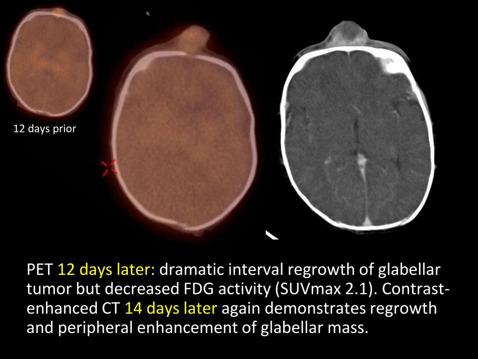
PET 12 days later: dramatic interval regrowth of glabellar tumor but decreased FDG activity (SUVmax 2.1). Contrast-enhanced CT 14 days later again demonstrates regrowth and peripheral enhancement of glabellar mass.
12 days prior
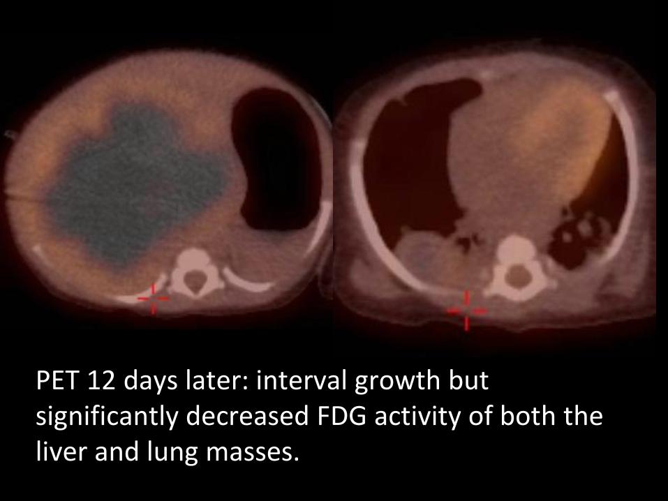
PET 12 days later: interval growth but significantly decreased FDG activity of both the liver and lung masses.
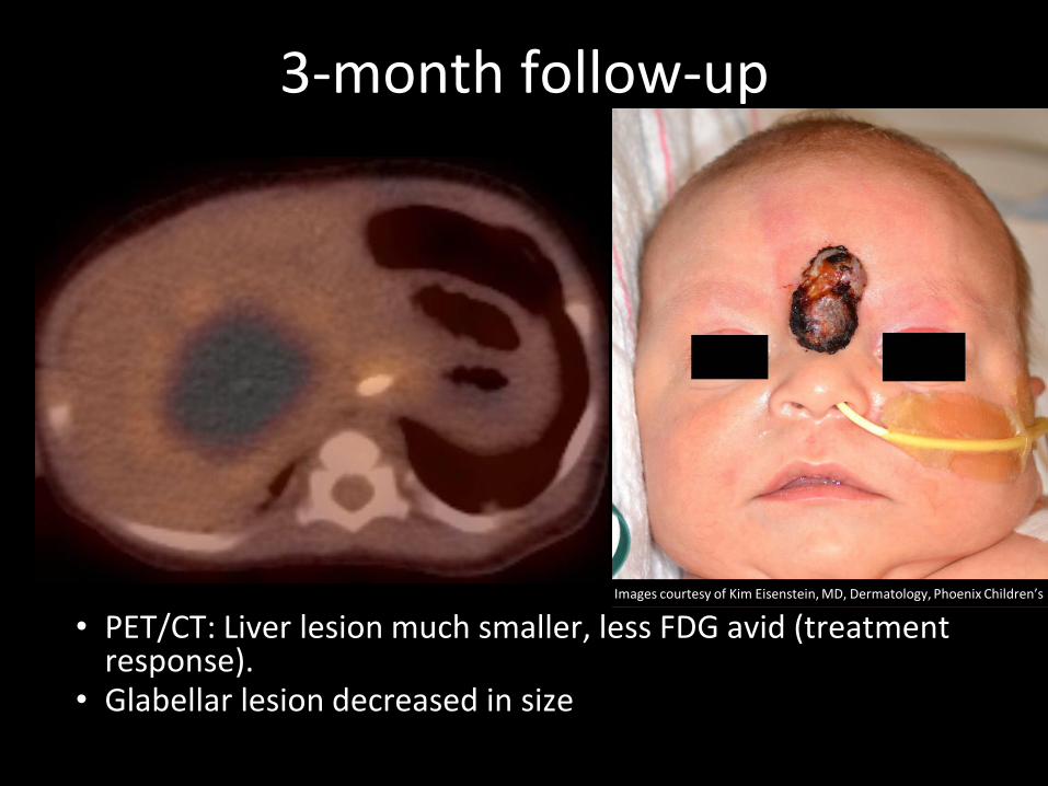
• PET/CT: Liver lesion much smaller, less FDG avid (treatment response).
• Glabellar lesion decreased in size
Images courtesy of Kim Eisenstein, MD, Dermatology, Phoenix Children’s
3-month follow-up
Treatment
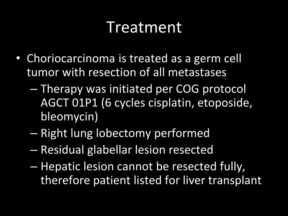
• Choriocarcinoma is treated as a germ cell tumor with resection of all metastases
– Therapy was initiated per COG protocol AGCT 01P1 (6 cycles cisplatin, etoposide, bleomycin)
– Right lung lobectomy performed
– Residual glabellar lesion resected
– Hepatic lesion cannot be resected fully, therefore patient listed for liver transplant
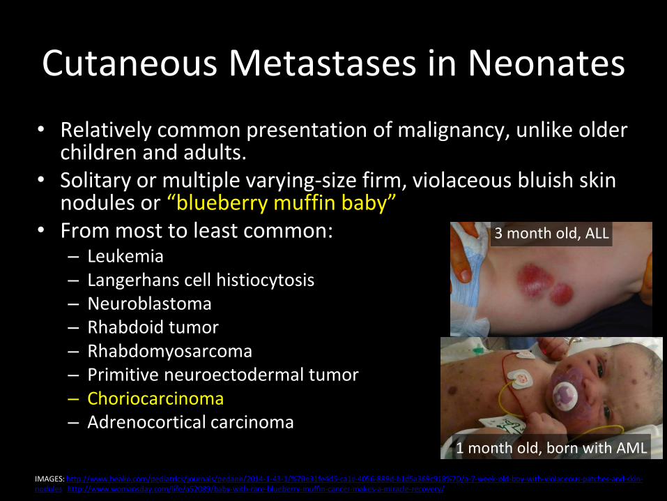
Cutaneous Metastases in Neonates
• Relatively common presentation of malignancy, unlike older children and adults.
• Solitary or multiple varying-size firm, violaceous bluish skin nodules or “blueberry muffin baby”
• From most to least common: – Leukemia – Langerhans cell histiocytosis – Neuroblastoma – Rhabdoid tumor – Rhabdomyosarcoma – Primitive neuroectodermal tumor – Choriocarcinoma – Adrenocortical carcinoma
IMAGES: http://www.healio.com/pediatrics/journals/pedann/2014-1-43-1/%7Be31fe4d5-ca1c-4056-889d-b1d5a369c918%7D/a-7-week-old-boy-with-violaceous-patches-and-skin-nodules http://www.womansday.com/life/a52089/baby-with-rare-blueberry-muffin-cancer-makes-a-miracle-recovery/
3 month old, ALL
1 month old, born with AML
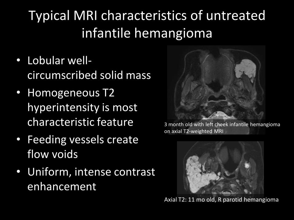
Typical MRI characteristics of untreated infantile hemangioma
• Lobular well-circumscribed solid mass
• Homogeneous T2 hyperintensity is most characteristic feature
• Feeding vessels create flow voids
• Uniform, intense contrast enhancement
3 month old with left cheek infantile hemangioma on axial T2-weighted MRI
Axial T2: 11 mo old, R parotid hemangioma
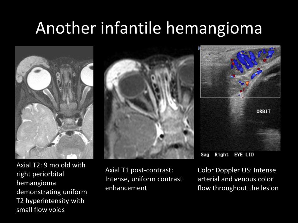
Another infantile hemangioma
Axial T2: 9 mo old with right periorbital hemangioma demonstrating uniform T2 hyperintensity with small flow voids
Axial T1 post-contrast: Intense, uniform contrast enhancement
Color Doppler US: Intense arterial and venous color flow throughout the lesion
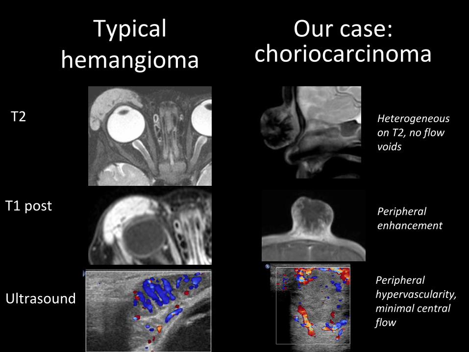
Typical hemangioma
T2
T1 post
Ultrasound
Our case: choriocarcinoma
Heterogeneous on T2, no flow voids
Peripheral enhancement
Peripheral hypervascularity, minimal central flow
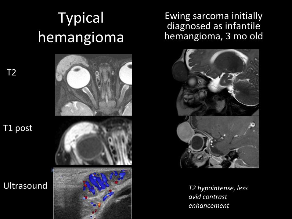
Typical hemangioma
T2
T1 post
Ultrasound
Ewing sarcoma initially diagnosed as infantile
hemangioma, 3 mo old
T2 hypointense, less avid contrast enhancement
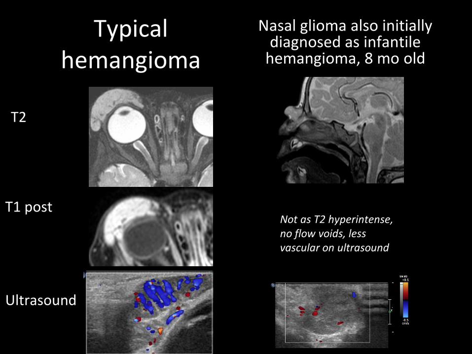
Typical hemangioma
T2
T1 post
Ultrasound
Nasal glioma also initially diagnosed as infantile
hemangioma, 8 mo old
Not as T2 hyperintense, no flow voids, less vascular on ultrasound
Further Discussion on Choriocarcinoma
• Clinical presentation: The unusual predilection of infantile malignancies for cutaneous metastases allows their early discovery and creates vivid clinical stigmata not easily forgotten. While relatively common in infancy, such presentations are uncommon in children and adults. The unsightly cutaneous metastasis in our case hastened the diagnosis and life-saving treatment.
• Known Mimic: The misdiagnosis of infantile choriocarcinoma for infantile hemangioma has occurred previously, as described in a 2014 case report by Brooks and Nolting also with a solitary cutaneous metastasis on the face.
Further Discussion on Choriocarcinoma
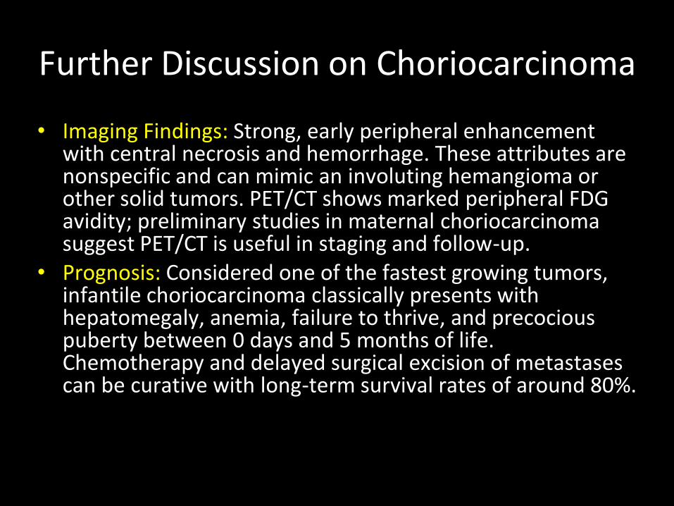
• Imaging Findings: Strong, early peripheral enhancement with central necrosis and hemorrhage. These attributes are nonspecific and can mimic an involuting hemangioma or other solid tumors. PET/CT shows marked peripheral FDG avidity; preliminary studies in maternal choriocarcinoma suggest PET/CT is useful in staging and follow-up.
• Prognosis: Considered one of the fastest growing tumors, infantile choriocarcinoma classically presents with hepatomegaly, anemia, failure to thrive, and precocious puberty between 0 days and 5 months of life. Chemotherapy and delayed surgical excision of metastases can be curative with long-term survival rates of around 80%.
Conclusion
• Beware the potential of solitary cutaneous metastases to mimic hemangioma both clinically and radiographically
• Typical MRI appearance of infantile hemangioma includes T2 hyperintensity, flow voids, and uniform contrast enhancement
• Atypical features can suggest alternate diagnosis • Early diagnosis and treatment for infantile
choriocarcinoma is critical for survival! • PET/CT may be useful for staging and follow-up
Sources
• Hart I et al, Cutaneous metastases in neonates: a review. Pediatric Dermatology, 28:2, 85-93, 2011.
• Yarris JP et al, Roy Hertz, MD (1909-2002): The cure of choriocarcinoma and its impact on the development of chemotherapy for cancer. Gynecologic Oncology, 89:2, 193-198, 2003.
• Tidy JA et al, Presentation and management of choriocarcinoma after nonmolar pregnancy. The British Journal of Obstetrics and Gynaecology, 102:9, 715–719, 1995.
• Brooks T, Nolting L, Cutaneous Manifestation of Metastatic Infantile Choriocarcinoma. Case Reports in Pediatrics. Volume 2014, 3 pages, http://dx.doi.org/10.1155/2014/104652
• Van der Hoef M et al, Solitary infantile choriocarcinoma of the liver: MRI findings. Pediatric Radiology. 34:10, 820-823, 2004.
• Mapelli P et al, Role of 18F-FDG PET in the management of gestational trophoblastic neoplasia, Eur. J. Nuc. Med. and Mol. Imaging, 40:4, 505–513, 2013.
• Yoon JM et al, Treatment of infantile choriocarcinoma of the liver. Pediatric Blood Cancer, 49:1, 99-102, 2007.
• McNally OM et al, Successful treatment of mother and baby with metastatic choriocarcinoma, International Journal of Gynecological Cancer, 12:4, 394–398, 2002.


![Choriocarcinoma syndrome complicating a mixed testicular ...choriocarcinoma are very rare (0, 3% of all GCT) [8]. βHCG is always secreted by choriocarcinoma and plays an important](https://img.pdfslide.us/doc/110x75/5e366cd2a1f24370d80dcb00/choriocarcinoma-syndrome-complicating-a-mixed-testicular-choriocarcinoma-are.jpg)