Embed Size (px)
Citation preview
CURRENT DIAGNOSTIC, PHARMACEUTIC AND RECONSTRUCTIVE
SURGICAL METHODS IN THE MANAGEMENT OF FACIAL NERVE PALSY
Ph.D. Thesis
Gabriella Kecskés M.D.
Department of Otolaryngology, Head- and Neck Surgery
University of Szeged
Szeged
2012

1
CURRENT DIAGNOSTIC, PHARMACEUTIC AND RECONSTRUCTIVE
SURGICAL METHODS IN THE MANAGEMENT OF FACIAL NERVE PALSY
Ph.D. Thesis
Gabriella Kecskés M.D.
Department of Otolaryngology, Head- and Neck Surgery
University of Szeged
University of Szeged, Faculty of Medicine
Clinical Medical Sciences Doctoral School
Ph.D. Program:
Clinical and Experimental Research for Reconstructive and Organ-sparing Surgery
Program director: Prof. Dr. Jenı Czigner D.Sc.
Supervisor: Prof. László Rovó Ph.D.
Department of Oto-Rhino-Laryngology, and Head and Neck Surgery
Faculty of Medicine, University of Szeged
2012

2
PUBLICATIONS related to the PhD Thesis
I.Kecskés G, Jóri J, Rovó L.Egyszerő sebészi módszerek a paralyticus ectropium és
lagophtalmus kezelésére.Fül-, Orr-, Gégegyógy 2012, megjelenés alatt
II.Kecskes G, Jóri J, O’Reilly BF, Viharos L, Rovó L. Clinical assessment of a new
computerised objective method of measuring facial palsy. Clin. Otolaryngol. 2011
Aug;36(4):313-9.
IF: 1,561(2010)
III.Kecskes G, Jóri J, O’Reilly BF, Sztanó B, Viharos L, Kiss JG, Rovó L. A glasgow-i
arcidegbénulás elemzı skála – új, objektív értékelési lehetıség. Fül-, Orr-, Gégegyógy.
2010;56(4):156-163.
IV.Kecskes G, Herman P, Kania R, Salvan D, El Bakkouri W, Tran Ba Huy P, Sauvaget E.
Lengthening temporalis myoplasty versus hypoglossal-facial nerve coaptation in the
surgical rehabilitation of facial palsy: evaluation by medical and nonmedical juries and
patient-assessed quality of life. Otol Neurotol. 2009 Feb;30(2):217-22
IF: 1.435

3
PUBLICATIONS not directly related to the PhD Thesis
I. Fekete-Szabo G, Berenyi I, Kecskés G, Urban E, Nagy E.Aerobic and anaerobic
bacteriology of chronic adenoid disease in children. Int J Pediatr Otorhinolaryngol.
2010;74(11):1217-1220.
IF: 1.069
II.Kecskés G, Rovó L, Ragó P, Katona M, Tornyos Sz, Majoros V, Jóri J. Respiratory
distress caused by congenital mixed (lymphoid-venous) vascular hamartoma. IJPORL
Extra2010 Nov 22
IF: 1,069
III. Rovó L, Kecskés G, Ragó P, Katona M, Tornyos Sz, Majoros V, Jóri J.
Légzési elégtelenséget okozó congenitalis kevert (lymphoid-venosus) vascularis
hamartoma.Fül-, Orr-, Gégegyógy.2009;55(4):177-182.
IV. Fekete-Szabó G, Berényi I, Kecskés G, Madani S, Pozsár I, Bereczki Cs. Otogen
abducens paresis gyermekkorban. Fül-, Orr-, Gégegyógy. 2007;53(3):125-129.
V.Fekete Szabó G, Berényi I, Kecskés G. Adenotómia elvégzése szemellenırzés mellett.
Fül-, Orr-, Gégegyógy.2007;53(1):30-33.

4
TABLE OF CONTENTS
1 Introduction......................................................................................................................... 8
1.1 The facial nerve ........................................................................................................... 8
1.2 The primary function of the facial nerve ..................................................................... 8
1.3 The facial nerve palsy-aetiology and pathophysiology ............................................... 8
1.4 Classification of the facial nerve palsy........................................................................ 9
1.5 Measurement of facial movements, the facial nerve grading systems ...................... 10
1.6 New objective methods for evaluating the facial nerve palsy................................... 11
1.7 Conservative management of facial nerve palsy ....................................................... 11
1.7.1 Bell’s palsy......................................................................................................... 11
1.7.2 Management of the eye ...................................................................................... 12
1.8 Surgical management of the facial nerve palsy.........................................................12
1.8.1 Dynamic and static facial reanimation ............................................................... 12
1.8.2 Reanimation of the eyelid function ....................................................................13
2 AIMS OF THE THESIS ................................................................................................... 14
2.1 Introduce a new diagnostic and therapeutic protocol................................................ 14
2.2 Introduce a new, objective facial grading system......................................................14
2.3 Compare the different surgical methods of the facial reanimation in case of
irreversible facial nerve palsy.................................................................................... 14
2.4 Introduced new management options in the treatment of paralytic lagophtalmus and
ectropion.................................................................................................................... 15
3 METHODS ....................................................................................................................... 16
3.1 Facial Palsy Questionnaire ........................................................................................ 16
3.2 Subjective and objective evaluation of facial nerve palsy......................................... 16
3.2.1 Subjective facial nerve grading scales ............................................................... 17
3.2.2 Objective facial nerve grading scales.................................................................19

5
3.2.3 Glasgow objective facial nerve grading system................................................. 20
3.3 Study population........................................................................................................ 22
3.3.1 Glasgow Facial Palsy Scale................................................................................ 22
3.3.2 Facial reanimation .............................................................................................. 22
3.3.3 Lateral canthopexy and upper lid gold weight implant...................................... 23
3.4 Surgical techniques.................................................................................................... 26
3.4.1 Hypoglossal-facial nerve anastomosis ................................................................. 0
3.4.2 Temporalis myoplasty ........................................................................................ 26
3.4.3 Eyelid reanimation ............................................................................................. 27
3.5 Evaluation.................................................................................................................. 28
3.5.1 GFPS versus classical grading systems.............................................................. 28
3.5.2 Facial nerve reanimation surgery ....................................................................... 29
3.5.3 Eyelid reanimation ............................................................................................. 30
4 RESULTS ......................................................................................................................... 31
4.1 Comparison between Glasgow Facial Nerve Palsy scale and four classical, widely
used objective and subjective facial grading scales.................................................. 31
4.2 Comparison of different types of hypoglosso-facial nerve anastomosis and
temporalis myoplasty ................................................................................................ 32
4.2.1 General Characteristics of the Population.......................................................... 32
4.2.2 Evaluation by the Medical Jury.......................................................................... 32
4.2.3 Evaluation by the Nonmedical Jury ................................................................... 33
4.2.4 Patient Evaluation .............................................................................................. 34
4.2.5 Prognostic Factors .............................................................................................. 34
4.2.6 Comparison of the Grading Systems.................................................................. 35
4.3 Gold eyelid weight and lateral canthopexy ............................................................... 35
5 DISCUSSION................................................................................................................... 36
6
5.1 Comparison between Glasgow Facial Nerve Palsy scale and four classical, widely
used objective and subjective facial grading scales.................................................. 36
5.1.1 Synopsis of the key findings .............................................................................. 36
5.1.2 Strengths and weaknesses .................................................................................. 36
5.2 Comparison of different types of hypoglosso-facial nerve anastomosis and
temporalis myoplasty ................................................................................................ 37
5.3 Comparison between lateral tarsorraphy and upper lid gold weight implant in the
treatment of paralytic lagophtalmus.......................................................................... 39
6 CONCLUSIONS and NEW RESULTS ...........................................................................41
6.1 Glasgow Facial Palsy Score ...................................................................................... 41
6.2 Facial reanimation methods....................................................................................... 41
6.3 Eyelid reanimation in facial nerve palsy ................................................................... 41
6.4 New results ................................................................................................................ 41
6.5 Future......................................................................................................................... 42
7 ABBREVIATIONS .......................................................................................................... 44
8 APPENDIX....................................................................................................................... 46
8.1 Facial Palsy Questionnaire ........................................................................................ 46
8.2 Facial palsy protocol.................................................................................................. 50
9 ACKNOWLEDGEMENTS.............................................................................................. 52
10 KORSZERŐ DIAGNOSZTIKUS, PHARMACOLÓGIAI ES HELYREÁLLÍTÓ
SEBÉSZI MÓDSZEREK AZ ARCIDEGBÉNULÁS KEZELÉSÉBEN......................... 54
10.1 Bevezetés ................................................................................................................... 54
10.2 Korszerő diagnosztikai és terápiás protokoll............................................................. 54
10.3 Az arcidegbénulás osztályozása ................................................................................ 54
10.4 Az arcidegbénulás sebészi rekonstrukciója ............................................................... 55
10.5 Az arcidegbénulás szemészeti szövıdményei és azok kezelése................................ 56
10.6 Új eredmények........................................................................................................... 57

7
10.7 Jövı............................................................................................................................ 57
11 REFERENCES ................................................................................................................. 58
8
1 INTRODUCTION
1.1 The facial nerve
The facial nerve consists of a motor and a sensory part, the latter being frequently described
as the nervus intermedius (pars intermedii of Wrisberg). The nerve contains 10 000 fibers out
of which7 000 are myelinated motor fibres. Most of the motor fibers travel to the
extratemporal portion of the facial nerve and innervate the muscle of the face, scalp, and
auricle, the buccinator and platysma, the stapedius, the stylohyoideus, and posterior belly of
the digastricus. The remaining 3 000 fibers branch off prior to the stylomastoid foramen and
provide autonomic and special sensory innervations to the salivary and lacrimal glands as
well asconvey tastesensations from the anterior two-thirds of the tongue. They also provide
sensory fibers to the posterior aspect of the external auditory canal.
1.2 The primary function of the facial nerve
The primary function of the facial nerve is to express voluntary behaviour and spontaneous
emotions via innervating twenty-three facial muscles on each side of the face.Facial
expressions are ranging from phasic, short-lasting or even flash-like muscle contractions
representing momentary emotions or voluntary acts, up to tonic, long-standing muscle
activation representing moods or dispositions. Damage to the facial nerve affects all muscles
of the facial expression thus facial paralysis is one of the most devastating peripheral nerve
injuries. Patients suffer serious functional, cosmetic and psychological problems with
impaired ability to communicate both verbally and non-verbally. The loss of oral competence
may lead to drooling and difficulty with articulation.Loss of eye sphincter function, especially
in the absence of tearing, can lead to blurred vision, exposure keratopathy and corneal
ulceration. The most dramatic impact of the paralysis is however its psychological effect
which may lead toisolation and fear of interaction with others.
1.3 The facial nerve palsy-aetiology and pathophysiology
Facial nerve palsy could be temporary or permanent and could manifest itselfin partial
weakness to total paralysis of the mimic muscles.The grade of dysfunction depends on the
aetiology, the localization and degree of the nerve injury.Trauma, herpes zoster oticus,
polyneuritis, Borrelia, tumor, diabetes mellitus and surgery are known etiologic and/or
concomitant factors involved in the disease.

9
When the relevant cause of palsy is idiopathic, the condition is known as Bell’s palsynamed
after Sir Charles Bell (1774-1842).In the first half of the 19thcentury, he discovered the
function of the facial nerve and attracted the attention of the medical world to facial paralysis.
Although he has long been considered to be the first to describe idiopathic facial paralysis,
recent publications1 concluded that Cornelis Stalpart van der Wiel was the first to record
Bell's palsy in 1683. In 1804 and 1805, Evert Jan Thomassen à Thuessink (1762-1832)
published what appears to be the first known extensive study on idiopathic peripheral facial
paralysis2. He believed that idiopathic peripheral facial paralysis was caused by 'rheumatism'
or exposure to cold. Despite the fact that many other etiological theories have since been
proposed, this hypothesis persists even today.
1.4 Classification of the facial nerve palsy
There is no single classification system that can describe all the many variations of nerve
injuries. Most systems attempt to correlate the degree of injury with symptoms, pathology and
prognosis. In 1943, Seddon3 introduced a classification of nerve injuries based on three main
types of nerve fiber injury and whether there is continuity of the nerve. The three types are:
axonotmesis, neuropraxia and neurotmesis (Table 1.)
Table 1. Seddon’s classification of periferial nerve injuries
Neuropraxia Axonotmesis Neurotmesis Pathological Anatomical continuity Essential damage
Preserved Selective demyelination
Preserved Nerve fibers interrupted
May be lost Complete disorganisation
Clinical Motor paralysis Muscle atrophy Sensory paralysis Autonomic paralysis
Complete Very little Usually much sparing Usually much sparing
Complete Progressive Complete Complete
Complete Progressive Complete Complete
Electrical phenomena Reaction of degeneration Nerve conduction distal Motor-unit action potential Fibrillation
Present Preserved Absent Occasionally detectable
Present Absent Absent Present
Present Absent Absent Present
Recovery Surgical repair Rate of recovery March of recovery Quality
Not necessary Rapid, days or weeks No order Perfect
Not necessary 1-2 mm/day According to order of innervations Perfect
Essential 1-2 mm/d after repair According to order of innervations Always imperfects
In 1953 Sunderland 4created a more detailed classification system describing the severity of
the nerve injury, summarized here:

10
• 1st degree(Seddon’s neuropraxia): electrical conduction is blocked but axoplasmic
flow continues bidirectionally. Usually it is result of compressive leasion or mild
trauma. This is the least severe form of nerve injury, usually with complete recovery.
• 2nd degree (Seddon’s axonotmesis): axonal continuity is lost and wallerian
degeneration sets in distally. Mainly seen in crush injury.The prognosis is good
because of the preservation of the endoneurinum.
• 3rd degree: the endoneurinal tube is disrupted.
• 4th degree: the endoneurinum and the perineurinum are disrupted and fascicles are no
longer segregated.
• 5th degree: the endoneurinum, the perineurinum and epineurinum are disrupted.
Wallerian degenerationand loss of endoneurial structure characterized these lesions. This form
of injury has variable and sometimes unpredictable outcome.
To predict the prognosis it is essential to assess the degree of palsy. The classical test for this
is the electroneurography (ENoG).It is non-invasive andrelatively easy to use, although it can
involve various errors, relating particularly to the use of the surface electrode and to the
comparison of the paretic and normal side. Jóri et al5have proved that measuring the facial
nerve conduction velocity is a more reliable method for prognostic consideration; however
because of its invasibility it is not justified for all patients, but can be very useful in bilateral
palsy.
Another feature affecting the clinical appearance, unique to facial nerve injury, is the effect of
aberrant facial nerve regeneration. This faulty rerouting results in secondary defects such as
synkinesis, hemifacial spasm, contracture, hyperacusis, crocodile tears and dysgeusia. The
phenomenons of secondary defects contribute to overall disfigurement and decreased quality
of life therefore theyare difficult to ignore in an overall assessment of the facial nerve
function6,7.Despite of their prominent nature some grading systems do not address secondary
defects.
1.5 Measurement of facial movements, the facial nerve grading systems
When assessing the function of the facial nerve, it is important to measure disability from the
onset to various stages of recovery and to detect changes over time or after treatment.In the

11
past few decades several, internationally accepted systems have been proposed by different
authors, yet most of the existing grading systems are subjective. Due to the lack of objectivity,
overall assessment of the facial function in a consistent manner has proven to be difficult.
The subjective scales have two main types. Gross scales with an overall impression of facial
nerve function have been proposed by House-Brackmann6,8, May et al 9and Peitersen10.
Regional weighted and unweighted scales – like the Yanagihara11 and the
Sunnybrook12Grading Scale – evaluate different areas of the face, grade each individual facial
movement and then summarize the grades.The main drawback of these scales is that – as with
any evaluation involving subjective examination – interobserver variability is particularly
difficult to overcome in the assessment of the face:subtle differences in skin wrinkles and
surface contour may confound the application of distinct gradations.
In an attempt to address the shortcomings of subjective scales several objective scales – based
exclusively on measurement – were developed like the Stennert13, the Burres-Fisch14 and the
Nottingham systems15. However, they often involve the need for precise measurements and
mathematical calculations, are complicatedand time-consuming thus impractical to be used in
the everyday otolaryngology practice.
1.6 New objective methods for evaluating the facial nerve palsy
Anobjective international standardised method, which is easy to perform at a low cost and
with a minimal requirement regarding time and equipments can bea useful clinical tool to
monitor clinical changes in subjects with facial palsy. It can also be used to measure results in
randomised trials of the treatment of facial palsy and to assess post-operative facial palsy
when performing clinical audit after skull base surgery.
The Glasgow Facial Palsy Scale (GFPS) is a recently developed, objective and quantitative
assessment of facial palsy, developed by Brian O’Reilly et al.16,17. It is based on the digital
processing of a video recording. Based on the authors’ experience this method provides a
faster and simpler measurement opportunity compared to earlier objective methods.
1.7 Conservative management of facial nerve palsy
1.7.1 Bell’s palsy
Bell's palsy is the most common acute mononeuropathy and is the most common cause of
acute facial nerve paralysis. The largest (18 trials involving 2786 patients) current systematic

12
review and meta-analysis for pharmacologic treatment concluded thatcorticosteroids
effectively reduce the risk of an unfavourable outcome in Bell palsy18. Antiviral agents, when
administered concurrently with corticosteroids, may result in additional benefit.Evidence of
the effectiveness of corticosteroids appears to be stronger than that of antiviral drugs, and they
tend to be most effective when given within three days of the appearance of symptoms.
Another large randomized, double-blind, placebo controlled multicenter study made between
2001-2006 by Mats Engström et al 19showed that patients who received corticosteroid had a
shorter time of complete recovery and their outcome at 12 months were more favourable than
that of those who did not receive prednisolon. Valaciclovir was not proven to be effective and
did not influence corticosteroid treatment.
1.7.2 Management of the eye
The most dangerous complication of facial nerve injuries is the paralysis of the orbicularis
oculi muscle with implication for the lid closure. Normal eyelid closure consists in 85% of the
upper lids lowering and in 15% of the lower lid elevation. The lagophtalmus and the
consequent corneal exposure with the interruption of the tear film may result already in short
term in various ophtalmologic complications from simple ocular discomfort to the loss of
vision. Therefore it is of paramount importance in case of facial nerve injury (due to any
cause) that the management of the paralysis includes provisions for adequate corneal
coverage. In case of temporary palsy the prior ophthalmologic treatment is conservative and
symptomatic such as ophthalmic drops and ointment, moisture chambers and taping of the
lower lid into proper position20. In case of severe lagophtalmus temporary lid loading using
external weights can be useful21.
1.8 Surgical management of the facial nerve palsy
1.8.1 Dynamic and static facial reanimation
Severe and long-standing facial paralysis leads to a significant deterioration in quality of life
(QOL) owing to serious esthetic and psychological impairments. Surgical rehabilitation aims
at restoring the symmetry of the face not only during rest but also during emotional and
voluntary motions such as smiling and eye closure. Different procedures of dynamic
reanimation have been attempted, such as nerve transposition with the hypoglossal nerve22,23,
24, 25, 26, 27 orwith the accessorius nerve28; cross-face nerve graft 29, 30 that could beassociated to
the „babysitter” technique 31, muscle transposition (temporalis, masseter) 32, 33, 34, 35, and free
13
muscle flap 36, 37, 38, 39, 40,41. Lengthening temporalis myoplasty and hypoglossal-facial (XII-
VII) coaptation are both popular techniques, although no procedure achieves perfectcosmetic
results. Evaluation of the results remainscontroversial because surgeons tend to overestimate
thesuccess of the surgery. Results are heterogeneous astheir analysis involves different
grading systems andthe available scales were developed to grade facialpalsy rather than the
result of facial rehabilitation.
1.8.2 Reanimation of the eyelid function
Surgical intervention may be required for patients whose temporary palsy did not recover
after several month and the eyelid closure is still not complete, whom have failed the medical
therapy and have ophthalmologic complications and in case of definitive palsy. In Hungary
lateral tarsorraphy is the classical and most frequently used surgical method to provide
corneal coverage. Although this procedure is easy to perform, it has many limitations:
provides poor corneal protection, the result is cosmetically unsatisfactory and limits the
vision42. Another simplesurgical method which can be applied as a temporary solution is the
central tarsorraphy which provides a good corneal coverage, but its aesthetic outcome is
worsewhile causesmore significant visual limitation than lateral tarsorraphy. In the past
decade the management of the lagophtalmus has significantly improved worldwide. The most
popular and widely used static procedure is the upper lid gold weight implantation which can
be coupled with lateral canthopexy in case of notable ectropion.

14
2 AIMS OF THE THESIS
2.1 Introduce a new diagnostic and therapeutic protocol
Our goal is to build a standard facial nerve palsy questionnaire which is available in everyday
practice, simple enough for use in an ordinary Hungarian ENT department and covers the
following areas:
1. Patient’s data
2. Questions about aetiology and risk factors
3. Result of physical examinations and facial tests
4. Result of objective and subjective measurements to evaluate the grade of the palsy
5. Which treatment the patientreceived
6. Results of the regular follow-ups
At the same time we also set off to provide a new therapeutic protocol to standardize the
treatment of the acute unilateral facial nerve palsy.
2.2 Introduce a new, objective facial grading system
The Glasgow Facial Palsy Score is a recently developed objective computerised method for
the evaluation of the facial nerve function. By introducing this new facial grading system for
the first time in Hungary, our objective was to compare the results obtained from this method
with those obtained by traditional clinical methods accepted in different parts of the world.
2.3 Compare the different surgical methods of the facial reanimation in case of
irreversible facial nerve palsy
The purpose was to compare end-to-end and end-to-side XII-VII coaptation with lengthening
temporalis myoplasty in the rehabilitation of facial palsy. The results were graded by 2 panels,
one of ear, nose, and throat surgeons and plastic surgeons, and the other a nonmedical jury,
using the above grading systems and other evaluations of the face. In addition, patients self-
assessed QOL after the surgery.

15
2.4 Introduced new management options in the treatment of paralytic lagophtalmus
and ectropion
In the past decade the management of the lagophtalmus has significantly improved worldwide
and the most popular and widely used static procedure is the upper lid gold weight
implantation which can be coupled with lateral canthopexy in case of notable ectropion. The
authors refer about their first experience with these two surgical methods in Hungary.

16
3 METHODS
3.1 Facial Palsy Questionnaire
In the current medical practice usage of protocols and questionnairesis becoming more and
more common. This also applies to the management of the facial nerve palsy where there are
several international examples for the usage of such standard protocols, amongst others, in
France. At present in Hungary the evaluation and treatment methods of the facial nerve palsy
are strongly dependent on the individual preferences and unique experience of the person the
patient first meets. This ad-hoc approach makes it nearly impossible to scientifically evaluate
the efficiency of the applied treatment methods, it leaves a greater margin for errors and on
the end lessens the patient’s chances of recovery. Consequently, thereis a significant need to
standardize the diagnostic steps and the therapy applied by developingand applying a new
protocol.
I set out to develop this new protocol using my international experience as a baseline.My aim
was to include in this protocol the criteria mentioned above (section 2.1): aetiology, risk
factors, case history, result of the physical examination, facial test and grading system and
treatment’s options. As the patients need to be followed up, the questionnaire (Appendix 1)
also included a follow up section. The questionnaire was part of theseparately created
treatment protocol which was made available for all residents and nurses in the in-patient
department. As many patients with sudden, unilateral facial palsy arrive during the night and
week-end, the protocol let all medical staff know what they should do with the
patient(Appendix 2).Thisprotocol also helped our scientific work to compare the therapeutic
outcome of the different patient populations.
3.2 Subjective and objective evaluation of facial nerve palsy
In the Facial Palsy Questionnaire I used three subjective and two objective staging systems to
assess the grade of the facial palsy at the first examination and later on to measure the
recovery at the regular follow-up visits.
17
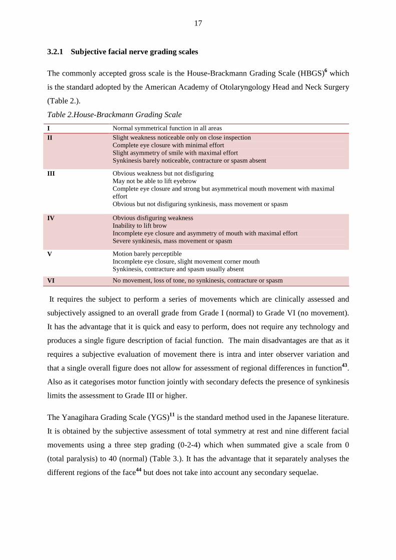
3.2.1 Subjective facial nerve grading scales
The commonly accepted gross scale is the House-Brackmann Grading Scale (HBGS)6 which
is the standard adopted by the American Academy of Otolaryngology Head and Neck Surgery
(Table 2.).
Table 2.House-Brackmann Grading Scale
I Normal symmetrical function in all areas
II Slight weakness noticeable only on close inspection Complete eye closure with minimal effort Slight asymmetry of smile with maximal effort Synkinesis barely noticeable, contracture or spasm absent
III Obvious weakness but not disfiguring May not be able to lift eyebrow Complete eye closure and strong but asymmetrical mouth movement with maximal effort Obvious but not disfiguring synkinesis, mass movement or spasm
IV Obvious disfiguring weakness Inability to lift brow Incomplete eye closure and asymmetry of mouth with maximal effort Severe synkinesis, mass movement or spasm
V Motion barely perceptible Incomplete eye closure, slight movement corner mouth Synkinesis, contracture and spasm usually absent
VI No movement, loss of tone, no synkinesis, contracture or spasm
It requires the subject to perform a series of movements which are clinically assessed and
subjectively assigned to an overall grade from Grade I (normal) to Grade VI (no movement).
It has the advantage that it is quick and easy to perform, does not require any technology and
produces a single figure description of facial function. The main disadvantages are that as it
requires a subjective evaluation of movement there is intra and inter observer variation and
that a single overall figure does not allow for assessment of regional differences in function43.
Also as it categorises motor function jointly with secondary defects the presence of synkinesis
limits the assessment to Grade III or higher.
The Yanagihara Grading Scale (YGS)11 is the standard method used in the Japanese literature.
It is obtained by the subjective assessment of total symmetry at rest and nine different facial
movements using a three step grading (0-2-4) which when summated give a scale from 0
(total paralysis) to 40 (normal) (Table 3.). It has the advantage that it separately analyses the
different regions of the face44 but does not take into account any secondary sequelae.
18
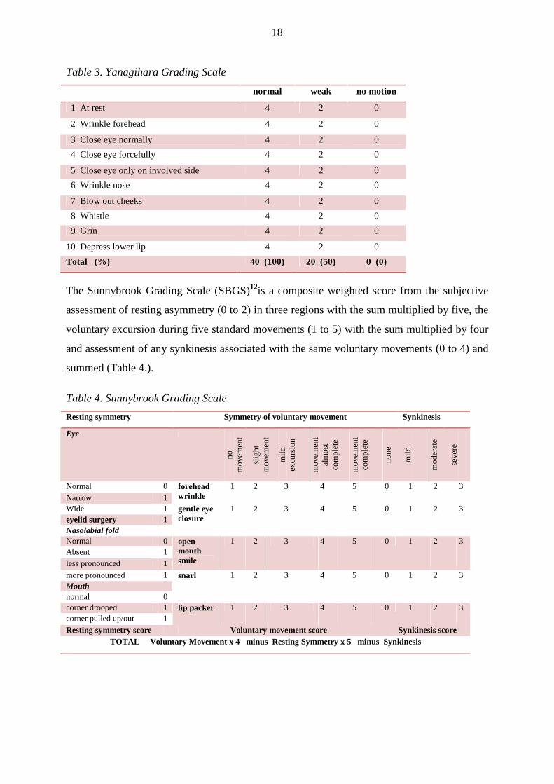
Table 3. Yanagihara Grading Scale
normal weak no motion
1 At rest 4 2 0
2 Wrinkle forehead 4 2 0
3 Close eye normally 4 2 0
4 Close eye forcefully 4 2 0
5 Close eye only on involved side 4 2 0
6 Wrinkle nose 4 2 0
7 Blow out cheeks 4 2 0
8 Whistle 4 2 0
9 Grin 4 2 0
10 Depress lower lip 4 2 0
Total (%) 40 (100) 20 (50) 0 (0)
The Sunnybrook Grading Scale (SBGS)12is a composite weighted score from the subjective
assessment of resting asymmetry (0 to 2) in three regions with the sum multiplied by five, the
voluntary excursion during five standard movements (1 to 5) with the sum multiplied by four
and assessment of any synkinesis associated with the same voluntary movements (0 to 4) and
summed (Table 4.).
Table 4. Sunnybrook Grading Scale
Resting symmetry Symmetry of voluntary movement Synkinesis
Eye
no
m
ove
men
t
slig
ht
mo
vem
ent
mild
ex
curs
ion
mo
vem
ent
alm
ost
co
mp
lete
mo
vem
ent
com
ple
te
no
ne
mild
mo
der
ate
seve
re
Normal 0 Narrow 1
forehead wrinkle
1 2 3 4 5 0 1 2 3
Wide 1 eyelid surgery 1 Nasolabial fold
gentle eye closure
1 2 3 4 5 0 1 2 3
Normal 0 Absent 1 less pronounced 1
open mouth smile
1 2 3 4 5 0 1 2 3
more pronounced 1 Mouth normal 0
snarl 1 2 3 4 5 0 1 2 3
corner drooped 1 corner pulled up/out 1
lip packer 1 2 3 4 5 0 1 2 3
Resting symmetry score Voluntary movement score Synkinesis score TOTAL Voluntary Movement x 4 minus Resting Symmetry x 5 minus Synkinesis

19
The final score is produced by subtracting the asymmetry and synkinesis scores from the
voluntary movement score giving a scale from 0 to a normal result of 100. Because of the
large number of assessments and gradings this system is more sensitive to small changes in
function and synkinesis in the different regions and has been shown to have good
reproducibility45. It is however time consuming and as all the assessments are subjective it has
the same disadvantages as the House-Brackmann Grading Scale and the Yanagihara Grading
Scale.
3.2.2 Objective facial nerve grading scales
The Stennert-Limberg-Frentrup Scale (SLFS)13is commonly used in the German literature and
has separate scores for paralysis and secondary defects (Table 5.).
Table 5. Stennert- Limberg- Frentrup Grading Scale
Motility score 0-10 Secondary defect score
At Rest Hyperacusis yes no eyelid difference <3 mm >3mm Dysgeusia yes no ec ectropion No Yes Synkinesis nasolabial fold less pronounced
No Yes
angle mouth depressed <3 mm >3mm Voluntary Movement
forehead eye nasolabial fold mouth chin
yes no
forehead wrinkle >50% Yes No more than 3 regions yes no lagophthalmos moderate effort
No Yes Spasm
lagophthalmos maximum effort
No Yes Moderate yes no
upper and lower canine teeth visible
Yes No Severe yes no
all 2nd upper incisor visible
Yes No Disturbing yes no
Philtrum/angle ditance shortening with whistle
>50% <50% Lacrimation
< 30% yes no <30% on complete
eye closure yes no
0% yes no
Contracture yes no
Crocodile tears yes no Paresis index: Secondary defect index:

20
The paralysis score is obtained by a combination of comparisons with the normal side of the
resting tone in the four regions of the face plus comparisons of six motility assessments
during voluntary movements each with score of 0 for similar and 1 for significantly worse
than the normal side. Added together a result of 0 is normal and 10 a total paralysis. The
secondary defect score is obtained by assessing the presence or absence of seven separate
symptoms. As most of the assessments result in a binary “Yes/No” value, the scale has low
observer variability but is time consuming to use.
3.2.3 Glasgow objective facial nerve grading system
The Glasgow Facial Palsy Scale (GFPS) is a recently developed objective method of
measuring both the House-Brackmann grading and the movement in the different regions of
the face. A computer programme is used to automatically measure the pixel changes in both
sides of the face produced during 5 standard movements. Using a Sony Handycam DCR-
PC105E camera each subject had a video recorded while performing the five movements:
raise eyebrow, close eye gently, close eye tightly, screw-up nose and full smile. The data from
the camera was passed via a FireWire cable with 4pin 1394 FireWire sockets to a 1.73-GHz
Laptop preloaded with the Glasgow Facial Palsy Scale program which analysed the pixel
changes in the video. The pixel changes on the palsied side of the face are compared to those
in the corresponding regions of the normal side.
Specially trained Artificial Neural Networks are then used to assess the relationship of the
pixel changes to the clinical grading of not only the House-Brackmann overall function but
also the function in the different regions of the face17. By using this system it is possible to
produce a consistent objective measurement of the overall House-Brackmann grading and
also the movement of the different regions of the face. For ease of interpretation in a clinical
setting the results are presented in a form similar to an audiogram with the regional standard
movements rather than frequencies along the x axis and the degree of palsy rather than
decibels on the y axis. A Facogram graph can then be produced in a similar time taken to
produce an audiogram. Examples of a moderate and a severe palsy are shown in Fig. 1.

21
Fig.1. Glasgow Facogram
a: Left severe facial palsy - HBGS Vb: Left moderate facial palsy - HBGS II
The programme is automated and as there is no requirement for facial markers, head fixation
or special lighting the assessment can be carried out in a clinical environment by an
audiometrician or audiovisual technician. A later version of the program will accept an input
from any camera that Windows XP or higher recognises and has a MiniDV digitaltape format
output via a FireWire.

22
3.3 Study population
3.3.1 Glasgow Facial Palsy Scale
Over a six month period 40 consecutive subjects with a unilateral facial palsy attending a
tertiary referral clinic were recruited for testing. The subjects were 28 females and 12 males
aged between 8 and 82 years with a mean age of 52. The aetiology of the facial palsies is
shown inTable 6.
Table 6. Aetiology of palsy
Aetiology Number
Bell’s palsy 29
Postoperative 6
Ramsay-Hunt syndrome 4
Temporal bone fracture 1
Total 40
Consent was obtained to store and analyse video recordings of the subjects.
3.3.2 Facial reanimation
42 patients underwent reanimation surgery for facial palsy from 1998 to 2005 in the
Lariboisiere Hospital in Paris. Patients had complete and irreversible facial palsy largely
secondary to cranial base surgery or secondary to middle ear cholesteatoma surgery, parotid
tumor extirpation, or temporal bone fracture. The cause was idiopathic in one case (Table 7.).

23
Table 7. Etiology of the facial palsy in 42 patients
Aetiology End-to-end End-to-side Myoplasty
Vestibular schwannoma 8 11 2
Facial nerve schwannoma 2 1 0
Geniculate ganglion hemangioma 0 1 2
Other intracranial tumors 2 1 3
Middle ear cholesteatoma 3 1 0
Parotid surgery 0 0 2
Temporal bone fracture 1 1 0
Idiopathic 0 0 1
Total 16 16 10
Facial rehabilitation involved lengthening temporalis myoplasty(n = 10) or XII-VII coaptation
(n = 32) by either classic end-to-end (n = 16) or end-to-side coaptation with interpositional
jump graft (technique of May et al.24; n = 16). Indications for myoplasty were a facial palsy
occurring after parotid tumor extirpation or for the long-standing facial palsy. For this reason,
mean duration of facial palsy (interval between the onset of the palsy and rehabilitation) was
higher in patients undergoing myoplasty than coaptation (6.4±10.33 versus 0.82±1.4 yr,
respectively). The mean follow-up was 115.75±56 months for the end-to-end coaptation,
38.1±15 months for the end-to-side coaptation, and 43.9±26 months for myoplasty.
3.3.3 Lateral canthopexy and upper lid gold weight implant
Between July 2009 and December 2009 we performed lateral canthopexy and upper lid gold
weight implantation on three patients.
The first patient was a 69-year-old male patient with right malignant parotid tumour
(undifferentiated spindle cell carcinoma) and preoperatively intact facial function. In March
2008 we performed a total parotidectomy and right selective neck dissection of lymph node I-
IV with the sacrifice of the facial nerve. The patient is clinically tumor-free since the
intervention. Due to the development of lagophtalmus he used, on a daily basis, artificial tears
and corneotrophical gels, protective eye glasses and occlusive dressing for the night. His
lagophtalmus associated with a gradually more and more pronounced ectropion lead to
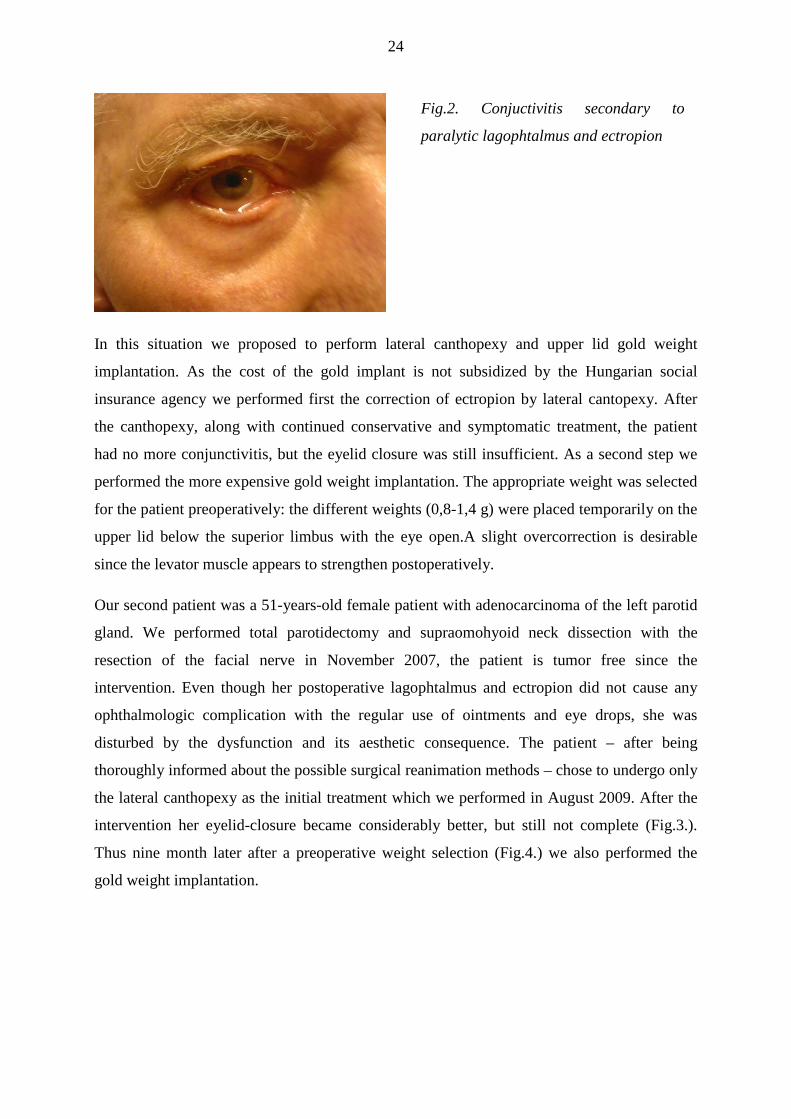
recurrent conjunctivitis (Fig.2).
24
In this situation we proposed to perform lateral canthopexy and upper lid gold weight
implantation. As the cost of the gold implant is not subsidized by the Hungarian social
insurance agency we performed first the correction of ectropion by lateral cantopexy. After
the canthopexy, along with continued conservative and symptomatic treatment, the patient
had no more conjunctivitis, but the eyelid closure was still insufficient. As a second step we
performed the more expensive gold weight implantation. The appropriate weight was selected
for the patient preoperatively: the different weights (0,8-1,4 g) were placed temporarily on the
upper lid below the superior limbus with the eye open.A slight overcorrection is desirable
since the levator muscle appears to strengthen postoperatively.
Our second patient was a 51-years-old female patient with adenocarcinoma of the left parotid
gland. We performed total parotidectomy and supraomohyoid neck dissection with the
resection of the facial nerve in November 2007, the patient is tumor free since the
intervention. Even though her postoperative lagophtalmus and ectropion did not cause any
ophthalmologic complication with the regular use of ointments and eye drops, she was
disturbed by the dysfunction and its aesthetic consequence. The patient – after being
thoroughly informed about the possible surgical reanimation methods – chose to undergo only
the lateral canthopexy as the initial treatment which we performed in August 2009. After the
intervention her eyelid-closure became considerably better, but still not complete (Fig.3.).
Thus nine month later after a preoperative weight selection (Fig.4.) we also performed the
gold weight implantation.
Fig.2. Conjuctivitis secondary to
paralytic lagophtalmus and ectropion

25
The third patient was a 51-years-old man who went through two parotid surgeries because of
recidive pleomorphic adenoma of the right parotid gland: lateral lobectomy in 1995 and total
parotidectomy in 2006 in abroad. Since the second intervention he had definitive facial nerve
palsy. We first met him because of a new recidive of his pleomorphic adenoma which we
removed in September 2009. We performed lateral canthopexy and gold weight implant in
December 2009 because of incomplete eye closure (Fig.5.).
Fig 4. Preoperative weight
selection
Fig.3. Eyelid closure after lateral
canthopexy
Fig.5. Preoperative eyelid closure
26
3.4 Surgical techniques
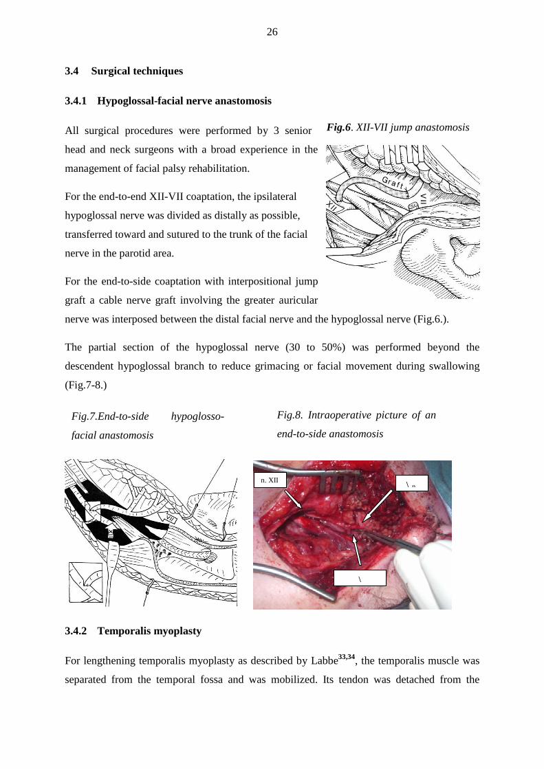
3.4.1 Hypoglossal-facial nerve anastomosis
All surgical procedures were performed by 3 senior
head and neck surgeons with a broad experience in the
management of facial palsy rehabilitation.
For the end-to-end XII-VII coaptation, the ipsilateral
hypoglossal nerve was divided as distally as possible,
transferred toward and sutured to the trunk of the facial
nerve in the parotid area.
For the end-to-side coaptation with interpositional jump
graft a cable nerve graft involving the greater auricular
nerve was interposed between the distal facial nerve and the hypoglossal nerve (Fig.6.).
The partial section of the hypoglossal nerve (30 to 50%) was performed beyond the
descendent hypoglossal branch to reduce grimacing or facial movement during swallowing
(Fig.7-8.)
3.4.2 Temporalis myoplasty
For lengthening temporalis myoplasty as described by Labbe33,34, the temporalis muscle was
separated from the temporal fossa and was mobilized. Its tendon was detached from the
Fig.7.End-to-side hypoglosso-
facial anastomosis
Fig.6. XII-VII jump anastomosis
Fig.8. Intraoperative picture of an
end-to-side anastomosis
n. XII \ n.
\

27
coronoid process and sutured to the subcutaneous tissues of the superior lip and to the
modiolus through a nasolabial incision, respecting the deep arterial temporal pedicles (Fig.9).
3.4.3 Eyelid reanimation
Lateral canthopexy: in local infiltration anaesthesia a small subciliary incision was made in
the lower lid crease laterally, the lateral palpebral ligament was identified and firmly attached
by non-absorbable suture to the periosteum on the inner aspect of the orbital rimto shorten
and tighten the lower rid (Fig 10.). To finish the surgery and to decrease the skin excess
wedge excision of the lateral lower eyelid was performed.
Upper eyelid gold weight implant: the pre-existing eyelid crease is marked 3-4 mm from the
upper lid margin with fine-tip marking pen just slightly wider than the width of the gold
weight. In local anaesthesia we perform a 2,5 cm incision through the skin and subcutaneous
Fig.10. The lateral palpebral ligament
is identified and attached by non-
absorbable suture to the periosteum on
the inner aspect of the orbital rim to
shorten and tighten the lower rid
Fig.9.Temporalis myoplasty- technique of Labbé

28
tissue and by blunt dissection we create a pocket which will accommodate the weight between
the orbicularis oculi muscle and the tarsus (Fig.11.). The gold weight is placed then in this
pocket and fixed to the tarsus with 5-0 synthetic monofilament suture through each of the
three holes. The wound is closed in two layers using 5-0 polyglactin for the deep layer and
running 6-0 nylon suture for the skin.
3.5 Evaluation
3.5.1 GFPS versus classical grading systems
3.5.1.1 Medical jury
The videos were then also individually assessed by 3 independent ENT Specialists (Drs K.G.,
R.L: and SZ.B.) who graded each subject using the subjective House-Brackmann Grading
Scale, Yanagihara Grading Scale and Sunnybrook Grading Scale and the objective Stennert-
Limberg-Frentrup Scale.
3.5.1.2 Statistical analysis
The results obtained from the subjects with the computerised Glasgow Facial Palsy Scale
assessment were compared to the 4 standard clinical methods of assessing facial palsy by
plotting the results on individual scatterplots. A One-sample Kolmogorov-Szmirnoff test was
applied to assess the presence of a normal distribution. The Pearson correlation coefficient
was measured in the presence of a normal distribution and the Spearman correlation
coefficient measured in the presence of an abnormal distribution (Statistica 8.0 software). In
both methods the linear correlation coefficient (r) measures the strength and direction of any
relationship between the two variables.
Fig. 11. 2,5 cm incision through the
skin and subcutaneous tissue and by
blunt dissection creation of a pocket
which will accommodate the weight
between the orbicularis oculi muscle
and the tarsus

29
3.5.2 Facial nerve reanimation surgery
3.5.2.1 Medical and non-medical jury
For the evaluation of the results of surgery, a video of the patient was recorded with the face
at rest, during voluntary motion of the 10 groups of facial muscles, during expression of the 6
main emotions (happiness, sadness, anger, disgust, surprise, and fear), and during a short free
conversation that allowed evaluation of spontaneous expression. Medical and nonmedical
juries then evaluated the recording. The medical jury consisted of three ear, nose, and throat
surgeons and two plastic surgeons. The nonmedical jury included four people selected
according to their ability to analyze the cosmetic appearance of a face; a cameraman, a
filmmaker, anesthetician, and an artist painter. Both juries were blinded to the procedure used.
The medical jury evaluated the facial rehabilitation using 4 classic systems to grade the face:
House-Brackmann8, Sunnybrook12, Yanagihara11, and Freyss22,46(Tables 2-4, 8.).
Table 8. Freyss score for the motility of 10 muscular groups (0-3)
G1 Frontales muscle
G2 Corrugator muscle
G3 Procerus muscle
G4 Orbicularis oculi muscle
G5 Levator labii muscle
G6 Zygomaticus muscle
G7 Orbicularis oris muscle
G8 Buccinator muscle
G9 Mentalis muscle
G10 Depressor labii inferioris muscle
3.5.2.2 Patient’s evaluation
Other evaluations involved scoring the face at rest and during voluntary motions and
emotional motions on a scale from 1 to 10. The nonmedical jury used the last 3measures to
evaluate the face. We mailed 2 well-established questionnaires, the Facial Disability Index
(FDI)47, 48 and the Glasgow Benefit Inventory (GBI)49, as well as a Quality of life(QOL)
questionnaire developed in our institution, to each patient(Table 9).
30
Table 9. Institution’s QOL questionnaire
1. Disability in professional life(0 = no handicap to 10 = severe handicap)
□ Before reanimation □After surgery 2. Disability in private life (0 = no handicap to 10 = severe handicap)
□ Before reanimation □ After surgery 3. Index of satisfaction(0 = not satisfied to 10 = very satisfied)
□ 4. Was the surgery useful?
□Yes □ No. Why? 5. Would you undergo the operation again?
□ Yes □ No. Why?
The FDI asks 10 questions regarding physical and social activities such as the ability to brush
teeth,eat, and speak; additional questions ask regarding cornea protection, isolation,
irritability, social activity and sleeping disorders. The GBI evaluates the effects of surgery
with 15 questions regarding social well-being and 3 regarding medical side effects. The
institution’s questionnaire evaluates disability in T5 professional and private life and general
satisfaction.
3.5.2.3 Statistical evaluation
The sex, age, and number of patients in the 3 treatment groups were compared (analysis of
variance [ANOVA] and χ2). Then, ANOVA and Fisher’s exact test were used to analyze the
relationship between facial palsy duration and quality of recovery. Treatment effects were
compared by the lapse time between the surgical procedure and the initial evidence of
recovery, scores on the 5 grading scales, and the other scores given by the medical and
nonmedical juries and the self-assessment by the patients. A p<0.05 was considered
statistically significant. The predictive value for the ability of the grading system to
discriminate the different procedures was also analyzed.
3.5.3 Eyelid reanimation
Statistical analysis is planned when the study population reach at least 15 patients.
31
4 RESULTS
4.1 Comparison between Glasgow Facial Nerve Palsy scale and four classical, widely
used objective and subjective facial grading scales
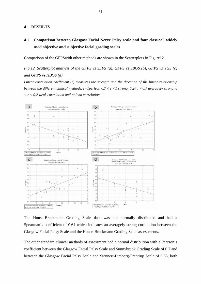
Comparison of the GFPSwith other methods are shown in the Scatterplots in Figure12.
Fig.12. Scatterplot analysis of the GFPS vs SLFS (a), GFPS vs SBGS (b), GFPS vs YGS (c)
and GFPS vs HBGS (d)
Linear correlation coefficient (r) measures the strength and the direction of the linear relationship
between the different clinical methods. r=1perfect, 0.7 ≤ r <1 strong, 0.2≤ r <0.7 averagely strong, 0
< r < 0.2 weak correlation and r=0 no correlation.
The House-Brackmann Grading Scale data was not normally distributed and had a
Spearman’s coefficient of 0.64 which indicates an averagely strong correlation between the
Glasgow Facial Palsy Scale and the House-Brackmann Grading Scale assessments.
The other standard clinical methods of assessment had a normal distribution with a Pearson’s
coefficient between the Glasgow Facial Palsy Scale and Sunnybrook Grading Scale of 0.7 and
between the Glasgow Facial Palsy Scale and Stennert-Limberg-Frentrup Scale of 0.65, both

32
showing an averagely strong correlation. The correlation between the Glasgow Facial Palsy
Scale and Yanagihara Grading Scale was the strongest with a Pearson coefficient of 0.72.
Table 10. Interobserver variability
Values of kappa can range from -1.0 to 1.0, -1.0= perfect disagreement below chance, 0.0= agreement equal to
chance, and 1.0= perfect agreement above chance. Kappa of 0 .70 or above=s adequate interrater agreement.
House Brackmann Grading Scale
Yanagihara Grading Scale
Sunnybrook Grading Scale
Stennert Grading Scale
Percent of overall agreement
0,73 0,92 0,90 0,92
Siegel and Castellan's Fixed-marginal kappa
0,66 0,92 0,90 0,90
Randolph's Free-marginal kappa
0,68 0,92 0,90 0,91
The consistency of the results from the three assessors is shown in Table 10. There was a low
interobserver variation for all the scales apart from House-Brackmann. The reason for this is
likely that this scale has the lowest number of options available when making the subjective
decision on the the degree of palsy.
4.2 Comparison of different types of hypoglosso-facial nerve anastomosis and
temporalis myoplasty
4.2.1 General Characteristics of the Population
Mean age did not differ between the groups (45.7±15.8 yr for the coaptation group versus
51.7±17.7 yr for the myoplasty group; p= 0.26), nor did sex distribution differ (p= 0.41).
Mean delay for detecting the first signs of recovery did not significantly differ between the
myoplasty group (2±1.1 mo) and the end-to-end coaptation group (5.2±3.5 mo) but it was
significantly longer for the end-to-side coaptation group (9.5±6.9 mo;p= 0.017).
Approximately half of the patients underwent facial physiotherapy after surgery.
4.2.2 Evaluation by the Medical Jury
Results of the grading systems for each procedure are reported in Table 11. The medical jury
rated end-to-side coaptation significantly better than myoplasty by the Sunnybrook grading
system (p = 0.03) and end-to-end coaptation better than myoplasty by the Freyss and
Yanagihara grading systems (p = 0.018 and p = 0.024, respectively). The jury also rated both
end-to-end and end-toside coaptation better than myoplasty by the HBGS (p = 0.037 and p =
0.026, respectively). Thus, end-to-end and end-to-side XII-VII coaptation performed better
33
than myoplasty according to all grading scales.For the face at rest, the mean score for all
procedure groups was 6.5/10, but the score for either coaptation type was not significantly
different from that of myoplasty (6.8/10 versus 5.4/10).
Table11. Medical jury mean scores by the 5 grading scales for hypoglossal-facial coaptation
and lengtheningtemporalis myoplasty
House-Brackmann
May Freyss Sunnybrook Yanagihara
End-to-end 3.4a 2.8 6,4a 30,1 14,2a
End-to-side 3,3 a 3,1 5,2 32,7a 12,7
Myoplasty 3.9a 2.6 3.4a 20.8a 9,9a ap <0.05 between procedures
For the face during voluntary motion, the mean score for all procedures was 4.9/10 and during
expression of emotions was 4.8/10 with no significant difference between groups (p = 0.91) as
shown in Table 12.
Table 12. Medical and nonmedical jury mean scores for the face at rest and during voluntary
motions and expression of emotions for hypoglossal-facial coaptation and lengthening
temporalis myoplasty.
Medical jury Non-medical jury At rest Voluntary
motion Emotions At rest Voluntary
motion Emotions
End-to-end coaption
6,8a 4,9 5,0 7,7a 5,8a 6,0a
End-to-side coaption
6,8a 4,9 4,8 7,9a 6,0a 5,8
Myoplasty 5,4a 4,8 4,6 6,7a 4,6a 4,7a ap <0.05 between procedures
4.2.3 Evaluation by the Nonmedical Jury
For the face at rest, the nonmedical jury gave a meanscore of 7.2/10 for all procedures; the
score was significantlyhigher for the end-to-end and end-to-side coaptationgroups than for the
myoplasty group (7.7/10, 7.9/10, and 5.6/10, respectively; p= 0.004 and p= 0.001;Table 12.).
The jury noted that disharmony of the face was moreobvious for the myoplasty group and that
voluntarymotions were stronger with both coaptation groupsthan with the myoplasty group
(5.8/10, 6/10, and 4.7/10,respectively; p= 0.02 and p= 0.006). The jury rankedthe expression

34
of emotions 6/10 for the end-to-end coaptation group and 4.7/10 for the myoplastygroup, with
a significant difference between these 2groups (p= 0.02).
4.2.4 Patient Evaluation
All patients felt less disabled after surgery thanbefore with respect to physical and social
impairment,but patients still had some complaints. Indeed, themean score of the FDI was
56/100±27.5 for the physical portion and 69/100±23 for the social portion, with nosignificant
difference according to procedure. The GBI results showed a net improvement regardless of
procedure (mean score = + 12±20 [max, + 50; min,-50]) (Table 13).
Table 13. Patients’ mean scores for QOL questionnaires for hypoglossal-facial coaptation
and lengtheningtemporalis myoplasty
FDI physical (/100)
FDI social (/100)
GBI total (-50/+50)
End-to-end coaptation
53 (±30.4) 68.3 (±22.6) 11.6 (±22.6)
End-to-side coaptation
62.7 (±14.9) 69.5 (±23) 8.6 (±20.9)
Myoplasty 52.8 (±32.7) 81.8 (±8.3) 19.5 (±18.6)
On our institution’s questionnaire, patients consideredthat their daily and professional lives
weresignificantly improved (p<0.001 and p<0.02, respectively; Table 14.). In most cases,
patients were satisfied,would consent to surgery again, and thought that surgeryhad been
useful.
Table 14.Patients’ mean scores for the institution’s QOL questionnaire for hypoglossal-facial
coaptation andlengthening temporalis myoplasty
Disability in professional lifebefore surgery (0-10)
Disability in professional life after surgery (0-10)
Disability private life before surgery (0-10)
Disability private life after surgery (0-10)
Index satisfaction (0-10)
Surgery was useful
Would undergo the operation again
Mean score 3,41 1,38 6,75 4,76 6,34 92% 82% End-to-end coaptation
4,43 1,24 6,22 4,58 6,28 92% 71%
End-to-side coaptation
2,42 2,37 7,18 4,95 6,4 92% 91%
Myoplasty 3,85 2,06 8,88 4,88 7,5 89% 89%
4.2.5 Prognostic Factors
For each surgical procedure, the time between the onsetof the facial palsy and the
rehabilitation procedure didnot influence the functional results by the HBGS.
35
4.2.6 Comparison of the Grading Systems
No significant difference was found in scores betweenthe 4 grading systems.
4.3 Gold eyelid weight and lateral canthopexy
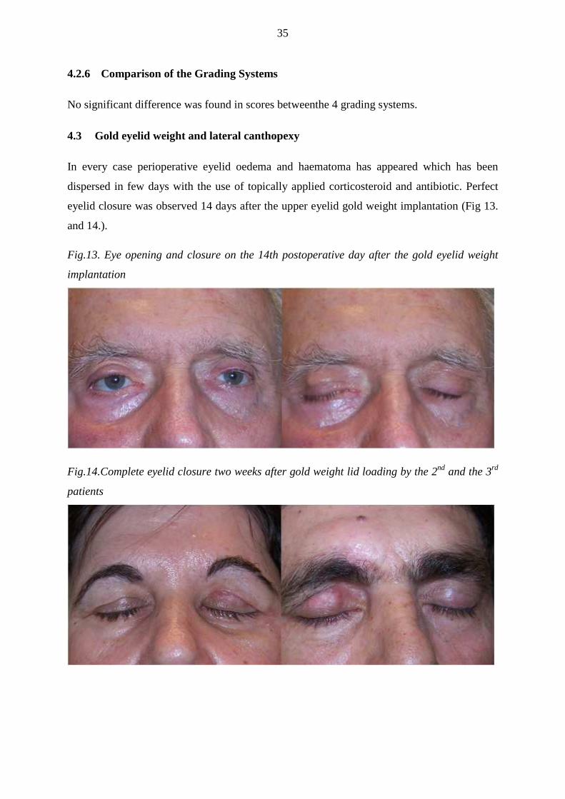
In every case perioperative eyelid oedema and haematoma has appeared which has been
dispersed in few days with the use of topically applied corticosteroid and antibiotic. Perfect
eyelid closure was observed 14 days after the upper eyelid gold weight implantation (Fig 13.
and 14.).
Fig.13. Eye opening and closure on the 14th postoperative day after the gold eyelid weight
implantation
Fig.14.Complete eyelid closure two weeks after gold weight lid loading by the 2nd and the 3rd
patients
36
5 DISCUSSION
5.1 Comparison between Glasgow Facial Nerve Palsy scale and four classical, widely
used objective and subjective facial grading scales
5.1.1 Synopsis of the key findings
There is a moderately strong correlation between the Glasgow Facial Palsy Scale and House-
Brackmann Grading Scale which is to be expected as the artificial neural networks used to
produce the Glasgow Facial Palsy Scale are trained with House-Brackmann Grading Scale
results assessed by clinicians. The strongest relationship was found between the Glasgow
Facial Palsy Scale and Yanagihara Grading Scale which underlines the detailed regional
information measured in both systems. The Glasgow Facial Palsy Scale showed a strong
correlation with the Sunnybrook Grading Scale and a moderately strong correlation with
Stennert-Limberg-Frentrup Scale demonstrating the sensitivity of this objective method when
assessing overall facial nerve function.
5.1.2 Strengths and weaknesses
The ideal grading scale for facial nerve function should be objective and simple to measure
but have a strong intra and inter observer reliability. It should take into account symmetry at
rest, both regional spontaneous and voluntary facial movements and secondary defects. There
is no such classification that includes all these criteria but several subjective8,11,12 and
objective 13,50,51 classifications have been proposed.
The Glasgow Facial Palsy Scale is an objective quantitative evaluating method based on the
computer analyses of pixel changes during a digital video recording of facial movements
thereby eliminating the subjectivity of the observer. The process is quick and has modest
technological requirements using a basic laptop computer and a domestic digital video
camera. The program is available on the web as open source software. The facogram which is
produced automatically demonstrates the individual regional facial nerve functions and can be
stored electronically or in a printed form in the case record.

37
Its drawbacks are that it does not evaluate secondary defects such as synkinesis or tearing but
it could be supplemented by simultaneous use of the Stennert’s secondary defect score. As the
paralyzed side’s movement is compared to that on the normal side it cannot be performed in
presence of bilateral palsy. The identification of the different regions relies on a normal
position of the pupils therefore it cannot be used in the presence of strabismus, oculomotor
palsy or an artificial eye.
This thesis concentrated on demonstrating the ability of the objective computerised method to
obtain similar results to the accepted clinical methods rather than its ability to track clinical
change in the individual patient which has been demonstrated in a previous study17.
5.2 Comparison of different types of hypoglosso-facial nerve anastomosis and
temporalis myoplasty
The largely used techniques for the reanimation of complete and irreversible facial palsy are
XII-VII coaptation and lengthening temporalis myoplasty. Classic end-to-end XII-VII
coaptation requires a viable distal facial stump and a nonatrophic musculature. Because it
induces lingual hemiatrophy, which may sometimes be disabling, some authors have
proposed a partial section of the XII nerve with interposition of a greater auricular nerve graft
(jump)24. XII-VII coaptation never restores spontaneity to the face. Lengthening temporalis
myoplasty33,34 claims overall results superior to the XII-VII coaptation, but no procedure
achieves perfect cosmetic results. However, evaluation of the results remains controversial in
part because the analysis has involved different grading systems developed to grade facial
palsy not facial rehabilitation.
We aimed to compare the results of end-to-end and end-to-side XII-VII coaptation and
lengthening temporalis myoplasty as assessed by an expert jury using the four most accepted
facial grading systems, by a nonmedical jury and by the patients’ own QOL assessments.
XII-VII coaptation, whatever the type, yielded significantly better results than myoplasty,
regardless of gradingsystems used by the medical jury and scores given by the nonmedical
jury. The most significant and discriminating factor of the myoplasty procedure was an
obvious disharmony of the lower face caused by the visibility of the nasolabial scar, the
inescapable overcorrection of the superior lip, and the absence of inferior lip rehabilitation
which led to a deviation of the inferior lip toward the healthy side. However, the medical jury
rated the smile more spontaneouswith myoplasty than with XII-VII coaptation.

38
The grading systems used could account for the differences, although not significant, we
found between both types of XII-VII coaptation. If the grading system took into account
synkinesis, as does the Sunnybrook system, results were better with end-to-side than end-to-
end coaptation. On the contrary, if the grading system did not take into account synkinesis,
such as the Freyss and Yanagihara systems, end-to-end coaptation led to better results. Thus,
the difference between the results of the procedures depended on scoring synkinesis in the
grading system. End-to-end coaptation – reinnervation by the whole hypoglossal nerve –
leads to high motility,but also to adverse effects, such as synkinesis and mass
movements23,27,52.In contrast, end-to-side coaptation leads to a weaker muscle tone, less
synkinesis and mass movement and longer recovery because of axonal loss and fibrosis due to
the presence of secondary coaptation23,53,54.
This finding could explain the conclusions of the medical jury who gave better scores for
patients with a strong emotional and spontaneous expression if they had undergone end-to-
end rather than end-to-side coaptation. Because of the stronger muscle tone provided by the
full reinnervation, the mouth is less attracted toward the healthy side. However, patients with
a weaker facial expression require less muscle tone thus they had a satisfying outcome with
the end-to-side coaptation because of the less important secondary effects. This therefore
shows that choosing the most appropriate surgical method must take into account the
spontaneous expression of the face.
Patient QOL was improved in general, regardless of the technique, meaning that rehabilitation
was guaranteed (55 and our study). However, the scores from the three patient questionnaires
were moderate, so rehabilitation was not perfect. Interestingly, these questionnaires could not
discriminate between the effects of XII-VII coaptation and myoplasty. Two biases impair the
study of QOL in facial rehabilitation: the GBI questionnairedoes not focus on the analysis of
facial motions and patients have difficulties in properly evaluating the benefit of a
reconstructive procedure performed at the same time or soon after ablative surgery, which, in
most cases, causes facial palsy and also affects QOL.
Comparison of the different grading systems have shown good
correlation6,14,56,,57,58,59,60especially for grading voluntary movements61. Surprisingly, we
found that results with the different grading systems were comparable with nonmedical jury
evaluations, showing that XII-VII coaptation led to better results than myoplasty. However,
these grading systems are not perfectly adapted for the evaluation of facial reanimation,
39
because secondary healing defects such as synkinesis and mass movements are not well
described: forehead motility is evaluated, but it is never reanimated and no system evaluates
emotional motions.
5.3 Comparison between lateral tarsorraphy and upper lid gold weight implant in the
treatment of paralytic lagophtalmus
Restoration of the eyelid animation and aesthetics are the major component of surgical
managementof long-term facial nerve palsy. Tarsorraphy has been the traditionally used
method in Hungary because of its simplicity. However besides limiting the vision and
offering an insufficient corneal coverage, the procedure may lead to unappealing cosmetic
effects. Following the release of the tarsorraphy notching of the eyelid margin or ectropion
may occur.
The gold eyelid weight introduced 60 years ago by Sheehan et al62 is the most widely applied
surgical method internationally. Gold weights can be safely implanted in an outpatient setting
with local anesthesia. This easy and effective method has also the advantage of being
reversible without leaving any defects, thus can also be used for patients with temporary
palsy. It reanimates only the paralyzed upper lid; therefore it should be completed if necessary
with a lower lid tightening procedure, as it was previously described. To obtain a good result
adequate preoperative evaluation is compulsory to determine the optimal size, weight and
position of the implant. Custom-made weights are far cheaper and produce a much more
aesthetic result than commercially manufactured gold implants. Besides gold20,63 the
implanted material can be tantalum62, platinum64or platinum-iridium65. The more popular gold
weight which was also used in our department provides satisfactory lid closure in case ofthe
vast majority(between 70 to 100% in relevant literature) of patients42,66,67.Sönmez et al 68
reported that the method offers an aesthetical postoperative appearance from the patient’s
point of view which was our experience as well. Complications like infection or allergic
reaction can occur but have been found infrequent69.Possible long-term complications are the
following: upper eyelid pseudoptosis, under correction, migration, extrusion and astigmatism
due to nonconformity of gold weight to the corneal slope. A German meta-analysis 70
investigated publications and presentations of this topic before 2005 and has found the
following complication rates: astigmatismus 11,5%, migration 6,4%, extrusion 6,8%,
infection 7%. According to Rofagha63 et al postoperative complications are few, but after 5
years the incidence of weight exposure increases to around 10 percent. Most of the possible

40
complications can be avoided by proper surgical techniques and a good understanding of
periocular anatomy. Pseudoptosis and under correction will not occur either when due
attention is given to finding the appropriate weight before intervention while the potential for
migration can be lessened by using appropriate tarsal fixation. In our study no complication
occurred.
Although gold weight implants are still the most common, since 1999 flexible platinum chain
implants has been available as an alternative to the rigid gold implant. Schrom et al70 are
convinced that the platinum chain matches much more the anatomic prerequisites of the upper
eye lid and postoperative complications are less frequent. Further lid loading method is the
palpebral spring introduced by Morel-Fatio et al in196471. According to Terzis et al gold
weight and palpebral spring are both efficient in restoring motion to the paretic upper eyelid,
but the palpebral spring is more so despite the frequent need for revisions72. It is an effective
treatment for lagophthalmos, even though has a high rate of extrusion73.
To restore the maximal eyelid function in addition to the upper eyelid weight implant the
management of lower lid drooping is crucialas well. When the patient cannot afford the price
of the gold weight and have only a small problem of occlusion, canthopexy alone can be
considered as the primary intervention. Merely the tightening of the lower lid can already
enhance the occlusion. Evidently gold weight lid loading can be always performed later on if
necessary.
In our interventions we opted not to use the commercially available implants due to their high
cost and the sometimes cumbersome and expensive ways of importing them to Hungary.
Instead, we used customized 99,99% pure gold implant manufactured by a private jeweller.
41
6 CONCLUSIONS AND NEW RESULTS
6.1 Glasgow Facial Palsy Score
The most common situation for a clinician wishing to measure and record facial weakness is
the clinical monitoring of Bell’s palsy. This objective programme is ideal for this and also
applicable when comparing results of treatment in double blind trials and in the clinical audit
of skull base surgery. The authors are preparing a new study to monitor clinical changes in
subjects with facial palsy by comparing the results obtained from the objective computerised
method with the results obtained by the standard subjective clinical methods of the House-
Brackmann Scale, Yanagihara, Sunnybrook grading scales and the objective clinical Stennert-
Limberg-Frentrup scale.
6.2 Facial reanimation methods
Fully restoring facial function and emotions after facial palsy remains challenging. The choice
of the appropriate surgical rehabilitation procedure must rely on a detailed analysis
comprising the facial palsy duration, the cause of the facial palsy, the presence of other cranial
nerve injuries, the spontaneous expression of the healthy side, and the motivation of the
patient. We suggest that XII-VII coaptation should be preferred over temporalis myoplasty for
facial palsy whenever possible because it provides better results with the face at rest while
motions with both procedures are comparable.
6.3 Eyelid reanimation in facial nerve palsy
As the funtional and cosmetic results of the combination of gold eyelid weight and lateral
canthopexy significantly surpass those of the lateral tarsorraphy this should be the primary
treatment in case of paralytic lagophtalmus. Customized gold eye weights offer a good and lot
cheaper alternative against commercially available ones.
6.4 New results
The conclusion of our paper on thecurrent diagnostic, pharmaceutic and reconstructive
surgical methods in the management of facial nerve palsy is that there is ahuge demand for a
facial nerve palsy protocol to compare the results of different workgroups and methods. The
international literature available on this topic reflects the same problem: different patient
42
groups are evaluated and treated with different methods so metaanalysis of the results is not
possible. In an attempt to address this gap:
• We have introduced a suitable complex questionnaire containingaetiology, risk
factors, case history,result of the physical examination, facial test, different subjective
and objective measurements and treatment options.
• We have established a new therapeutic protocol to standardize the treatment of the
acute unilateral facial nerve palsy.
• For the first time in Hungary, we used a new, quick, objective, quantitative method –
the Glasgow Facial Palsy Score – which could be easily applied to daily clinical
routine (II,III).
• Based on myforeign clinical experience and derived from the surgical results of our
French colleagues we compared three different facial reanimation techniques (IV).
• For the first time in Hungary,we introduced and accomplished the use of simple
surgical methods already proven abroad for the treatment of lagophtalmus and
ectropion secondary to facial nerve palsy(I).
6.5 Future
For the future we have the following ideas to realize:
• We would like to develop a new facial palsy score with which the results of the facial
reanimation could be also analysed.
o We propose that the facial function should be evaluated according to
� the symmetry at rest at the fronto-orbital (mention of the eye surgery),
nasal and labial regions,
� the voluntary motions (eye closure, smile, whistle, lower lip depression
and grin),
� the 6 main emotions and
� the spontaneity.

43
o Secondary defects such as synkinesis, mass movements, hemispasm and the
side effects (tongue motility, problems of swallowing and mastication) must be
also described.
• We would like to continue to perform eyelid reanimation surgery and do statistical
analysis of our long-term results

44
7 ABBREVIATIONS
ANOVA: Analysis of variance
ENT: Ear-Nose-Throat
ENoG: Electroneuronography
FDI: Facial Disability Index
GBI:Glasgow Benefit Inventory
GFPS: Glasgow Facial Palsy Scale
XII-VII: Hypoglossal-Facial
HBGS: House-Brackmann Grading Scale
QOL: Quality of life
SBGS: Sunnybrook Grading Scale
SLFS: Stennert-Limberg-Frentrup Scale
YGS: Yanagihara Grading Scale

45

46
8 APPENDIX
8.1 Facial Palsy Questionnaire
Beteg neve: Életkora: Azonosítója: Címe: Foglalkozás: Elérhetıség: Mikor következett be a FP: Elsı orvosi ellátás idıpontja: Anamnézis: Láz influenzaszerő tünetegyüttes stressz koponyatrauma kullancscsípés huzat vagy meghőlés (hidegben tartósan tartózkodás) egyéb:……………………………………………………………………………………… KÍSÉRİ TÜNETEK: Csecsnyúlvány feletti fájdalom nem igen Bırkiütés nem igen Arcduzzanat nem igen Könnyezés nem igen Szemszárazság nem igen Ízérzés zavar nem igen Hyperacusis nem igen Fülzúgás nem igen Halláscsökkenés nem igen (kezdetben >24h) Forgó jellegő szédülés/bizonytalanség érzet nem igen (kezdetben >24h) Nystagmus nem igen Facialis hypoesthesia nem igen (kezdetben >24h) Fejfájás nem igen (kezdetben >24h) Nyakfájdalom nem igen Dysphonia nem igen Egyéb neurológiai tünet: …………………………………………………………………. Herpeses anamnézis: nem igen Herpes fertızést felvetı klinikai tünetek: labialis hypoesthesia nem igen labialis dysesthesia nem igen eruptio nem igen Korábbi FP epizód: nem igen Ideje: Oldal: Maradványtünetek: KORÁBBI ANAMNÉZIS-EGYÉB BETEGSÉGEK Gastroduodenalis ulcus nem igen Alkoholos májcirrhosis nem igen Immunszupprimált állapot (transzplantatio, HIV, vérképzırsz.bet.) nem igen Pszichosis nem igen
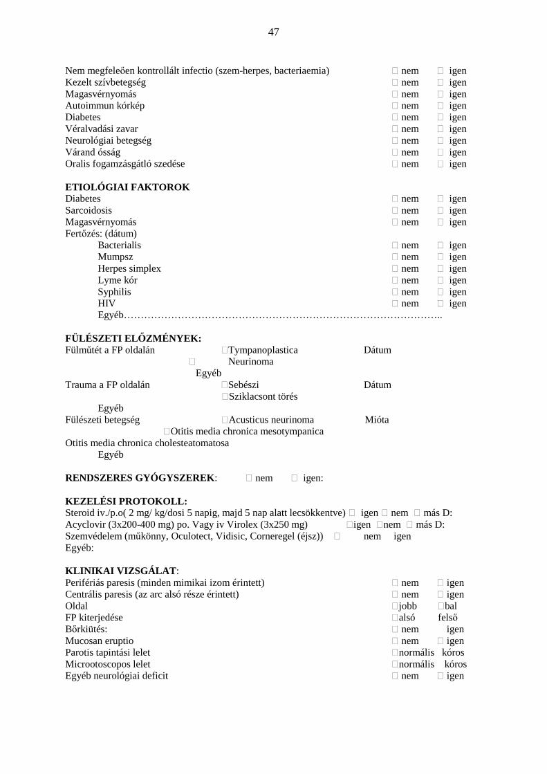
47
Nem megfeleıen kontrollált infectio (szem-herpes, bacteriaemia) nem igen Kezelt szívbetegség nem igen Magasvérnyomás nem igen Autoimmun kórkép nem igen Diabetes nem igen Véralvadási zavar nem igen Neurológiai betegség nem igen Várand ósság nem igen Oralis fogamzásgátló szedése nem igen ETIOLÓGIAI FAKTOROK Diabetes nem igen Sarcoidosis nem igen Magasvérnyomás nem igen Fertızés: (dátum) Bacterialis nem igen Mumpsz nem igen Herpes simplex nem igen Lyme kór nem igen Syphilis nem igen
HIV nem igen Egyéb…………………………………………………………………………………..
FÜLÉSZETI EL İZMÉNYEK: Fülmőtét a FP oldalán Tympanoplastica Dátum Neurinoma Egyéb Trauma a FP oldalán Sebészi Dátum Sziklacsont törés Egyéb Fülészeti betegség Acusticus neurinoma Mióta Otitis media chronica mesotympanica Otitis media chronica cholesteatomatosa Egyéb RENDSZERES GYÓGYSZEREK: nem igen: KEZELÉSI PROTOKOLL: Steroid iv./p.o( 2 mg/ kg/dosi 5 napig, majd 5 nap alatt lecsökkentve) igen nem más D: Acyclovir (3x200-400 mg) po. Vagy iv Virolex (3x250 mg) igen nem más D: Szemvédelem (mőkönny, Oculotect, Vidisic, Corneregel (éjsz)) nem igen Egyéb: KLINIKAI VIZSGÁLAT : Perifériás paresis (minden mimikai izom érintett) nem igen Centrális paresis (az arc alsó része érintett) nem igen Oldal jobb bal FP kiterjedése alsó felsı Bırkiütés: nem igen Mucosan eruptio nem igen Parotis tapintási lelet normális kóros Microotoscopos lelet normális kóros Egyéb neurológiai deficit nem igen

48
3-7 NAP KÖZÖTTI VIZSGÁLATI EREDMÉNYEK NET jobb bal Schirmer teszt jobb bal Tympanometria jobb bal Stapedius reflex jobb igen csökkent nem (nincs stapedius-beszédaud.) bal igen csökkent nem Audiogram jobb bal Zajban végzett beszédaudiometria: jobb bal Egyéb vizsgálatok BERA: jobb: bal: Otoneurológiai vizsgálat: jobb: bal: Calroikus vestibularis ingerlés jobb: bal: House-Brackmann : Stennert: Yanagihara: Sunnybrook: Facogramm: homlok szem N szem erıs nasolabialis redı szájzug
House-Brackmann skála:
I. normális facialis funkció II. min.elmaradás a mozgásokban, elıfordulhat
enyhe synkinesis, de nyugalomban normál tónus homlokráncolás: kp.-jó ; szemzárás puhán is normális ; szájzug:kis aszimmetria
III. észrevehetı, de nem torzító asszimmetria, mérsékelt, nem torzító synkinesis, kontraktura, hemifacialis spasmus, nyugalomban normál tónus és szimmetria homlokráncolás: enyhe-kp. szemzárás erıltetve normális szájzug:max.erılködés mellett kis elmaradás
IV. Egyértelmő gyengeség vagy torzító asszimmetria, kifejezett synkinesis-kontraktúra vagy hemifacialis spasmus nyugalomban normál tónus és szimmetria homlokráncolás: nincs; szemzárás:inkomplett; szájzug: elmarad V. Alig észrevehetı mozgás, nyugalomban is asszimetria Homlokráncolás: nincs; szemzárás: inkomplett; Szájzug: minimális mozgás
VI. Nincs mozgás
Stennert-Limberg-Frentrup skála
Motilitás score (0-10) Maradvány tünet score
Nyugalmi dysacusis van nincs
Szemrés differencia < 3mm > 3mm ízérzés zavar van nincs
Entropium nincs van Synkinesis
Nasolabialis redı elsímult nem igen homlok igen nem
Szájszeglet ptosisa < 3mm > 3mm szem igen nem
Motilitás nasolabialis redı igen nem
Homlokráncolás szájszeglet igen nem
(redıképz.ill. szemöldökemelés >50%) igen nem áll igen nem
lagophtalmus mérs.innerv nincs van több mint 3 régió között
max.innerv. nincs van Pseudospasmus
fogmutatás mérsékelt igen nem
szemfog fent és lent látható igen nem kifejezett igen nem
2..metszı fent teljes ssélességében igen nem beteget zavarja igen nem
szájcsücsorítés Könnyelválasztás nem igen
philtrum-szájszeglet megrövidülése >50% <50% < 30%
ellenoldalhoz képest <30%, szemhéjzárás lehet
0%
Contractura nem igen
Krokodilkönny nem igen
Paresis index: Maradványtünet index:
Sunnybrook skála:
Nyugalomban, Akaratlagos mozgások Synkinesis ellenoldalhoz képest ellenoldalhoz képest izomtevékenység foka
Szem nincs kis enyhe majdnem normál nincs kis kp. súlyos
mozgás mozgás elmozdulás teljes mőködés mőködés Normál 0 Keskenyebb 1 Szélesebb 1 homlokráncolás 1 2 3 4 5 0 1 2 3 Szemmőtét 1 Nasolabialis redı Normál 0 puha szemzárás 1 2 3 4 5 0 1 2 3 Hiányzik 1 Kevésbé kifejezett 1 Kifejezettebb 1 nyitott szájas mosoly 1 2 3 4 5 0 1 2 3 Szájzug Normál 0 orrot felhúz(vicsorog) 1 2 3 4 5 0 1 2 3 Szájzug le 1 Szájzug fel 1 csücsörít 1 2 3 4 5 0 1 2 3
Nyugalmi összpont x 5: akaratlagos mozgás összpont x 4: Synkinesis összpont: Végeredmény (0-100) : akaratlagos- nyugalmi- synkinesis :
Yanagihara skála: Normál Gyenge Hiányzó
1 Nyugalomban 4 2 0
2 Homlokráncolás 4 2 0
3 Normál szemzárás 4 2 0
4 Erıltetett szemzárás 4 2 0
5 Kacsintás az érintett oldalon 420
6 Orr felhúzása 4 2 0
7 Arc felfúvása 42 0
8 Fütyülés 4 2 0
9 Széles mosoly 4 2 0
10 Alsó ajak lehúzása 4 2 0
Összpontszám (%) 40(100) 20(50 0(0)

49
15 NAPOS KONTROLL House-Brackmann : Stennert: Yanagihara: Sunnybrook: Facogramm: homlok szem N szem erıs nasolabialis redı szájzug 30 NAPOS KONTROLL NET: jobb bal Laboreredményekben eltérés nem igen: Serológiai pozititás: nem igen MRI nem igen: CT nem igen: House-Brackmann : Stennert: Yanagihara: Sunnybrook: Facogramm: homlok szem N szem erıs nasolabialis redı szájzug Egyéb: 4 HÓNAPOS KONTROLL Objektív javulás: Maradvány tünetek: Synkinesis nem igen szem-száj száj-szem Hemifacialis spasmus nem igen Kontraktúra nem igen House-Brackmann : Stennert: Yanagihara: Sunnybrook: Facogramm: homlok szem N szem erıs nasolabialis redı szájzug Szubjektív tünetek: Ízérzés zavara nem igen: Hyperacusis nem igen: Szemszárazság nem igen: Egyéb megjegyzés: 1 ÉVES KONTROLL Objektív javulás: Maradvány tünetek: Synkinesis nem igen szem-száj száj-szem Hemifacialis spasmus nem igen Kontraktúra nem igen House-Brackmann : Stennert: Yanagihara: Sunnybrook: Facogramm: homlok szem N szem erıs nasolabialis redı szájzug Szubjektív tünetek: Ízérzés zavara nem igen: Hyperacusis nem igen: Szemszárazság nem igen: Facialis rehabilitáció: ideganastomosis myoplastica temporalis
szemmőtét (mi) Egyéb megjegyzés:

50
8.2 Facial palsy protocol
TERÁPIA:
• Solu-Medrol 1,5-2 mg/kg 5 napig
� 80 kg-ig 125 mg
� 80-110 kg-ig 165 mg (125+40 mg), 4. és 5. napon 125 mg
� 110 kg felett 250 mg, 4 és 5.napon 125 mg
• Gyomorvédelem: Panogastin este 40 mg vagy Quamatel 2x20 mg, 250 mg dózistól ill.
pozitív gastritises-ulcusos anamnézis esetén Panogastin 2x40 mg v.Quamatel 2x40 mg
• Kálium R napi 1 tbl
A beteg bentfekvıként maradjon az elsı 1 vagy két éjszaka, utána hazajárhat.
Diabeteses beteg minden esetben végig maradjon bent.
VIZSGÁLATOK BENTFEKVÉS ALATT:
• Facialis skála felvétele (KG)
• Videó (KG-SZB)
• Audiológia (szerda13-15 h)
• Otoneurológia (spontán tünetek és calorikus ingerlés): szerda 13-15 h
• Schirmer teszt -1. vagy 2. napon
• Tympanometria és stapedius reflex – 1. vagy 2. napon
� Stapedius rfx hiányában zajban végzett beszédaudiometria és
magasfrekvenciákon végzett küszöbvizsgálat szükséges
• NET (4.-5.napon)
Egyéb vizsgálatok, kóros audiológiai lelet esetén:
• BERA (kedd vagy szerda 11-13h)
• OAE (kedd vagy szerda 11-13h)
OTTHONÁBAN:
• Steroid leépítése: Medrol 64-32-16-8-4 mg
• Panogastin este 40 mg 28 napig
• Kálium R 1x1 tbl 5 napig a steroiddal

51
Kontroll: 15.nap, 1 hó, 4 hó, 1 év
52
9 ACKNOWLEDGEMENTS
The work with this dissertation has been extensive and trying, but in the first place exciting,
instructive, and fun. Without help, support, and encouragement from several persons, I would
never have been able to finish.
First of all, I would like to express my gratitude and thanks to my supervisor,Professor László
Rovó, for having faith in me from the very beginning, for encouraging and supporting my
work and for being a constant inspiration guiding me to a deeper understanding of scientific
work.His support was and still is invaluable.
I wish to expressmy gratitude to Professor Jóri József, the former Head of Department of Oto-
Rhino-Laryngology, Head and Neck Surgery who provided me with the opportunity to begin
my scientific career.
I am very thankful to Imre Berényi for introducing me to the world of otorhinolaryngology,
for helping me to learn the adequate treatment strategies in paediatric otorhinolaryngology
and for making it possible for me to seize the opportunity and further expand my knowledge
and skills in Paris. This is where all of my work culminating in this paper originates from.
I will also give special thanks to my friend Elisabeth Sauvaget and Brian O’Reilly for a
fruitful collaboration as co-authors.
Thanks to my colleagues at the Department of Oto-Rhino-Laryngology, Head and Neck
Surgery and the Department of Pediatric Oto-Rhino-Laryngology for the supportive and good
work atmosphere.
Last but not least I wish to thankmy family, especially my mother and my husbandfor their
constant support and encouragement with love and understanding. Special thanks to my
husband, Marcellfor helping me to improve the English manuscript of my publications and
PhD thesis.

53

54
10 KORSZERŐ DIAGNOSZTIKUS, PHARMACOLÓGIAI ES HELYREÁLLÍTÓ
SEBÉSZI MÓDSZEREK AZ ARCIDEGBÉNULÁS KEZELÉSÉBEN
10.1 Bevezetés
A nervus facialis károsodása miatt a mimikai izmok az etiológiától, a folyamat
lokalizációjától és súlyosságától függıen részben, vagy összességükben, eltérı mértékben
bénulhatnak. A motoros bénuláshoz, az ideg összetett mőködésébıl következıen, a vegetatív
funkciók (könny- es nyálelválasztás, ízérzés) károsodása is társulhat. A klinikai képet késıbb
tovább árnyalhatják a maradványtünetek: a synkinesis, a hemispasmus, a contractura, a
hyperacusis, a gustolacrimalis reflex, valamint a dysgeusia. Tudományos értekezésem az
arcidegbénulás korszerő diagnosztikus, pharmacológia és helyreállító sebészi módszereivel és
az ezen területen bevezetett új eljárásokról számol be.
10.2 Korszerő diagnosztikai és terápiás protokoll
Célom volt egy olyan egységes arcidegbénulás kérdıív létrehozása, amely gyorsan és
egyszerően alkalmazható a mindennapi hazai fül-orr-gégészeti gyakorlatban és mindenki
számára elérhetı. A kérdıív a következıeket foglalja magában: beteg adatok, anamnézis,
etiológia, rizikófaktorok, fizikális vizsgálat, facialis tesztek eredménye, arcidegbénulás
szubjektív és objektív osztályozása (facialis skálák), az alkalmazott terápia és a bénulás
kimenetelének alakulása a rendszeres kontrollok alapján. A kérdıívvel egy idıben egy
korszerő terápiás protokoll is bevezetésre került, egységesítve az egyoldali akut
arcidegbénulás kezelését.
10.3 Az arcidegbénulás osztályozása
Az arcidegbénulás összetett klinikai képének pontos leírása nehéz, bár fontos feladat, hiszen a
bénulás súlyossága, a kezelésre adott válasz értékelése alapvetı fontossággal bír a terápia
meghatározásában. Az utóbbi évtizedekben számos, nemzetközileg is elfogadott osztályozás
született, melyek legtöbbje azonban szubjektív, ugyanakkor a jelenleg használt objektív
méréseken alapuló skálák alkalmazása bonyolult, idıigényes, nehezen illeszthetı be a
mindennapi fül-orr-gégészeti gyakorlatba.
A tézisben beszámolok egy új, az arcmozgások digitális videó felvételének számítógépes
feldolgozásán alapuló objektív értékelési módszerrıl, a Brian O’Reilly és munkatársai által

55
kidolgozott Glasgow Facial Palsy Score-ról és a vele szerzett elsı hazai tapasztalatainkról. A
Szegedi Tudományegyetem Fül-Orr-Gégészeti és Fej-Nyaksebészeti Klinikáján 40 egymást
követıen diagnosztizált, arcidegbénulásban szenvedı betegen hasonlítottuk össze a módszert
a korábbi szubjektív (House-Brackmann, Yanagihara és Sunnybrook skála) és objektív
osztályozások (Stennert-Limberg-Frentrup) arcmozgásokat értékelı eredményeivel.
A GFPS eredményei jól korreláltak a többi hagyományos osztályozás eredményeivel. A
legerısebb összefüggést a Yanagihara skálával találtunk. A vizsgálat gyors, kis eszközigényő,
kivitelezéséhez elegendı egy egyszerő számítógép és egy digitális videókamera. A paresis
súlyosságát régiónként ábrázoló görbe jól reprodukálható, a beteg dokumentációjában
rögzíthetı, elsı felmérésre, késıbbi követésre, pre- illetve postoperatív bénulás
dokumentálására egyaránt alkalmas. A módszer hiányossága, hogy a maradvány-tüneteket
nem képes analizálni, ez korrigálható a Stennert-féle maradványtünet score-ral történı
együttes használat esetén. A program a bénult oldal mozgását az épnek vélt ellenoldalihoz
hasonlítja, így kétoldali paresis, illetve korábban lezajlott ellenoldali bénulás esetén nem
alkalmazható.
10.4 Az arcidegbénulás sebészi rekonstrukciója
A sebészi reanimáció célja az arc szimmetriájának visszaállítása nem csak nyugalomban,
hanem a spontán és akaratlagos mozgások során egyaránt, külön hangsúlyt fektetve a
szemzárásra és mosolyra. Világszerte számos különbözı technikával próbálják megvalósítani
dinamikus rehabilitációt: a n.hypoglossussal, a n.accessoriussal vagy az ellenoldali facialissal
képzett ideganastomosis, m. temporalis vagy m. masseter izomtranszpozíció és szabad
izomlebeny átültetés.
1998-2005 között a párizsi Lariboisiere Egyetemi Klinikán 42 betegnél történt maradandó
arcidegbénulás miatt facialis reanimáció. A betegek többségénél a bénulás koponyaalapi
tumorok sebészi eltávolítást követıen alakult ki. Egyéb kiváltó okok: középfül cholesteatoma
sanatio, parotis tumor eltávolítás, traumás sziklacsonttörés illetve egy esetben kiváltó okot
nem találtunk. Az alkalmazott facialis reanimációs technika end-to-end vagy end-to-side XII-
VII ideganastomosis vagy temporalis izomtranszpozíció volt.
A tudományos értekezésben a fenti reanimációs technikák eredményét hasonlítjuk össze egy
ötfıs, fül-orr-gégészekbıl és plasztikai sebészekbıl álló szakértıi valamint egy négyfıs,
mővészi látásmóddal rendelkezı nem orvosi bizottság segítségével. Kiegészítésként a betegek

56
kérdıívek segítségével saját maguk is értékelték az életminıség változását a mőtét után. Az
orvosokból álló bizottság négy ismert facialis skálát használt az arcidegfunkció felmérésére:
House-Brackmann, Sunnybrook, Yanagihara és Freyss.
A szakértı és nem szakértı bizottság egyaránt azt találta, hogy mindkét hypoglosso-facialis
anastomosis jobb eredményt nyújt, mint a temporalis izom leforgatása. A legjelentısebb
különbség az arc alsó felének diszharmóniája izomtranszpozíció esetében, a feltőnı
nasolabialis heg, az elkerülhetetlen felsıajak túlkorrekció és alsó ajak rehabilitáció hiánya
miatt. Az ideganastomosisok esetén az end-to-end technika jobb izomtónust biztosít, de
gyakoribb a synkinesis, mint az end-to-side technikánál. A páciensek a sebészi módszertıl
függetlenül minden esetben javulásról számoltak be.
10.5 Az arcidegbénulás szemészeti szövıdményei és azok kezelése
Definitív arcidegbénulás esetén nagyon fontos a mőködési zavarok minimalizálása és
elsısorban a szemzárás biztosítása. Klasszikusnak mondható sebészi eljárás a paralytikus
lagophthalmus kezelésében hazánkban a lateralis tarsorrhaphia. Elınye, hogy egyszerően
kivitelezhetı és a beteget anyagilag nem terheli meg. Hátránya, hogy sem funkcionális, sem
esztétikai szempontból nem ad kielégítı eredményt. A külföldi tapasztalatok szerint
kedvezıbb eredmények érhetık el a szemhéjzárást elısegítı aranysúly implantatióval és az
alsó szemhéj ectropiumának korrekciójára szolgáló lateralis canthopexiával. Célom ezen
eljárások hazai bemutatása és az elsı tapasztalatok értékelése.
2009. július és december között klinikánkon három betegnél végeztünk lateralis canthopexiát
és aranysúly implantatiót. A arcidegbénulás mindhárom esetben maradandó volt, két esetben
malignus parotis tumor eltávolítását, egy esetben recidiváló pleiomorph adenoma többszöri
mőtétét követıen alakult ki. A két mőtétet kombináltan alkalmazva szemhéj occlusioja már a
mőtétek után két héttel kitőnı volt.
Arcidegbénulás következményeként kilakuló lagophthalmus és ectropium esetén szemész
szakorvossal együttmőködve kell a konzervatív kezelést végezni és szükség esetén a
megfelelı mőtétet indikálni. Célszerő mőtéti eszköztárunkat az egyszerő canthopexiával és
aranysúly beültetéssel, illetve ezek kombinációjával bıvíteni.

57
10.6 Új eredmények
Fenti eredményeinket és tapasztalatainkat összegezve elmondható, hogy hatalmas igény van
egy egységes facialis protokollra, mely lehetıvé teszi a különbözı munkacsoportok
eredményeinek összehasonlítását. Munkánkban törekedtünk többek között ennek az őrnek a
betöltésére:
• Ismertetésre került egy összetett, de a mindennapi klinikai gyakorlatban könnyen
alkalmazható kérdıívet, amely magában foglalja a következıket: beteg adatok,
anamnézis, etiológiai és rizikó faktorok, fizikális vizsgálat, facialis tesztek, szubjektív
és objektív osztályozások, az alkalmazott kezelés és a követés.
• Klinikánkon a fenti kérdıívvel párhuzamos bevezettük egy új terápiás protokollt, ezzel
egységesítve az akut, egyoldali arcidegbénulás kezelését.
• Hazánkban és Európában elıször bevezetésre került napi klinikai rutin részeként, egy
új, egyszerő, objektív és kvantitatív osztályozás (Glasgow Facial Palsy Score) a
facialis paresis felmérésében: A módszer eredményeit összehasonlítottuk a nemzetközi
gyakorlatban használt egyéb szubjektív és objektív skála eredményeivel.
• Külföldön végzett klinikai gyakorlatom alapján beszámoltam a három facialis
reanimációs módszerrıl és azok eredményeirıl.
• A magyarországi fül-orr-gégészeti gyakorlatban elsıként alkalmaztunk egy külföldön
elismert, egyszerő sebészi módszert a definitív facialis paresis esetében kialakuló
lagophthalmus és ectropion kezelésében.
10.7 Jövı
A jövıben a továbbiakat szeretném megvalósítani:
• Kidolgozni és a nemzetközi gyakorlatba is bevezetni egy új arcidegbénulást
osztályozó módszert, amely segítségével objektíven kiértékelhetıvé válnak a facialis
reanimáció eredményei is.
• További szemhéjzáró mőtéteket kivitelezni és megfelelı esetszám esetén
statisztikailag is kiértékelni eredményeinket.
58
11 REFERENCES
1. van de Graaf RC, Nicolai JP.Bell's palsy before Bell: Cornelis Stalpart van der Wiel's
observation of Bell's palsy in 1683.Otol Neurotol. 2005 Nov;26(6):1235-8.
2. van de Graaf RC, IJpma FF, Nicolai JP, Werker PM. Bell's palsy before Bell: Evert Jan
Thomassen à Thuessink and idiopathic peripheral facial paralysis.J Laryngol Otol. 2009
Nov;123(11):1193-8.
3. Seddon HJ. Three types of nerve injury. Brain 1943;66:237-288.
4. Sunderland S, Cossar DF. The structure of the facial nerve. Anat Rec. 1953;116:147-162.
5. Jóri J, Székely L, Kiss JG, Tóth F. The prognostic value of facial nerve conduction velocity
in patients with Bell's palsy. Clin Otolaryngol Allied Sci. 1998;23:520-3.
6. House JW. Facial nerve grading sytems. Laryngoscope 1983;93:1056-69.
7. Irwing RM, Viani L, Hardy DG et al. Nervus intermedius function after vestibular
schwannoma removal. Clinical features and pathophysiological mechanism.Laryngoscope
1995;105:809-13.
8. House JW, Brackmann DE. Facial nerve grading system. Otolaryngol Head Neck Surger.
1985;93:146-7.
9. May M, Blumenthal F, Taylor FH. Bell’s palsy: surgery based on prognostic indication and
result. Laryngoscope 1981;91:2092-103.
10. Peitersen E. Spontaneous course of Bell’s palsy. Proceedings of the Third International
Symposium of Facial nerve surgery, Zurich, 1976. In Fisch U, ed. Facial Nerve Surgery.
Amstelveen, The Netherlands: Kugler Medical Publication, 1977;337-43.
11. Yanagihara N. Grading of facial palsy. Proceedings of the Third International Symposium
of Facial Nerve Surgery, Zurich, Switzerland: Kugler Medical Publications, 1976.
12. Ross BR, Fradet G, Nedzelski JM. Development of a sensitive clinical facial grading
system. Eur Arch Otorhinolaryngol. 1994;S180-1.

59
13. Stennert E, Limberg CH, Frentrup KP. An index for paresis and defective healing –an
easily applied method for objectively determining therapetic results in facial paresis HNO
1977;238-45.
14. Burres S, Fisch U. The comparison of facial grading systems. Arch Otolaryngol Head
Neck Surg. 1986;112:755-8.
15. Murty GE, Diver JP, Kelly PJ, et al. The Nottingham System: objective assessment of
facial nerve function int he clinic. Otolaryngol Head Neck Surg. 1994;110:156-61.
16. He S, Soraghan JJ, O'Reilly B. Objective grading of facial paralysis using Local Binary
Patterns in video processing. Conf ProcIEEE Eng Med Biol Soc.2008;4805-8.
17. O'Reilly BF, Soraghan JJ, McGrenary S, He S. Objective Method of Assessing and
Presenting the House-Brackmann and Regional Grades of Facial Palsy by Production of a
Facogram Otol Neurotol. 2010 Apr;31(3):486-91.
18. Thaera GM, Wellik KE, Barrs DM, Dunckley ED, Wingerchuk DM, Demaerschalk BM.
Are corticosteroid and antiviral treatments effective for bell palsy? A critically appraised
topic. Neurologist 2010 Mar;16(2):138-40.
19. Engström M, Berg T, Stjernquist-Desatnik A, Axelsson S, Pitkäranta A, Hultcrantz M,
Kanerva M, Hanner P, Jonsson L. Prednisolone and valaciclovir in Bell's palsy: a randomised,
double-blind, placebo-controlled, multicentre trial. Lancet Neurol. 2008 Nov;7(11):993-1000.
20. Choo PH, Carter SR, Seiff SR. Upper eyelid gold weight implantation in the Asian patient
with facial paralysis. Plastic and Reconstructive Surgery 2000;105(3):855-59.
21. Lee V, Currie Z, Collin JRO: Ophtalmic management of facial nerve palsy.
Eye2004;18(12):1225-1234.
22. Briche T, Poncet JL, Diard JP, et al. Rehabilitation of the paralyzed face by hypoglossal-
facial nerve anastomosis. An analysis of 7cases. Ann Otolaryngol Chir Cervicofac.
1999;116:228-36.
23. Hammerschlag PE. Facial reanimation with jump interpositional graft hypoglossal facial
anastomosis and hypoglossal facial anastomosis: evolution in management of facial paralysis.
Laryngoscope 1999;109:1-23.

60
24. May M, Sobol SM, Mester SJ. Hypoglossal-facial nerve interpositional- jump graft for
facial reanimation without tongue atrophy. Otolaryngol Head Neck Surg. 1991;104:818-25.
25. Saeed SR, Ramsden RT. Rehabilitation of the paralysed face:results of facial nerve
surgery. J Laryngol Otol. 1996;110:922-5.
26.Darrouzet V, Guerin J, Bebear JP. New technique of side-to-end hypoglossal-facial nerve
attachment with translocation of the infratemporal facial nerve. J Neurosurg. 1999;90:27-34.
27. Yetiser S, Karapinar U. Hypoglossal-facial nerve anastomosis: a meta-analytic study. Ann
Otol Rhinol Laryngol. 2007;116:542-9.
28. Székely T. Anmerkungen zur Chirirgie des N.Facialis. ORL Nova 1991;1:53-55.
29. Anderl H. Cross-face nerve transplantation in facial palsy. Ann Chir Gynaecol.
1982;71:70-6.
30. Galli SK, Valauri F, Komisar A. Facial reanimation by cross-facial nerve grafting: report
of five cases. Ear Nose Throat J. 2002;81:25-9.
31. Mersa B, Tiangco DA,Terzis JK. Efficacy of the „baby-sitter”procedure after prolonged
denervation. J Reconstr Microsurg. 2000;16:27-35.
32. Baker DC, Conley J. Regional muscle transposition for rehabilitation of the paralyzed
face. Clin Plast Surg. 1979;6:317-31.
33. Labbe D. Lengthening of temporalis myoplasty and reanimation of lips. Technical notes.
Ann Chir Plast Esthet. 1997;42:44-7.
34. Labbe D, Huault M. Lengthening temporalis myoplasty and lip reanimation. Plast
Reconstr Surg. 2000;105:1289-97.
35. Schauss F, Schick B, Draf W. Regional muscle flap-plasty and adjuvant measures for
rehabilitation of the paralyzed face.Laryngorhinootologie 1998;77:576-81.
36. Aviv JE, Urken ML. Management of the paralyzed face with microneurovascular free
muscle transfer. Arch Otolaryngol Head Neck Surg. 1992;118:909-12.
37. Doi K, Hattori YT, Tan SH, Dhawan V. Basic science behind functioning free muscle
transplantation. Clin Plast Surg. 2002;29:483-95.

61
38. Kimata Y, Sakuraba M, Hishinuma S, Ebihara S, Hayashi R, Asakage T. Free
vascularized nerve grafting for immediate facial nerve reconstruction. Laryngoscope
2005;115:331-6.
39. Kumar PA, Hassan KM. Cross-face nerve graft with free-muscle transfer for reanimation
of the paralyzed face: a comparative study of the single-stage and two-stage procedures. Plast
Reconstr Surg. 2002;109:451-62.
40. Terzis JK, Noah ME. Analysis of 100 cases of free-muscle transplantation for facial
paralysis. Plast Reconstr Surg. 1997;99:1905-21.
41. Takushima A, Harii K, Asato H, Momosawa A, Okazaki M. Onestage reconstruction of
facial paralysis associated with skin/soft tissue defects using latissimus dorsi compound flap.
J Plast Reconstr Aesthet Surg. 2006;59:465-73.
42.May M. Gold weight and wire spring implants as alternatives to tarsorraphy. Arch
Otolaryngol Head Neck Surg. 1987;113(6):656-660.
43. de Ru JA, Braunius WW, van Benthem PP, Busschers WB, Hordijk GJ. Grading facial
nerve function: why a new grading system, the MoReSS, should be proposed. Otol Neurotol.
2006;27(7):1030-6.
44. Satoh Y, Kanzaki J, Yoshihara S. A comparison and conversion table of „the House-
Brackmann facial nerve grading systems” and „the Yanagihara grading system” Auris Nasus
Larynx 2000;27(3):207-212
45. Kayhan FT, Zurakowski D, Rauch SD. Toronto facial grading system: interobserver
reliability. Otolaryngol Head Neck Surg. 2000;122(2):212-5.
46. Vacher S, Hadjean E, Monteil JP, et al. Hypoglossofacial anastomois.Analysis and results
on 12 cases and comparison with results of facio facial anastomosis. Ann Otolaryngol Chir
Cervicofac.1982;99:303-12.
47. VanSwearingen JM, Brach JS. The Facial Disability Index: reliability and validity of a
disability assessment instrument for disorders of the facial neuromuscular system. Phys Ther.
1996;76: 288-98.
48. Brach JS, VanSwearingen J, Delitto A, Johnson PC. Impairment and disability in patients
with facial neuromuscular dysfunction. Otolaryngol Head Neck Surg. 1997;117:315-21.

62
49. Robinson K, Gatehouse S, Browning GG. Measuring patient benefit from
otorhinolaryngological surgery and therapy. Ann Otol Rhinol Laryngol. 1996;105:415-22.
50. Lindstrom C.J. Objective facial motion analysis in patients with facial nerve disfunction.
Laryngoscope 2002;112(7 Pt 1):1129-47.
51. Sargent EW, Fadhli OA, Cohen RS. Measurement of facial movement with computer
software. Arch Otolaryngol Head Neck Surg. 1998;124(3):313-8.
52. Linnet J, Madsen FF. Hypoglosso-facial nerve anastomosis. Acta Neurochir. (Wien)
1995;133:112-5.
53. Atlas MD, Lowinger DS. A new technique for hypoglossal-facial nerve repair.
Laryngoscope 1997;107:984-91.
54. Flores LP. Surgical results of the hypoglossal-facial nerve jump graft technique. Acta
Neurochir.(Wien) 2007;149:1205-10.
55. Sood S, Anthony R, Homer JJ, Van Hille P, Fenwick JD. Hypoglossal-facial nerve
anastomosis: assessment of clinicalresults and patient benefit for facial nerve palsy following
acoustic neuroma excision. Clin Otolaryngol Allied Sci. 2000;25:219-26.
56. Berg T, Jonsson L, Engstrom M. Agreement between the Sunnybrook, House-Brackmann,
and Yanagihara facial nerve grading systems in Bell’s palsy. Otol Neurotol.2004;25:1020-6.
57. Croxson G, May M, Mester SJ. Grading facial nerve function:House-Brackmann versus
Burres-Fisch methods. Am J Otol.1990;11:240-6.
58. Ikeda M, Nakazato H, Hiroshige K, Abiko Y, Sugiura M. To what extent do evaluations
of facial paralysis by physicians coincide with self-evaluations by patients: comparison of the
Yanagihara method, the House-Brackmann method, and self-evaluation by patients. Otol
Neurotol.2003;24:334-8.
59. Kanerva M, Poussa T, Pitkaranta A. Sunnybrook and House–Brackmann. Facial grading
systems: intrarater repeatability and interrater agreement. Otolaryngol Head Neck Surg.
2006;135:865-71.
60. Smith IM, Murray JA, Cull RE, Slattery JA. Comparison of facial grading systems. Clin
Otolaryngol Allied Sci. 1992;17:303-7.
63
61. Coulson SE, Croxson GR, Adams RD, O’Dwyer NJ. Reliability of the „Sydney”,
„Sunnybrook”, and „House Brackmann” facial grading systems to assess voluntary movement
and synkinesis after facial nerve paralysis. Otolaryngol Head Neck Surg. 2005;132:543-9.
62. Sheehan JE. Progress in correction of facial palsy with tantalum wire and mesh. Surgery
1950;27(1):122-5.
63. Rofagha S, Seiff SR. Long-term results for the use of gold eyelid load weights in the
management of facial paralysis. Plast Reconstr Surg. 2010;125(1):142-9.
64. Silver AL, Lindsay RW, Cheney ML, Hadlock TA. Thin-profile platinum eyelid
weighting: a superior option in the paralyzed eye. Plast Reconstr Surg. 2009;123(6):1697-
703.
65. Mohr A. Pretarsal platinum-iridium implants for therapy of lagophthalmos in facial
paralysis. Preliminary results. Ophthalmologe 1997;94(8):600-2.
66. Kartush JM, Linstrom CJ, McCann PM, Graham MD. Early gold weight eyelid
implantation for facial paralysis. Otolaryngol Head Neck Surg. 1990;103(6):1016-23.
67. Snyder MC, Johnson PJ, Moore GF, Ogren FP. Early versus late gold weight implantation
for rehabilitation of the paralyzed eyelid. Laryngoscope 2001;111(12):2109-13.
68.Sönmez A, Oztürk N, Durmus N, Bayramicli M, Numanoglu A. Patients’ perpectives on
the ocular symptoms of facial paralysis after gold weight implantation. J Plast Reconstr
Aesthet Surg.2008;61(9):1065-8.
69. Jobe RP. The use of gold weights in the upper eyelid. Br J Plast Surg. 1993;46:343-346.
70. Thomas Schrom. Habilitationsschrift zur Erlangung der Lehrbefähigung für das Fach:
Entwicklung und Evaluierung eines Lidimplantates zur chirurgischen Korrektur des
Lagophthalmus bei peripherer Fazialisparese. Hals-, Nasen-, Ohrenheilkunde vorgelegt der
Medizinischen Fakultät der Charité, Universitätsmedizin Berlin, 2005September.
71. Morel-Fatio D, Lalardrie JP. Palliative surgical treatment of facial paralysis. The palpebral
spring. Plast Reconstr Surg. 1964 May;33:446-56.
72. Terzis JK, Kyere SA. Experience with the gold weight and palpebral spring in the
management of paralytic lagophthalmos. Plast Reconstr Surg. 2008;121(3):806-15.

64
73. McNeill JI, Oh YH. An improved palpebral spring for the management of paralytic
lagophthalmos. Ophthalmology. 1991;98(5):715-9.