Embed Size (px)
Citation preview

HYPOTHESIS Open Access
Cortical cytasters: a highly conserved developmentaltrait of Bilateria with similarities to CtenophoraMiguel Salinas-Saavedra* and Alexander O Vargas
Abstract
Background: Cytasters (cytoplasmic asters) are centriole-based nucleation centers of microtubule polymerizationthat are observable in large numbers in the cortical cytoplasm of the egg and zygote of bilaterian organisms. Inboth protostome and deuterostome taxa, cytasters have been described to develop during oogenesis from vesiclesof nuclear membrane that move to the cortical cytoplasm. They become associated with several cytoplasmiccomponents, and participate in the reorganization of cortical cytoplasm after fertilization, patterning the antero-posterior and dorso-ventral body axes.
Presentation of the hypothesis: The specific resemblances in the development of cytasters in both protostome anddeuterostome taxa suggest that an independent evolutionary origin is unlikely. An assessment of published dataconfirms that cytasters are present in several protostome and deuterostome phyla, but are absent in the non-bilaterianphyla Cnidaria and Ctenophora. We hypothesize that cytasters evolved in the lineage leading to Bilateria and werealready present in the most recent common ancestor shared by protostomes and deuterostomes. Thus, cytasters wouldbe an ancient and highly conserved trait that is homologous across the different bilaterian phyla. The alternativepossibility is homoplasy, that is cytasters have evolved independently in different lineages of Bilateria.
Testing the hypothesis: So far, available published information shows that appropriate observations have beenmade in eight different bilaterian phyla. All of them present cytasters. This is consistent with the hypothesis ofhomology and conservation. However, there are several important groups for which there are no currentlyavailable data. The hypothesis of homology predicts that cytasters should be present in these groups. Increasingthe taxonomic sample using modern techniques uniformly will test for evolutionary patterns supporting homology,homoplasy, or secondary loss of cytasters.
Implications of the hypothesis: If cytasters are homologous and highly conserved across bilateria, their potentialdevelopmental and evolutionary relevance has been underestimated. The deep evolutionary origin of cytasters alsobecomes a legitimate topic of research. In Ctenophora, polyspermic fertilization occurs, with numerous spermentering the egg. The centrosomes of sperm pronuclei associate with cytoplasmic components of the egg andreorganize the cortical cytoplasm, defining the oral-aboral axis. These resemblances lead us to suggest thepossibility of a polyspermic ancestor in the lineage leading to Bilateria.
Keywords: cytasters, cytoplasmic asters, parthenogenesis, polyspermic fertilization, bilaterian, ctenophores, microtu-bule network, centrioles.
BackgroundThe zygote of Bilateria is known to have well-differen-tiated and independent cytoskeletal domains of microtu-bules that divide the cytoplasm in two regions, theectoplasmic (cortical) and endoplasmic (inner) domains
(Figure 1) [1-5]. The endoplasmic domain presents asingle aster (monoaster, black aster in Figure 1), whoseradially running microtubule fibers are nucleated fromthe centrosome [6]. Upon fertilization, the sperm cen-triole becomes the new endoplasmic centrosome (thematernal centrosome is no longer observable)a. Thesperm-derived centrosome then reorganizes the endo-plasmic domain: movements of inner cytoplasm polarize
* Correspondence: [email protected] of Ontogeny and Phylogeny, Department of Biology, Faculty ofScience, University of Chile. Las Palmeras, Ñuñoa, Casilla 653, Santiago, Chile
Salinas-Saavedra and Vargas EvoDevo 2011, 2:23http://www.evodevojournal.com/content/2/1/23
© 2011 Salinas-Saavedra and Vargas; licensee BioMed Central Ltd. This is an Open Access article distributed under the terms of theCreative Commons Attribution License (http://creativecommons.org/licenses/by/2.0), which permits unrestricted use, distribution, andreproduction in any medium, provided the original work is properly cited.
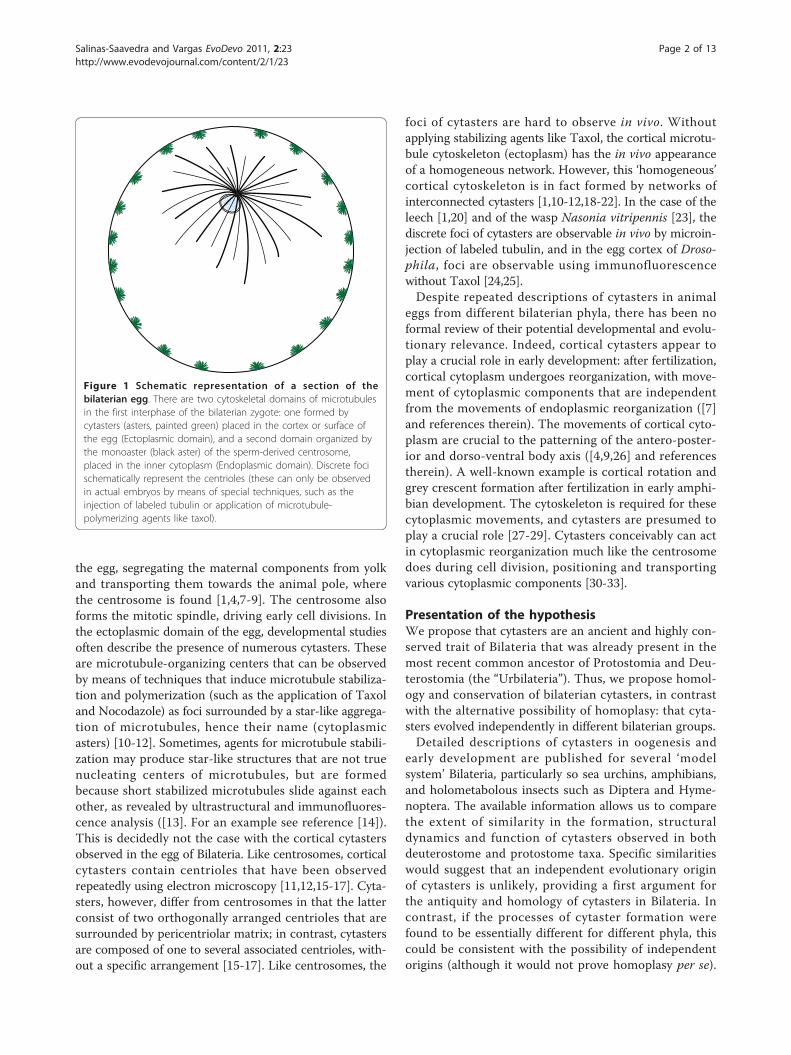
the egg, segregating the maternal components from yolkand transporting them towards the animal pole, wherethe centrosome is found [1,4,7-9]. The centrosome alsoforms the mitotic spindle, driving early cell divisions. Inthe ectoplasmic domain of the egg, developmental studiesoften describe the presence of numerous cytasters. Theseare microtubule-organizing centers that can be observedby means of techniques that induce microtubule stabiliza-tion and polymerization (such as the application of Taxoland Nocodazole) as foci surrounded by a star-like aggrega-tion of microtubules, hence their name (cytoplasmicasters) [10-12]. Sometimes, agents for microtubule stabili-zation may produce star-like structures that are not truenucleating centers of microtubules, but are formedbecause short stabilized microtubules slide against eachother, as revealed by ultrastructural and immunofluores-cence analysis ([13]. For an example see reference [14]).This is decidedly not the case with the cortical cytastersobserved in the egg of Bilateria. Like centrosomes, corticalcytasters contain centrioles that have been observedrepeatedly using electron microscopy [11,12,15-17]. Cyta-sters, however, differ from centrosomes in that the latterconsist of two orthogonally arranged centrioles that aresurrounded by pericentriolar matrix; in contrast, cytastersare composed of one to several associated centrioles, with-out a specific arrangement [15-17]. Like centrosomes, the
foci of cytasters are hard to observe in vivo. Withoutapplying stabilizing agents like Taxol, the cortical microtu-bule cytoskeleton (ectoplasm) has the in vivo appearanceof a homogeneous network. However, this ‘homogeneous’cortical cytoskeleton is in fact formed by networks ofinterconnected cytasters [1,10-12,18-22]. In the case of theleech [1,20] and of the wasp Nasonia vitripennis [23], thediscrete foci of cytasters are observable in vivo by microin-jection of labeled tubulin, and in the egg cortex of Droso-phila, foci are observable using immunofluorescencewithout Taxol [24,25].Despite repeated descriptions of cytasters in animal
eggs from different bilaterian phyla, there has been noformal review of their potential developmental and evolu-tionary relevance. Indeed, cortical cytasters appear toplay a crucial role in early development: after fertilization,cortical cytoplasm undergoes reorganization, with move-ment of cytoplasmic components that are independentfrom the movements of endoplasmic reorganization ([7]and references therein). The movements of cortical cyto-plasm are crucial to the patterning of the antero-poster-ior and dorso-ventral body axis ([4,9,26] and referencestherein). A well-known example is cortical rotation andgrey crescent formation after fertilization in early amphi-bian development. The cytoskeleton is required for thesecytoplasmic movements, and cytasters are presumed toplay a crucial role [27-29]. Cytasters conceivably can actin cytoplasmic reorganization much like the centrosomedoes during cell division, positioning and transportingvarious cytoplasmic components [30-33].
Presentation of the hypothesisWe propose that cytasters are an ancient and highly con-served trait of Bilateria that was already present in themost recent common ancestor of Protostomia and Deu-terostomia (the “Urbilateria”). Thus, we propose homol-ogy and conservation of bilaterian cytasters, in contrastwith the alternative possibility of homoplasy: that cyta-sters evolved independently in different bilaterian groups.Detailed descriptions of cytasters in oogenesis and
early development are published for several ‘modelsystem’ Bilateria, particularly so sea urchins, amphibians,and holometabolous insects such as Diptera and Hyme-noptera. The available information allows us to comparethe extent of similarity in the formation, structuraldynamics and function of cytasters observed in bothdeuterostome and protostome taxa. Specific similaritieswould suggest that an independent evolutionary originof cytasters is unlikely, providing a first argument forthe antiquity and homology of cytasters in Bilateria. Incontrast, if the processes of cytaster formation werefound to be essentially different for different phyla, thiscould be consistent with the possibility of independentorigins (although it would not prove homoplasy per se).
Figure 1 Schematic representation of a section of thebilaterian egg. There are two cytoskeletal domains of microtubulesin the first interphase of the bilaterian zygote: one formed bycytasters (asters, painted green) placed in the cortex or surface ofthe egg (Ectoplasmic domain), and a second domain organized bythe monoaster (black aster) of the sperm-derived centrosome,placed in the inner cytoplasm (Endoplasmic domain). Discrete focischematically represent the centrioles (these can only be observedin actual embryos by means of special techniques, such as theinjection of labeled tubulin or application of microtubule-polymerizing agents like taxol).
Salinas-Saavedra and Vargas EvoDevo 2011, 2:23http://www.evodevojournal.com/content/2/1/23
Page 2 of 13

Different mechanisms of cytaster formation would sug-gest that cytasters could form easily under different bio-logical conditions, supporting the argument forhomoplasy.
Developmental similarities in protostomes anddeuterostomesIn both protostome and deuterostome taxa, cytasters firstbecome visible during oogenesis (Figure 2). Their cen-trioles have been observed to develop from CentriolarPrecursor Bodies (CPBs) associated with the nuclearenvelope, which act as ‘seeds’ for centrioles, as describedfor Hymenoptera [34], Echinodermata [15,35-39], andMammalia ([40-43]). These ‘seeds’ probably correspondto centrin buds, accumulations of centrin proteins asso-ciated with the outer surface of the nuclear envelope,which have been well studied in mammalian culture cells([44,45] and references therein). The first step in theontogeny of cytasters is that numerous membranous ele-ments containing CPB ‘seeds’ detach from the nuclearenvelope and move from the cytoplasm to the oocytecortex (2 in Figure 2). These membranous elements arecalled accessory nuclei in insects [34], annulate lamellaein sea-urchin [36] and sea-cucumber [38], multivesicularaggregates in mouse [40], and small vesicles in rabbit[42]. Initiation, assembly, and development of the cen-triole and aster of cytasters begin after the breakdown ofthe membrane of the nucleus (germinal vesicle) duringmeiosis (3 in Figure 2), when CPBs recruit maternal pro-teins from the oocyte cytoplasm and become centrioles(4 to in Figure 2). Diverse cytoplasmic components(mitochondria, endoplasmic reticulum, granular material,ribosomes, proteins, maternal mRNA, membranous ele-ments, and others) then become associated with cytastersconcomitant to the progress of meiosis [15,34,40]. New
cytasters are also produced by centriole duplication offully formed cytasters [24]. The formation of cytasters iscompletely independent in timing and place from centro-some duplication and nuclear division in the cell cycle[15,46]. After fertilization, and immediately following thefirst mitosis, cytasters begin to lose their distinct radialconfiguration and gradually revert back to small astral(star-like) areas that diminish considerably in size, even-tually losing most of their characteristic astral features(3a in Figure 3). The centriole becomes no longer visibleby electron microscopy, and is integrated within the ecto-plasmic network of microtubules [15,37,47,48].The fact that all the specific developmental processes
above have been described in both protostome and deuter-ostome taxa suggests that cytasters did not originate inde-pendently in each lineage but are homologous, havingalready been present in their most recent common ances-tor (the Urbilateria). The specific developmental pathwaysalso suggest that cytasters are not easily formed underdifferent biological processes and conditions. A clear pre-diction of the hypothesis of homology is that most basicgroups of Bilateria should also conserve these specificsimilarities. In contrast, if taxa with cytasters are nestedwithin groups that otherwise lack cytasters, this wouldprovide evidence of homoplasy, with cytasters appearingindependently in different lineages. The absence of cyta-sters, or their modification, in turn, can be proven to be asecondarily derived condition, if it occurs in taxa that arenested within groups that otherwise exhibit the presenceand general developmental pattern of cytasters. By exam-ining published evidence from non-bilaterian outgroups,such as Ctenophora and Cnidaria, we can also testwhether cytasters originated exclusively in the lineageleading to Bilateria, or in the ancestors of a larger group ofEumetazoa. To this purpose, we reviewed published data
Progress of Oogenesis
ne
1 2 3 5 64
Female Pronucleus “Accessory Nuclei” Centriolar Precursor Body (CPB) Juvenile Centiole of the Cytaster Microtubules of the Aster Cytaster Polar Body
Figure 2 The development of cytasters. During oogenesis (1 to 6), multiple accessory nuclei (purple) detach from the nuclear envelope (ne)and migrate towards the egg surface (2). The nuclear envelope breaks down (dashed line, 3) and the centriolar precursor bodies (CPB, redpoints), which are placed in these membranous elements (3 and 4), develop into the centrioles (red circles) of the cytasters, whichsimultaneously nucleate microtubules of the asters (green lines, 5). At the end of oogenesis (6), cytasters (green) have surrounded the eggcortex. Arabic numbers represent the sequence of events (not stages of meiosis). The polar body indicates the end of the first meiosis.
Salinas-Saavedra and Vargas EvoDevo 2011, 2:23http://www.evodevojournal.com/content/2/1/23
Page 3 of 13

Developmental Progress
fp
cytsRCCCM
fp
sns
SPZ
mn
mc
1 2 3
a
b
c
Cnidaria
Ctenophora
Bilateria
Female Pronucleus
Reduced Centriole(RC)
CytasterSperm Sperm Pronuclear Zone (SPZ)
Cortical Cytoplasmatic Module (CCM)
Figure 3 Schematic illustration of early development in Cnidaria, Ctenophora, and Bilateria. In bilaterian eggs (1 to 3a), cytasters (cyts)are present in the oocyte (during and at the end of oogenesis; 1c). When the egg is activated by fertilization (1a), cytasters reorganize theectoplasm around them forming the cortical cytoplasmic modules (CCM, dashed circle) and mitosis starts. Immediately following mitosis (3a),cytasters begin to lose their distinct radial configurations, gradually reverting back to small astral areas that diminish considerably in size (dashedline), possibly becoming reduced centrioles (RC). Bilaterian cytasters have a similar role to that of sperm pronuclei in ctenophores. In Ctenophora(1 to 3b), previous to the entry of sperm (1b) no cytoplasmic movements are observed. When supernumerary sperm (sns) enter the egg (2b),cytoplasm reorganizes into several sperm pronuclear zones (SPZ, dashed circle) and the female pronucleus (fp) chooses one sperm (doublearrow). In this moment cell division starts and supernumerary sperm asters diminish in size and become integrated to the microtubule network(dashed line; 3b). In Cnidaria, normal development (1 to 3c) requires a microtubule network (mn) exclusively organized by the maternalcentrosome (mc; 1c), and after fertilization (2c), by the sperm-derived centrosome. All maternal determinants necessary for normal developmentare transported towards the animal pole (arrows in 1c), where the nucleus and centrosome are found. Fertilized eggs of cnidarians contain aresidual microtubule network at the time of first mitosis (3c).
Salinas-Saavedra and Vargas EvoDevo 2011, 2:23http://www.evodevojournal.com/content/2/1/23
Page 4 of 13
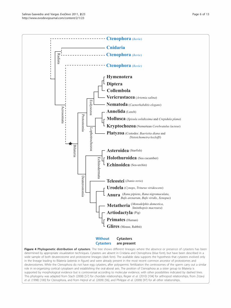
reporting the presence or absence of cytasters in differentanimal species. Although some of the older descriptionsare rudimentary, modern observations with plenty ofstructural detail are available for several taxa. The techni-ques used and structures observed are summarized inAdditional file 1. This bibliographical review allows identi-fication of the more important gaps in information andfuture directions of research that will allow further testingof the hypothesis of homology.The available information reveals a diversity of bilaterian
phyla in which egg cytasters have been reported (seeFigure 4), as summarized in Additional file 1. Both Deuter-ostomia and Protostomia are well represented by severallineages, including some that diverged very early withineach group, as we describe below. The available informa-tion is also summarized in Additional file 1.
Cytasters in protostomesWithin protostomes, it is well accepted that two mainlineages exist, the Lophotrochozoa and the Ecdysozoa.The Lophotrochozoa includes well-studied phyla such asMollusca and Annelida. In zygotes of the leech (an anne-lid) the cortical cytoplasm is populated by numerous inter-connected cytasters that together constitute the wholecortex microtubule network. This ectoplasmic cytoskeletaldomain is observable in the egg before fertilization, fromthe meiotic phase onwards, when no monoaster has yetbeen formed, which confirms its formation is independentof the monoaster [1]. The reorganization and translocationof the ectoplasmic cytoskeleton is linked to the dynamicsof cytasters (see [20]). No studies have yet confirmed (ordiscarded) the initial formation of cytasters from nuclearvesicles (See Additional file 1). In the mollusk Crepidulaplana, as in the aforementioned groups, cytasters are inthe egg cortex, surrounded by cytoplasmic components,closely associated with vesicles (presumably of nuclear ori-gin), and their formation is not related to the centrosome[49] (See Additional file 1). Another mollusk in whichcytasters have been reported is Spisula solidissima [50].These observations are important since the lineage leadingto annelids and mollusks (the Lophotrochozoa) divergedfrom that leading to insects (the Ecdysozoa) towards theorigins of Protostomia. Other Lophotrochozoa in whichcytasters have been observed (although in less detail) arethe Cestoda Baerietta diana and Distoichometra kozloffi[51], and the Nemertean Cerebratulus lacteus [52].Within the Ecdysozoa, information about cytasters is
available in great detail for holometabolous insects likeHymenoptera (as discussed above) and Diptera. They havealso been described in Collembolla, indicating that cyta-sters were already present at the origin of the Hexapoda[17,23-25,53,54]. In other arthropods, cytasters have beendescribed in the eggs of the Vericrustacean Artemia salina[5]. The description of cytasters in the nematode
Caenorhabditis elegans [55] is important, because Nema-todes diverged early from all other Ecdysozoan lineages[56]. We conclude that the available evidence is consistentwith the homology and conservation of egg cytasters inProtostomes, according to a good sample of lineages span-ning both Ecdysozoa and Lophotrochozoa.
Cytasters in deuterostomesThe deuterostomes are split into two main groups, theAmbulacraria and the Chordata [56,57]. Within theAmbulacraria, cytasters are especially well-studied in seaurchins, as discussed above ([15,16,18,21,58,59] and refer-ences therein). In sea urchins, interconnected cytasters[21] take part in cytoplasmic rotation (cortical reorganiza-tion over 16°C) when the egg is fertilized [58]. Cytastersare also present in other echinoderms such as starfish[39], sea cucumber [38], and sand-dollar [35,60]. Withinthe chordates, available information is almost entirelyrestricted to amphibians and mammals. Otherwise, one ofus has confirmed the presence of cytasters in the egg cor-tex of the teleost Danio rerio, the zebrafish (MS personalobservation). In the amphibian Cynops (a newt), the cyta-sters are distributed around the whole unfertilized egg cor-tex, and form a coarse microtubule network in parallelarray, except around the meiotic spindle at the animalpole (cytoplasm is restricted to the animal pole, because ofthe high yolk content at the vegetal pole; [48]). This paral-lel array correlates with the direction of cortical rotation.Cytoplasmic asters also have been described in the oogen-esis of the salamander Triturus viridescens [61] and severalanurans (see Additional file 1), related to cortical andgerm plasm movements [11,12,27,62-65].In mammals, as expected, the cytasters surround the
cortex of the unfertilized egg [22,47,66-69] and are inde-pendent of the sperm derived-centrosome. Cytastershave been described in the oocytes of human [70], pig[43,71], rabbit [42], opossum [72] and marsupial rat [73].Observations in marsupials are especially interesting,since this lineage diverged early within mammals. TheGlires (mouse and rabbit) deserve special attentionbecause they are derived: their cytasters do not containmature centrioles. Rather, the nuclear vesicles (multivesi-cular aggregates) have been observed to contain CPBssurrounded by pericentriolar matrix [40-42]. The absenceof a mature centriole has led to the description of thesecytasters as acentriolar [19,74], but it must be kept inmind that CPB can also organize microtubule polymeri-zation (even in absence of pericentriolar matrix [15]).Navara [75] reported that no cytasters are present in thecow. If this is not an artifact, and the cow is really lackingcytoskeletal organizers in the cortical cytoplasm, thiswould represent a secondary loss of cytasters in evolu-tion: Phylogenetically, the cow is firmly nested withinmammals that do have cytasters.
Salinas-Saavedra and Vargas EvoDevo 2011, 2:23http://www.evodevojournal.com/content/2/1/23
Page 5 of 13
Protostomia
Deuterostom
ia
Bilateria
Ctenophora (Beröe)
Cnidaria
HymenoteraDipteraCollembola
Nematoda (Caenorhabditis elegans)
Annelida (Leech)
Platyzoa (Cestodes: Baerietta diana and Distoichometra kozloffi)
Kryptochozoa (Nemertean Cerebratulus lacteus)
Mollusca (Spisula solidissima and Crepidula plana)
Echinoidea (Sea-urchin)
Artiodactyla (Pig)
Acrosom
ataR
adiata
Ctenophora (Beröe)
Ctenophora (Beröe)
Vericrustacea (Artemia salina)
Holothuroidea (Sea cucumber)
Asteroidea (Starfish)
Teleostei (Danio rerio)
Urodela (Cynops, Triturus viridescens)
Anura (Rana pipiens, Rana nigromaculata, Bufo arenarum, Bufo viridis, Xenopus)
Metatheria (Monodelphis domestica, Sminthopsis macroura)
Glires (Mouse, Rabbit)
Primates (Human)
EcdysozoaLophotrochozoa
Echinodermata
Without Cytasters
Cytasters are present
Figure 4 Phylogenetic distribution of cytasters. The tree shows different lineages where the absence or presence of cytasters has beendetermined by appropriate visualization techniques. Cytasters are absent in Cnidaria and Ctenophora (blue font), but have been described in awide sample of both deuterostome and protostome lineages (dark font). The available data supports the hypothesis that cytasters evolved onlyin the lineage leading to Bilateria (asterisk in figure) and were already present in the most recent common ancestor of protostomes anddeuterostomes. While the Ctenophora do not have egg cytasters, after polyspermic fertilization the centrosomes of the sperm carry out a similarrole in re-organizing cortical cytoplasm and establishing the oral-aboral axis. The position of Ctenophora as a sister group to Bilateria issupported by morphological evidence but is controversial according to molecular evidence, with other possibilities indicated by dashed lines.This phylogeny was adapted from Stach (2008) [57] for chordate relationships, Regier et al. (2010) [104] for arthropod relationships, from Zrzavýet al. (1998) [100] for Ctenophora, and from Hejnol et al. (2009) [56], and Philippe et al. (2009) [97] for all other relationships.
Salinas-Saavedra and Vargas EvoDevo 2011, 2:23http://www.evodevojournal.com/content/2/1/23
Page 6 of 13

Cytasters in absence of the centrosome: a common rolethroughout BilateriaIn the early development of Bilateria, the centriole of thesperm provides the centrosome, which drives the migra-tion, encounter, and fusion of the sperm and egg pronucleias well as the early cycles of cell division. However, insome cases where the sperm-derived centrosome is absent,these functions are carried out by cytasters. As we havedescribed above, the Glires differ from other mammalsbecause their cytasters do not have mature centrioles (seeabove). Glires are also derived in that the oocyte has nocentriole [69], and there is no sperm-derived centrosomein the fertilized egg. In the absence of a sperm-derivedcentriole to form the centrosome, the only comparableorganizers are cytasters, which take over its function inguiding the migration of the pronuclei towards the centerof the egg [67]. Cytasters in Glires also organize and formthe mitotic spindle, allowing the cell cycles of early mousedevelopment [19,22,67-69,74,76] in the absence of a cen-trosome. Subsequently, at the blastocyst stage, in each cella cytaster develops into a mature centriole and becomes acentrosome ([19,40,42] and references therein). Thisdemonstrates that despite being described as acentriolar,the cytasters of Glires can be functional as cytoplasmicorganizers and have the potential to become the centro-some. The functional takeover of centrosomal functionsby cytasters has also been repeatedly observed in severalbilaterian phyla in cases in which the sperm-derivedcentrosome may be absent yet development proceeds.This is the case for many Hexapoda that present naturalparthenogenesis [17,23-25,53,54] and is also true forexperimentally induced parthenogenesis in eggs of theVericrustacean Artemia salina [5]. In sea urchin and sanddollar, when eggs are artificially activated (without fertili-zation), the cytasters form a structure resembling a mitoticspindle (bipolar but without asters; [59]) and developmentproceeds parthenogenetically [35,37]. Cytasters also takeover the function of early cell divisions when parthenogen-esis is artificially induced in the pig and rabbit [42,43]. Inthe salamander Triturus, in experiments on androgenicdevelopment, eggs are fertilized in the absence of thesperm-derived centrosome, so cytasters must organize theearly cell divisions [61]. In all the cases mentioned above,cortical reorganization and cell divisions proceed normally,in the absence of a sperm-derived centrosome. Thisdemonstrates that cytasters can organize and move cyto-plasmic components much like a centrosome, which sup-ports the strong inference that cytasters are crucial for thereorganization of cortical cytoplasm and axis establish-ment. A potential for replacing centrosomal functions inearly development, and a capacity to eventually becomecentrosomes, is another specific trait of cytasters that isubiquitous across distantly related bilaterian phyla (seeAdditional file 1).
Development without cytasters: Cnidaria and CtenophoraA review of published evidence from basic groups ofEumetazoa supports the notion that cytasters are an exclu-sively bilaterian trait. Outside of Bilateria, in Cnidaria andCtenophora, no cytasters are formed during the process ofoogenesis, as confirmed by immunofluorescence andmicrotubule polymerization techniques [14,77] (Figure 4).In cnidarians, animal-vegetal (a-v) polarity of the oocyte isgenerated during oogenesis and is present in the oocytebefore fertilization, with the animal pole at the site ofemission of the polar bodies [77,78]. Maternal determi-nants that specify the germ line (for example, Nvvas1, andNvnos2 RNA [79]) and oocyte polarity (CheFz1 RNA [77])are transported throughout the entire cytoplasm involvingmicrotubules [77,79], and not in the ectoplasm alone as inBilateria [3,8,80-86]. There is no independent ectoplasmicnetwork of microtubules, nor did we find any descriptionsuggesting the differentiation of cortical cytoplasm in cni-darians. In this case, the polarization of the egg is directedby the centrosome associated with the meiotic spindle.Transportation and reorganization of maternal determi-nants in cnidarians begins during oogenesis and is com-pleted after fertilization, mediated by a single microtubulenetwork that is required for their transport (1c in Figure3) [77]. This microtubule network has no obvious polarity,other than the position at the animal pole of the maternalcentrosome of the meiotic spindle during oogenesis. Atthe moment of fertilization, the maternal centrosome isabsent and the sperm-derived centrosome localizes at theanimal pole (2c in Figure 3) [77]. At the end of each meio-tic and mitotic cycle, when the centrosome ceases itsactivity, the entire microtubule network is depolymerized[77], in contrast with bilaterian eggs, in which the corticalnetwork remains. The position of the centrosome of themeiotic/mitotic spindle (both before and after fertilization)is associated with the direction of transportation of mater-nal determinants during ooplasmic segregation. Undernormal conditions, all maternal determinants necessaryfor normal development are transported towards the ani-mal pole (1c in Figure 3) [78], where the nucleus and cen-trosome of the meiotic/mitotic spindle are found [77].When centrifuged, the centrosome can be moved to aposition that is offset from the nucleus. In this case, mater-nal determinants are now transported towards the newposition of the centrosome (rather than the nucleus). Thepresence of a single microtubule network nucleated by asingle centrosome in the egg [77] confirms that cnidariansdo not have an independent ectoplasmic network ofmicrotubules comparable to that formed in bilaterians andctenophores. Thus, the process of redistribution of mater-nal determinants in cnidarian eggs involves only the meio-tic/mitotic spindle in cytoplasmic reorganization, ratherthan multiple cytasters. In this sense, we may comparecnidarian development to the endoplasmic domain of
Salinas-Saavedra and Vargas EvoDevo 2011, 2:23http://www.evodevojournal.com/content/2/1/23
Page 7 of 13

Bilateria, which is also reorganized upon fertilization bythe microtubule network associated with the sperm-derived centrosome. In contrast, in both Ctenophora andBilateria, reorganization of cortical cytoplasm is indepen-dent of the centrosome.
Cortical reorganization without cytasters: The role ofpolyspermy in CtenophoraCytasters are absent in the eggs of ctenophores (Figure 4)as confirmed by immunofluorescence and microtubulepolymerization techniques [14]. However, cortical cyto-plasmic movements after fertilization occur that closelyresemble those of bilaterian ectoplasm (1 to 3b in Figure3). The ctenophore Beroe ovata has physiological poly-spermic fertilization [87,88] taking place immediatelybefore or during formation of the first polar body (1 to 2bin Figure 3) [89]. After several spermatozoa enter the egg,cytoplasmic components become associated with thesupernumerary sperm cells, each forming a spherical zonecalled Sperm Pronuclear Zone (SPZ; 2b in Figure 3), thatconsists of cortical granules, mitochondria, endoplasmicreticulum, and other cytoplasmic components, includingthe nuclear envelope of the sperm pronucleus. Each SPZis organized by the centriole associated with each malepronucleus [89]. The formation of this new cortical cyto-plasmic configuration is carried out by microtubule-mediated waves and is of utmost importance in establish-ing the oral-aboral axis [14], which takes place after thefusion of pronuclei and first cleavage cycle [14,89]. In thepolyspermic Ctenophora, it is the female pronucleus ofBeroe that migrates to join a stationary male pronucleus,choosing one (2b in Figure 3). At this site, the zygotenucleus forms, first mitosis occurs and the first cleavagefurrow starts [14,89]. As development and cell divisionadvances, the nuclear envelopes of the supernumerarymale pronucleus break down, their DNA is degraded, andthe giant asters nucleated by their associated centrosomesshrink and are no longer visible, presumably becomingintegrated with the microtubule network (3b in Figure 3)[14]. The centrioles of zygote centrosomes retain theirmature form.
Testing the hypothesisThe specific developmental similarities of cytasters acrossBilateria provide compelling evidence of homology, ahypothesis that is also supported by the available data onphylogenetic distribution of the presence of cytasters. Pub-lished information on the presence of cytasters covers avery good taxonomic sample: 20 orders, representing 15classes and 8 phyla (see Figure 4, Additional file 1). Forperspective, we can consider how genomic structure andmolecular-developmental aspects are typically known forpoor taxonomic samples of only a few model species.Good taxon sampling is crucial to any hypotheses of
homology, because it allows testing for evolutionarypatterns such as conservation, homoplasy, and secondaryloss. We found no evolutionary pattern that would sup-port the independent origin of cytasters in different bila-terian phyla. We found a single (possible) case of absenceof cytasters: The cow. Further confirmation of this case isimportant, since this would be a clear case of a secondaryloss of cytasters in evolution. Also within mammals, theGlires are interesting since they show how small differ-ences may evolve in a specific lineage (lack of sperm-derived centrosome, cytasters with immature centrioles),while at the same time conserving the general pattern offormation and function of cytasters. In all, published avail-able data is overwhelmingly consistent with homology andconservation of cytasters across bilateria. Homologydirectly implies that cytasters should be observable in sev-eral groups for which there is currently no available pub-lished information, that are represented in Figure 5 bygrey branches (Dark branches represent groups in whichthe presence of cytasters has already been documented).Within the Deuterostomes, data are missing for the phylaHemichordata, Urochodata and Cephalochordata. WithinVertebrata, data are missing for several basic groups offishes: Cyclostomata, Chondrichthyes, Chondrostei, Holos-tei, Actinistia, and Dipnoi. Within Tetrapoda, no data areavailable for Monotremata and Reptilia (including birds).In Protostomes, data are missing for several importantphyla: Chaetognatha, Bryozoa, Brachiopoda, Kinorhyncha,Loricifera, Priapulida, Onychophora, Tardigrada, Chelicer-ata, Myriapoda, and Oligostraca (Pancrustaceans). In somegroups, like Platyzoa [51] and Kryptochozoa [52], theavailable information about cytasters was described duringthe early twentieth century by observations of fixed andstained eggs made under the light microscope. Updateddescriptions are desirable to confirm that these groupsreally present cytasters. We found no available informationabout the presence or absence of cytasters in the eggs ofPlacozoa and Porifera. The documented absence of cyta-sters in the other non-bilaterian phyla Cnidaria and Cte-nophora suggests that cytasters should also be absent inPlacozoa and Porifera. If so, this would support the notionthat cytasters only evolved in the lineage leading to Bila-teria. For all the phyla mentioned above, new studies onthe presence or absence of cytasters can benefit from uni-form application of modern techniques, ensuring soundcomparison of data across phyla.
Implications of the hypothesisCortical cytoplasmic modules are an ancient and highlyconserved Bilaterian traitBased on the observations in several different bilateriananimals, cytasters are much more than star-like centersof microtubule polymerization. They have a specificontogeny, developing from precursor bodies (possibly
Salinas-Saavedra and Vargas EvoDevo 2011, 2:23http://www.evodevojournal.com/content/2/1/23
Page 8 of 13

Protostomia
Deuterostom
ia
Bilateria
HymenoteraDipteraCollembola
Nematoda
Annelida
Platyzoa
Kryptochozoa
Mollusca
Echinoidea
Artiodactyla
Vericrustacea
HolothuroideaAsteroidea
Teleostei
UrodelaAnura
Metatheria
GliresPrimates
EcdysozoaLophotrochozoa
Echinodermata
OligostracaMyriapodaChelicerata
Arthropoda
BrachiopodaNemertea
OnychophoraTardigrada
Kinorhyncha
Priapulida
Bryozoa
Loricifera
Chaetognatha
Reptilia
Hemichordata
CephalochordataUrochordata
Chondrichthyes
HolosteiChondrostei
Dipnoi
Cyclostomata
Tetrapoda
Vertebrata
Monotremata
Actinistia
Cytasters are present
No available published information
Figure 5 Bilaterian groups expected to present cytasters. Cytasters have been described in a wide sample of both deuterostome andprotostome phyla (dark branches). Grey branches are used for phyla for which we found no available published information. More detailedstudies are necessary to clarify the presence or absence of cytasters in these missing phyla, using modern techniques uniformly applied acrossdifferent taxa. This phylogeny was adapted from Stach (2008) [57] for chordate relationships, Regier et al. (2010) [104] for arthropod relationships,and from Hejnol et al. (2009) [56], and Philippe et al. (2009) [97] for all other relationships.
Salinas-Saavedra and Vargas EvoDevo 2011, 2:23http://www.evodevojournal.com/content/2/1/23
Page 9 of 13

centrin buds) found in fragments of nuclear membrane(often described as accessory nuclei or vesicles) thatmove to the egg cortex and become associated to severalcytoplasmic components, conforming complex corticalmodules (Figure 2, Additional file 1). Thus, to go beyondthe notion contained in the descriptive term cytaster, wewish to forward the notion and acronym of CorticalCytoplasmatic Modules (CCM) for the entire assemblageof centriole (or, as in mouse and rabbit, centriolar pre-cursor) associated with the membranous element, asterand associated cytoplasmic components: mitochondria,endoplasmic reticulum, granular material, ribosomes,proteins, maternal mRNA, membranous elements, andothers.
Are CCMs inherited in cytoplasm?The development of CCMs brings up interesting questionsabout their inheritance. An intriguing possibility suggestedby Kallenbach and Mazia [37] is that the oocyte cytastersupon disappearing may produce themselves the CPBs(seeds) that become allocated to the nuclear surface. Thus,cytaster reduction may be an important source of seedsthat become centrioles of new CCMs during oogenesisb. InBilateria, during the process of cell division, the nuclearmembrane (from which vesicles of CCM’s are formed) dis-integrates into fragments, which thereafter allocate to thecytoplasm of both resulting cells [30,32,33,90]. Thus, it iseasy to conceive how CPBs could be inherited throughcytoplasm containing these fragments. CPBs may always bepresent in the nuclear membrane, but only become cyta-sters during the process of oogenesis. If so, artificial induc-tion of cytaster formation is conceivable. This seems to bethe case in cultured mammalian cells, where formation ofcytasters similar to that in oogenesis is induced upon theexperimental ablation of the centrosome [44] and whencells are arrested in S-phase [45]. In these cell cultures, thecentrioles of cytasters develop from seeds in vesicles of thenuclear membrane that move towards the cortical cyto-plasm. These seeds in the nuclear membrane have beenshown to be centrin buds containing alpha/gamma-tubulinand centrin 2. Like centrioles, these seeds are capable ofself-replication [45], and are probably the same as the CPBseeds observed in oogenesis. Further research is requiredto establish if new centrin buds can only be formed byreplication, or can also be assembled from isolated centrinproteins [44,91]. If new CPBs only form by replication,they can only be inherited through cytoplasm.
A polyspermic ancestor?The early development of Bilateria can be compared toother Eumetazoan outgroups in order to make inferencesabout the origin of CCMs. In this regard, the capacity ofthe sperm-derived centrosome to re-organize the egg
cytoplasm (as observed in Cnidaria and the endoplasm ofBilateria) is linked in the case of the polyspermic Cteno-phora to the establishment of a well-differentiated ecto-plasmic domain, which is required for the establishmentof the oral-aboral axis. Compelling resemblances existbetween the CCMs of bilaterian eggs and the aforemen-tioned SPZ of Ctenophora. The SPZ and CCM both pos-sess a microtubule-nucleating centriole (and aster)initially associated with the nuclear membrane (spermpronucleus of SPZ, and vesicles of nuclear membrane oraccessory nuclei in CCM), which becomes surrounded byassociated cytoplasmic components of the egg. Both arerequired for the early developmental processes of ecto-plasmic reorganization and axes establishment, and bothhave a similar developmental fate, reducing the aster andcentrioles, which become incorporated in the microtu-bule network (see above; compare illustration 2-3b and2-3a in Figure 3). As discussed above, in the absence ofthe sperm-derived centrosome, CCMs are capable of tak-ing over its role in the migration, encounter, and fusionof the sperm and egg pronuclei [92]. In the ctenophoreBeroe ovata, when the unfertilized egg is artificially acti-vated (SPZs are absent) the female pronucleus migratesrandomly in the cytoplasm throughout the egg [14,93].When the egg of Beroe is fertilized, SPZs drive the migra-tion, encounter, and fusion of pronuclei [93]. As dis-cussed above, when bilaterian oocytes are artificiallyactivated (no sperm enter the egg), they undergo the nor-mal process of segregation and redistribution of cyto-plasm, without reactivation of meiosis ([30,33] M.Salinas-Saavedra, personal observation in zebrafish andsea urchin) and development often continues partheno-genetically [17,24,35,48,53,54,73,94,95]. In contrast, cyto-plasmic reorganization and cell division is disrupted inartificially activated eggs of ctenophores, demonstratingthat SPZs are required for the same processes carried outby CCMs in Bilateria.The remarkable similarities to ctenophore SPZs listed
above suggest that the development and function ofCCMs are derived from early developmental processessimilar to those in the polyspermic Ctenophora. Morespecifically, we propose that the origin of CCMs involvedthe acquisition by the female germ line of the capacity toproduce numerous centriole-based modules of ecto-plasm, a process that previously was required for the exo-genous contribution of supernumerary sperm. BecauseCPBs can be inherited through cytoplasm, it is possiblethat the female germline acquired numerous CPBsdirectly from the reduction of sperm centrioles. An inter-esting point to observe in future studies is the distribu-tion of germline determinants (like vasa and nanos)during the development of polyspermic Ctenophores.According to our hypothesis, we expect a cortical
Salinas-Saavedra and Vargas EvoDevo 2011, 2:23http://www.evodevojournal.com/content/2/1/23
Page 10 of 13

localization of these markers associated with the microtu-bules of SPZs, similar to the distribution observed in bila-terian eggs, associated to microtubules of the egg cortex.The phylogenetic relationships of Ctenophora to other
metazoa are currently controversial [56,96-102]. However,Ctenophora has often been suggested to be a sister groupto Bilateria [96,99-102], especially on the basis of the mor-phological evidence. If so, this would support the notionthat polyspermy could have been present in the mostrecent common ancestor of Ctenophora and Bilateria, andthe lack of a differentiated cortical cytoplasm, as in cnidar-ians, may represent a primitive condition for the egg (witha single microtubule network).The complex egg of Bilateria and its reorganization are
crucial in the selective distribution of cytoplasmic domainsduring cleavage, leading to body axis patterning. Despitethe well-acknowledged importance of egg cytoplasmicdomains and their movements [1,4], their evolutionary ori-gin is seldom discussed and thus seems largely mysterious.In this sense, a new explicative framework emerges whenwe consider the possibility that these cytoplasmic move-ments may derive from a complex, polyspermic fertiliza-tion, like that of Ctenophora, leading to the differentiationof an ectoplasmic domain and the cytoplasmic movementsof body axis patterning. More detailed research in Cteno-phora and Cnidaria is bound to be informative about theearly evolutionary history of cytoplasmic reorganization.
EndnotesaPreviously, during the formation of the egg, the endo-plasm is organized by the germinal vesicle-derived cen-trosome. In the literature, authors discussing the fate ofthe oocyte-derived centrosome state that it is missing atthe end of meiosis [30,53].
bThis notion is supported by the experimental injec-tion of centrioles (isolated from adult tissues) into theegg of Xenopus. These centrioles become reduced andcease to be observable, but upon fertilization, anincreased number of CCMs shows up at the site ofinjection ([103] and references therein). This suggeststhe injected centrioles provided an increased number ofcentriole precursor bodies at the injection site.
Additional material
Additional file 1: Characteristics of cytasters in different taxa. Tablesthat summarize the available information on cytasters in Protostomia andDeuterostomia, indicating observed structural aspects and the techniquesused. The bold font represents taxa in which the specific developmentalpathway of cytaster formation is well documented.
List of abbreviationsCCM: cortical cytoplasmatic module; CPB: centriolar precursor body; cyts:cytasters; fp: female pronucleus; mc: maternal centrosome; mn: microtubule
network; RC: reduced centriole; sns: supernumerary sperm; SPZ: spermpronuclear zone.
AcknowledgementsThis work was supported by FONDECYT grant 11080258 to AOV. The authorsthank Jorge Mpodozis, Luis Ossa-Fuentes, Juan Salazar, Sebastian Jaramillo,Jõao F. Botelho, Uziel Carrera, and members of ‘Rayo’ lab for helpfuldiscussions.
Authors’ contributionsMS and AOV wrote the article and were responsible for comparative andevolutionary interpretation of the data. MS made the figures and additionalfile material. Both authors read and approved the final version of themanuscript.
Competing interestsThe authors declare that they have no competing interests.
Received: 12 August 2011 Accepted: 1 December 2011Published: 1 December 2011
References1. Cantillana V, Urrutia M, Ubilla A, Fernández J: The complex dynamic
network of microtubule and microfilament cytasters of the leech zygote.Dev Biol 2000, 228:136-149.
2. Gard DL, Cha BJ, Schroeder MM: Confocal immunofluorescencemicroscopy of microtubules, microtubule-associated proteins, andmicrotubule-organizing centers during amphibian oogenesis and earlydevelopment. Curr Top Dev Biol 1996, 31:383-431.
3. Theusch EV, Brown KJ, Pelegri F: Separate pathways of RNA recruitmentlead to the compartmentalization of the zebrafish germ plasm. Dev Biol2006, 292:129-141.
4. Sardet C, Paix A, Prodon F, Dru P, Chenevert J: From oocyte to 16-cellstage: cytoplasmic and cortical reorganizations that pattern the ascidianembryo. Dev Dyn 2007, 236:1716-1731.
5. Gross F: Memoirs: cleavage of blastomeres in the absence of nuclei. JCell Sci 1936, S2-79:57-72.
6. Fernández J, Olea N, Téllez V: Formation of the male pronucleus,organization of the first interphase monaster, and establishment of aperinuclear plasm domain in the egg of the glossiphoniid leechtheromyzon rude. Dev Biol 1994, 164:111-122.
7. Fuentes R, Fernández J: Ooplasmic segregation in the zebrafish zygoteand early embryo: Pattern of ooplasmic movements and transportpathways. Dev Dyn 2010, 239:2172-2189.
8. Fernández J, Valladares M, Fuentes R, Ubilla A: Reorganization ofcytoplasm in the zebrafish oocyte and egg during early steps ofooplasmic segregation. Dev Dyn 2006, 235:656-671.
9. Newman SA: Animal egg as evolutionary innovation: a solution to the‘embryonic hourglass’ puzzle. J Exp Zool B Mol Dev Evol 2011.
10. Hornick JE, Bader JR, Tribble EK, Trimble K, Breunig JS, Halpin ES,Vaughan KT, Hinchcliffe EH: Live-cell analysis of mitotic spindle formationin taxol-treated cells. Cell Motil Cytoskeleton 2008, 65:595-613.
11. Van Assel S, Brachet J: Métabolisme des acides nuclféiques et desprotéines et formation de cytasters dans les oeufs d’amphibiens sousléaction de l’eau lourde. J Embryol Exp Morphol 1968, 19:261-272.
12. Van Assel S, Brachet J: Formation de cytasters dans les œufs deBatraciens sous l’action de l’eau lourde. J Embryol Exp Morphol 1966,15:143-151.
13. Verde F, Berrez JM, Antony C, Karsenti E: Taxol-induced microtubule astersin mitotic extracts of Xenopus eggs: requirement for phosphorylatedfactors and cytoplasmic dynein. J Cell Biol 1991, 112:1177-1187.
14. Houliston E, Carré D, Johnston JA, Sardet C: Axis establishment andmicrotubule-mediated waves prior to first cleavage in Beroe ovata.Development 1993, 117:75-87.
15. Kallenbach RJ: Ultrastructural analysis of the initiation and developmentof cytasters in sea-urchin eggs. J Cell Sci 1985, 73:261-278.
16. Kuriyama R, Borisy GG: Cytasters induced within unfertilized sea-urchineggs. J Cell Sci 1983, 61:175-189.
17. Riparbelli MG, Tagu D, Bonhomme J, Callaini G: Aster self-organization atmeiosis: a conserved mechanism in insect parthenogenesis? Dev Biol2005, 278:220-230.
Salinas-Saavedra and Vargas EvoDevo 2011, 2:23http://www.evodevojournal.com/content/2/1/23
Page 11 of 13
18. Harris PJ, Clason EL, Prier KR: Tubulin polymerization in unfertilized sea-urchin eggs induced by elevated temperature. J Cell Sci 1989, 93:9-17.
19. Schuh M, Ellenberg J: Self-organization of MTOCs replaces centrosomefunction during acentrosomal spindle assembly in live mouse oocytes.Cell 2007, 130:484-498.
20. Fernández J, Cantillana V, Ubilla A: Reorganization and translocation ofthe ectoplasmic cytoskeleton in the leech zygote by condensation ofcytasters and interactions of dynamic microtubules and actin filaments.Cell Motil Cytoskeleton 2002, 53:214-230.
21. Harris PJ, Clason EL: Conditions for assembly of tubulin-based structuresin unfertilized sea urchin eggs. Spirals, monasters and cytasters. J Cell Sci1992, 102:557-567.
22. Tang C-JC, Hu H-M, Tang TK: NuMA expression and function in mouseoocytes and early embryos. J Biomed Sci 2004, 11:370-376.
23. Tram U, Sullivan W: Reciprocal inheritance of centrosomes in theparthenogenetic Hymenopteran Nasonia vitripennis. Curr Biol 2000,10:1413-1419.
24. Riparbelli MG, Callaini G: Drosophila parthenogenesis: a model for denovo centrosome assembly. Dev Biol 2003, 260:298-313.
25. Riparbelli MG, Callaini G: Drosophila parthenogenesis: a tool to deciphercentrosomal vs acentrosomal spindle assembly pathways. Exp Cell Res2008, 314:1617-1625.
26. Jesuthasan S, Stähle U: Dynamic microtubules and specification of thezebrafish embryonic axis. Curr Biol 1997, 7:31-42.
27. Dettlaff TA: Action of actinomycin and puromycin upon frog oocytematuration. J Embryol Exp Morphol 1966, 16:183-195.
28. Ressom R, Dixon K: Relocation and reorganization of germ plasm inXenopus embryos after fertilization. Development 1988, 103:507-518.
29. Houliston E, Elinson RP: Patterns of microtubule polymerization relatingto cortical rotation in Xenopus laevis eggs. Development 1991,112:107-117.
30. Schatten G: The Centrosome and Its Mode of Inheritance: The Reductionof the Centrosome during Gametogenesis and Its Restoration duringFertilization. Dev Biol 1994, 165:299-335.
31. Palazzo RE, Vogel JM, Schnackenberg BJ, Hull DR, Wu X: Centrosomematuration. Curr Top Dev Biol 1999, 49:449-470.
32. Bornens M: Organelle positioning and cell polarity. Nat Rev Mol Cell Biol2008, 9:874-886.
33. Wilson P: Centriole inheritance. Prion 2008, 2:9-16.34. Ferree PM, McDonald K, Fasulo B, Sullivan W: The origin of centrosomes in
parthenogenetic hymenopteran insects. Curr Biol 2006, 16:801-807.35. Chambers R: The formation of the aster in artificial parthenogenesis. J
Gen Physiol 1921, 4:33-39.36. Kallenbach RJ: ’De novo’ centrioles originate at sites associated with
annulate lamellae in sea-urchin eggs. Biosci Rep 1982, 2:959-966.37. Kallenbach RJ, Mazia D: Origin and maturation of centrioles in association
with the nuclear envelope in hypertonic-stressed sea urchin eggs. Eur JCell Biol 1982, 28:68-76.
38. Miyazaki A, Kato K, Nemoto S: Role of microtubules and centrosomes inthe eccentric relocation of the germinal vesicle upon meiosis reinitiationin sea-cucumber oocytes. Dev Biol 2005, 280:237-247.
39. Shirai H, Hosoya N, Sawada T, Nagahama Y, Mohri H: Dynamics of mitoticapparatus formation and tubulin content during oocyte maturation instarfish. Dev Growth Differ 1990, 32:521-529.
40. Calarco P: Centrosome precursors in the acentriolar mouse oocyte.Microsc Res Tech 2000, 49:428-434.
41. Szollosi D, Calarco P, Donahue R: Absence of centrioles in the first andsecond meiotic spindles of mouse oocytes. J Cell Sci 1972, 11:521-541.
42. Szöllosi D, Ozil J-P: De novo formation of centrioles inparthenogenetically activated, diploidized rabbit embryos. Biol Cell 1991,72:61-66.
43. Kim NH, Simerly C, Funahashi H, Schatten G, Day BN: Microtubuleorganization in porcine oocytes during fertilization and parthenogenesis.Biol Reprod 1996, 54:1397-1404.
44. La Terra S, English C, Hergert P, McEwen B, Sluder G, Khodjakov A: The denovo centriole assembly pathway in HeLa cells. J Cell Biol 2005,168:713-722.
45. Collins E, Hornick J, Durcan T, Collins N, Archer W, Karanjeet K, Vaughan K,Hinchcliffe E: Centrosome biogenesis continues in the absence ofmicrotubules during prolonged S-phase arrest. J Cell Physiol 2010,225:454-465.
46. Gard D, Hafezi S, Zhang T, Doxsey S: Centrosome duplication continues incycloheximide-treated Xenopus blastulae in the absence of a detectablecell cycle. J Cell Biol 1990, 110:2033-2042.
47. Schatten G, Simerly C, Asai DJ, Szöke E, Cooke P, Schatten H: Acetylatedalpha-tubulin in microtubules during mouse fertilization and earlydevelopment. Dev Biol 1988, 130:74-86.
48. Iwao Y, Yasumitsu K, Narihira M, Jiang J, Nagahama Y: Changes inmicrotubule structures during the first cell cycle of physiologicallypolyspermic newt eggs. Mol Reprod Dev 1997, 47:210-221.
49. Conklin E: Mitosis and amitosis. Biol Bull 1917, 33:396-436.50. Kuriyama R: Effect of taxol on first and second meiotic spindle formation
in oocytes of the surf clam, Spisula solidissima. J Cell Sci 1986, 84:153-164.51. Douglas L: The development of organ systems in nematotaeniid
cestodes. III. Gametogenesis and embryonic development in Baeriettadiana and Distoichometra kozloffi. J Parasitol 1963, 49:530-558.
52. Yatsu N: Experiments on the development of egg fragments inCerebratulus. Biol Bull 1904, 6:123-136.
53. Karr TL: Centrosome inheritance: a central ‘in-egg-ma’ solved? Curr Biol2001, 11:R21-24.
54. Riparbelli M, Giordano R, Callaini G: Centrosome inheritance in theparthenogenetic egg of the collembolan Folsomia candida. Cell TissueRes 2006, 326:861-872.
55. Hyman AA, White JG: Determination of cell division axes in the earlyembryogenesis of Caenorhabditis elegans. J Cell Biol 1987, 105:2123-2135.
56. Hejnol A, Obst M, Stamatakis A, Ott M, Rouse GW, Edgecombe GD,Martinez P, Baguna J, Bailly X, Jondelius U, Wiens M, Müller WE, Seaver E,Wheeler WC, Martindale MQ, Giribet G, Dunn CW: Assessing the root ofbilaterian animals with scalable phylogenomic methods. Proc Biol Sci2009, 276:4261-4270.
57. Stach T: Chordate phylogeny and evolution: a not so simple three-taxonproblem. J Zool 2008, 276:117-141.
58. Schroeder T, Battaglia D: “Spiral asters” and cytoplasmic rotation in seaurchin eggs: induction in Strongylocentrotus purpuratus eggs byelevated temperature. J Cell Biol 1985, 100:1056-1062.
59. Henson JH, Fried CA, McClellan MK, Ader J, Davis JE, Oldenbourg R,Simerly CR: Bipolar, anastral spindle development in artificially activatedsea urchin eggs. Dev Dyn 2008, 237:1348-1358.
60. Fry H: Conditions determining the origin and behavior of central bodiesin cytasters of Echinarachnius eggs. Biol Bull 1928, 54:363-395.
61. Kaylor C: Studies on experimental haploidy in salamander larvae: II.cytological studies on androgenetic eggs of Triturus viridescens. Biol Bull1941, 81:402-419.
62. Keppel D, Dawson A: Effects of colchicine on the cleavage of the frog’segg (Rana pipiens). Biol Bull 1939, 76:153-161.
63. Kubota T: A regional change in the rigidity of the cortex of the egg ofRana nigromaculata following extrusion of the second polar body.Development 1967, 17:331-340.
64. Skoblina MN: Role of karyoplasm in the emergence of capacity of eggcytoplasm to induce DNA synthesis in transplanted sperm nuclei. JEmbryol Exp Morphol 1976, 36:67-72.
65. Manes ME, Barbieri FD: On the possibility of sperm aster involvement indorso-ventral polarization and pronuclear migration in the amphibianegg. J Embryol Exp Morphol 1977, 40:187-197.
66. Calarco-Gillam PD, Siebert MC, Hubble R, Mitchison T, Kirschner M:Centrosome development in early mouse embryos as defined by anautoantibody against pericentriolar material. Cell 1983, 35:621-629.
67. Maro B, Howlett S, Webb M: Non-spindle microtubule organizing centersin metaphase II-arrested mouse oocytes. J Cell Biol 1985, 101:1665-16672.
68. Schatten H, Schatten G: Motility and centrosomal organization during seaurchin and mouse fertilization. Cell Motil Cytoskeleton 1986, 6:163-175.
69. Palacios MJ, Joshi HC, Simerly C, Schatten G: Gamma-tubulinreorganization during mouse fertilization and early development. J CellSci 1993, 104:383-389.
70. Terada Y, Hasegawa H, Ugajin T, Murakami T, Yaegashi N, Okamura K:Microtubule organization during human parthenogenesis. Fertil Steril2009, 91:1271-1272.
71. Katayama M, Zhong Z, Lai L, Sutovsky P, Prather RS, Schatten H:Mitochondrial distribution and microtubule organization in fertilized andcloned porcine embryos: implications for developmental potential. DevBiol 2006, 299:206-220.
Salinas-Saavedra and Vargas EvoDevo 2011, 2:23http://www.evodevojournal.com/content/2/1/23
Page 12 of 13

72. Breed WG, Simerly C, Navara CS, VandeBerg JL, Schatten G: Microtubuleconfigurations in oocytes, zygotes, and early embryos of a marsupial,Monodelphis domestica. Dev Biol 1994, 164:230-240.
73. Merry NE, Johnson MH, Gehring CA, Selwood L: Cytoskeletal organizationin the oocyte, zygote, and early cleaving embryo of the stripe-faceddunnart (Sminthopsis macroura). Mol Reprod Dev 1995, 41:212-224.
74. Schatten H, Sun Q-Y: Centrosome dynamics during mammalian oocytematuration with a focus on meiotic spindle formation. MolecularReproduction and Development 2011.
75. Navara C: Microtubule organization in the cow during fertilization,polyspermy, parthenogenesis, and nuclear transfer: The role of thesperm aster. Dev Biol 1994, 162:29-40.
76. Schatten G, Simerly C, Schatten H: Microtubule configurations duringfertilization, mitosis, and early development in the mouse and therequirement for egg microtubule-mediated motility during mammalianfertilization. Proc Natl Acad Sci USA 1985, 82:4152-4156.
77. Amiel A, Houliston E: Three distinct RNA localization mechanismscontribute to oocyte polarity establishment in the cnidarian Clytiahemisphaerica. Dev Biol 2009, 327:191-203.
78. Fritzenwanker JH, Genikhovich G, Kraus Y, Technau U: Early developmentand axis specification in the sea anemone Nematostella vectensis. DevBiol 2007, 310:264-279.
79. Extavour CG, Pang K, Matus DQ, Martindale MQ: vasa and nanosexpression patterns in a sea anemone and the evolution of bilateriangerm cell specification mechanisms. Evol Dev 2005, 7:201-215.
80. Wu H-R, Chen Y-T, Su Y-H, Luo Y-J, Holland LZ, Yu J-K: Asymmetriclocalization of germline markers Vasa and Nanos during earlydevelopment in the amphioxus Branchiostoma floridae. Dev Biol 2011,353:147-159.
81. Yabe T, Ge X, Pelegri F: The zebrafish maternal-effect gene cellular atollencodes the centriolar component sas-6 and defects in its paternalfunction promote whole genome duplication. Dev Biol 2007, 312:44-60.
82. Pelegri F: Maternal factors in zebrafish development. Dev Dyn 2003,228:535-554.
83. Howley C, Ho RK: mRNA localization patterns in zebrafish oocytes. MechDev 2000, 92:305-309.
84. Carré D, Djediat C, Sardet C: Formation of a large Vasa-positive germgranule and its inheritance by germ cells in the enigmaticChaetognaths. Development 2002, 129:661-670.
85. Sardet C, Prodon F, Dumollard R, Chang P, Chênevert J: Structure andfunction of the egg cortex from oogenesis through fertilization. Dev Biol2002, 241:1-23.
86. Sardet C, Dru P, Prodon F: Maternal determinants and mRNAs in thecortex of ascidian oocytes, zygotes and embryos. Biol Cell 2005, 97:35-49.
87. Gould MC, Stephano JL: Polyspermy prevention in marine invertebrates.Microsc Res Tech 2003, 61:379-388.
88. Carré D, Rouvière C, Sardet C: In vitro fertilization in ctenophores: spermentry, mitosis, and the establishment of bilateral symmetry in Beroeovata. Dev Biol 1991, 147:381-391.
89. Carré D, Sardet C: Fertilization and early development in Beroe ovata.Dev Biol 1984, 105:188-195.
90. Bornens M: Centrosome composition and microtubule anchoringmechanisms. Curr Opin Cell Biol 2002, 14:25-34.
91. Kilmartin J: Sfi1p has conserved centrin-binding sites and an essentialfunction in budding yeast spindle pole body duplication. J Cell Biol 2003,162:1211-1221.
92. Lindeman R, Pelegri F: Vertebrate maternal-effect genes: Insights intofertilization, early cleavage divisions, and germ cell determinantlocalization from studies in the zebrafish. Mol Reprod Dev 2010,77:299-313.
93. Houliston E, Carré D, Chang P, Sardet C: Cytoskeleton and ctenophoredevelopment. Curr Top Dev Biol 1995, 31:41-63.
94. Kaufman M: Early Mammalian Development: Parthenogenetic StudiesCambridge University Press; 1983.
95. Riparbelli MG, Stouthamer R, Dallai R, Callaini G: Microtubule organizationduring the early development of the parthenogenetic egg of thehymenopteran Muscidifurax uniraptor. Dev Biol 1998, 195:89-99.
96. Wallberg A, Thollesson M, Farris J, Jondelius U: The phylogenetic positionof the comb jellies (Ctenophora) and the importance of taxonomicsampling. Cladistics 2004, 20:558-578.
97. Philippe H, Derelle R, Lopez P, Pick K, Borchiellini C, Boury-Esnault N,Vacelet J, Renard E, Houliston E, Quéinnec E: Phylogenomics revivestraditional views on deep animal relationships. Curr Biol 2009, 19:706-712.
98. Ryan J, Pang K: The homeodomain complement of the ctenophoreMnemiopsis leidyi suggests that Ctenophora and Porifera diverged priorto the ParaHoxozoa. EvoDevo 2010, 1:9.
99. Nielsen C, Eibye-Jacobsen D: Cladistic analyses of the animal kingdom.Biol J Linnean Soc 1996, 57:385-410.
100. Zrzavý J, Mihulka S, Kepka P, Bezděk A, Tietz D: Phylogeny of the Metazoabased on morphological and 18S ribosomal DNA evidence. Cladistics1998, 14:249-285.
101. Peterson K, Eernisse D: Animal phylogeny and the ancestry of bilaterians:inferences from morphology and 18S rDNA gene sequences. Evol Dev2001, 3:170-205.
102. Nielsen C: Six major steps in animal evolution: are we derived spongelarvae? Evol Dev 2008, 10:241-257.
103. Nadezhdina ES, Skoblina MN, Fais D, Chentsov YS: Exclusively juvenilecentrioles in Xenopus laevis oocytes injected with preparations ofmature centrioles. Microsc Res Tech 1999, 44:430-434.
104. Regier J, Shultz J, Zwick A, Hussey A, Ball B, Wetzer R, Martin J,Cunningham C: Arthropod relationships revealed by phylogenomicanalysis of nuclear protein-coding sequences. Nature 2010,463:1079-1083.
doi:10.1186/2041-9139-2-23Cite this article as: Salinas-Saavedra and Vargas: Cortical cytasters: a highlyconserved developmental trait of Bilateria with similarities to Ctenophora.EvoDevo 2011 2:23.
Submit your next manuscript to BioMed Centraland take full advantage of:
• Convenient online submission
• Thorough peer review
• No space constraints or color figure charges
• Immediate publication on acceptance
• Inclusion in PubMed, CAS, Scopus and Google Scholar
• Research which is freely available for redistribution
Submit your manuscript at www.biomedcentral.com/submit
Salinas-Saavedra and Vargas EvoDevo 2011, 2:23http://www.evodevojournal.com/content/2/1/23
Page 13 of 13