Embed Size (px)
Citation preview

Dissertation
zum Erwerb des Doctor of Philosophy
(Ph.D.) an der Medizinischen Fakultät der
Ludwig-Maximilians-Universität zu München
Doctoral Thesis for the awarding of a Doctor of Philosophy
(Ph.D.) at the Medical Faculty of
Ludwig-Maximilians-Universität, Munich
vorgelegt von
submitted by Perpetua Ibekwe
____________________________________
aus (Geburtsort)
born in (place of birth)
Enugu, Nigeria
____________________________________ am (Tag an dem die Dissertation abgeschlossen wurde)
submitted on (day of finalization of the thesis) 24 April 2014
__________________
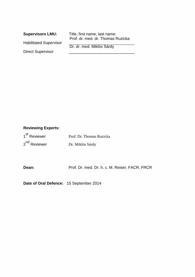
Supervisors LMU: Title, first name, last name:
Habilitated Supervisor Prof. dr. med. dr. Thomas Ruzicka
______________________________
Direct Supervisor
Dr. dr. med. Miklós Sárdy
______________________________
Reviewing Experts:
1st
Reviewer Prof. Dr. Thomas Ruzicka
2nd
Reviewer Dr. Miklós Sárdy
Dean: Prof. Dr. med. Dr. h. c. M. Reiser, FACR, FRCR
Date of Oral Defence: 15 September 2014

Correlation of Malassezia species with clinical characteristics of pityriasis versicolor
Affidavit
Ibekwe, Perpetua Surname, first name
University of Abuja Teaching Hospital Gwagwalada Street
PMB 228, Abuja Zip code, town
Nigeria Country
I hereby declare, that the submitted thesis entitled
Correlation of Malassezia species with clinical characteristics of pityriasis versicolor Thesis Title
Thesis Title (cont.)
Thesis Title (cont.)
is my own work. I have only used the sources indicated and have not made
unauthorised use of services of a third party. Where the work of others has been
quoted or reproduced, the source is always given. The submitted thesis or parts thereof have not been presented as part of an
examination degree to any other university. I further declare that the electronic version of the submitted thesis is congruent with
the printed version both in content and format.
Abuja, 23 April 2014
Place, Date Signature PhD Student

DEDICATION
To God Almighty, with whom all things are possible.

ii
ACKNOWLEDGEMENTS
I wish to express my sincere and heartfelt gratitude to my supervisors and teachers,
Prof Dr. med. Dr. h.c.mult. Thomas Ruzicka, Dr. dr. med. Miklós Sárdy, Assoc. Prof.
Adebola Ogunbiyi for their immense support, encouragement and invaluable advice
towards the completion of this work.
I appreciate the Head of Department of Microbiology, National Hospital Abuja Dr.
K.C Iregbu, for granting permission and also providing working space for me in the
laboratory and the Laboratory Scientists: Mrs. Izama Elizabeth Ushang and Mrs.
Simon Christy Kimwa for their assistance with the preparation of the culture media
used in this study.
Also very well appreciated are Dr R. Besch and Claudia Kammerbauer for allowing
me to use their laboratory at the Dermatology Hospital Ludwig Maximilian University,
Munich and teaching me most of what I know on PCR.
I am not also forgetting Dr. Smith, Mrs Aniekan and the Director Prof. Ujah of
Nigerian Institute for Medical Research (NIMR) Lagos; for I would not have been
able to complete the molecular analysis of my work if not for their help.
My thanks also go to Prof. Andrew Finlay and his team for granting me permission to
use the Children’s Dermatology Life Quality Index questionnaire free of charge and
also to Prof. Mirhendi for his advice on the interpretation of the RFLP-PCR
Special thanks to the members of FCT Secondary School Board, Principals, Head
teachers and school nurse of the various schools visited for the cooperation I was
given and also the students for enthusiastically consenting to be my study
participants.
I am grateful to the Head of the Centre for International Health Prof. Hölscher and his
amiable team Dr. Günter Fröschl, Miss Bettina Prüller and Andrea Kinigadner for
their dedication and constant support. I also wish to acknowledge the following
agencies for funding my PhD programme: the BMZ, DAAD and Exceed. My work
was also supported by the grant obtained from Wissenschaftliches
Herausgeberkollegium der Münchener Medizinischen Wochenschrift.
I sincerely appreciate my husband Dr. T. S. Ibekwe for his invaluable advice, support,
love and understanding and, my adorable children Chinonye, Onyekachi, Ikechukwu
and Ogechukwu for enduring my frequent and long absences during the course of
this programme and still making me a very proud mother. To my wonderful parents,
siblings, and in-laws; thanks for being there.

iii
TABLE OF CONTENTS
PAGE
Dedication i
Acknowledgements ii
Table of contents iii
List of Tables vii
List of Figures viii
List of Appendices x
List of Abbreviations xi
Curriculum vitae 111
List of Publications 112
ABSTRACT 1
CHAPTER ONE: INTRODUCTION 2
1.1 Background 2
1.2 Literature review 3
1.2.1 PV and climate 3
1.2.2 PV and age 3
1.2.3 PV and skin hydration/sebum 4
1.2.4 PV and immune status 5
1.2.5 PV and genetic predisposition 6
1.2.6 PV and other predisposing factors 7
1.2.7 Prevalence of PV in Nigeria 8
1.3 Aetiology of PV 8
1.3.1 History of taxonomy of Malassezia 9
1.3.2 Other diseases associated with Malassezia 11
1.4 Pathogenesis of PV 14
1.4.1 Stratum corneum and PV 16
iv
1.4.2 Mechanism for hypo- and hyperpigmentation of PV 17
1.4.3 Histology of PV lesions 18
1.4.4 Malassezia and healthy skin 19
1.4.5 Malassezia in animals 20
1.4.6 Predominant Malassezia species in PV 20
1.5 Clinical features of PV 21
1.6 Differential diagnosis of PV 21
1.7 Diagnostic investigations in PV management 22
1.7.1 Direct Examination 22
1.7.2 Microscopic Evaluation 22
1.7.3 Culture characteristics 23
1.7.4 Biochemical test 24
1.8 Identification of Malassezia species 24
1.8.1 Morphological and physiological identification methods 24
1.8.2 Molecular identification methods 26
1.8.3 RFLP-PCR method 27
1.9 Treatment of PV 29
1.9.1 Goal of treatment 29
1.9.2 Topical treatment 30
1.9.3 Systemic treatment 32
1.9.4 Herbal treatment 33
1.9.5 Prophylactic treatment 33
CHAPTER TWO: RATIONALE AND OBJECTIVES 34
2.1 Rationale of study 34
2.2 Research questions 35
2.3 Objectives of study 35
CHAPTER THREE: SUBJECTS, MATERIALS AND METHODS 36
v
3.1 Study design 36
3.2 Study location 36
3.3 Study population 38
3.4 Sample size determination 38
3.4.1 Inclusion criteria 39
3.4.2 Exclusion criteria 39
3.5 Selection technique 39
3.6 Data collection instrument 42
3.7 Clinical evaluation 42
3.8 Skin scraping 43
3.9 Isolation and identification of Malassezia 45
3.9.1 DNA preparation: heat - freeze method 45
3.9.2 PCR procedure 45
3.9.3 Enzyme digestion 46
3.10 List of materials 48
3.10.1 List of equipment 48
3.10.2 List of supplies and reagents 49
3.11 Statistical analysis 50
3.12 Ethical certificate 51
CHAPTER FOUR: RESULTS 52
4.1 Clinical characteristics of subjects 52
4.1.1 Age of subjects 53
4.1.2 Demography of students 54
4.1.3 Personal grooming of the students 55
4.1.4 Family relations with PV 56
4.2 Clinical features and characteristics of PV 58
4.2.1 Age of onset of PV 58

vi
4.2.2 Predisposing factors of PV 59
4.2.3 Symptoms and signs of PV 60
4.2.4 Mode of treatment 66
4.3 Health related quality of life 67
4.4 Culture and microscopy results 73
4.5 Malassezia species identified 76
CHAPTER FIVE: DISCUSSION 79
5.1 Clinical characteristics of the subjects 79
5.2 Clinical features of PV 80
5.3 Health related quality of life 84
5.4 Identification of Malassezia species 86
5.5 Conclusion 87
5.6 Study limitations 88
CHAPTER SIX: RECOMMENDATIONS 89
6.0 Recommendations 89
REFERENCES 90
vii
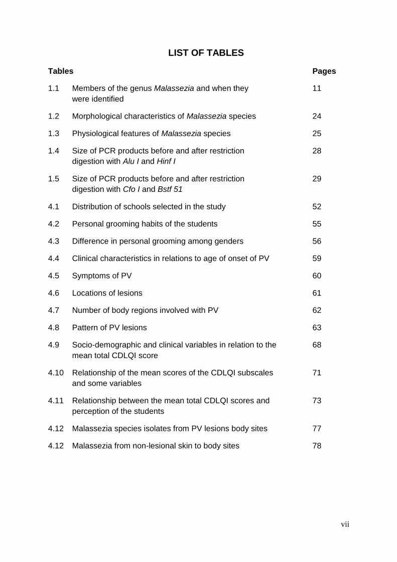
LIST OF TABLES
Tables Pages
1.1 Members of the genus Malassezia and when they 11
were identified
1.2 Morphological characteristics of Malassezia species 24
1.3 Physiological features of Malassezia species 25
1.4 Size of PCR products before and after restriction 28
digestion with Alu I and Hinf I
1.5 Size of PCR products before and after restriction 29
digestion with Cfo I and Bstf 51
4.1 Distribution of schools selected in the study 52
4.2 Personal grooming habits of the students 55
4.3 Difference in personal grooming among genders 56
4.4 Clinical characteristics in relations to age of onset of PV 59
4.5 Symptoms of PV 60
4.6 Locations of lesions 61
4.7 Number of body regions involved with PV 62
4.8 Pattern of PV lesions 63
4.9 Socio-demographic and clinical variables in relation to the 68
mean total CDLQI score
4.10 Relationship of the mean scores of the CDLQI subscales 71
and some variables
4.11 Relationship between the mean total CDLQI scores and 73
perception of the students
4.12 Malassezia species isolates from PV lesions body sites 77
4.12 Malassezia from non-lesional skin to body sites 78

viii
LIST OF FIGURES
Pages
1.1 DNA palindrome EcoR I and Sma I 28
2.1 Map of Nigeria showing latitude and longitude 34
3.1 Map of Nigeria with 36 states and Abuja 36
3.2 Abuja Area Councils 37
3.3 One of the schools used in the study 41
3.4 School clinic where samples were collected 41
3.5 Opsite transparent dressing used for sample collection 43
3.6 “Spaghetti and meatball” microscopy of PV scales 44
3.7 Culture of Malassezia in mDA 44
3.8 In preparation to commence PCR 46
3.9 PCR machine and Gel electrophoresis apparatus 47
3.10 Staining with EB and Gel documentation system 47
4.1 Age distribution of the students 53
4.2 Class distribution of the students 54
4.3 Family members with PV 57
4.4 Recurring and first episode of PV 58
4.5 Symptoms of PV 60
4.6 Exposed and unexposed PV location 61
4.7 Colour of PV 62
4.8 Hyperpigmented PV on mandibular region of the face 63
4.9 Hypopigmented PV on shoulders/upper arm 64
4.10 Hyperpigmented PV located at the back 64
4.11 Extensive PV on face, back and upper arm 65
4.12 Follicular PV at the shoulders 65
4.13 Close-up of follicular PV 66

ix
4.14 Mode of treatment utilized by the students 66
4.15 The mean of the subscales of CDLQI 69
4.16 Culture results of Malassezia species 73
4.17 Unipolar budding microscopy of Malassezia 75
4.18 26S rDNA PCR 77
4.19 Digestion with CfoI showing M. furfur and M. restricta 78
4.20 Digestion with CfoI showing M. furfur and M. sympodialis 78
4.21 Digestion with BstCI showing M. furfur only 78
x
LIST OF APPENDICES
Pages
APPENDIX I Parents/patients information and 101
consent form
APPENDIX II Sample questionnaire 104
APPENDIX III National Hospital Abuja Ethical Clearance 108
APPENDIX IV Ludwig Maximillian University Munich 109
Ethical Clearance
APPENDIX V Permission from FCT Secondary School 110
Education Board

xi
LIST OF ABBREVIATIONS
PV - Pityriasis versicolor
M - Malassezia
CDLQI - Children dermatology life quality index
QoL - Quality of life
RFLP - Restricted fragment length polymorphism
HRQoL - Health related quality of life
TEWL - Trans epidermal water loss
UV - Ultraviolet
AD - Atopic dermatitis
IgE - Immunoglobulin E
SC - Stratum corneum
PCR - Polymerase chain reaction
RNA - Ribonucleic acid
DNA - Deoxyribonucleic acid
RAPD - Random amplification of polymorphic DNA
AFLP - Amplified fragment length polymorphism
tFLP - terminal fragment length polymorphism
SSCP - Single strand conformation polymorphism
DGGE - Denaturing gradient gel electrophoresis
FDA - Federal drug regulation agency
FCT - Federal capital territory
LMU - Ludwig Maximilian University
NIMR - Nigerian Institute for Medical Research
SS - Senior secondary
GSS - Government senior secondary
ANOVA - Analysis of variance
1
ABSTRACT
Background: Pityriasis versicolor (PV) is a superficial mycosis, highly prevalent
among teenagers living in the tropics. Malassezia is the etiological agent of PV.
Fourteen species have been identified worldwide. So far, the debate is on which
species is more prevalent and how the clinical presentations of PV differ
geographically.
Objective: To investigate the most prevalent Malassezia causing PV in Nigerian
students, describe its clinico-epidemiological characteristics and estimate its effect
on health related quality of life (HRQoL).
Methodology: Students were recruited from senior secondary schools within the
period of May 2012 to May 2013. Their clinical characteristics were described and
scales obtained from PV lesions and non-lesions. The Children’s Dermatology Life
Quality Index questionnaire was used to assess HRQoL. Malassezia species were
identified using a molecular method. Results were analysed using SPSS package
version 16.
Results: The students recruited were 304. They had an average age of 16 years.
The age of onset of PV was influenced by a positive family history, socioeconomic
status, the daily use of petrolatum and concomitant presence of dandruff. The
recurrence of PV and the color of the lesions which was majorly hypopigmented,
were not related to differences in personal hygiene or family history of PV. The
location of the lesions was predominately on face while the extent of distribution of
the lesion was significantly linked with pruritus and dysesthesia. A moderate effect
(25% impairment) on the students’ HRQoL was observed. Only three species were
identified. The most prevalent was Malassezia furfur. Finally, no distinct clinical
feature was linked with a specific species.
Conclusion: PV has negative effect on the life of teens. Apart from climate, genetics
and hyperhidrosis, its presentation could also be influenced by dandruff, routine
application of petrolatum and socioeconomic status. Malassezia furfur is the main
causative species of PV in Nigerian students.
Keywords: pityriasis versicolor; Malassezia species; senior secondary school
students; quality of life; tropical environment
Word count: 25,791

2
CHAPTER ONE
INTRODUCTION
1.1 Background
Pityriasis versicolor (PV) is a superficial fungal infection of the skin. It is also known
as tinea versicolor, previously as dermatomycosis furfuracea, tinea flava and
achromia parasitica.(1) In Nigeria, the common names for PV include “eczema” in
English language,(2) “Ifo” in Yoruba language, “Makenkero” in Hausa language and
“Ngwo” in Ibo language; the three major languages spoken by Nigerians.
It is a chronically recurring fungal infection despite the easy availability of antifungal
medications; however, there are no guidelines acceptable for long term treatment. It
is majorly asymptomatic but cosmetically not acceptable. Many affected individuals
usually do not seek medical attention, thus its prevalence might be more than those
inferred in the literature. PV occurrence is worldwide and covers most geographic
locations. It is most prevalent in hot, humid climate(3) where it is seen all year round.
In temperate regions, it is noticed more easily during summer when the skin tends to
tan in light-coloured individuals. The disease affects males and females alike, most
commonly young adults.
It is non-contagious and highly prevalent in individuals with genetic predisposition,
hyperhidrosis, poor general health, and immune-compromised states but not in
pregnancy and diabetes mellitus.(3) The lesions are described characteristically as
multiple hypopigmented and hyperpigmented macules; these may coalesce into
large, irregular patches, and have a fine scaly appearance(4) which can become
obvious by stretching the skin (Zeliri’s sign). Although the lesions may be
asymptomatic, some patients complain of pruritus and tingling sensation. Meanwhile,
in a majority of patients, the lesions are disfiguring, embarrassing and restrict choice
of clothing.
Malassezia species are the causative organisms of PV. They are lipophilic yeasts
which are of the normal skin microflora. They convert from their saprophytic yeast
form to the pathologic mycelial morphology to cause disease.(5) They are richly
located in the sebum-rich body areas such as the face, chest, back and upper arm
due to their requirement of lipid to grow.(6) Diagnosis of PV is essentially clinical.
Examination with Wood’s light may reveal a yellowish or golden fluorescence. Direct
microscopy of scales with potassium hydroxide shows hyphae and spores; the
characteristic “spaghetti and meatballs” appearance. Culture of Malassezia requires
a lipid rich media while molecular analysis as well as physiological characteristics
can be used to identify the various species.
There are various treatment options for PV which could be applied via topical and
oral routes; however, relapse is common due to the importance of both exogenous
and endogenous factors that aid the development of PV.

3
1.2 Literature review
1.2.1 PV and climate
Pityriasis versicolor (PV) occurs worldwide. However, its prevalence is influenced
both by environmental and endogenous factors. An important environmental factor is
the climate. PV affects more people living in the tropics than those in temperate
regions. The tropical environment is rich in heat and moisture which are important
elements that attribute to the development of PV.(7)
In order to assess the effect of climate on the prevalence of PV; a quick literature
search using “prevalence of PV” as the search object with focus on studies from
various regions of the world was carried out. The regions were grouped according to
the updated Köppen-Geiger climate classification by Peel et al.(8) which classified
world climate into 5 types; namely type A – tropical, type B – arid, type C –
temperate, type D – cold and type E – polar climate. This search, however, revealed
only a handful of prevalence studies on PV. Selecting randomly 3 countries each
from the same climate type showed the following: In tropical countries of Brazil,
Central African Republic and Thailand; prevalence of PV were documented as
13.2%,(9) 16.6%(10) and 17%,(11) respectively. In arid countries of Tunisia, Saudi
Arabia and Libya, prevalence of PV were 21.6%,(12) 25.8%(13) and 27.8%,(14)
respectively.
In Malawi, a country in southern Africa with a subtropical climate type A, 8% of the
people examined had extensive PV which the authors described as PV involving 3 or
more regions of the body while 9% was the prevalence for mild disease among the
people who were aged 15 - 24 years.(15) Also in the subtropical climate of Santo
Andre in Brazil where the average all year high temperature is below 300 C, a
prevalence of 3.1%(16) was documented.
Available data from temperate climate are quite limited. In Italy a prevalence of 2.1%
was observed in 1024 young sailors(17) while the famous study from Sweden by
Hellgren et al. showed a prevalence of 0.5% in males and 0.3% in females.(18)
There were no studies of PV documented in type E polar climate regions of the world
such as Norway or Siberia. It is obvious from the above statistics that highest
prevalence of PV is observed in arid countries, areas which have high temperatures
all year round.
1.2.2 PV and Age
The common endogenous factors linked with PV include age, poor immune status
and genetic predisposition. The age factor contributes significantly to the onset and
duration of PV. The age distribution of individuals with PV in most epidemiological
studies falls within the puberty period, 12 to 40years.(10, 11, 15, 19, 20) These are
usually students and young adults. The high prevalence of PV in this age group is
linked with the increased presence of androgen which stimulates sebum production.
Also, the rise in the level of physical activity leading to increased sweating and the
4
probability of harbouring the causative yeast as part of skin flora is higher in this age
group. However, studies assessing factors known to hasten or delay the age of
onset of PV in these predisposed individuals are few. Apart from genetic
predisposition (21), data is lacking on possible associated factors that may affect the
age of onset of PV. Another research question yet to be adequately answered is with
regards to the natural course of the infection. Although death has not been
associated with PV, the degree of morbidity suffered by patients with PV as a result
of chronicity and/or recurrence of the infection is unknown. Also are age and
genetics the only factors that influence the course of PV?
PV has rarely been reported in infants less than 2 years of age and in most of these
cases the infants were premature and placed in intensive care following birth. In one
report of PV in a 2-week-old infant; the authors suggest that the hot, humid
environment of the incubator may have been a contributing factor,(22) although the
sebum level was not assessed. Premature neonates are more often found to have
the causative yeast on their skin, regardless of whether they develop PV lesions.
These yeasts as part of their skin microbiota may have been picked up via
nosocomial route because these infants are handled more frequently by health care
personnel than babies born at term.(23) In an epidemiological study of PV in the
pediatric age group by Jena et al.(24) conducted in Cuttack, a tropical region in India,
PV in children aged up to 14 years accounted for about 31% of the total cases of PV
seen in a 2-year period with about 4.8% cases presenting in infancy. This high
prevalence may suggest that hot humid climate or environment increases the
prevalence of PV in children and even infants.(24)
Furthermore, it is unusual for elderly individuals to suffer from PV. This could be due
to the reduction in sebum production that occurs with increasing age(25) although
studies on PV in this age group is sparse. Similar to the prevalence of PV in the
pediatric age group, environmental factors play a significant role in the prevalence of
PV in the geriatric age group. Thus, the study by Di Silverio et al(26) showed the
presence of Malassezia yeasts and hyphae in about 40.4% of hospitalized elderly
patients aged over 60 years. They had the scaling and hyperpigmented patches of
PV. The possible predisposing factors included heat and sweat, especially as their
clothes were not changed frequently, and the frequency of bath taken may have
been reduced in some cases.(26) There was no relationship observed between PV
and underlying illness of these hospitalized patients, thus the presence of this skin
disorder was not related to the immunocompromised state of the individuals.
1.2.3 PV and skin hydration/sebum
The combination of hydration and sebum contribute to the cause of PV by providing
a conducive growth milieu for the etiological yeasts.(7) Studies measuring the
difference in the stratum corneum humidity status and sebum excretion rate in PV
lesions as compared with non-lesional skin or healthy individuals are quite limited.
Investigation of the skin characteristics of PV patients as compared with healthy
5
controls using the non-invasive MPA-5 by Park et al. showed higher skin humidity,
increased sebum excretion rate and increased trans-epidermal water loss (TEWL)
values in PV patients(7). The authors deduced that higher humid status and sebum
excretion of the skin form part of the factors that encouraged the growth of the
etiological yeast of PV while the increase in TEWL is a consequence from skin
barrier disruption caused by the interaction between the yeasts and skin barrier
materials. They also noted no significant difference in these measured factors
between the hypopigmented and hyperpigmented skin lesions.
This result is in contrast with a study by Lee et al.(27) who observed reduced skin
hydration status in lesional skin compared with adjacent infection-free skin. They
argued that the reduced hydration level of the PV lesions was as a result of the
alteration of the skin biophysical properties by the etiological yeasts on the body. The
study unlike Park et al. did not measure the sebum excretion rate nor did they
compare skin features of the affected individuals with healthy subjects. On the other
hand, Burke’s study done many years(28) measured lipid levels in PV lesional skin
and healthy skin (through a different methodology), observed a higher skin lipid level
in PV skin but this was not a consistent feature on all lesions. Meanwhile, Nazzaro-
Porro et al. observed higher levels of specific lipoperoxide values derived from the
oxidation of different skin lipid classes in PV lesions than in normal controls and
deduced that these lipoperoxides may play an important role in the pathogenesis of
PV(29) particularly in hypopigmented PV lesions; this was confirmed by De Luca et
al.(30)
In neonates, sebum secretion by the skin is elevated due to maternally transferred
androgen; this decreases significantly during childhood but starts to rise again during
puberty and reaches its maximum in young adults.(25) Sebum production decreases
in menopausal females but remains stable with increasing age in males.(25) The role
of skin hydration and sebum production in PV is clear and the above studies have
shown that this role may not be due to a combined effect; however, studies are
needed to determine which of these two factors is more important and also the
minimum level of sebum production and skin hydration at which PV is expected to
develop. This could possibly influence the use or non-use of emollient in predisposed
individuals.
1.2.4 PV and immune status
The relationship between the immune status and PV is not linear. For example, PV is
not particularly more prevalent in patients with human immunodeficiency virus (HIV)
and the clinical presentations also do not differ when compared with non-HIV
patients.(31, 32) An individual with a low immune status does not necessarily
develop the infection. The main factor predisposing an immunocompromised
individual to PV remains unknown; probably genetics and hormone status driving
sebum production play a determining role. Only a few studies of some diseases have
shown a clear relationship between PV and a low immune state. An example is the

6
increased PV prevalence when compared to controls observed in renal transplant
patients(33, 34) and chronic kidney disease patients;(35, 36) while the contrast is
observed in diabetic patients in which case-control studies show a relatively low
prevalence of PV.(37-39) Probably, the development of PV in chronic kidney disease
patients may be related to the accumulation of or failure to excrete an unknown
endogenous factor. It could also be due to a disruption in skin barrier mechanism or
changes in the skin milieu. Since studies on hydration status of the stratum corneum
in patients on dialysis revealed a low hydration state and increased TEWL which in
one study correlated with the complaints of pruritus in these patients.(40, 41) Sebum
excretion rate in these patients are yet to be documented in the literature.
According to the minireview by Tragiannidis et al.(42) Malassezia yeasts, that is
etiological agents of PV, found in immunocompromised patients such as diabetics,
patients with haematological malignancies, bone marrow transplantation, and solid
organ transplantation, Crohn’s disease and AIDS are known more often to cause
severe skin conditions and systemic diseases. These severe skin conditions
included Malassezia folliculitis, seborrheic dermatitis, catheter-related fungaemia and
sepsis and a variety of deeply invasive infections.(42) PV was clearly not mentioned.
This suggests the ability of these yeasts to invade the body to a deeper level when
the individual is immunocompromised as compared to its very superficial state in PV.
Meanwhile, PV infection is not dependent on the cellular or humoral immune status
of the affected individual. Data from Saadatzadeh et al.(43) which assessed the cell-
mediated immune response to the active (mycelial) phase of Malassezia in PV
patients as compared with age- and sex-matched controls revealed a similar immune
response between the two study groups. There was no deficiency in cell-mediated
immune response observed in the diseased patients. On the humoral immune
response of PV patients compared with age- and sex-matched controls study
conducted by Ashbee et al.(44) there was no statistically significant difference in
measured total immunoglobulins produced in response to Malassezia furfur by both
groups. The result of both studies supports the hypothesis that PV infection is not
directly linked to the patient’s immune status.(43, 44)
1.2.5. PV and genetic predisposition
PV is not a contagious disease and the lack of report of conjugal cases(23) among
discordant couples who have been living together for a prolonged length of time
combined with the tendency of PV to occur in at least one other member of the
family unit lay credence to genetic predisposition being one of the important
endogenous factors that contribute to the development of PV. Genetic models
described by Hafez and later confirmed by He et al. revealed that PV patients fulfil
the criteria for multifactorial (genetic-environmental) mode of inheritance.(21, 45)
Meanwhile, inconsistencies with single-gene recessive or dominant mode of
inheritance were shown as well as rejection of the environmental and non-
transmitted genetic models of inheritance.(21)

7
Available literature from various studies have revealed very strong positive family
history of PV among patients, ranging from 21.1% to 38.3%(21, 45-47) families. He
et al calculated the heritability of PV in first-degree relatives to be 48.13%, second-
degree to be 40.11% and third-degree to be 27.2%.(21) This shows a risk reduction
with distance in family ties, thus justifying the multifactorial nature of the disease.
Additionally, the role of environmental factor cannot be over-emphasized since the
heritability of PV is by and large lower than 70%.(45) In general, these studies also
agreed with the finding that patients with a positive family history of PV had an earlier
onset and a longer duration of the disease. A chance of recurrence was also higher,
compared with individuals without a family history.
1.2.6 PV and other predisposing factors
Other factors that may predispose individuals to PV include hyperhidrosis, use of
oral contraceptives, malnutrition, pregnancy, skin occlusion and prolonged use of
systemic corticosteroid. Evidence supporting the relationship between oral
contraceptives use, malnutrition and PV is quite weak and not properly documented.
Although hyperhidrosis is generally accepted as one of the predisposing factors to
PV(23), studies have produced conflicting results. While profuse sweating was
considered on the one hand, in the studies by Tarazooie and He et al. as one of the
strong endogenous factors mediating the development of PV;(21, 48) on the other
hand, this was not supported in the studies by Burke and Ingordo et al.(17, 49) were
no significant association between PV and hyperhidrosis was observed.
Even though the skin colonization by Malassezia yeasts is increased in the third
trimester and postpartum period, the frequency of PV in pregnancy has been shown
not to differ significantly from that of the general population.(50)
PV studies in patients on prolonged steroid use are sparse. The incidence of PV in
corticosteroid-treated patients as observed a long time ago by Boardman et al. was
16%.(51) Similarly a significant relationship was observed by Burke(49) between
increased systemic cortisol levels that are found in Cushing’s syndrome patients as
well as those on systemic glucocorticoid therapy and PV development. The possible
explanation for this interaction is not yet proven by research especially as this
relationship with PV was not observed among patients applying topical
glucocorticoids. An experimental work on steroids and Malassezia yeasts did not
show any cause and effect relationship. Steroids did not encourage or increase the
growth of the yeasts in culture. Moreover, topical glucocorticoids can induce a
negative nitrogen balance and reduction in pH when applied on the skin.(49) This
could in turn impede the growth of the fungi.
Some studies could not relate personal hygiene with the development of pityriasis
versicolor(46, 47) although this is in contrast to a general perception by most people
that PV results from an inadequate personal grooming of the patients.(52) This
perception could increase stress and feeling of being stigmatized in these patients. A
8
recent work on skin infections and infestations among Nigerian prison inmates
showed that PV formed 27% of all 178 infections, second only to infections by
dermatophytes.(53) Skin infection was significantly associated with the frequency of
bath, frequency of soap usage and frequency at which their clothing was changed.
These confirm a possible relationship between personal hygiene and skin infections
and infestations of which PV was included. They also observed a significant
influence caused by accommodation arrangements namely overcrowding and poor
ventilation on skin infections. It would have been interesting to know if the prison
environment (hot and humid) was considered also as a possible predisposing factor.
Meanwhile, this study differs with a similar study of PV among Indian prisoners in
which no significant difference in PV prevalence and clinical features was observed
when compared with the general population.(54)
1.2.7 Prevalence of pityriasis versicolor in Nigeria.
Studies on PV in Nigeria are quite limited but documented prevalence of PV range
from 6.7% in Northwestern region,(55) a region closest to the Sahara desert and
higher temperatures all year round to 3.7%(56) in Southern region of the country
which has a cooler climate and closest to the ocean. However, both studies had
limitations. The first study was hospital-based which does not usually give a true
prevalence of skin diseases in the community and the second was conducted in a
primary school where majority of the pupils were below the adolescent age, the peak
age of PV development.
A prevalence of 4.6% was observed by Uneke et al. in an epidemiological survey of
tinea capitis and PV infections among school children in South-east Nigeria.(57)
While in the Southwestern region, a prevalence of 4.7% was observed, also among
school children.(58) In both studies, an epidemiological comparison of PV was made
with dermatophyte infections, in which the prevalence of dermatophytosis was
significantly higher than PV (51.8% vs 4.6% and 15.2% vs 4.7%) while the children
with PV were significantly older than those with tinea capitis. The peak age group for
tinea was 3 - 7 years while for PV was 14 - 17 years.(57, 59)
In neighbouring countries of Nigeria, few prevalent studies of PV have been
conducted. Examples include Mali which is located to the north of the country. There
Faye et al.(60) observed a group of children aged 15 years and below in rural
communities of Mali for non-leprous hypochromic patches. A prevalence of 4.1%
was documented, of which PV had the highest frequency of 39.4%. Studies from
other West African countries like Ghana, Togo, Benin and Liberia could not be found
in the literature.
1.3 Aetiology of pityriasis versicolor
PV is a superficial cutaneous infection caused by the fungus of the genus
Malassezia, one of the numerous dimorphic fungi that infect humans and animals.
This microorganism form part of the normal skin flora when it exists in the
9
saprophytic or yeast state. It causes PV most times when it changes to the hyphal or
mycelial state; the mechanism by which this change of state is conducted has
remained unclear.
Meanwhile, epidemiological study of Malassezia yeast found on normal skin of
individuals from tropical environment has shown increased chance of hyphal
formation. This leads to the conclusion that the mycelial state of Malassezia per se
may not always indicate the presence of a disease.(3) Another group argues the
possibility of “overt” infection since these hyphae were isolated from non-lesional
skin scales of same patients who had PV infection. Additionally, the percentage of
hyphae isolated from skin of healthy individuals is negligible in comparison to
samples obtained from PV lesions.(61) The reason why Malassezia yeasts have
been implicated as aetiological agent of PV is the increased likelihood of obtaining a
positive fungal culture from skin specimens taken from PV lesions than from clinically
unaffected skin areas of either the same individual or matched healthy controls.(48,
61, 62)
1.3.1 History and taxonomy of Malassezia
After several decades of continuous reclassification, Malassezia yeasts are presently
placed in the Phylum Basidiomycota, subphylum Ustilaginomycotina, class
Exobasidiomycetes, order Malasseziales and family Malasseziaceae.(61) There
were disagreements as to when the organism was first grown, the optimal culture
medium, the relationship between the different morphological and colonial variants of
the organism, the genus to which it should be assigned and with what name, and the
role it plays in a variety of cutaneous diseases.
The pathological agent of PV was first described in 1846 by Eichstedt.(63) This
fungus was later named Microsporum furfur by Robin Baillon in 1853, as he thought
it was identical to the dermatophyte Microsporum auduoinii. In 1874, Malassez
further described the shape (round, oval) and budding nature of this organism and
differentiated it from dermatophytes. Robin Baillon in recognition of this work and the
furfuraceous (consisting of or covered with flaky particles) nature of the PV lesions
proposed the first nomenclature Malassezia furfur in 1889.(64)
However, 15 years later Sabouraud described a group of budding yeast cells without
hyphal elements observed in and isolated from normal skin and scalp. He proposed
the genus Pityrosporum.(65) Thus two genera were established: Pityrosporum, as
the yeast form found on normal skin flora and Malassezia as the mycelia form found
in disease state.
Panja in 1927 was able to successfully isolate the organism from PV lesions and he
also suggested that both yeast and mycelia forms may be the different expression of
one organism by proposing the merging of both genera into one.(64) This suggestion
was not generally accepted as there were no scientific ways of proving that this
relationship exists. Most important was the difficulty in culturing the organism in vitro.

10
The reason for the difficulty was later explained by Benham in 1939. She observed
that the organism required a “fatty substance” to grow.(64) Once this lipid
requirement was established it paved way for the formulation of various culture
media that reliably recovered and maintained the organism thus enabling much work
to be done on the taxonomy, physiology and biochemistry of these yeasts especially
in the discovery of new species for each genus.
By 1984 mycologists restricted the genus to just 2 valid species: a human one, M.
furfur, and an animal one, M. pachydermatis. The old name Pityrosporum was
rejected (nomen rejiciendum) because it was considered a synonym of Malassezia,
a term already in use and so given priority according to the rules of taxonomic
nomenclature.(66)
Despite all these studies and more, the taxonomy and nomenclature of Malassezia
species was marred with confusion and chaos until 1995 when the advancement in
molecular techniques allowed physiological and ultrastructural studies describing the
characteristics of each species. Using these techniques Guillot and Gueho analyzed
104 different isolates of Malassezia, and subsequently defined seven species of
Malassezia: M. furfur, M. sympodialis, M. obtusa, M. globosa, M. restricta, M.
slooffiae, and M. pachydermatis.(67) In recent years, using the combination of
biochemical, physiological, morphological and molecular techniques 7 additional
species have been identified: M. dermatis, M. japonica (isolated from humans with
atopic dermatitis), M. yamatoensis (in seborrheic dermatitis), M. equina, M. caprae,
M. nana and finally M. cuniculi (last 4 isolated from various animals).(68)
The current members of the genus Malassezia with their dates of identification as
adapted from the review by Crespo-Erchiga et al.(66) are highlighted in Table 1.1
below.

11
Table 1.1 Members of the Genus Malassezia and dates of identification(66)
First Studies (3spp) 1996 Review (7spp) The Genus in 2014 (14spp)
M. furfur 1889
P. ovale 1913
P. orbiculare 1951
M. pachydermatis1935
P. pachydermatis1925
P. canis 1955
M. sympodialis 1990
M. furfur 1889
M. pachydermatis 1935
M. sympodialis 1990
M. globosa 1996
M. obtusa 1996
M. restricta 1996
M. slooffiae 1996
M. furfur 1889
M. pachydermatis 1935
M. sympodialis 1990
M. globosa 1996
M. obtusa 1996
M. restricta 1996
M. slooffiae 1996
M. dermatis 2002
M. japonica 2003
M. nana 2004
M. yamatoensis 2004
M. caprae 2007
M. equine 2007
M. cuniculi 2010
1.3.2 Other diseases associated with Malassezia
PV is not the only mycosis caused by Malassezia yeasts. There are other diseases,
both cutaneous and systemic, in which they have been implicated and a discussion
on Malassezia and PV will not be complete without a brief mention of the role played
by these yeasts in these diseases. In some, the association remains controversial
and the pathogenesis unclear.
The systemic infections caused by Malassezia include intravenous catheter induced
fungemia, endocarditis, interstitial pneumonia and peritonitis in patients undergoing
continuous ambulatory peritoneal dialysis.(61) Most of these systemic infections
were said to be as a result of the fungus being introduced into the blood stream or

12
the peritoneum via the skin during these procedures. The rate of infection correlates
with the length of time the catheters were kept in place. Malassezia can cause
systemic infection in people of all ages particularly those with immunosuppression
and venous catheter either central or peritoneal.(42) Affected individuals present with
persistent fever, no response to antibiotics and a high degree of clinical suspicion is
required for timely management.(61)
For cutaneous diseases such as seborrheic dermatitis, pityriasis capitis and
psoriasis, the role played by Malassezia is not completely clear. Controversial roles
of Malassezia has been associated with also the following: neonatal cephalic
pustulosis, atopic dermatitis, onychomycosis, sinusitis, confluent and reticulated
papillomatosis of Gougerot-Carteaud and otitis externa.(61)
Seborrheic dermatitis is a recurring disease commonly seen in sebum-rich areas of
the skin, such as scalp, eyebrows, paranasal folds, upper chest and back. It is
characterised by erythema and scaling.(61) It commonly affects neonates and
infants, Parkinson’s disease patients and also immunocompromised individuals.(42)
Current data available do not sufficiently define the pathological features of
Malassezia that leads to the development and exacerbation of seborrheic dermatitis,
but it has been considered as a deeper level of infection in comparison with PV(61).
The yeasts were isolated from the dermatitis lesions by various studies (69, 70) and
anti-fungal medications form part of the treatment plan for seborrheic dermatitis.
Malassezia (Pityrosporum) folliculitis is a benign cutaneous disorder, characterised
by follicular papules and subcorneal pustules most commonly located on the face,
chest, back and upper arms. It is often pruritic and could be misdiagnosed as
acne.(3) Direct microscopy of pustules usually reveals an abundance of Malassezia
yeasts and the absence of other microorganisms. Histology is also helpful in
diagnosis where a periodic acid-schiff (PAS) or methenamine silver staining
technique will show the budding yeasts within the keratinous material of dilated hair
follicles. A study in Korea showed a predominance of M. restricta in patients with
Malassezia folliculitis.(71)
Confluent and reticulate papillomatosis of Gougerot-Carteaud is a rare cutaneous
disorder characterised by confluent, grayish-brown, hyperkeratotic papules often
symmetrically located on the trunk.(3) It responds to topical and systemic anti-
fungals, although in combination with other modes of treatment. Studies on this skin
disorder are few. However, M. furfur and M. sympodialis have been isolated from the
lesions.(3)
Neonatal cephalic pustulosis is a benign disorder commonly seen in neonates. It is
characterised by non-follicular papulopustules found on the face and neck of
newborns. Direct microscopy of the pustules have shown Malassezia yeasts,
although the exact pathogenesis is not well known, this disease responds well to
topical ketoconazole therapy.(61)

13
Pityriasis capitis (dandruff) also known as mild seborrheic dermatitis is a subclinical
inflammatory condition of the scalp associated with episodic, recurrent or constant
scaling of the scalp. It is more prevalent in young adults and may be aggravated by
environmental factors such as dust, hair cosmetics, UV irradiation, and airborne
irritants. Malassezia yeasts have been isolated from dandruff scalp(69) and the
condition is known to improve on reducing the population of Malassezia on the scalp
with proper antifungal treatment(72). Meanwhile, a study has demonstrated the
contribution of other fungi apart from Malassezia such as Filobasidium and
Acremonium species in the aetiopathogenesis of dandruff.(73)
Atopic dermatitis (AD) is a chronic relapsing inflammatory skin disease characterised
by severely itchy, red, dry, and crusted skin affecting all age groups but most
commonly the paediatric age group. Pathogenetically, it results from a combination
of defective skin barrier and inappropriate immune responses against both genetic
and environmental factors. Malassezia yeasts exacerbate AD especially in those
with dermatitis lesions involving mainly the head and neck regions.(61, 64, 74)
These patients have been shown to have a higher IgE titre to Malassezia and also a
more positive skin prick test to Malassezia allergen than healthy subjects.(64, 74, 75)
M. furfur, M. globosa, M. restricta and M. sympodialis are the most common species
isolated from atopic dermatitis skin lesions.(75, 76) Likewise, the addition of
antifungals to patients’ management has resulted in rapid treatment response. In the
interim, exact roles played by the fungi in atopic dermatitis remains under intense
investigation.
Psoriasis is a chronic inflammatory immune-mediated disorder that affects the skin
and joints. The skin lesions are characterised by erythematous scaly papules and
plaques. Although the aetiology of psoriasis remains elusive, Malassezia yeasts
have been reported as one of the microorganisms that can exacerbate this disease
condition. Some studies have isolated M. furfur, M. globosa, M. restricta and M.
japonica from surfaces of psoriatic lesions.(77, 78) One of these studies was able to
demonstrate pseudohyphae in active psoriatic lesions and none was observed in
stable lesions.(77) The mechanism is still not clear since it is difficult to understand if
the pseudohyphae developed as a result of the reduced barrier function and low
immune defence observed in exacerbated lesions or if the presence of the yeasts
contributed directly to the exacerbation. Nevertheless, these patients are known to
respond more quickly when topical antifungals are included in their management.(78)
Malassezia yeasts have been demonstrated to induce onychomycosis in normal and
immunocompromised patients.(79, 80) This association remains contentious
especially as these lipophilic yeasts are known not to normally colonize nails and
besides nails are poor sources of lipids. Also these yeasts do not have keratolytic
properties as dermatophytes. Zhao et al. attributed the process whereby Malassezia
can cause onychomycosis to a change in the microenvironment induced by the local
application of steroids.(79)

14
1.4 Pathogenesis of pityriasis versicolor
The pathogenesis of PV is yet to be completely explained. However, various studies
on PV development concur on the interdependence of the following factors:
Malassezia skin colonization, sebum production and individual predisposition. PV
infection is caused by the lipophilic dimorphic yeasts named Malassezia. These
yeasts colonize the stratum corneum (SC) and are among the organisms that make
up the skin microbiota.(1, 72) The microorganisms of the skin flora are quite diverse
and are made up of bacteria, viruses, protozoa and fungi. Some are transient that is
they are detected as a result of transient hand/body carriage from contamination or
transmission events (examples include adenovirus and rotavirus) while others are
resident. The resident flora are persistent and cannot be completely washed away;
examples of these group of organisms include Malassezia, Staphylococcus
epidermidis, Corynebacterium, Demodex folliculorum, just to mention a few.(81)
The skin becomes colonized by microbes soon after birth. These microbes are
usually from the environment and they subsequently interact with the epithelial cells
of the newborn leading to microbial colonization and co-existence.(82) Most of these
microbes are beneficial, commensals or neutral, however, a few do become
pathogenic and harmful to the skin or body in general.(82) The commensal or
pathogenic nature of an organism is argued to depend on the immune system of the
individual rather than the inherent properties of the microbe.(82) Of equal importance
is the interaction among species. In order to survive, a microbe has to compete with
other microbes of the normal flora for available nutritional elements and space. By so
doing, they try to keep the skin flora in a stable state and resist abrupt changes in the
community structures. This process in the long run also protect against the growth of
pathogenic microbes that may cause disease.(81, 82)
Various skin structures may harbor their own type of microbes. Such that the stratum
corneum, keratinocytes, hair shaft and follicle, sweat glands, apocrine glands and
sebaceous glands may each have their own type of unique microorganisms which
colonize there. Malassezia yeasts reside mainly in the stratum corneum, though
some authors suspect their presence in the hair follicles which could act as a
reservoir and thus accounts for the recurrence of PV after treatment.(47, 73)
Meanwhile, the stratum corneum provides nutrients in form of lipids secreted by the
sebaceous glands. Sebum production is increased in humans at puberty and so is
PV. This also explains the reason for the predilection of the yeasts for sebum-rich
regions of the body. Sebum lipids are postulated as essential ingredients that
support the growth of these yeasts while the sebum triglycerides are degraded by
Malassezia-derived lipases to produce proinflammatory unsaturated fatty acids, an
antimicrobial property that limits the types of microorganisms that can co-exist on the
skin.(83)
Since these Malassezia yeasts exist as skin flora on healthy skin, the development
of PV in certain individuals indicates an interactive role played by other factors which

15
could have altered the ecosystem of the skin, and thus a resultant change in the
yeasts’ microbial state. This interactive process was well described in the conceptual
framework developed by Rosenthal et al.(82) Most important of these factors that
could predispose individuals to develop PV is the presence of high temperature and
high relative humidity. There are also behavioral characteristics of the individual such
as cosmetic use and host demographic characteristics such as seborrhea and
hyperhidrosis. Of equal importance is the genetic susceptibility of the individual.(21)
All of these driving factors interact to some degree to influence the pathogenesis of
PV.
Although PV is a cutaneous infection, it is associated with little or no inflammation
unlike other Malassezia-associated diseases such as Malassezia folliculitis and
seborrheic dermatitis. Malassezia are dimorphic yeasts, that is, they are able to exist
in both yeast and mycelial (hyphal) forms. The mechanism by which individual’s
endogenous as well as environmental factors influence the yeasts to convert to the
hyphal state, thus causing PV is also not well established. Moreover, studies
investigating the absence of an active immune response by the host to the presence
of Malassezia and possible factors that mediate the immune balance in the host
between the commensal and pathogenic state of the yeasts are lacking in the
literature. A more likely hypothesis has been linked to the lipophilic nature of the
yeasts cell wall. Lipids reduce phagocytosis of Malassezia by neutrophils.
Phagocytosis is an important nonspecific immune mechanism for removal of
microorganisms from the body. The uptake of the yeasts by these inflammatory cells
is efficiently higher when they are killed than when they are alive.(64)
Presently there is little information on the relative pathogenicity or virulence factors of
Malassezia species. The limitation by the body’s neutrophils in killing the organism
may account for its persistence in predisposed individuals. A study described by
Ashbee and Evans(64) specified that only 5% of the Malassezia cells taken up by
neutrophils were actually killed in an in vitro complement-dependent process, and
this killing ability by the same neutrophils was increased to 23% when the yeasts
were pretreated with ketoconazole. This was in contrast with the killing of 30% - 50%
Candida albicans yeast cells and 80% of the cells of other fungal genera. Another
hypothesis proposed for this limitation was attributed to the production of azelaic acid
by Malassezia when it is grown in the presence of oleic acid or olive oil. Azelaic acid
scavenges oxygen radicals, thus preventing oxidative killing of the organism by the
phagocytes. Likewise is the protective function of lipids richly found within the cell
wall of the organism; this reduces their uptake by and subsequent activation of
neutrophils and so protecting them from being phagocytosed.(64) M. furfur produces
pityriarubins which is protective against neutrophilic activity. The lack of inflammation
seen in PV has been attributed to pityriarubin through its role in down regulation of
the immune response.(84)
Immunomodulatory properties of Malassezia have been demonstrated in mice and in
vitro. In mice, Malassezia through the up-regulation of macrophages protect against
16
infection and malignancy(85) while in vitro it reduces the production of cytokines
such as interleukin 1, interleukin 6 and tumour necrosis factor-alpha.(86) Cellular
and humoral immune reactions to Malassezia have been measured in healthy
individuals despite their having no evidence of Malassezia-associated diseases;
these activities were age-related with higher levels observed in young adults and
lower levels in children and the elderly.(43, 44, 87) This poses some difficulties in
studies on the immune pathogenesis of Malassezia-associated disorders and it will
be necessary to conduct these studies as age- and sex-matched case-control
studies in order to appropriately interpret the results. Meanwhile studies have not
shown any deficiency in cell-mediated immunity to Malassezia in patients with
PV.(43, 64)
1.4.1 Stratum corneum (SC) and PV
The human skin is made of the epidermal, dermal and subcutaneous layers. The
stratum corneum (SC) is the final differentiated product of the epidermis.(88) In its
healthy state, the SC forms a protective barrier against various environmental insults
including pathogenic microbes, damaging irradiation, and potentially toxic
xenobiotics and it also maintains skin/body hydration by preventing water loss.(83,
88) A disruption of the skin barrier structure and function has been observed in
PV.(27) These disruptions include an increase in transepidermal water loss (TEWL)
and a significant reduction in skin hydration. The resultant effect of these disruption
processes leads probably to the pathologically increased fragility of PV lesional skin
surface.(61)This can be demonstrated in the “evoked-scale” sign(61) also called
“Zeliri’s sign”(89). This sign is elicited when on stretching or scraping the surface of a
PV lesion, scales become more visible. The precise pathogenetic mechanism by
which Malassezia contributes to this characteristic feature of PV has several
explanations. Some hypotheses are discussed below.
The SC is the most superficial layer of the skin that is solely responsible for the
characteristic features of PV which is scaling and dyspigmentation. To fully
understand the pathogenesis of PV, it is important to understand the structure and
organization of the SC and how these properties are altered during PV. The SC is a
multilayered tissue composed of anucleated, flattened cells which are surrounded by
multiple lamellar sheets of lipids.(83) It has also been described as a “unique
sophisticated biosensor” that signals the underlying epidermis to respond to external
stresses.(90) SC has been likened, in the simplest terms, to a brick wall (these are
the terminally differentiated keratinocytes also called corneocytes) linked by
intercellular cement (this is composed of a continuous matrix of specialized
lipids).(90) The corneocytes provide protection against injuries while the
intercorneocyte lipids which are mainly broad sheets of ceramides produced from
lamellar bodies in the granular layer, serve as water barrier.(88) SC is formed during
the process of keratinization of which the final act is desquamation. This is the
orderly release of single corneocytes at the skin surface. Desquamation is facilitated
by several proteolytic enzymes that degrade corneodesmosomes which are the
17
protein structures that fasten neighboring corneocytes together both in the plane of
the SC layer and adjacent layers.(90) These proteolytic enzymes are controlled in
the end by water activity and the pH of the SC.(83) Another possible facilitator of the
desquamation process is sebum. The presence of triglycerides and short-chain fatty
acids, as structural barrier lipids of the SC may disrupt the organization in the
superficial layer of the intercellular lipid (intercellular cement).(83) Triglycerides and
short-chain fatty acids are produced and released primarily by the sebaceous gland.
Desquamation process can be facilitated by the presence of yeasts in the SC. Three
distinct forms of Malassezia yeasts were recognized in the SC by Piérard et al.(91)
These include round spores or conidia, budding spores and mycelium. Meanwhile,
the mycelial elements are regularly observed to be located within the corneocytes.
They are usually the invasive form of most yeast. Additionally, the keratinous content
(mainly the tonofilaments) of these yeasts invaded cells are replaced by “amorphous,
moderately electron-dense material” which were observed by Borgers et al.(92) as
lipid in nature. Similar amorphous material was observed in between keratinocytes
(extracellular compartment); most often in form of several sized spheres. Its
keratinocyte origin is supported by the certainty of frequent membrane ruptures of
swollen invaded keratinocytes. The degradation of the tonofilaments in the
keratinocytes is speculated to be via the activity of Malassezia yeast enzymes. The
replacement by lipids serve as nutritive material, which were demonstrated to be
essential for growth initiation and maintenance of this lipophilic yeast in the SC.(92)
Meanwhile this work done by Borgers et al. is yet to be confirmed as a similar study
by del Palacio-Hernanz et al.(93) who studied the ultrastructure of PV lesions before
and after treatment with cyclopiroxolamine could not observe these intracellular
amorphous materials but rather severe necrosis of the cytoplasm. The reason was
attributed possibly to the short fixation time by the later authors (2 weeks vs 2-3
days).
In conclusion, although the exact structural alterations of the SC are still unknown,
the possible combination of frequent membrane ruptures of invaded keratinocytes
and the activities of the Malassezia yeasts enzymes may contribute to the increased
fragility of the lesional SC which is evident in the “Zeliri’s sign” or “evoked-scale” sign
of PV.
1.4.2 Mechanism for hypo- and hyperpigmentation of PV lesion
Some studies have been done to explain the reason why PV lesions are either
hypopigmented or hyperpigmented. Some have tried to answer this question by
observing the relationship between Malassezia and melanocytes. Study by Karaoui
et al.(94) confirmed earlier research in which no difference in number of melanocytes
between PV skin whether hypopigmented or hyperpigmented and normal skin was
found; but rather observed a difference in the dispersion and arrangement of
melanosomes by the melanocytes.

18
In hypopigmented skin, the melanosomes were individually dispersed and fewer in
number while in hyperpigmented skin, they were sequestered in most cells.(94) This
was later collaborated by Galadari et al.(95) but refuted by Dotz et al.(96) who
examined biopsy specimens from lesions of hyperpigmented PV involving a
vitiliginous skin and found absence of both melanosomes and melanocytes. This
study increased the doubt of a possible role play by melanocytes in the
dyspigmentation of PV lesions.
The explanation behind the interaction between Malassezia causing PV and
melanocytes was further attempted by Krämer et al.(97) who suggests an indole
alkaloid produced by M. furfur from tryptophan called melassezin as the likely cause
of the hypopigmented lesions. Melassezin induces melanocytes to undergo
apoptosis and thus reduces melanin production but this may not explain the
hyperpigmentation of PV lesions. Other reasons for the hypopigmentation of some
PV lesions have been connected to the production of lipoperoxides from oxidation of
skin lipid by the yeast. Since a higher lipoxygenase and lipoperoxidase activity was
measured in hypopigmented lesions positive for fungal hyphae and spores than
controls.(29, 30)
Another speculation in view of the hypopigmentation of PV lesion worthy of note to
mention is the role of the lipid-like material produced by the Malassezia yeast in the
stratum corneum. The hypochromia is said to be as a result of filtering of the
ultraviolent light provided by the “lipid” screen.(92) This “lipidification” of the SC is
clearly documented only with Malassezia yeast infection and thus differentiates it
from other fungal infections. Also this lipid process is not influenced by antifungal
medications such as itraconazole. For this reason the hypopigmentation of PV
lesions persists for 3 – 4 weeks after disappearance of viable fungi from the skin
since normal epidermal regeneration takes about 4 weeks.(92)
Meanwhile, another school of thought has attributed the color change of PV to the
thickness of the stratum corneum. In both pigmentary states, the thickness of the
stratum corneum differs with hyperpigmented skin being thicker than hypopigmented
skin and both being thicker than that of uninvolved skin.(94)
1.4.3 Histology of PV lesions
Electron microscopy of skin scales from PV lesions has revealed the clustering of
yeast cells and short hyphae in nest-like cavities.(98) The hyphae may be seen
perforating the outer layer of the stratum corneum perpendicularly.(93) Malassezia
species reproduce asexually by monopolar, enteroblastic budding from a
characteristic broad or narrow base depending on the species. Separation leaves a
prominent bud scar through which successive daughter cells emerge.(64) In lesional
skin, particularly around hair follicles and acrosyringium, the clustering of yeast cells
and mycelial hyphae is larger in the superficial part of the stratum corneum than

19
deeper layers. Furthermore in healthy skin, the clusters are mainly made up of yeast
cells and are fewer in number (98, 99).
Histology of skin biopsy from PV patients can be accessed via the Hematoxylin-
eosin stain or the PAS stain. Basically, the histology has shown a slight to moderate
hyperkeratosis with “basket weave” stratum corneum, acanthosis, and vacuolization
of epidermal cells.(99) There is also increased or normal basal layer pigmentation.
Occasionally, melanin incontinence may be seen.(61) Gaitanis et al.(61) describe
two types of PV that may be observed histopathologically from lesional skin biopsy;
these are the inflammatory and non-inflammatory PV. The non-inflammatory PV is
characterized by mild or almost absent infiltration of the dermis by superficial
perivascular inflammatory cells while in the inflammatory PV, there is a moderately
dense infiltration in the upper dermis of perivascular inflammatory cells. These
mononuclear infiltrates are characteristically Langerhans cells, T lymphocytes and
occasionally plasma cells.(95) Meanwhile, numerous budding yeast cells and short
hyphae can be viewed in the stratum corneum and only the hyphal and not the spore
form of the yeast are observed within the cells.(1) Unusual histological features
include the presence of dilated blood vessels in relation to the extremely rare
erythematous PV and papillomatosis like acanthosis nigricans.(99)
1.4.4 Malassezia and healthy skin
The presence of Malassezia yeasts on healthy skin of humans was described since
the second half of the 19th century, and it soon became evident that colonization
density was associated with age and differences in the activity of sebaceous glands
in different areas of the body. However, there are no differences observed between
the Malassezia species identified from healthy and diseased skin.(74)
Malassezia, as part of the body flora can be isolated from the sebum-rich areas of
the skin, particularly the chest, back, and head regions. A study done by Leeming(6)
examined clinically normal skin at 20 different sites over the entire body surface.
Malassezia species were recovered from every subject from the chest, midline back,
scalp, ear, and upper inner thigh. The highest mean population densities occurred on
the chest, ear, upper back, forehead, and cheeks. Some differences in carriage rates
were noted between females and males, with higher population densities from the
lower trunk and upper thigh of males.
In a study by Lim et al. on the distribution of the different Malassezia species on
normal human skin,(100) M. restricta was seen to predominate on the scalp, M.
sympodialis on the trunk and M. globosa was widely and equally distributed in all
seborrheic areas. When the number of colonies isolated from healthy individuals was
compared with those from PV, seborrheic dermatitis, AD and psoriasis patients; the
number was comparable with PV patients but significantly smaller in seborrheic
dermatitis, AD and psoriasis.(72, 74)

20
1.4.5 Malassezia in animals
Malassezia is rarely isolated from the environment because it is mostly confined to
the skin of warm blooded animals. This is probably because its distribution in nature
has not been well researched.
Veterinarians’ investigation of these yeasts on the skin of several animals
demonstrates the role of M. pachydermatis (and to a lesser extent, M. furfur, M.
obtusa, and M. sympodialis) as the causative agents of otitis externa in cats and
dogs. M. pachydermatis is also known to cause Malassezia dermatitis in dogs. This
chronic skin disorder with similar clinical features to seborrheic dermatitis in humans
is characterized by relatively demarcated scaly erythematous patches located at the
sebum-rich regions of the animal.(101) Other clinical features that could be
associated with Malassezia dermatitis in animals include pruritus, lichenification,
excoriations and alopecia.
Meanwhile, studies in many other animals have shown that specific species of
Malassezia, just as in humans, also form parts of their skin microbiota. Such that M.
equine is commonly isolated from the skin of monkeys, pigs, bears, birds, and
horses. M. caprae specifically colonises goats while M. nana colonises cattle and
cats.(66) The most recent addition to Malassezia species, M. cuniculi was isolated
from rabbits.(68)
1.4.6 Predominant Malassezia species in PV
The predominant causative Malassezia species of PV have been demonstrated from
different countries. Studies in Spain by Crespo Erchiga et al.(102) were the first to
show M. globosa as the most prevalent species in PV lesions. M. globosa was also
the most prevalent species observed in Japan by Nakabayashi et al.(70) in Spain by
Aspiroz et al.(103) and in India by Dutta et al.(104) Also in Iran by Tarazooie et al.(48)
and Bosnia-Herzegovina by Prohic et al.(105) In particular, Aspiroz et al.(103)
demonstrated greater lipase and esterase production in M. globosa strains than in
other species of the genus, a fact that may explain the greater level of pathogenicity
associated with this species on human skin, and may also support its hypothetical
role in the etiology of PV. However, contrasting results were obtained in Canada by
Gupta et al. where a predominance of M. sympodalis was observed although noted
is the important difference in culture medium and methodology used when compared
with aforementioned authors.(3) Other countries where M. sympodialis was observed
to predominate in PV lesions include Brazil(106, 107) and Argentina.(20)
It is also noteworthy to state that some PV studies conducted in tropical or
subtropical climates regions have shown a clear predominance of M. furfur.
Examples are studies in Madagascar, Panama, Brazil and later Indonesia by
Kristanty et al.(108) These results may confirm the over eighty-year-old hypothesis
which postulates that differences in species of Malassezia which infect different
individuals are based on distinct geographic distribution.
21
1.5 Clinical features of pityriasis versicolor
PV lesions are characterised by discrete confluent, scaly, dyspigmented, irregular
macules.(3) These lesions may be hypopigmented or hyperpigmented and an
individual may have both types of lesions. The hyperpigmented lesions vary in colour
from pink or tan to dark brown or black.(4)
The distribution of the lesions generally parallels the density of sebaceous glands
and common sites of affectation include the chest, face and back; flexural
involvement of PV is rare.(7, 46) Morphologically the common patterns of PV lesions
could be macular with dyspigmented areas, follicular which is usually multiple
hypopigmented macules surrounding hair follicles, confluent which are composed of
multiple macules arranged very close together and in an irregular pattern and finally
the macules could be tear-drop like which is known as the guttate pattern.(47)
In literature, unusual and rare forms PV lesions have also been described. These
include:
a. Inverse tinea versicolor(5) in which the characteristic PV lesions are mainly
located in the axillae, groin or perineum. Differential diagnosis of this form of
PV will include dermatophyte infections, erythrasma and seborrheic dermatitis.
b. Pityriasis versicolor atrophicans in which the lesions are atrophic,
erythematous and asymptomatic. Some lesions may have minute surface
teleangiectasia. The topography of PV atrophicans generally follows that of
common PV and it partially or completely resolves with appropriate antifungal
therapy. The possible differential diagnosis of this form of PV includes
anetoderma, acne scars, and macular atrophy. Histology is required to make
diagnosis.(109)
c. Pityriasis versicolor pseudoatrophicans in which the typical hypo- and
hyperpigmented PV lesions coexist with atrophic patches. These atrophic
patches are iatrogenic and secondary to prolong topical corticosteroid therapy.
The lesions resolve with the suspension of the steroid use.(109)
d. Blaschkoid pityriasis versicolor(110) was described as a rare variant in which
the PV lesions are distributed along Blaschko’s lines.
e. Pityriasis versicolor rubra is a red variant of PV in which the lesions are
erythematous and has overlying teleangiectasia which can be seen through a
capillaroscopy. Lesions are distributed in sebum-rich body areas and improve
with antifungal treatment.(111)
1.6 Differential diagnosis of pityriasis versicolor
Pityriasis versicolor is relatively easy to diagnose especially in dark skinned
individuals although the varying clinical presentation of the lesions may be confusing

22
to an inexperienced physician.(4) Possible differential diagnosis of PV will include
pigmentary disorders such as vitiligo, idiopathic guttate hypomelanosis and melasma;
scaling is usually absent in these disorders. Other diseases to exclude are pityriasis
alba, Hansen’s disease, pityriasis rosea, pityriasis rotunda, hypo- or hyperpigmented
mycosis fungoides, secondary syphilis, lentigo solaris, piebaldism and post-
inflammatory hyperpigmentation.(23) Pityriasis rotunda is a rare disorder of
keratinization characterized by a persistent, hyperpigmented or hypopigmented,
geometrically perfect circular patches of dry ichthyosiform scaling with no
inflammatory changes.(112) Pityriasis rotunda lesions may be associated with
malignancies and liver diseases and it can be misdiagnosed as PV. In addition,
some of the diseases also caused by Malassezia such as seborrheic dermatitis and
confluent and reticulated papillomatosis of Gourgerot and Carteaud may co-exist and
or resemble PV; thus making diagnosis more difficult.(70)
1.7 Diagnostic investigations in PV management
1.7.1 Direct examination
On direct examination of the lesions, Zeliri’s sign may be elicited. The sign is positive
if stretching of the skin reveals the fine desquamation of the lesions. This is due to
the slight collapse of the keratin.(89) However, this sign has a low efficacy in clinical
diagnosis with high false negative results.(89)
Wood’s light examination may also help in the diagnosis of PV. Wood’s light is
produced with a high pressure mercury lamp emitting radiation between 320nm and
400nm (ultraviolet A). It is absorbed by melanin and it produces characteristic
fluorescence in pathologic conditions, thus making it a useful diagnostic tool in
pigmentary and some cutaneous disorders. The examination should be conducted in
a dark environment so the fluorescence can be well appreciated. The fluorescence
of PV lesions tends to be bright yellow or golden yellow.(23) A positive response has
been observed in only about a third of cases.(113) This examination does not
confirm PV diagnosis.(26)
Most recently added is a possible bedside test in the PV diagnosis called “in vivo
gram staining of tinea versicolor”.(114) The technique involves the use of gentian
violet to stain PV lesions. PV is present when the infected area becomes more
prominent on cleaning off the stain with alcohol. This effect has not been observed in
other pigmenting disorders. It was noticed that gentian violet forms a covalent bond
with thioredoxin reductase 2 which is produced by Malassezia. The accentuation
could also be due to alteration in either the host or fungal lipids by the stain.(114) A
validation study is needed to assess the level of efficacy of this method.
1.7.2 Microscopic evaluation
Microscopy of the PV scales adds a distinguishing feature to its diagnosis and helps
differentiate it from the various skin disorders that may have similar clinical features

23
to PV. The scales for light microscopy are usually taken from the centre of lesions,
where a large number of the yeasts are expected to be obtained.(113) Skin scraping
may be collected directly onto a glass slide with a blade or the edge of a second
glass slide.
Alternatively, a scotch tape stripping technique can be used.(76) The tape is placed
on a glass slide and examined under the microscope. The keratin and debris of the
skin scales are first dissolved with the use of 10-20% potassium hydroxide and then
stained with methylene blue, parker ink or lacto-phenol blue to encourage clear
viewing of the fungal element.(102) The characteristic “spaghetti and meat balls”
appearance of the fungus is usually observed.
1.7.3 Culture characteristics
Malassezia is lipophilic and requires lipid medium to grow in vitro; cultures have to
be grown on selective media, except for M pachydermatis which can also grow on
standard media (Sabouraud dextrose).(102, 115) The 2 media commonly used to
isolate Malassezia are modified Dixon agar (mDixon agar), first described by Van
Abbe in 1964 and LN medium described by Leeming and Notman in 1987.(68)
Modified Dixon Agar medium comprises 36 g malt extract, 10 g bacteriological
peptone, 20 g desiccated ox bile, 10 ml Tween 40, 2 ml glycerol, 2 g oleic acid, 0.5 g
chloramphenicol and 0.5 g cycloheximide(116) while the LN medium comprises 10 g
bacteriological peptone, 0.1 g yeast extract, 5 g glucose, 8 g desiccated ox bile, 1 ml
glycerol, 0.5 g glycerol monostearate, 0.5 g Tween 60, 10 ml whole fat cow milk, 0.5
g chloramphenicol and 0.5 g cycloheximide.(68) These are added in 1 L of
demineralized water. The pH is adjusted to 6.0 and 12 - 15 g of agar is added before
the mixture is sterilized by autoclaving at 1150 C for 15 min, and aliquot as required.
Other media used in the culture of Malassezia include Sabouraud agar plus olive oil,
CHROM agar(117). Growth of the yeast on these agars is observed on an average
of 5-10 days. The mDixon medium permits better visualization and isolation of the
colonies, which is very important when, as is common, 2 or more species are found
in the same lesion(118). Incubation temperature is also an important factor, since
several species (among them M. globosa and M. restricta) do not grow above 36o C.
Thus, cultures should be incubated in ovens between 30o C and 35o C, and the
dishes wrapped in plastic bags to ensure suitable humidity and prevent the medium
from drying out.(63) The yeast form of the fungus predominates in culture though
hyphae may also be seen.
The morphology of the grown colony varies according to the species of Malassezia.
Its surface could be dull or glistening, smooth or rough, convex or flat with slightly
folded or grooved edges. Texture could be friable, coarse or hard. The colour varies
from cream to white. Sometimes a slight central elevation could be visualized.
24
Microscopy of the cultured yeasts that is a colony also varies with species. The cells
are usually unipolar in appearance with a broad or narrow base bud. The cells could
be large or small, spherical or cylindrical, ovoid or globose. These variable
macroscopic and microscopic features have been implored in the identification of the
different species of the organism.
1.7.4 Biochemical tests
These are usually carried out on the cultured specimen and not directly on the scales.
1. Catalase reaction: A positive reaction is seen when there is production of
gas bubbles on adding a drop of hydrogen peroxide on a tiny part of the
cultured specimen. Only M. restricta gives a negative reaction.
2. Tween assimilation test: Malassezia species grow differently around
Tween compounds, basically Tween 20, 40, 60 and 80.
3. Esculin Test: The splitting of Esculin reveals the presence of β-
glucosidase activity of Malassezia species. This is positive with M. furfur,
M. sloofiae and M. sympodialis.
4. Assimilation of glycine: This is positive in M. furfur only.
1.8 Identification of Malassezia species
The Malassezia species can be identified based on their macroscopic/microscopic
features, and physiological characteristics and also the use of molecular techniques.
The macroscopic features of the predominant colonies include the shape, size,
colour, texture, and the characteristics of the medium around them. While the
microscopic features of the cultured yeast after staining include the morphology, size
and nature of the budding base. To assess physiological properties of Malassezia
species; catalase and β-glucosidase reactions (esculin test), and Tween test using
10%, 0.5%, 0.5% and 0.1% of Tween 20, 40, 60, and 80, respectively, are done.
This method is usually time-consuming, though it is cheap and does not require
sophisticated equipment.
1.8.1 Morphological and physiological identification methods
The macro-/microscopic characteristics as well as the physiological features of some
Malassezia species as described by Gueho et al(63) are summarized below:
Table 1.2. Morphological characteristics of Malassezia species
Species Nature of colony Microscopy
M. furfur Dull, smooth or slightly folded
with convex elevations (average
Large, oval, cylindrical or
spherical cells, broad base

25
diameter 5mm); soft/friable
texture
bud
M. pachydermatis Dull, convex sometimes
umbonate, cream in colour
(average diameter 5mm);
soft/friable texture
Small ovoid cells, broad
base bud leaving a
prominent budding scar
M. sympodialis Glistening smooth, flat or with
slight central elevation (average
diameter 5mm); soft texture
Ovoid to globose cells,
narrow base bud. Repetitive
or sympodial budding occur.
M. globosa Raised, folded and rough
(average diameter 4mm);
coarse and brittle texture
Spherical cells, narrow base
bud
M obtuse Smooth and flat (average
diameter 4mm); sticky texture
Large cylindrical cells
M restricta Dull surface; smooth to rough
edges (average diameter 3mm);
hard and brittle texture
Spherical or oval cells,
narrow base bud
M slooffiae Rough surface with fine grooves
(average diameter 3mm);
coarse texture
Short cylindrical cells with
buds formed on a broad
base.
Table 1.3. Physiological features of Malassezia species
Species
on
SDA
32
°C
37
°C
40
°C
10
%
Tw
20
0.5
%
Tw
40
0.5
%
Tw
60
0.1
%
Tw
80
Esculin Catalase
reaction
M.
pachydermatis
+ + + + + + + + ± +
M. sympodialis _ + + + _ + + + +
M. globosa _ + ± _ _ _ _ _ +
M. dermatis _ + + + + + + _ +
Growth on
mDixon at
26
M. furfur _ + + + + + + _ +
M. slooffiae _ + + + ± + _ _ +
M. obtusa _ + ± _ _ _ _ + +
M. restricta _ + + _ _ _ _ _ _
M. japonica _ + + _ _ + _ + +
mDixon (modified Dixon agar); Tw (Tween); +, positive; _, negative; ±, weakly
positive; SDA (Sabouraud dextrose agar).
1.8.2 Molecular identification methods
Molecular techniques in Malassezia species identification was developed in the mid-
1990s particularly the rRNA sequencing analysis. This has accelerated
investigations into Malassezia epidemiology and pathobiology. It also encouraged
research on the association of Malassezia species with regard to specific geographic
locations(4). The probe used in these techniques to identify Malassezia could be
culture and non-culture based that is, samples can be collected from skin and
cultured before performing the molecular analysis or the analysis can be done
directly from patient skin scales.(119) The difference is that the yeast yield when
done directly may be insufficient for the molecular analysis while the drawback for
the culture based analysis is the possibility of missing out some species especially
slow growing species or those that are outgrown in culture by contaminating fungi.
The four molecular techniques already in use to detect and identify Malassezia
species include:
1. DNA sequence analysis. This is first method implored in Malassezia
species identification. It involves the nucleotide sequence analysis of the
obtained ribosomal DNA gene of the yeast and the result is
phylogenetically compared with distant or closely related species.
2. Biotyping using Api 20 NE and ApiZym enzymes. This has received very
little attention(120) and has so far being used to identify six of the 14
species of Malassezia. It includes the use of enzyme activity profiles of the
different species. Common enzymes studied include catalase, esculin,
lipase, phospholipase, proteinase, hyaluronidase and chondroitin-
sulfatase.(120)
3. Chromosomal analysis using pulsed field gel electrophoresis (PFGE). This
is the use of the heterogeneity in chromosomal number and patterns to
identify Malassezia species. The method is time consuming,

27
methodologically demanding and the recently described species
karyotypes are yet to be analysed.(120)
4. Polymerase chain reaction (PCR) – based methods. These methods
require the development and use of species-specific probes and are very
common methods implored in Malassezia species identification. They
consist of the Random Amplification of Polymorphic DNA (RAPD) and
DNA fingerprinting; Restriction Fragment Length Polymorphism (RFLP)
analysis and the Amplified Fragment Length Polymorphism (AFLP)
analysis. In these methods a band pattern is generated per strain which is
compared with available band patterns. Other PCR – based methods are
the Terminal Fragment Length Polymorphism (tFLP) analysis in which
amplicon lengths are compared; Denaturing gradient gel electrophoresis
(DGGE) and single strand conformation polymorphism (SSCP) analysis in
which the migration nature of single stranded DNA of species are
compared. Also included is the species identification by the Luminex
Technology whereby the probes are bound to latex fluorescent beads and
species detected with the use of the Luminex analyser. The Real-time and
multiplex PCR(120, 121) are additional PCR-based methods used in the
identification of Malassezia species.
1.8.3 Restriction Fragment Length Polymorphism (RFLP) PCR method.
RFLP is a molecular biology technique that exposes the dissimilarity in homologous
DNA sequences by identifying the presence of fragments of different lengths
following digestion of this same DNA sample with specific restriction enzymes.
Basically in RFLP analysis, the DNA sample is digested by restriction enzymes and
the products of this digestion called restriction fragments are separated according to
their lengths by gel electrophoresis. This method apart from identification of
strains/species of microorganisms, has been used in various genetic studies for the
location of mutations, generation of human linkage maps, identification of disease
genes and in DNA fingerprinting where solutions to paternity cases, criminal justice
can be applied.(122)
Restriction enzymes are bacterial derived enzymes that recognize and cleave
specific sequences in DNA to produce fragments. A common example of a restriction
enzyme is EcoR I which is isolated from the bacterium Escherichia coli. This enzyme
recognizes the sequence 5´-GAATTC-3´. Whenever EcoR I find this sequence in a
DNA molecule, it cuts between the G and the first A. Another example is Sma I
isolated from Serratia marcescens which recoqnizes 5´-CCCGGG-3´sequence and
cuts between the last C and the first G. See figure 1.1.
Since each enzyme recognizes one specific DNA sequence, each enzyme will digest
a particular DNA molecule only at that specific point. Thus a DNA molecule that has
just one 5´-GAATTC-3´sequence and is to be digested by EcoR I will be cut once

28
and if the same molecule has three 5´-CCCGGG-3´sequences that is recognized by
Sma I, it will be cut thrice, producing 3 DNA fragments. These 3 fragments may have
different length, that is base pairs, and they usually add up to the original size of the
molecule.
Figure 1.1 showing a DNA palindrome of the recognition sequence for EcoR I
and Sma I with arrows indicating cut sites
Studies in the identification of Malassezia species from PV lesions using PCR-RFLP
method have used specific primers to amplify the 26S rDNA and the 5.8S rDNA-ITS2
regions of the fungi. These amplified DNA sequences were then digested with
specific restriction enzymes.(119, 123) The digested fragments were observed to
give species-specific patterns which were comparable with known DNA sequences
of the different species. Seven species can be identified with the 5.8S rDNA
amplification product using Alu I and Hinf I restriction enzymes while 11 species can
be identified with the 26S rDNA amplification product using Cfo I and BstF 51
enzymes. See Table 1.6 and 1.7 below
Table 1.4. Size of PCR products before and after restriction digestion with Alu I
and Hinf I
Malassezia
species
5.8S rDNA-ITS2
amplification product
(bp)
Alu I digest (bp) Hinf I digest (bp)
M. furfur 509 268, 241 483, 18, 8
M. globosa 430 241,173,16 412, 10, 8
M. obtusa 506 No restriction site 480, 18, 8
EcoR I Sma I
5´--------------GAATTC-----------3´ 5´----------CCCGGG------------3´
3´--------------CTTAAG-----------5´ 3´----------GGGCCC------------5´
Restriction site for EcoRI Restriction site for SmaI
-------G AATTC-------- ------------CCC GGG--------
-------CTTA G-------- ------------GGG CCC--------
“Sticky” end “Blunt” end

29
M. slooffiae 486 368,118 403, 83
M. restricta 410 No restriction site 392, 18, 8
M. sympodalis 374 No restriction site 356, 10, 8
M. pachydermatis 483 374,109 457, 18, 8
Table 1.5. Size of PCR products before and after restriction digestion with Cfo I
and BstF 51
BstF 51 digest (bp) Cfo I digest (bp) 26S rDNA
amplification
product (bp)
Malassezia
species
No restriction site 250, 109, 87, 64, 74 580 M. slooffiae
400, 180 250, 113, 109, 59,
30,19,2
580 M. furfur
No restriction site 250, 153, 109, 29,
19, 18, 2
580 M. obtusa
500, 70 250, 221, 97, 580 M. pachydermatis
480, 100 457, 127 580 M. globosa
500, 70 No restriction site 580 M. restricta
No restriction site 359, 199,21 580 M. dermatis
No restriction site 250, 197, 62, 47, 21 580 M. nana
No restriction site 165, 113, 108, 85,
59, 30, 19,2
580 M. yamatoensis
400, 180 357, 197, 21 580 M. sympodialis
No restriction 250, 220, 104, 5 580 M. japonicum
1.9 Treatment of pityriasis versicolor
1.9.1 Goal of treatment
The goal of treatment of PV is to achieve both clinical and mycological cure. Clinical
cure is the disappearance of the scaling and dyspigmented lesions of PV while
mycological cure is achieved when the mycelial state of the causative yeasts,

30
Malassezia can no longer be identified in the lesions. Consequently, they have
returned to their skin flora status. This is the state in which they are not associated
with disease. However, assessment of an effective treatment is challenging since the
yeasts remain on the skin surface a few weeks after cell death while the reversal of
pigmentation takes a slow course up to several weeks therefore giving the
impression of no response.
Treatment of PV could be via the topical and/or systemic route. Almost all the drugs
available are not specifically developed to treat PV but can also be used to treat
other superficial dermatomycosis both Malassezia and non-Malassezia associated.
These medications are largely safe with minimal side effects and some are cost
effective. However the high recurrence of the infection and patients’ adherence to
treatment could affect management.
1.9.2 Topical treatment
There are numerous topical medications available to treat PV; they are classified into
the non-specific and the specific topical anti-fungal agents. The application of topical
medications is easy although patient compliance may affect their effectiveness. The
non-specific drugs are the oldest and first available treatment for PV and they do not
have any direct antifungal activity but generally act by removing infected superficial
epidermis and prevent invasion of newly formed ones.(124) There are also non-
fungal specific forms of treatment that have been added most recently in the
literature. Examples of some of the non-specific topical antifungal agents are
highlighted below; most were reviewed by Gupta et al.(99)
1. 2.5% Selenium sulphide which could be used as a lotion, cream or shampoo
is one of the first and oldest treatment options of PV. It is also effective though
associated with a high relapse rate.
2. There is 5% or 10% benzoyl peroxide gel which contains propylene. This
agent is effective after 3 weeks. Its mode of action may be due to the
propylene glycol which is a keratolytic agent.
3. Whitfield’s ointment consists of 3% salicylic acid and 6% benzoic acid. The
combination of these two components gives the ointment its keratolytic
property.
4. The effectiveness of the shampoo containing 1% zinc pyrithione in PV
treatment is usually noticeable after 2 weeks.
5. 50% propylene glycol in water is also a non-specific antifungal medication
used in the treatment of PV. The lesions are said to clear when used alone,
twice daily for 2 weeks.
6. Sulphur and salicylic acid shampoos or creams have shown good efficacy in
the treatment of PV.

31
7. Nitric oxide liberating cream has also been used in PV treatment. A 10 day
twice daily application is effective.(125)
8. 0.2 mol/liter aqueous cycloserine solution was used in a 3-patient study. It
was observed to give complete healing and rapid pigment correction.(126)
More studies with a larger study population are required to confirm this
observation.
9. 5-aminolevulinic acid photodynamic therapy has also been used in PV
treatment but it is limited to regionally confined lesions,(127) thus cannot be
used for extensive PV.
10. Adapalene gel was used recently in a PV treatment clinical trial and it showed
no clinically effective difference with ketoconazole.(128)
The specific anti-fungal agents used in PV treatment are mainly the azoles and
allylamines. They work by inhibiting enzymes involved in the synthesis of the fungal
cell membrane. Specifically, they inhibit the biosynthesis of ergosterol, the main
sterol in the fungal cell membrane. The azole derivatives also distort the synthesis of
triglycerides and phospholipids which are components of enzymes involved in
oxidative and perioxidative enzyme activities. The accumulation of highly toxic levels
of hydrogen peroxide may contribute to the deterioration of subcellular organelles
and cell necrosis commonly observed when these medications are used.(129) Some
of the specific topical antifungal medications used in the treatment of PV highlighted
below include the following, all of which were reviewed by Gupta et al.(124)
1. 2% ketoconazole shampoo or cream. This is quite effective and is retained for
a long time in stratum corneum.
2. 1% bifonazole cream, spray, solution and gels produces irreversible changes
in the yeasts. A 3-day treatment is highly effective.
3. 1% clotrimazole cream or solution is a broad spectrum imidazole used
commonly in superficial fungal infections.
4. 2% miconazole cream is effective when used twice a day for 2 weeks.
5. 1% econazole foaming solution, cream or shampoo is effective when used
once daily.
6. 1% or 2% sertaconazole cream is also an effective PV medication.
7. 2% fenticonazole lotion is effective in PV.
8. 1% tioconazole lotion or solution is a 1-substituted imidazole with shown
effectiveness in PV treatment.
9. 2% fluconazole shampoo is considered effective in PV

32
10. 1% terbinafine solution, emulsion and cream also contains propylene glycol. It
is highly lipophilic and has long lasting effect.
11. 1% ciclopirox-olamine solution or cream is a substituted pyridine, unrelated to
the imidazole derivatives but more effective than clotrimazole. It also has anti-
inflammatory properties.
1.9.3 Systemic treatment
Systemic treatment of PV is known to increase patient compliance since the
treatment is more convenient and less time consuming than topical medications.
This is the preferred mode of treatment for extensive PV which is PV affecting large
skin surface areas. Systemic treatment has been used as prophylaxis in recurring
cases.
Common drugs used include:
1. Ketoconazole 200 mg/day for 5, 10 and 28 days.(124) It is the first significant
oral imidazole developed to treat many fungal infections. Some studies have
recommended a single dose of 400 mg.(130, 131) Ketoconazole can be
excreted through the sweat glands and this is the major route by which it
rapidly reaches the stratum corneum. Thus, patients should not bathe for 12
hours after taking the drug.(130) It is a cytochrome P-450 enzyme inhibitor.
Although no side effects have been documented with its use in PV treatment,
oral use of ketoconazole is being discouraged by the United States Federal
Drug regulation Agency (FDA). This is why there is need to discover a more
accessible and affordable replacement.
2. Terbinafine used orally has not shown good results in treating PV as the level
of its antifungal properties are quite low in the stratum corneum in patients
with PV since it is not excreted through the sweat glands as ketoconazole or
sebum as itraconazole.(4)
3. Itraconazole has been shown in vitro to be active against Malassezia by
inhibiting its transformation into the mycelial form from the yeast form and
also induces cell wall abnormalities.(132) It is extensively excreted in the
sebum and persists in the basal layer of the epidermis (the reservoir effect).
Most fungal pathogens respond to a plasma concentration of 100 ng/ml of
itraconazole(133) which translates to 5 days of 200 mg daily. However, some
clinical trials have shown an effective treatment of PV with itraconazole at a
single dose of 400 mg/day and 200 mg/day for 7 days.(132)
4. Fluconazole is used in the systemic treatment of PV at an effective weekly
dose of 400 mg, 300 mg and 150 mg for as long as 2 to 4 weeks.(124) This is
because of its slow elimination rate from the skin. It selectively inhibits fungal
cytochrome P-450 enzymes than other azoles and thus has a better safety
profile.(134)
33
5. Pramiconazole, a broad-spectrum triazole antifungal has been shown to be a
convenient short duration therapy for PV. It is effective at a 200 mg or 400 mg
taken once or a 200 mg daily dose for 2 or 3 days.(135)
1.9.4 Herbal treatment of pityriasis versicolor
This involves the use of plant extracts. Some have been studied in vitro and shown
to be effective for PV treatment; however evidence of efficacy is yet to be evaluated
by randomized controlled clinical trials. Examples include Xanthorrhizol extracted
from the rhizome of Curcuma xanthorrhiza and found to kill 100% of M. furfur and M.
pachydermatis inoculated cells.(136)
Australian tea tree oil is an essential oil of Melaleuca alternifolia that has shown good
in vitro activity against six common Malassezia species.(137)
Other plant extracts shown to have therapeutic properties in PV include Acalypha
wilkesiana,(138) Cassia alata,(139) Ilex paraguariensis(140) and Artemisia
abrotanum.(141)
1.9.5 Prophylactic treatment of pityriasis versicolor
The importance of above mentioned endogenous and exogenous predisposing
factors increases the tendency of PV to recur especially in individuals living in
tropical environment and who have a genetic predisposition. This could be quite
distressing and frustrating to both the patients and their physicians(142)
necessitating the need for the development of an effective guideline for prophylactic
treatment.
A study from Israel in which the patients with PV were followed up for up to 2 years
showed a relapse rate of 60%(143) although this rate could be as high as 90% in
some cases.(4) Patients with PV can have 4 or more relapsing episodes within 12
months despite adequate treatment.(144) Also the number of relapsing episodes is
not significantly linked to the extent of distribution of the lesions.
Oral therapy has been used in clinical trials at variable doses and methods as a form
of prophylaxis in PV patients. Patients were followed-up for variable length of time.
Examples include the use of ketoconazole at 400 mg single dose per month and
followed up for a year or 200 mg per day for 3 consecutive days per month and
followed up for up to 15 months; both studies showed low rate of recurrence.(144,
145) Also is the use of 400 mg single dose per month for 6 months of itraconazole
which was shown to significantly delay recurrence than placebo.(146) However,
there are no consensus guidelines available for the prophylactic treatment of PV.
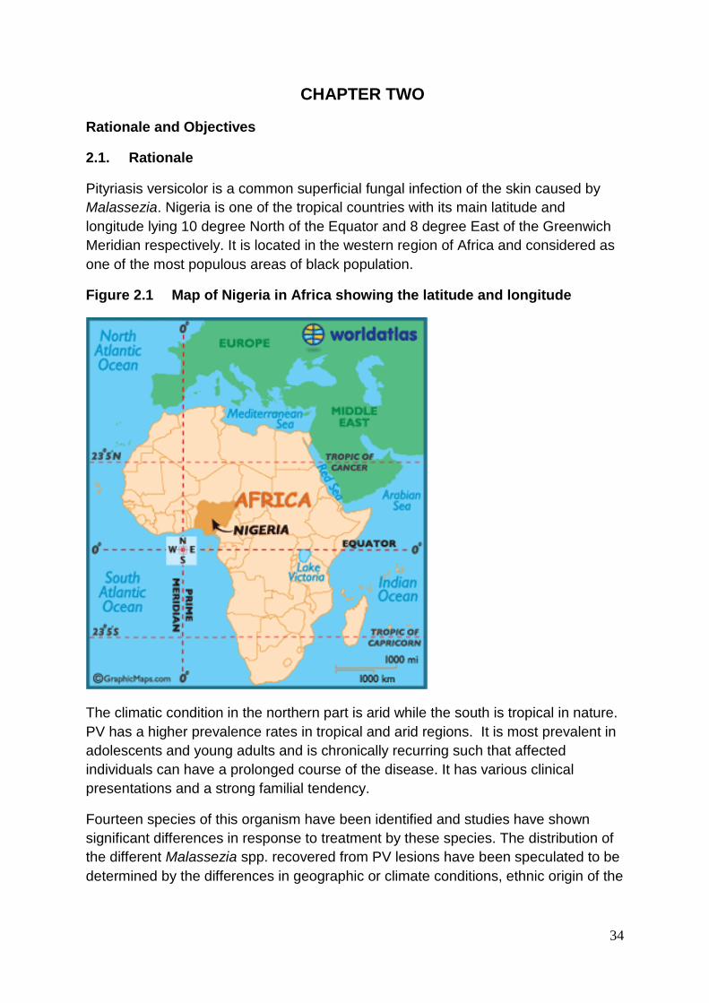
34
CHAPTER TWO
Rationale and Objectives
2.1. Rationale
Pityriasis versicolor is a common superficial fungal infection of the skin caused by
Malassezia. Nigeria is one of the tropical countries with its main latitude and
longitude lying 10 degree North of the Equator and 8 degree East of the Greenwich
Meridian respectively. It is located in the western region of Africa and considered as
one of the most populous areas of black population.
Figure 2.1 Map of Nigeria in Africa showing the latitude and longitude
The climatic condition in the northern part is arid while the south is tropical in nature.
PV has a higher prevalence rates in tropical and arid regions. It is most prevalent in
adolescents and young adults and is chronically recurring such that affected
individuals can have a prolonged course of the disease. It has various clinical
presentations and a strong familial tendency.
Fourteen species of this organism have been identified and studies have shown
significant differences in response to treatment by these species. The distribution of
the different Malassezia spp. recovered from PV lesions have been speculated to be
determined by the differences in geographic or climate conditions, ethnic origin of the
35
population, body site, specimen collection and identification methods.(108) And yet,
studies from the tropical regions of the world are very few in number.
A summary of several studies done to determine the causative species in most PV
lesions have shown M. globosa, M. sympodialis and M. furfur as the three dominant
Malassezia species.(108) There is also a major geographical location difference
among these species with M globosa being more prevalent in temperate regions and
M furfur in tropical regions of the world, although the other Malassezia species do
exist. The predominant species in West Africa has not well been studied. The clinical
characteristics of PV among Africans need to be updated.
The rationale for this study is to see if there is any association between the clinical
features and causative species of Malassezia in this environment which may
influence treatment.
2.2 Research question(s) and/ or hypotheses
1. Which of the Malassezia species isolated is more prevalent in Nigeria?
2. Is there an association between the species and clinical characteristic?
3. How does PV affect the quality of life of affected individuals?
2.3 Objectives
To identify the existing Malassezia species in Nigerian students with pityriasis
versicolor and to correlate the various species with the clinical characteristics of the
lesions
Aim of study:
1. To identify the prevailing species of Malassezia causing PV.
2. To describe the clinical characteristics of PV.
3. To describe attitude and health related quality of life effect of PV on affected
subjects.
Objectives:
1. To identify the most prevalent species of Malassezia in Nigerian subjects.
2. To determine if the clinical morphology of PV lesions is species specific.
3. To identify possible predisposing factors contributing to development of PV.
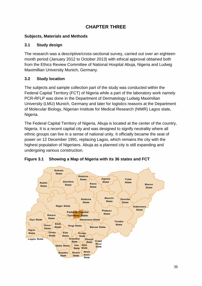
36
CHAPTER THREE
Subjects, Materials and Methods
3.1 Study design
The research was a descriptive/cross-sectional survey, carried out over an eighteen
month period (January 2012 to October 2013) with ethical approval obtained both
from the Ethics Review Committee of National Hospital Abuja, Nigeria and Ludwig
Maximillian University Munich, Germany.
3.2 Study location
The subjects and sample collection part of the study was conducted within the
Federal Capital Territory (FCT) of Nigeria while a part of the laboratory work namely
PCR-RFLP was done in the Department of Dermatology Ludwig Maximilian
University (LMU) Munich, Germany and later for logistics reasons at the Department
of Molecular Biology, Nigerian Institute for Medical Research (NIMR) Lagos state,
Nigeria.
The Federal Capital Territory of Nigeria, Abuja is located at the center of the country,
Nigeria. It is a recent capital city and was designed to signify neutrality where all
ethnic groups can live in a sense of national unity. It officially became the seat of
power on 12 December 1991, replacing Lagos, which remains the city with the
highest population of Nigerians. Abuja as a planned city is still expanding and
undergoing various construction.
Figure 3.1 Showing a Map of Nigeria with its 36 states and FCT

37
It is divided into six area councils. The area councils include Bwari, Gwagwalada,
Municipal (city center) Area Council, Kuje, Abaji and Kwali. The estimated population
of Abuja for 2013 by the 9th edition of the Demographia World Urban Areas of March
2013 is about 2.36 million making it the fourth largest city after Lagos, Kano and
Ibadan. Unlike other cities in Nigeria, Abuja is not dominated by any tribe or ethnicity
and the native people who lived there before it became the country’s capital are
considered a minority group.
Abuja’s climate is tropical wet and dry under the Köppen climate classification.(8) It
experiences three seasonal weather conditions annually. The first is the rainy
season from April to October, when the atmospheric temperatures range from 220 C
to 300 C. The second is called the dry season (without any rains) from November to
March, when the temperatures vary from as low as 120 C to as high as 400 C. Within
the dry season, in December/January there is usually a short period of the harmattan
season caused by the northeast trade winds which blows in fine dust particles from
the Sahara into the Gulf of Guinea. It is characterized by dust haze and an intensity
of cold as well as dry weather. According to Wikipedia, the dust haze can impair
visibility and sun exposure for several days; atmospheric temperature can be as low
as 50 C while humidity may drop to 15 percent.
Most of the laboratory work for the study namely the culture media preparation,
incubation of the fungi, DNA extraction and storage of extracted culture were done at
the National Hospital Abuja one of the tertiary hospitals located in Abuja.
Figure 3.2 Showing Abuja Area Councils

38
3.3 Study population
The study population consisted of senior secondary (SS) students from year one to
three (final year) who had clinical features of PV as well as positive culture growth of
the fungus from the lesions. The students were consecutively selected from the
Government Secondary Schools (GSS) in Abuja. They included a wide range of
Nigerians from different ethnic groups and social backgrounds.
For a student to be in the senior secondary school, he or she must have spent at
least 6 years in primary school, passed the National Common Entrance Examination
to enter the Junior Secondary School and after at least 3 years of junior secondary
education, must pass the Junior Secondary School Certificate Examination in order
to begin his or her senior secondary education. The minimum time spent in this
education is three years. Due to the above reasons, the expected age range of the
students in this study population (SS one to three) should lie between 13 up to 16
years. However, this may not be the case as the age range of the students in each
class varied widely with some students being as old as 22 years. Some students
may have commenced their education later than others or took a break from school
or are slow in learning. The reasons are usually socioeconomically related.
Meanwhile, the rate limiting step used in the students’ placements include mainly the
School Entrance, Certificate and Class Examinations. Therefore, students are placed
in classes based on the level of their intellectual capacity as shown by their success
at these examinations. Age, especially when older, does not usually play a major
role.
The schools selected were randomly distributed within the six area councils of Abuja.
Permission was obtained from the FCT Secondary School Management Board in
order to gain access to the schools (appendix V). The Board controls 57 GSS
distributed randomly in all the area councils. Some area councils have more schools
than the others and a bigger student population. For example Abaji and Kwali have 3
and 4 senior secondary schools, respectively, while Municipal Area council has 22.
These schools are attended by both girls and boys alike and comprise mostly of Day
schools while the Boarding schools are just 5 in number.
3.4 Sample size determination
The sample size calculation was done based on the prevalence rate of 6.7%
(prevalence of PV in Northwestern Nigeria).(55) Using the formula
n = 2zpq/d2
n = sample size
p = 0.067 (prevalence of PV in Northwestern Nigeria)
q = 1 - p

39
d = 5% (degree of accuracy desired)
The calculated sample size was 96.1
Samples could only be collected when the schools were open. Basically most of the
first year senior secondary students are registered and admitted late into the first
term since they have to await the result of their junior secondary school certificate
examinations and some may have to change/transfer schools. Also from the second
term of the session, the third year students commence their senior secondary school
certificate examinations. Therefore at any point in time about 20 to 30% of the
students may not be assessable for the research. Furthermore, there were students
who could refuse to participate. To compensate for the above reasons, a new
sample size using 30% was calculated as
corrected sample size = 96.1 divided by (1-0.30). Thus new sample size is 137.3
Considering that we had to analyze the identification of Malassezia species in
relation to the clinical features of PV, and using the culture based technique, a
growth rate of 50% is expected.(100) This increases the minimum sample size to
275 [that is calculated from 137.3 divided by (1-0.5)].
Students were recruited into the study based on their satisfaction of the inclusion
criteria (shown below). A total of 304 students were included in this study.
3.4.1 Inclusion Criteria
1. Students with clinical features of pityriasis versicolor and whose skin
scraping’s culture result showed growth of the Malassezia fungus.
2. Students whose parents and self, voluntarily consented to participate in
the study.
3.4.2 Exclusion Criteria
1. Students who claim to have had PV but do not currently have clinical
evidence of PV.
2. Students with PV who have used any topical antifungal medications in the
previous 2 weeks or systemic antifungal agents in the previous 1 month.
3. Those who are under 18 years of age whose parents did not give consent
to the study.
4. Students who are immunocompromised or chronically ill.
40
3.5 Selection Technique
The study was conducted from January 2012 to October 2013. The students were
recruited from May 2012 to May 2013 during the period the schools were in session.
There are three terms of 2 to 3 months period within a school year. Recruitment was
not done during the holidays.
Average number of students per class was estimated at 40. Average number of
classes per school-year was estimated as 6. Assuming that within the study period,
second year students and either first or third year students will be available for the
study; the average student population per school who may be available for the study
was then estimated as 480.
Using a prevalence of 6.7%, expected average number of students with PV per
school is 32. Therefore, visit to a minimum of 10 schools was required in order to
attain the calculated sample size. These schools were selected from the pool of 57
schools by simple random sampling. On visit to the schools, the Principal was
informed about the study and the letter from the FCT board presented to him or her.
The Principal then directed the head teacher in charge of health and the school
nurse to assist with the study. A convenient date when the study could begin was
usually selected by the school authorities. The study period in each school (this
includes timing for students’ education, collection of consent and skin scrapping of
PV lesions) was on average seven days and it was dependent on the student
population.
The students were addressed and information about pityriasis versicolor provided to
all the students who were present in their classes and on the Assembly Ground. The
students with PV who voluntarily presented themselves were given the consent form
and parents/patients information form (appendix I) for subsequent presentation to
their parents/guardians. The following days, the students whose parents have
already signed the consent form and who agreed to participate in the study as well
as those who did not have any of the exclusion criteria stated above were examined
at the School Clinic.
They were examined by the dermatologist and allotted a number. They filled the
questionnaire and skin scraping was collected and grown. Questionnaires not
properly or completely filled by the student especially with regards to key aspects of
the study were discarded.

41
Figure 3.3 Picture of one of the schools used in the study
Figure 3.4 Picture of the school clinic where samples were collected
42
3.6 Data Collection Instrument
Information was obtained from the students using a pre-tested questionnaire
(appendix II). The questions were simple and easy to understand. The questionnaire
was divided into four sections: section A for collection of demographic data, section
B on history related to PV infection including personal grooming habits, after school
activity and family history of PV. Section C was questions from the Children’s
Dermatology Life Quality index (CDLQI). All ten questions of the CDLQI were
obtained with the permission of copyright holders, Prof. Andrew Finlay and Dr.
Lewis-Jones of Cardiff University, United Kingdom. It consisted of questions
structured under 6 headings: symptoms and feeling (questions 1 and 2), leisure
(questions 4, 5 and 6), school or holiday (question 7), personal relationship
(questions 3 and 8), sleep (question 9) and treatment (question 10). Section D
contained records of the clinical pattern and distribution of the PV lesions as well as
the result of laboratory investigations. This section was completed by the principal
investigator. The questionnaire was administered in English language which is the
language spoken and also used for teaching in the schools. Knowledge of the
English language is a prerequisite for admission into any of the schools.
Nevertheless, since there was a possibility that some students may not have a clear
understanding of the questions, provision was made to assist them with easier
interpretations. This, however, was rarely utilized.
3.6 Clinical Evaluation
All the eligible students recruited into the study filled in the questionnaire. Thereafter
a detailed physical examination was conducted in a well lit room with particular focus
on the skin lesions. Photographs of some lesions were also taken.
PV was diagnosed as macules of different shapes which may coalesce into large
irregular patches. They may have overlying thin scales which can be made more
obvious by stretching the skin (Zileri’s sign). The size of the lesions varied from a
few millimeters to large confluent areas.
The colours of the skin lesions were hypopigmented or hyperpigmented. Some
subjects had a combination of the two. The lesions were distributed on the face,
upper trunk, upper arms and back.
The sites of involvement were divided into regions: face (perioral, around the nose,
behind ears, cheeks, and forehead), chest, back, upper arm and others which
includes lower limbs, scalp, and perineum. The number of regions involved were
counted and grouped as localized when one region was involved and extensive
when three or more regions were involved. This was used to grade the severity of
infection.
43
3.7. Skin Scrapings
Skin scrapings were collected from the clinical lesions with the use of OpSite
transparent dressing (3 by 4 cm; Smith and Nephew Medical Ltd, Hull, United
Kingdom) as described by Sugita and Saad et al.(76, 147) This was then placed on a
glass slide for onward transfer to the laboratory. The site of scrapings was
documented. Scrapings from adjacent healthy skin from the same student (as
controls) were also collected. This was also collected with Opsite dressing. In
subjects with PV involvement of more than two regions, samples were collected from
the most accessible. In schools where a room with a dark background was available,
wood’s light examination was also done. A part of the collected scrapings was used
for direct microscopic examination with 20% potassium hydroxide plus lactophenol
blue.
A piece of the dressing was placed immediately on modified Dixon agar media
consisting of 3.6% malt extract (Sigma-Aldrich Ltd), 0.6% peptone (Sigma-Aldrich), 2%
desiccated ox bile (Sigma-Aldrich), 1% Tween 40 (Sigma-Aldrich), 0.2% glycerol, 0.2%
oleic acid (Sigma-Aldrich), 1.2% agar (Sigma-Aldrich), 0.05 g/dL chloramphenicol
and 0.05 g/dL cycloheximide (Sigma-Aldrich)(63). The medium was used within a
week of preparation and the cultures incubated at 34°C for a maximum of seven
days. The macroscopic feature of the fungal growth was compared with pictures of
Malassezia species found in textbook written by Crespo Erchiga et al.(3)
In some culture plates, there was contamination by other fungi species. Sometimes
these species grew quite fast (within two days) and completely covered up the
culture plate, preventing growth of the Malassezia yeasts. In these cases sub-culture
of the growing Malassezia yeasts was done on to a fresh culture plate. Catalase test
and microscopy of culture was also matched to confirm Malassezia species growth.
The morphology of the colonies was documented and two or three of these were
selected and placed in microtubes and stored in -800 C freezer until use. The storage
period varied from one to 10 months.
Figure 3.5 OpSite transparent dressing used for scale collection

44
Figure 3.6 “Spaghetti and meatball” microscopy (x4) of PV scales
Figure 3.7 Culture of Malassezia on mDA

45
3.8. Isolation and identification of colonies compatible with Malassezia
This was done using the PCR-RFLP identification method as described by Mirhendi
et al.(123) There was some modification with regard to the extraction of genomic
DNA from the culture. This was extracted using the heating and freezing method.
3.8.1 DNA preparation: Heat-freeze method
One ml of sterile water was added to microtubes containing Malassezia colony which
was obtained from the -800 C freezer. It was then heated to 950 C in a dry heat block
for 10 minutes. It was immediately transferred to a bowl containing liquid nitrogen
and frozen. This heating and freezing sequence was repeated three times and the
samples were allowed to thaw at room temperature and homogenized by vortex for
10 seconds. There was no further DNA extraction, samples were used as prepared.
3.8.2 PCR Procedure
Primers that aim to amplify the 26S rDNA of Malassezia which has been verified to
identify at least 11 species with the use of RFLP-PCR was used.(7, 71, 123, 148)
Their sequences were: forward, 5’ – TAACAAGGATTCCCCTAGTA and reverse, 5’ –
ATTACGCCAGCATCCTAAG. A final volume of 50 µL for each sample was used to
perform PCR amplification. Each reaction contained 1 µL of the above prepared
DNA sample, 10 µL of 5X Herculase buffer, 0.5U Herculase Hotstart DNA
Polymerase (Agilent Technologies, Stratagene), 100mM each deoxynucleoside
triphosphate (dNTPs), 10µM of each primer and 1µL of 100% DMSO. The rest of the
volume was made up with DNA - free water. The mixture was done on ice with the
help of a master-mix.
The amplification carried out at the Department of Dermatology Ludwig Maximilian
University Munich, Germany was performed with the Stratagene RoboCycler 40
PCR machine while that done at the Department of Molecular Biology Nigerian
Institute of Medical Research Lagos, Nigeria was carried out with the Eppendorf
Mastercycler PCR machine using the same program. The program consisted of an
initial denaturation step at 940 C for 5 minutes, followed by 40 cycles of denaturation
step at 940 C for 45 seconds, annealing step at 550 C for 45 seconds and an
extension step at 720 C for 60 minutes, thereafter a final extension at 720 C for 7
minutes.
Amplified products were visualized with the use of 1.5% (w/v) agarose (Sigma-
Aldrich) gel electrophoresis in TBE (Tris HCl, boric acid, EDTA) buffer and stained
with RedSafe Nucleic Acid Staining Solution (iNtRON Biotechnology).
The agarose was first boiled in microwave until the crystals were completely
dissolved. On cooling to a level when the bottom of flask can be comfortably touched,
5 µL of RedSafe was added and mixed before pouring into tray. Pouring was done
from the edge to avoid bubbles. Formed bubbles were quickly removed. A comb was
placed at one end and middle of the tray, then the agarose was left to solidify.

46
Following the solidification of the agarose, the combs were carefully removed.
Fifteen microliter of each product plus three microliter of 6X gel loading buffer were
then loaded into the pockets created by the combs in the gel. The electrodes were
attached and left to run for one and half hours at a voltage of 100 V. Thereafter, the
DNA bands were visualized and photographed under UV transillumination. The
products successfully amplified by the primers were all expected to produce a single
band of approximately 580 base pairs.
3.8.3 Enzyme digestion
Digestion of products with positive band was done with the Cfo I (Roche Applied
Science) and BtsC I (New England BioLabs) restriction enzymes as described by
other workers.(7, 71, 123) Digestion was performed with a final reaction volume of
20 µL with 10 U and later 1.25 U of the enzymes. Incubation was at 370 C for 3 hours.
Digested products were viewed with the use of 2.2% agarose gel electrophoresis in
TBE buffer. The electrophoresis was done also at 100 V but for one hour only.
Subsequently the gel was placed in a chamber containing one µg/ml of ethidium
bromide. This was left for 20 minutes, then, placed in distilled water chamber for 5
minutes before being read under UV transillumination.
The bands generated were of variable predictable sizes depending on the
Malassezia species. The base pair length used for identification is as in Table1.7
above. Nine species can be clearly distinguished from one another using the Cfo I
enzyme. These include M. furfur, M. pachydermatis, M. globosa, M. obtuse, M.
restricta, M. slooffiae, M. nana, M. japonica, and M. yamatoensis; however, M.
sympodialis and M. dermatis were shown to produce a similar pattern, which are
357bp, 197bp, 21bp and 359bp, 199bp and 21bp respectively. Therefore, these two
species were further differentiated using the enzyme BtsC I which is an isoschizomer
(recognizes the same sequence) of BstF 51.
Figure 3.8. In preparation to commence PCR showing the ice tray,
microtubes, DNA-free water and the polymerase and enzymes

47
Figure 3.9. The Stratagene RoboCycler 40 PCR machine and the loaded gel
operating at 100 V
Figure 3.10. Staining with Ethidium Bromide and the Gel Documentation
System where the DNA bands were visualized and photographed
under UV transillumination

48
3.9 List of materials
3.9.1 List of equipment:
1. Sellotape
2. Opsite flexi transparent adhesive 10cmx10cm roll (Smith and Nephew GmbH)
3. Microscope slides. Cat. Number 7104
4. Petri dishes 100 mm x 15 mm
5. Autoclave machine
6. Measuring scale
7. Binder Incubator
8. Thermocool Fridge
9. -800 C freezer
10. Vortex machine
11. Dry heat block
12. Liquid Nitrogen
13. 2ml Eppendorf tubes
14. 1.5 Eppendorf tubes
15. Tube racks
16. Filtered pipette tips
17. Unfiltered pipette tips
18. Pipettes 20 µL, 200 µL, 1000 µL
19. PCR tubes
20. Stratagene RoboCycler 40 PCR Machine
21. Eppendorf Mastercycler
22. Gel electrophoresis apparatus
23. Bio-Rad Gel Doc 2000 System
24. Conical flask
25. Siemens Microwave
26. Microscope
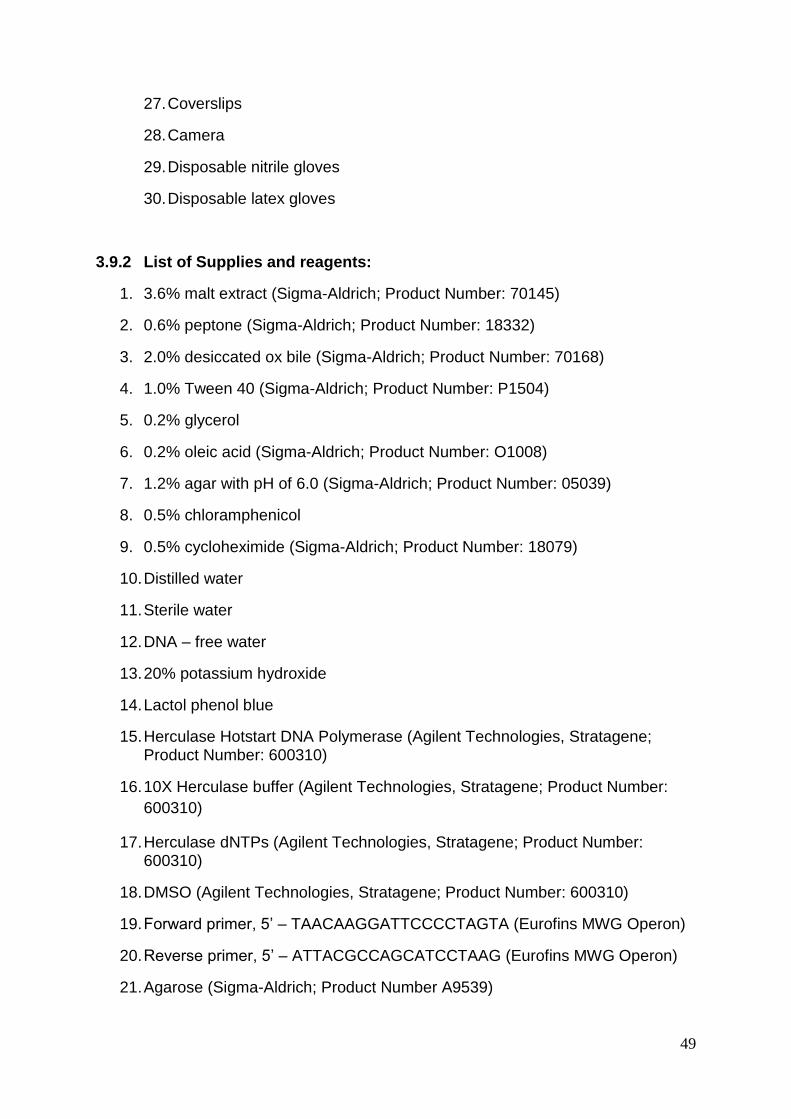
49
27. Coverslips
28. Camera
29. Disposable nitrile gloves
30. Disposable latex gloves
3.9.2 List of Supplies and reagents:
1. 3.6% malt extract (Sigma-Aldrich; Product Number: 70145)
2. 0.6% peptone (Sigma-Aldrich; Product Number: 18332)
3. 2.0% desiccated ox bile (Sigma-Aldrich; Product Number: 70168)
4. 1.0% Tween 40 (Sigma-Aldrich; Product Number: P1504)
5. 0.2% glycerol
6. 0.2% oleic acid (Sigma-Aldrich; Product Number: O1008)
7. 1.2% agar with pH of 6.0 (Sigma-Aldrich; Product Number: 05039)
8. 0.5% chloramphenicol
9. 0.5% cycloheximide (Sigma-Aldrich; Product Number: 18079)
10. Distilled water
11. Sterile water
12. DNA – free water
13. 20% potassium hydroxide
14. Lactol phenol blue
15. Herculase Hotstart DNA Polymerase (Agilent Technologies, Stratagene; Product Number: 600310)
16. 10X Herculase buffer (Agilent Technologies, Stratagene; Product Number:
600310)
17. Herculase dNTPs (Agilent Technologies, Stratagene; Product Number: 600310)
18. DMSO (Agilent Technologies, Stratagene; Product Number: 600310)
19. Forward primer, 5’ – TAACAAGGATTCCCCTAGTA (Eurofins MWG Operon)
20. Reverse primer, 5’ – ATTACGCCAGCATCCTAAG (Eurofins MWG Operon)
21. Agarose (Sigma-Aldrich; Product Number A9539)
50
22. RedSafe Nucleic Acid Staining Solution ( iNtRON Biotechnology; Product Number: 21141)
23. Ethidium bromide solution
24. 6X gel loading buffer (Sigma-Aldrich; Product Number: G7654)
25. DNA SmartLadder – 200 to 10000 base pairs (Eurogentec; Product Number: 08A31-2)
26. Low molecular weight DNA ladder – 25 to 766 base pairs (New England BioLabs; Product Number: N3233S)
27. Cfo I (Hha I) enzymes and buffer (Roche Applied Science; Product Number: 10688541001
28. BtsC I enzymes and buffer ( New England BioLabs; Product Number: R0647S)
29. Icebox
30. TBE buffer: Tris base 54 g, boric acid 27.5 g and EDTA 3.72 g in 1 L
3.10 Statistical analysis
Data generated from the study were entered, coded, and analyzed using the
Statistical Package for Social Sciences (SPSS Inc, Chicago, IL) software version
16.0. Descriptive variables were presented using frequency tables, pie charts and
bar charts as appropriate. The student’s t-test was used to test for statistical
difference between the age of the students and gender. It was also used between
age of onset of PV and some clinical variables which included residence of the
students, presence of PV in a nuclear family member, daily use of petroleum jelly,
daily use of body cream, episodes of PV infection (first episode vs recurring),
presence of dandruff and regular animal contact.
Chi-square and Fisher exact tests as appropriate were used to test for independence
between age group and the use of bleaching creams, having dandruff, recurrence of
PV, residence of the students and presence of a family member with PV. Odds ratio
was used to analyze the relationship between having a positive family history of PV
and recurring PV. Chi-square was also used to test for independence between
genders and some personal grooming habits as well as some clinical features. Odds
ratio was used to analyze the relationship between genders and routine use of
Vaseline and bleaching/toning creams.
For the analysis of the health-related quality of life (HRQoL), a statistical difference
between the total CDLQI score in relation to some clinical variable was analysed with
the use of the independent student’s t-test. The clinical variables included age,
gender, episodes of PV infection, family history of PV, site affected (exposed vs
unexposed), number of body regions affected by PV, color of PV lesions and the
presence of additional symptoms. Also used were the Spearman’s rank coefficient,
one way analysis of variance ANOVA and the Fisher’s least significant difference
51
(LSD) post hoc analysis as appropriate to find the correlation between the students’
year in school and number of regions affected by PV.
Chi-square test and Fisher exact test as appropriate were used to test for the
association between the identified Malassezia species and the clinical variables
mentioned above. A P-value of <0.05 was considered significant.
3.11 Ethical Certificate
Approval was obtained from the National Hospital Abuja Ethics Review Committee
(appendix III) and the Ethics Commission of the Ludwig Maximilian University
Munich Germany (appendix IV). Permission was also obtained from the FCT
Secondary School Education Board Abuja (appendix V). Only students who were
adequately informed and whose consent was obtained were included in the study.

52
CHAPTER FOUR
Results
4.1 Clinical characteristics of subjects
A total of 304 students with clinical features of pityriasis versicolor (PV) were
recruited from 10 senior secondary schools into the study between May 2012 and
May 2013. Out of these, 175 skin samples of PV grew from the mDA culture medium.
This produced a 57.6% growth rate. Mode of distribution is as shown in Table 4.1
Table 4.1. Distribution of schools selected in the study
The table shows the number of students recruited in each school and the number
whose skin scraping had a positive culture growth. Government senior secondary
(GSS) Kwali had the highest number of students with PV while GSS Wuse had the
lowest number. There was no statistically significant correlation between the number
of students recruited and number with positive culture growth.
Schools Number of Students with PV
recruited
Number with positive culture
growth
GSS Gwarinpa 9 4
GSS Garki 20 12
GSS Jabi 18 10
ADSS Maitama 18 8
GSS Nyanya 42 24
GSS Lugbe 53 35
GSS Kuje 27 20
GSS
Gwagwalada
50 27
GSS Wuse 8 5
GSS Kwali 59 29
Total 304 175
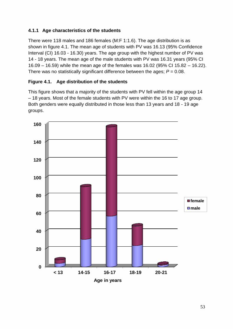
53
4.1.1 Age characteristics of the students
There were 118 males and 186 females (M:F 1:1.6). The age distribution is as
shown in figure 4.1. The mean age of students with PV was 16.13 (95% Confidence
Interval (CI) 16.03 - 16.30) years. The age group with the highest number of PV was
14 - 18 years. The mean age of the male students with PV was 16.31 years (95% CI
16.09 – 16.59) while the mean age of the females was 16.02 (95% CI 15.82 – 16.22).
There was no statistically significant difference between the ages; P = 0.08.
Figure 4.1. Age distribution of the students
This figure shows that a majority of the students with PV fell within the age group 14
– 18 years. Most of the female students with PV were within the 16 to 17 age group.
Both genders were equally distributed in those less than 13 years and 18 - 19 age
groups.
0
20
40
60
80
100
120
140
160
< 13 14-15 16-17 18-19 20-21
Age in years
female
male

54
4.1.2 Demography of students About half of the students recruited (51.6%) were in their second year (SS2) while the percentage of students in third and final year was the lowest at 10.9%. There was no significant difference between the male and female according to their classes, P = 0.51. The distribution is shown in Figure 4.2 Figure 4.2. Class distributions of the students The least number were students in senior secondary class 3 (SS3).
The tribe of students recruited numbered up to 46. The tribes were from the different
ethnic groups and state of origin found in Nigeria. However, the majority were Ibo
(26.9%), next was Yoruba (15.8%) then Igala (7.6%) and there were about 15% of
the students who did not fill in their ethnic origin.
On the area of residence which was evenly divided into urban and rural areas, most
of the students were recruited from Kwali and Gwagwalada Area Councils; see Table
4.1 above. One of the schools had only 8 students with PV. This school GSS Wuse
is located right in the city centre.
Most of the students lived with their parents (78.9%) while the rest were residing with
a Guardian. There were about 16% of the fathers and 26% of the mothers who had
no formal education.
The occupation of the parents was varied; however, trading was the occupation with
the highest frequency by both parents.
The students had diverse and multiple afterschool activities. These included
household chores, playing football, cooking, reading, watching television, dancing,
and singing. Others helped in the farms and shops of their parents. A few loved to
play volleyball, basketball, table tennis, jog, swim and or visit friends.
114
157
33
SS1
SS2
SS3

55
4.1.3 Personal grooming of the students
Table 4.2. The frequency of respondents with regard to their personal
grooming
Personal grooming of the students Frequency in Percentage (%)
Daily bath 100%
Use Tap water daily 75.7%
Use toilet soap 48%
Use medicated antibacterial soap 51%
Use body creams 67.8%
Use petrolatum (Vaseline® jelly) 51%
Use bleaching and toning creams 14.5%
Use talcum powder 32.2%
Share body towel 34.2%
Sweat excessively during the day 39.8%
Sweat excessively at night 32.2%
Notice worsening of PV and thinks PV is
related to the climate or seasons
46%
Toilet soaps were the ordinary bathing soaps commonly used included “Lux”, “Joy”,
“Premier”, Johnson baby soap etc. The medicated antibacterial soaps frequently
used were “Dettol”, “Tetmosol”, “Delta” and “Lifebouy”. The common body creams
used by the students included “Familia”, “Jergens”, “Nivea”, “Cocoa butter”. The
beaching/toning creams are body moisturizers containing hydroquinone, highly
potent steroids and/or mercury. These included “Tura”, “Fair and white”, “Funbact A”,
“Dermovate”, “Rapid white”, “Skin light” and “Maxi tone”
All the students took daily baths but not all used Tap water as routine source of
water. More students used body creams than petrolatum, commonly called
Vaseline® as their regular body moisturizer. Almost 40% of the students sweat
excessively during the day while 14.5% use toning/bleaching creams. About half of
the students recruited believed their PV infection is climate and seasons related
especially when the atmospheric temperature is at its peak during the dry season.
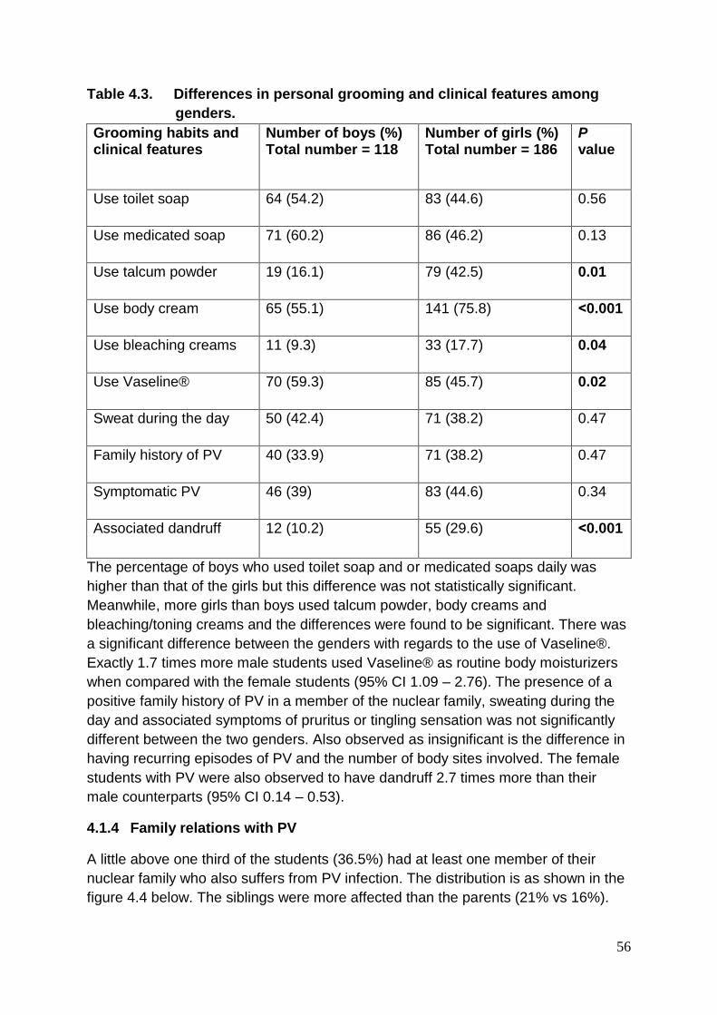
56
Table 4.3. Differences in personal grooming and clinical features among
genders.
Grooming habits and clinical features
Number of boys (%) Total number = 118
Number of girls (%) Total number = 186
P value
Use toilet soap 64 (54.2) 83 (44.6) 0.56
Use medicated soap 71 (60.2) 86 (46.2) 0.13
Use talcum powder 19 (16.1) 79 (42.5) 0.01
Use body cream 65 (55.1) 141 (75.8) ˂0.001
Use bleaching creams 11 (9.3) 33 (17.7) 0.04
Use Vaseline® 70 (59.3) 85 (45.7) 0.02
Sweat during the day 50 (42.4) 71 (38.2) 0.47
Family history of PV 40 (33.9) 71 (38.2) 0.47
Symptomatic PV 46 (39) 83 (44.6) 0.34
Associated dandruff 12 (10.2) 55 (29.6) ˂0.001
The percentage of boys who used toilet soap and or medicated soaps daily was
higher than that of the girls but this difference was not statistically significant.
Meanwhile, more girls than boys used talcum powder, body creams and
bleaching/toning creams and the differences were found to be significant. There was
a significant difference between the genders with regards to the use of Vaseline®.
Exactly 1.7 times more male students used Vaseline® as routine body moisturizers
when compared with the female students (95% CI 1.09 – 2.76). The presence of a
positive family history of PV in a member of the nuclear family, sweating during the
day and associated symptoms of pruritus or tingling sensation was not significantly
different between the two genders. Also observed as insignificant is the difference in
having recurring episodes of PV and the number of body sites involved. The female
students with PV were also observed to have dandruff 2.7 times more than their
male counterparts (95% CI 0.14 – 0.53).
4.1.4 Family relations with PV
A little above one third of the students (36.5%) had at least one member of their
nuclear family who also suffers from PV infection. The distribution is as shown in the
figure 4.4 below. The siblings were more affected than the parents (21% vs 16%).
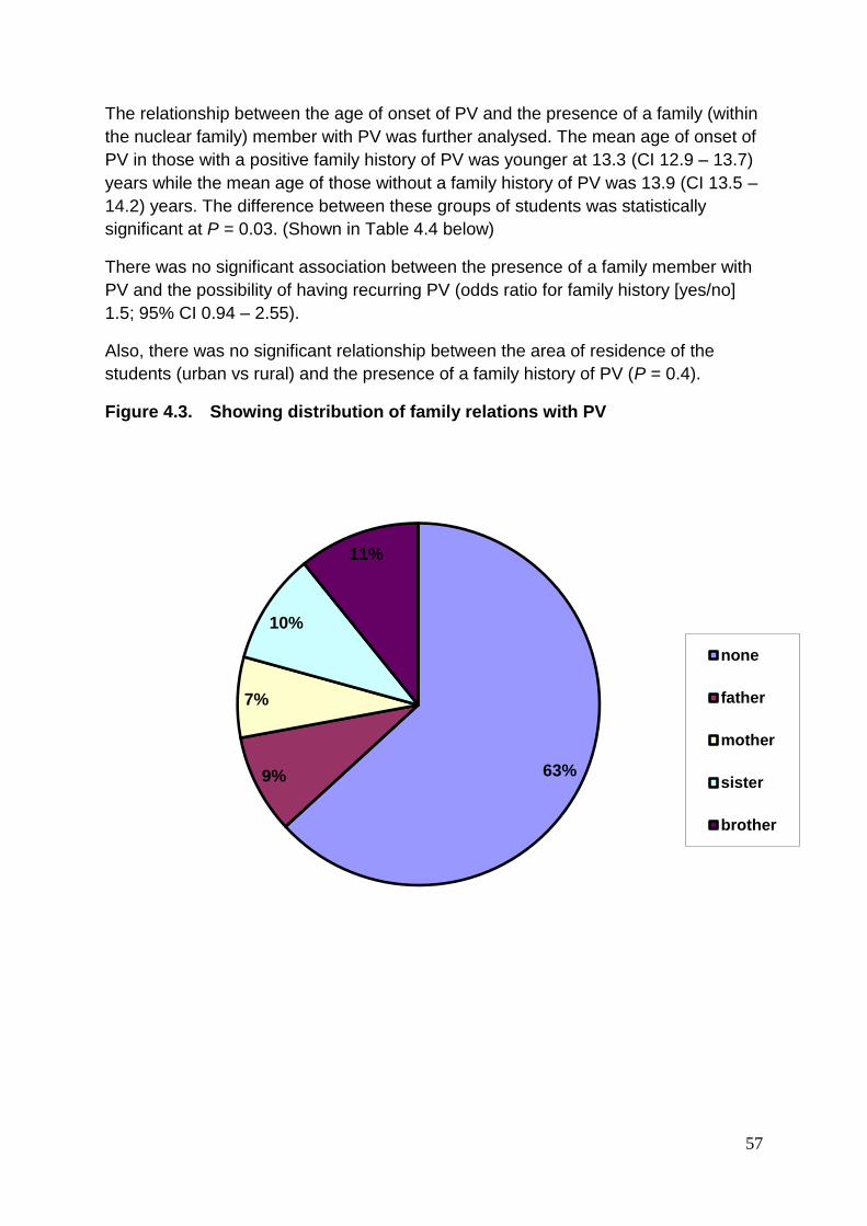
57
The relationship between the age of onset of PV and the presence of a family (within
the nuclear family) member with PV was further analysed. The mean age of onset of
PV in those with a positive family history of PV was younger at 13.3 (CI 12.9 – 13.7)
years while the mean age of those without a family history of PV was 13.9 (CI 13.5 –
14.2) years. The difference between these groups of students was statistically
significant at P = 0.03. (Shown in Table 4.4 below)
There was no significant association between the presence of a family member with
PV and the possibility of having recurring PV (odds ratio for family history [yes/no]
1.5; 95% CI 0.94 – 2.55).
Also, there was no significant relationship between the area of residence of the
students (urban vs rural) and the presence of a family history of PV (P = 0.4).
Figure 4.3. Showing distribution of family relations with PV
63% 9%
7%
10%
11%
none
father
mother
sister
brother

58
4.2 Clinical features and characteristics of PV
4.2.1. Age of onset of PV
The average age of onset of PV among the students was 13.7 years (95% CI 13.4 –
13.9 years). The median was 14 years while the minimum age of onset was 5 years
and the maximum was 19 years. The mean age in males was 13.9 years (C.I. 13.5 –
14.4 years) and the females were younger at 13.5 years (13.1 – 13.9 years). The
difference in the means was not statistically significant, P = 0.15.
The area of residence by the students was divided into rural and urban. The rural
areas are regions more likely to have untarred roads and lower cost of housing as
well as food. People with lower income usually reside here thus they are more
populated than the urban region. These areas are also known as satellite towns. The
rural regions selected in this study included Gwagwalada, Kwali, Nyanya, Kuje and
Mararaba. The urban areas are close to or within the city centre. They are less
populated because of the higher cost of housing and living expenses. About 157
(53.6%) students were from the satellite towns while those from the city centre were
147 (46.4%). The students who resided in the rural part of Abuja developed PV at a
younger age (mean age of onset was found to be 13.3; 95% CI 12.8 – 13.6 years)
compared with students living in the city centre [mean age was 14.2 (95% C.I. 13.8 –
14.6) years]. The difference between the 2 means was statistically significant, P =
0.001. See Table 4.4.
A large number of the students (69.1%) had recurring episodes of PV infection while
for 94 students (30.9%); there was no history of recurrence. There was no
statistically significant difference between the students who had recurring and first
episode of PV with regards to age, sex, age of onset, family history, symptoms,
colour and locations of lesions. See Figure 4.4.
Figure 4.4. Number of students with recurring and first episode of PV
94
210
first PV episode
recurring PV

59
Table 4.4. The clinical characteristics in relations to the age of onset of PV
Mean age of onset
of PV in years
(95% CI)
Mean age of onset of
PV in years (95% CI)
P
value
Residence of student Satellite towns
13.2 (12.8 – 13.6)
City centre
14.2 (13.8 – 14.6)
0.001
Presence of nuclear family
member with PV
Positive
13.3 (12.9 – 13.7)
Negative
13.9 (13.5 – 14.2)
0.03
Daily use of Vaseline®
jelly
Yes
13.3 (12.8 – 13.7)
No
14.1 (13.7 – 14.5)
0.004
Daily use of body cream Yes
13.6 (13.2 – 13.9)
No
13.8 (13.3 – 14.3)
0.43
Difference between first
and recurring episodes
First episode of PV
13.8 (13.3 – 14.3)
Repeat episode of PV
13.6 ( 13.3 – 13.9)
0.5
Difference between those
with/without dandruff
Those with dandruff
12.6 ( 11.9 – 13.2)
Those without dandruff
14.0 ( 13.7 – 14.3)
0.001
Regular animal contact Yes
13.4 (12.9 – 13.95)
No
13.7 (13.4 – 14.04)
0.37
Also shown in this table is that students who routinely used Vaseline® or had
associated history of dandruff developed PV at a significantly younger age than their
counterpart who did not use Vaseline® or did not have dandruff. Meanwhile the
younger age of onset of PV in students who used body creams, or had repeated
episodes of PV or regular animal contact was not statistically significant.
4.2.2. PV and possible predisposing factors
Only 44 students (14.5%), majority of which were females admitted the use of skin
toning/bleaching creams. A little over half (54.5%) of this group of students were
either 16 or 17 years. Also the most frequent age of onset of PV in students who
used bleaching/toning creams was observed in the 13 to 16 age group which was
not different from the general student population (P = 0.18). Overall, the average age
of students who used bleaching/skin toning products were older (16.3 vs 16.1 years)
and the average age of onset was also older when compared with students who did
not use bleaching/toning creams (14.3 vs 13.5 years).
About 20% of the students had regular contact with animals either as house pets or
kept livestock, however, this did not affect the age of onset of PV when compared
with those without regular animal contact.
Also 22% of the respondents had dandruff (pityriasis capitis) in addition to PV. PV in
this group of students appeared at a younger age than those without dandruff (P =

60
0.001). Meanwhile, there was no significant association observed between the
students who had dandruff and those who had recurrence of PV infection (P = 0.55).
4.2.3 Symptoms and Signs of PV
Table 4.5. Showing the percentage distribution of complaints experienced
by the students.
Complaints Number of students Percentage (%)
Skin lesions 304 100%
Symptomatic:
Pruritus
Tingling sensation
130
106
24
42.8%
34.9%
7.9%
Asymptomatic 174 57.2%
Symptomatic complaints were documented for those students who said their PV
lesions were pruritic or gave them tingling sensation. Interestingly, it was observed
that those who sweat excessively during the day were twice likely to be symptomatic
than those who did not have hyperhidrosis. This was statistically significant. The
odds ratio for not symptomatic/symptomatic was 0.58, 95% CI 0.37 – 0.93.
Statistically significant differences when compared to other clinical characteristics
between patients who were symptomatic and asymptomatic were not observed.
Figure 4.5. Showing the percentage distribution of the complaints
experienced by the students.
0 50 100 150 200 250 300 350
Skin lesions
Asymptomatic
Symptomatic
304
174
130
symptoms of PV
No. of students

61
Table 4.6. Location of PV lesions
Location Number of students Percentage (%)
Back 101 24.2
Face 189 45.3
Chest 57 13.7
Upper arm 27 6.5
Neck 18 4.3
Others 25 6.0
This was categorized by body regions such as face, back, etc and not by the number
or count of PV lesions. Affectation of the face had the highest frequency at 45.3%
while the neck had the lowest at 4.3%. “Others” included scalp, nasal creases, lower
limbs and abdomen. Some students had involvement of more than one body region.
Figure 4.6. Exposed and unexposed PV location
A chart showing the number of students with PV lesions located at an exposed part
of the body mainly face, neck, scalp, nasal creases and unexposed part of the body
mainly chest, back and upper arms. About a third of the students had the PV lesions
on an unexposed part of the body.
73%
27%
exposed
unexposed

62
Table 4.7. Number of body regions infected with PV
Number of body region
involved with PV
Frequency Percentage (%)
1 208 68.4
2 71 23.4
3 21 6.9
4 4 1.3
Exactly 8.2% of the students had extensive PV (involvement of 3 or more body
regions). A complaint of pruritus or tingling sensation was statistically associated with
students who had extensive PV (χ2 = 8.237, P = 0.02). Most of the students had just
one body region involved (68.4%).
Figure 4.7. Colour of PV
The pigmentations of the lesions observed were hypo- and hyperpigmented. Only 5
students were observed to have a combination of both.
14.5%
85.5%
Hyperpigmented Hypopigmented
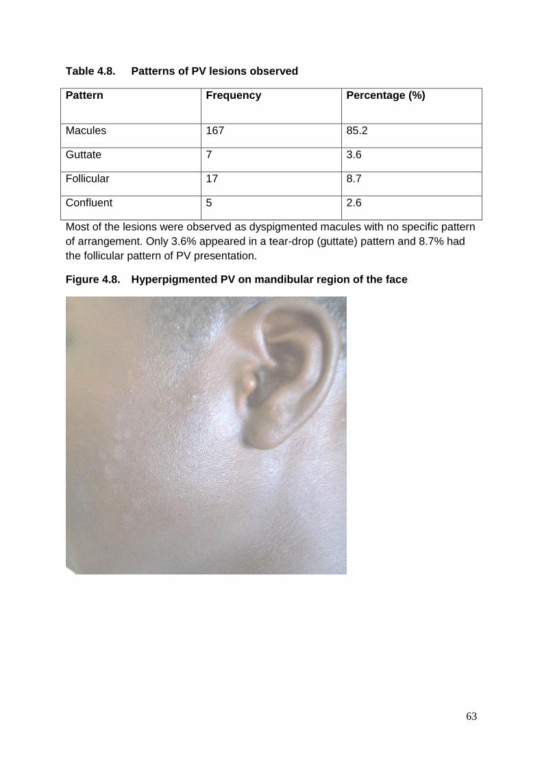
63
Table 4.8. Patterns of PV lesions observed
Pattern Frequency Percentage (%)
Macules 167 85.2
Guttate 7 3.6
Follicular 17 8.7
Confluent 5 2.6
Most of the lesions were observed as dyspigmented macules with no specific pattern
of arrangement. Only 3.6% appeared in a tear-drop (guttate) pattern and 8.7% had
the follicular pattern of PV presentation.
Figure 4.8. Hyperpigmented PV on mandibular region of the face

64
Figure 4.9. Hypopigmented PV on shoulders/upper arm
Figure 4.10. Hyperpigmented PV located at the back
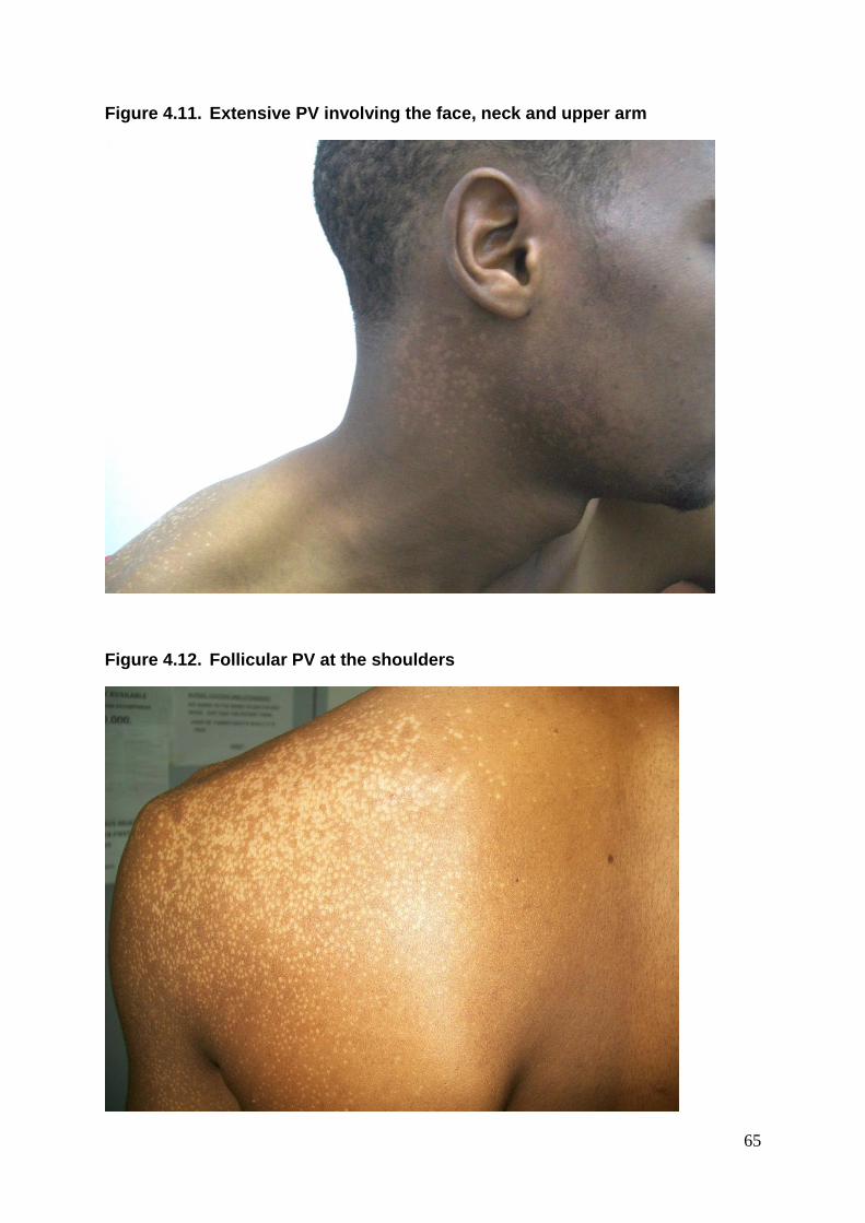
65
Figure 4.11. Extensive PV involving the face, neck and upper arm
Figure 4.12. Follicular PV at the shoulders

66
Figure 4.13. Close-up of the follicular PV lesions, notice how each hair shaft
is surrounded by hypopigmented macule
4.2.4 Mode of treatment implored by the students
Figure 4.14. Mode of treatment utilized by the students
34
15
20
112
45
78
0 20 40 60 80 100 120
drugs
herbs
hydraulic fluids
antifungal creams
others
none
number of students

67
Most of the students (36.8%) used antifungal creams to treat their PV infection while
25.7% of the students did not try any form of treatment. 6.6% of the students used
hydraulic fluids, mainly brake fluids and 4.9% used herbs as a form of treatment. The
drugs used by the students were mainly antifungal medications taken via oral route
such as ketoconazole, fluconazole and itraconazole. The “others” mode of treatment
implored by the students were classified as inappropriate for PV management. They
included antibacterial medications both topical and systemic, antiseptic soaps,
“Nixoderm”, “Skin light” body cream, “Biocoten”, “Funbact-A”.
4.3 Health related quality of life (HRQoL) of affected students with PV
Table 4.9. Socio-demographic and clinical variables in relation to the mean
total CDLQI score
Variables Mean total CDLQI (95% CI)
Total score: 7.39 (6.73-8.06)
P value
Mean CDLQI by gender
Male: n=118 (38.8%)
Female: n=186 (61.2%)
7.36 (6.23 - 8.49)
7.41(6.58 - 8.23)
0.95
Mean CDLQI by age group
≤13 years old: n=8 (2.6%)
>13years old: n=296 (97.6%)
4.75 (3.35 - 6.15)
7.46 (6.78 - 8.14)
0.002
Mean CDLQI by residence
Urban: n=141(46.4)
Rural: n=163 (53.6%)
7.11(6.14 - 8.08)
7.64 (6.72 - 8.56)
0.433
Mean CDLQI by school year
First year: n=114(37.5%)
Second year: n=157(51.6%)
Third year: n=33 (10.9%)
7.27(6.17-8.37)
7.32 (6.39 - 8.25)
8.12 (6.07 - 10.17)
0.505
Mean CDLQI by age of onset
<13 years: n=91(29.9%)
≥13 years: n=213 (70.1%)
7.41(6.18 - 8.83)
7.38 (6.59 - 8.18)
0.977

68
Mean CDLQI by episode of PV
First episode: n=94 (30.9%)
Recurring PV: n=210 (69.1%)
8.85 (7.57 - 10.14)
6.74 (5.98 - 7.50)
0.006
Mean CDLQI by family history
Positive: n=111(36.5%)
Negative: n=193 (63-5%)
8.41(7.27 - 9.55)
6.80 (5.99 - 7.62)
0.021
Mean CDLQI by site
Exposed region: n=221 (72.7%)
Unexposed: n=83 (27.3%)
7.26 (6.47 - 8.05)
7.73 (6.48 - 8.99)
0.534
Mean CDLQI by number of body
region affected
One site: n=208 (68.4%)
More than one site: n=96
(31.6%)
6.78 (6.05 - 7.51)
8.72 (7.35 - 10.08)
0.014
Mean CDLQI by color of lesions
Hypopigmented: n=260 (85.5%)
Hyperpigmented: n=44 (14.5%)
7.24 (6.54 - 7.94)
8.30 (6.26 - 10.33)
0.271
Mean CDLQI by associated
symptoms with PV
None: n=175 (57.6%)
Present: n=129 (42.4%)
6.34 (5.53 - 7.15)
8.81 (7.73 - 9.89)
<0.001
In this study, the overall mean of total CDLQI was 7.39 (95% CI 6.73 - 8.06). In
relation to some socio-demographic and clinical variables (Table 4.8.), the mean was
significantly higher in those older than 13 years (P = 0.002). Girls had higher mean
scores than boys and those who live in rural parts of Abuja had also higher mean
scores but both differences were not statistically significant. Students with first
episode of PV and a positive family history of PV had higher mean total CDLQI
which was significantly different from the students with recurring episodes of PV as
well as a negative family history (P = 0.006; P = 0.021 respectively). Although the
mean total CDLQI increased with higher classes, those in third and final year senior

69
secondary school had the highest score and those in first year the lowest. No
statistically significant correlation was observed (rs = 0.04, P = 0.505).
Interestingly, the mean total CDLQI scores were higher in those whose PV first
appeared when they were less than 13 years old and in those whose PV were
located on a frequently unexposed body region such as chest, back or upper arm.
Students with hyperpigmented PV lesions also had higher CDLQI scores (Table 4.8.).
These differences were, however, not statistically significant when compared with
students whose PV infection began when they were older than 13 years or had the
lesions on exposed body region such as face and neck or who had hypopigmented
PV lesions respectively. Further on the mean total score of CDLQI, the students who
had PV on more than one body region and who had additional symptoms associated
with PV had statistically significant higher scores. Further bivariate analysis showed
the number of body regions affected by PV correlated positively with the mean total
CDLQI (rs = 0.1, P = 0.021). The one way analysis of variance (ANOVA) revealed
that students with three or more body regional involvement had significantly impaired
QoL when compared with those who had just one body region affectation (F = 5.293,
df = 2, P = 0.006)
The mean of the CDLQI subscales are shown in Figure 4.16. The leisure axis had
the highest score (1.70), followed by the symptoms and feeling axis (1.07) while
holiday or school and sleep were closely tied as the axis with the least scores.
Figure 4.15. The mean of the subscales of CDLQI among the students.
1.56
1.84
0.73
0.5
0.49
0.84
0 0.2 0.4 0.6 0.8 1 1.2 1.4 1.6 1.8 2
symptoms and feelings (axes 1 and 2)
leisure (axes 4,5 and 6)
personal relationships (axes 3 and 8)
school or holidays (axis 7)
sleep (axis 9)
treament (axis 10)
mean score of CDLQI subscales
70
The significant results from the mean total CDLQI were further analysed at the level
of the subscales in order to know which axis contributed significantly to the quality of
life score. This is shown in Table 4.9. The leisure category had the highest CDLQI
among the subscales. It was significantly higher in those who were having the first
episode of PV infection, who had more than one body region affected, who had a
positive family history of PV and who had additional symptoms associated with the
lesions. Further bivariate analysis showed the number of body region affected by PV
correlated positively with the leisure subscale (rs = 0.1, P = 0.043). However, the
one-way ANOVA was not significant (F = 2.806, df = 2, P = 0.06).
Essentially, the mean score of the students whose PV was pruritic or gave tingling
sensation was significantly higher in all subscale except the treatment category when
compared with those without additional symptoms. The mean score of those older
than 13 years was significantly higher in the personal relationship and also
symptoms and feeling category however, not in the other subscales. The score of
those whose PV lesions were located on an unexposed body part was significantly
higher in the sleep category but not in other subscales. Meanwhile there was no
statistically significant difference observed in the treatment category.
Table 4.10. Relationship of the mean scores of the CDLQI subscales and some
variables
Variable (no of
students)
S and F
Mean
Leisure
Mean
PR
Mean
S or H
Mean
Sleep
Mean
Tx
Mean
Sex:
Male (118)
Female(186)
1.07
1.07
P=0.468
1.77
1.66
P=0.574
0.58
0.46
P=0.413
0.47
0.52
P=0.628
0.47
0.51
P=0.743
0.82
0.85
P=0.774
Age group
≤ 13 years(8)
>13 years(296)
0.73
1.08
P=0.177
1.10
1.72
P=0.049
0.08
0.52
P=<0.001
0.25
0.50
P=0.420
0.25
0.50
P=0.382
0.62
0.85
P=0.522
Family history
Yes (111)
No (193)
1.20
0.99
2.04
1.51
0.57
0.47
0.58
0.45
0.56
0.46
0.92
0.80
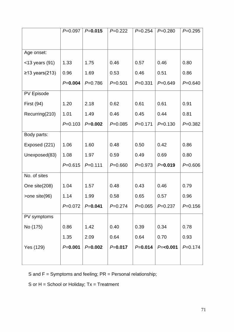
71
S and F = Symptoms and feeling; PR = Personal relationship;
S or H = School or Holiday; Tx = Treatment
P=0.097 P=0.015 P=0.222 P=0.254 P=0.280 P=0.295
Age onset:
<13 years (91)
≥13 years(213)
1.33
0.96
P=0.004
1.75
1.69
P=0.786
0.46
0.53
P=0.501
0.57
0.46
P=0.331
0.46
0.51
P=0.649
0.80
0.86
P=0.640
PV Episode
First (94)
Recurring(210)
1.20
1.01
P=0.103
2.18
1.49
P=0.002
0.62
0.46
P=0.085
0.61
0.45
P=0.171
0.61
0.44
P=0.130
0.91
0.81
P=0.382
Body parts:
Exposed (221)
Unexposed(83)
1.06
1.08
P=0.615
1.60
1.97
P=0.111
0.48
0.59
P=0.660
0.50
0.49
P=0.973
0.42
0.69
P=0.019
0.86
0.80
P=0.606
No. of sites
One site(208)
>one site(96)
1.04
1.14
P=0.072
1.57
1.99
P=0.041
0.48
0.58
P=0.274
0.43
0.65
P=0.065
0.46
0.57
P=0.237
0.79
0.96
P=0.156
PV symptoms
No (175)
Yes (129)
0.86
1.35
P=0.001
1.42
2.09
P=0.002
0.40
0.64
P=0.017
0.39
0.64
P=0.014
0.34
0.70
P=<0.001
0.78
0.93
P=0.174

72
Questions were asked on how the students with PV perceived themselves when
compared with other students. These questions included if they felt ashamed having
PV, if they have low self-confidence and if having PV has prevented them from
making friends. Their response was analysed with the mean of the total CDLQI. The
results showed that slightly less than two-third of the students felt ashamed (64.0%)
and acknowledged having low self-confidence (60.4%) while about one-sixth of the
students (16.2%) agreed that PV did prevent them from making friends. The mean
total CDLQI scores were higher in these three categories; however, the difference
was only significant in those with low self-confidence and those who had difficulty
making friends (Table 4.11 below.).
Table 4.11. Relationship between the mean total CDLQI scores and
perception of the students
Variables Mean total CDLQI P-value
Feel ashamed having PV
Yes: n=71(64.0%)
No: n=40 (36.0%)
8.85 (7.5 - 10.18)
6.82 (4.89 - 8.76)
0.80
Has low self-confidence
Yes: n=67 (60.4%)
No: n=44 (39.6%)
9.16 (7.81 - 10.52)
6.52 (4.72 - 8.33)
0.019
PV prevents from making
friends
Yes: n=18 (16.2%)
No: n=93 (83.8)
14.78 (11.93 - 17.63)
6.83 (5.81 - 7.84)
<0.001

73
4.4. Culture and microscopy results
Figure 4.16. (a) Culture results of Malassezia species showing white glistening
smooth surface and grooved edges. The texture was soft
(b) Showing cream coloured glistening smooth surface colonies and
slight central elevation which is characteristic of M. sympodialis

74
(c) This is in keeping with M. restricta showing colonies with dull
surfaces, smooth edges and average diameter of 3mm. The
texture was hard and brittle.
(d) These colonies are characteristic of M. furfur with smooth surface,
convex elevations, average diameter of 5mm and soft texture

75
Figure 4.17. Microscopy of Malassezia culture showing the unipolar, enteroblastic
budding from a characteristic broad base of large, oval cells.
Separation leaves a prominent bud scar through which successive
daughter cells emerge. (x400 magnification)
(a)
(b)

76
4.5. Malassezia species identified
Only three species were identified in this study; namely M. furfur, M. sympodialis and
M. restricta. There were 175 samples collected from PV lesions which grew on the
modified Dixon agar. Out of this number 143 (81.7%) were identified as M. furfur, 9
(5.1%) were M. restricta while 4 (2.3 %) were M. sympodialis. Exactly 10.9% of the
cultured samples did not show an identifiable band on PCR despite several repeats
done on DNA extraction.
The species distribution according to body site is as shown in Table 4.12 below. The
PV lesions on the face had a 50% species identification rate while the back lesions
had a higher yield at 61.2%. These two were the only sites where the three identified
species were all isolated. The site with the highest positive species identification
from culture was the scalp (80%) followed by the chest (68.6%) but just two species,
M. furfur and M. restricta, could be isolated from these locations. Meanwhile, only
one species, M. furfur was isolated from the upper arms lesions. Further, analysis
did not find any significant correlation between the species identified and clinical
features of affected students.
Skin samples were also collected from normal (non-lesional) skin. The scales
collected were limited to the sebum-rich body regions. A total of 110 samples were
collected, out of which growth was established in 58 (52.7%) cultures. M. furfur was
identified in 45 (77.6%), M. restricta in 6 (10.4%) while no PCR band was seen in
7(12%). The species distribution according to body site is shown in Table 4.13 below.
M. restricta was isolated only from the head and neck region and similar with species
isolation from PV lesions, the face had about 50% species identification rate. This
was lower than the percentage yield obtained from other body regions.
Table 4.12. Distribution of Malassezia species isolated from PV lesions
according to body sites
Body site M. furfur M. sympodialis M. restricta No growth
face 79 1 3 82
back 25 2 3 19
chest 23 0 1 11
Upper arm 8 0 0 12
neck 6 1 0 5
scalp 2 0 2 1
Total 143 4 9 129
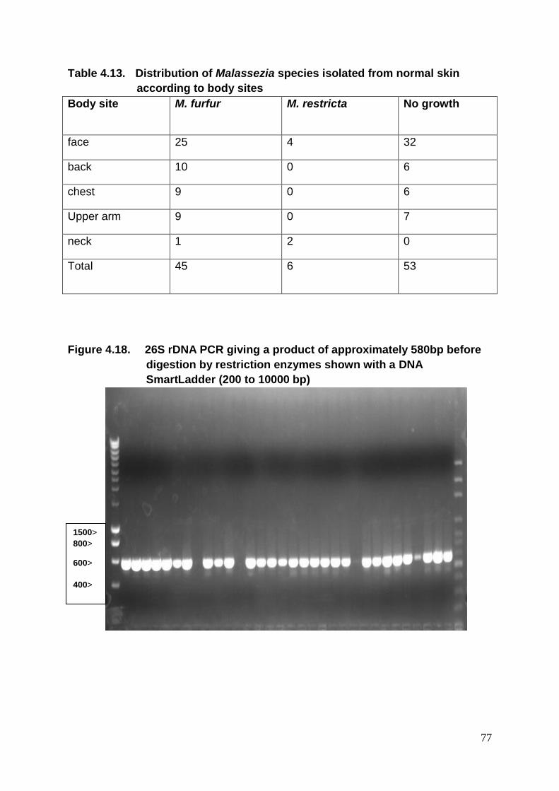
77
Table 4.13. Distribution of Malassezia species isolated from normal skin
according to body sites
Body site M. furfur M. restricta No growth
face 25 4 32
back 10 0 6
chest 9 0 6
Upper arm 9 0 7
neck 1 2 0
Total 45 6 53
Figure 4.18. 26S rDNA PCR giving a product of approximately 580bp before
digestion by restriction enzymes shown with a DNA
SmartLadder (200 to 10000 bp)
1500>
800> 600>
400>
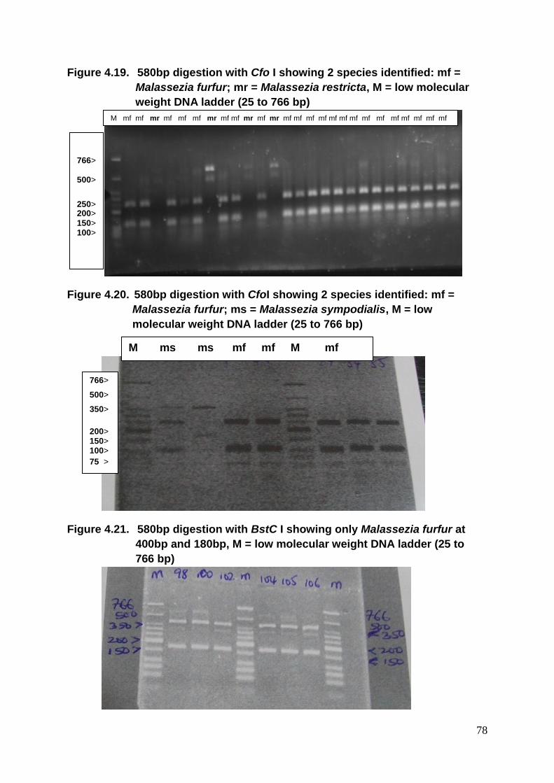
78
Figure 4.19. 580bp digestion with Cfo I showing 2 species identified: mf =
Malassezia furfur; mr = Malassezia restricta, M = low molecular
weight DNA ladder (25 to 766 bp)
Figure 4.20. 580bp digestion with CfoI showing 2 species identified: mf =
Malassezia furfur; ms = Malassezia sympodialis, M = low
molecular weight DNA ladder (25 to 766 bp)
Figure 4.21. 580bp digestion with BstC I showing only Malassezia furfur at
400bp and 180bp, M = low molecular weight DNA ladder (25 to
766 bp)
M mf mf mr mf mf mf mr mf mf mr mf mr mf mf mf mf mf mf mf mf mf mf mf mf mf mf
766> 500> 250> 200> 150> 100>
M ms ms mf mf M mf
mf mf
766>
500>
350>
200> 150> 100>
75 >

79
CHAPTER FIVE
Discussion
5.1. Clinical characteristics of the participants
The average age of students with PV observed in this study was 16 years and the
age group with the highest number of students was 14 up to 18 years. This is similar
to previous studies conducted in Brazil, Dar es Salaam and Bangkok, where the
highest prevalence of PV was observed in patients aged between 10 and 19 years,
16 and 19 years and 12 and 21 years respectively.(10, 11, 19, 56, 89) It also agrees
with past observation of PV as an infection of older teens/adolescents when
compared with dermatophyte infections.(4, 14, 57, 89) The reason can be attributed
to the increase in activity of sebaceous glands(15, 25) which is linked with increased
release of hormones especially androgens in this age group.(10, 149)
There was no significant difference between age and gender among the participants.
Also more females than males were observed with PV in this study (M:F 1:1.6).
Some studies have shown increased prevalence of PV in females;(11, 57, 89) while
other reports suggest that there is no sex predilection.(3, 4) A major factor that could
have contributed to the higher prevalence of PV in females in this study was the
eagerness of the females to be participants. The other contributing factors may be
the higher use by the female population of occlusive cosmetics namely talcum
powders as well as skin lightening creams, some of which may also contain
corticosteroid which is known to impair the integrity of the skin immune surveillance
against superficial infections. Daily use of cosmetics has been observed to increase
concentration of carbondioxide in the epidermis thus inducing a change in the
microflora, producing low skin pH and encourage growth of the Malassezia yeast.(89)
This study showed that personal grooming habits such as having a daily bath did not
prevent the development of PV in this environment. All of the students had at least
one daily bath, about two-third used tap water (running water) and more than half of
the students used soap to wash daily. This is always expected from school children
and is a rule usually enforced by their teachers and the Principal of the school.
Studies from India and the Central African Republic did not observe, similar to this
study, any direct association with regards to personal hygiene and the development
or spread of PV infection.(10, 46, 47) However, this is in contrast to a study in this
environment on prevalence of skin infections and infestations among prison in-mates
where a significant association was observed with poor hygiene.(53) Meanwhile, the
humid and hot conditions of the prisons compared to the community may have been
a confounding factor.
There was a higher prevalence of PV among those living in low income residential
areas of Abuja where overcrowding, resulting in poor ventilation and higher
environmental temperatures could be quite common. Majority of the parents were
traders which can be regarded as a low income occupation. Some studies have

80
documented a higher prevalence of PV in patients from low socioeconomic
background; however, similar with results from this study there was no statistically
significant difference observed by Belec et al. in Central African Republic(10) and
Jena et al. in India.(24) According to Belec et al, the clinical characteristics of PV did
not differ distinctly within country or by the type of urbanization in which the patient
lived.(10, 150)
This study suggested hyperhidrosis as an important factor in the development of PV.
Although most of the students after school activity were not sports related, some of
them were involved in other activities that could lead to excessive sweating such as
farming and street trading. Most importantly, sweating is common in our environment
as gadgets that cool the temperature such as air-conditioning and fans are not
readily available. Presently electricity is also erratic. The average atmospheric
temperature recording in the time course of the study was as high as 380 C which is
a strong predisposing environmental factor to PV development.(3, 17, 48, 53, 149)
None of the students had associated chronic systemic diseases or were in any
immunocompromised state.
A family history was associated with the development of PV in this study. About one
third of the students had at least one member of the nuclear family with a history of
PV. This was observed more among siblings than parents. A slightly higher result
was obtained by Rao et al. (38.3%) in India(47) while lower percentages of 22.2%
and 25%, respectively, were observed by Hafez et al.(21) and Ghosh et al.(46)
Genetic factor has been linked to increased sebum production in those infected with
PV.(1, 7, 149) This combines with environmental factors to predispose individuals to
PV. Meanwhile students with the presence of a family history of PV were significantly
younger than those without a family history (13.3 vs 13.9 years). This was expected
and also agrees with previous observations.(21, 45, 47) Unpredictably, the
relationship between recurring PV and having a positive family history was not
observed in this study unlike in above mentioned studies. It is worthy to note that
having a family history of PV was not influenced by socioeconomic status or gender.
5.2 Clinical features of PV
The average age of onset of PV in this study was 13.7 years. This is the mean age
when an increase in sebaceous activity is observed.(25) Although females with PV
developed the disorder at a slightly younger age than males, this difference was not
significant. Besides, study by Luebberding et al. observed a significant difference in
sebum production in both genders, with the production always higher and stable with
increasing age in the males and progressively decreases over lifetime in the
females.(151) Meanwhile females begin puberty at an earlier age than males.
The area of residence of participants from this study had an effect on the age of
onset of PV. The students who lived in the low income residential part of Abuja with
poor availability of amenities including housing and increased population were

81
observed to develop PV at a younger mean age of 13.3 years in contrast to those
living in the more expensive city centre region of Abuja. Belec et al.(10) compared
the characteristics of PV patients and non-PV patients with their place of residence
and found no significant difference between the two groups. In contrast, this study
compared place of residence within PV patients. The earlier age of onset in this
group of students could be related to poor living conditions and possible persistence
of poor ventilation and high environmental temperatures; however, further research
would be needed to confirm this finding. Meanwhile, studies looking at factors that
could affect the age of onset of PV need to be included in the literature.
There are four main classes of moisturizers based on their mechanism of action.
They include the occlusives, humectants, emollients and protein rejuvenators.
Occlusives provide exogenous barrier for the skin and marked reduction in
transepidermal water loss (TEWL). A well know prototype of occlusive moisturizers is
petrolatum. Petrolatum was shown to have an occlusive effect on the skin, stimulate
skin lipid biosynthesis and permeate all levels of the stratum corneum in a research
by Ghadially et al.(152) These processes were observed on normal skin and have
been confirmed by Patzelt(153) and Stamatas et al.(154) However, studies with
regard to the relationship of petrolatum and Malassezia infection are sparse.
Koyama et al. was able to demonstrate a florid growth of Malassezia furfur but not of
Malassezia globosa in a culture plate containing petrolatum.(155) Humectants
increase water content of the skin by enhancing water absorption from the dermis
into the epidermis. The absorbed water can easily evaporate into the environment,
thus producing increased TEWL. The most common humectant is glycerol or
glycerine which is a major component of most of the body creams used by the
participants. In this study, about 51% of the students mainly males applied
petrolatum namely Vaseline® on a daily basis. These students developed PV at a
younger age than those who did not use Vaseline® (13.3 vs 14.1 years). This
difference was found to be statistically significant P = 0.004. Meanwhile, a
statistically significant difference was not observed among those who applied body
creams routinely.
Dandruff also known as pityriasis capitis or mild seborrheic dermatitis is a non-
inflammatory scaling condition of the scalp(73) which commonly affects postpubertal
individuals and has been associated with Malassezia, Acremonium, Penicillium and
Cryptococcus species hence the good results obtained after treatment with
antifungals. Non microbial causes of dandruff include dust, heat, cosmetic use
etc.(73) Studies on the pathogenesis and management of this very common scalp
disorder are few and inconclusive. This study observed a very high percentage (22%)
of PV students with concomitant pityriasis capitis; higher than were observed by
Ghosh and Rao et al. who observed 10% and 11.6%, respectively.(46, 47)
Generally, not all individuals with Malassezia on their skin have dandruff(156) and
also the level of Malassezia on the normal scalp tends to be doubled that on dandruff
scalp.(73) In this study the students who had pityriasis capitis were found to have an

82
earlier age of onset of PV infection (12.6 vs 14.0 years; p=0.001). The reason for this
association is, however, unclear and may be related to non-studied endogenous
factors that could predispose these individuals at an earlier age to fungal infections.
Although the information on the age of onset of pityriasis capitis by this group of
students was not obtained, it would be interesting to know if there is any relationship
with the age of onset of PV. Meanwhile, there were no significant differences
observed between those who had pityriasis capitis and recurring PV; thus the fact
that an individual has dandruff does not predispose him/her to recurring episodes of
PV. Probably this could be because the etiological agent of the pityriasis capitis was
not necessarily the source of the PV. Further studies are needed to clarify this.
Animals have been observed to have Malassezia infections although the species
differ slightly from those that infect man,(66, 68) a study have shown that the
possible transfer of zoophilic Malassezia pachydermatis to human skin is quite
rare.(157) A small number of the students had regular contact with animals (20%)
and there was no relationship between having animal contact and PV infection from
this study.
On the use of bleaching products by the students, 14.5% of them acknowledged
their use. These were mainly female students. This is predictable as females are
known to be more beauty conscious, thus use more cosmetic products than males. It
is also known that females tend to be disturbed by blemishes on the skin. However,
the clinical features of PV in this group of students did not differ significantly from the
other students in the study. According to case-control study reports by some authors,
pityriasis versicolor is not as common as dermatophyte infections in population who
use bleaching products.(158, 159)
Pruritus and tingling sensation are major symptoms associated with PV. Slightly less
than half of the students in this study had pruritus/tingling sensation (42.8%). Similar
percentages were also reported by Ingordo et al. in Italian sailors, Rao et al. in India
and Morais et al. in Brazil.(17, 46, 160) It was also observed in this study that the
students who sweat excessively are more likely to have associated symptoms of
pruritus and tingling sensation. Although this association was not found in literature,
since research in this area is sparse, excessive perspiration could also be
associated with pruritus as seen in disorders of keratinization. It could also act as a
confounding factor in PV studies. Another reason for the pruritus could be due to the
effect of dehydration of the stratum corneum from the increased perspiration. This is
corroborated by reviews that have shown PV to be more pruritic when lesions are
exposed to high temperatures. Meanwhile a higher skin humidity provides a better
growth environment in the skin for Malassezia yeasts.(7) A study by Santana et al.
correlated pruritus in PV patients with skin type and observed that pruritus was more
frequent in patients with dehydrated epidermis.(89)
Recurrence of PV is commonly reported. About two-third of the students had
recurring episodes. This is similar to reports by Framil and Morais et al.(144, 160)

83
who noted recurrence rate of 67.4% and 52.6%, respectively. However, this study
did not find significant differences among these students when analysed with regard
to their use of petrolatum and presence of excessive sweating, as well as having a
positive family history of PV which had been previously linked to recurrence in PV. It
is possible that endogenous factors may predispose individuals to recurrent/relapse
episodes of PV and not only the presence of environmental factors such as
hyperhidrosis, application of body oils and occlusive dressing.(4) Rao et al. attributed
the reason for the high recurrence rate in PV infection to the presence of Malassezia
in individual’s hair follicles.(47) There is need for studies investigating the actual
factors that predispose individuals to PV recurrence. This is grossly not explored in
the literature.
The lesions seen in our study participants were hypopigmented macules which
usually start perifollicularly as seen in Figure 4.13. This corroborates Rao et al.
speculation on the presence of Malassezia in individual’s hair follicles. The lesions
then coalesce to form larger macules. Some of the lesions had a brawny scaly
appearance especially at areas which were not accessible to scrubbing during a bath.
Lesions on the face had minimal scaling. Hypopigmentation of PV lesions is a
common presentation in a darker-skinned population. Akin findings were reported in
studies conducted in Africa, Brazil, India and Bangkok.(10, 11, 46, 47, 60, 89, 160)
The reason still remains unclear. This study, comparable with other authors did not
observe any correlation between the colour of the lesions with personal hygiene,
clinical features and presence of a family history.
Most of the lesions were located on the face. This confirms literature reports
describing facial distribution of PV lesions as common mostly in children and black
people,(10, 24, 56, 89, 149, 161) in contrast to the rarity of facial involvement in the
white skinned population. In this study, frequent sites involved by PV were the face,
back and chest. Parallel findings were noted in studies by Ghosh and Rao et al.(46,
47) Extensive PV which can be described as PV involvement of 3 or more body
regions was observed in 8.2% of the students. This is comparable with the study by
Pönnighaus et al. in Malawi who observed 8.0%(15) and Terragni et al. in Italy who
observed 8.9%(149) but lower than figure obtained by Jena et al. in India who
observed extensive PV in 45 (16.6%) children.(24) In this study, students with
extensive PV were significantly associated with PV symptoms of pruritus and tingling
sensation. No known similar study has compared PV symptoms with extent of
distribution of lesions. Macular pattern of PV lesions as well as follicular, confluent
and guttate patterns was observed in this study as were observed by other
authors.(46, 47) Inverse distribution of PV was not detected.
Medications used by the students were mostly over-the-counter antifungal products.
Most of these products are sold in combination with highly potent steroids.
Meanwhile 10.5% of the students used folk remedies to treat PV, particularly
hydraulic (brake) fluid which has been observed by some studies in Nigeria and
Botswana as a common means utilized by less educated individuals to treat most

84
skin diseases at home.(52, 162) The students acknowledged clearance of the
lesions on application of hydraulic fluid, though, there was usually a recurrence. This
clearance could be due to propylene glycol a common base stock for hydraulic fluids.
Propylene glycol is one of the several non-specific topical antifungal agents available
for PV treatment. However, due to the other chemical compounds also found in
hydraulic fluids, these students need to be informed that dermal exposure to these
fluids could lead to signs of skin irritation.
5.3 Health related quality of life (HRQoL) of students with PV
The total quality of life score observed in this study was 7.39 (95%CI 6.73-8.06)
which is approximately 25% impairment in HRQoL. This figure is close to the total
CDLQI score (7.17±3.03) observed in a Nigerian community cross-sectional study of
children with tinea infection.(163) Comparing this effect with the comparative study
done by Beattie et al.(164) the level of impairment (25%) caused by PV was higher
than urticaria (20%) and acne (18%) but lower than psoriasis (30.6%) and atopic
dermatitis (30.5%).
According to the health behavior in school aged children (HBSC) 2009/2010 report,
age, gender, socioeconomic, and environmental differences are significant social
determinants of health and well-being among young people.(165) In this study, there
was a significant increase in HRQoL impairment among the students with PV who
were older than 13 years and those who had PV among member(s) of their nuclear
family. This was similarly observed in the study by Akinboro et al. where the CDLQI
score was significantly higher in children with tinea capitis older than 13 years.(163)
The increase in impairment observed in the students with PV who also had a family
member with PV could be due to the additional stress they felt having someone they
care about with a skin disorder. In this study, the difference in gender, social
environment and level of education did not have a significant effect on their quality of
life.
A large percentage of students had recurring PV. They had a lower HRQoL
impairment than those having PV for the first time. This was unpredictable since
chronic or recurring skin diseases are expected to produce a negative impact on
individual’s psychosocial well-being as seen in the study on hand dermatitis and
psoriasis.(166, 167) Perhaps, since the comparison is between individuals with
recurring disease and first timers, the anxiety and sensitivity to peoples’ opinion felt
by the recurring disease individuals had waned with time. More studies are needed
to further evaluate this aspect because the recurring nature of PV has been
generally assumed to contribute to the psychosocial well-being of patients.(142)
It is interesting to observe that students with PV lesions on unexposed parts of the
body had lower quality of life than those who had on exposed body parts although
this difference was not statistically significant. This observation was similarly
documented in the paper of Ludwig et al.(168) in which no significant difference in

85
quality of life was found associated with the sites of skin lesions. Correspondingly,
quality of life did not differ significantly between students having hypopigmented PV
and hyperpigmented PV. This could be because the skin is regarded as a whole and
indivisible organ; hence no matter the region of the body affected or the colour of the
lesion, the presence of a skin disorder is a cause of embarrassment and anxiety for
the patient.(167, 169)
Although the percentage of students with PV who had reduced self-confidence and
were ashamed of the lesions was high, the significant impairment in quality of life
associated with low self-confidence and reduced ability to make friends is, however,
inconclusive as a comparative study with students without PV would be required.
The HRQoL impairment correlated positively with the number of body regions
afflicted with PV. The increase in number of involved sites could be related with the
disease severity which has been shown to correlate with patients’ quality of life.(163,
168, 169)
Students who had pruritus or tingling sensation associated with PV had significantly
higher CDLQI scores. Pruritus is an important factor affecting QoL,(170) thus it is
expected that it would impair quality of life and this should be taken into
consideration when treating patients with PV.
The leisure category contributed mostly to the quality of life impairment observed in
this study. Since the burden of skin disease encompasses psychosocial and financial
consequences on the patients, their families and the society,(171) it is obvious from
this study that PV was a contributor to the psychosocial burden of the students. The
treatment category was third in rank as factors that impair quality of life but it did not
make a statistically significant impact. Generally, skin diseases that itch, have
unacceptable appearance or not easily treatable tend to significantly impair social
relations, psychological status and day-to-day activities of patients.(167, 171)
Unfortunately, a lot of individuals underestimate the effect that PV has on the quality
of life of adolescents in the Tropics. This should be noted and addressed in its
management.
In summary, quality of life determinants in students with PV observed in this study
included age of the patient, number of episodes of PV infection, existence of a family
member with PV, number of body regions affected and the presence of associated
pruritus or tingling sensation. The gender, level of education and the type of
neighborhood where the students reside did not significantly contribute to their
quality of life. Overall, this study has demonstrated the negative impact which PV
has on the quality of life among adolescents and more studies are being called
especially from areas with high PV prevalence as this knowledge can lead to
improved patient management.
86
5.4 Identification of Malassezia species
In this study, three species were identified from PV lesions namely, Malassezia furfur,
Malassezia restricta and Malassezia sympodialis, of which 81.7% was composed of
M. furfur. A higher percentage of M. furfur (77.6%) was also observed on non-
lesional skin samples. Two species only were identified from non-lesional skin (M.
furfur and M. restricta). This observation is quite different from most studies on the
identification of Malassezia species from PV. These studies have indicated M.
globosa as the most prevalent causative species.(62, 70, 102, 105, 172-175) They
were studies from temperate and mediterranean climates of the world, namely,
Spain, Japan, Bosnia-Herzegovina, Israel, Tunisia and Italy. The few investigations
where M. furfur have been most prevalent were from tropical and subtropical
climates such as Thailand, Indonesia, Brazil and Venezuela.(11, 108, 176, 177)
Furthermore, the method of identification used by these studies from tropical
climates was based on the physiological and biochemical characteristics of the
cultured yeast while this study used the molecular analysis method. This difference
should not affect results obtained just as a study by Romano et al. did not observe
differences in the identified Malassezia species when comparing conventional
culture-based with the molecular based methods.(173)
Further studies from Brazil(106, 107) and Argentina(20) observed M. sympodialis as
the most prevalent species in PV and this was consistently followed by M. furfur. Yet,
Crespo-Erchiga et al.(66) agree with the more than 80 years old hypothesis which
postulates that a second species other than M. globosa (mostly observed in
temperate climates) could be predominant in warmer and more humid climates. This
study is subject to their call for larger and reliable studies from a tropical climate to
test this hypothesis. Not only is M. furfur the most prevalent species in PV lesions, it
was also the most prevalent on non-lesional skin. By this result, we can conclude
that M. furfur is the causative agent of PV in Nigerian students. It is possible to
observe so very little number of Malassezia species per study as was seen in a
report from Israel where M. globosa was mostly detected in the lesional and non-
lesional skin of patients with PV at a rate of 97.3% and 80.8%, respectively.(175)
Prevalent studies on identification of Malassezia species from sub-Saharan Africa
are sparse. The study in Sudan on the molecular epidemiology of Malassezia in
patients with PV was restricted to the identification of M. globosa and M.
restricta.(147) Saad et al. looked at the level of colonization of only these 2 species
using a molecular-based, culture-independent method while comparing diseased
with healthy individuals. Further studies are required to observe the possible
prevalence of all Malassezia species and how they compare with the above
mentioned species and thus determine the most prevalent causative species of PV
in sub-Saharan Africa.
In this study, the second most prominent species identified though at very low
percentage was M. restricta which is in contrast also with most studies where M.
87
sympodialis is, in order of frequency, the second most common species isolated
from culture.(102, 108, 176) Also different in this study is the failure to identify M.
sympodialis from healthy skin. M. sympodialis has been observed to predominate on
healthy skin of the trunk, both in non-lesional areas in PV patients and healthy
controls.(66, 72, 157) The reason could be related to the absence of M. globosa
which is known to be associated almost always with M. sympodialis.(20, 66)
A comparable report in this study with other authors on the identification of
Malassezia species is the higher culture and isolation rate of Malassezia obtained
from the scalp (80%) and chest (68.6%) in contrast with other regions of the body.
Also similar is the more common recovery of M. restricta from the upper regions of
the body (scalp, face and neck) than the lower body.(76, 178, 179) The correlation of
the different species isolated with the clinical characteristics of the participants as
well as the PV lesions did not reveal any significant difference. This is in line with all
reported studies on the identification of Malassezia species from patients with PV.
No distinct feature has been linked with a specific species.
In this study, the recovery rate of Malassezia species from cultures both from
lesional and non-lesional sites were 57.6% and 52.7%, respectively, which was most
comparable to the study by Lim et al.(100) but lower than the yield observed by
Tarazooie et al.(48) and Nakabayashi et al.(70) This was not predicted as the study
participants were within the age group when the highest positive culture rate was
expected. (76, 179) The difference could be related to the sampling technique used
and the growth conditions of the culture. Similar to the study conducted by Tarazooie
et al.(48) only single species per culture were identified in this study unlike many
other studies where more than one species could be recovered from a sample.
Some of the cultures were covered up by faster growing species of other fungi. It
was difficult to maintain a pure culture and to discriminate a species from a mixed
sample, thus, some Malassezia species might have been lost after several
subcultures.
5.5 Conclusion
PV was more prevalent in older teenagers and females. The clinical presentation
was similar to those of other studies with the mean age at first presentation of
infection observed at 13.7 years. The age of onset of PV was significantly affected
by a positive history of PV in genetically related family members, socioeconomic
status, and the daily use of petrolatum and concomitant presence of pityriasis capitis
(dandruff). The recurrence of PV among the students and the colour of the lesions
were not linked with inadequate personal grooming methods, clinical features or
family history of PV. The facial location of the lesions was more prevalent, a location
similar with those of other studies of children and black population while the extent of
distribution of the lesion was associated significantly with presence of pruritus and
dysesthesia.

88
The quality of life determinants in students with PV observed in this study included
age of the patient, number of episodes of PV lesions, presence of a family member
with PV, number of body regions affected and the presence of associated pruritus or
dysesthesia. The gender, level of education and the type of neighborhood where the
students lived did not significantly contribute to their quality of life while a moderate
effect on the students’ HRQoL was observed.
Only three species were identified from this study with the main prevalent species
being Malassezia furfur which was observed in both lesional and non-lesional skin
samples. Finally, as observed in other studies, no correlation was found between the
species and clinical features of PV lesions.
5.6 Limitations of the study
1. This was a cross-sectional study. Most of the information used were as
related by the students.
2. Some students did not come forward because they did not want to be
recognized as having PV.
3. Poor culture yield of the samples; this could have affected the number in
species identified as the RFLP-PCR was done on culture results.
4. The skin scales yield even with the use of the Sellotape was poor in some
cases. Most were affected by sweat which caused poor Sellotape - skin
adhesion as well as absent yield on skin scrapping as samples were collected
in the heat of the afternoon. Most of the lesions especially the facial PV had
little or no scales. Also the use of sponge and daily bath of the students could
have resulted in the poor skin scales yield.
5. There were several contaminations from fast growing species of other fungi
which covered up the Malassezia species and their subsequent loss after
several subcultures.
89
CHAPTER SIX
Recommendations
1. RFLP-PCR can be used even in tropical climate to identify the different
species of Malassezia.
2. Since the culture based PCR yields a little above 50% of species identification,
and the direct skin scale based PCR requires a good amount of scales which
could be affected by sweat and personal hygiene practices, a better method
of sample collection needs to be developed to adequately study the presence
of PV Malassezia species in a tropical environment.
3. PV had a negative effect on the quality of life of these students; an effective
management guideline should be developed and if possible with focus on how
the factors affecting onset of PV can be controlled. This could reduce its
prevalence.
4. Most of the students took daily body bath. There is need for public awareness
to remove the general believe that PV was associated with poor personal
hygiene and thus reduce the self-stigmatization of patients.
5. There is prerequisite for more studies to look into other factors that could
affect the age of onset of PV. This may probably explain why the infection
affects certain individuals and spare others even when they share similar
environment.

90
References
1. Sunenshine PJ, Schwartz RA, Janniger CK. Tinea versicolor. International Journal of Dermatology. 1998;37(9):648-55.
2. Okoro AN. Tinea versicolor, not eczema. Nigerian Medical Journal. 1973;3(1):47-51.
3. Crespo Erchiga V, Hay R. Pityriasis versicolor and other Malassezia skin diseases In: Boekhout T, Gueho E, Mayser P, Velegraki A (Hrsg) Malassezia and the skin science and clinical practice. Springer, Berlin; 2010.
4. Gupta AK, Batra R, Bluhm R, Faergemann J. Pityriasis versicolor. Dermatologic Clinics. 2003;21(3):413-29.
5. Ryu HW, Cho JW, Lee KS. Pityriasis versicolor on penile shaft in a renal transplant recipient. Annals of Dermatology. 2012;24(3):345-7.
6. Leeming JP, Notman FH, Holland KT. The distribution and ecology of Malassezia furfur and cutaneous bacteria on human skin. The Journal of Applied Bacteriology. 1989;67(1):47-52.
7. Park HJ, Lee YW, Choe YB, Ahn KJ. Skin Characteristics in patients with pityriasis versicolor using non-invasive method, MPA5. Annals of Dermatology. 2012;24(4):444-52.
8. Peel MC, Finlayson BL, McMahon TA. Updated world map of the Köppen-Geiger climate classification. Journal of Hydrology Earth System Sciences. 2007;4(2):439-73.
9. Bechelli LM, Haddad N, Pimenta WP, Pagnano PM, Melchior E, Fregnan RC, et al. Epidemiological survey of skin diseases in schoolchildren living in the Purus Valley (Acre State, Amazonia, Brazil). Dermatologica. 1981;163(1):78-93.
10. Belec L, Testa J, Bouree P. Pityriasis versicolor in the Central African Republic: a randomized study of 144 cases. Journal of Medical and Veterinary Mycology. 1991;29(5):323-9.
11. Imwidthaya P, Thianprasit M, Srimuang S. A study of pityriasis versicolor in Bangkok (Thailand). Mycopathologia. 1989;105(3):157-61.
12. Khorchani H, Haouet H, Amri M, Zanned I, Babba H, Azaiz R. Epidemiological and clinical profile of superficial mycoses in the Monastir region (Tunisia). Retrospective studz (1991-1994) of 3578 cases. Archives de l'Institut Pasteur de Tunis. 1996;73(3-4):179-84.
13. Venugopal PV, Venugopal TV. Superficial mycoses in Saudi Arabia. The Australasian Journal of Dermatology. 1992;33(1):45-8.
14. Ellabib MS, Khalifa ZM. Dermatophytes and other fungi associated with skin mycoses in Tripoli, Libya. Annals of Saudi Medicine. 2001;21(3-4):193-5.
15. Ponnighaus JM, Fine PE, Saul J. The epidemiology of pityriasis versicolor in Malawi, Africa. Mycoses. 1996;39(11-12):467-70.
16. Martins EL, Goncalves CA, Mellone FF, Paves L, Tcherniakovsky M, Montes MN, et al. Medicina Cutanea Ibero-latino-Americana. 1989;17(5):287-91.
17. Ingordo V, Naldi L, Colecchia B, Licci N. Prevalence of pityriasis versicolor in young Italian sailors. The British Journal of Dermatology. 2003;149(6):1270-2.
18. Hellgren L, Vincent J. The incidence of tinea versicolor in central Sweden. Journal of Medical Microbiology. 1983;16(4):501-2.
19. Komba EV, Mgonda YM. The spectrum of dermatological disorders among primary school children in Dar es Salaam. BMC Public Health. 2010;10:765.
91
20. Giusiano G, Sosa ML, Rojas F, Vanacore ST, Mangiaterra M. Prevalence of Malassezia species in pityriasis versicolor lesions in northeast Argentina. Revista Iberoamericana de Micologia. 2010;27(2):71-4.
21. He SM, Du WD, Yang S, Zhou SM, Li W, Wang J, et al. The genetic epidemiology of tinea versicolor in China. Mycoses. 2008;51(1):55-62.
22. Wyre HW, Jr., Johnson WT. Neonatal pityriasis versicolor. Archives of Dermatology. 1981;117(11):752-3.
23. Gupta AK, Bluhm R, Summerbell R. Pityriasis versicolor. Journal of the European Academy of Dermatology and Venereology 2002;16(1):19-33.
24. Jena DK, Sengupta S, Dwari BC, Ram MK. Pityriasis versicolor in the pediatric age group. Indian Journal of Dermatology, Venereology and Leprology. 2005;71(4):259-61.
25. Zouboulis CC, Boschnakow A. Chronological ageing and photoageing of the human sebaceous gland. Clinical and Experimental Dermatology. 2001;26(7):600-7.
26. Di Silverio A, Mosca M, Brandozzi G, Gatti M. Pityriasis versicolor in the aged: a clinical investigation and epidemiological survey in 190 elderly hospitalized patients. Mycopathologia. 1989;105(3):187-90.
27. Lee WJ, Kim JY, Song CH, Jung HD, Lee SH, Lee SJ, et al. Disruption of barrier function in dermatophytosis and pityriasis versicolor. The Journal of Dermatology. 2011;38(11):1049-53.
28. Burke RC. Investigations in tinea versicolor: lipid and amino acid studies. The Yale Journal of Biology and Medicine. 1962;35:206-21.
29. Nazzaro-Porro M, Passi S, Picardo M, Mercantini R, Breathnach AS. Lipoxygenase activity of Pityrosporum in vitro and in vivo. The Journal of Investigative Dermatology. 1986;87(1):108-12.
30. De Luca C, Picardo M, Breathnach A, Passi S. Lipoperoxidase activity of Pityrosporum: characterisation of by-products and possible role in pityriasis versicolor. Experimental Dermatology. 1996;5(1):49-56.
31. Aly R, Berger T. Common superficial fungal infections in patients with AIDS. Clinical Infectious Diseases 1996;22(2):S128-32.
32. Lohoue Petmy J, Lando AJ, Kaptue L, Tchinda V, Folefack M. Superficial mycoses and HIV infection in Yaounde. Journal of the European Academy of Dermatology and Venereology 2004;18(3):301-4.
33. Gulec AT, Demirbilek M, Seckin D, Can F, Saray Y, Sarifakioglu E, et al. Superficial fungal infections in 102 renal transplant recipients: a case-control study. Journal of the American Academy of Dermatology. 2003;49(2):187-92.
34. Shuttleworth D, Philpot CM, Salaman JR. Cutaneous fungal infection following renal transplantation: a case control study. The British Journal of Dermatology. 1987;117(5):585-90.
35. Falodun O, Ogunbiyi A, Salako B, George AK. Skin changes in patients with chronic renal failure. Saudi Journal of Kidney Diseases and Transplantation. 2011;22(2):268-72.
36. Tajbakhsh R, Dehghan M, Azarhoosh R, Haghighi AN, Sadani S, Zadeh SS, et al. Mucocutaneous manifestations and nail changes in patients with end-stage renal disease on hemodialysis. Saudi Journal of Kidney Diseases and Transplantation. 2013;24(1):36-40.
37. Pavlović MD, Milenković T, Dinić M, Mišović M, Daković D, Todorović S, et al. The prevalence of cutaneous manifestations in young patients with type 1 diabetes. Diabetes Care. 2007;30(8):1964-7.
92
38. Garcia-Humbria L, Richard-Yegres N, Perez-Blanco M, Yegres F, Mendoza M, Acosta A, et al. (38). Investigacion Clinica. 2005;46(1):65-74.
39. Wahid Z, Nasreen S, Usman G, Ahmed I. Frequency of pityriasis versicolor in patients with uncontrolled type 2 diabetes attending a tertiary care hospital. Journal Liaquat University of Medical Health Sciences. 2013;12(01):03.
40. Kato A, Hamada M, Maruyama T, Maruyama Y, Hishida A. Pruritus and hydration state of stratum corneum in hemodialysis patients. American Journal of Nephrology. 2000;20(6):437-42.
41. Morton CA, Lafferty M, Hau C, Henderson I, Jones M, Lowe JG. Pruritus and skin hydration during dialysis. Nephrology, Dialysis, Transplantation. 1996;11(10):2031-6.
42. Tragiannidis A, Bisping G, Koehler G, Groll AH. Minireview: Malassezia infections in immunocompromised patients. Mycoses. 2010;53(3):187-95.
43. Saadatzadeh MR, Ashbee HR, Cunliffe WJ, Ingham E. Cell-mediated immunity to the mycelial phase of Malassezia spp. in patients with pityriasis versicolor and controls. The British Journal of Dermatology. 2001;144(1):77-84.
44. Ashbee HR, Fruin A, Holland KT, Cunliffe WJ, Ingham E. Humoral immunity to Malassezia furfur serovars A, B and C in patients with pityriasis versicolor, seborrheic dermatitis and controls. Experimental Dermatology. 1994;3(5):227-33.
45. Hafez M, el-Shamy S. Genetic susceptibility in pityriasis versicolor. Dermatologica. 1985;171(2):86-8.
46. Ghosh SK, Dey SK, Saha I, Barbhuiya JN, Ghosh A, Roy AK. Pityriasis versicolor: a clinicomycological and epidemiological study from a tertiary care hospital. Indian Journal of Dermatology. 2008;53(4):182-5.
47. Rao GS, Kuruvilla M, Kumar P, Vinod V. Clinico-epidermiological studies on tinea versicolor. Indian Journal of Dermatology, Venereology and Leprology. 2002;68(4):208-9.
48. Tarazooie B, Kordbacheh P, Zaini F, Zomorodian K, Saadat F, Zeraati H, et al. Study of the distribution of Malassezia species in patients with pityriasis versicolor and healthy individuals in Tehran, Iran. BMC Dermatology. 2004;4:5.
49. Burke RC. Tinea versicolor: susceptibility factors and experimental infection in human beings. The Journal of Investigative Dermatology. 1961;36:389-402.
50. Zampino MR, Osti F, Corazza M, Virgili A. Prevalence of pityriasis versicolor in a group of Italian pregnant women. Journal of the European Academy of Dermatology and Venereology. 2007;21(9):1249-52.
51. Boardman CR, Malkinson FD. Tinea versicolor in steroid-treated patients. Incidence in patients with chronic ulcerative colitis and regional enteritis treated with corticotropin and corticosteroids. Archives of Dermatology. 1962;85:44-52.
52. Ifebuzor DC, Mabuza LH, Malete NH, Govender I. The perceptions of parents about the skin conditions of their children presenting with comorbid fungal skin infections in Francistown, Botswana. African Journal of Primary Health Care and Family Medicine. 2013;5(1):Art.#459,6pages
53. Oninla OA, Onayemi O. Skin infections and infestations in prison inmates. International Journal of Dermatology. 2012;51(2):178-81.

93
54. Salahi-Moghaddam A, Davoodian P, Jafari A, Nikoo MA. Evaluation of pityriasis versicolor in prisoners: a cross-sectional study. Indian journal of Dermatology, Venereology and Leprology. 2009;75(4):379-82.
55. Onayemi O, Isezuo SA, Njoku CH. Prevalence of different skin conditions in
an outpatients’ setting in north‐western Nigeria. International Journal of Dermatology. 2005;44(1):7-11.
56. Akpata LE, Gugnani HC, Utsalo SJ. Pityriasis versicolor in school children in Cross River State of Nigeria. Mycoses. 1990;33(11-12):549-51.
57. Uneke C, Ngwu B, Egemba O. Tinea capitis and pityriasis versicolor infections among school children in the South-Eastern Nigeria: The public health implications. The Internet Journal of Dermatology. 2006;4(2).
58. Ogunbiyi AO, Owoaje E, Ndahi A. Prevalence of skin disorders in school children in Ibadan, Nigeria. Pediatric Dermatology. 2005;22(1):6-10.
59. Ogunbiyi AO, Omigbodun Y, Owoaje E. Prevalence of skin disorders in school children in southwest Nigeria. International Journal of Adolescent Medicine and Health. 2009;21(2):235-41.
60. Faye O, N'Diaye HT, Keita S, Traore AK, Hay RJ, Mahe A. High prevalence of non-leprotic hypochromic patches among children in a rural area of Mali, West Africa. Leprosy Review. 2005;76(2):144-6.
61. Gaitanis G, Magiatis P, Hantschke M, Bassukas ID, Velegraki A. The Malassezia genus in skin and systemic diseases. Clinical Microbiology Reviews. 2012;25(1):106-41.
62. Salah SB, Makni F, Marrakchi S, Sellami H, Cheikhrouhou F, Bouassida S, et al. Identification of Malassezia species from Tunisian patients with pityriasis versicolor and normal subjects. Mycoses. 2005;48(4):242-5.
63. Guého E, Midgley G, Guillot J. The genus Malassezia with description of four new species. Antonie van Leeuwenhoek. 1996;69(4):337-55.
64. Ashbee HR, Evans EG. Immunology of diseases associated with Malassezia species. Clinical Microbiology Reviews. 2002;15(1):21-57.
65. Marcon MJ, Powell DA. Human infections due to Malassezia spp. Clinical Microbiology Reviews. 1992;5(2):101-19.
66. Crespo-Erchiga V, Gomez-Moyano E, Crespo M. Pityriasis versicolor and the yeasts of genus Malassezia. Actas Dermo-sifiliograficas. 2008;99(10):764-71.
67. Guillot J, Hadina S, Gueho E. The genus Malassezia: old facts and new concepts. Parassitologia. 2008;50(1-2):77-9.
68. Cafarchia C, Gasser RB, Figueredo LA, Latrofa MS, Otranto D. Advances in the identification of Malassezia. Molecular and cellular probes. 2011;25(1):1-7.
69. Affes M, Salah SB, Makni F, Sellami H, Ayadi A. Molecular identification of Malassezia species isolated from dermatitis affections. Mycoses. 2009;52(3):251-6.
70. Nakabayashi A, Sei Y, Guillot J. Identification of Malassezia species isolated from patients with seborrhoeic dermatitis, atopic dermatitis, pityriasis versicolor and normal subjects. Medical Mycology. 2000 Oct;38(5):337-41.
71. Ko JH, Lee YW, Choe YB, Ahn KJ. Epidemiologic study of Malassezia yeasts in patients with Malassezia folliculitis by 26S rDNA PCR-RFLP analysis. Annals of Dermatology. 2011;23(2):177-84.
72. Gupta AK, Batra R, Bluhm R, Boekhout T, Dawson TL, Jr. Skin diseases associated with Malassezia species. Journal of the American Academy of Dermatology. 2004;51(5):785-98.

94
73. Park HK, Ha MH, Park SG, Kim MN, Kim BJ, Kim W. Characterization of the fungal microbiota (mycobiome) in healthy and dandruff-afflicted human scalps. Public Library of Science one. 2012;7(2):e32847.
74. Faergemann J. Atopic dermatitis and fungi. Clinical Microbiology Reviews. 2002;15(4):545-63.
75. Zhang E, Tanaka T, Tajima M, Tsuboi R, Kato H, Nishikawa A, et al. Anti-Malassezia-specific IgE antibodies production in Japanese patients with head and neck atopic dermatitis: relationship between the level of specific IgE antibody and the colonization frequency of cutaneous Malassezia species and clinical severity. Journal of Allergy. 2011;2011:645670.
76. Sugita T, Suto H, Unno T, Tsuboi R, Ogawa H, Shinoda T, et al. Molecular analysis of Malassezia microflora on the skin of atopic dermatitis patients and healthy subjects. Journal of Clinical Microbiology. 2001;39(10):3486-90.
77. Gomez-Moyano E, Crespo-Erchiga V, Martinez-Pilar L, Godoy Diaz D, Martinez-Garcia S, Lova Navarro M, et al. Do Malassezia species play a role in exacerbation of scalp psoriasis? Journal de Mycologie Medicale. 2014 Jan 8.
78. Rudramurthy SM, Honnavar P, Chakrabarti A, Dogra S, Singh P, Handa S. Association of Malassezia species with psoriatic lesions. Mycoses. 2014 Mar 21.
79. Zhao Y, Li L, Wang JJ, Kang KF, Zhang QQ. Cutaneous malasseziasis: four case reports of atypical dermatitis and onychomycosis caused by Malassezia. International journal of dermatology. 2010;49(2):141-5.
80. Ertam I, Aytimur D, Alper S. Malassezia furfur onychomycosis in an immunosuppressed liver transplant recipient. Indian Journal of Dermatology, Venereology and Leprology. 2007;73(6):425-6.
81. Fredricks DN. Microbial ecology of human skin in health and disease. The Journal of Investigative Dermatology. 2001 Dec;6(3):167-9.
82. Rosenthal M, Goldberg D, Aiello A, Larson E, Foxman B. Skin microbiota: microbial community structure and its potential association with health and disease. Infection, Genetics and Evolution Journal. 2011 Jul;11(5):839-48.
83. Turner GA, Hoptroff M, Harding CR. Stratum corneum dysfunction in dandruff. International Journal of Cosmetic Science. 2012;34(4):298-306.
84. Kramer HJ, Kessler D, Hipler UC, Irlinger B, Hort W, Bodeker RH, et al. Pityriarubins, novel highly selective inhibitors of respiratory burst from cultures of the yeast Malassezia furfur: comparison with the bisindolylmaleimide arcyriarubin A. Chembiochem: a European Journal of Chemical Biology. 2005;6(12):2290-7.
85. Takahashi M, Ushijima T, Ozaki Y. Biological activity of Pityrosporum. II. Antitumor and immune stimulating effect of Pityrosporum in mice. Journal of the National Cancer Institute. 1986;77(5):1093-7.
86. Walters CE, Ingham E, Eady EA, Cove JH, Kearney JN, Cunliffe WJ. In vitro modulation of keratinocyte-derived interleukin-1 alpha (IL-1 alpha) and peripheral blood mononuclear cell-derived IL-1 beta release in response to cutaneous commensal microorganisms. Infection and Immunity. 1995;63(4):1223-8.
87. Cunningham AC, Ingham E, Gowland G. Humoral responses to Malassezia furfur serovars A, B and C in normal individuals of various ages. The British Journal of Dermatology. 1992;127(5):476-81.

95
88. Marks R. The stratum corneum barrier: the final frontier. The Journal of Nutrition. 2004;134(8):2017S-21S.
89. Santana JO, de Azevedo FL, Filho PC. Pityriasis versicolor: clinical-epidemiological characterization of patients in the urban area of Buerarema-BA , Brazil. Anais Brasileiros de Dermatologia. 2013;88(2):216-21.
90. Harding CR. The stratum corneum: structure and function in health and disease. Dermatologic Therapy. 2004;17(1):6-15.
91. Pierard J, Dockx P. The ultrastructure of tinea versicolor and Malassezia furfur. International Journal of Dermatology. 1972;11(2):116-24.
92. Borgers M, Cauwenbergh G, Van de Ven MA, del Palacio Hernanz A, Degreef H. Pityriasis versicolor and Pityrosporum ovale. Morphogenetic and ultrastructural considerations. International Journal of Dermatology. 1987;26(9):586-9.
93. del Palacio-Hernanz A, Guarro-Artigas J, Figueras-Salvat MJ, Esteban-Moreno J, Lopez-Gomez S. Changes in fungal ultrastructure after short-course ciclopiroxolamine therapy in pityriasis versicolor. Clinical and Experimental Dermatology. 1990;15(2):95-100.
94. Karaoui R, Bou-Resli M, Al-Zaid NS, Mousa A. Tinea versicolor: ultrastructural studies on hypopigmented and hyperpigmented skin. Dermatologica. 1981;162(2):69-85.
95. Galadari I, el Komy M, Mousa A, Hashimoto K, Mehregan AH. Tinea versicolor: histologic and ultrastructural investigation of pigmentary changes. International Journal of Dermatology. 1992;31(4):253-6.
96. Dotz WI, Henrikson DM, Yu GS, Galey CI. Tinea versicolor: a light and electron microscopic study of hyperpigmented skin. Journal of the American Academy of Dermatology. 1985;12(1 Pt 1):37-44.
97. Kramer HJ, Podobinska M, Bartsch A, Battmann A, Thoma W, Bernd A, et al. Malassezin, a novel agonist of the aryl hydrocarbon receptor from the yeast Malassezia furfur, induces apoptosis in primary human melanocytes. Chembiochem: a European Journal of Chemical Biology. 2005;6(5):860-5.
98. Lee KH, Kim YG, Bang D, Kim YA. Scanning electron microscopy of Malassezia furfur in tinea versicolor. Yonsei Medical Journal. 1989;30(4):334-8.
99. Janaki C, Sentamilselvi G, Janaki VR, Boopalraj JM. Unusual observations in the histology of Pityriasis versicolor. Mycopathologia. 1997;139(2):71-4.
100. Lim SH, Kim YR, Jung JW, Hahn HJ, Lee YW, Choe YB, et al. A comparison study between culture based technique and Op-site non-culture based technique for identifying Malassezia yeasts on normal skin. Korean Journal of Medical Mycology. 2012;17(4):217-29.
101. Machado ML, Ferreiro L, Ferreira RR, Corbellini LG, Deville M, Berthelemy M, et al. Malassezia dermatitis in dogs in Brazil: diagnosis, evaluation of clinical signs and molecular identification. Veterinary Dermatology. 2011;22(1):46-52.
102. Crespo Erchiga V, Ojeda Martos AA, Vera Casano A, Crespo Erchiga A, Sanchez Fajardo F. Isolation and identification of Malassezia spp. in pytiriasis versicolor, seborrheic dermatitis and healthy skin. Revista Iberoamericana de Micologia. 1999;16(S):S16-21.
103. Aspiroz C, Ara M, Varea M, Rezusta A, Rubio C. Isolation of Malassezia globosa and M. sympodialis from patients with pityriasis versicolor in Spain. Mycopathologia. 2002;154(3):111-7.
96
104. Dutta S, Bajaj AK, Basu S, Dikshit A. Pityriasis versicolor: socioeconomic and clinico-mycologic study in India. International Journal of Dermatology. 2002;41(11):823-4.
105. Prohic A, Ozegovic L. Malassezia species isolated from lesional and non-lesional skin in patients with pityriasis versicolor. Mycoses. 2007 Jan;50(1):58-63.
106. Framil VM, Melhem MS, Szeszs MW, Corneta EC, Zaitz C. Anais Brasileiros de Dermatologia. 2010;85(1):111-4.
107. Petry V, Tanhausen F, Weiss L, Milan T, Mezzari A, Weber MB. Identification of Malassezia yeast species isolated from patients with pityriasis versicolor. Anais Brasileiros de Dermatologia. 2011;86(4):803-6.
108. Krisanty RI, Bramono K, Made Wisnu I. Identification of Malassezia species from pityriasis versicolor in Indonesia and its relationship with clinical characteristics. Mycoses. 2009;52(3):257-62.
109. Tellechea O, Cravo M, Brinca A, Robalo-Cordeiro M. Pityriasis versicolor atrophicans. European Journal of Dermatology. 2012;22(2):287-8.
110. Tan C, Zhu WY, Min ZS. Blaschkoid pityriasis versicolor. Mycoses. 2010;53(4):366-8.
111. Maeda M, Makimura KC, Yamaguchi H. Pityriasis versicolor rubra. European Journal of Dermatology. 2002;12(2):160-4.
112. Hasson I, Shah P. Pityriasis rotunda. Indian Journal of Dermatology, Venereology and Leprology. 2003;69(1):50-1.
113. Savin R. Diagnosis and treatment of tinea versicolor. The Journal of Family Practice. 1996;43(2):127-32.
114. Spence-Shishido A, Carr C, Bonner MY, Arbiser JL. In vivo Gram staining of tinea versicolor. JAMA Dermatology. 2013;149(8):991-2.
115. Kindo AJ, Sophia SK, Kalyani J, Anandan S. Identification of Malassezia species. Indian Journal of Medical Microbiology. 2004;22(3):179-81.
116. Guillot J, Breugnot C, de Barros M, Chermette R. Usefulness of modified Dixon's medium for quantitative culture of Malassezia species from canine skin. Journal of Veterinary Diagnostic Investigation. 1998;10(4):384-6.
117. Kaneko T, Makimura K, Onozaki M, Ueda K, Yamada Y, Nishiyama Y, et al. Vital growth factors of Malassezia species on modified CHROMagar Candida. Medical Mycology. 2005 Dec;43(8):699-04.
118. Neves RP, Magalhães OMC, Silva MLd, Souza-Motta CMd, Queiroz LAd. Identification and pathogenicity of Malassezia species isolated from human healthy skin and with macules. Brazilian Journal of Microbiology. 2005;36(2):114-7.
119. Gaitanis G, Velegraki A, Frangoulis E, Mitroussia A, Tsigonia A, Tzimogianni A, et al. Identification of Malassezia species from patient skin scales by PCR-RFLP. Clinical Microbiology and Infection. 2002;8(3):162-73.
120. Sugita T, Boekhout T, Velegraki A, Guillot J, Hadina S. Epidemiology of Malassezia-Related skin diseases In: Boekhout T, Gueho E, Mayser P, Velegraki A (Hrsg) Malassezia and the skin science and clinical practice. Springer, Berlin; 2010.
121. Vuran E, Karaarslan A, Karasartova D, Turegun B, Sahin F. Identification of Malassezia species from pityriasis versicolor lesions with a new multiplex PCR method. Mycopathologia. 2014;177(1-2):41-9

97
122. Loenen WA, Dryden DT, Raleigh EA, Wilson GG, Murray NE. Highlights of the DNA cutters: a short history of the restriction enzymes. Nucleic Acids Research. 2014;42(1):3-19
123. Mirhendi H, Makimura K, Zomorodian K, Yamada T, Sugita T, Yamaguchi H. A simple PCR-RFLP method for identification and differentiation of 11 Malassezia species. Journal of Microbiological Methods. 2005;61(2):281-4.
124. Gupta AK, Kogan N, Batra R. Pityriasis versicolor: a review of pharmacological treatment options. Expert Opinion on Pharmacotherapy. 2005;6(2):165-78.
125. Jowkar F, Jamshidzadeh A, Pakniyat S, Namazi MR. Efficacy of nitric oxide-liberating cream on pityriasis versicolor. The Journal of Dermatological Treatment. 2010;21(2):93-6.
126. Mayser P, Rieche I. Rapid reversal of hyperpigmentation in pityriasis versicolor upon short-term topical cycloserine application. Mycoses. 2009;52(6):541-3.
127. Kim YJ, Kim YC. Successful treatment of pityriasis versicolor with 5-aminolevulinic acid photodynamic therapy. Archives of Dermatology. 2007;143(9):1218-20.
128. Shi TW, Ren XK, Yu HX, Tang YB. Roles of adapalene in the treatment of pityriasis versicolor. Dermatology. 2012;224(2):184-8.
129. Borgers M. Mechanism of action of antifungal drugs, with special reference to the imidazole derivatives. Reviews of Infectious Diseases. 1980;2(4):520-34.
130. Jacobs PH. Ketoconazole use in tinea versicolor. The Western Journal of Medicine. 1987;147(4):457.
131. Bhogal CS, Singal A, Baruah MC. Comparative efficacy of ketoconazole and fluconazole in the treatment of pityriasis versicolor: a one year follow-up study. The Journal of Dermatology. 2001;28(10):535-9.
132. Wahab MA, Ali ME, Rahman MH, Chowdhury SA, Monamie NS, Sultana N, et al. Single dose (400 mg) versus 7 day (200 mg) daily dose itraconazole in the treatment of tinea versicolor: a randomized clinical trial. Mymensingh Medical Journal. 2010;19(1):72-6.
133. Cauwenbergh G. Mycoses. 1994;37(2):27-33. 134. Amer MA. Fluconazole in the treatment of tinea versicolor. Egyptian
Fluconazole Study Group. International Journal of Dermatology. 1997;36(12):940-2.
135. Faergemann J, Todd G, Pather S, Vawda ZF, Gillies JD, Walford T, et al. A double-blind, randomized, placebo-controlled, dose-finding study of oral pramiconazole in the treatment of pityriasis versicolor. Journal of the American Academy of Dermatology. 2009;61(6):971-6.
136. Rukayadi Y, Hwang JK. In vitro anti-Malassezia activity of xanthorrhizol isolated from Curcuma xanthorrhiza Roxb. Letters in Applied Microbiology. 2007;44(2):126-30.
137. Hammer KA, Carson CF, Riley TV. In vitro activities of ketoconazole, econazole, miconazole, and Melaleuca alternifolia (tea tree) oil against Malassezia species. Antimicrobial Agents and Chemotherapy. 2000;44(2):467-9.

98
138. Oyelami OA, Onayemi O, Oladimeji FA, Ogundaini AO, Olugbade TA, Onawunmi GO. Clinical evaluation of Acalypha ointment in the treatment of superficial fungal skin diseases. Phytotherapy Research. 2003;17(5):555-7.
139. Damodaran S, Venkataraman S. A study on the therapeutic efficacy of Cassia alata, Linn. leaf extract against pityriasis versicolor. Journal of Ethnopharmacology. 1994;42(1):19-23.
140. Filip R, Davicino R, Anesini C. Antifungal activity of the aqueous extract of Ilex paraguariensis against Malassezia furfur. Phytotherapy Research. 2010;24(5):715-9.
141. Brodin K, Alahyar H, Hedner T, Sterner O, Faergemann J. In vitro activity of artemisia abrotanum extracts against Malassezia Spp., Candida albicans and Staphylococcus aureus. Acta Dermato-venereologica. 2007;87(6):540-2.
142. Pantazidou A, Tebruegge M. Recurrent tinea versicolor: treatment with itraconazole or fluconazole? Archives of Disease in Childhood. 2007;92(11):1040-2.
143. Alteras I, Sandbank M, Segal R. Two years of follow-up of oral ketoconazole therapy in 60 cases of pityriasis versicolor. Dermatologica. 1987;175(3):142-4.
144. Framil VM, Melhem MS, Szeszs MW, Zaitz C. New aspects in the clinical course of pityriasis versicolor. Anais Brasileiros de Dermatologia. 2011;86(6):1135-40.
145. Rausch LJ, Jacobs PH. Tinea versicolor: treatment and prophylaxis with monthly administration of ketoconazole. Cutis. 1984;34(5):470-1.
146. Faergemann J, Gupta AK, Al Mofadi A, Abanami A, Shareaah AA, Marynissen G. Efficacy of itraconazole in the prophylactic treatment of pityriasis (tinea) versicolor. Archives of Dermatology. 2002;138(1):69-73.
147. Saad M, Sugita T, Saeed H, Ahmed A. Molecular epidemiology of Malassezia globosa and Malassezia restricta in Sudanese patients with pityriasis versicolor. Mycopathologia. 2013;175(1-2):69-74.
148. Oh BH, Song YC, Lee YW, Choe YB, Ahn KJ. Comparison of nested PCR and RFLP for identification and classification of Malassezia yeasts from healthy human skin. Annals of Dermatology. 2009;21(4):352-7.
149. Terragni L, Lasagni A, Oriani A, Gelmetti C. Pityriasis versicolor in the pediatric age. Pediatric Dermatology. 1991;8(1):9-12.
150. Berry M, Khachemoune A. Extensive tinea versicolor mimicking Pityriasis rubra pilaris. Journal of Drugs in Dermatology. 2009;8(5):490-1.
151. Luebberding S, Krueger N, Kerscher M. Skin physiology in men and women: in vivo evaluation of 300 people including TEWL, SC hydration, sebum content and skin surface pH. International Journal of Cosmetic Science. 2013;35(5):477-83.
152. Ghadially R, Halkier-Sorensen L, Elias PM. Effects of petrolatum on stratum corneum structure and function. Journal of the American Academy of Dermatology. 1992;26(3):387-96.
153. Patzelt A, Lademann J, Richter H, Darvin ME, Schanzer S, Thiede G, et al. In vivo investigations on the penetration of various oils and their influence on the skin barrier. Skin Research and Technology. 2012;18(3):364-9.
99
154. Stamatas GN, de Sterke J, Hauser M, von Stetten O, van der Pol A. Lipid uptake and skin occlusion following topical application of oils on adult and infant skin. Journal of Dermatological Science. 2008;50(2):135-42.
155. Koyama T, Kanbe T, Kikuchi A, Tomita Y. Effects of topical vehicles on growth of the lipophilic Malassezia species. Journal of Dermatological Science. 2002;29(3):166-70.
156. Pierard-Franchimont C, Xhauflaire-Uhoda E, Pierard GE. Revisiting dandruff. International Journal of Cosmetic Science. 2006;28(5):311-8.
157. Prohic A, Kasumagic-Halilovic E. Identification of Malassezia pachydermatis from healthy and diseased human skin. Medicinski arhiv. 2009;63(6):317-9.
158. Mahe A, Ly F, Aymard G, Dangou JM. Skin diseases associated with the cosmetic use of bleaching products in women from Dakar, Senegal. The British Journal of Dermatology. 2003;148(3):493-500.
159. Adebajo SB. An epidemiological survey of the use of cosmetic skin lightening cosmetics among traders in Lagos, Nigeria. West African Journal of Medicine. 2002;21(1):51-5.
160. Morais PM, Cunha Mda G, Frota MZ. Clinical aspects of patients with pityriasis versicolor seen at a referral center for tropical dermatology in Manaus, Amazonas, Brazil. Anais Brasileiros de Dermatologia. 2010;85(6):797-03.
161. Kallini JR, Riaz F, Khachemoune A. Tinea versicolor in dark-skinned individuals. International Journal of Dermatology. 2014;53(2):137-41.
162. Arikpo G, Eja M, Enene E, Okon S, Enyi-Idoh K, Etim S. Petroleum distilates use in folk medicine in South Eastern Nigeria. The Internet Journal of Health. 2010;11(1).
163. Akinboro AO, Olasode OA, Onayemi O, Mejiuni AD. The impacts of tinea capitis on quality of life: a community based cross sectional study among Nigerian children. Clinical Medicine Insights: Dermatology. 2013;6:9-17
164. Beattie PE, Lewis-Jones MS. A comparative study of impairment of quality of life in children with skin disease and children with other chronic childhood diseases. The British Journal of Dermatology. 2006;155(1):145-51.
165. Currie C. et al. Social determinants of health and well-being among young people. Health Behaviour in School-aged Children (HBSC) study: international report from the 2009/2010 survey. Copenhagen, WHO Regional Office for Europe, 2012 (Health policy for Children and Adolescents, No 6).
166. Charan UP, Peter CV, Pulimood SA. Impact of hand eczema severity on quality of life. Indian Dermatology Online Journal. 2013;4(2):102-5.
167. Ibler KS, Jemec GB. Cumulative life course impairment in other chronic or recurrent dermatologic diseases. Current Problems in Dermatology. 2013;44:130-6.
168. Ludwig MW, Oliveira Mda S, Muller MC, Moraes JF. Quality of life and site of the lesion in dermatological patients. Anais Brasileiros de Dermatologia. 2009;84(2):143-50.
169. Ahmed A, Leon A, Butler DC, Reichenberg J. Quality-of-life effects of common dermatological diseases. Seminars in Cutaneous Medicine and Surgery. 2013;32(2):101-9.
100
170. Świnoga M, Kłos M, Miniszewska J, Zalewska-Janowska A. Health-related quality of life in dermatological and allergo-dermatological patients. Advances in Dermatology and Allergology. 2012;29:69-73.
171. Basra MK, Shahrukh M. Burden of skin diseases. Expert Review of Pharmacoeconomics & Outcomes Research. 2009;9(3):271-83.
172. Trabelsi S, Oueslati J, Fekih N, Kammoun MR, Khaled S. Identification of Malassezia species from Tunisian patients with pityriasis versicolor. La Tunisie Medicale. 2010;88(2):85-7.
173. Romano C, Mancianti F, Nardoni S, Ariti G, Caposciutti P, Fimiani M. Identification of Malassezia species isolated from patients with extensive forms of pityriasis versicolor in Siena, Italy. Revista Iberoamericana de Micologia. 2013;30(4):231-4.
174. Shah A, Koticha A, Ubale M, Wanjare S, Mehta P, Khopkar U. Identification and speciation of malassezia in patients clinically suspected of having pityriasis versicolor. Indian Journal of Dermatology. 2013;58(3):239.
175. Lyakhovitsky A, Shemer A, Amichai B. Molecular analysis of Malassezia species isolated from Israeli patients with pityriasis versicolor. International Journal of Dermatology. 2013;52(2):231-3.
176. Miranda KC, de Araujo CR, Soares AJ, de Aquino Lemos J, Souza LK, do Rosario Rodrigues Silva M. Identification of Malassezia species in patients with pityriasis versicolor in Goiania-GO. Revista da Sociedade Brasileira de Medicina Tropical. 2006;39(6):582-3.
177. Gonzalez-Moran E, Rodriguez-Valero S, Del Monte ML, Briceno M, Sintjago S, Mesa LM, et al. Isolation and identification of Malassezia species isolated from healthz skin of malnourished and eutrophic children cared for in daycare centers in Venezuela. Investigacion Clinica. 2009;50(2):145-52.
178. Jang SJ, Lim SH, Ko JH, Oh BH, Kim SM, Song YC et al. The investigation on the distribution of Malasseyia yeasts on the normal Korean skin bz 26S rDNA PCR-RFLP. Annals of Dermatology. 2009;21(1):18-26.
179. Gupta AK, Kohli Y, Summerbell RC, Faergemann J. Quantitative culture of Malasseyia species from different body sites of individuals with or without dermatoses. Medical Mycology. 2001;39(3):243-51.
101
APPENDIX I Patient information on the study: "Correlation of Malassezia species with clinical characteristics of pityriasis versicolor" Principal investigator: Dr. Perpetua Ibekwe [MBBS (Ib), FMCP] National Hospital, Abuja Nigeria Tel: +2348033192661 Email: [email protected] Abuja, February 05 2013 Dear Participant, You have volunteered to participate in the above named study. Please read the patient information carefully. You can also request for more information from your doctor. Pityriasis versicolor known commonly in Nigeria as “eczema” or “Ugwa” in igbo, “Ifo in yoruba”, “Makenkero” in hausa is that light- or dark- colored, flat, usually asymptomatic rash found on ones face, upper chest and upper back. It can easily return after treatment. This rash is actually caused by a fungus called Malassezia and it resides normally in the outer part of your skin. This fungus can be found in everyone but for some unconfirmed reason it causes this skin infection in only some people. There are 13 types, also called species of Malassezia which are distributed based on climatic conditions, ethnic origin, and body site of the affected individual. There are studies conducted in a few countries showing the different species of Malassezia available there but none yet from our country Nigeria. The species of Malassezia found on one’s skin lesion are known to play a role in the clinical characteristics of pityriasis versicolor. Our aim is to describe the clinical characteristics of your skin lesion, that is the pityriasis versicolor and to identify the associative Malassezia. To this end, we wish to obtain skin scale samples from the region which has the rash by the use of swabs/sellotape. I assure you that this method is safe and does not bring you to any bodily harm. The scales are cultured in a special medium where we expect this fungus to grow. These grown fungal cells are further analyzed by observing their physical characteristics through the microscope. These will be done at the laboratory of the Department of Microbiology, National Hospital, Abuja. Afterwards, we will carry out molecular analysis of the same fungal cells to identify their species. This part will be done at the laboratory of the dermatology hospital of the Ludwig Maximilian University, Munich, Germany. The data samples (skin scrape/cultured fungus/isolated material) will be destroyed not later than one year after their evaluation. Despite the isolation of genetic material from the fungi, no genetic studies of human genes are being made. Participation is voluntary and you may withdraw your consent at any time and without notice.
102
We will greatly appreciate your voluntary participation in this research as it will support our efforts to broaden our knowledge on pityriasis versicolor and Malassezia from an environment such as ours and thus improve on better patient management. For inquiries, please contact us at any time. Dr. Perpetua Ibekwe Informed consent on the study: "Correlation of Malassezia species with clinical characteristics of pityriasis versicolor" Patient Name: Patient Address: I have been informed verbally and in writing about the above named study by the physician named below. I have read through the written patient information. I also understand the voluntary nature of my participation and am aware of my right to revoke my consent at any time without prejudice. My consent is being requested to collect skin samples from the pityriasis versicolor lesions on my body. The nature of the investigations to be carried out on the samples has been explained to me. I have noticed that genetic material will be isolated and analyzed only from the fungal cells grown from the skin sample. The extracted biological material from fungi on my skin is destroyed completely in a year. I have a copy of the full Patient Information for this study and a signed copy of this consent form. _________________________ ________________________ Date, signature of participant Signature of the doctor ____________________________ Date, signature of parent/guardian

103
Data protection statement In this study, the rules on medical confidentiality and privacy are respected. The collected data will not include personal details (i.e., neither your name nor your initials or date of birth will be used), just some medical details related to pityriasis versicolor. An identification of the participant from the fungal sample will be impossible (all data will be irreversibly anonymized). For this reason, in case you wish to withdraw your consent after anonymization, the data cannot be identified and destroyed any more. Only the principal investigator, Dr. Perpetua Ibekwe will have access to the original medical data. The documents will be in paper form at the Department of Microbiology, National Hospital, Abuja, Nigeria. It will be kept for up to 10 years after completion or termination of the study in a room inaccessible to unauthorized persons. In the case of publications of the study results, the confidentiality of personal information is guaranteed by the anonymization. I hereby agree for my medical data to be collected in this study and to be stored in paper form in the Department of Microbiology, National Hospital, Abuja. They may be used according to the information given in the patient information above. I hereby agree that the medical data collected from my medical information and skin sample can be reported in accordance with the agreed patient information. __________________________ ____________________ Date, signature of participant signature of the doctor ____________________________ Date, signature of parent/guardian

104
APPENDIX II
QUESTIONAIRE ON PITYRIASIS VERSICOLOR
(please fill up or tick where appropriate)
1. ID No.__________
2. Sex M__ F___
3. Age______
4. Where do you live? ___________________________________________
5. Do you live in a Flat, Bungalow, Duplex, Face-me-I-face-you, or_______(circle
one only)
6. How many rooms are in your house _______
7. How many people in total live in your house _______
8. Tribe ______________ State of origin___________
9. Class in the school _________________
10. Do you live with your parents_____ or guardian_____
11. Level of education of parents/guardian: University/college of ducation/polytechnic
Secondary school
Primary school
None
12. Occupation (please be specific) of
Mother:_____________
Father:_______________
Guardian:____________
13. What do you normally do after school: Do sports___, Do household chores____
Watch TV____, Go to the farm_____
Visit friends__, Have afternoon naps__
Read novels___ Sell things in market___
Others (pls specify)____________
14. Personal habits: Tick the appropriate answers (you can tick more than one)
I take my bath everyday: YES NO
I use tap water to bath: YES NO
I use sponge to scrub my body: YES NO
I use ordinary toilet soap to bath: YES NO
I use medicated soap to bath: YES NO
I use talcum powder everyday: YES NO
I use body cream after bathing: YES NO
I use skin toning/bleaching body creams: YES NO
I use vaseline: YES NO
I share my towels and cloths: YES NO
I sleep alone on my bed: YES NO
I sweat a lot during the day: YES NO
I sweat a lot at night: YES NO
The cloths I wear make me sweat: YES NO
I have PV during the hamattan season____ and/or rainy season____

105
15. My PV is worse during the hamattan season____ and/or rainy season______
16. I noticed the rash when I was __________ years old
17. Which parts of your body do you have PV____________________________
18. Are you ashamed of the lesions? Yes____ No____
19. Does the presence of the lesions reduce your self-confidence? Yes___ No___
20. Has the lesions prevented you from making friends? Yes____ No_____
21. Should the lesions be treated? Yes___ No___
22. What did you use to treat your PV: drugs__, herbs__,
battery water__, engine oil__
antifungal creams___ others____
23. Course of the lesions. Please tick the approriate answers (you can tick more
than one)
The lesions continue to stay without treatment_______
The lesions continue to stay even with treatment_____
The lesions returns after treament____
The lesions disappears without treatment and then returns_____
24. The lesions cause itching sensation___ tingling sensation ___ no
sensation___
25. There is someone in my family who has PV. Yes____ No_____
If yes, is it your: Father / Mother / brothers / sisters (circle the appropriate
ones)
26. Do you have any other sickness? Yes / No If yes ___________________
27. Do you have dandruff? Yes____ No____
28. Do you have a House pet? Yes / No If yes Dog/Cat/Others___________
29. Do you have regular contact with animals Yes____ No____
30. Over the last week, how itchy, "scratchy", Very much □
sore or painful has your skin been? Quite a lot □
Only a little □
Not at all □
31. Over the last week, how embarrassed Very much □
or self-conscious, upset or sad have you Quite a lot □
been because of your skin? Only a little □
Not at all □
32. Over the last week, how much have your Very much □
skin affected your friendships? Quite a lot □
Only a little □
Not at all □
33. Over the last week, how much have you changed Very much □
or worn different or special clothes Quite a lot □
because of your skin? Only a little □
Not at all □

106
34. Over the last week, how much have your Very much □
skin trouble affected going out, playing, Quite a lot □
or doing hobbies? Only a little □
Not at all □
35. Over the last week, how much have you Very much □
avoided football or other sports because Quite a lot □
of your skin trouble? Only a little □
Not at all □
36. Last week, If school time: Over the Prevented school □
was it last week, how much did Very much □
school time? your skin problem affect your Quite a lot □
school work? Only a little □
OR Not at all □
was it If holiday time: How much Very much □
holiday time? over the last week, have your Quite a lot □
skin problem interfered with Only a little □
your enjoyment of the holiday? Not at all □
37. Over the last week, how much trouble Very much □
have you had because of your skin with Quite a lot □
other people calling you names, teasing, Only a little □
bullying, asking questions or avoiding you? Not at all □
38. Over the last week, how much have your sleep Very much □
been affected by your skin problem? Quite a lot □
Only a little □
Not at all □
39. Over the last week, how much of a Very much □
problem has the treatment for your Quite a lot □
skin been? Only a little □
Not at all □
Please check that you have answered EVERY question. Thank you.
©AY Finlay, GK Khan, April 1992 www.dermatology.org.uk.

107
EXAMINATION ( This will be done by me)
40. Colour of rash Hypopigmented_____ Hyperpigmented_____
41. Pattern of lesion: Macular___
Follicular___
Confluent___
Guttate_____
42. Location of rash ________________________________________________
43. Other Dermatological disorders present____________________________
44. Skin scraping location: Lesion_____________
Nonlesion_________
45. Microscopy: predominant morphology______
size______
budding base______
46. Wood’lamp: Positive_________ Negative________
47. Culture Macroscopy: Shape_________
Size_______
Color consistency__________
Characteristics of surrounding media____________
48. Species identified ____________________________________________

108
APPENDIX III
109
APPENDIX IV

110
APPENDIX V

112
LIST OF PUBLICATIONS
1. Ibekwe P. U (2012). Textbook on Prevalence of HHV8 in AIDS
Patients with Kaposi's sarcoma", ISBN 978-3-8473-3610-5,
Lambert Publishers Germany
2. Ibekwe, P.U. (2012). Social stigmatization of two sisters with
Lamellar ichthyosis. Int. J. Dermatol. Jan;51(1):67-8. doi:
10.1111/j.1365-4632.2011.05060x
3. Ibekwe P.U (2011). Kaposi’s Sarcoma in HIV-infected men and
women in Nigeria. AIDS Patients Care and STDs.
Nov;25(11):635-7.
4. Ibekwe P.U (2011). Dermatological manifestations of HIV in
Nigerian Patients. Access Dermatology. October 2011
5. Ibekwe P.U (2011). Immunohistochemical detection of Human
Herpes virus-8 in epidemic Kaposi’s sarcoma. Presented at the
World Congress of Dermatology Korea(Seoul) 2011) and
published in the book of Abstracts
6. Ibekwe, P.U (2010). Psoriasis co-existing with vitiligo in a female
patient with human immunodeficiency virus infection.
Kosmetische Medizin ; Vol 31:68-72
7. Ibekwe, P.U (2008). Migraine and Meniere’s disease: two
different phenomena with frequently observed concomitant
occurrences. J NatlMed assoc; Vol 100(3):334-338
8. Ibekwe, P. U (2007). Paediatric Otorhinolaryngology
Emergencies: A Tropical Country’s Experience. Emerg Med
Australasia; Vol. 19(1):76-77.
9. Ibekwe, P. U (2005). Spectrum of Otorhinolaryngology
Emergengies in the Elderly in Ibadan Nigeria. Journal of
Medicine; Vol 114 (4): 411-414


![either psoriasis [711, vitiligo [79], alopecia areata [67], Acne vulgaris [66] or pityriasis versicolor [63] were selected for the illness behavior study. The eligibility criteria](https://img.pdfslide.us/doc/110x75/5e30a186e645bc47ac420e94/either-psoriasis-711-vitiligo-79-alopecia-areata-67-acne-vulgaris-66-or.jpg)