Embed Size (px)
Citation preview
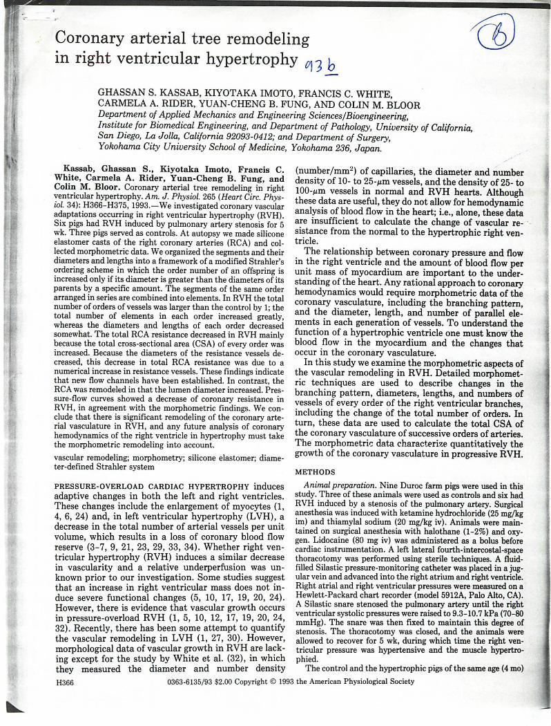
Coronary arterial tree remodelingin right ventricular hypertrophy ^ y
GHASSAN S. KASSAB, KIYOTAKA IMOTO, FRANCIS C. WHITE,CARMELA A. RIDER, YUAN-CHENG B. FUNG, AND COLIN M. BLOORDepartment of Applied Mechanics and Engineering Sciences/Bioengineering,Institute for Biomedical Engineering, and Department of Pathology, University of California,San Diego, La Jolla, California 92093-0412; and Department of Surgery,Yokohama City University School of Medicine, Yokohama 236, Japan.
Kassab, Ghassan S., Kiyotaka Imoto, Francis C.White, Carmela A. Rider, Yuan-Cheng B. Fung, andColin M. Bloor. Coronary arterial tree remodeling in rightventricular hypertrophy. Am. J. Physiol. 265 (Heart Circ. Physiol. 34): H366-H375, 1993.—We investigated coronary vascularadaptations occurring in right ventricular hypertrophy (RVH).Six pigs had RVH induced by pulmonary artery stenosis for 5wk. Three pigs served as controls. At autopsy we made siliconeelastomer casts of the right coronary arteries (RCA) and collected morphometric data. We organized the segments and theirdiameters and lengths into a framework of a modified Strahler'sordering scheme in which the order number of an offspring isincreased only if its diameter is greater than the diameters of itsparents by a specific amount. The segments of the same orderarranged in series are combined into elements. In RVH the totalnumber of orders of vessels was larger than the control by 1; thetotal number of elements in each order increased greatly,whereas the diameters and lengths of each order decreasedsomewhat. The total RCA resistance decreased in RVH mainlybecause the total cross-sectional area (CSA) of every order wasincreased. Because the diameters of the resistance vessels decreased, this decrease in total RCA resistance was due to anumerical increase in resistance vessels. These findings indicatethat new flow channels have been established. In contrast, theRCA was remodeled in that the lumen diameter increased. Pressure-flow curves showed a decrease of coronary resistance inRVH, in agreement with the morphometric findings. We conclude that there is significant remodeling of the coronary arterial vasculature in RVH, and any future analysis of coronaryhemodynamics of the right ventricle in hypertrophy must takethe morphometric remodeling into account.vascular remodeling; morphometry; silicone elastomer; diameter-defined Strahler system
PRESSURE-OVERLOAD CARDIAC HYPERTROPHY inducesadaptive changes in both the left and right ventricles.These changes include the enlargement of myocytes (1,4, 6, 24) and, in left ventricular hypertrophy (LVH), adecrease in the total number of arterial vessels per unitvolume, which results in a loss of coronary blood flowreserve (3-7, 9, 21, 23, 29, 33, 34). Whether right ventricular hypertrophy (RVH) induces a similar decreasein vascularity and a relative underperfusion was unknown prior to our investigation. Some studies suggestthat an increase in right ventricular mass does not induce severe functional changes (5, 10, 17, 19, 20, 24).However, there is evidence that vascular growth occursin pressure-overload RVH (1, 5, 10, 12, 17, 19, 20, 24,32). Recently, there has been some attempt to quantifythe vascular remodeling in LVH (1, 27, 30). However,morphological data of vascular growth in RVH are lacking except for the study by White et al. (32), in whichthey measured the diameter and number densityH366
(number/mm2) of capillaries, the diameter and numberdensity of 10- to 25-/zm vessels, and the density of 25- to100-Mm vessels in normal and RVH hearts. Althoughthese data are useful, they do not allow for hemodynamicanalysis of blood flow in the heart; i.e., alone, these dataare insufficient to calculate the change of vascular resistance from the normal to the hypertrophic right ventricle.
The relationship between coronary pressure and flowin the right ventricle and the amount of blood flow perunit mass of myocardium are important to the understanding of the heart. Any rational approach to coronaryhemodynamics would require morphometric data of thecoronary vasculature, including the branching pattern,and the diameter, length, and number of parallel elements in each generation of vessels. To understand thefunction of a hypertrophic ventricle one must know theblood flow in the myocardium and the changes thatoccur in the coronary vasculature.
In this study we examine the morphometric aspects ofthe vascular remodeling in RVH. Detailed morphometric techniques are used to describe changes in thebranching pattern, diameters, lengths, and numbers ofvessels of every order of the right ventricular branches,including the change of the total number of orders. Inturn, these data are used to calculate the total CSA ofthe coronary vasculature of successive orders of arteries.The morphometric data characterize quantitatively thegrowth of the coronary vasculature in progressive RVH.METHODS
Animal preparation. Nine Duroc farm pigs were used in thisstudy. Three of these animals were used as controls and six hadRVH induced by a stenosis of the pulmonary artery. Surgicalanesthesia was induced with ketamine hydrochloride (25 mg/kgim) and thiamylal sodium (20 mg/kg iv). Animals were maintained on surgical anesthesia with halothane (1-2%) and oxygen. Lidocaine (80 mg iv) was administered as a bolus beforecardiac instrumentation. A left lateral fourth-intercostal-spacethoracotomy was performed using sterile techniques. A fluid-filled Silastic pressure-monitoring catheter was placed in a jugular vein and advanced into the right atrium and right ventricle.Right atrial and right ventricular pressures were measured on aHewlett-Packard chart recorder (model 5912A, Palo Alto, CA).A Silastic snare stenosed the pulmonary artery until the rightventricular systolic pressures were raised to 9.3-10.7 kPa (70-80mmHg). The snare was then fixed to maintain this degree ofstenosis. The thoracotomy was closed, and the animals wereallowed to recover for 5 wk, during which time the right ventricular pressure was hypertensive and the muscle hypertro-phied.
The control and the hypertrophic pigs of the same age (4 mo)0363-6135/93 $2.00 Copyright © 1993 the American Physiological Society

CORONARY ARTERIAL REMODELING IN CARDIAC HYPERTROPHY
and weight (31 ± 2 kg) were then prepared for morphometricstudy. Each animal was anesthetized as described in the aboveprotocol, intubated, and ventilated with room air using a Harvard mechanical respirator. A midline sternotomy was performed, and the aorta was cannulated. Arterial pressure wasmonitored via a carotid artery; right ventricular pressure wasmeasured at the termination ofthe study by inserting a catheterinto the right ventricle via the jugular vein. KC1 was given toarrest the heart, and then we perfused the heart with a car-dioplegic solution, as previously described (16). The right coronary artery (RCA) was cannulated, whereas the left anteriordescending coronary artery (LAD) and left circumflex coronaryartery (LCX) were ligated.
Determination of pressure-flow relationship of RCA artery. Tocompare the resistances to blood flow of the control and hypertrophic right ventricles we determined the pressure-flow (P-Q)relationships in these hearts with the setup shown in Fig. 1. Arigid tube with a lumen diameter of 0.14 cm and a length of 98cm was used to perfuse the RCA and served as a flowmeter inthe interim. For the flows used in these experiments, the maximum Reynolds number was <400, so the effect of turbulencewas negligible. In calibrating the tube as a flowmeter, the inletand outlet pressures were measured with pressure transducerson a Hewlett-Packard strip-chart recorder while the flows weremeasured by collecting the volumes of flow in a graduated cylinder for a given period of time. The fluid used to measure theP-Q curve of the RCA was the cardioplegic solution describedpreviously (16). Figure 1 shows how the rigid tube was incorporated into our isolated heart preparation (stopcock closed totube 1 and opened to tube 2). In the RCA experiments, thepressure changes were imposed with a regulator according toselected loading and unloading ramps. The pressures P, and P2were recorded on a Hewlett-Packard strip-chart recorder. Flowswere calculated from the pressure differences, P- — P2, according to the calibrated tube flow resistance. The inlet pressure tothe RCA is P2 minus a small pressure drop correction for the12-cm-long cannulating catheter with a diameter of, for example, 0.14 cm. The pressure difference across the right ventriclecircuit was P2 minus the outlet venous pressure, which was zeroor atmospheric. The RCA was preconditioned with several load-
£ iSILICONEEIASTOMER
CARDIOPLEGICSOLUTION
HB5QUCTCR
THREE-WAYSTOPCOCK
PRESSUREMANCMEIER
HYPOTHERMICSALINE SOLtTTICN
Fig. 1. Schematic diagram of perfusion apparatus. RV and LV, right andleft ventricles, respectively; 1 and 2 are tubes 1 and 2; P- and P2 are inletand outlet pressures of the rigid tube, respectively.
ing and unloading cycles before recording the final data. In thisway, the RCA P-Q curve was obtained.
Determination of myocardial region perfused by RCA ventricular branches. Our morphometric data were measured frompolymer casts of the RCA vasculature, which was perfused witha silicone elastomer (CP-101, Flow Technology), catalyzed with6.2% stannous 2-ethylhexoate and 3.1% ethyl silicate in a liquidstate under controlled pressures and then allowed to solidify ina specific period of time at a static pressure of 80 mmHg. Theprotocol for casting the RCA was identical to that describedpreviously (16).
In this study, we were interested in the morphometry of thenormal and hypertrophied RCA branches that supply the rightventricle. Hence we dissected the RCA cast to identify the atrialbranches and tied a suture around every atrial branch beforecorrosion of the myocardium. A suture was also tied around theposterior descending artery (RCA-PDA) that supplies the posterior interventricular septum and posterior portions of the leftventricle. Only the right ventricular branches (RCA-RVB) weremeasured in the morphometric analysis. The two atria were cutaway from the casted heart at the level of the atrioventricularvalves. The right ventricular free wall was then excised andweighed. The left ventricle (including the septum) was alsoweighed. However, the right ventricular free wall mass is greaterthan the myocardial mass supplied by the RCA-RVB, sincesome branches of the LAD and LCX supply the posterior rightventricular wall. Because the LAD and LCX arteries were notperfused with elastomer, a border was found between the elastomer-perfused and nonperfused right ventricular myocardium.This was determined by removing myocardial plugs along theborder, sectioning, clearing, and viewing them under the microscope. Hence the region perfused by RCA-RVB was measured.
Histological and cast studies of RCA ventricular branches. Forhistological studies of smaller coronary arteries of orders 1-4,12plugs of myocardial tissue were removed from the right ventricles of 3 RVH pigs and 8 plugs from the right ventricles of 2control pigs. Each transmural plug was ~4x4 mm in crosssection and extended from epicardium to endocardium. Themethod of preparation of histological sections was identical tothat described previously (16). Figure 2 shows an example of acleared histological section taken 4.7 mm from the epicardialsurface of a hypertrophic right ventricle.
For cast studies, the same hearts used for the histologicalstudies of the microvasculature were corroded after removal oftissue plugs for histological sections. We corroded the tissuewith a 30% KOH solution for several days. The corroded tissuewas washed away with soap and water. The veins were carefullypruned away, leaving the RCA and its branches intact withclusters of capillaries. The casts of RCA beds in normal andhypertrophic right ventricles are shown in Fig. 3.
Using methods developed in the earlier study of Kassab et al.(16), we measured the arterial trees from histological specimensof the right ventricular free wall and the right ventricularbranches from the polymer casts ofthe RCA. We assigned ordernumbers to arterial vessels in histological specimens and corroded casts according to the diameter-defined Strahler systemdescribed by Kassab et al. (16) and measured the diameters andlengths, combined segments of the same order but connected inseries into elements, counted the number of elements in eachorder, and obtained their connectivity matrices.
Mathematical description of branching pattern, order number,and connectivity matrix. To better describe the branching pattern ofthe arteries in RVH, we introduced three innovations ina companion study (16). In brief, a diameter-defined Strahlersystem is used, whereby capillaries are defined as vessels oforder 0. The smallest arteries (arterioles) are identified as vessels of order 1. Arterioles are identified by their tortuosity, andcapillaries are identified by their nontreelike topology (15). Two

Fig. 2. Photomicrograph of arterioles (a) in hypertrophic pig RV. This 70-^m-thick section taken ~4.7 mm fromepicardial surface shows several arterioles feeding a capillary bed and a nearby venule (v) draining it.
order 1 vessels meet to form a vessel of order 2, if its diameteris larger than that of the order 1 vessels by a certain amount; orremain at order 1, if the new diameter is not large enough. Twoorder 2 vessels meet to yield a vessel of order 3, if the diametercriterion is satisfied; or remain at order 2, if the diameter is notlarge enough, and so on. The diameter criterion is the dividingline between vessels of order n and vessels of order n + 1
[(Dn_, + An_,) + (Dn-An)]/2on the left, and
[(D„ + An) + (Dn + 1-An + ,)]/2on the right, where Dn is the mean diameter of vessels of ordern. and A., is the standard deviation nf the HiameterQ nf vpccoIc r»forder n. This definition eliminates the overlap of histograms ofdiameters of all vessels in successive orders and enables anaccurate analog circuit to be constructed for hemodynamics.The second innovation is to combine all vessel segments of agiven order but connected in series into elements. The thirdinnovation is to describe asymmetric branching by a connectivity matrix, C(m,n), whose element in row m and column n is theratio of the total number of elements of order m that arisedirectly from parent elements of order n divided by the totalnumber of elements of order n.
RESULTS
We induced RVH by stenosis of the pulmonary arteryfor 5 wk. The mean and peak systolic pressures of theright ventricle increased to three to four times the control
levels. The right ventricle-to-left ventricle weight ratiodoubled over the 5 wk.
Table 1 summarizes the heart weights, blood pressures,and morphometric measurements of the two control andthree hypertrophic right ventricles. Morphometric measurements were made on all histological specimens fromall five right ventricles. We conducted completely detailedpolymer-cast measurements on one control right ventricle and one hypertrophic right ventricle. We examinedthe variability between the two control hearts and thethree hypertrophic hearts by analyzing the morphometricdata from each heart separately. The morphometric data
metric data for the two control hearts were combined.Likewise, the morphometric data from the three hypertrophic hearts for each respective order were combinedbefore conducting the statistical analysis shown in Tables2-6.
The P-Q relation of the rigid tube used as a flowmeterwas fitted by the method of least squares. The result isPressure drop (mmHg)= 417 (mmHg • ml"1-s"1) x flow (ml/s) - 0.512 (mmHg)with a correlation coefficient of 0.999. Figure 4 shows theP-Q relationship of the RCA. The two curves for each

CORONARY ARTERIAL REMODELING IN CARDIAC HYPERTROPHY
Fig. 3. Casts of right coronary artery (RCA) in control (left) and hypertrophic (right) pig RV. Atrial branches andposterior descending artery were excised.
heart correspond to loading and unloading of pressureand were fitted with second-order polynomials by theleast-squares method.
The RCA and RCA-RVB arteries of control and hypertrophic right ventricles are shown in Fig. 5. The average diameters and lengths of the vessel segments aremarked. For the trunk of the RCA, excluding the RCA-PDA, the average diameters and lengths were 2,469 ± 242and 2,988 ± 1,689 um, respectively, in the control rightventricle (N = 16 vessel segments) and 2,926 ± 320 and3,484 ± 1,641 um, respectively, in the hypertrophic rightventricle (N = 22 vessel segments). The total lengths ofthe RCA trunk with segments in series as an element,excluding the RCA-PDA, were 47.8 mm in the controlright ventricle and 76.7 mm in the hypertrophic rightventricle. A total of 16 subtrees, i.e., RCA-RVB arisenfrom the RCA trunk, excluding the RCA-PDA, in control
right ventricles and 23 RCA-RVB in hypertrophic rightventricles is shown in Fig. 5.
Tables 2 and 3 show the mean diameters and lengths ofvessel segments and elements in each order of vessels inthe right ventricular branches of the RCA of control andhypertrophic right ventricles, respectively. A segment is avessel between two successive nodes of bifurcation,whereas an element is a combination of segments of thesame order in series. These tables show that a total of 10and 11 orders of vessels lie between the coronary capillaries and the largest right ventricular branches for thecontrol and hypertrophic right ventricles, respectively.
Figure 6 shows the relationship between the mean vessel diameter and the order number for the elements oftheRCA-RVB in control and hypertrophic right ventricles,respectively. Figure 7 shows the relationship between the
Table 1. Hemodynamic data and morphometricspecimens from control and hypertrophic pig RV
PiK, , . ■ , . B l ood P ressu re ,W e , g h t , g m m H g Morphometry From
RV RV/LV Peg™*1 RV Aorta Histology Cast
AB
CDE
21.118.5
52.358.761.0
Control RV0 . 2 9 1 2 . 5 2 3 / 3 1 3 0 / 1 0 50 . 2 9 1 4 . 8 2 6 / 1 11 0 / 9 0
Hypertrophic RV0 . 6 7 4 1 . 9 9 5 / 0 1 6 5 / 1 2 00 . 6 3 3 0 . 5 7 3 / 0 1 0 5 / 8 50 . 6 9 4 4 . 8 6 0 / 0 1 3 0 / 9 0
YesYes
YesYesYes
YesNo
YesNoNo
Blood pressure values are systolic/diastolic. LV and RV, left andright ventricles, respectively.
S 2.0" n Control RVJ J J A H y p e r t r o p h i c R V
g 1.5:
3 1.0"-
0 10 20 30 40 50 60 70 80 90100Coronary In le t P ressure (mmHg)
Fig. 4. Pressure-flow relation of RCA in control and hypertrophic pigRV.

H370 CORONARY ARTERIAL REMODELING IN CARDIAC HYPERTROPHY
■hi'
— u i
n*—» _, H\ o—» en . H*
01o ~* to 09 t o
K> 0*1 - •o t aMCl
t o - UI •O CJ -4U l - » to LJ 09 o iN> Kl in ** oO OS Ui —» U i ansr* • U i o*» U i > (nto — -4 t o ooo eg vo VO ra00 - J
(2359,524)
(2225,2536)327
(2196,2062)
(2350,5267)
Atrial Branch(2656,4298)
>> r r
H*SI
U Ir r ui
—» VOH* UI "»•u p * * U i o to
UI —' I - U i U i WMClaA9*
« A -» eo i U i o -* O l V O o•• to ca O O l «J - « o
„^ U I U Ito 10*1 Dl
to at o
**■■**•*to >U i -»•*». - 3 t o U I IO • j00 VO-» to O -ft. to o e .- J 00—' o sr to UI -4 ***** A» J - w
U Io i •Ch Ol **■*
U i o i • * ■ * ' he> \o ft
(2827,5462(273,421)1
(2828,2621(969,580)J
(2873,2818(377,233)K(2917,1851)
(413,2057)L(2780,828
(2824,6329)(167,1294)G
(2506,1052)(2567,1638)
(2427,2633)
Poster iorDescendingAr tery
(2877,1586)(440 ,895)0
(2687,1308)
(2623,4183)(336,2812)Q
(2684,2852)
(2770,3658)
(295,932)T(2734,4134)
(2580,
(1540,1673)V(2355,6251)
Fig. 5. Sketches of RCAs marked with measured diameters and lengths (in um) of vessel segments in control andhypertrophic pig RV.
Pos te r io rDescendingAr tery
Table 2. Diameters and lengths of vessel segments and elementsin each order of vessels in control pig RV arterial branches
Segments ElementsOrder
Diameter, um n Length, mm n Diameter, jxm n Length, mm n1 9.7±0.95 823 0.073±0.048 383 9.5±0.84 99 0.138+0.092 992 13.6±1.5 599 0.083±0.055 348 13.4±1.1 95 0.149±0.115 953 19.8±2.4 398 0.087±0.062 266 18.8+1.7 52 0.172±0.103 524 30.1±4.5 767 0.180±0.170 343 29.8±3.6 60 0.332±0.210 605 59.8±9.2 533 0.368±0.311 313 49.5±6.5 50 1.23±0.800 506 102±20.9 441 0.601±0.461 347 97.0±13.9 46 2.24±1.20 467 201±36.9 203 0.723±0.625 192 191±26.3 47 2.55±1.88 478 385±70.8 76 1.11±0.804 70 370±53.2 11 5.18±4.65 119 701±126 53 1.47±1.07 52 669±45.0 5 U.9±6.71 5
10 1,016±107 10 2.25±0.995 10 1,007 2 9.62 2Values are means ± SD; n, no. of vessels measured.

CORONARY ARTERIAL REMODELING IN CARDIAC HYPERTROPHY
Table 3. Diameters and lengths of vessel segments and elementsof each order of RV arterial branches of RV hypertrophic pig
Segments ElementsDiameter, um
9.7±1.013.2±1.217.9±1.625.9±3.040.0±5.565.6±11.0121±23.1224±37.0396±79.2837±175
1,350±122
Length, mm
0.071±0.0510.074±0.0510.081±0.0610.170±0.170.342±0.280.457±0.360.576±0.470.707±0.53
1.00±0.771.54+1.102.50±2.03
Values are means ± SD; n, no. of vessels measured.
mean vessel element length and the order number for theRCA-RVB in control and hypertrophic right ventricles,respectively. The curves in Fig. 6 obey Horton's law (13,16). The ratio of the diameters of successive orders ofarteries is a constant independent of n and is called the"diameter ratio." The diameter ratio is given by the antilog ofthe slope ofthe lines of Fig. 6. The mean elementlength also obeys Horton's law but with a discontinuity inthe slope at order 3 (Fig. 7). The mean element diameter
Diameter, -/m
9.5±0.9213.0±1.217.7-+1.525.3±3.139.6±5.165.4±9.6124+19.7238±27.8404±57.4881+126
1,325
Length, mm
0.105±0.0840.137±0.100.174±0.140.237±0.180.546±0.391.07±0.762.15+1.412.82±1.924.29±3.9416.6±10.7
16.3
10000□ Control RVA Hypertrophic RV
OrderFig. 6. Relation between average diameters of vessel elements in successive orders of vessels and order number of vessels in RV branches(RCA-RVB) of control and hypertrophic pig RV.
100000
10000
ratios of RCA-RVB in control and hypertrophic rightventricles are 1.73 and 1.67, respectively, whereas themean element length ratios of RCA-RVB in control andhypertrophic ventricles are 1.89 and 1.89, respectively, fororders 4-10 but are 1.12 and 1.29, respectively, for ordersl—o.
The ratio of the segment number and the element number (S/E) is presented in Table 4. It can be plotted as afunction of n for each tree and fitted by a fourth-orderpolynomial. The fitting is shown in Fig. 8. The S/E ratiohas the physical meaning of the average number of vesselsegments in series. The largest orders are most asymmetric. The degree of asymmetry decreases with a decrease inthe order number in control and hypertrophic right ventricles.
The connectivity matrices of the RCA-RVB in controland hypertrophic right ventricles are given in Tables 5and 6, respectively. The total number of arterial elementsof each order can be computed from information on thenumber of intact and cut elements and the connectivitymatrix as illustrated in the appendix of our companionreport (16). Similar analysis is carried out for each RCA-RVB, whose location along the RCA trunk is shown inFig. 5. The total number of elements is then summed overall RCA-RVB trees at each order to give the total number
Table 4. S/E for each order of vessels in RV arterialbranches of control and RV hypertrophic pig
O Control RVA Hypertrophic RV
Control RV Hypertrophic RV
Order
and hypertrophic pig RV.
1 1.93±0.99 101 1.55±0.87 1482 1.92+1.0 95 1.78±0.95 1763 2.08+1.1 60 1.96+1.2 16?•1 2.10+1.6 60 1.36±0.76 1575 3.20±1.9 50 1.54±0.81 2596 3.83±2.3 46 2.25+1.3 3877 3.57±2.9 47 3.80±2.5 2508 5.18±5.0 11 3.74±3.1 1099 8.20±4.1 5 3.98±3.4 40
10 5.5 2 7.86±5.9 711 7.0 2/alues are means ± SD; r , no. of c segme
order to total no. of elements in that order.

CORONARY ARTERIAL REMODELING IN CARDIAC HYPERTROPHY
□ Control RVA Hypertrophic RV
£ 2|£ 5? !3en z
O r d e rFig. 8. Relation between average number of segments in series in successive orders of vessels and order number of vessels in RCA-RVB ofcontrol and hypertrophic pig RV.
of elements at that order. The results, means ± propagated errors, are presented in Table 7. If the total numberof arterial segments of each order is desired, one need onlyto multiply the number of elements by the ratio of thenumber of segments to the number of elements presentedin Table 4.
When the number of elements of order n is normalizedwith respect to the perfused myocardial mass given in
Table 1 and plotted against n on a semilog paper (Fig. 9),the data fit a straight line. Thus the number of vesselsincreases as an inverse geometric sequence in accordancewith Horton's law (13). The ratio ofthe numbers of vessels of successive orders is a constant independent of nand is called the "branching ratio." The branching ratio isgiven by the antilog of the absolute value of the slope ofthe lines of Fig. 9. The element branching ratios of RCA-RVB in control and hypertrophic right ventricles are 3.13and 2.98, respectively.
The data on the diameters, lengths, and number ofelements are used to compute the total CSAs and bloodvolumes, as shown previously (16). The total CSAs arenormalized with respect to the perfused myocardial massgiven in Table 1, plotted in Fig. 10, and fitted with fifth-order polynomials by the least-squares method. Similarly,the total blood volumes in all elements of a given order arenormalized with respect to the perfused myocardial massand are shown in Fig. 11.
DISCUSSIONWe experimentally determined the resistance of the
RCA in control and hypertrophic right ventricles by measuring their P-Q relationships in the isolated heart preparation, as shown in Fig. 4. The inverse of the slope ofthese curves represents the total resistance to flow of the
Table 5. Connectivity matrix for RV arterial branches of control pig
0 2.84±0.101 0.642±0.085 0.133±0.041 0.017±0.017 01 0.099±0.030 2.22±0.081 0.800±0.108 0.233±0.069 0.520±0.050 0.174+0.056 0.170+0.076 0 0 0
0.063±0.029 2.13±0.084 1.10+0.157 0.580+0.111 0.522±0.131 0.213±0.067 0 273±0195 0 0' 0 .033+0.023 1.65±0.091 1.14+0.183 0.630±0.105 0.511±0.128 0.364±0.152 0.600±0 245 00.167±0.048 1.96±0.148 1.11+0.159 1.00±0.182 1.09±0.494 0.400±0.245 0
0.200±0.057 2.33±0.143 1.17±0.170 1.27±0.573 2.00±0.707 1.00.152±0.053 1.81+0.138 1.73±0.604 3.40±0.927 1.0
0.064±0.036 2.09±0.342 2.20±0.860 0.500 1 .40±0 .245 2 .5
i o ° "Values are means ± SE. An element (m,n) in mth row and nth column is ratio of total no. of elements of order m that spring directly from parent
elements of order n divided by total no. of elements of order n.
Table 6. Connectivity matrix for RV arterial branches of RV hypertrophic pig
0 2 . 5 5 ± 0 . 0 7 5 0 . 6 0 8 ± 0 . 0 6 6 0 . 4 2 6 ± 0 . 0 7 7 0 . 0 1 3 ± 0 . 0 0 9 0 0 01 0.041±0.023 2.14±0.076 0.660±0.066 0.115±0.029 0.077±0.017 0.049±0.014 0.060±0.017 0.055±0.02 0 0 0
0.057±0.017 2.03±0.072 0.650±0.059 0.309±0.038 0.181±0.025 0.128±0.023 0.064±0.030 0.125±0.053 0 03 0.099±0.031 1.61±0.065 0.741±0.049 0.411±0.032 0.404±0.047 0.257±0.069 0.125±0.064 0.143±0.143 0
0.025±0.016 1.39±0.045 0.755±0.046 0.692±0.061 0.404±0.067 0.575±0.179 0.286±0.286 0.500.042±0.014 1.80±0.044 1.05±0.074 0.945±0.121 0.875±0.172 0.714±0.286 1.0
0.109±0.017 2.50±0.080 1.39±0.127 1.08±0.228 2.86±0.705 3.50.180±0.027 2.05±0.103 0.850±0.195 1.86±0.705 3.5
0.073±0.025 1.92±0.166 3.71±1.27 1.00.050±0.035 3.57±1.27 1.0
0 "Values are means ± SE. An element (m,n) in mth row and nth column is ratio of total no. of elements of order m that spring directly from parent
elements of order n divided by total no. of elements of order n.

CORONARY ARTERIAL REMODELING IN CARDIAC HYPERTROPHY
Table 7. Total number of vessel elementsin each order of RV arterial branchesof control and RV hypertrophic pig
Number of Vessel ElementsControl RV Hypertrophic RV
c <? 0.008o £.♦_ O)
0.006-
□ Control RVA Hypertrophic RV
22+182±16
271±961,021±4183,039±1,5356,676±3,838
19,608±12,70555,720±40,093
1160±3
180+15626±66
2,369±3015,749±868
10,960+2,07725,963±6,67066,086±20,280
167,335±61,689Values are means ± SE, expressed in no. of vessel elements.
RCA circuit. We found that the flow (volume/time) resistance through the RCA decreased in RVH (Fig. 4).
The diameter of the trunk of the RCA increased withRVH. The diameters of the RCA-RVB arising from theRCA also increased (Figs. 3 and 5). The length of thetrunk, which crowns the base of the heart, increased inRVH. The number of RCA-RVB arising from the trunkalso increased in RVH. These results are consistent withthe angiogenesis described by White et al. (32) in which amoderate DNA synthesis was found in smooth musclecells.
The mean diameters of the vessel segments and elements were slightly smaller, at each order, in RVH (Fig.6). Progressing from the smallest arteriole to the largestRCA-RVB in the hypertrophied right ventricle required11 orders. In contrast, this progress in the control rightventricle required only 10 orders. This reflects both theincreased diameter ofthe largest RCA-RVB in RVH andthe remodeling of the branching patterns of the RCAarterial circuit. The remodeling ofthe branching patternsis best seen in S/E (Fig. 8). The RVH arterial trees aremore symmetrical at any order than those of the corresponding control hearts. The segment and elementlengths are decreased in RVH (Fig. 7). Angiogenesis ofarterioles, active DNA synthesis, in this RVH model inthe early stages of hypertrophy was previously demon-
?104■■■■«r 103 □ Control RV
A Hypertrophic RV
n £ 10"1r- HI 10-2
OrderFig. 10. Relation between calculated total cross-sectional area of arterialbranches in each order and order number for RCA-RVB of control andhypertrophic pig RV.
strated with tritiated thymidine labeling (34). Angiogenesis in RVH contributed to the increased number of vessels (Fig. 9). However, the increase in the number of smallarterioles is most important, since those arterioles are themajor sites of vascular resistance (8, 14, 28).
The total CSA of the RCA-RVB is larger in hypertrophic than control right ventricles. This increase inCSA is due to the large increase in the number of parallelelements. However, the CSA-to-mass ratio of RCA-RVBis similar in both control and hypertrophic right ventricles (Fig. 10). The volume ofthe RCA-RVB is also largerin RVH. Again, when we normalize the volume of theRCA-RVB for the mass perfused, we find some compensation (Fig. 11). The mass-normalized accumulative volume decreases 11% in RVH. In conclusion, the RCA-RVB appears to adapt in the hypertrophic right ventriclethrough compensatory vascular growth and remodeling.
Studies by Breisch et al. (6, 7) and Tomanek et al. (30)have shown that left ventricles subjected to pressure overload that induces ventricular hypertrophy of >50% haveincreased total coronary perfusion even though minimalcoronary resistance increases. Furthermore, this increasein total flow correlated with increased numbers and CSAsof small arterioles, showing that angiogenesis can occur inthe left ventricle subjected to pressure overload. Thus
□ Control RVA Hypertrophic RV
.00001O r d e r Order
c o n t r o l a n d h y p e r t r o p h j c p i g R V . h y p e r t r o p h i c p i g R V .

"1861 'A8L-Q8Z, -8F 'S^H 'OMQ -AqdojuadAq JBjnoujusA iqSuoujuaauoa ajaAas ifliM S3AJB0 paziaqisauBun ui aAjasaj iBfnasBA
"6Z.6I 68S-I8S m 'S^H MQ 'S9Apjo ui pboij3ao ajns•sajdjgmou-jnaA iqSu ajnos Suunp uoipunj iBipjBDoAra puB mojj
f P-»-Iina "A P-*B3sia 3 O 'W 'J.BqouBi»i 61 ' mA, v „ '1861 '888H-188H :(6jensAyj *«J0 jjoa//) oi*S IOIS&ij p my -saiuod pazipqisauBun
"9iSZ,l-68-SOa 5UBJ0 U0P-Bpunoj aouapg ibuoi:jbn sn P"* :(qBSSB->r *g q oj) tg-06 d-qsMoqaj
•Suqapora -aj oij;auioqdjoui aq; ;unooos o;ui ajp?; ;snm ajoij;uaA;qSu oiqdoj;jadA~q jo uoi^mojio Ajbuojoo jo sisA"jbub ajn;-njj 'dpuyaaA %Vf8u oiqdojuackq aq; ui ajn;BjnosBA jbtj
jaoipjDj "^qdojuadXq jBinauiuaA luSu T„„qiiM auiMS SunoA- snopsuoa ui BipjBoAqoBj Suunp uoqnqujsip '8IP HAH u! saSB3J0ap a0UB;sisaj AJBUOJOO paMOqSmojj pus a/uasaj JO^iposBA ajbuojoo iBJnuisuBjj, j-j *.n?qouBp\[ AI SI9Pom 9S9lll m0*IJ pauiBiqo SUOUBJBdajd ;jB8q pa;BJOSI
"8661 'S98H-098H '■(*£ ptsKyj ojiq jjoa//) uiojj saAjno ^-<j jnQ "Japjo qosa uiq;iM s;uaraaja pssaA "~ "*■ PH-vb --UBUOJ03 Sid jo AjpuioqdjojM jauBJBd jo jaquinu aq; puB pssaA jo japjo qoBa io sv<n
•0661 '0Sa?a UBS ^HS^SL Zm^£££S " IS S SaSH9I°U- °*, "* HAH " ^^^ ^^ Std at/? til saua^y -Gm/o/oo aV7 /0 ^amoyduoyq *s *0 -qBssBfl -gl P"9^ VOH I^oj, "(HAH u\ ®®P*° II °1 !<>i;uoo aq; ui"1661 '88Z,-92i =89 say outj -s^bd aAisuauadAq ui sjapjo oi uiojj) ajoij;uaA ;q3ij oiqdoj;jadA~q aq; ui sjapjo 30UB1SIS3J JBinnSRAfuriTiiT f-rBtmnu-. -,^i,^,f,.,. ._' «_* in i^nmnn Tmn- n„- ,,. „„»,„ '
-TBHTW mBq,SBa**i*0«3uiduiB1*0*H«*H.B3Ins1BUBH *n -paq icpiOOTA aAH'VOH «&"ni sjassaA jo japjo qoBa
•xso,oqdjouj aA^uBnb 4ipSSfc ,B9oS,slqZPA^:sSq™ *B ^^l PTO «^niBip pasBajoap aABq sapu;uaA ;qSu-uiBjp jisq; Pub sureajis jo luamaqsAap TBuoisoig *a *a 'uojjoH ci °.lIdoj:lJ'3dAq ;Bq; paMoqs s;jnsai jnQ *HAH 9Aissaj3ojd
•Z.86I 'IW-9-I :i -jddns '9L uoij SiiLmp saSuBqo paq jB|nosBA [Bua^re a^buojod ;qgu aq; ui-D/rjoJ?0 -3AJ3S3J ajbuojod jo m3ia jboi^ud v 'a 'I T 'nuBiujjoH 'z\ *0]J poo{q o; aouB;sisaj ;sq; paMoqs aM (gg) A\sci6u9X&
-uy tuvuouoo ^amdopAap pu^oo SGSiySS TaTT,n . . """"^ ^ T ABUIQSa3u^0 JHOlSopq^d
•1861 */,6^-88t W -say ouir) -apu-JiaA Aj9A 8JB WP" ^B SS9lun «"ojiun ajB 'stsauaSoiSuB?q3ij ;bd aq*j jo pboij3ao sjnsssjd sAisssjSojd oiuojqo -snDJBjM PUB uoRBZHBinasBAoau XjTBpadsa 'suoi;B;dBpB JBjnosBA
-uy ^vuouoo -luauKlo^p .Bja.Bqoo 2S^3^SSi iamo Q , """"^ 8q T fl"l"IWP W*WIWl
•1861 */,6^-88t W -say -aSp -apu-JiaA Aj9A 9JB WP" ^B SS9lun u"ojiun ajB 'stsauaSoiSuB?q3u ;bd sqj jo pBousAO sjnsssjd SAisssjSojd oiuojqo -snDJBjM PUB uoRBZHBin3SBAOau ^JBIOadsa 'SUOi;B;dBpB JBjnOSBA•q *w pus 'ipaBqjqa *0 *P ^ausuiox T 'H "O **iadooo Ol -^buojoo ;sq; s;saSSns siqj," -jA g o; oiii f uiojj SuiSubi
•oeeiH-weiH 'in w«Hd ™o v*»m sss vmau r™v 'IfB 3u!iCl!rAJ°sS]d u- IBnm-s SBM B-m^DSI J0^9SU0 9^^ •XqdojuadXq obiPjb3 pabnpui-uoiroawadXiJ [Buaj u.uoisnjjCd Pub I9¥B ^U9radoI9A9P PW<W>3 AJBUOJOO ;Bq; paMOqs (XI)Aj?ainoqdjoui jBjnDSBAOJoi^ ssiaM -H *H Pub «-w *0 «iuiuii3 6 'IB ^9 st-c-0a ^Bua:)9H 'HAH QAissajSojd ui paAjasqo uoi;
9861 '881H -Bsuaduioo JBjnosBA aq; ui a*oj A"a->{ b A"B*d p|noa auounoq -6LLK -m jojstyj ojiq ?JDau) IS3 ^ois/C^ •/> toy -apu-juaA q^wojS jo laAai aq; 'nBaiBid b SuiuoBaj ao Dinous asBudyai SuuBaq ui aouBisisaj JBjnosBA Xjbuojod io iinnnmncm mn-, .. « -...._ l__lL __„ + l »«SH^« "H p[iioqb abBqa
"9861 '88'H -6LLR :(03 lojstyj ojjq jjvbH) \qz -jots^yj P wy -apu^uaAysj SupBsq ui aouFjsisaj JBjnosBA Ajbuojoo jo uoiinquisip" jbj™-SBAOJDij/I -snojBjM "T "IM PUB -uiBqiSBa "I j *-w *Ai 'uBiiiqo *8... __,- .. '9861 'L9SI-6SSI •09 Pisriyj -pay p -ajniBinosBAOJoiui puB mojj pooiq io uoiiBiaj -inn r •lCiTHr.tiT-»Hfii -»^,»,,n^ «^ ' ' r '^J • lt^^ t ■ l - —Cl Id >TY !_* If**"* ubujj5[oW Q W «ouiuiiN *3 a -a^q^ 3 j --y a 'qosiaja i
'1^861 'Sf-8-888 :iS 'jsaauj qirj -Xpnjs puoipunj »ub pun-pans b :Xqdoju8dXq pbojjsao sjnsssjd jo sotisusjDBJBqo
'861 Z.0I-IOI =88 'Sung oojoyx UUV -^qd-0JU3dXq iBpiourjuaA iqSu 3iBJ3poui miM sSop SunoX ui 3AJ3S3J iraBtinnoai *"t>,,,,. .,..-, . .„<a . _ .Cl *¥»T */T
q^&ojS aAipB Jiaq; qSnoq;jv -s;mpB SunoA" ajaM sSidinQ -paounouojd ajoui A"jqBqojd sbm sisauaSoiSuB 'passaJS-oid HAH SB 3SBqd q^v\ojS 8Ai;0B ub ui ajaM sSop asaq;
mojj poojq Ajbuojoo ui asBajoui ub si aiaq; 'Aja^iB Ajbu-ouijnd aq; jo SuipuBq q;iM saiddnd ui ';Bq; UMoqs aABq
-jnp jo BiuiajadAq 9At;0Baj Suunp joj;uoo q;iM pajBduioopasBajoap mojj poojq Ajbuojoo JBjnou;uaA ;qSu japora Sop
i - sa^ooA"ra aq; sb sjaAaj joj;uoo ;b aAjasai mojjpoojq suib;uibui ;Bq; sjnooo ySO JBjnosBA ui assajoui ub in"- *""qq .#..... 1H ■ »gKt*ff y IBJKil j uazjuajy 'a '3 'pqojA n l "j* u 'aqoBa -f -uaA aq; uaqM aAjasaj mojj poojq Ajbuojoo ui sasBajoap'o;
■« pu» -^^^^i^S^^S^SS* a|q!V,8Dsns SS3' *3nm S! 3'D^U3A »t* «b '^»™ «i
ysi ipiM sSop ui sspjsxs Suijnp mojj pooiq iBipjBOOAW *uubuioh 'SSui^asO a pub 'TaqojA. *H *x 'ouoa-iv "d 'Pa Z-'x "f 'H *aqoBa -g fso^ u? paonpoj;ui ajB sio;obj ajnssaiduou jaq;o aouis.,„„ ._ t I66I '09SIH-2S9IH 'J9JJ.P *^Bra saSuBqo aq; '(uoisuauadA"q onauaS jo 'oiraa; .(62 /ojs^vj 0JJ0 UV3H) 09S 7*»^Vc/ V W '^bj ui ajnpBj -SA"s 'jBUaj 'S'd) SUBaui IOT10 A*0 DaonDUI SI uat iiatiAASH=^!5fV-2??^^!W*» P™ ***** •*—2 toL ajar am uo «£^ SSSlSS.'Si
«wl 'I98H-9S8H :(8i 7^^c/ wj "°S- 8I9qM Su?was B U- Jnoo° su0IWdBpB JBjnosBA asaq;WHntZ loisKyj p -my -;bj aq; ui asiaiaza snonusjp Xq psonp V^B P9^0U 9C1 Pinoqs ;j •(« '6S <gS '6 «q «o) iOj;uO0 UBU; -UI AudOJljadAU JBinDUlU3A Hl9n in *rt-ia..,o.,H,«,.T ..?■--- .1 un^ imnmn, t^„»„ __o ___ . ' l . n_fpub prBuoaow a s ^oiA8TA.jq3aa*0.d<.d.B8J3AUV -T o; 8np AlIJBUI,jd 8AJ9S9J M0[J poqq ^uoioo ui asBajo
saoNaaajaH '9P B s* 9I9ll^ ^H* 99iSb HAT pa^npui-pBojjaAo a'jnssaid•»«*» r™.„., u- 3uHjnDD0 suoi;B;dBpB JBjnosBA Ajbuojoo io satpmq C66I AJBnjq3J 8l uu0J puy u, p3,dao3B :266I XjBiuqa^ „ p3Apo3H -sajoijauB JJBUIS JO q^ojS aq; Xq SJU00O
„( 2190-86036 VO 'BlJOf Bq "jq ubui|i0 oog6 'aujotpajw jo iooqoS 30UB;sisaj AJBUOJOO JBUIlUlUI 10 80UBUa;uiBUI 8U1 DUB 'SIS QSOn iOopiRBj jo Tdaa 'jooia • w 0 :s,S3nbaj 1Uud3j J0J ss3jpPV -auaSoiSuB a;Bjnuii;S ubo AqdoiuadAq pBojjaAO-ajnssaj'd
AHdOHjnadAH oviqhvo ni ONiiaaoiAiaa iviaaxav ahvnohooum

C O R O N A R Y A R T E R I A L R E M O D E L I N G I N C A R D I A C H Y P E R T R O P H Y H 3 7 5
21. Marcus, M L., T. M Mueller, J A. Gascho, and R. E. 28. Tillmanns, H., M. Steinhausen, H. Leinberger, H. Thede-
!~ tC aC. ^er t rophy secondary to hyper ten- ran , and W. Kub ler. Pressure measurements in the te rmina lsion on the coronary cjrculation. Am. J. Cardiol. 44: 1023-1028, vascular bed of the epimyocardium of rats and cats. Circ Res 49-1202-1211, 1981.
Effects of experimental right ventricular hypertrophy on myocardial blood flow in conscious dogs. J. Clin. Invest. 64: 421-4271979.
23. Oaieefe, D. D., J. I. E. Hoffmann, R. Cheitlin, M. J.O'Neill, J. R. Allard, and E. Shapkin. Coronary blood flow inexperimental canine left ventricular hypertrophy. Circ. Res. 43:43-51, 1978.
24. Olivetti, G., R. Ricci, C. Lagraste, E. Maniga, E. Sonne-blick, and P. Anversa. Cellular basis of wall remodeling inlongterm pressure overload induced right ventricular hypertrophyin rats. Circ. Res. 63: 648-657, 1988.
25. Rakusan, K. Quantitative morphology of capillaries of the heart.Number of capillaries in animal and human hearts under normaland pathological conditions. Methods Achiev. Exp. Pathol. 5: 272-285, 1971.
26. Rakusan, K. Microcirculation in the stressed heart. In: TheStressed Heart, edited by M. J. Legato. Boston, MA: Nijhoff, 1987,p. 107-123.
27. Rakusan, K., and P. Wicker. Morphometry of the small arteries and arterioles in the rat heart: effects of chronic hypertensionand exercise. Cardiovasc. Res. 24: 278-284, 1990.
Schreiber, C. L. Eastham, and M. L. Marcus. Morphometryof canine coronary arteries, arterioles, and capillaries during hypertension and left ventricular hypertrophy. Circ. Res. 58: 38-461986.
30. Tomanek, R. J., K. A. Schalk, M. L. Marcus, and D. G.Harrison. Coronary angiogenesis during long-term hypertensionand left ventricular hypertrophy in dogs. Circ. Res. 65: 352-3591989.
31. Vatner, S. F. Reduced subendocardial myocardial perfusion asone mechanism for congestive heart failure. Am. J. Cardiol. 62:94E-98E, 1988.
32. White, P., Y. Nakatani, L. Nimmo, and C. Bloor. Compensatory angiogenesis in compensated, progressive right ventricularhypertrophy. Am. J. Cardiovasc. Pathol. 4: 46-61, 1992.
33. White, F. C, M. D. McKirnan, E. A. Breisch, B. D. Guth,Y. M. Liu, and C. M. Bloor. Adaptation ofthe left ventricle toexercise-induced hypertrophy. J. Appl. Physiol. 62: 1097-11101987.
34. White, F. C, T. M. Sanders, T. Peterson, and C. M. Bloor.Ischemic myocardial injury after exercised stress in the pressureoverloaded heart. Am. J. Pathol. 97: 473-488, 1979.