Embed Size (px)
Citation preview
Vol. 55, No. 10
Corky Root of Lettuce Caused by Strains of a Gram-NegativeBacterium from Muck Soils of Florida, New York, and Wisconsin
ARIENA H. C. VAN BRUGGEN,* PHILIP R. BROWN, AND KENNETH N. JOCHIMSEN
Department of Plant Pathology, University of California at Davis, Davis, California 95616
Received 25 May 1989/Accepted 18 July 1989
Slow-growing bacteria similar to the bacterium causing lettuce corky root (CR) in California (strain CA1)were isolated from muck soils of Florida, New York, and Wisconsin, using lettuce seedlings as bait. All strainswere tested for reaction with polyclonal antibodies produced against strain CAl and for pathogenicity on
CR-susceptible (Salinas) and CR-resistant (Green Lake) lettuce cultivars in a greenhouse. Five strains fromFlorida, three from New York, and three from Wisconsin induced severe CR symptoms on Salinas and mildsymptoms on Green Lake. All strains were gram-negative, aerobic, oxidase positive, and catalase positive andreduced nitrate to ammonia. Whole-cell fatty acid compositions were similar for all strains and resembled thatof Pseudomonas paucimobilis. Since this fatty acid pattern is unique, it is suggested that CR of lettuce is causedby strains of the same bacterium in Florida, New York, Wisconsin, and California.
Corky root (CR) of lettuce has been reported in California(20), Florida (12, 13), New York (14, 15), Ontario (6),Wisconsin (1, 21), and Italy (7, 8). The symptoms initiallyconsist of yellow bands on young roots and develop laterinto dark greenish brown and corked areas covering most ofthe taproot and main laterals. Infected roots become brittleand break off easily (23). The etiology of this disease was
controversial for many years. In the 1960s and 1970s, CRsymptoms were attributed to phytotoxic components, suchas ammonia (11, 15, 17) or toxins liberated from decompos-ing lettuce debris (2, 7, 8, 14). Before that time, similarsymptoms had been attributed to biotic agents, namely,Botrytis cinerea (16), a Pythium sp. (C. I. Hannon, Ph.D.thesis, Cornell University, Ithaca, N.Y., 1955.), Pseudomo-nas rhizoctonia (22), and Xanthomonas vitians (4). How-ever, Koch's postulates were not fulfilled until recently,when it was shown that a slow-growing, gram-negativebacterium caused CR of lettuce in California (23).
This bacterium has not been reported as the pathogencausing CR in areas outside California. Toxic substancesfrom decomposing lettuce debris were suspected to be thecause of CR in Ontario (L. V. Busch and J. A. Carpenter,Can. Phytopathol. Soc. Proc. 31:23, 1964), Wisconsin (2),New York (J. P. Hartnett and J. W. Lorbeer, Phytopathol-ogy 58:1053, 1968), and Italy (7, 8). However, a bacterialcausal agent could not be excluded in those areas, becauseexperiments involving decomposing lettuce debris were per-formed under field conditions or in the greenhouse with fieldsoil for decomposition of lettuce residue. In Wisconsin, a
secondary amine was purified from lettuce debris whichcaused root necrosis of lettuce seedlings (2) but not thetypical corkyness. Autoclaved crude extract, however, didcause typical CR symptoms, indicating that the toxic sub-stance was heat stable (2). This toxic substance could havebeen produced by a bacterium similar to that causing lettuceCR in California, because a heat-stable toxin was isolatedfrom culture filtrate of the CR bacterium (J. Kao, D. H.Mitten, and C. A. Milich, Phytopathology 76:844, 1986).The objectives of the research reported here were to
demonstrate the presence of a gram-negative bacterium in
* Corresponding author.
CR-prone soil from Florida, New York, and Wisconsinsimilar to the bacterium causing lettuce CR in California andto compare various strains for virulence on resistant andsusceptible lettuce cultivars and for culture, physiological,and chemical characteristics.
MATERIALS AND METHODSSoil samples. Samples of corked lettuce roots and attached
soil were collected in June 1987 from Oswego County, NewYork. Soil samples from Florida and Wisconsin were pro-vided by Victor Guzman and Luis Sequeira in May andSeptember 1987, respectively. All samples originated frommuck soils in which CR of lettuce had been a problem inprevious years.
Isolation procedures. Isolations from soil were made with2- to 3-week-old lettuce seedlings, cultivar Salinas, as bait.Soil suspensions were made by mixing 50 g of soil from eachlocation in 75 ml of distilled water plus 3 drops of Tween 20.The suspensions were stirred for 10 to 15 min and filteredthrough six layers of cheesecloth. Suspension (5 ml) was
dispensed at the stem base of each of five 2- to 3-week-oldSalinas lettuce seedlings in a greenhouse. At 3 to 4 weeksafter inoculation, the plants were uprooted and isolationswere made from yellow or corked areas on the roots as
described previously (23) but without surface sterilizationwith 0.5% sodium hypochlorite. Slow-growing colonies thatappeared similar to those of CR bacteria from Californiawere transferred to S medium (23).
Antibody tests. To distinguish potential CR strains fromother slow-growing bacteria, all isolates with colonies simi-lar to those of the CR bacterium were tested for theirreaction with polyclonal antibodies. The antibodies were
produced by immunizing New Zealand White rabbits withintact and sonicated cells of the first strain of the CRbacterium from California (strain CA1) (23). Serum was
precipitated in 40% ammonium sulfate, dialyzed againstphosphate-buffered saline (0.01 M phosphate, pH 7.6, 0.14M NaCl), and purified in a DEAE-cellulose column.
Cultures (5 days old) of the CR strains in S broth were
sonicated for 30 to 45 s with a Microson ultrasonic celldisruptor model MS-50 equipped with a CM-1 convertor at80% power to break up clumps (Heat Systems Electronics,
2635
APPLIED AND ENVIRONMENTAL MICROBIOLOGY, Oct. 1989, p. 2635-26400099-2240/89/102635-06$02.00/0Copyright © 1989, American Society for Microbiology
Dow
nloa
ded
from
http
s://j
ourn
als.
asm
.org
/jour
nal/a
em o
n 18
Jan
uary
202
2 by
14.
53.1
62.8
4.
2636 VAN BRUGGEN ET AL.
Inc., Farmingdale, N.Y.). The concentration of bacteria insuspension was assessed with a spectrophotometer at 650nm (Spec 20; Bausch & Lomb, Inc., Rochester, N.Y.). Eightdilutions were prepared of each strain in Tris-buffered saline(TBS) (10 mM Tris hydrochloride, 150 mM NaCl, pH 8.0)and applied onto a nitrocellulose membrane with a vacuummanifold (Hybri-dot manifold; Bethesda Research Labora-tories, Gaithersburg, Md.). The dilutions amounted to 2 x10⁸, 5 x 10⁷, 1 x 10⁷, 1 x 10⁶, 1 x 10⁵, 1 x 10⁴, 1 x 10³, and1 x 102 cells per dot. Rhizobium meliloti (8D15; C. I. Kado,University of California at Davis) was applied in the sameamounts as a negative control. The blot was air dried for atleast 4 h and then dried at 50°C for 1 h. The blot was soakedin TBST (TBS plus 0.05% Tween 20) and incubated inblocking solution (TBST plus 1% bovine serum albumin,fraction 5) at 37°C with shaking. After 30 min, the blot wasrinsed in TBST and incubated in a 1:100-diluted anti-CRstrain CAl immunoglobulin G solution in TBST for 3 to 4 hat 37°C with shaking. All nonreacted antibody was rinsedaway by three successive 7-min washes with shaking in anexcess of TBST. Anti-CAl was detected by incubating theblot for 2 h at 37°C with a 1: 1,000 dilution of proteinA-alkaline phosphatase conjugate (Sigma Chemical Co., St.Louis, Mo.). The blot was rinsed in TBST as before. ProteinA-alkaline phosphatase conjugate was detected by a colorreaction with Nitro Blue Tetrazolium (0.66% [vol/vol] of a
50-mg/ml solution) and 5-bromo-4-chloro-3-indolyl phos-phate (0.33% [vol/vol] of a 50-mg/ml solution) in AP buffer(100 mM Tris chloride, 100 mM NaCl, 5 mM MgCl2, pH 9.5).Reaction of each CR bacterium strain with the antibody wasrated subjectively (0 to 6 scale) by comparison with thereaction of CAl and the negative control.
Pathogenicity tests. Preliminary pathogenicity tests weredone with all strains that reacted with the antibodies and hada colony morphology and growth rate similar to those of theCR bacterium from California (23). Sterile distilled water (10ml) was pipetted onto plates with 7- to 10-day-old purecultures, and 2 to 3 ml of suspension was dispensed at thestem bases of two or three 2-week-old Salinas lettuce seed-lings for each strain.
Extensive pathogenicity tests to fulfill Koch's postulateswere performed in a greenhouse with 12 strains that causedsymptoms in preliminary tests, as described previously for aCalifornia strain of the CR bacterium (CA1) (23). Inoculawere prepared by centrifuging 5-day-old cultures in S me-dium broth for 20 min at 9,150 x g and suspending the pelletsin the same amount of distilled water. Bacterial concentra-tions were estimated with a spectrophotometer and wereadjusted to (1.7 ± 0.7) x 109 CFU/ml. Lettuce seedlings (2weeks old) in vermiculite were inoculated by dispensing 3 mlof bacterial suspension or distilled water (controls) at thestem bases of each plant. Plants of each treatment wereplaced on saucers in insect-proof cages 50 cm apart to avoidcross contamination by fungus gnats and water splashing.There were six plants per treatment. The plants were wa-tered alternately with 20 to 25 ml of distilled water, half-strength Hoagland solution, or 0.005 M Ca(NO3)2 + 0.005 MKNO3. Minimum and maximum temperatures were 16.8 +2.1 and 27.9 ± 1.5°C, respectively. Daylight was extended to14 h by fluorescent tubes. Four weeks after inoculation, theplants were uprooted and scored for CR severity on a 0 to 9scale, based on percentages of the taproot infected (5).Isolations were made from three inoculated and controlplants as described previously (23), and reisolated colonieswere again tested for reaction with polyclonal antibodies andpathogenicity on lettuce seedlings. Three plants were dried
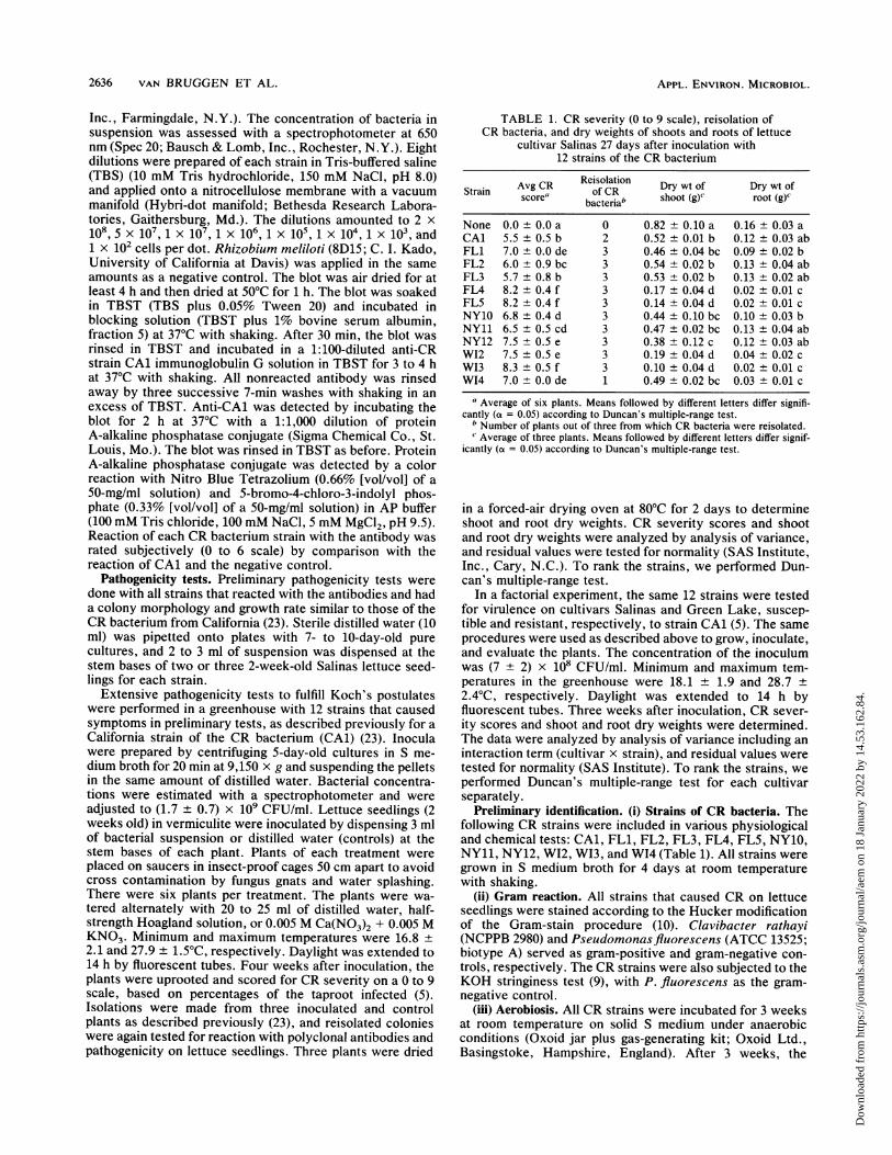
TABLE 1. CR severity (0 to 9 scale), reisolation ofCR bacteria, and dry weights of shoots and roots of lettuce
cultivar Salinas 27 days after inoculation with12 strains of the CR bacterium
Strain Avg CR Reisolation Dry wt of Dry wt ofStrain scorea ofCR shoot (g) Droot(g)wbacteriabrot()None 0.0 ± 0.0 a 0 0.82 ± 0.10 a 0.16 ± 0.03 aCAl 5.5 ± 0.5 b 2 0.52 ± 0.01 b 0.12 ± 0.03 abFL1 7.0 ± 0.0 de 3 0.46 ± 0.04 bc 0.09 ± 0.02 bFL2 6.0 ± 0.9 bc 3 0.54 ± 0.02 b 0.13 ± 0.04 abFL3 5.7 ± 0.8 b 3 0.53 ± 0.02 b 0.13 ± 0.02 abFL4 8.2 0.4 f 3 0.17 ± 0.04 d 0.02 ± 0.01 cFL5 8.2 ± 0.4 f 3 0.14 ± 0.04 d 0.02 ± 0.01 cNY10 6.8 ± 0.4 d 3 0.44 ± 0.10 bc 0.10 ± 0.03 bNY11 6.5 ± 0.5 cd 3 0.47 ± 0.02 bc 0.13 ± 0.04 abNY12 7.5 ± 0.5 e 3 0.38 ± 0.12 c 0.12 ± 0.03 abW12 7.5 ±0.5 e 3 0.19 ± 0.04 d 0.04 ± 0.02 cW13 8.3 ±0.5 f 3 0.10 ± 0.04 d 0.02 ± 0.01 cW14 7.0 ± 0.0 de 1 0.49 ± 0.02 bc 0.03 ± 0.01 c
a Average of six plants. Means followed by different letters differ signifi-cantly (a = 0.05) according to Duncan's multiple-range test.
b Number of plants out of three from which CR bacteria were reisolated.c Average of three plants. Means followed by different letters differ signif-
icantly (a = 0.05) according to Duncan's multiple-range test.
in a forced-air drying oven at 80°C for 2 days to determineshoot and root dry weights. CR severity scores and shootand root dry weights were analyzed by analysis of variance,and residual values were tested for normality (SAS Institute,Inc., Cary, N.C.). To rank the strains, we performed Dun-can's multiple-range test.
In a factorial experiment, the same 12 strains were testedfor virulence on cultivars Salinas and Green Lake, suscep-tible and resistant, respectively, to strain CAl (5). The sameprocedures were used as described above to grow, inoculate,and evaluate the plants. The concentration of the inoculumwas (7 ± 2) x 108 CFU/ml. Minimum and maximum tem-peratures in the greenhouse were 18.1 ± 1.9 and 28.7 +2.4°C, respectively. Daylight was extended to 14 h byfluorescent tubes. Three weeks after inoculation, CR sever-ity scores and shoot and root dry weights were determined.The data were analyzed by analysis of variance including aninteraction term (cultivar x strain), and residual values weretested for normality (SAS Institute). To rank the strains, weperformed Duncan's multiple-range test for each cultivarseparately.
Preliminary identification. (i) Strains of CR bacteria. Thefollowing CR strains were included in various physiologicaland chemical tests: CA1, FL1, FL2, FL3, FL4, FL5, NY10,NY11, NY12, W12, W13, and W14 (Table 1). All strains weregrown in S medium broth for 4 days at room temperaturewith shaking.
(ii) Gram reaction. All strains that caused CR on lettuceseedlings were stained according to the Hucker modificationof the Gram-stain procedure (10). Clavibacter rathayi(NCPPB 2980) and Pseudomonasfluorescens (ATCC 13525;biotype A) served as gram-positive and gram-negative con-trols, respectively. The CR strains were also subjected to theKOH stringiness test (9), with P. fluorescens as the gram-negative control.
(iii) Aerobiosis. All CR strains were incubated for 3 weeksat room temperature on solid S medium under anaerobicconditions (Oxoid jar plus gas-generating kit; Oxoid Ltd.,Basingstoke, Hampshire, England). After 3 weeks, the
APPL. ENVIRON. MICROBIOL.
Dow
nloa
ded
from
http
s://j
ourn
als.
asm
.org
/jour
nal/a
em o
n 18
Jan
uary
202
2 by
14.
53.1
62.8
4.
ISOLATION OF BACTERIUM CAUSING CORKY ROOT OF LETTUCE 2637
plates were incubated under aerobic conditions at 27°C for 1week and checked for growth.
(iv) Oxidase test. Oxidase activity was tested by themethod of Kovacs (10), using 4-day-old cultures of 12 strainsof the CR bacterium in S broth and 1-week-old cultures onsolid S medium. Cultures (1 day old) of P. fluorescens andPseudomonas syringae pv. syringae (5D4214 from C. I.Kado) on S medium were positive and negative controls,respectively.
(v) Catalase test. Catalase production was determined asdescribed before (10). To rule out the possibility of a weakcatalase reaction going unnoticed, active liquid cultures(about 3 x 10⁹ CFU/ml) were mixed 1:1 (vol/vol) with 3%hydrogen peroxide and the production of oxygen was mon-itored over 10 min at 1-min intervals with an oxygen-sensitive electrode (P5 oxygen sensor; Jensen Instruments,Tacoma, Wash.) (3). Cultures (2 days old) of P. fluorescensand Streptococcus lactis (ATCC 19435) on S medium servedas positive and negative controls, respectively. Oxygenevolution of the bacterial cultures was compared by analyz-ing intercepts and slopes of individual linear regressions ontime by analysis of variance combined with contrast analysisand Duncan's multiple-range test (SAS Institute). For P.fluorescens, oxygen production was measured for 30 to 50 sat 5-s intervals until the percent oxygen started to level off.Regression on time was only performed over the initial,linear part of the curve.
(vi) Nitrate reduction. Reduction of nitrate to nitrite wasdetermined by mixing 1 drop of 4-day-old cultures of 12 CRstrains in S broth with 2 to 3 drops of 12 N H2SO4 and 2drops of starch iodide solution [as in reference 9, but Z(I)2was used instead of KI]. Reduction to ammonia was de-tected by mixing 1 drop of 4-day-old CR culture with 1 dropof Nessler reagent (10). Production of nitrogen gas wasmonitored by incubating a liquid culture in a culture tubewith a submerged gas-trap tube. Escherichia coli (DH1 fromC. I. Kado) served as a positive control for the conversion ofnitrate to nitrite, P. fluorescens served as a positive controlfor reduction to ammonia, and Pseudomonas solanacearumwas the positive control for denitrification to N2.
(vii) Fatty acid proffles. Whole-cell fatty acid profiles werecompared for all 12 CR strains. Cultures were grown for 4 to5 days in S broth on a shaker at 28°C. Fatty acids wereextracted and methylated by the procedure of Moore et al.(18). Fatty acid methyl esters were separated and analyzedby gas chromatography-mass spectrometry at the Facilityfor Advanced Instrumentation, University of California atDavis, on a 30-m DB-225 capillary column (J. and W.Scientific, Folsom, Calif.). Column temperatures wereraised from 70 to 250°C at 60°C/min. Peaks were analyzed byretention times and by chemical ionization on a mass spec-trophotometer (Trio-2 gas chromatograph-mass spectrome-ter; VG Masslab, Altrincham, United Kingdom).
RESULTS
Isolations. Isolations from soil with lettuce seedlings asbait resulted in 11 strains from Florida, 5 from New York,and 3 from Wisconsin. Eleven strains (FL1, FL2, FL3, FL4,FL5, NY10, NY11, NY12, W12, W13, and W14) wereretained for pathogenicity tests and characterization. Thecolony color of most strains was creamy white when ob-served from above and yellowish when observed againstfluorescent lights. One strain (WI4) was dull yellow, produc-ing a brown diffusible pigment on S medium. On solid Smedium, CR colonies were relatively compact and dislodged
1 3 3 4 5 4 7 S 9 10 11 12 13 14
C f:^:40 -07'' .- :
V~~~~~~~~~~~~~~~~~~~~~~~~~~7
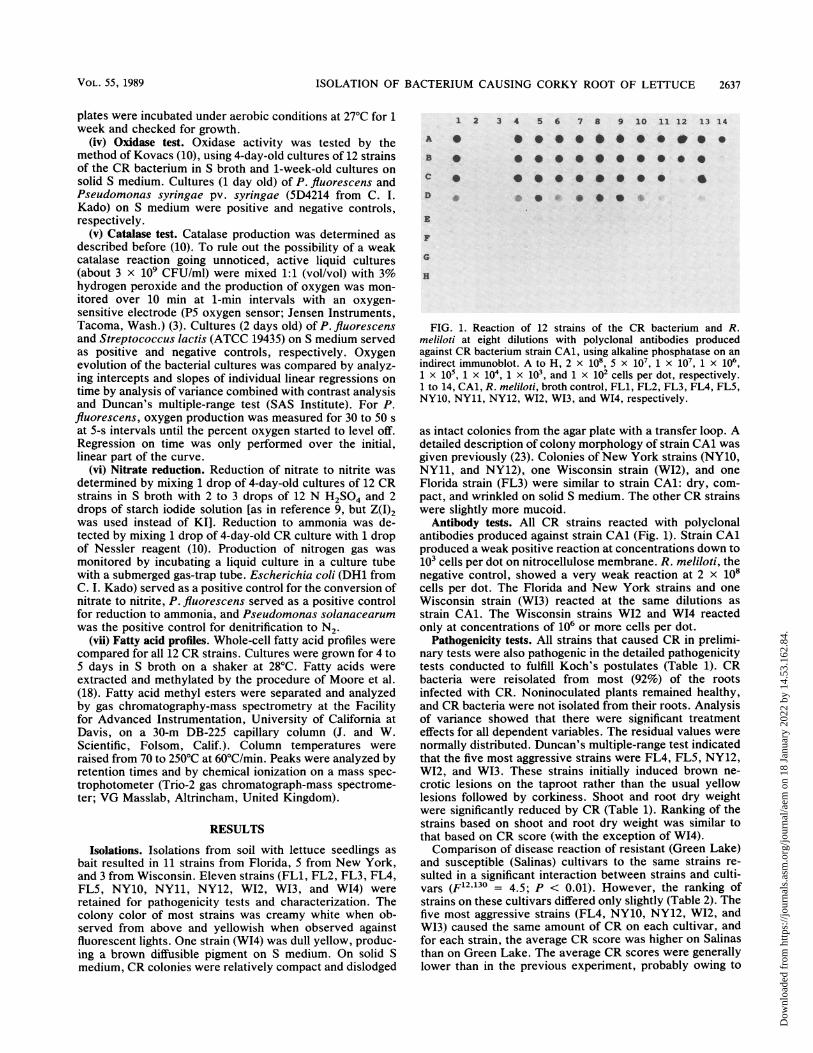
FIG. 1. Reaction of 12 strains of the CR bacterium and R.meliloti at eight dilutions with polyclonal antibodies producedagainst CR bacterium strain CAl, using alkaline phosphatase on anindirect immunoblot. A to H, 2 x 10⁸, 5 x 10⁷, 1 X 10⁷, 1 X 106,1 X X05,X104, 1 XiO', and 1 X 10² cells per dot, respectively.1 to 14, CAl, R. meliloti, broth control, FL1, FL2, FL3, FM4, FL5,NY10, NY11, NY12, W12, WI3, and WI4, respectively.
as intact colonies from the agar plate with a transfer loop. Adetailed description of colony morphology of strain CAl wasgiven previously (23). Colonies of New York strains (NY1o,NY11, and NY12), one Wisconsin strain (W12), and oneFlorida strain (FL3) were similar to strain CAl: dry, com-pact, and wrinkled on solid S medium. The other CR strainswere slightly more mucoid.
Antibody tests. All CR strains reacted with polyclonalantibodies produced against strain CAl (Fig. 1). Strain CAlproduced a weak positive reaction at concentrations down to103 cells per dot on nitrocellulose membrane. R. meliloti, thenegative control, showed a very weak reaction at 2 x 10⁸cells per dot. The Florida and New York strains and oneWisconsin strain (W13) reacted at the same dilutions asstrain CAl. The Wisconsin strains W12 and WC4 reactedonly at concentrations of 106 or more cells per dot.
Pathogenicity tests. All strains that caused CR in prelimi-nary tests were also pathogenic in the detailed pathogenicitytests conducted to fulfill Koch's postulates (Table 1). CRbacteria were reisolated from most (92%) of the rootsinfected with CR. Noninoculated plants remained healthy,and CR bacteria were not isolated from their roots. Analysisof variance showed that there were significant treatmenteffects for all dependent variables. The residual values werenormally distributed. Duncan's multiple-range test indicatedthat the five most aggressive strains were FL4, FL5, NY12,W12, and W13. These strains initially induced brown ne-crotic lesions on the taproot rather than the usual yellowlesions followed by corkiness. Shoot and root dry weightwere significantly reduced by CR (Table 1). Ranking of thestrains based on shoot and root dry weight was similar tothat based on CR score (with the exception of W14).Comparison of disease reaction of resistant (Green Lake)
and susceptible (Salinas) cultivars to the same strains re-sulted in a significant interaction between strains and culti-vars (F'12"130 = 4.5; P < 0.01). However, the ranking ofstrains on these cultivars differed only slightly (Table 2). Thefive most aggressive strains (FL4, NY10, NY12, W12, andWI3) caused the same amount of CR on each cultivar, andfor each strain, the average CR score was higher on Salinasthan on Green Lake. The average CR scores were generallylower than in the previous experiment, probably owing to
VOL. S5, 1989
Dow
nloa
ded
from
http
s://j
ourn
als.
asm
.org
/jour
nal/a
em o
n 18
Jan
uary
202
2 by
14.
53.1
62.8
4.
2638 VAN BRUGGEN ET AL.
TABLE 2. CR severity (0 to 9 scale), reisolation of CR bacteria, and dry weights of shoots and roots of lettuce cultivarsSalinas and Green Lake 21 days after inoculation with 12 strains of the CR bacterium
Cultivar Strain Avg CR Reisolation of Dry wt of Dry wt ofscore' CR bacteria' shoot (g)C root (mg)'
Salinas None 0.1 ± 0.4da 1 0.12 ± 0.01 abcd 16.0 ± 1.6 abcCAl 2.5 ± 1.7 bcd 3 0.15 ± 0.06 a 21.7 ± 9.9 abFL1 1.7 +0.8 b 3 0.14 ± 0.01 ab 24.7 ± 0.5 aFL2 1.3 ± 0.5 b 3 0.13 +0.02 abc 17.0 ± 5.4 abcFL3 1.5 ± 0.6 b 1 0.13 +0.03 abc 18.7 ± 2.9 abcFL4 4.7 ± 0.8 f 3 0.10 ± 0.01 bcd 16.3 ± 1.2 abcFL5 3.2 +0.8 cde 3 0.09 ± 0.02 cd 16.3 ± 2.6 abcNY10 3.3 ± 1.4 de 3 0.13 ± 0.01 abc 19.0 ± 5.0 abcNY11 1.5 ± 1.0 b 3 0.10 ± 0.01 bcd 11.7 ± 2.9 bcNY12 4.7 ± 0.8 f 3 0.13 ± 0.01 abc 19.3 ± 1.2 abcW12 3.5 ± 0.6 de 3 0.10 ± 0.01 abcd 14.7 ± 2.5 abcW13 4.2 ± 0.8 ef 3 0.11 ± 0.03 abcd 18.0 ± 5.0 abcW14 2.2 ± 1.0 bc 1 0.08 ± 0.01 d 9.7± 3.7 c
Green Lake None 0.0 ± 0.0 a 0 0.22 ± 0.04 ab 18.0 ± 5.9 cCAl 0.5 ± 0.8 bc 3 0.19 ± 0.06 abcd 30.7 ± 5.4 abFL1 1.2 ± 0.4 cde 3 0.21 ± 0.01 abc 30.3 ± 2.5 abFL2 1.2 ± 0.4 cde 3 0.21 ± 0.01 abc 31.0 ± 2.9 abFL3 0.8 ± 0.4 bcd 3 0.20 ± 0.01 abcd 28.3 ± 2.4 abcFL4 1.8 ± 1.0 efg 3 0.18 ± 0.02 bcd 26.0 ± 9.1 abcFL5 1.5 ± 0.6 cde 3 0.16 ± 0.00 d 23.3 ± 5.8 bcNY10 2.3 ± 0.5 gh 3 0.23 ± 0.01 a 37.7 ± 2.6 aNY11 1.3 ± 0.5 def 1 0.20 ± 0.01 abcd 32.7 ± 3.9 abNY12 1.8 ± 0.8 efg 3 0.21 ± 0.04 abc 35.0 ± 7.8 abW12 2.0 ± 0.6 fgh 3 0.18 ± 0.02 abcd 23.7 ± 4.7 bcW13 2.7 ± 0.5 h 3 0.21 ± 0.01 abc 33.0 ± 2.9 abW14 0.2 ± 0.4 ab 2 0.17 ± 0.00 cd 27.3 ± 2.5 abc
a Average of six plants. Means followed by different letters differ significantly (a = 0.05) according to Duncan's multiple-range test.b Number of plants out of three from which CR bacteria were reisolated.C Average of three plants. Means followed by different letters differ significantly (a = 0.05) according to Duncan's multiple-range test.d One plant with very slight yellowing.
lower concentrations of the inoculum. One uninoculatedplant of the Salinas cultivar became contaminated, and one
colony of the CR bacterium was isolated from that plant. Theoverall percentage of reisolation from infected plants was90%. Shoot and root dry weights were significantly affectedby cultivar and strain, but there was no significant interac-tion. The ranking according to Duncan's multiple-range testwas not consistent with that for CR scores. The dry weightsof the Green Lake cultivar were significantly higher thanthose of the Salinas cultivar (F1 52 = 225; P < 0.01).
Preliminary identification. All CR strains tested withHucker Gram stain were gram negative. However, the KOHstringiness test was variable for the CR strains (Table 3). AllNew York strains, two Florida strains, and one Wisconsinstrain showed a negative reaction (no stringiness), as did theCalifornia strain (CA1). The ability to form slimy strings inKOH was negatively correlated to the dryness of the strainson plates and the extent of flocculation in broth.None of the CR strains were able to grow when held under
anaerobic conditions for 3 weeks. However, the same cul-tures started to grow normally after removal from theanaerobiosis jar.The oxidase test was positive for all strains (Table 3).The catalase test was negative or very weakly positive
when visually assessed on plates or in broth. Oxygen evo-lution as measured with an oxygen electrode was muchslower for the CR strains than for the positive control (P.fluorescens), but the slopes of regression of percent oxygenon time were significantly higher for all CR strains than for S.lactis (P < 0.01). Thus, the catalase test was weakly positivefor all CR strains (Table 3).
All the strains produced nitrite and ammonia from nitrate,but none produced nitrogen gas (Table 3).The fatty acid profiles were very similar for all CR strains
(Table 4). They consisted of several saturated and unsatura-
TABLE 3. Reaction of strains of the CR bacterium and controlstrains (P. fluorescens, P. syringae, P. solanacearum,E. coli, and S. lactis) to the KOH stringiness, oxidase,
catalase, and nitrate reductase testsa
KOH Nitrate reductionStrain Oxidase Catalasebtest NN02 NH3 N2
P. fluorescens + + + - + NTP. syringae NT - NT NT NT NTP. solanacearum NT NT NT (+) + +E. coli NT - + + + -S. lactis NT NT - NT NT NTCAl - + (+) + + -
FL1 (+) + (+) + + -FL2 (+) + (+) + + -FL3 - + NT + + -FL4 (+) + (+) + + -FLS - + NT + + -NY10 - + (+) + + -NY11 - + NT + + -NY12 - + (+) + + -W12 - + (+) + + -W13 (+) + NT + + -W14 + + (+) + + -
a +, Positive reaction; -, negative reaction; (+), weak positive reaction;NT, not tested.
b Oxygen production measured with an oxygen electrode.
APPL. ENVIRON. MICROBIOL.
Dow
nloa
ded
from
http
s://j
ourn
als.
asm
.org
/jour
nal/a
em o
n 18
Jan
uary
202
2 by
14.
53.1
62.8
4.
ISOLATION OF BACTERIUM CAUSING CORKY ROOT OF LETTUCE 2639
TABLE 4. Fatty acid composition of whole cells of 12 strains of the CR bacterium as identified bygas chromatography-mass spectrophotometrya
% Fatty acid in:Fatty acid
CAl FLL1 FL2 FL FL4 FL5 NY10 NY1 NY12 W12 W13 W14
12:0-2-OH Tr13:0-2-OH Tr14:0-2-OH 6.1 8.4 8.5 6.9 7.7 5.9 5.7 9.1 4.1 7.5 5.6 6.015:0-2-OH 1.312:0 Tr Tr Tr Tr Tr Tr Tr Tr Tr Tr14:0 0.6 0.6 0.4 1.0 0.5 0.3 Tr 0.5 0.6 0.4 Tr 2.115:0 0.4 Tr Tr Tr 1.616:0 13.2 12.7 12.9 15.1 9.7 11.1 14.1 11.1 9.8 10.0 7.4 18.016:1 10.8 15.4 10.4 13.9 9.6 12.1 12.1 11.8 12.2 10.6 7.6 7.117:0 0.4 1.317:1 3.5 Tr Tr 0.6 1.9 14.3 0.918:1 56.5 58.5 63.0 61.0 71.4 68.2 62.3 62.7 64.2 69.2 57.7 65.918:1-10 CH3 5.9 4.2 4.2 Tr 0.5 2.4 5.3 3.7 6.4 2.1 2.7 Tr19:0-cyclo 0.2 Tr 0.3 Tr 0.7 Tr Tr Tr 0.8 0.3 Tr Tr
a Trace amounts not included in the calculation of the percentages.
ted straight-chain fatty acids, 2-hydroxy fatty acids, one
cyclopropane fatty acid, and one methylated fatty acid.3-Hydroxy fatty acids were not detected. The main straight-chain fatty acids had even numbers of carbon atoms (12:0,14:0, 16:0, 16:1, and 18:1) with the exception of 17:1, whichoccurred in most strains from California, New York, andWisconsin but not in those from Florida (except for a trace inFL1). CAl and WI3 were the only strains with measurableamounts of 15:0 and 17:0. The main hydroxy fatty acid was
14:0-2-OH for all strains. All strains also had 10-methyl 18:1fatty acid, and cyclopropane 19:0.
DISCUSSIONIn a previous paper (23), we demonstrated that infectious
CR of lettuce was caused by a gram-negative bacterium inCalifornia. In the current report, it was shown that similarbacteria resided in CR-prone soils from New York, Florida,and Wisconsin and that these bacteria induced CR on lettuceseedlings in the greenhouse. Although we have not tested theability of these bacteria to cause CR under field conditions inthe areas from which the soil originated (New York, Florida,and Wisconsin), it is likely that infectious CR is caused bybacteria similar to the California strain CAl.
All CR strains were pathogenic on the susceptible cultivarSalinas and to a lesser extent on the resistant cultivar GreenLake. The latter cultivar was developed in Wisconsin forresistance to CR at the time when the etiology of the diseasewas still unknown (21). Green Lake was later shown to beresistant to the California strain CAl of the CR bacterium(5). Green Lake and breeding lines derived from it were alsoresistant to CR in Florida (12, 13). These observations lendweight to our proposal that CR in Wisconsin and Florida iscaused by bacteria similar to those that cause CR in Califor-nia.
Resistance to CR in Green Lake and other cultivars andbreeding lines is conferred by a single recessive gene (5).When resistant and susceptible lettuce cultivars were inoc-ulated with various strains of the CR bacterium, there was a
statistically significant interaction between strains of the CRbacterium and cultivars in their effect on CR severity.However, disease severity on Green Lake was lower thanthat on Salinas for all strains, and the five most aggressivestrains were the same strains for Salinas and Green Lake.We conclude that thus far no differential races of the CRbacterium have been detected.
All bacterial strains that induced CR were gram negativewith Hucker Gram stain. The KOH stringiness test wasvariable. The New York strains and one Florida strainshowed a negative reaction, as did the first California strain(CA1) (23). This indicated that these strains might be grampositive, but we demonstrated that strain CAl containslipopolysaccharide (23). Thus, the KOH stringiness test isnot recommended for CR bacteria. Results with the tradi-tional catalase test (visual observation of oxygen production)were also variable. This may be related to a relatively slowgrowth rate and possibly a slow metabolism. With the use ofan oxygen electrode, we demonstrated that all strains of theCR bacterium were catalase positive. All CR strains reactedpositively in the oxidase and nitrate reductase tests.The fatty acid profiles were similar, consisting of several
saturated and unsaturated straight-chain fatty acids, 2-hy-droxy fatty acids, one cyclopropane fatty acid, and onemethylated fatty acid. The most characteristic peak of thefatty acid profiles was that of a hydroxy fatty acid (14:0-2-OH). A large peak of this component is characteristic forPseudomonas paucimobilis (19; M. Sasser, personal com-munication). However, the results of several other physio-logical and biochemical tests (unpublished data) indicate thatthe CR bacterium may not be the same as P. paucimobilis.No 3-OH fatty acids were detected. According to Oyaizuand Komagata (19), this is unusual for gram-negative bacte-ria. Another characteristic peak was that for 10-methyl 18:1fatty acid, which has been reported for Rhizobium species(M. Sasser, personal communication). Since this fatty acidpattern is unique, it is likely that CR of lettuce is caused bystrains of the same bacterium in Florida, New York, Wis-consin, and California.
ACKNOWLEDGMENTS
We thank Dan Jones of the Facility for Advanced Instrumenta-tion, University of California at Davis, for the analysis of fatty acids.We are grateful to Myron Sasser of Microbial ID, Newark, Del., forperforming some fatty acid analysis (to be published elsewhere) andfor suggesting a potential relatedness between the CR bacterium andP. paucimobilis. We thank C. I. Kado for providing bacterialcultures.
This work was supported by the California Iceberg LettuceResearch Board.
VOL. 55, 1989
Dow
nloa
ded
from
http
s://j
ourn
als.
asm
.org
/jour
nal/a
em o
n 18
Jan
uary
202
2 by
14.
53.1
62.8
4.
2640 VAN BRUGGEN ET AL.
LITERATURE CITED1. Amin, K. S., and L. Sequeira. 1966. Role of certain factors in the
etiology of corky root rot of lettuce. Phytopathology 56:1047-1053.
2. Amin, K. S., and L. Sequeira. 1966. Phytotoxic substances fromdecomposing lettuce residues in relation to the etiology of corkyroot rot of lettuce. Phytopathology 56:1054-1061.
3. Auling, G., M. Reh, C. M. Lee, and H. G. Schlegel. 1978.Pseudomonas pseudoflava, a new species of hydrogen-oxi-dizing bacteria: its differentiation from Pseudomonas flava andother yellow-pigmented, gram-negative, hydrogen-oxidizingspecies. Int. J. Syst. Bacteriol. 28:82-95.
4. Brown, N. A. 1918. Some bacterial diseases of lettuce. J. Agric.Res. 13:367-388.
5. Brown, P. R., and R. W. Michelmore. 1988. The genetics ofcorky root resistance in lettuce. Phytopathology 78:1145-1150.
6. Busch, L. V., and G. L. Barron. 1963. Root rot of head lettucein Ontario. Can. J. Plant Sci. 43:166-173.
7. D'Ercole, N. 1981. La suberosi radicale della lattuga. ColtureProtette 10:35-38.
8. D'Ercole, N. 1981. La suberosi radicale della lattuga. Terra eVita 41:40-41.
9. Fahy, P. C., and G. J. Persley. 1983. Plant bacterial diseases. Adiagnostic guide. Academic Press, Sydney.
10. Gerhardt, P., R. G. E. Murray, R. N. Costilow, E. W. Nester,W. A. Wood, N. R. Krieg, and G. B. Phillips (ed.) 1981. Manualof methods for general bacteriology. American Society forMicrobiology, Washington, D.C.
11. Grogan, R. G., and F. W. Zink. 1956. Fertilizer injury and itsrelationship to several previously described diseases of lettuce.Phytopathology 46:416-422.
12. Guzman, V. L. 1981. Yield and quality response of crispheadlettuce cultivars to seeding dates and farms in south Florida
organic soils. Proc. Fla. State Hortic. Soc. 94:182-185.13. Guzman, V. L. 1984. South Bay and Raleigh. Two crisphead
lettuce cultivars resistant to corky root rot for organic soils.Circular S-310. Agricultural Experiment Stations, Institute ofFood and Agricultural Science, University of Florida, Gaines-ville.
14. Hartnett, J. P., and J. W. Lorbeer. 1971. The production of anoninfectious lettuce root rot under controlled environmentaland soil conditions. Phytopathology 61:1153-1158.
15. Hoff, J. K., and A. G. Newhall. 1960. Corky root rot of iceberglettuce on the mucklands of New York. Plant Dis. Rep. 44:333-339.
16. MacNeill, B. H. 1953. A Botrytis root rot condition in lettuce.Plant Dis. Rep. 37:618-619.
17. Marlatt, R. B. 1955. Brown stele of lettuce. Plant Dis. Rep.39:827-828.
18. Moore, C. J., H. Mawhinney, and P. J. Blackall. 1987. Differ-entiation of Bordetella avium and related species by cellularfatty acid analysis. J. Clin. Microbiol. 25:1059-1062.
19. Oyaizu, H., and K. Komagata. 1983. Grouping of Pseudomonasspecies on the basis of cellular fatty acid composition and thequinone system with special reference to the existence of3-hydroxy fatty acids. J. Gen. Appl. Microbiol. 29:17-40.
20. Patterson, C. L., R. G. Grogan, and R. N. Campbell. 1986.Economically important diseases of lettuce. Plant Dis. 70:982-987.
21. Sequeira, L. 1970. Resistance to corky root rot in lettuce. PlantDis. Rep. 54:754-758.
22. Thomas, R. C. 1922. A bacterial rosette disease of lettuce. OhioAgric. Exp. Stn. Bull. 359:197-214.
23. van Bruggen, A. H. C., R. G. Grogan, C. P. Bogdanoff, andC. M. Waters. 1988. Corky root of lettuce in California causedby a gram-negative bacterium. Phytopathology 78:1139-1145.
APPL. ENVIRON. MICROBIOL.
Dow
nloa
ded
from
http
s://j
ourn
als.
asm
.org
/jour
nal/a
em o
n 18
Jan
uary
202
2 by
14.
53.1
62.8
4.