Embed Size (px)
Citation preview
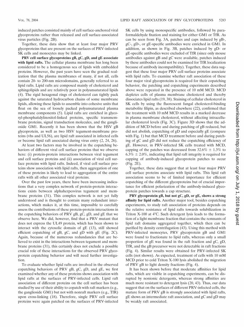
JOURNAL OF VIROLOGY, May 2004, p. 5279–5287 Vol. 78, No. 100022-538X/04/$08.00�0 DOI: 10.1128/JVI.78.10.5279–5287.2004Copyright © 2004, American Society for Microbiology. All Rights Reserved.
Copatching and Lipid Raft Association of Different Viral GlycoproteinsExpressed on the Surfaces of Pseudorabies Virus-Infected Cells
Herman W. Favoreel,1,2* Thomas C. Mettenleiter,3 and Hans J. Nauwynck1
Laboratory of Virology1 and Laboratory of Immunology,2 Faculty of Veterinary Medicine, Ghent University,Merelbeke, Belgium, and Federal Research Centre for Virus Diseases of Animals, Insel Riems, Germany3
Received 19 September 2003/Accepted 26 January 2004
Pseudorabies virus (PRV) is a swine alphaherpesvirus that is closely related to human herpes simplex virus(HSV). Both PRV and HSV express a variety of viral envelope glycoproteins in the plasma membranes ofinfected cells. Here we show that at least four major PRV glycoproteins (gB, gC, gD, and gE) in the plasmamembrane of infected swine kidney cells and monocytes seem to be linked, since monospecific antibody-inducedpatching of any one of these proteins results in copatching of the others. Further, for all four PRV glycopro-teins, monospecific antibody-induced patches were enriched in GM1, a typical marker of lipid raft microdo-mains, but were excluded for transferrin receptor, a nonraft marker, suggesting that these viral proteins mayassociate with lipid rafts. However, only gB and, to a lesser extent, gE were found in lipid raft fractions by usingdetergent floatation assays, indicating that gC and gD do not show strong lipid raft association. Addition ofmethyl-�-cyclodextrin (MCD), a cholesterol-depleting agent that is commonly used to disrupt lipid rafts, onlyslightly reduced copatching efficiency between the different viral proteins, indicating that other factors, perhapstegument-glycoprotein interactions, may be important for the observed copatching events. On the other hand,MCD strongly reduced polarization of the antibody-induced viral glycoprotein patches to a cap structure, agE-dependent process that has been described for specific PRV- and HSV-infected cells. Therefore, we hy-pothesize that efficient gE-mediated capping of antibody-antigen patches may require the lipid raft-associatedsignal transduction machinery.
Pseudorabies virus (PRV) is a swine alphaherpesvirus that isclosely related to the prototypical herpes simplex virus (HSV).Like that of HSV, the PRV envelope contains at least 10different viral envelope glycoproteins, designated glycoproteinB (gB), gC, gD, gE, gH, gI, gK, gL, gM, and gN. Upon infec-tion of a susceptible cell, newly synthesized viral envelopeglycoproteins travel through the Golgi network and are subse-quently incorporated in the plasma membrane, rendering thecells recognizable for virus-specific antibodies.
Earlier, we and others have shown that the interactionbetween PRV or HSV polyclonal serum antibodies and theviral proteins on the cell surface initiates intriguing, cell type-dependent redistribution processes of the antibody-antigencomplexes (8, 10, 39). Addition of polyclonal serum immuno-globulin G (IgG) to PRV-infected swine kidney (SK) cells orHSV-infected human embryonic lung fibroblasts (HEL cells)and human larynx epidermoid carcinoma (Hep-2) cells leads toaggregation of the viral cell surface proteins into patches,which subsequently polarize to one side of the cell to form acap (10, 39), sometimes followed by shedding of the caps (10).In PRV-infected blood monocytes, on the other hand, patchesof viral cell surface proteins are rapidly internalized and donot cap (8). The exact function of these processes is not fullyunderstood, although there are strong indications that theymay be important for alphaherpesviruses to enhance virus sur-vival in the face of an antibody response. First, the internal-ization of patches of viral cell surface proteins in PRV-infected
monocytes has been shown to interfere with efficient antibody-dependent lysis of the infected monocytes (45). Further,capping of antibody-antigen patches has been suggested tobe related to antibody-dependent enhancement of cell-to-cell spread of HSV (39). Also, it has been shown for PRV-infected monocytes that the interaction with virus-specific an-tibodies ultimately leads to suppression of intracellular viralprotein levels, perhaps even leading to a quiescent, persistentform of infection (11).
Both types of redistribution of the antibody-induced patchesof viral cell surface proteins, capping and internalization, havebeen shown to be initiated by specific viral cell surface pro-teins. In both PRV- and HSV-infected cells, efficient cappinghas been shown to depend on the presence of viral protein gE,whereas internalization has been demonstrated to be initiatedmainly by viral proteins gB and gD and to a lesser extent gE (8,10, 39). Exactly how these viral proteins initiate the redistri-bution processes is not fully understood, but it has been shownthat tyrosine-based amino acid motifs in the cytoplasmic tailsof PRV gB and gE are of crucial importance for internalizationand capping, respectively (7, 9). In addition, PRV gE-mediatedcapping has been suggested to require specific tyrosine kinasesignaling (9).
Interestingly, the addition of PRV- or HSV-specific serumantibodies does not result in exclusive capping or internaliza-tion of the viral proteins that initiate these processes but alsoresults in that of the other viral cell surface proteins that arerecognized by the immune serum (8, 10, 39). One explanationfor this massive change in surface viral glycoprotein distribu-tion initiated by single or a few viral proteins may be that(some of) the major viral envelope proteins that are present onthe surfaces of PRV- or HSV-infected cells are somehow
* Corresponding author. Mailing address: Laboratories of Virologyand Immunology, Faculty of Veterinary Medicine, Ghent University,Salisburylaan 133, 9820 Merelbeke, Belgium. Phone: 32 9 264 73 74.Fax: 32 9 264 74 95. E-mail: [email protected].
5279
on July 1, 2018 by guesthttp://jvi.asm
.org/D
ownloaded from
linked, allowing one or a few viral proteins to initiate lateralmovement of many, if not all, of the viral proteins in the cellsurface.
The aim of the present study was therefore (i) to studywhether such a putative link between several of the major viralcell surface proteins indeed does exist, by examining whetherpatching of single viral proteins on the cell surface by theaddition of monospecific antibodies to PRV-infected cellsleads to copatching of other viral proteins, and (ii) if so, toobtain indications about the nature of such a link between thedifferent viral cell surface proteins.
MATERIALS AND METHODS
Antibodies and reagents. Mouse monoclonal antibodies directed against gB(1C11), gD (13D12), and gE (18E8) were all described earlier (34) and were usedat a dilution of 1/30 (1C11) or 1/100 (13D12 and 18E8). Mouse monoclonalanti-gC antibody was kindly provided by A. Brun and used at a dilution of 1/100.Polyclonal monospecific antibodies were used at a dilution of 1/50 and werekindly provided by S. Brockmeier (swine anti-gD) and K. Bienkowska-Szewczyk(rabbit anti-gE). Mouse anti-transferrin receptor (anti-TfR) was purchased fromZymed Laboratories, Inc. (San Francisco, Calif.) and diluted 1/100. Biotinylatedcholera toxin B subunit (Sigma Chemical Co., St. Louis, Mo.) was used at adilution of 1/100. Fluorescein isothiocyanate (FITC)-labeled goat anti-mouseantibodies (used at a dilution of 1/100) and Texas red-labeled goat anti-mouseand anti-rabbit antibodies and streptavidin (all used at a dilution of 1/50) were allpurchased from Molecular Probes (Eugene, Oreg.). Texas red-labeled goat anti-swine antibodies (Jackson Immunologicals, West Grove, Pa.) were used at adilution of 1/50. Biotinylated sheep anti-mouse antibodies (Amersham Bio-sciences, Buckinghamshire, United Kingdom) were used at a dilution of 1/100,and peroxidase-conjugated streptavidin-biotin complex (Amersham Biosciences)was used at a dilution of 1/200. Methyl-�-cyclodextrin (MCD) and Triton X-100were purchased from Sigma.
Copatching experiments. Isolation of porcine blood monocytes, in vitro culti-vation of SK cells and monocytes, and in vitro PRV inoculation of SK cells andmonocytes were carried out as described before (8, 10). PRV strains 89V87,Kaplan, Kaplan gE-gI null, Kaplan ICP18.5 null, and Kaplan UL49 null wereused, and all were described before (13, 21, 29, 30, 33). At 13 h postinfection(p.i.), cells were washed three times in phosphate-buffered saline (PBS), followedby incubation for 30 min at 37°C with primary antibodies (monoclonal anti-gB,-gC, -gD, and/or -gE antibodies or polyclonal rabbit anti-gE or swine anti-gDantibodies as indicated). Afterwards, cells were washed twice in PBS, and incu-bated with the appropriate secondary antibodies (FITC-labeled anti-mouse oranti-rabbit antibodies) for 30 min at 37°C. The cells were then either parafor-maldehyde fixed (2% paraformaldehyde, 10 min, room temperature) or, forlive-cell imaging, washed in ice-cold PBS. Cell were then washed in ice-cold PBSand incubated with polyclonal swine anti-gD antibodies, polyclonal rabbit an-ti-gE antibodies, mouse monoclonal anti-TfR antibody, or biotinylated choleratoxin B subunit for 1 h on ice. Thereafter, cells were washed twice in ice-cold PBSand incubated for 1 h on ice with Texas red-labeled antibodies (anti-swine,anti-rabbit, or anti-mouse) or Texas red-labeled streptavidin. Afterwards, cellswere washed twice in ice-cold PBS and resuspended in 40 �l of ice-cold PBS. Forfixed cells, cells were mounted in a glycerin-PBS solution (0.9:0.1, vol/vol) with2.5% 1,4-diazabicyclo(2.2.2)octane (Janssen Chimica, Beerse, Belgium) on amicroscope slide and analyzed by confocal microscopy. For the analysis of livecells, two coverslips were mounted on a 3-aminopropyltriethoxysilane-coatedmicroscope slide, leaving a 0.5-cm gap between the two coverslips. The gap was
filled with 15 �l of cell suspension and covered with a third coverslip. Cells wereimmediately analyzed by confocal microscopy.
Confocal microscopy. Cells were analyzed by using a TCS SP2 laser scanningspectrum confocal system (Leica Microsystems GmbH, Heidelberg, Germany),using an argon 488-nm laser line and a Gre/Ne 543-nm laser line to excite FITCand Texas red, respectively. To avoid signal overlap, FITC and Texas red imageswere taken separately, and images stained with FITC only and Texas red onlyserved as controls. Images were merged by using Leica confocal and ConfocalAssistant software.
Lipid raft floatation assay. Isolation of porcine blood monocytes, in vitrocultivation of SK cells and monocytes, and in vitro PRV inoculation (PRV strain89V87) of SK cells and monocytes were carried out as described before (8, 10).At 13 h p.i., cells were washed three times in ice-cold TNE (25 mM Tris HCl, 150mM NaCl, 5 mM EDTA [pH 6.7]), and subsequently 107 cells were lysed for 30min on ice, with regular shaking, in 1 ml of lysis buffer (1% Triton X-100 in TNEsupplemented with complete protease inhibitor cocktail [Roche DiagnosticsGmbH, Mannheim, Germany]). Afterwards, the lysate was homogenized bybeing passed 20 times through an 18-gauge needle on a 1-ml syringe and subse-quently mixed with 2 ml of ice-cold 60% Optiprep (Nycomed-Pharam, Oslo,Norway). This mixture was put at the bottom of a Beckman SW41 ultracentrifugetube (Beckman, Munich, Germany), overlaid with 5 ml of ice-cold 35% Optiprepin TNE and 3 ml of ice-cold 5% Optiprep in TNE, and centrifuged at 200,000 �g at 4°C for 20 h. Ten fractions from the top to the bottom of the tube werecollected and diluted 1:2 in 2� concentrated nonreducing sodium dodecyl sul-fate-polyacrylamide gel electrophoresis (SDS-PAGE) loading buffer. Fifteen mi-croliters of each diluted fraction was then subjected to SDS-PAGE and Westernblotting. The blots were blocked in PBS with 0.1% Tween 20 (Sigma) (PBS-T)supplemented with 5% membrane blocking agent (Amersham Biosciences) andwashed twice in PBS-T. Afterwards, blots were incubated with different mono-clonal antibodies (anti-gB, -gC, -gD, -gE, or -TfR) or biotinylated cholera toxinsubunit B in PBS-T for 1 h, washed three times in PBS-T, and incubated withbiotinylated sheep anti-mouse antibodies and/or peroxidase-conjugated strepta-vidin-biotin complex for 1 h, with three washing steps between each incubation.Finally, blots were washed three times in PBS-T and revealed with 3,3�-diami-nobenzidine (Sigma).
RESULTS
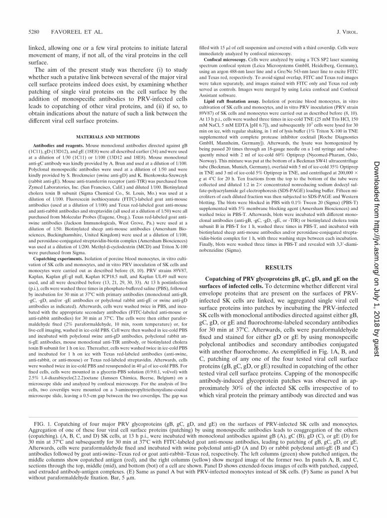
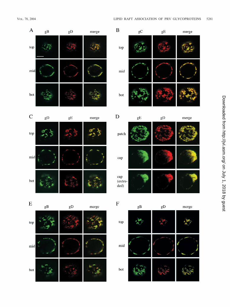
Copatching of PRV glycoproteins gB, gC, gD, and gE on thesurfaces of infected cells. To determine whether different viralenvelope proteins that are present on the surfaces of PRV-infected SK cells are linked, we aggregated single viral cellsurface proteins into patches by incubating the PRV-infectedSK cells with monoclonal antibodies directed against either gB,gC, gD, or gE and fluorochrome-labeled secondary antibodiesfor 30 min at 37°C. Afterwards, cells were paraformaldehydefixed and stained for either gD or gE by using monospecificpolyclonal antibodies and secondary antibodies conjugatedwith another fluorochrome. As exemplified in Fig. 1A, B, andC, patching of any one of the four tested viral cell surfaceproteins (gB, gC, gD, or gE) resulted in copatching of the othertested viral cell surface proteins. Capping of the monospecificantibody-induced glycoprotein patches was observed in ap-proximately 30% of the infected SK cells irrespective of towhich viral protein the primary antibody was directed and was
FIG. 1. Copatching of four major PRV glycoproteins (gB, gC, gD, and gE) on the surfaces of PRV-infected SK cells and monocytes.Aggregation of one of these four viral cell surface proteins (patching) by using monospecific antibodies leads to coaggregation of the others(copatching). (A, B, C, and D) SK cells, at 13 h p.i., were incubated with monoclonal antibodies against gB (A), gC (B), gD (C), or gE (D) for30 min at 37°C and subsequently for 30 min at 37°C with FITC-labeled goat anti-mouse antibodies, leading to patching of gB, gC, gD, or gE.Afterwards, cells were paraformaldehyde fixed and incubated with swine polyclonal anti-gD (A and D) or rabbit polyclonal anti-gE (B and C)antibodies followed by goat anti-swine–Texas red or goat anti-rabbit–Texas red, respectively. The left columns (green) show patched antigen, themiddle columns show copatched antigen (red), and the right columns (yellow) show merged image of the former two. In panels A, B, and C,sections through the top, middle (mid), and bottom (bot) of a cell are shown. Panel D shows extended-focus images of cells with patched, capped,and extruded antibody-antigen complexes. (E) Same as panel A but with PRV-infected monocytes instead of SK cells. (F) Same as panel A butwithout paraformaldehyde fixation. Bar, 5 �m.
5280 FAVOREEL ET AL. J. VIROL.
on July 1, 2018 by guesthttp://jvi.asm
.org/D
ownloaded from
VOL. 78, 2004 LIPID RAFT ASSOCIATION OF PRV GLYCOPROTEINS 5281
on July 1, 2018 by guesthttp://jvi.asm
.org/D
ownloaded from
also accompanied by cocapping of the other viral proteins, asillustrated in Fig. 1D. Similarly, patching of either gB, gC, gD,or gE on PRV-infected monocytes also resulted in copatchingof the other viral cell surface proteins (Fig. 1E and data notshown).
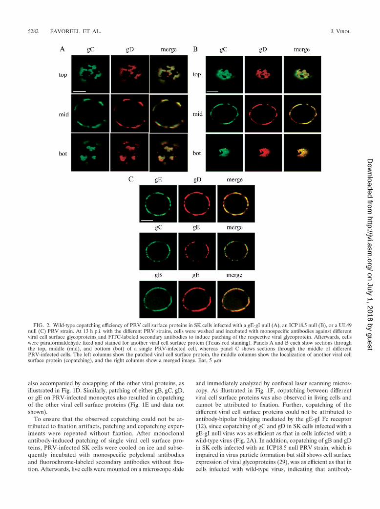
To ensure that the observed copatching could not be at-tributed to fixation artifacts, patching and copatching exper-iments were repeated without fixation. After monoclonalantibody-induced patching of single viral cell surface pro-teins, PRV-infected SK cells were cooled on ice and subse-quently incubated with monospecific polyclonal antibodiesand fluorochrome-labeled secondary antibodies without fixa-tion. Afterwards, live cells were mounted on a microscope slide
and immediately analyzed by confocal laser scanning micros-copy. As illustrated in Fig. 1F, copatching between differentviral cell surface proteins was also observed in living cells andcannot be attributed to fixation. Further, copatching of thedifferent viral cell surface proteins could not be attributed toantibody-bipolar bridging mediated by the gE-gI Fc receptor(12), since copatching of gC and gD in SK cells infected with agE-gI null virus was as efficient as that in cells infected with awild-type virus (Fig. 2A). In addition, copatching of gB and gDin SK cells infected with an ICP18.5 null PRV strain, which isimpaired in virus particle formation but still shows cell surfaceexpression of viral glycoproteins (29), was as efficient as that incells infected with wild-type virus, indicating that antibody-
FIG. 2. Wild-type copatching efficiency of PRV cell surface proteins in SK cells infected with a gE-gI null (A), an ICP18.5 null (B), or a UL49null (C) PRV strain. At 13 h p.i. with the different PRV strains, cells were washed and incubated with monospecific antibodies against differentviral cell surface glycoproteins and FITC-labeled secondary antibodies to induce patching of the respective viral glycoprotein. Afterwards, cellswere paraformaldehyde fixed and stained for another viral cell surface protein (Texas red staining). Panels A and B each show sections throughthe top, middle (mid), and bottom (bot) of a single PRV-infected cell, whereas panel C shows sections through the middle of differentPRV-infected cells. The left columns show the patched viral cell surface protein, the middle columns show the localization of another viral cellsurface protein (copatching), and the right columns show a merged image. Bar, 5 �m.
5282 FAVOREEL ET AL. J. VIROL.
on July 1, 2018 by guesthttp://jvi.asm
.org/D
ownloaded from
induced patches consisted mainly of cell surface-anchored viralglycoproteins rather than released and cell surface-associatedvirus particles (Fig. 2B).
Together, these data show that at least four major PRVglycoproteins that are present on the surfaces of PRV-infectedSK cells and monocytes may be linked.
PRV cell surface glycoproteins gB, gC, gD, and gE associatewith lipid rafts. The cellular plasma membrane has long beenconsidered to be a homogenous sea of lipids with embeddedproteins. However, the past years have seen the gradual real-ization that the plasma membranes of many, if not all, cellscontain 20- to 200-nm microdomains, generally referred to aslipid rafts. Lipid rafts are composed mainly of cholesterol andsphingolipids and are relatively poor in polyunsaturated lipids(4). The rigid hexagonal rings of cholesterol can tightly packagainst the saturated hydrocarbon chains of some membranelipids, allowing these lipids to assemble into cohesive units thatfloat on the sea of loosely packed polyunsaturated plasmamembrane components (5). Lipid rafts are enriched in glyco-syl-phosphatidylinositol-linked proteins, specific transmem-brane proteins, signal transduction molecules, and the gangli-oside GM1. Recently it has been shown that the HSV gBglycoprotein, as well as two HSV tegument-membrane pro-teins (vhs and UL56), are lipid raft associated in infected cellsor become lipid raft associated during virus entry (2, 24, 26).
At least two factors may be involved in the copatching be-haviors of different viral cell surface proteins that we observehere: (i) protein-protein interactions between viral tegumentand cell surface proteins and (ii) association of viral cell sur-face proteins with lipid rafts. Indeed, if viral cell surface pro-teins show association with lipid rafts, then aggregation of oneof these proteins is likely to lead to aggregation of the entirerafts with all other associated viral proteins.
Over the past few years, there have been increasing indica-tions that a very complex network of protein-protein interac-tions exists between alphaherpesvirus tegument and mem-brane proteins (31). This complex network is not yet fullyunderstood and is thought to contain many redundant inter-actions, which makes it, at this time, impossible to carefullyassess the contributions of these protein-protein interactions inthe copatching behaviors of PRV gB, gC, gD, and gE that weobserve here. We did, however, find that a PRV mutant thatdoes not express the UL49 protein, which has been shown tointeract with the cytosolic domain of gE (13), still showedefficient copatching of gB, gC, and gD with gE (Fig. 2C).Again, because of the numerous redundancies that are be-lieved to exist in the interactions between tegument and mem-brane proteins (31), this certainly does not exclude a possiblecrucial role of these interactions for the observed PRV glyco-protein copatching behavior and will need further investiga-tion.
To evaluate whether lipid rafts are involved in the observedcopatching behaviors of PRV gB, gC, gD, and gE, we firstexamined whether any of these proteins shows association withlipid rafts at the surfaces of PRV-infected cells. Lipid raftassociation of different proteins on the cell surface has beenstudied by use of their ability to copatch with raft markers (e.g.,the ganglioside GM1) but not with nonraft markers (e.g., TfR)upon cross-linking (18). Therefore, single PRV cell surfaceproteins were again patched on the surfaces of PRV-infected
SK cells by using monospecific antibodies, followed by para-formaldehyde fixation and staining for either GM1 or TfR. Ascan be seen from Fig. 3A, patches and caps induced by gB-,gC-, gD-, or gE-specific antibodies were enriched in GM1. Inaddition, as shown in Fig. 3B, patches induced by gD- orgE-specific antibodies were excluded of TfR (since only mouseantibodies against gB and gC were available, patches inducedby these antibodies could not be examined for TfR localizationbecause of antibody incompatibility). Together, these data sug-gest that these four major PRV cell surface proteins associatewith lipid rafts. To examine whether raft association of thesefour major viral glycoproteins is required for their copatchingbehavior, the patching and copatching experiments describedabove were repeated in the presence of 10 mM MCD. MCDdepletes cells of plasma membrane cholesterol and therebydissociates lipid rafts (19, 38). Staining of cellular cholesterol inSK cells by using the fluorescent fungal cholesterol-bindingmetabolite filipin, as described elsewhere (22), confirmed thatthe treatment with 10 mM MCD results in a marked decreasein plasma membrane cholesterol, without affecting intracellu-lar cholesterol levels (Fig. 3C). Figure 3D shows that the ad-dition of MCD before and during patching of gB reduced, butdid not abolish, copatching of gD and especially gE (comparewith Fig. 1) but that MCD treatment before and during patch-ing of gC and gD did not reduce the copatching efficiency ofgE. However, in PRV-infected SK cells treated with MCD,capping of the patches was decreased from 32.6% � 1.3% to6.3% � 2.0%, indicating that lipid raft integrity is required forcapping of antibody-induced glycoprotein patches to occur(Fig. 2E).
Together, these data suggest that at least four major PRVcell surface proteins associate with lipid rafts. This lipid raftassociation seems to be of limited importance for efficientcopatching of the different glycoproteins but of crucial impor-tance for efficient polarization of the antibody-induced glyco-protein patches towards a cap structure.
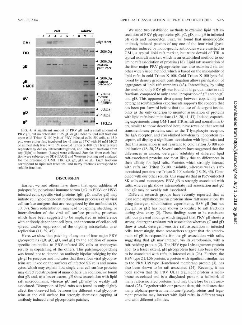
PRV glycoprotein gB, but not gC, gD, or gE, shows a strongaffinity for lipid rafts. Another major tool, besides copatchingexperiments, to study raft association of proteins depends onthe relative resistance of lipid rafts towards solubilization inTriton X-100 at 4°C. Such detergent lysis leads to the forma-tion of a light membrane fraction that contains the remnants oflipid raft domains aggregated together, which then can bepurified by density centrifugation (43). Using this method withPRV-infected monocytes, PRV glycoprotein gB and GM1were found to fractionate to lipid rafts, whereas only a smallproportion of gE was found in the raft fraction and gC, gD,TfR, and the gB precursor were not detectable in raft fractions(Fig. 4). Similar results were obtained for PRV-infected SKcells (not shown). As expected, treatment of cells with 10 mMMCD prior to cold Triton X-100 lysis abolished the migrationof PRV gB to light density fractions (Fig. 4).
It has been shown before that moderate affinities for lipidrafts, which are visible in copatching experiments, can be dis-rupted by nonionic detergents, whereas strong affinities aremuch more resistant to detergent lysis (20, 43). Thus, our datasuggest that on the surfaces of different PRV-infected cells, themature form of PRV gB is strongly associated with lipid rafts,gE shows an intermediate raft association, and gC and gD maybe weakly raft associated.
VOL. 78, 2004 LIPID RAFT ASSOCIATION OF PRV GLYCOPROTEINS 5283
on July 1, 2018 by guesthttp://jvi.asm
.org/D
ownloaded from
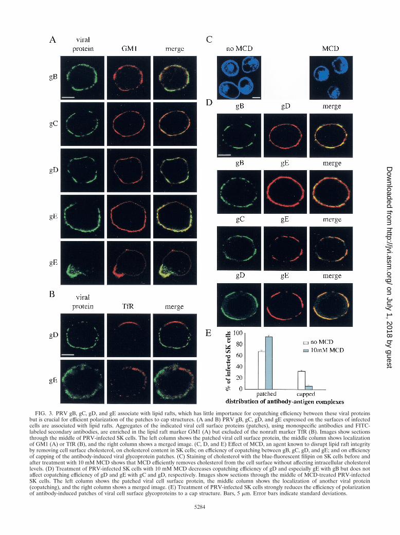
FIG. 3. PRV gB, gC, gD, and gE associate with lipid rafts, which has little importance for copatching efficiency between these viral proteinsbut is crucial for efficient polarization of the patches to cap structures. (A and B) PRV gB, gC, gD, and gE expressed on the surfaces of infectedcells are associated with lipid rafts. Aggregates of the indicated viral cell surface proteins (patches), using monospecific antibodies and FITC-labeled secondary antibodies, are enriched in the lipid raft marker GM1 (A) but excluded of the nonraft marker TfR (B). Images show sectionsthrough the middle of PRV-infected SK cells. The left column shows the patched viral cell surface protein, the middle column shows localizationof GM1 (A) or TfR (B), and the right column shows a merged image. (C, D, and E) Effect of MCD, an agent known to disrupt lipid raft integrityby removing cell surface cholesterol, on cholesterol content in SK cells; on efficiency of copatching between gB, gC, gD, and gE; and on efficiencyof capping of the antibody-induced viral glycoprotein patches. (C) Staining of cholesterol with the blue-fluorescent filipin on SK cells before andafter treatment with 10 mM MCD shows that MCD efficiently removes cholesterol from the cell surface without affecting intracellular cholesterollevels. (D) Treatment of PRV-infected SK cells with 10 mM MCD decreases copatching efficiency of gD and especially gE with gB but does notaffect copatching efficiency of gD and gE with gC and gD, respectively. Images show sections through the middle of MCD-treated PRV-infectedSK cells. The left column shows the patched viral cell surface protein, the middle column shows the localization of another viral protein(copatching), and the right column shows a merged image. (E) Treatment of PRV-infected SK cells strongly reduces the efficiency of polarizationof antibody-induced patches of viral cell surface glycoproteins to a cap structure. Bars, 5 �m. Error bars indicate standard deviations.
5284
on July 1, 2018 by guesthttp://jvi.asm
.org/D
ownloaded from
DISCUSSION
Earlier, we and others have shown that upon addition ofpolyspecific, polyclonal immune serum IgG to PRV- or HSV-infected cells, specific viral proteins (gB, gD, and/or gE) mayinitiate cell type-dependent redistribution processes of all viralcell surface antigens that are recognized by the antibodies (8,10, 39). Such redistribution may lead to capping, shedding, orinternalization of the viral cell surface proteins, processeswhich have been suggested to be implicated in interferencewith antibody-dependent cell lysis, enhancement of cell-to-cellspread, and/or suppression of the ongoing intracellular virusreplication (11, 39, 45).
Here, we show that patching of any one of four major PRVglycoproteins (gB, gC, gD, and gE) by the addition of mono-specific antibodies to PRV-infected SK cells or monocytesresults in copatching of the others. This patching-copatchingwas found not to depend on antibody bipolar bridging by thegE-gI Fc receptor and indicates that these four viral glycopro-teins are linked on the surfaces of infected SK cells and mono-cytes, which may explain how single viral cell surface proteinsmay direct redistribution of many others. In addition, we foundthat gB and, to a lesser extent, gE show association with lipidraft microdomains, whereas gC and gD may be weakly raftassociated. Disruption of lipid rafts was found to only slightlyaffect the observed link between the different viral glycopro-teins at the cell surface but strongly decreased capping ofantibody-induced viral glycoprotein patches.
We used two established methods to examine lipid raft as-sociation of PRV glycoproteins gB, gC, gD, and gE in infectedSK cells and monocytes. First, we found that monospecificantibody-induced patches of any one of the four viral glyco-proteins induced by monospecific antibodies were enriched inGM1, a typical lipid raft marker, but were devoid of TfR, atypical nonraft marker, which is an established method to ex-amine raft association of proteins (18). Lipid raft association ofthe four major PRV glycoproteins was also examined via an-other widely used method, which is based on the insolubility oflipid rafts in cold Triton X-100. Cold Triton X-100 lysis fol-lowed by density gradient centrifugation allows purification ofaggregates of lipid raft remnants (43). Interestingly, by usingthis method, only PRV gB was found in large quantities in raftfractions, compared to only a small proportion of gE and no gCand gD. This apparent discrepancy between copatching anddetergent solubilization experiments supports the concern thathas been put forward before that the use of detergent insolu-bility as the only criterion to monitor association of proteinswith lipid rafts has limitations (18, 20, 41, 43). Indeed, copatch-ing experiments using GM-1 and TfR as raft and nonraft mark-ers, similar to those described here, have revealed that severaltransmembrane proteins, such as the T lymphocyte receptor,the IgA receptor, and cross-linked low-density lipoprotein re-ceptor, all display a significant association with lipid rafts andthat this association is not resistant to cold Triton X-100 sol-ubilization (18, 20, 25). Several authors have suggested that thedifferences in anionic detergent solubility of different lipidraft-associated proteins are most likely due to differences intheir affinity for lipid rafts. Proteins which strongly interactwith rafts are Triton X-100 insoluble, whereas weakly raft-associated proteins are Triton X-100 soluble (18, 20, 43). Com-bined with our other results, this suggests that in PRV-infectedSK cells and monocytes, PRV gB is strongly associated withrafts, whereas gE shows intermediate raft association and gCand gD may be weakly raft associated.
Different research groups have recently reported that atleast some alphaherpesvirus proteins show raft association. Byusing detergent solubilization experiments, HSV gB (but notgC, gD, or gH) has been shown to localize to raft fractionsduring virus entry (2). These findings seem to be consistentwith our present findings which suggest that PRV gB shows astrong, detergent-resistant raft association whereas gC and gDshow a weak, detergent-sensitive raft association in infectedcells. Interestingly, those researchers suggest that the ectodo-main of gB is responsible for the gB association with rafts,suggesting that gB may interact, via its ectodomain, with araft-residing protein (2). The HSV type 1 vhs tegument proteinand, to a lesser extent, gH glycoprotein have also been shownto be associated with rafts in infected cells (26). Further, theHSV type 2 UL56 protein, a protein with significant similaritiesto the PRV Us9 type II-anchored membrane protein (3), hasalso been shown to be raft associated (24). Recently, it hasbeen shown that the PRV UL11 tegument protein is mem-brane associated and is a diacylated protein, a hallmark ofmany raft-associated proteins, and may therefore be raft asso-ciated (23). Together with our present data, this indicates thatmany alphaherpesvirus membrane (glyco)proteins and tegu-ment proteins may interact with lipid rafts, in different waysand with different affinities.
FIG. 4. A significant amount of PRV gB and a small amount ofPRV gE, but no detectable PRV gC or gD, float to lipid raft fractionsupon cold Triton X-100 lysis of PRV-infected cells. SK cells, at 13 hp.i., were either first incubated for 45 min at 37°C with 10 mM MCDor immediately lysed with 1% ice-cold Triton X-100. Cell lysates wereseparated by density ultracentrifugation, and different fractions fromtop (light) to bottom (heavy) were collected. Samples from each frac-tion were subjected to SDS-PAGE and Western blotting and analyzedfor the presence of GM1, TfR, gB, gC, gD, or gE. Light fractionscorrespond to lipid raft fractions, and heavy fractions correspond tosoluble fractions.
VOL. 78, 2004 LIPID RAFT ASSOCIATION OF PRV GLYCOPROTEINS 5285
on July 1, 2018 by guesthttp://jvi.asm
.org/D
ownloaded from
Although the exact determinants that direct specific proteinsto lipid rafts are very diverse and still largely unresolved, itseems unlikely that all of these alphaherpesvirus proteins con-tain specific signals that link them directly to lipid rafts. Al-though it is speculative, an attractive alternative explanationmay be that the numerous protein-protein interactions whichare believed to exist between tegument proteins and the cyto-plasmic domains of viral membrane (glyco)proteins (31) mayallow a specific subset of strongly raft-associated membrane(glyco)proteins and tegument proteins to serve as nucleationsites to concentrate many of the others indirectly to rafts.Indeed, it is known that proteins, such as Fc gamma receptors,can be indirectly raft associated through their affinity for otherraft-associated proteins (16). Importantly, since lipid raft for-mation and protein association with lipid rafts occur in theGolgi complex (14), such a putative glycoprotein and tegumentprotein concentrating function of lipid rafts may also be sig-nificant for efficient alphaherpesvirus budding and particle for-mation, as has been hypothesized recently (17, 23, 26). In thiscontext, it is interesting that lipid rafts have recently beenshown to serve as budding platforms for several envelopedviruses, such as human immunodeficiency virus, Ebola virus,influenza virus, and measles virus (1, 28, 35, 36, 40), and thatsuch lipid raft-mediated budding has been hypothesized toexplain pseudotyping of different viruses, including HSV (37).Further, the viral glycoprotein distribution in the HSV virionenvelope has been shown to be nonrandom, which has beenhypothesized to be caused by association of viral glycoproteinswith lipid rafts (17). In addition, we have found that, like forhuman immunodeficiency virus (15, 27), cholesterol in thePRV envelope is required for infectivity, since depletion ofcholesterol from PRV preparations by using 10 mM MCDcompletely abolished PRV’s ability to successfully infect sus-ceptible cells (data not shown). Therefore, it will be interestingto dissect the exact role of lipid rafts during alphaherpesvirusparticle formation.
We found that disruption of lipid raft integrity had only aminor effect on the observed copatching between different viralcell surface glycoproteins. This indicates that lipid rafts may beredundant for maintaining viral cell surface protein links, but itdoes not exclude the possibility that lipid rafts may be ofimportance in establishing these links. Indeed, as mentionedabove, lipid rafts may serve as nucleation sites to collect andaggregate many glycoproteins and perhaps other viral proteinsduring the process of the establishment of a link between thedifferent viral cell surface proteins. Further research will benecessary to evaluate this. It is likely that the complex networkof interactions between alphaherpesvirus tegument and mem-brane glycoproteins (31), as mentioned above, is involved inboth establishing and maintaining links between the differentviral cell surface proteins. However, this complex network isnot yet fully understood, which makes it at this time extremelydifficult to carefully examine the exact roles of these interac-tions in the copatching between different PRV cell surfaceproteins that we observe here. We did find that a PRV mutantthat does not express the UL49 protein, which has been shownto interact with the cytosolic domain of gE (13), still showedefficient copatching of gB, gC, and gD with gE, but because ofthe many redundancies that are thought to exist in the tegu-ment-glycoprotein interaction network (31), this does not nec-
essarily exclude a possible crucial role of these interactions forthe observed copatching behaviors of PRV cell surface glyco-proteins.
Disruption of lipid rafts by cholesterol depletion resulted ina strong reduction in polarization of the patches to a capstructure, a gE-mediated process that has been observed inspecific PRV- and HSV-infected cells (10, 39). Interestingly,lipid rafts have been shown to collect or recruit specific signaltransduction proteins at their cytoplasmic leaflet that are ofcritical importance in allowing capping of specific cellular sur-face proteins, such as the B-cell receptor (BCR) and T-cellreceptor (TCR) (32, 42, 43). Indeed, antigen-stimulated lipidraft association of the BCR and TCR has been shown to allowthese protein complexes to interact with raft-associated Srckinases, which subsequently phosphorylate specific tyrosine-based amino acid motifs (immunoreceptor tyrosine-based ac-tivation motifs) in the BCR and TCR as a first step in the signaltransduction cascade leading to capping of the BCR and TCRand subsequent lymphocyte activation (6). The present datamy therefore further support the hypothesis that has been putforward before (9) that the mechanism underlying capping ofcross-linked viral glycoproteins, mediated by PRV gE andHSV gE, may be related to capping of cross-linked BCR andTCR in lymphocytes. Indeed, like BCR and TCR capping,efficient PRV gE-mediated capping has been shown to dependon immunoreceptor tyrosine-based activation-like motifs inthe gE tail and on tyrosine kinase activity (9), and, as shownhere, it seems to depend on lipid raft integrity.
It has been shown before that during the first 4 h of infec-tion, PRV gB and gE, but not gC, undergo spontaneous en-docytosis in PRV-infected PK 15 cells (44). This endocytosis isinhibited at later times in infection (44). If, as we show here,gB, gC, and gE all are linked, partly through raft association,then how can gB and gE spontaneously internalize while gCremains on the cell surface? One possible hypothesis may bethat the complex interaction between lipid rafts, viral mem-brane (glyco)proteins, and tegument proteins that we suggestto exist may depend on sufficient levels of protein. Most struc-tural proteins (envelope proteins and tegument proteins) aremost abundantly expressed at later stages in infection (from 5to 6 h p.i. onwards). It may be that at earlier stages in infection,not all factors necessary to allow a link between the differentglycoproteins are present in sufficient amounts, thereby allow-ing endocytosis of some, but not all, major viral cell surfaceproteins. In support of this hypothesis, we found that whenpatching gC on the surfaces of PRV-infected SK cells, gDcopatching is less pronounced at 5 h p.i. than at 13 h p.i. (datanot shown).
In conclusion, we show that at least four major PRV glyco-proteins (gB, gC, gD, and gE) on the plasma membranes ofinfected cells coaggregate when incubated with monospecificantibodies, indicating that they are linked. Further, we haveindications that these four viral glycoproteins all may be asso-ciated with lipid rafts, although raft association of gC and gDis likely to be very weak. Lipid raft association was found tohave only minor importance in allowing the observed copatch-ing events but to be required for efficient capping of the anti-body-induced patches of viral glycoproteins, indicating a pos-sible involvement of lipid raft-associated transmembranesignaling events during this process.
5286 FAVOREEL ET AL. J. VIROL.
on July 1, 2018 by guesthttp://jvi.asm
.org/D
ownloaded from
ACKNOWLEDGMENTS
We thank Carine Boonen for excellent assistance in many experi-ments; Fernand De Backer, Chantal Vanmaercke, Geoffrey Labarque,Peter Meerts, and Steven Van Gucht for help in isolating porcineblood monocytes; and Geert Van Minnebruggen, Peter Delputte, Kris-tin Geenen, and the other lab members for fruitful discussions. Wealso thank S. Brockmeier for swine anti-gD antibodies, K. Bienkowska-Szewczyk for rabbit anti-gE antibodies, and A. Brun for monoclonalanti-gC antibodies.
This research was supported by a Concerted Research Action Fundof the Research Council of Ghent University.
REFERENCES
1. Bavari, S., C. M. Bosio, E. Wiegand, G. Ruthel, A. B. Will, T. W. Geisbert,M. Hevey, C. Schmaljohn, A. Schmaljohn, and M. J. Aman. 2002. Lipid raftmicrodomains: a gateway for compartmentalized trafficking of Ebola andMarburg viruses. J. Exp. Med. 195:593–602.
2. Bender, F. C., J. C. Whitbeck, M. Ponce de Leon, H. Lou, R. J. Eisenberg,and G. H. Cohen. 2003. Specific association of glycoprotein B with lipid raftsduring herpes simplex virus entry. J. Virol. 77:9542–9552.
3. Brideau, A. D., B. W. Banfield, and L. W. Enquist. 1998. The Us9 geneproduct of pseudorabies virus, an alphaherpesvirus, is a phosphorylated,tail-anchored type II membrane protein. J. Virol. 72:4560–4570.
4. Brown, D. A., and E. London. 1998. Functions of lipid rafts in biologicalmembranes. Annu. Rev. Cell Dev. Biol. 14:111–136.
5. Brown, D. A., and E. London. 2000. Structure and function of sphingolipid-and cholesterol-rich membrane rafts. J. Biol. Chem. 275:17221–17224.
6. Dykstra, M., A. Cherukuri, H. W. Sohn, S. J. Tzeng, and S. K. Pierce. 2003.Location is everything: lipid rafts and immune cell signaling. Annu. Rev.Immunol. 21:457–481.
7. Favoreel, H. W., G. Van Minnebruggen, H. J. Nauwynck, L. W. Enquist, andM. B. Pensaert. 2002. A tyrosine-based motif in the cytoplasmic tail ofpseudorabies virus glycoprotein B is important for both antibody-inducedinternalization of viral glycoproteins and efficient cell-to-cell spread. J. Virol.76:6845–6851.
8. Favoreel, H. W., H. J. Nauwynck, H. M. Halewyck, P. Van Oostveldt, T. C.Mettenleiter, and M. B. Pensaert. 1999. Antibody-induced endocytosis ofviral glycoproteins and major histocompatibility complex class I on pseudo-rabies virus-infected monocytes. J. Gen. Virol. 80:1283–1291.
9. Favoreel, H. W., H. J. Nauwynck, and M. B. Pensaert. 1999. Role of thecytoplasmic tail of gE in antibody-induced redistribution of viral glycopro-teins expressed on pseudorabies-virus-infected cells. Virology 259:141–147.
10. Favoreel, H. W., H. J. Nauwynck, P. Van Oostveldt, T. C. Mettenleiter, andM. B. Pensaert. 1997. Antibody-induced and cytoskeleton-mediated redis-tribution and shedding of viral glycoproteins, expressed on pseudorabiesvirus-infected cells. J. Virol. 71:8254–8261.
11. Favoreel, H. W., G. R. Van de Walle, H. J. Nauwynck, T. C. Mettenleiter, andM. B. Pensaert. 2003. Pseudorabies virus (PRV)-specific antibodies suppressintracellular protein levels in PRV-infected monocytes. J. Gen. Virol. 84:2969–2973.
12. Frank, I., and H. M. Friedman. 1989. A novel function of the herpes simplexvirus type 1 Fc receptor: participation in bipolar bridging of antiviral immu-noglobulin G. J. Virol. 63:4479–4488.
13. Fuchs, W., B. G. Klupp, H. Granzow, C. Hengartner, A. Brack, A. Mundt,L. W. Enquist, and T. C. Mettenleiter. 2002. Physical interaction betweenenvelope glycoproteins E and M of pseudorabies virus and the major tegu-ment protein UL49. J. Virol. 76:8208–8217.
14. Gkantiragas, I., B. Brugger, E. Stuven, D. Kaloyanova, X. Y. Li, K. Lohr, F.Lottspeich, F. T. Wieland, and J. B. Helms. 2001. Sphingomyelin-enrichedmicrodomains at the Golgi complex. Mol. Biol. Cell 12:1819–1833.
15. Graham, D. R., E. Chertova, J. M. Hilburn, L. O. Arthur, and J. E. Hildreth.2003. Cholesterol depletion of human immunodeficiency virus type 1 andsimian immunodeficiency virus with beta-cyclodextrin inactivates and per-meabilizes the virions: evidence for virion-associated lipid rafts. J. Virol.77:8237–8248.
16. Green, J. M., A. D. Schreiber, and E. J. Brown. 1997. Role for a glycanphosphoinositol anchor in Fc � receptor synergy. J. Cell Biol. 139:1209–1217.
17. Grunewald, K., P. Desai, D. C. Winkler, J. B. Heymann, D. M. Belnap, W.Baumeister, and A. C. Steven. 2003. Three-dimensional structure of herpessimplex virus from cryo-electron microscopy. Science 302:1396–1398.
18. Harder, T., P. Scheiffele, P. Verkade, and K. Simons. 1998. Lipid domainstructure of the plasma membrane revealed by patching of membrane com-ponents. J. Cell Biol. 141:929–942.
19. Ilangumaran, S., and D. C. Hoessli. 1998. Effects of cholesterol depletion by
cyclodextrin on the sphingolipid microdomains of the plasma membrane.Biochem. J. 335:433–440.
20. Janes, P. W., S. C. Ley, and A. I. Magee. 1999. Aggregation of lipid raftsaccompanies signaling via the T cell antigen receptor. J. Cell Biol. 147:447–461.
21. Kaplan, A. S., and A. E. Vatter. 1959. A comparison of herpes simplex virusand pseudorabies virus. Virology 7:394–407.
22. Keller, P., and K. Simons. 1998. Cholesterol is required for surface transportof influenza virus hemagglutinin. J. Cell Biol. 140:1357–1367.
23. Kopp, M., H. Granzow, W. Fuchs, B. G. Klupp, E. Mundt, A. Karger, andT. C. Mettenleiter. 2003. The pseudorabies virus UL11 protein is a virioncomponent involved in secondary envelopment in the cytoplasm. J. Virol.77:5339–5351.
24. Koshizuka, T., F. Goshima, H. Takakuwa, N. Nozawa, T. Daikoku, O. Koi-wai, and Y. Nishiyama. 2002. Identification and characterization of the UL56gene product of herpes simplex virus type 2. J. Virol. 76:6718–6728.
25. Lang, M. L., L. Shen, and W. F. Wade. 1999. �-Chain dependent recruitmentof tyrosine kinases to membrane rafts by the human IgA receptor Fc�R.J. Immunol. 163:5391–5398.
26. Lee, G. E., G. A. Church, and D. W. Wilson. 2003. A subpopulation oftegument protein Vhs localizes to detergent-insoluble lipid rafts in herpessimplex virus-infected cells. J. Virol. 77:2038–2045.
27. Manes, S., G. del Real, R. A. Lacalle, P. Lucas, C. Gomez-Mouton, S.Sanchez-Palomino, R. Delgado, J. Alcami, E. Mira, and A. C. Martinez.2000. Membrane raft microdomains mediate lateral assemblies required forHIV-1 infection. EMBO Rep. 1:190–196.
28. Manie, S. N., S. Debreyne, S. Vincent, and D. Gerlier. 2000. Measles virusstructural components are enriched into lipid raft microdomains: a potentialcellular location for virus assembly. J. Virol. 74:305–311.
29. Mettenleiter, T. C., A. Saalmuller, and F. Weiland. 1993. Pseudorabies virusprotein homologous to herpes simplex virus type 1 ICP18.5 is necessary forcapsid maturation. J. Virol. 67:1236–1245.
30. Mettenleiter, T. C., C. Schreurs, F. Zuckermann, and T. Ben-Porat. 1987.Role of pseudorabies virus glycoprotein gI in virus release from infectedcells. J. Virol. 61:2764–2769.
31. Mettenleiter, T. C. 2002. Herpesvirus assembly and egress. J. Virol. 76:1537–1547.
32. Moran, M., and M. C. Micelli. 1998. Engagement of GPI-linked CD48contributes to TCR signals and cytoskeletal reorganization: a role for lipidrafts in T cell activation. Immunity 9:787–796.
33. Nauwynck, H. J., and M. B. Pensaert. 1992. Abortion induced by cell-associated pseudorabies virus in vaccinated sows. Am. J. Vet. Res. 53:489–493.
34. Nauwynck, H. J., and M. B. Pensaert. 1995. Effect of specific antibodies onthe cell-associated spread of pseudorabies virus in monolayers of differentcell types. Arch. Virol. 140:1137–1146.
35. Nguyen, D. H., and J. E. Hildreth. 2000. Evidence for budding of humanimmunodeficiency virus type 1 selectively from glycolipid-enriched mem-brane lipid rafts. J. Virol. 74:3264–3272.
36. Ono, A., and E. O. Freed. 2001. Plasma membrane rafts play a critical role inHIV-1 assembly and release. Proc. Natl. Acad. Sci. USA 98:13925–13930.
37. Pickl, W., F. X. Pimentel-Muinos, and B. Seed. 2001. Lipid rafts andpseudotyping. J. Virol. 75:7175–7183.
38. Popik, W., T. M. Alce, and W. C. Au. 2002. Human immunodeficiency virustype 1 uses lipid raft-colocalized CD4 and chemokine receptors for produc-tive entry into CD4� T cells. J. Virol. 76:4709–4722.
39. Rizvi, S. M., and M. Raghavan. 2003. Responses of herpes simplex virus type1-infected cells to the presence of extracellular antibodies: gE-dependentglycoprotein capping and enhancement in cell-to-cell spread. J. Virol. 77:701–708.
40. Scheiffele, P., A. Rietveld, T. Wilk, and K. Simons. 1999. Influenza virusesselect ordered lipid domains during budding from the plasma membrane.J. Biol. Chem. 274:2038–2044.
41. Sedwick, C. E., and A. Altman. 2002. Ordered just so: lipid rafts and lym-phocyte function. Sci. STKE 122:RE2.
42. Simons, K., and E. Ikonen. 1997. Functional rafts in cell membranes. Nature387:569–572.
43. Simons, K., and D. Toomre. 2000. Lipid rafts and signal transduction. Nat.Rev. Mol. Cell Biol. 1:31–39.
44. Tirabassi, R. S., and L. W. Enquist. 1998. Role of envelope protein gEendocytosis in the pseudorabies virus life cycle. J. Virol. 72:4571–4579.
45. Van de Walle, G. R., H. W. Favoreel, H. J. Nauwynck, and M. B. Pensaert.2003. Antibody-induced internalization of viral glycoproteins and gE-gI Fcreceptor activity protect pseudorabies virus-infected monocytes from effi-cient complement-mediated lysis. J. Gen. Virol. 84:939–948.
VOL. 78, 2004 LIPID RAFT ASSOCIATION OF PRV GLYCOPROTEINS 5287
on July 1, 2018 by guesthttp://jvi.asm
.org/D
ownloaded from