Embed Size (px)
Citation preview
Confocal Microscope C2
C o n f o c a l M i c r o s c o p e
An essential microscopy laboratory insturument…
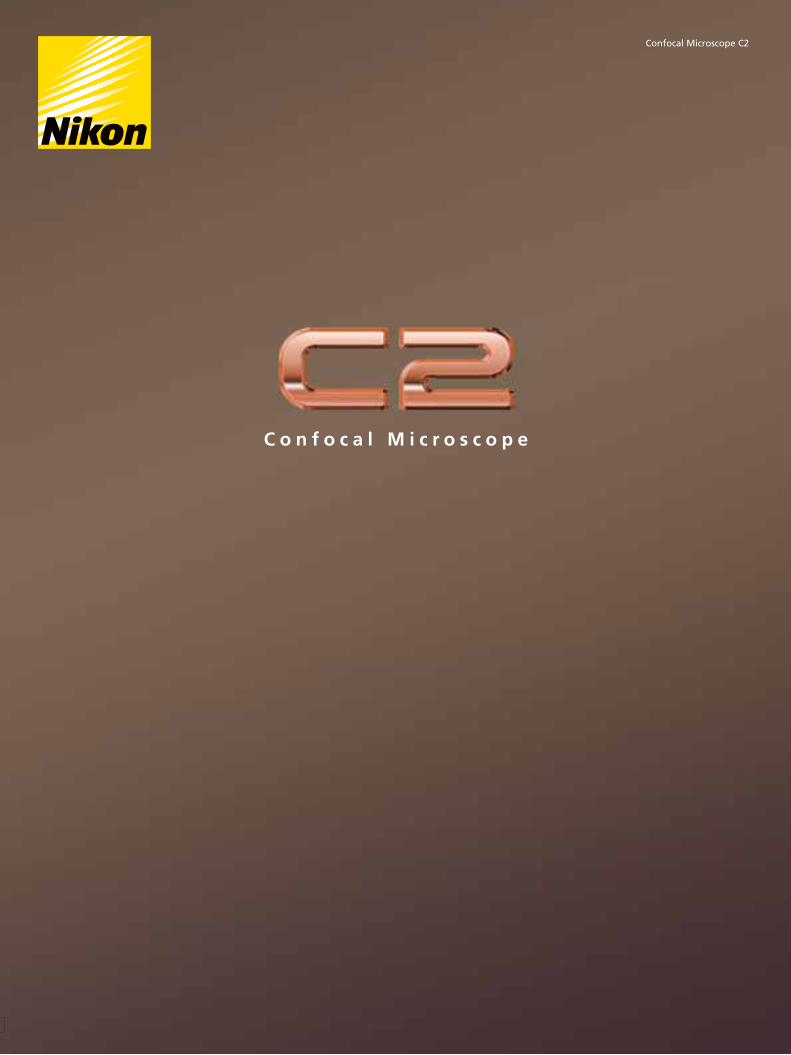
The C2 confocal microscope system comprises a new generation of Nikon confocal
instruments designed to be essential laboratory tools. Built on a reputation of
incredible stability and operational simplicity coupled with superior optical
technologies, the C2 with its host of functions and various analytical capabilities is the
perfect tool for a new microscope, or as a new accessory to a Nikon imaging system.
2 3
Confocal Microscope
The C2 employs Nikon’s proprietary imaging software NIS-Elements, which has a high reputation
for image acquisition in the camera market. NIS-Elements also allows the C2 to enjoy the same
operational ease as the high-grade confocal microscope system A1. The NIS-Elements software
that enhanced trust in Nikon’s microscopy imaging systems is a powerful reinforcement for the
C2’s usability, functionality, and broad analytical capability.
4 5
High efficiency scanning heads and detectors
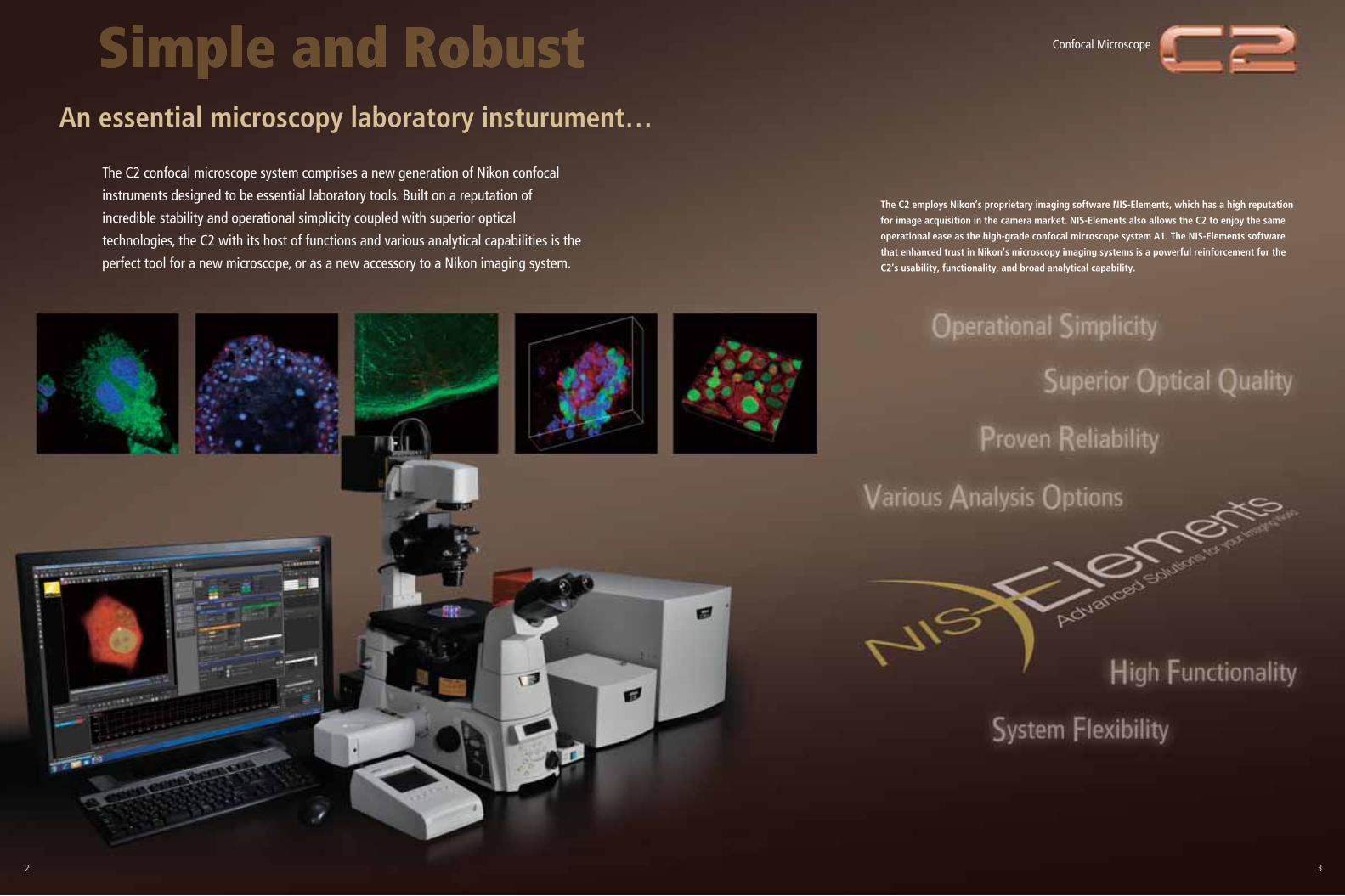
Nikon's unprecedented optics and a time-proven, highly efficient optical design provide the brightestand sharpest images, at the longest working distances.
Image QualityNikon's renowned imaging software NIS-Elements enables intuitive operation of all Nikon hardware devicesand peripheral. With a remarkably large number of analyzing functions for its class, the C2 supports yourroutine laboratory research activities.
High functionality
The C2 fits all Nikon microscopes with the smallest scan head footprint onthe market. The C2 employs high precision mirrors and optically ideal circularpinholes, enabling noiseless, high contrast and high quality confocal imaging,The spectral detector of the C2 enables high speed imaging usingsimultaneous 32-channel acquisition. Signal loss has been minimized whileimaging of fluorescence spectra in real colors is possible by host ofinnovations for accurately correcting spectral data.
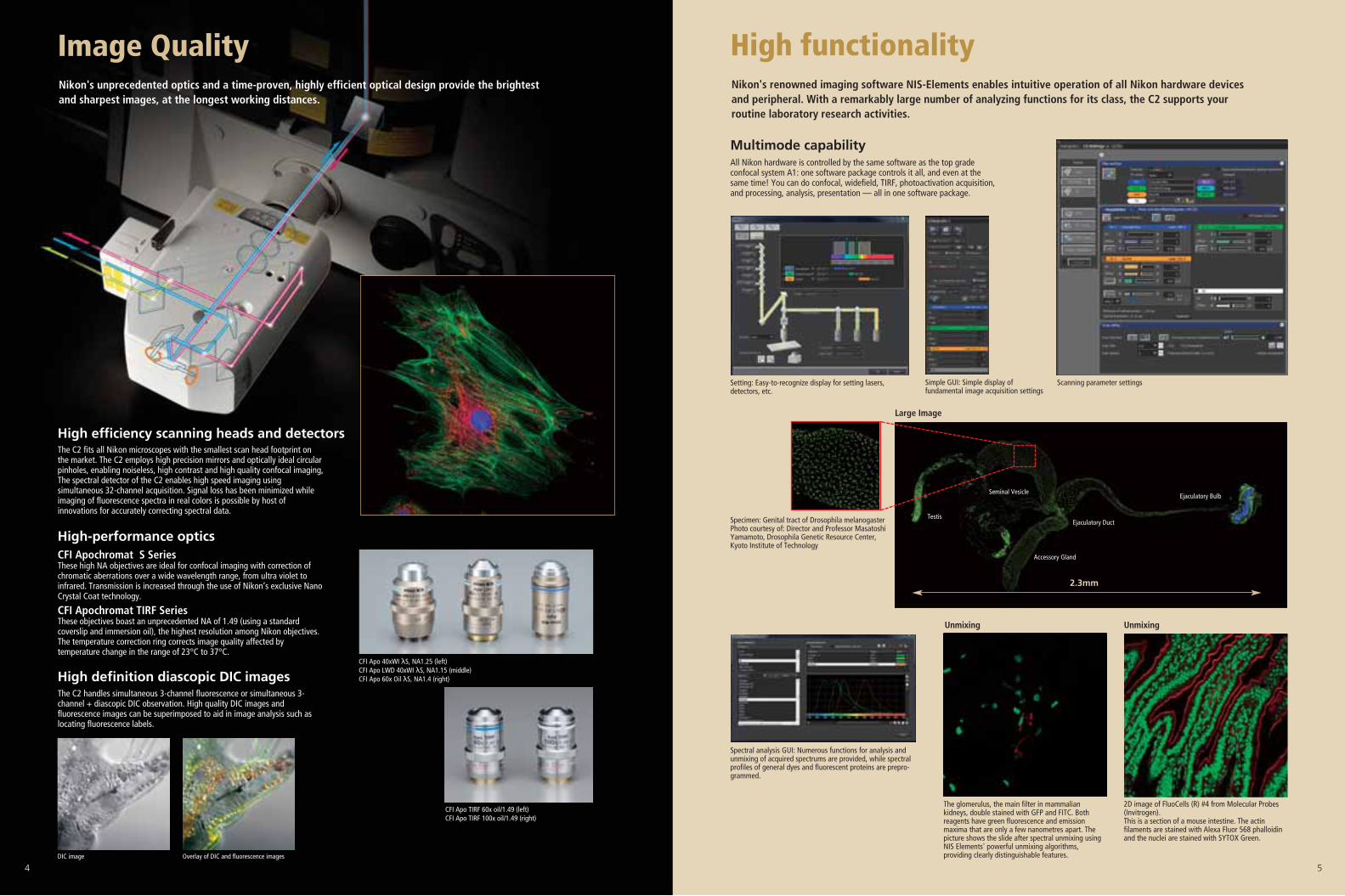
Multimode capabilityAll Nikon hardware is controlled by the same software as the top gradeconfocal system A1: one software package controls it all, and even at thesame time! You can do confocal, widefield, TIRF, photoactivation acquisition,and processing, analysis, presentation — all in one software package.
High-performance opticsCFI Apochromat S SeriesThese high NA objectives are ideal for confocal imaging with correction ofchromatic aberrations over a wide wavelength range, from ultra violet toinfrared. Transmission is increased through the use of Nikon’s exclusive NanoCrystal Coat technology.
CFI Apochromat TIRF SeriesThese objectives boast an unprecedented NA of 1.49 (using a standardcoverslip and immersion oil), the highest resolution among Nikon objectives.The temperature correction ring corrects image quality affected bytemperature change in the range of 23°C to 37°C.
High definition diascopic DIC imagesThe C2 handles simultaneous 3-channel fluorescence or simultaneous 3-channel + diascopic DIC observation. High quality DIC images andfluorescence images can be superimposed to aid in image analysis such aslocating fluorescence labels.
DIC image Overlay of DIC and fluorescence images
CFI Apo 40xWI λS, NA1.25 (left) CFI Apo LWD 40xWI λS, NA1.15 (middle) CFI Apo 60x Oil λS, NA1.4 (right)
CFI Apo TIRF 60x oil/1.49 (left)CFI Apo TIRF 100x oil/1.49 (right)
Scanning parameter settingsSimple GUI: Simple display offundamental image acquisition settings
Setting: Easy-to-recognize display for setting lasers,detectors, etc.
Spectral analysis GUI: Numerous functions for analysis andunmixing of acquired spectrums are provided, while spectralprofiles of general dyes and fluorescent proteins are prepro-grammed.
Specimen: Genital tract of Drosophila melanogasterPhoto courtesy of: Director and Professor MasatoshiYamamoto, Drosophila Genetic Resource Center,Kyoto Institute of Technology
The glomerulus, the main filter in mammaliankidneys, double stained with GFP and FITC. Bothreagents have green fluorescence and emissionmaxima that are only a few nanometres apart. Thepicture shows the slide after spectral unmixing usingNIS Elements' powerful unmixing algorithms,providing clearly distinguishable features.
2D image of FluoCells (R) #4 from Molecular Probes(Invitrogen). This is a section of a mouse intestine. The actinfilaments are stained with Alexa Fluor 568 phalloidinand the nuclei are stained with SYTOX Green.
2.3mm
Testis
Seminal Vesicle
Ejaculatory Duct
Ejaculatory Bulb
Accessory Gland
Large Image
Unmixing Unmixing
6 7
X position (pixel) X position (pixel)
L1L2L3L4
PC
Software
Diascopic Detector Unit
AOM Unit
Laser unit
Scanner set
Microscope
Detector unit
1st DM 1st DM
2nd, 3rd DM
Spectral Detector
C-LU3EX 3-laser Unit EX
Standard Epi-fl Detector (3-PMT)
LU-LR 4-laser Power Source Rack
C2 Scanning Head
C2si Scanning Head
AZ100FN1 90i/80i
Controller
LU4A 4-laser Unit
Ring Adapter L/SRing Adapter L/S Ring Adapter L/S
C2 Adapter C2 Adapter
Ring Adapter L/S
Z-focus ModuleZ-focus Module (for Ti-U)
Z-focus Module (for 80i)
Ti-E/U
6 7
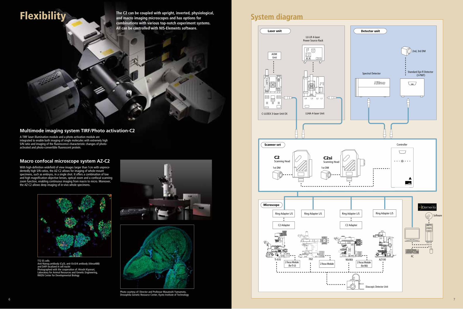
System diagramThe C2 can be coupled with upright, inverted, physiological,and macro imaging microscopes and has options forcombinations with various top-notch experiment systems.All can be controlled with NIS-Elements software.
Flexibility
A TIRF laser illumination module and a photo activation module areintegrated to enable both imaging of single molecules with extremely highS/N ratio and imaging of the fluorescence characteristic changes of photo-activated and photo-convertible fluorescent protein.
Multimode imaging system TIRF/Photo activation-C2
With high-definition widefield of view images larger than 1cm with unprece-dentedly high S/N ratios, the AZ-C2 allows for imaging of whole-mountspecimens, such as embryos, in a single shot. It offers a combination of lowand high magnification objective lenses, optical zoom and a confocal scanningzoom function, enabling continuous imaging from macro to micro. Moreover,the AZ-C2 allows deep imaging of in-vivo whole specimens.
Macro confocal microscope system AZ-C2
TT2 ES cells Anti-Nanog antibody (Cy3), anti-Oct3/4 antibody (Alexa488)and DAPI localized in cell nuclei Photographed with the cooperation of: Hiroshi Kiyonari,Laboratory for Animal Resources and Genetic Engineering,RIKEN Center for Developmental Biology
Photo courtesy of: Director and Professor Masatoshi Yamamoto,Drosophila Genetic Resource Center, Kyoto Institute of Technology
EnPrinted in Japan (1009-08)T Code No. 2CE-SCHH-1
This brochure is printed on recycled paper made from 40% used material.
Specifications and equipment are subject to change without any notice or obligationon the part of the manufacturer. June 2010 ©2010 NIKON CORPORATION
Monitor images are simulated.Sample images in this brochure were captured using the C1 confocal microscope system.Company names and product names appearing in this brochure are their registered trademarks or trademarks.N.B. Export of the products* in this brochure is controlled under the Japanese Foreign Exchange and Foreign Trade Law.Appropriate export procedure shall be required in case of export from Japan.*Products: Hardware and its technical information (including software)
WARNINGTO ENSURE CORRECT USAGE, READ THE CORRESPONDINGMANUALS CAREFULLY BEFORE USING YOUR EQUIPMENT.
NIKON CORPORATIONShin-Yurakucho Bldg., 12-1, Yurakucho 1-chome, Chiyoda-ku, Tokyo 100-8331, Japan phone: +81-3-3216-2375 fax: +81-3-3216-2385http://www.nikon.com/instruments/
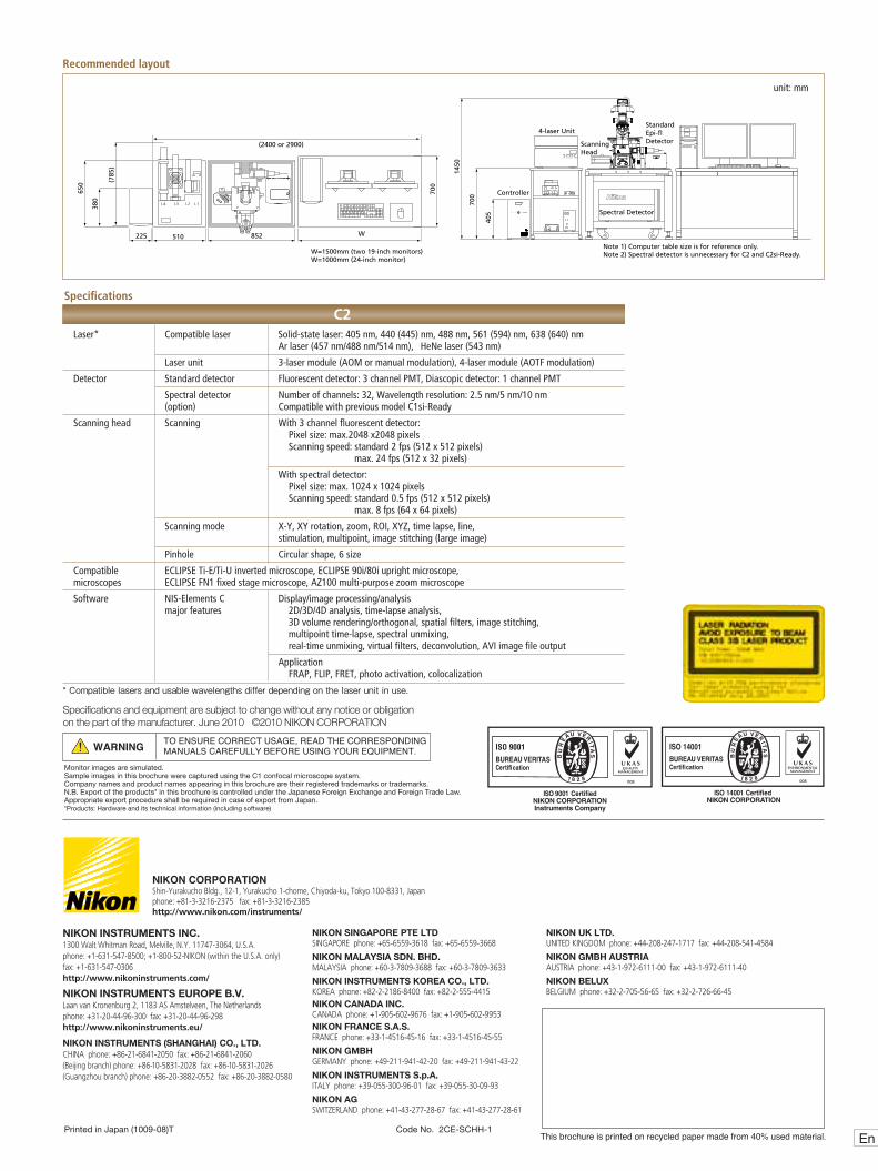
Laser* Compatible laser Solid-state laser: 405 nm, 440 (445) nm, 488 nm, 561 (594) nm, 638 (640) nm Ar laser (457 nm/488 nm/514 nm), HeNe laser (543 nm)
Laser unit 3-laser module (AOM or manual modulation), 4-laser module (AOTF modulation)
Detector Standard detector Fluorescent detector: 3 channel PMT, Diascopic detector: 1 channel PMT
Spectral detector Number of channels: 32, Wavelength resolution: 2.5 nm/5 nm/10 nm (option) Compatible with previous model C1si-Ready
Scanning head Scanning With 3 channel fluorescent detector: Pixel size: max.2048 x2048 pixels Scanning speed: standard 2 fps (512 x 512 pixels) max. 24 fps (512 x 32 pixels)
With spectral detector: Pixel size: max. 1024 x 1024 pixels Scanning speed: standard 0.5 fps (512 x 512 pixels) max. 8 fps (64 x 64 pixels)
Scanning mode X-Y, XY rotation, zoom, ROI, XYZ, time lapse, line, stimulation, multipoint, image stitching (large image)
Pinhole Circular shape, 6 size
Compatible ECLIPSE Ti-E/Ti-U inverted microscope, ECLIPSE 90i/80i upright microscope, microscopes ECLIPSE FN1 fixed stage microscope, AZ100 multi-purpose zoom microscope
Software NIS-Elements C Display/image processing/analysis major features 2D/3D/4D analysis, time-lapse analysis, 3D volume rendering/orthogonal, spatial filters, image stitching, multipoint time-lapse, spectral unmixing, real-time unmixing, virtual filters, deconvolution, AVI image file output
Application FRAP, FLIP, FRET, photo activation, colocalization
* Compatible lasers and usable wavelengths differ depending on the laser unit in use.
Specifications
C2
Recommended layout
380 700
650 (7
85)
405
225 510 W
1450
700
(2400 or 2900)
Controller
レーザユニット Scanning Head
Spectral Detector
Standard Epi-fl Detector
4-laser Unit
W=1500mm (two 19-inch monitors)W=1000mm (24-inch monitor)
Note 1) Computer table size is for reference only.Note 2) Spectral detector is unnecessary for C2 and C2si-Ready.
L1L2L3L4
L1 POWERL2L3L4
852
unit: mm
NIKON INSTRUMENTS INC.1300 Walt Whitman Road, Melville, N.Y. 11747-3064, U.S.A.phone: +1-631-547-8500; +1-800-52-NIKON (within the U.S.A. only)fax: +1-631-547-0306http://www.nikoninstruments.com/
NIKON INSTRUMENTS EUROPE B.V.Laan van Kronenburg 2, 1183 AS Amstelveen, The Netherlandsphone: +31-20-44-96-300 fax: +31-20-44-96-298http://www.nikoninstruments.eu/
NIKON INSTRUMENTS (SHANGHAI) CO., LTD.CHINA phone: +86-21-6841-2050 fax: +86-21-6841-2060(Beijing branch) phone: +86-10-5831-2028 fax: +86-10-5831-2026(Guangzhou branch) phone: +86-20-3882-0552 fax: +86-20-3882-0580
NIKON SINGAPORE PTE LTDSINGAPORE phone: +65-6559-3618 fax: +65-6559-3668
NIKON MALAYSIA SDN. BHD.MALAYSIA phone: +60-3-7809-3688 fax: +60-3-7809-3633
NIKON INSTRUMENTS KOREA CO., LTD.KOREA phone: +82-2-2186-8400 fax: +82-2-555-4415NIKON CANADA INC.CANADA phone: +1-905-602-9676 fax: +1-905-602-9953NIKON FRANCE S.A.S.FRANCE phone: +33-1-4516-45-16 fax: +33-1-4516-45-55
NIKON GMBHGERMANY phone: +49-211-941-42-20 fax: +49-211-941-43-22
NIKON INSTRUMENTS S.p.A.ITALY phone: +39-055-300-96-01 fax: +39-055-30-09-93
NIKON AGSWITZERLAND phone: +41-43-277-28-67 fax: +41-43-277-28-61
NIKON UK LTD. UNITED KINGDOM phone: +44-208-247-1717 fax: +44-208-541-4584
NIKON GMBH AUSTRIA AUSTRIA phone: +43-1-972-6111-00 fax: +43-1-972-6111-40
NIKON BELUXBELGIUM phone: +32-2-705-56-65 fax: +32-2-726-66-45