Embed Size (px)
Citation preview
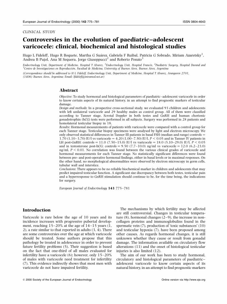
CLINICAL STUDY
Controversies in the evolution of paediatric±adolescentvaricocele: clinical, biochemical and histological studies
Hugo L Fideleff , Hugo R Boquete, Martha G SuaÂrez, Gabriela F Ruibal, Patricia G Sobrado, Miriam Azaretsky1,Andrea B Pujol, Ana M Sequera, Jorge Giuseppucci2 and Roberto Ponzio3
Endocrinology Unit, Department of Medicine, Hospital T Alvarez, 1Endocrinology Unit, Hospital FranceÂs, 2Paediatric Surgery, Hospital Durand and3Centro de Investigaciones en ReproduccioÂn, Facultad de Medicina, University of Buenos Aires, Buenos Aires, Argentina
(Correspondence should be addressed to H L Fideleff, Endocrinology Unit, Department of Medicine, Hospital T Alvarez, Aranguren 2701,(1406) Buenos Aires, Argentina; Email: [email protected])
Abstract
Objective: To study hormonal and histological parameters of paediatric±adolescent varicocele in orderto know certain aspects of its natural history, in an attempt to find prognostic markers of testiculardamage.Design and methods: In a prospective cross-sectional study, we evaluated 93 children and adolescentswith left unilateral varicocele and 29 healthy males as control group. All of them were classifiedaccording to Tanner stage. Scrotal Doppler in both testes and GnRH and human chorionicgonadotrophin (hCG) tests were performed in all subjects. Surgery was performed in 28 patients andhomolateral testicular biopsy in 18.Results: Hormonal measurements of patients with varicocele were compared with a control group foreach Tanner stage. Testicular biopsy specimens were analysed by light and electron microscopy. Weonly observed statistical differences in Tanner III patients in basal FSH (median and range) controls �1:70 (1.10±3.70) IU/l vs varicocele � 4:20 (1.00±7.50) IU/l, P , 0:05 and in Tanner IV patients inLH post-GnRH: controls � 11:0 (7.50±15.0) IU/l vs varicocele � 18:0 (5.10±29.0) IU/l, P , 0:05and in testosterone post-hCG: controls � 9:50 (7.7±10.0) ng/ml vs varicocele � 12:0 (6.2±23.0)ng/ml, P , 0:01: No correlation was found between the various clinical grades of varicocele andhormonal measurements for each Tanner stage. No statistically significant differences were foundbetween pre- and post-operative hormonal findings, either in basal levels or in maximal responses. Onthe other hand, no morphological abnormalities were observed by electron microscopy in germ cells,tubular wall and interstice.Conclusions: There appears to be no reliable biochemical marker in children and adolescents that maypredict impaired testicular function. A significant size discrepancy between both testes, testicular painand a hyperresponse to GnRH stimulation should continue to be, for the time being, the indicationsfor surgery.
European Journal of Endocrinology 143 775±781
Introduction
Varicocele is rare below the age of 10 years and itsincidence increases with progressive pubertal develop-ment, reaching 15±20% at the age of 14±15 years (1,2), a rate similar to that reported in adults (3, 4). Thereare some controversies over the age at which varicoceleshould be treated. Some authors propose that thispathology be treated in adolescence in order to preventfuture fertility problems (5). Their suggestion is basedon the fact that one-third of all males evaluated forinfertility have a varicocele (6); however, only 15±20%of males with varicocele need treatment for infertility(7). This evidence indirectly shows that most men withvaricocele do not have impaired fertility.
The mechanisms by which fertility may be affectedare still controversial. Changes in testicular tempera-ture (8), hormonal changes (2±9), the increase in non-collagen proteins and immunoglobulins found in thespermatic vein (7), production of `toxic substances' (10)and testicular hypoxia (7), have been proposed amongother causes. As regards hormonal changes, it is stillunknown whether they cause or result from gonadaldamage. The information available on circulatory flowalterations (11) and the onset of histological testicularinjuries is also limited (12).
The aim of our work has been to study hormonal,circulatory and histological parameters of paediatric±adolescent varicocele to know certain aspects of itsnatural history, in an attempt to find prognostic markers
ISSN 0804-4643European Journal of Endocrinology (2000) 143 775±781
q 2000 Society of the European Journal of Endocrinology Online version via http://www.eje.org
of testicular damage in order to select, in the future, thesubset of patients requiring surgical treatment.
Subjects and methods
Ninety-three children and adolescents with left uni-lateral varicocele, chronological ages (CA) rangingbetween 8.6 and 16.9 years (mean: 12.8 years), andTanner stages I±V were evaluated. All prepubertalpatients were referred by the Healthy Children Sectionand pubertal patients were referred by the AdolescentsSection. In all cases, the Valsalva manoeuvre was partof the routine clinical examination. None of thepatients had clinical symptoms consistent with varico-cele. Testicular volume was measured by Praderorchidometer. Patients were clinically classified accord-ing to the classification of Dubin & Amelar (13) inwhich grade 1 (G1) corresponds to the small varicocele(detected by Valsava manoeuvre), grade 2 (G2) to themoderate varicocele (detected by simple palpation) andgrade 3 (G3) to the large varicocele (palpable andvisible). The control group consisted of 29 healthymales with a CA between 9 and 30 years, and Tannerstages I±V, who were being followed up at ourEndocrinology Unit for growth and developmentcontrol.
In all subjects, a scrotal Doppler study was carriedout in both testes, in the standing and supine positions,using ultrasonic monitor vascular Doppler (MV-1,Tecny Med SRL, Buenos Aires, Argentina) with a5 MHz transducer. The study was performed at theoffice by one of the authors (M G S) in all outpatients atthe first visit. The sonographic findings were char-acterized as: negative or positive reflux.
A gonadotrophin-releasing hormone (GnRH) testwas performed in all subjects (100 mg i.v. bolus) withbasal measurements of luteinizing hormone (LH),follicle-stimulating hormone (FSH) and testosterone.Both gonadotrophins were also measured 30, 60 and90 min after GnRH injection. The administration ofhuman chorionic gonadotrophin (hCG) was started48 h later (at a dose of 1000 IU/day i.m. for 5 days)and a blood sample was taken on the 6th day fortestosterone measurement.
LH and FSH were measured by IRMA (Serono MaiaClone, Milan, Italy) and testosterone by RIA. The intra-and interassay coefficients of variation were: LH: 12.1and 15.2% (mean: 3.4 IU/l) and 5 and 7.5% (mean:16.5 IU/l); FSH: 9.2 and 15.5% (mean: 4 IU/l) and 5.8and 5.3% (mean: 13.7 IU/l); and testosterone: 10 and11.2% (mean: 0.85 ng/ml) and 4.5 and 3.7% (mean:4.4 ng/ml).
Corrective surgery was indicated in 28 patients, usingPalomo's technique (14); left testicular biopsy wasperformed intraoperatively in 18 of them. The biopsyspecimens were processed for both light and electronmicroscopy. Quantitative analysis was performed by lightmicroscopy determining the presence of tubular lumen,
the maturation state of Sertoli cells and the number ofspermatocytes and mature spermatids per cross-tubu-lar section in each biopsy. The volume density (VD) ofLeydig cells (i.e. the proportion of the total testicularvolume occupied by Leydig cells) was evaluated by thedifferential point-counting method (15) employing aZeiss point-counting eyepiece (Carl Zeiss, Oberkochen,WuÈ rtt, Germany). Since an estimate of testicularvolume was obtained, the absolute Leydig cells amount(mg) per testis could be calculated. The results of thestereological and cell-counting methods are shown inTable 2. Electron microscopy was used to analyseSertoli cell morphological maturation signs (tripartitenucleolus, folded nuclear membrane, intercellularjunctions), Sertoli cell abnormalities (vacuolation),tubular wall maturation (abnormal cells, basementmembrane thickening), spermatogonia (infantile oradult characteristics) and Leydig cells (identifiableprecursors, adult cells, involutional cells). The GnRHand hCG tests were repeated in 14 patients 6 monthsafter surgery. Informed consent was obtained from allsubjects and their parents for hormonal studies and forbiopsies when there was an indication for surgery. Thestudy protocol was submitted to the Alvarez HospitalEducation and Research Committee and to the EthicalCommittee.
The Mann±Whitney test was used to comparehormonal findings in patients with varicocele andcontrols for each Tanner stage. The Wilcoxon signedrank test was used for evaluation of pre-surgery vspost-surgery findings (16).
Results
Clinical findings
Out of 93 patients, 17 were at Tanner stage I (G1varicocele: ten; G2: two and G3: five), 18 at Tanner stageII (G1: five; G2: five and G3: eight), 16 at Tanner stage III(G1: four; G2: four and G3: eight), 29 at Tanner stage IV(G1: nine; G2: twelve and G3: eight) and 13 at Tannerstage V (G1: none; G2: six and G3: seven).
Out of 29 controls, nine were at Tanner stage I, fiveat stage II, five at stage III, five at stage IV and five atstage V.
As regards the Doppler study, all patients withvaricocele had positive reflux, while no reflux wasobserved in any of the subjects of the control group.
Out of 28 patients who underwent surgery (all of themG3 varicocele) three were at Tanner stage I, four at stageII, five at stage III, five at stage IV and five at stage V.
Out of 93 patients, 44 who had not undergonesurgery were clinically followed until they reached anadult testicular volume, with no worsening of varico-cele. The remaining 21 patients who had not beenoperated on were dropouts. As regards the testicularvolume of these 65 patients at the start, in prepubertalpatients the difference between the affected testis and
776 H L Fideleff and others EUROPEAN JOURNAL OF ENDOCRINOLOGY (2000) 143
www.eje.org
the contralateral testis did not exceed 50%; in pubertalpatients the difference between the two testes was lessthan 2 ml. Out of 28 operated patients, 13 showed afavourable evolution, reaching an adult testicularvolume. Fifteen of the operated subjects dropped outbetween 2 and 10 months after surgery, with notesticular evolution.
Biochemical findings
Basal levels of LH, FSH and testosterone and their
maximal response to GnRH and hCG respectively areshown in Table 1.
Basal FSH levels at Tanner stage III and the post-stimulation maximal response of LH and testosteronein stage IV were significantly higher than those ofthe control group. No statistically significant differ-ences in basal LH, basal testosterone and themaximal response of FSH were found betweensubjects with varicocele and the control group inany of the Tanner stages.
No relationship was found between the various
Table 1 Basal levels and maximal response of LH (LHB and LHMx) (IU/l) and FSH (FSHB and FSHMx) (IU/l) to GnRH, and basal levels andresponse of testosterone (TB and TPost) (ng/ml) to hCG in control subjects and varicocele patients at the different Tanner stages. Valuesare expressed as median and range.
Tanner stage Subjects LHB LHMx FSHB FSHMx TB TPost
I Controls �n � 9� 0.40(0.40±0.90)
2.15(0.63±12.0)
1.45(0.40±3.00)
4.20(2.40±5.90)
0.10(0.10±0.30)
3.20(1.20±4.90)
Varicocele �n � 16� 0.40(0.40±2.10)
3.80(1.10±13.0)
2.08(0.40±4.50)
4.80(2.60±10.0)
0.20(0.10±1.50)
2.50(0.80±11.50)
II Controls �n � 5� 1.25(0.40±1.90)
11.0(2.80±18.0)
1.80(0.40±3.10)
10.0(0.40±15.0)
1.50(0.10±2.30)
7.60(3.00±10.60)
Varicocele �n � 14� 1.60(0.40±3.30)
13.0(8.00±20.0)
2.90(0.85±5.80)
5.60(2.35±14.0)
1.20(0.35±4.70)
8.40(2.50±13.10)
III Controls �n � 5� 2.60(1.50±3.00)
13.5(7.60±16.5)
1.70(1.10±3.70)
4.20(1.60±5.80)
3.60(2.20±5.60)
9.50(5.40±13.00)
Varicocele �n � 14� 1.50(0.40±2.70)
15.0(5.60±28.0)
4.20*(1.00±7.50)
6.20(1.00±12.6)
3.40(0.25±5.80)
9.80(5.20±18.00)
IV Controls �n � 5� 1.40(0.75±3.10)
11.0(7.50±15.0)
3.60(2.20±7.10)
7.50(4.90±11.0)
3.35(3.20±6.80)
9.50(7.70±10.00)
Varicocele �n � 29� 2.40(0.75±7.20)
18.0*(5.10±29.0)
3.60(1.00±8.00)
6.00(2.50±37.0)
4.80(2.50±7.30)
12.00**(6.20±23.00)
V Controls �n � 5� 3.50(1.90±4.50)
18.0(11.0±23.6)
3.70(1.80±4.10)
5.00(3.80±6.60)
4.80(4.40±8.90)
12.00(8.00±17.00)
Varicocele �n � 9� 2.15(0.60±3.90)
14.0(10.0±17.0)
3.30(1.50±11.0)
6.00(2.40±16.0)
4.50(3.50±6.20)
11.45(6.60±14.00)
*P , 0:05; **P , 0:01: Mann±Whitney test.
Table 2 Paediatric±adolescent varicocele: histological findings by light microscopy.
Tannerstage
Testicularvolume (ml)
VD Leydigcells (%)
Leydigcells/testis (mg)
Maturespermatides/tubule
Spermatocytes/tubule Lumen Sertoli
Completespermatogenesis
I 3 0.07 2 0.0 0.0 2 Immature 2I 2 0.28 5 0.0 0.0 2 Immature 2II 4 0.60 24 7.1 0.0 2 Immature 2II 5 1.62 81 2.3 0.0 + Intermediate 2III 6 2.83 170 3.8 0.0 + Intermediate 2III 7±8 2.13 106 + 6.8 + Mature +III 7 1.56 109 +++ 0.7 + Mature +III 6 1.83 110 +++ 10.7 + Mature +III 8 1.69 135 + 9.0 + Intermediate +III 6 2.63 158 + 5.1 + Intermediate +V 15 1.92 288 +++ 20.6 + Mature +IV 9 3.23 290 +++ 12.3 + Mature +V 12 2.79 335 +++ 17.6 + Mature +V 11 3.07 338 +++ 14.7 + Mature +IV 10 3.76 376 +++ 18.7 + Mature +V 12 3.18 381 +++ 12.7 + Mature +IV 10 4.45 445 +++ 12.8 + Mature +V 20 2.58 516 +++ 23.0 + Mature +
Paediatric±adolescent varicocele 777EUROPEAN JOURNAL OF ENDOCRINOLOGY (2000) 143
www.eje.org
clinical grades of varicocele or Doppler patterns andhormonal findings for each Tanner stage �P � NS�:
Surgery
All operated patients �n � 28� had G3 varicocele. Inpubertal patients, the difference in testicular volumebetween the two testes was greater than 2 ml. Inprepubertal patients, a difference in size between theaffected testis and the contralateral testis was con-sidered when it was greater than 50%.
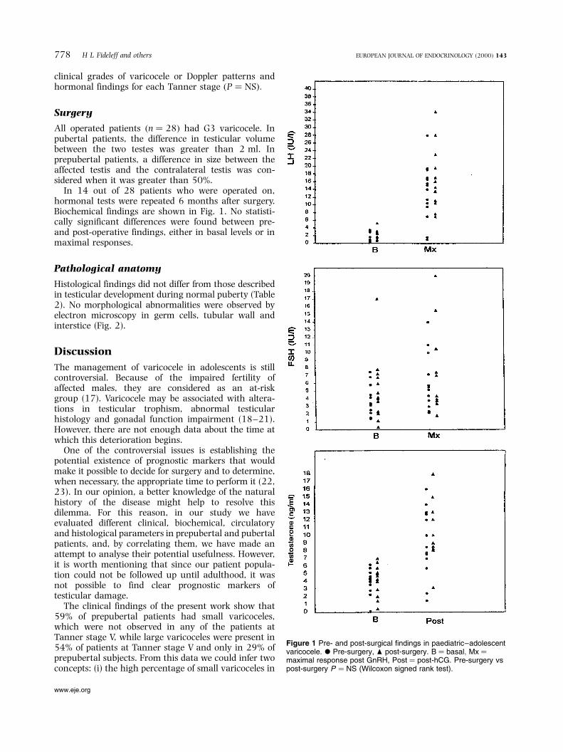
In 14 out of 28 patients who were operated on,hormonal tests were repeated 6 months after surgery.Biochemical findings are shown in Fig. 1. No statisti-cally significant differences were found between pre-and post-operative findings, either in basal levels or inmaximal responses.
Pathological anatomy
Histological findings did not differ from those describedin testicular development during normal puberty (Table2). No morphological abnormalities were observed byelectron microscopy in germ cells, tubular wall andinterstice (Fig. 2).
Discussion
The management of varicocele in adolescents is stillcontroversial. Because of the impaired fertility ofaffected males, they are considered as an at-riskgroup (17). Varicocele may be associated with altera-tions in testicular trophism, abnormal testicularhistology and gonadal function impairment (18±21).However, there are not enough data about the time atwhich this deterioration begins.
One of the controversial issues is establishing thepotential existence of prognostic markers that wouldmake it possible to decide for surgery and to determine,when necessary, the appropriate time to perform it (22,23). In our opinion, a better knowledge of the naturalhistory of the disease might help to resolve thisdilemma. For this reason, in our study we haveevaluated different clinical, biochemical, circulatoryand histological parameters in prepubertal and pubertalpatients, and, by correlating them, we have made anattempt to analyse their potential usefulness. However,it is worth mentioning that since our patient popula-tion could not be followed up until adulthood, it wasnot possible to find clear prognostic markers oftesticular damage.
The clinical findings of the present work show that59% of prepubertal patients had small varicoceles,which were not observed in any of the patients atTanner stage V, while large varicoceles were present in54% of patients at Tanner stage V and only in 29% ofprepubertal subjects. From this data we could infer twoconcepts: (i) the high percentage of small varicoceles in
Figure 1 Pre- and post-surgical findings in paediatric±adolescentvaricocele. X Pre-surgery, O post-surgery. B � basal; Mx �maximal response post GnRH, Post � post-hCG. Pre-surgery vspost-surgery P � NS (Wilcoxon signed rank test).
778 H L Fideleff and others EUROPEAN JOURNAL OF ENDOCRINOLOGY (2000) 143
www.eje.org
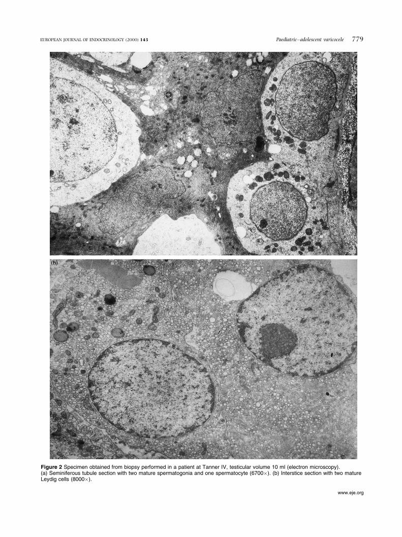
Figure 2 Specimen obtained from biopsy performed in a patient at Tanner IV, testicular volume 10 ml (electron microscopy).(a) Seminiferous tubule section with two mature spermatogonia and one spermatocyte (6700�). (b) Interstice section with two matureLeydig cells (8000�).
Paediatric±adolescent varicocele 779EUROPEAN JOURNAL OF ENDOCRINOLOGY (2000) 143
www.eje.org
prepuberty could explain, at least partially, the reasonwhy this pathology may be underdiagnosed in thisperiod, and (ii) the worsening of varicoceles betweenprepuberty and the final stage of sexual maturation.Because of the high dropout rate, no definitiveconclusions could be drawn on the follow-up. Probablythe high dropout rate may be attributed to the lack ofcomprehension of the importance in the parents and/orto non-cooperation of the general practitioners.
Some years ago, in a study on pubertal varicocele(11) we found a significant increase in LH response toGnRH and an increase in testosterone post-hCGstimulation, in some unilateral varicoceles. Never-theless, we pointed out that it would be advisable todivide the possible findings according to the stage ofsexual maturation. Later, while studying prepubertalpatients (24) we noticed that basal levels of LH and FSHin large varicoceles were significantly higher thanthose of the control group, with no hyperresponse toGnRH, and no alterations in basal and post-hCG levelsof testosterone. Our hypothesis was that those increasesmight represent autocrine and paracrine alterations inthe regulation of testicular function in those patients.In the present work, having increased the number ofpatients enrolled in the study, considering subjects withunilateral varicoceles ranging from prepuberty toTanner stage V, we noticed some changes in basallevels of FSH, and in the maximal response of LH andtestosterone post-stimulation in the midstages and finalstages of puberty. These changes did not follow aconsistent pattern when we made an attempt tocorrelate them with the clinical grades of varicocele.This would show that a certain clinical grade ofvaricocele does not necessarily imply a specific bio-chemical alteration, so that a hormonal evaluation isrequired for each individual case.
The evaluation performed in our patients 6 monthsafter surgery did not show significant hormonalchanges. This might be partly explained by the pubertalevolution of these patients, whose physiological hor-monal changes may have masked possible hormonalchanges between pre- and post-operative evaluations.
An important contribution of our work has been thestudy of biopsy specimens from our operated patientsby light and electron microscopy. Surprisingly, we didnot find alterations in the study with light microscopy,which was confirmed by the normal findings obtainedin the study with electron microscopy. This could bepossibly attributed to the fact that biopsies wereperformed at an early stage of varicocele evolution,and before the occurrence of injuries, which are partlytime-dependent. Histological lesions may possibly beevidenced during periods of more active spermatogen-esis. Moreover, some authors only found histologicalabnormalities in adolescents having a hyperresponsepost-GnRH stimulation (12). This suggests that thehormonal changes occurring in some cases mightprecede histological injuries and this could partly
explain the absence of histological damage in ourpatients.
In conclusion, varicocele is an heterogeneous entitywith marked individual variations. At present there isno reliable biochemical marker that may predictimpaired testicular function. Therefore, a significantsize difference between both testes, testicular pain anda hyperresponse to GnRH stimulation should continueto be, for the time being, the indications for surgery.Hormonal changes and histological injuries will be, to alarge extent, time-dependent, in many cases worseningduring puberty. Perhaps, in the future, the design ofnew research methodologies and the use of ultrasensi-tive biochemical assays will contribute to the prospec-tive characterization of the subset of patients at risk oftesticular damage.
Acknowledgements
We thank Carina Fideleff for her assistance in thecorrection of the English manuscript.
References1 Steeno OP. Varicocele in the adolescent. In Temperature and
Environmental Effects on the Testis, pp 295±321. Ed AWZorgniotti. New York: Plenum Press, 1991.
2 Castro-Magana M, Angulo M, Canas J & Uy J. Improvement ofLeydig cell function in male adolescents after varicocelectomy.Journal of Pediatrics 1989 115 809±811.
3 Hudson RW & Mckay DE. The gonadotropin response of menwith varicoceles to gonadotropin-releasing hormone. Fertility andSterility 1980 33 427±432.
4 World Health Organization. The influence of varicocele onparameters of fertility in a large group of men presenting toinfertility clinics. Fertility and Sterility 1992 57 1289±1293.
5 Laven JSE, Haans LCF, Mali WM, Velde ER, Wensing CJ &Eimers JM. Effects of varicocele treatment in adolescents: arandomized study. Fertility and Sterility 1992 58 756±762.
6 Niedzielski J, Paduch D & Raczynski P. Assessment of adolescentvaricocele. Pediatric Surgery International 1997 12 410±413.
7 Skoog SJ, Roberts KP, Goldstein M & Pryor JL. The adolescentvaricocele: what's new with an old problem in young patients?Pediatrics 1997 100 112±122.
8 Agger P. Scrotal and testicular temperature: its relation to spermcount before and after operation for varicocele. Fertility andSterility 1971 22 286±297.
9 Hudson RW. The endocrinology of varicoceles. Fertility andSterility 1988 49 199±208.
10 Takihara H, Sakatoku J & Cockett A. The pathophysiology ofvaricocele in male infertility. Fertility and Sterility 1991 55 861±868.
11 Fideleff HL, Boquete HR, Saskyn N, Zanchetti F, Ambiela R,Sobrado P.. et al. Pubertal varicocele: correlation between clinical.Doppler and hormonal findings. Fertility and Sterility 1993 59693±695.
12 Aragona F, Ragazzi R & Pozzan GB. Correlation of testicularvolume, histology and LH-RH test in adolescents with idiopathicvaricocele. European Urology 1994 26 61±66.
13 Dubin L & Amelar RD. Varicocele size and results of varico-celectomy in selected subfertile men with varicocele. Fertility andSterility 1970 21 606±609.
14 Palomo A. Radical cure of varicocele by a new technique:preliminary report. Journal of Urology 1949 61 604±607.
780 H L Fideleff and others EUROPEAN JOURNAL OF ENDOCRINOLOGY (2000) 143
www.eje.org
15 Weibel ER. Practical methods for biological morphometry. InStereological Methods, vol 1, pp 101±161. London: AcademicPress, 1979.
16 Siegel S. Non-Parametric Statistics for the Behavioral Sciences, edn5. New York: McGraw, 1990.
17 Lenzi A, Gandini L, Bagolan P, Nahum A & Dondero F. Spermparameters after early left varicocele treatment. Fertility andSterility 1998 69 347±349.
18 Kass EJ, Freitas JE & Bour JB. Adolescent varicocele: objectiveindications for treatment. Journal of Urology 1989 142 579±582.
19 Vasavada S, Ross J, Nasrallah P & Kay R. Prepubertal varicocele.Urology 1997 50 774±777.
20 Podesta ML, Gottlieb S, Medel R, Ropelato G, Bergada C &Quesada EM. Hormonal parameters and testicular volume inchildren and adolescents with unilateral varicocele: preoperativeand postoperative findings. Journal of Urology 1994 152 794±798.
21 Hudson RW. Free sex steroid and sex hormone-binding globulinlevels in oligozoospermic men with varicoceles. Fertility andSterility 1996 66 299±304.
22 Paduch DA & Niedzielski J. Repair versus observation inadolescent varicocele: a prospective study. Journal of Urology1997 158 1128±1132.
23 Sayfan J, Siplovich L, Koltun L & Benyamin N. Varicoceletreatment in pubertal boys prevents testicular growth arrest.Journal of Urology 1997 157 1456±1457.
24 Fideleff HL, Boquete HR, Ruibal GF, SuaÂrez MG, Sobrado PGV,Zanchetti F. et al. Varicocele in prepubertal boys evaluation ofclinical, Doppler and hormonal findings. Medicina 1996 56 679±682.
Received 26 April 2000Accepted 23 August 2000
Paediatric±adolescent varicocele 781EUROPEAN JOURNAL OF ENDOCRINOLOGY (2000) 143
www.eje.org