Embed Size (px)
Citation preview
Controversies in Cartilage repair and osteoarthritis: 2016
Sam A. Labib, MD Associate Professor of Orthopedic Surgery Editorial Board Member: Arthroscopy Journal Head Team Physician Oglethorpe Team Consultant: Georgia Tech/ Atlanta Falcons Emory Sports Medicine Center
COI Disclosure
Research Grant Support • Linvatec • Arthrex • Ossur Consultant: • Arthrex Inc • Arthrosurface Inc
OUTLINE
• WHAT IS A CARTILAGE DEFECT? • WHAT CAUSES OSTEO-ARTHRITIS? • WHAT IS JOINT PRESERVATION? • NEW TECHNIQUES • CASE PRESENTATIONS • DISCUSSSION • CONCLUSIONS
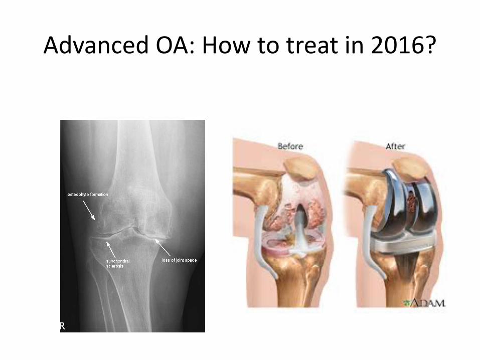
Advanced OA: How to treat in 2016?
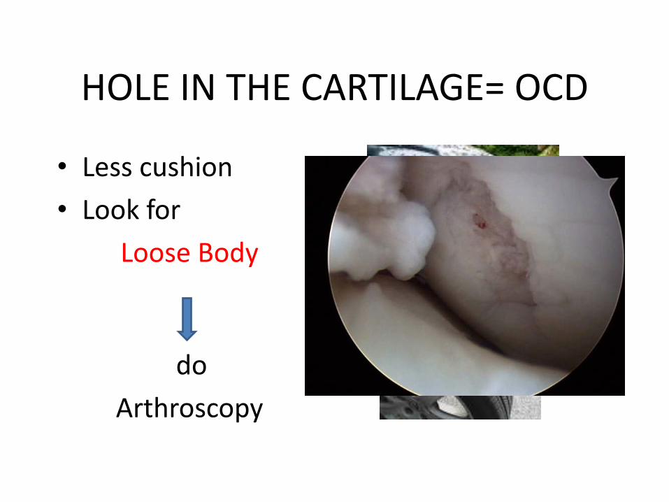
HOLE IN THE CARTILAGE= OCD
• Less cushion • Look for
Loose Body
do Arthroscopy
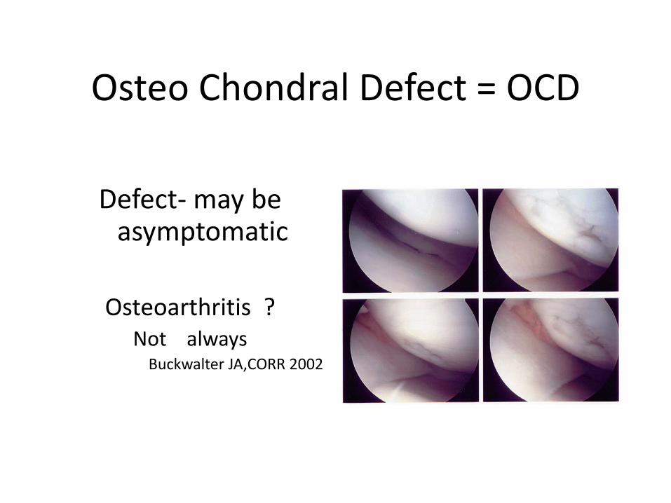
Osteo Chondral Defect = OCD
Defect- may be
asymptomatic
Osteoarthritis ? Not always
Buckwalter JA,CORR 2002
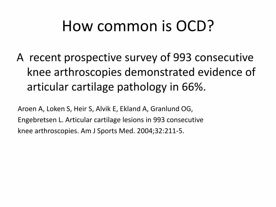
How common is OCD?
A recent prospective survey of 993 consecutive knee arthroscopies demonstrated evidence of articular cartilage pathology in 66%.
Aroen A, Loken S, Heir S, Alvik E, Ekland A, Granlund OG, Engebretsen L. Articular cartilage lesions in 993 consecutive knee arthroscopies. Am J Sports Med. 2004;32:211-5.
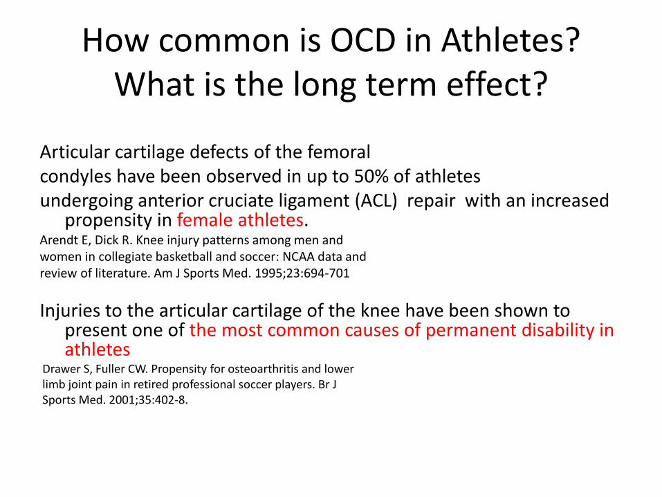
How common is OCD in Athletes? What is the long term effect?
Articular cartilage defects of the femoral condyles have been observed in up to 50% of athletes undergoing anterior cruciate ligament (ACL) repair with an increased
propensity in female athletes. Arendt E, Dick R. Knee injury patterns among men and women in collegiate basketball and soccer: NCAA data and review of literature. Am J Sports Med. 1995;23:694-701
Injuries to the articular cartilage of the knee have been shown to present one of the most common causes of permanent disability in athletes
Drawer S, Fuller CW. Propensity for osteoarthritis and lower limb joint pain in retired professional soccer players. Br J Sports Med. 2001;35:402-8.
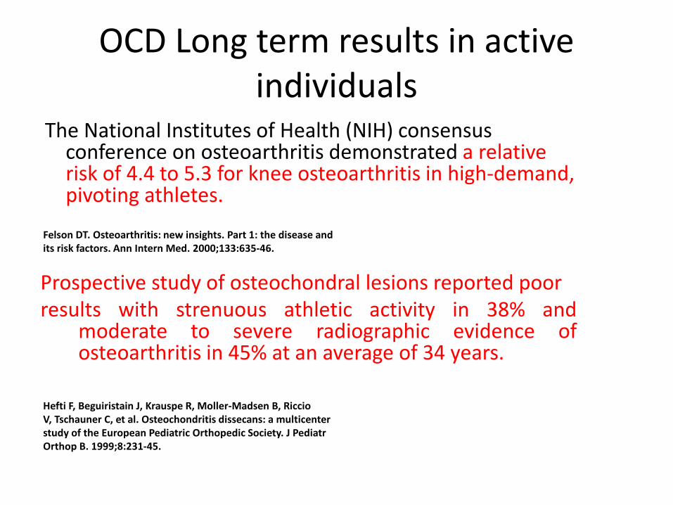
OCD Long term results in active individuals
The National Institutes of Health (NIH) consensus conference on osteoarthritis demonstrated a relative risk of 4.4 to 5.3 for knee osteoarthritis in high-demand, pivoting athletes.
Felson DT. Osteoarthritis: new insights. Part 1: the disease and its risk factors. Ann Intern Med. 2000;133:635-46.
Prospective study of osteochondral lesions reported poor results with strenuous athletic activity in 38% and
moderate to severe radiographic evidence of osteoarthritis in 45% at an average of 34 years.
Hefti F, Beguiristain J, Krauspe R, Moller-Madsen B, Riccio V, Tschauner C, et al. Osteochondritis dissecans: a multicenter study of the European Pediatric Orthopedic Society. J Pediatr Orthop B. 1999;8:231-45.
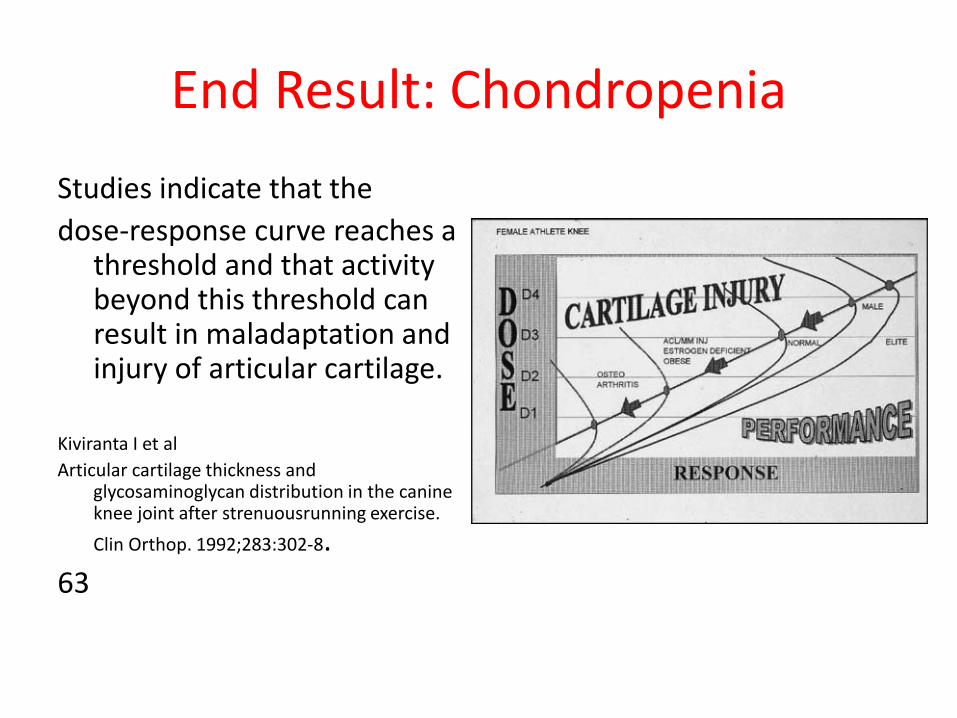
End Result: Chondropenia Studies indicate that the dose-response curve reaches a
threshold and that activity beyond this threshold can result in maladaptation and injury of articular cartilage.
Kiviranta I et al Articular cartilage thickness and
glycosaminoglycan distribution in the canine knee joint after strenuousrunning exercise. Clin Orthop. 1992;283:302-8.
63
Outcome of Untreated Traumatic Articular Cartilage Defects of The Knee: A Natural History
Study Shelbourne et al: JBJS-A, 85: 8-16 2003
• 101 ACL patients + Chondral defect: 48 medial & 53 Lateral-
No Cartilage Treatment. • Control group: ACL no Chondral defect. • IKDC rating/ X-rays @ mean 8.7 years. • Control group scored better on Subjective scale. 95.2 versus
92.8 ( lateral defect) • No difference in Objective data or X-rays • 79% return to high impact activity
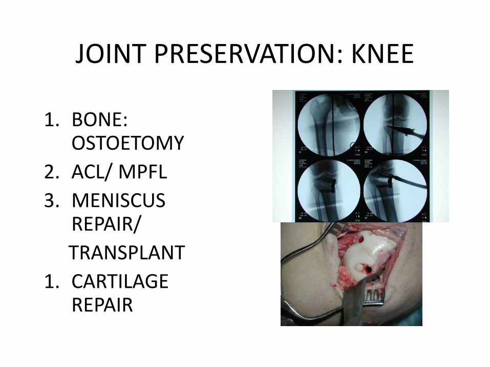
JOINT PRESERVATION: KNEE
1. BONE: OSTOETOMY
2. ACL/ MPFL 3. MENISCUS
REPAIR/ TRANSPLANT 1. CARTILAGE
REPAIR
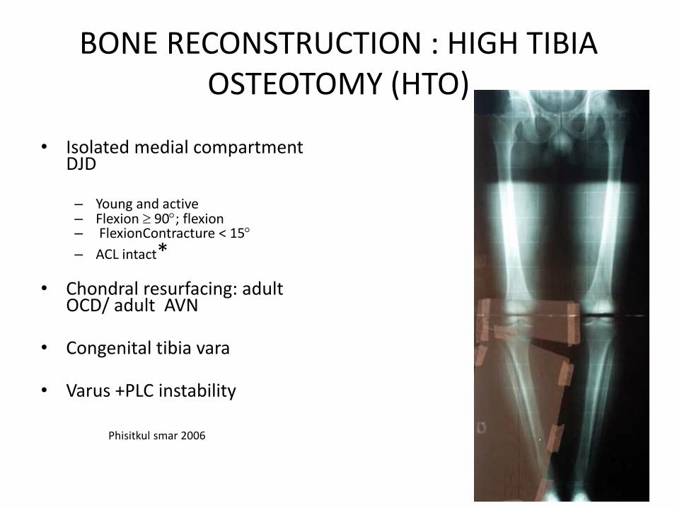
BONE RECONSTRUCTION : HIGH TIBIA OSTEOTOMY (HTO)
• Isolated medial compartment
DJD
– Young and active – Flexion t 90q; flexion – FlexionContracture < 15q – ACL intact*
• Chondral resurfacing: adult OCD/ adult AVN
• Congenital tibia vara
• Varus +PLC instability
Phisitkul smar 2006
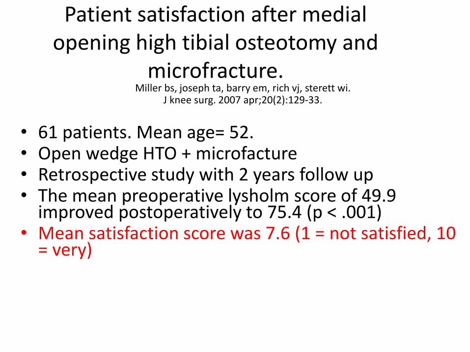
Patient satisfaction after medial opening high tibial osteotomy and
microfracture. Miller bs, joseph ta, barry em, rich vj, sterett wi.
J knee surg. 2007 apr;20(2):129-33. • 61 patients. Mean age= 52. • Open wedge HTO + microfacture • Retrospective study with 2 years follow up • The mean preoperative lysholm score of 49.9
improved postoperatively to 75.4 (p < .001) • Mean satisfaction score was 7.6 (1 = not satisfied, 10
= very)
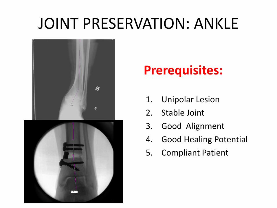
JOINT PRESERVATION: ANKLE
Prerequisites:
1. Unipolar Lesion 2. Stable Joint 3. Good Alignment 4. Good Healing Potential 5. Compliant Patient
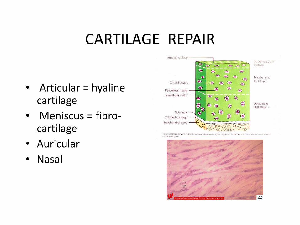
CARTILAGE REPAIR
• Articular = hyaline
cartilage • Meniscus = fibro-
cartilage • Auricular • Nasal
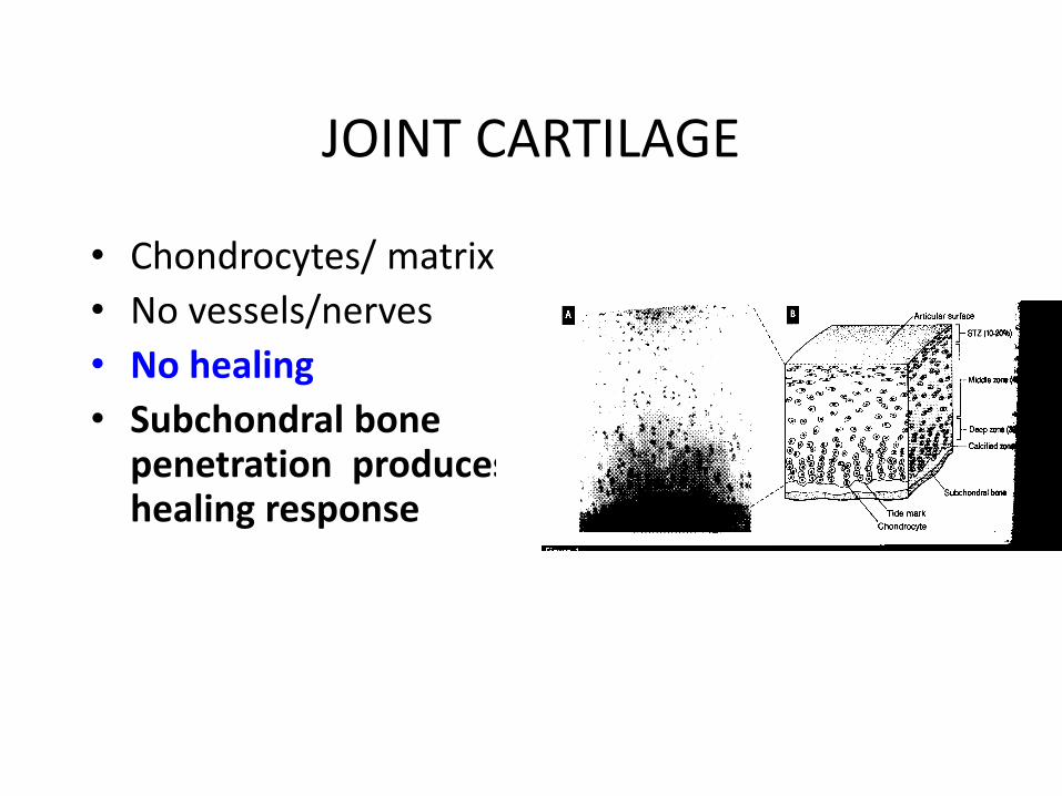
JOINT CARTILAGE
• Chondrocytes/ matrix • No vessels/nerves • No healing • Subchondral bone
penetration produces healing response
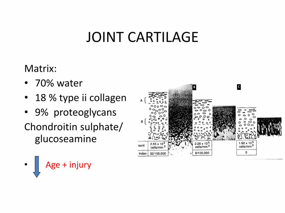
JOINT CARTILAGE
Matrix: • 70% water • 18 % type ii collagen • 9% proteoglycans Chondroitin sulphate/
glucoseamine • Age + injury
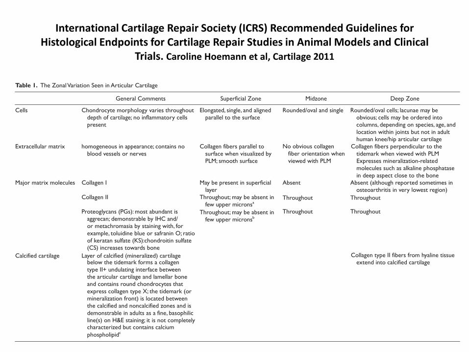
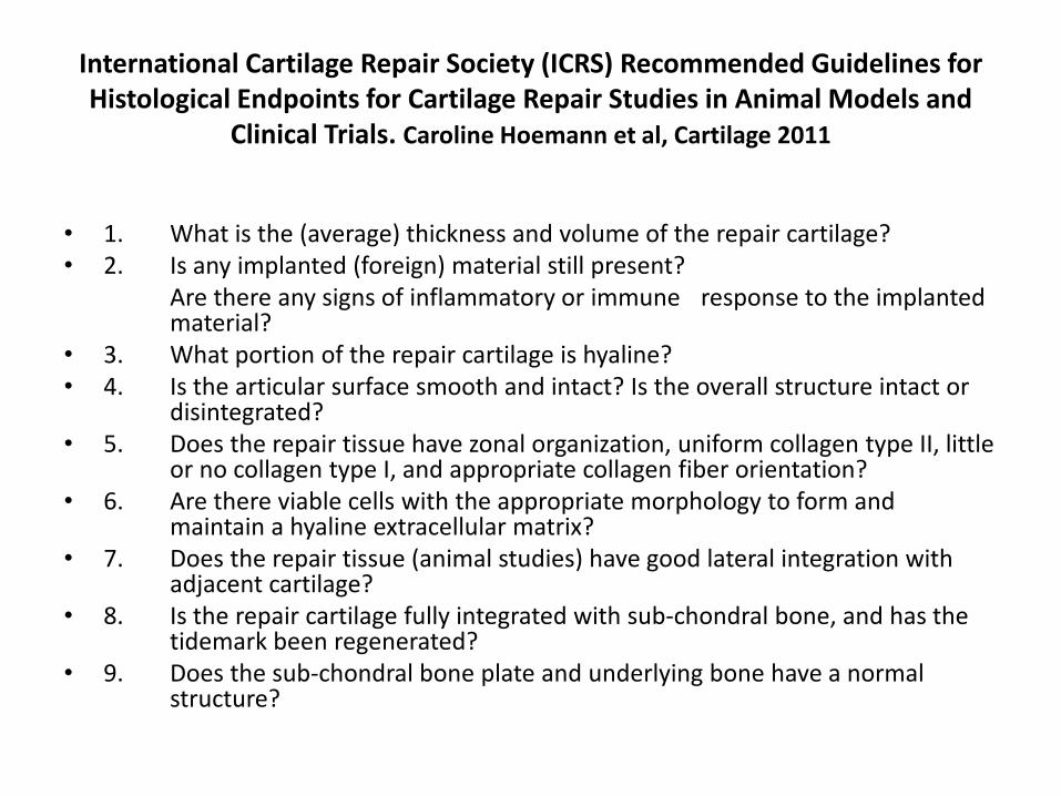
International Cartilage Repair Society (ICRS) Recommended Guidelines for Histological Endpoints for Cartilage Repair Studies in Animal Models and Clinical
Trials. Caroline Hoemann et al, Cartilage 2011
Table 1. The Zonal Variation Seen in Articular Cartilage
General Comments Superficial Zone Midzone Deep Zone
Cells Chondrocyte morphology varies throughout depth of cartilage; no inflammatory cells present
Elongated, single, and aligned
parallel to the surface
Rounded/oval and single Rounded/oval cells; lacunae may be
obvious; cells may be ordered into columns, depending on species, age, and location within joints but not in adult human knee/hip articular cartilage
Extracellular matrix homogeneous in appearance; contains no blood vessels or nerves
Collagen fibers parallel to surface when visualized by PLM; smooth surface
No obvious collagen fiber orientation when viewed with PLM
Collagen fibers perpendicular to the tidemark when viewed with PLM Expresses mineralization-related molecules such as alkaline phosphatase in deep aspect close to the bone
Major matrix molecules Collagen I May be present in superficial layer
Collagen II Throughout; may be absent in few upper micronsa
Absent Absent (although reported sometimes in osteoarthritis in very lowest region)
Throughout Throughout
Proteoglycans (PGs): most abundant is aggrecan; demonstrable by IHC and/ or metachromasia by staining with, for example, toluidine blue or safranin O; ratio of keratan sulfate (KS):chondroitin sulfate (CS) increases towards bone
Calcified cartilage Layer of calcified (mineralized) cartilage below the tidemark forms a collagen type II+ undulating interface between the articular cartilage and lamellar bone and contains round chondrocytes that express collagen type X; the tidemark (or mineralization front) is located between the calcified and noncalcified zones and is demonstrable in adults as a fine, basophilic line(s) on H&E staining; it is not completely characterized but contains calcium phospholipidc
Throughout; may be absent in few upper micronsb
Throughout Throughout
Collagen type II fibers from hyaline tissue extend into calcified cartilage
International Cartilage Repair Society (ICRS) Recommended Guidelines for Histological Endpoints for Cartilage Repair Studies in Animal Models and
Clinical Trials. Caroline Hoemann et al, Cartilage 2011
• 1. What is the (average) thickness and volume of the repair cartilage? • 2. Is any implanted (foreign) material still present? Are there any signs of inflammatory or immune response to the implanted material? • 3. What portion of the repair cartilage is hyaline? • 4. Is the articular surface smooth and intact? Is the overall structure intact or
disintegrated? • 5. Does the repair tissue have zonal organization, uniform collagen type II, little
or no collagen type I, and appropriate collagen fiber orientation? • 6. Are there viable cells with the appropriate morphology to form and
maintain a hyaline extracellular matrix? • 7. Does the repair tissue (animal studies) have good lateral integration with
adjacent cartilage? • 8. Is the repair cartilage fully integrated with sub-chondral bone, and has the
tidemark been regenerated? • 9. Does the sub-chondral bone plate and underlying bone have a normal
structure?
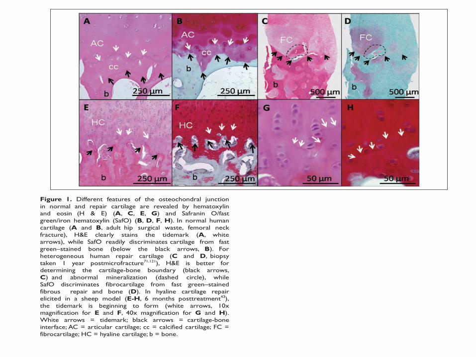
Figure 1. Different features of the osteochondral junction in normal and repair cartilage are revealed by hematoxylin and eosin (H & E) (A, C, E, G) and Safranin O/fast green/iron hematoxylin (SafO) (B, D, F, H). In normal human cartilage (A and B, adult hip surgical waste, femoral neck fracture), H&E clearly stains the tidemark (A, white arrows), while SafO readily discriminates cartilage from fast green–stained bone (below the black arrows, B). For heterogeneous human repair cartilage (C and D, biopsy taken 1 year postmicrofracture71,121), H&E is better for determining the cartilage-bone boundary (black arrows, C) and abnormal mineralization (dashed circle), while SafO discriminates fibrocartilage from fast green–stained fibrous repair and bone (D). In hyaline cartilage repair elicited in a sheep model (E-H, 6 months posttreatment43), the tidemark is beginning to form (white arrows, 10x magnification for E and F, 40x magnification for G and H). White arrows = tidemark; black arrows = cartilage-bone interface; AC = articular cartilage; cc = calcified cartilage; FC = fibrocartilage; HC = hyaline cartilage; b = bone.
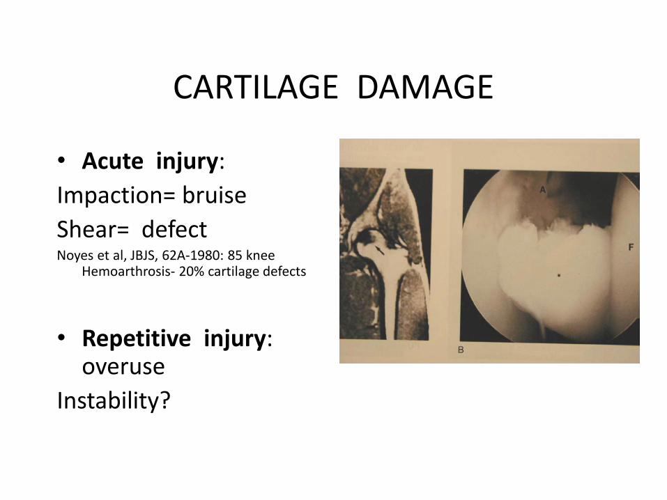
CARTILAGE DAMAGE
• Acute injury: Impaction= bruise Shear= defect Noyes et al, JBJS, 62A-1980: 85 knee
Hemoarthrosis- 20% cartilage defects
• Repetitive injury: overuse
Instability?
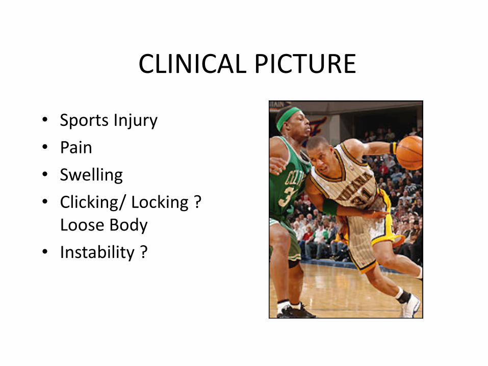
CLINICAL PICTURE
• Sports Injury • Pain • Swelling • Clicking/ Locking ?
Loose Body • Instability ?
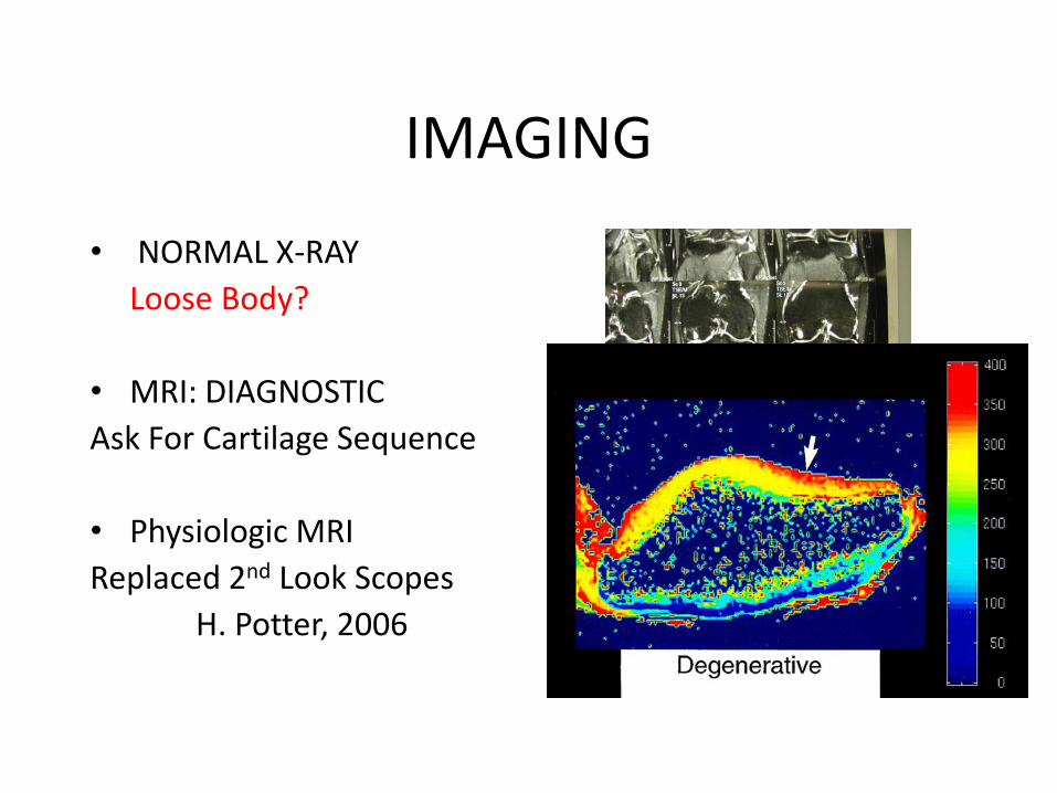
IMAGING
• NORMAL X-RAY Loose Body? • MRI: DIAGNOSTIC Ask For Cartilage Sequence • Physiologic MRI Replaced 2nd Look Scopes H. Potter, 2006
WHICH CARTILAGE DEFECT TO REPAIR?
• Symptomatic: Pain/ Mechanical • Joint effusion • Isolated (Multiple) – Unipolar (Femoral) • Joint: Stable ( +ACL) • Limb: No Deformity ( + HTO) • Patient: Younger Than 50 Years (Now 60 Ys ??)
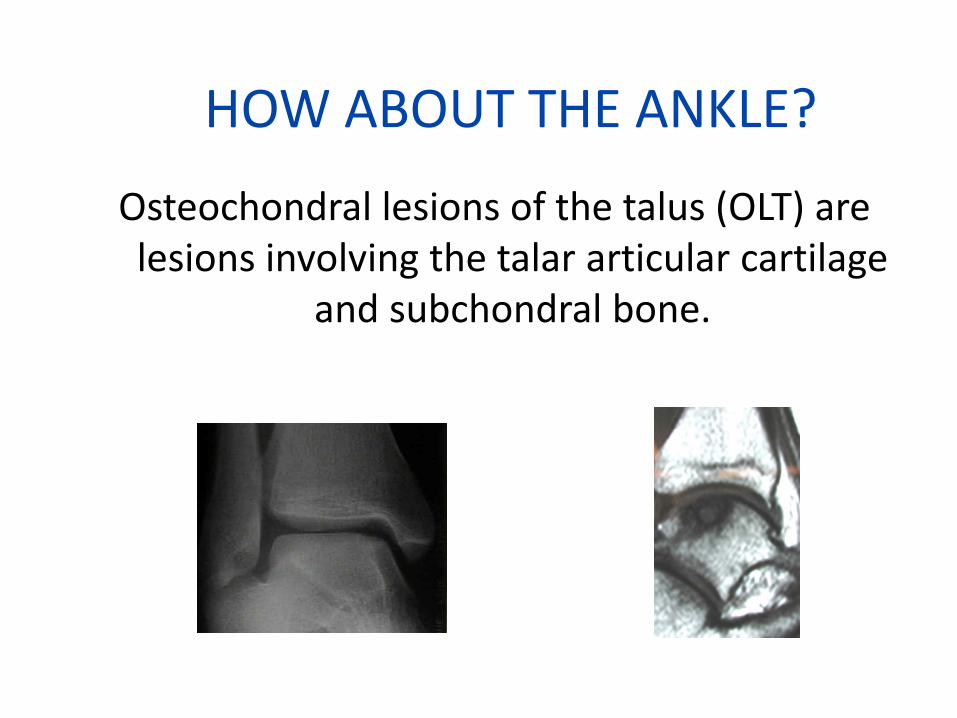
HOW ABOUT THE ANKLE? Osteochondral lesions of the talus (OLT) are lesions involving the talar articular cartilage
and subchondral bone.
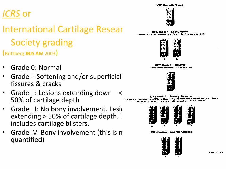
ICRS or International Cartilage Research
Society grading (Brittberg JBJS AM 2003) • Grade 0: Normal • Grade I: Softening and/or superficial
fissures & cracks • Grade II: Lesions extending down <
50% of cartilage depth • Grade III: No bony involvement. Lesions
extending > 50% of cartilage depth. This includes cartilage blisters.
• Grade IV: Bony involvement (this is not quantified)
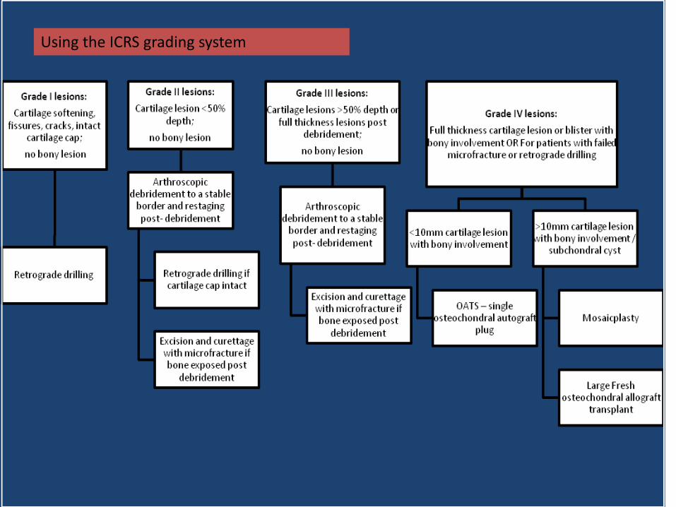
Using the ICRS grading system
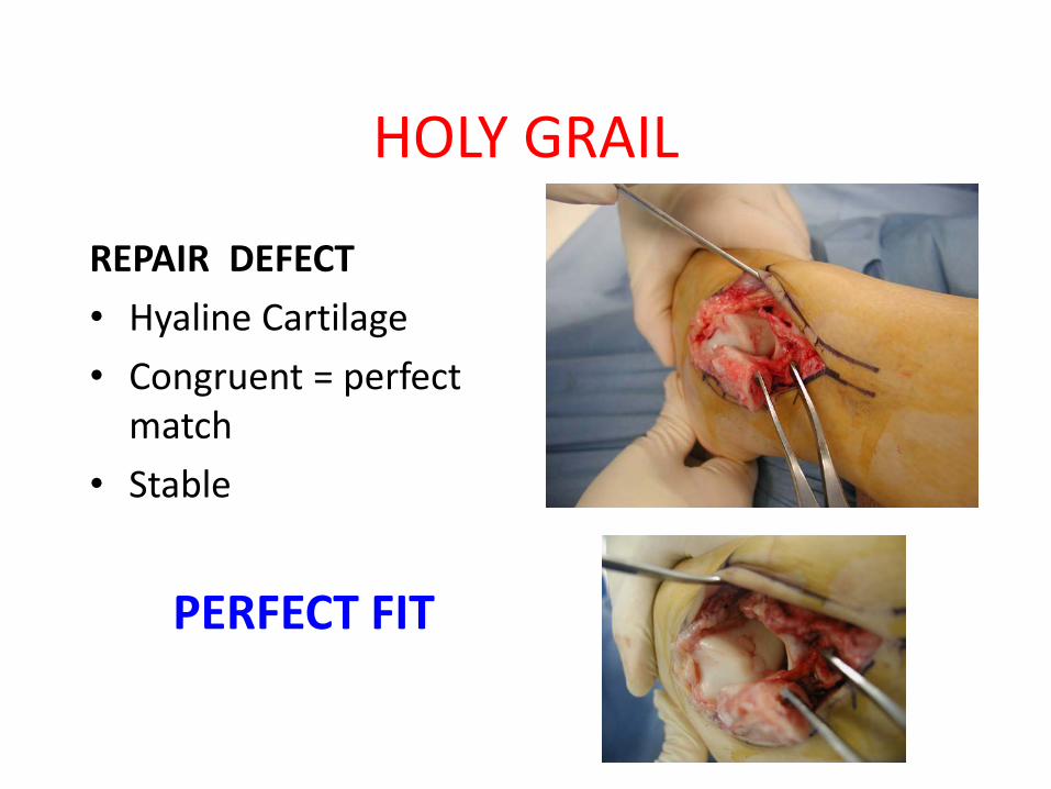
HOLY GRAIL
REPAIR DEFECT • Hyaline Cartilage • Congruent = perfect
match • Stable
PERFECT FIT
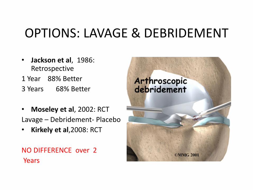
OPTIONS: LAVAGE & DEBRIDEMENT
• Jackson et al, 1986: Retrospective
1 Year 88% Better 3 Years 68% Better • Moseley et al, 2002: RCT Lavage – Debridement- Placebo • Kirkely et al,2008: RCT NO DIFFERENCE over 2 Years
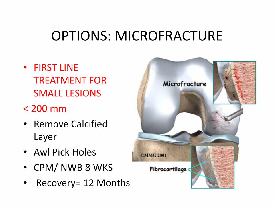
OPTIONS: MICROFRACTURE
• FIRST LINE TREATMENT FOR SMALL LESIONS
< 200 mm • Remove Calcified
Layer • Awl Pick Holes • CPM/ NWB 8 WKS • Recovery= 12 Months
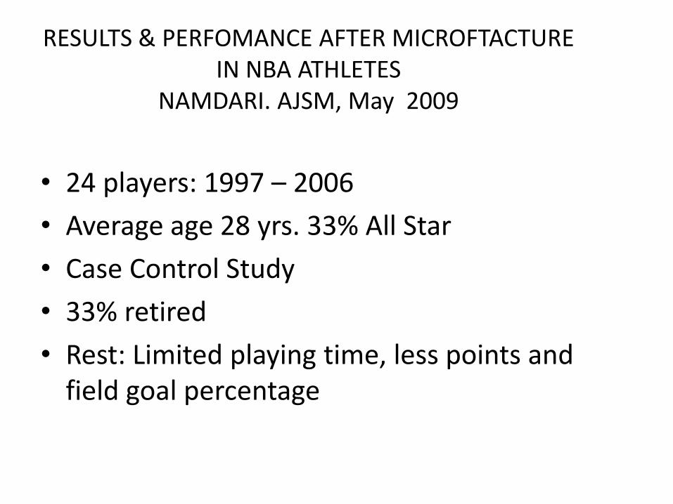
RESULTS & PERFOMANCE AFTER MICROFTACTURE IN NBA ATHLETES
NAMDARI. AJSM, May 2009
• 24 players: 1997 – 2006 • Average age 28 yrs. 33% All Star • Case Control Study • 33% retired • Rest: Limited playing time, less points and
field goal percentage
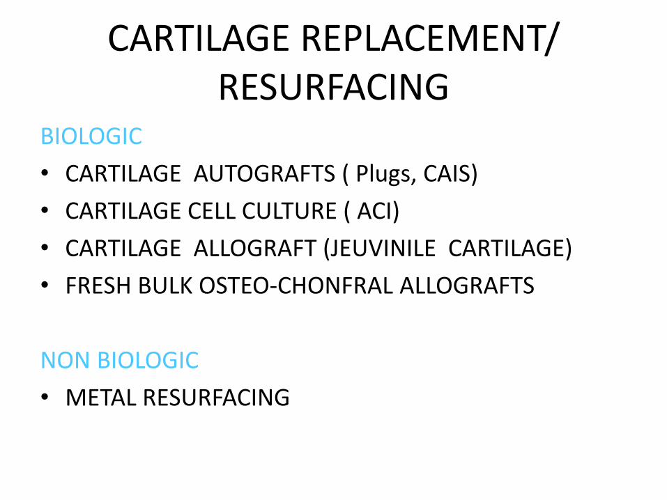
CARTILAGE REPLACEMENT/ RESURFACING
BIOLOGIC • CARTILAGE AUTOGRAFTS ( Plugs, CAIS) • CARTILAGE CELL CULTURE ( ACI) • CARTILAGE ALLOGRAFT (JEUVINILE CARTILAGE) • FRESH BULK OSTEO-CHONFRAL ALLOGRAFTS
NON BIOLOGIC • METAL RESURFACING
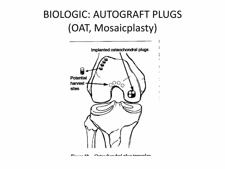
BIOLOGIC: AUTOGRAFT PLUGS (OAT, Mosaicplasty)
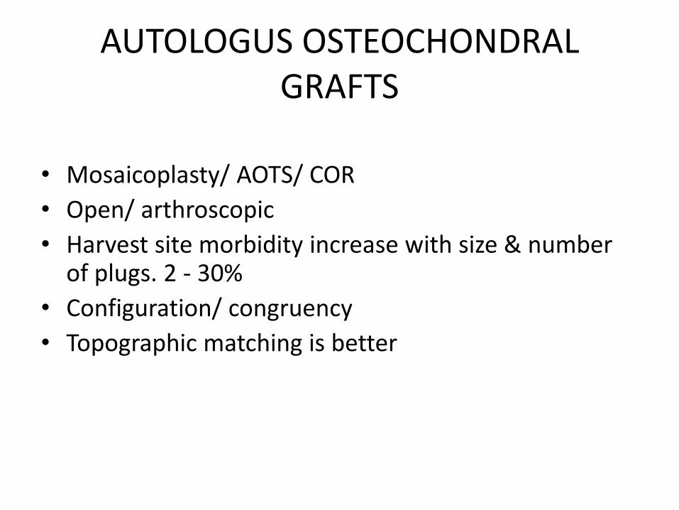
AUTOLOGUS OSTEOCHONDRAL GRAFTS
• Mosaicoplasty/ AOTS/ COR • Open/ arthroscopic • Harvest site morbidity increase with size & number
of plugs. 2 - 30% • Configuration/ congruency • Topographic matching is better
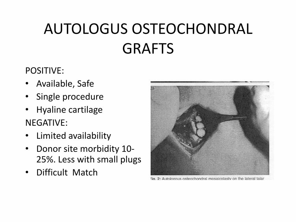
AUTOLOGUS OSTEOCHONDRAL GRAFTS
POSITIVE: • Available, Safe • Single procedure • Hyaline cartilage NEGATIVE: • Limited availability • Donor site morbidity 10-
25%. Less with small plugs • Difficult Match
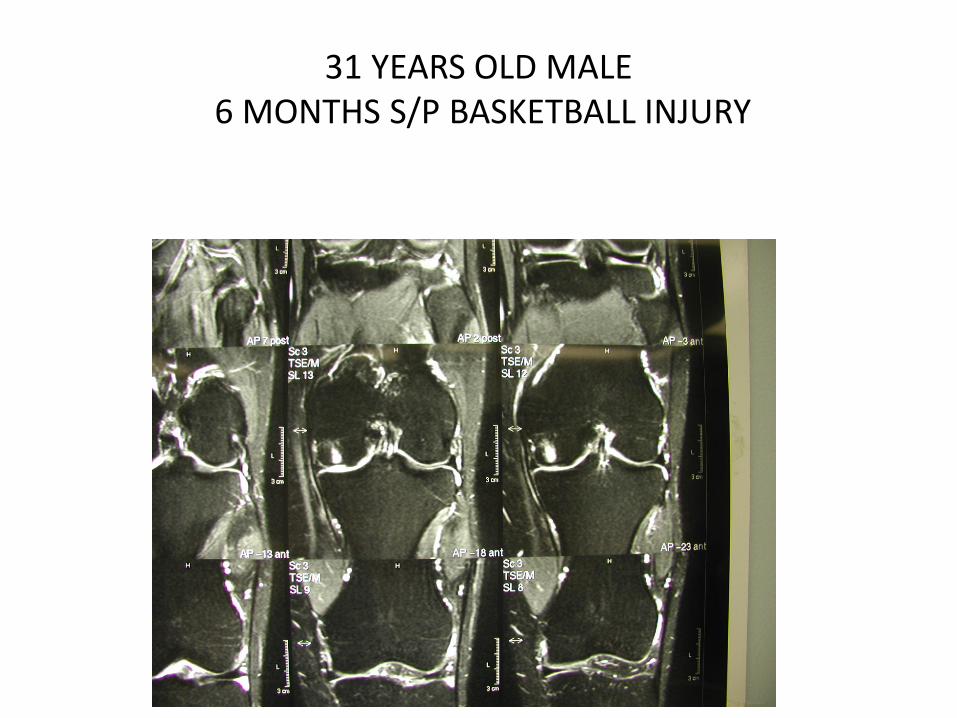
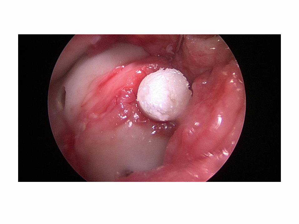
31 YEARS OLD MALE 6 MONTHS S/P BASKETBALL INJURY
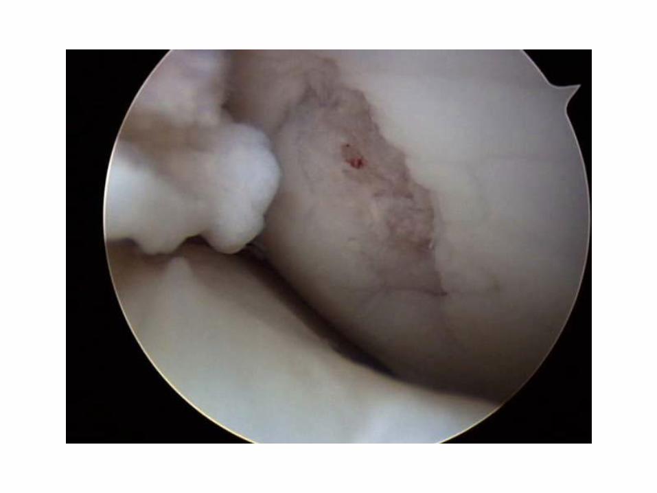
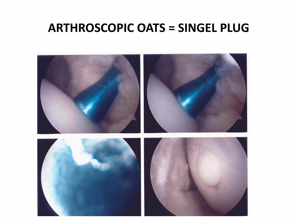
ARTHROSCOPIC OATS = SINGEL PLUG
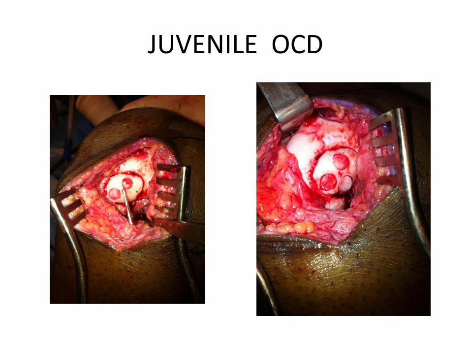
JUVENILE OCD
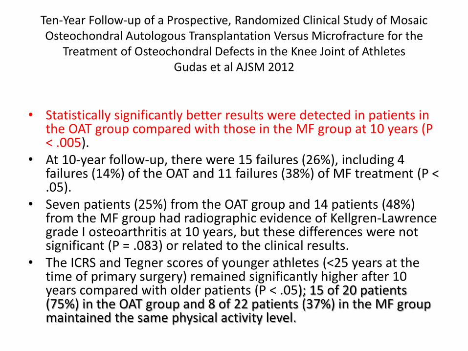
Ten-Year Follow-up of a Prospective, Randomized Clinical Study of Mosaic Osteochondral Autologous Transplantation Versus Microfracture for the
Treatment of Osteochondral Defects in the Knee Joint of Athletes Gudas et al AJSM 2012
• Statistically significantly better results were detected in patients in
the OAT group compared with those in the MF group at 10 years (P < .005).
• At 10-year follow-up, there were 15 failures (26%), including 4 failures (14%) of the OAT and 11 failures (38%) of MF treatment (P < .05).
• Seven patients (25%) from the OAT group and 14 patients (48%) from the MF group had radiographic evidence of Kellgren-Lawrence grade I osteoarthritis at 10 years, but these differences were not significant (P = .083) or related to the clinical results.
• The ICRS and Tegner scores of younger athletes (<25 years at the time of primary surgery) remained significantly higher after 10 years compared with older patients (P < .05); 15 of 20 patients (75%) in the OAT group and 8 of 22 patients (37%) in the MF group maintained the same physical activity level.
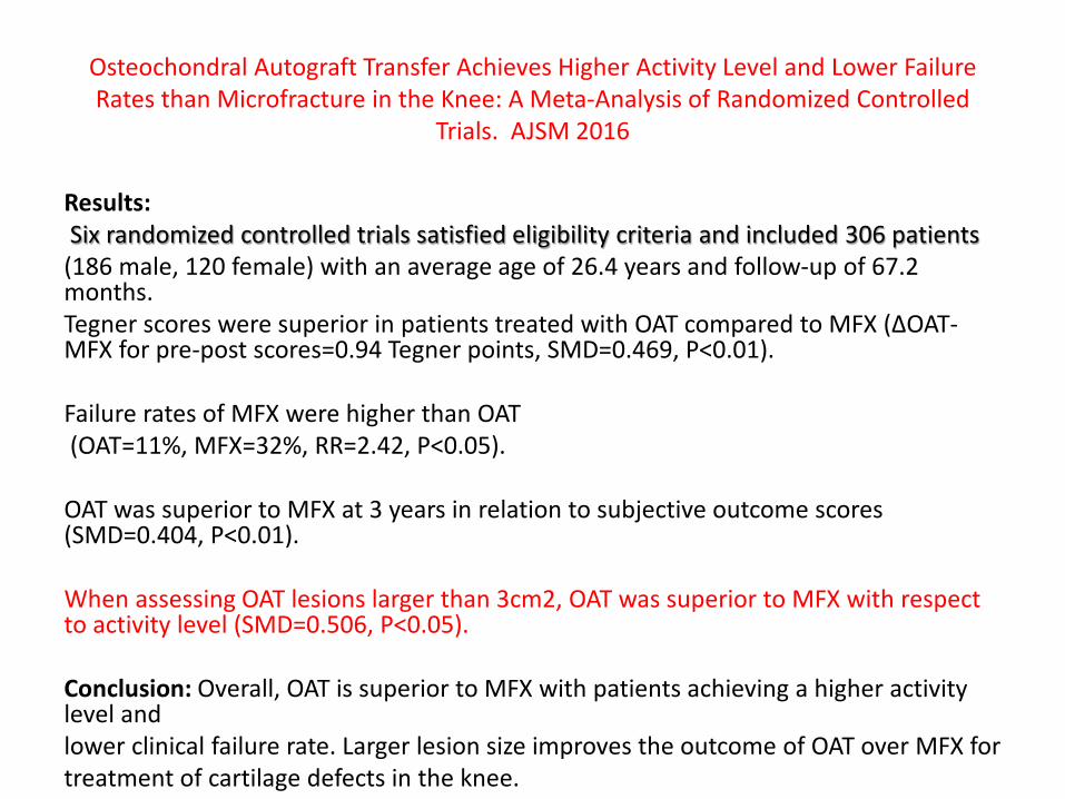
Osteochondral Autograft Transfer Achieves Higher Activity Level and Lower Failure Rates than Microfracture in the Knee: A Meta-Analysis of Randomized Controlled
Trials. AJSM 2016
Results: Six randomized controlled trials satisfied eligibility criteria and included 306 patients (186 male, 120 female) with an average age of 26.4 years and follow-up of 67.2 months. Tegner scores ǁĞƌĞƐƵƉĞƌŝŽƌŝŶƉĂƚŝĞŶƚƐƚƌĞĂƚĞĚǁŝƚŚKdĐŽŵƉĂƌĞĚƚŽD&y;ȴKd-MFX for pre-post scores=0.94 Tegner points, SMD=0.469, P<0.01). Failure rates of MFX were higher than OAT (OAT=11%, MFX=32%, RR=2.42, P<0.05). OAT was superior to MFX at 3 years in relation to subjective outcome scores (SMD=0.404, P<0.01). When assessing OAT lesions larger than 3cm2, OAT was superior to MFX with respect to activity level (SMD=0.506, P<0.05). Conclusion: Overall, OAT is superior to MFX with patients achieving a higher activity level and lower clinical failure rate. Larger lesion size improves the outcome of OAT over MFX for treatment of cartilage defects in the knee.
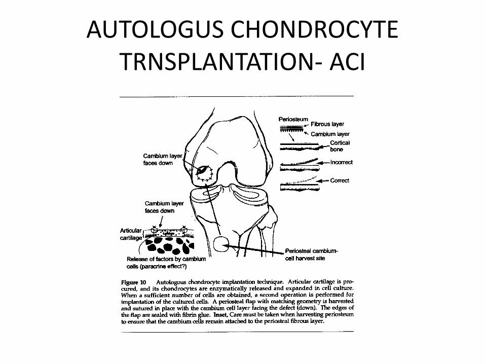
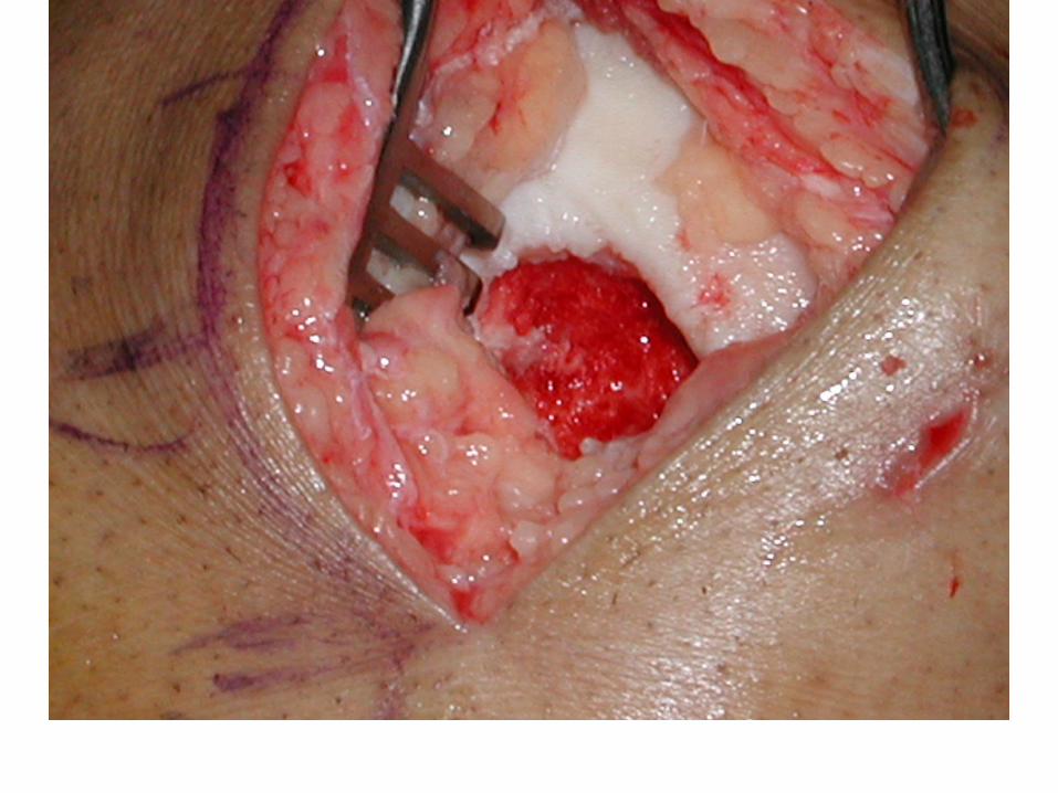
AUTOLOGUS CHONDROCYTE TRNSPLANTATION- ACI
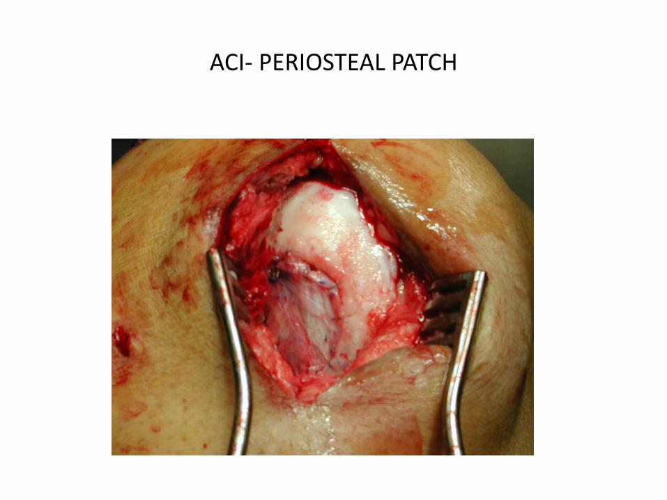
ACI- PERIOSTEAL PATCH
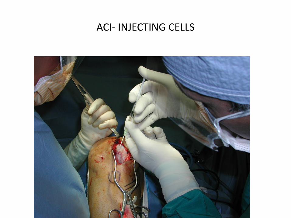
ACI- INJECTING CELLS
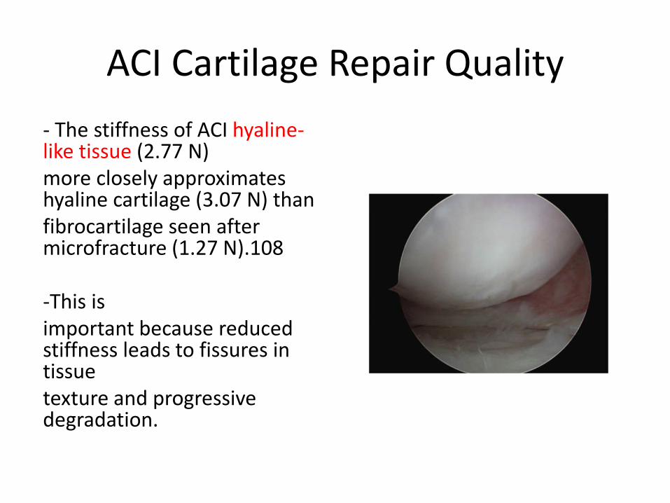
ACI Cartilage Repair Quality - The stiffness of ACI hyaline-like tissue (2.77 N) more closely approximates hyaline cartilage (3.07 N) than fibrocartilage seen after microfracture (1.27 N).108 -This is important because reduced stiffness leads to fissures in tissue texture and progressive degradation.
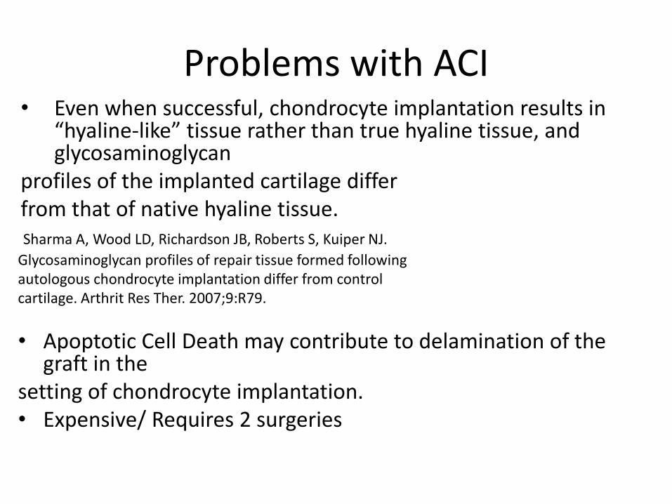
Problems with ACI • Even when successful, chondrocyte implantation results in
“hyaline-like” tissue rather than true hyaline tissue, and glycosaminoglycan
profiles of the implanted cartilage differ from that of native hyaline tissue. Sharma A, Wood LD, Richardson JB, Roberts S, Kuiper NJ. Glycosaminoglycan profiles of repair tissue formed following autologous chondrocyte implantation differ from control cartilage. Arthrit Res Ther. 2007;9:R79.
• Apoptotic Cell Death may contribute to delamination of the graft in the
setting of chondrocyte implantation. • Expensive/ Requires 2 surgeries
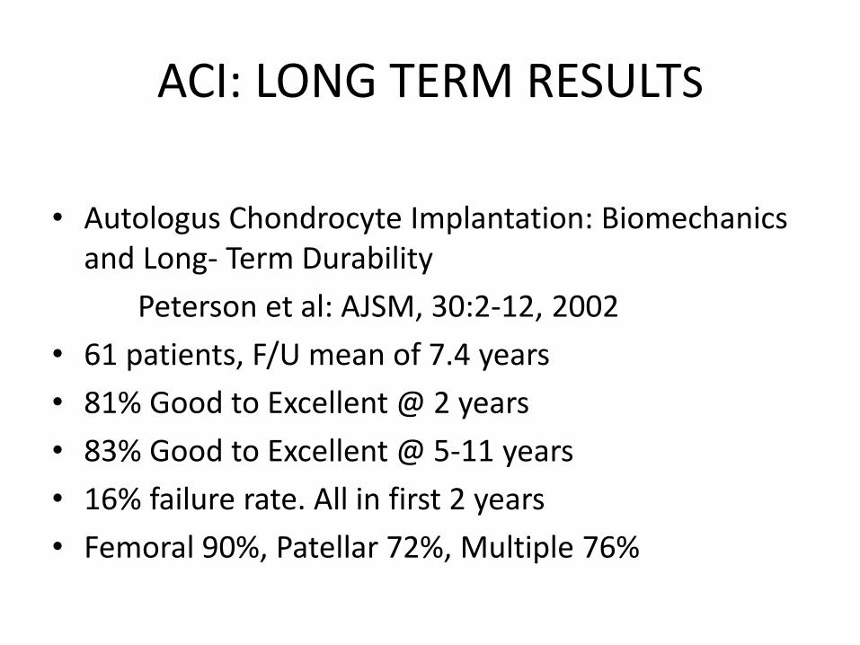
ACI: LONG TERM RESULTS
• Autologus Chondrocyte Implantation: Biomechanics and Long- Term Durability
Peterson et al: AJSM, 30:2-12, 2002 • 61 patients, F/U mean of 7.4 years • 81% Good to Excellent @ 2 years • 83% Good to Excellent @ 5-11 years • 16% failure rate. All in first 2 years • Femoral 90%, Patellar 72%, Multiple 76%
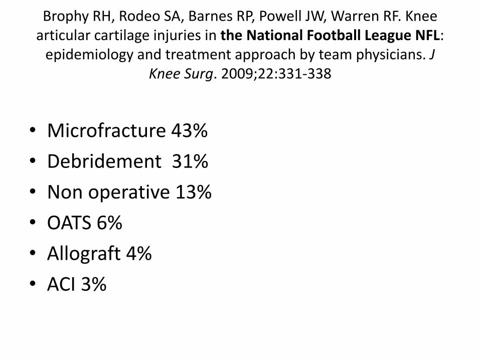
Brophy RH, Rodeo SA, Barnes RP, Powell JW, Warren RF. Knee articular cartilage injuries in the National Football League NFL:
epidemiology and treatment approach by team physicians. J Knee Surg. 2009;22:331-338
• Microfracture 43% • Debridement 31% • Non operative 13% • OATS 6% • Allograft 4% • ACI 3%
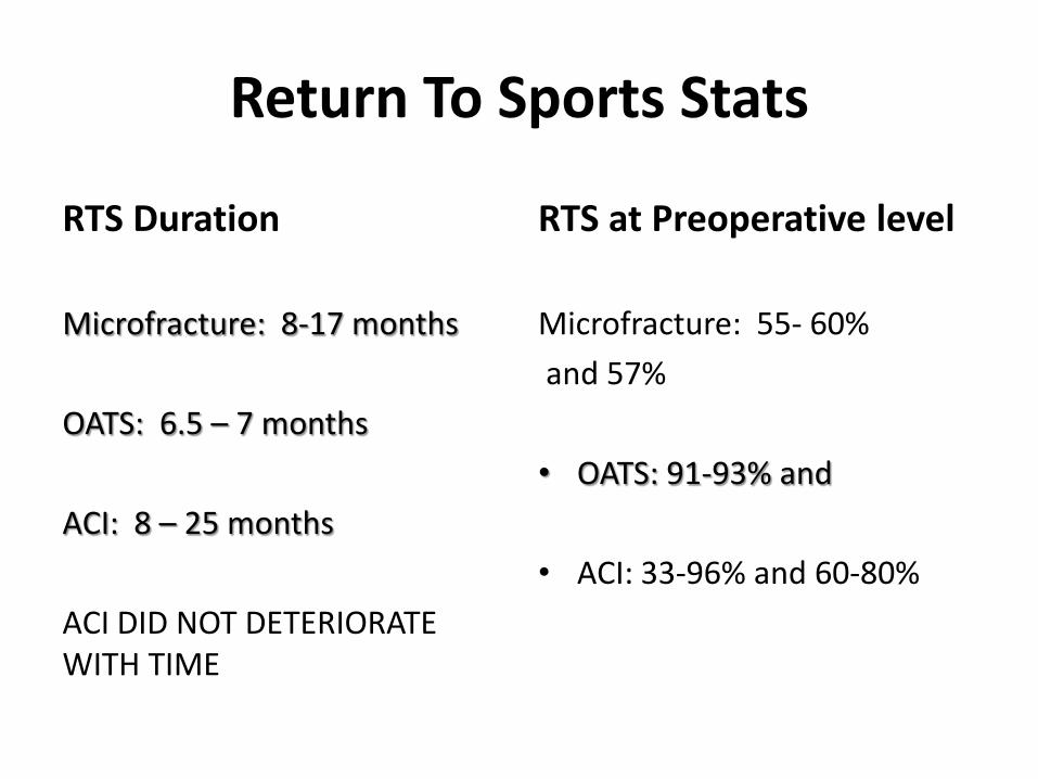
Return To Sports Stats
RTS Duration
Microfracture: 8-17 months OATS: 6.5 – 7 months ACI: 8 – 25 months ACI DID NOT DETERIORATE WITH TIME
RTS at Preoperative level
Microfracture: 55- 60% and 57%
• OATS: 91-93% and
• ACI: 33-96% and 60-80%
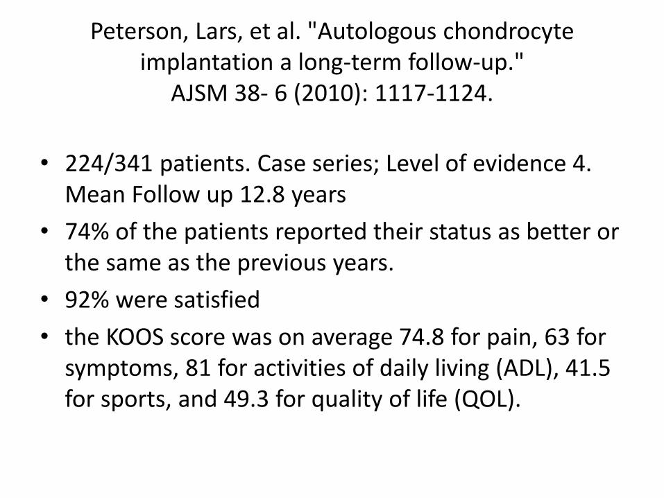
Peterson, Lars, et al. "Autologous chondrocyte implantation a long-term follow-up."
AJSM 38- 6 (2010): 1117-1124.
• 224/341 patients. Case series; Level of evidence 4. Mean Follow up 12.8 years
• 74% of the patients reported their status as better or the same as the previous years.
• 92% were satisfied • the KOOS score was on average 74.8 for pain, 63 for
symptoms, 81 for activities of daily living (ADL), 41.5 for sports, and 49.3 for quality of life (QOL).
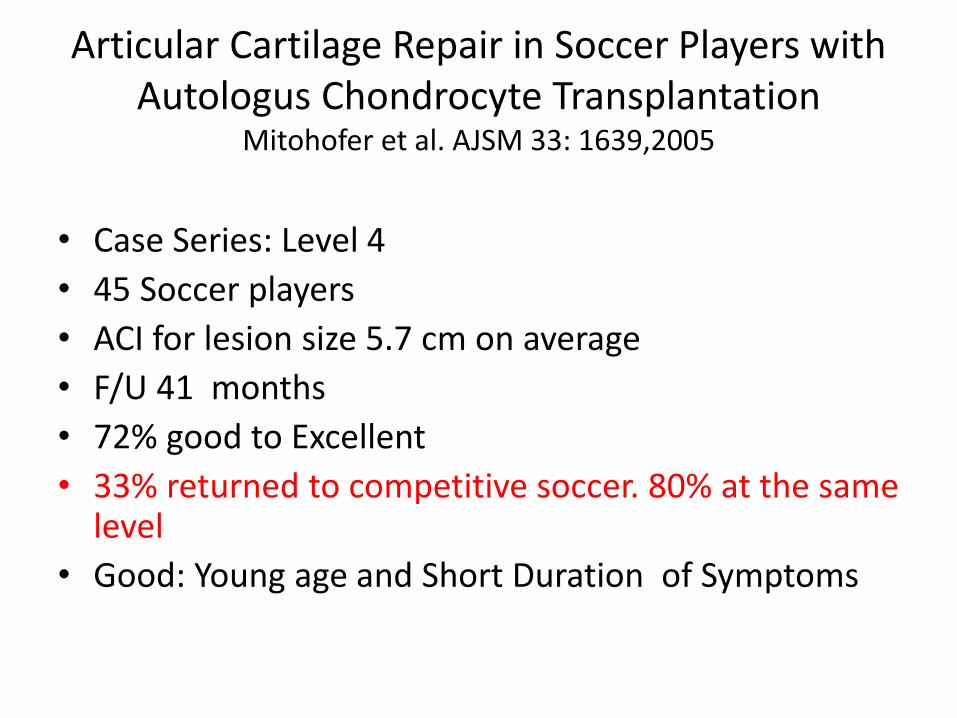
Articular Cartilage Repair in Soccer Players with Autologus Chondrocyte Transplantation
Mitohofer et al. AJSM 33: 1639,2005
• Case Series: Level 4 • 45 Soccer players • ACI for lesion size 5.7 cm on average • F/U 41 months • 72% good to Excellent • 33% returned to competitive soccer. 80% at the same
level • Good: Young age and Short Duration of Symptoms
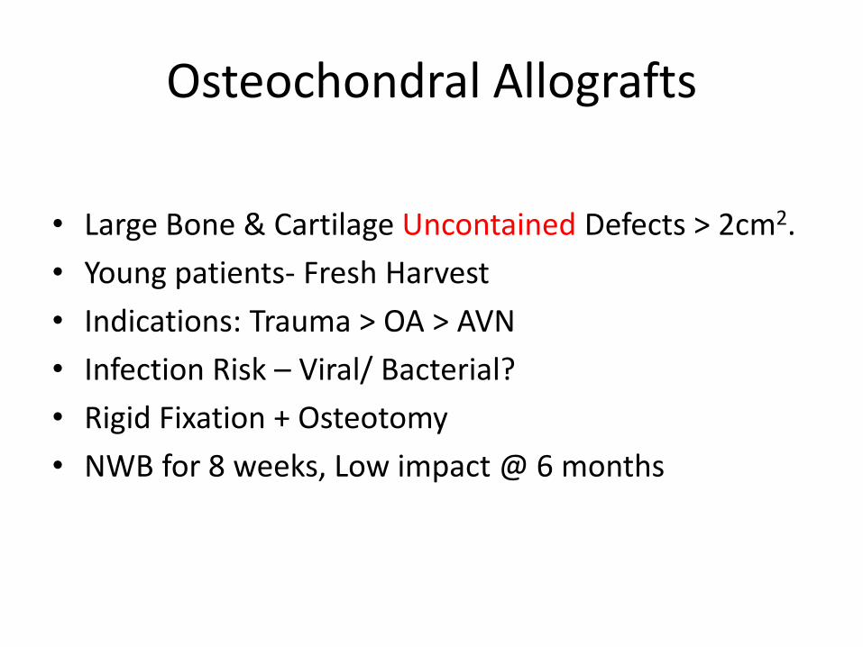
Osteochondral Allografts
• Large Bone & Cartilage Uncontained Defects > 2cm2. • Young patients- Fresh Harvest • Indications: Trauma > OA > AVN • Infection Risk – Viral/ Bacterial? • Rigid Fixation + Osteotomy • NWB for 8 weeks, Low impact @ 6 months
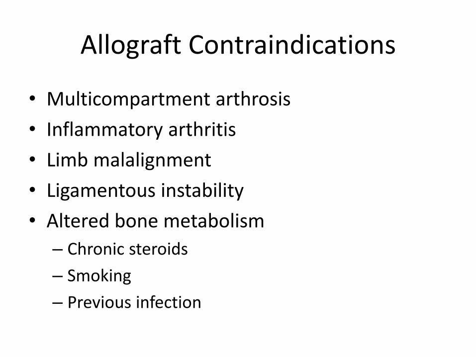
Allograft Contraindications
• Multicompartment arthrosis • Inflammatory arthritis • Limb malalignment • Ligamentous instability • Altered bone metabolism – Chronic steroids – Smoking – Previous infection
Osteochondral Allografts
• Hyaline cartilage is ideal for transplantation – Avascular – Metabolic needs met by diffusion from synovial
fluid – Aneural – Immunopriviledged – Viable chondrocytes able to survive hypothermic
storage
Osteochondral Allografts
• Osseous structure – Underlying structural support – Allows fixation to host – Originally vascularized – Cells do not survive hypothermic storage – Scaffold for creeping substitution
Graft Acquisition
• Age criterion: 15-40 years old • Joint surface passes visual inspection • Size match – Radiographs taken with a magnification marker – +/- 2 mm acceptable match
• Typically not HLA or blood type matched – Hyaline cartilage immunopriviledged – 50% individuals can develop anti-HLA antibodies
with unknown significance
Graft Safety
• Recovery, processing and testing established by American Association of Tissue Banks – Hold tissue for 14 days for microbiologic testing – No published data quantifying risk of transmission
for osteochondral allografts
• AAOS Musculoskeletal Allograft Tissue Safety Statement – 75th Annual Meeting, March 5-9, 2008
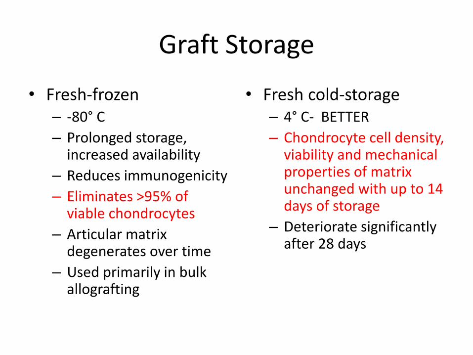
Graft Storage
• Fresh-frozen – -80° C – Prolonged storage,
increased availability – Reduces immunogenicity – Eliminates >95% of
viable chondrocytes – Articular matrix
degenerates over time – Used primarily in bulk
allografting
• Fresh cold-storage – 4° C- BETTER – Chondrocyte cell density,
viability and mechanical properties of matrix unchanged with up to 14 days of storage
– Deteriorate significantly after 28 days
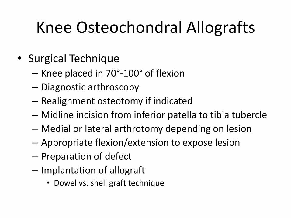
Knee Osteochondral Allografts
• Surgical Technique – Knee placed in 70°-100° of flexion – Diagnostic arthroscopy – Realignment osteotomy if indicated – Midline incision from inferior patella to tibia tubercle – Medial or lateral arthrotomy depending on lesion – Appropriate flexion/extension to expose lesion – Preparation of defect – Implantation of allograft
• Dowel vs. shell graft technique
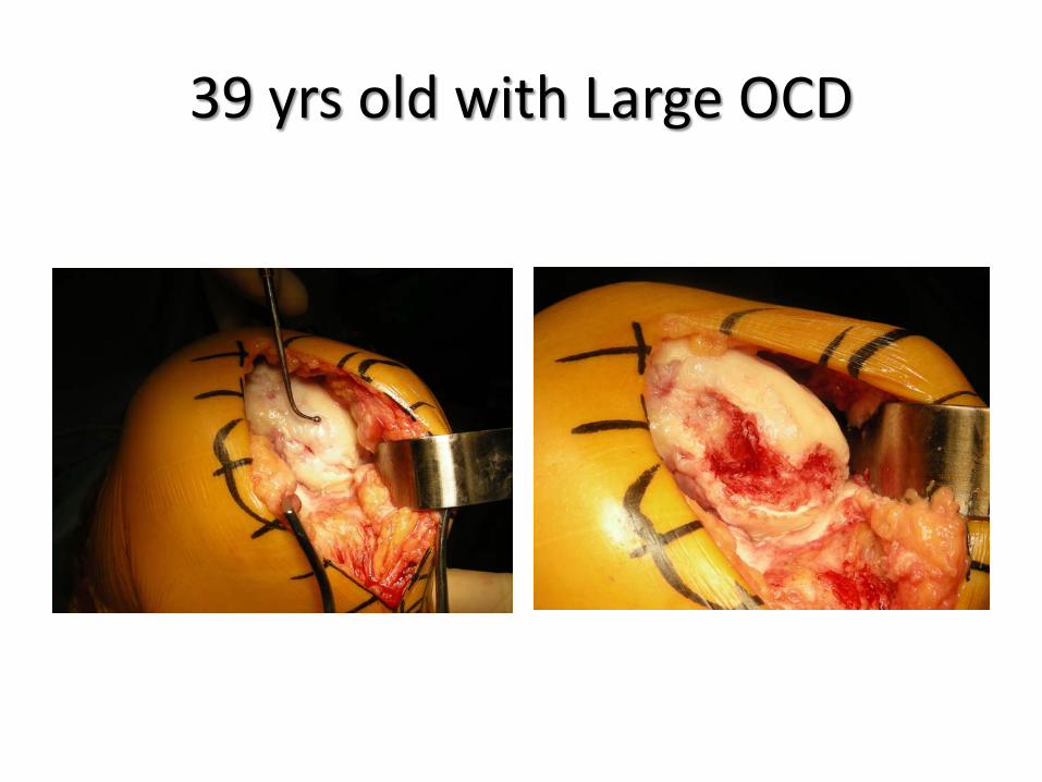
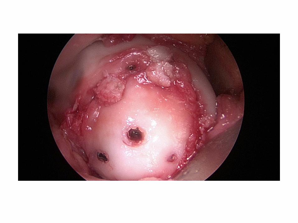
39 yrs old with Large OCD
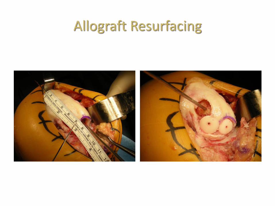
Allograft Resurfacing
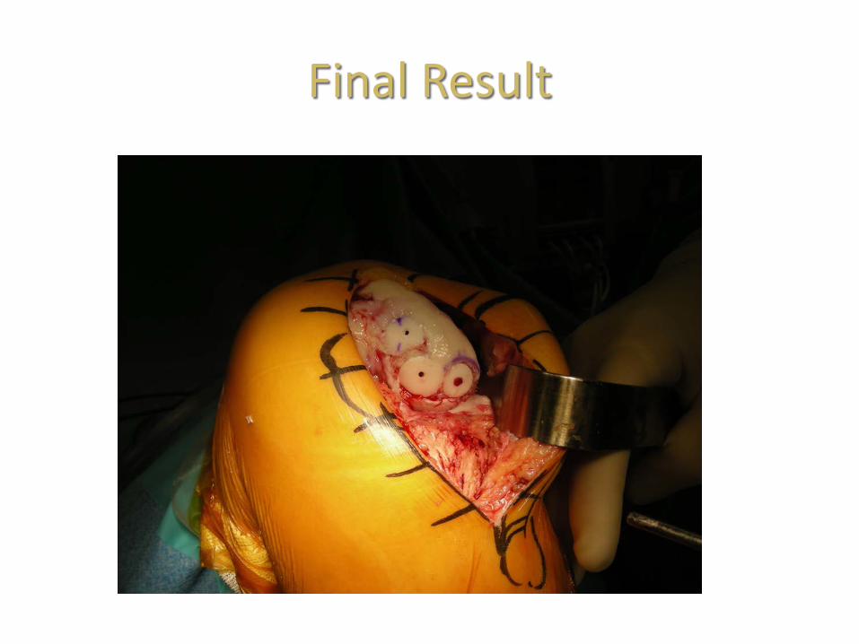
Final Result
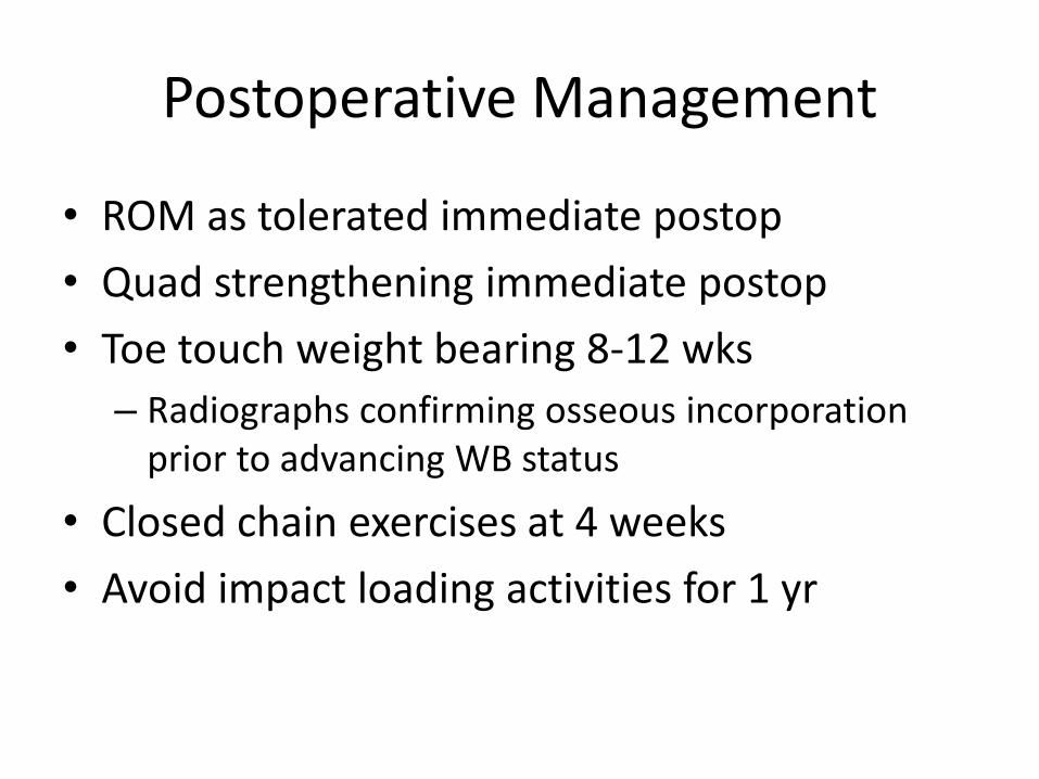
Postoperative Management
• ROM as tolerated immediate postop • Quad strengthening immediate postop • Toe touch weight bearing 8-12 wks – Radiographs confirming osseous incorporation
prior to advancing WB status
• Closed chain exercises at 4 weeks • Avoid impact loading activities for 1 yr
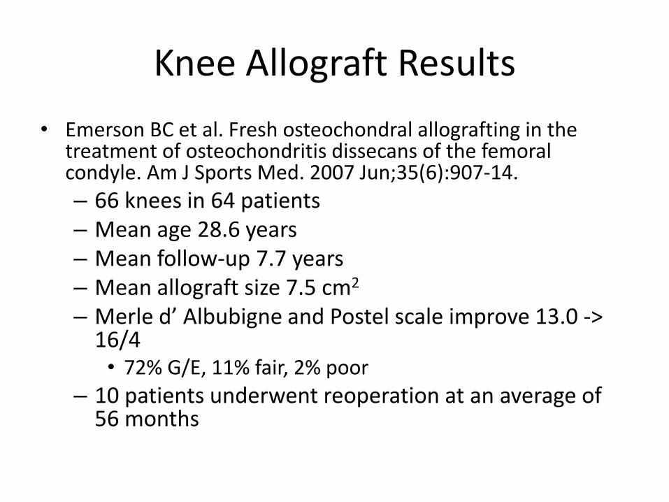
Knee Allograft Results • Emerson BC et al. Fresh osteochondral allografting in the
treatment of osteochondritis dissecans of the femoral condyle. Am J Sports Med. 2007 Jun;35(6):907-14. – 66 knees in 64 patients – Mean age 28.6 years – Mean follow-up 7.7 years – Mean allograft size 7.5 cm2
– Merle d’ Albubigne and Postel scale improve 13.0 -> 16/4 • 72% G/E, 11% fair, 2% poor
– 10 patients underwent reoperation at an average of 56 months
Knee Allograft Results
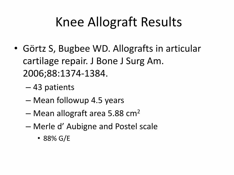
• Görtz S, Bugbee WD. Allografts in articular cartilage repair. J Bone J Surg Am. 2006;88:1374-1384. – 43 patients – Mean followup 4.5 years – Mean allograft area 5.88 cm2 – Merle d’ Aubigne and Postel scale • 88% G/E
Knee Allograft Results
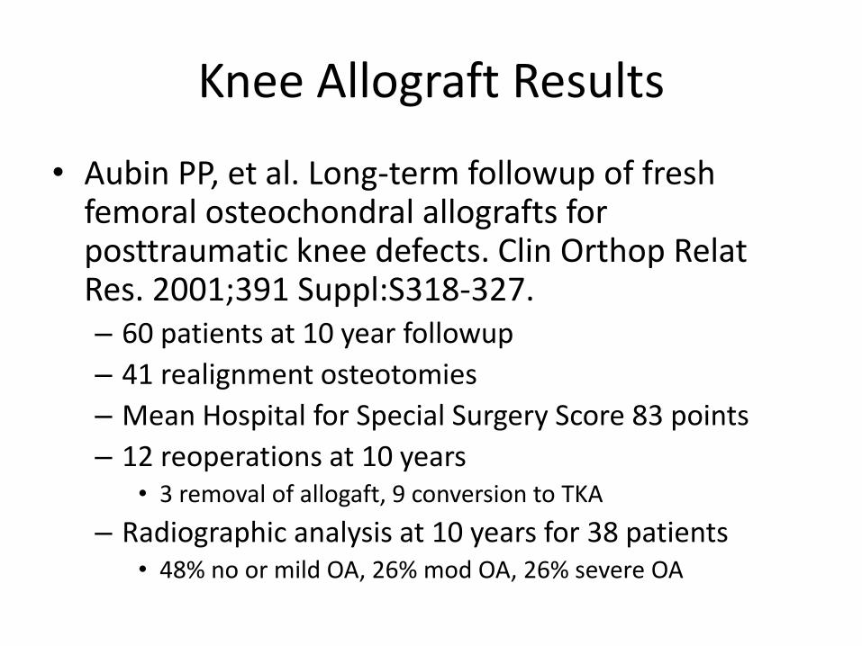
• Aubin PP, et al. Long-term followup of fresh femoral osteochondral allografts for posttraumatic knee defects. Clin Orthop Relat Res. 2001;391 Suppl:S318-327. – 60 patients at 10 year followup – 41 realignment osteotomies – Mean Hospital for Special Surgery Score 83 points – 12 reoperations at 10 years
• 3 removal of allogaft, 9 conversion to TKA – Radiographic analysis at 10 years for 38 patients
• 48% no or mild OA, 26% mod OA, 26% severe OA
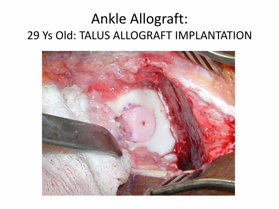
Ankle Allograft: 29 Ys Old: TALUS ALLOGRAFT IMPLANTATION
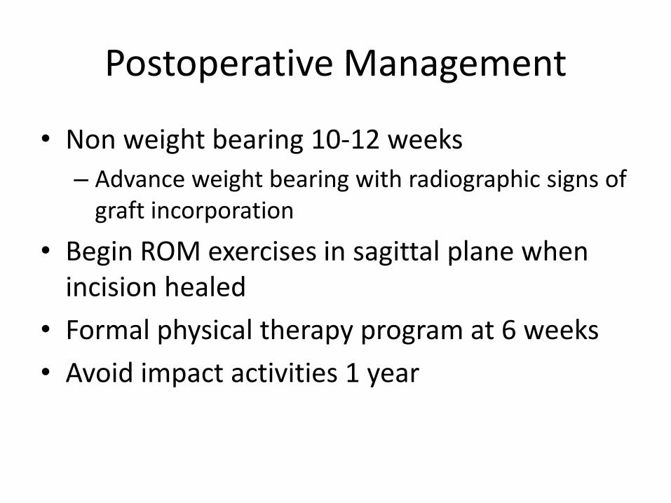
Postoperative Management
• Non weight bearing 10-12 weeks – Advance weight bearing with radiographic signs of
graft incorporation
• Begin ROM exercises in sagittal plane when incision healed
• Formal physical therapy program at 6 weeks • Avoid impact activities 1 year
Results
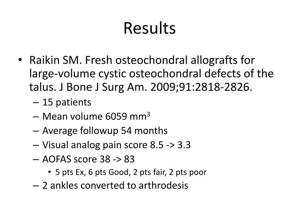
• Raikin SM. Fresh osteochondral allografts for large-volume cystic osteochondral defects of the talus. J Bone J Surg Am. 2009;91:2818-2826. – 15 patients – Mean volume 6059 mm3 – Average followup 54 months – Visual analog pain score 8.5 -> 3.3 – AOFAS score 38 -> 83
• 5 pts Ex, 6 pts Good, 2 pts fair, 2 pts poor – 2 ankles converted to arthrodesis
Results
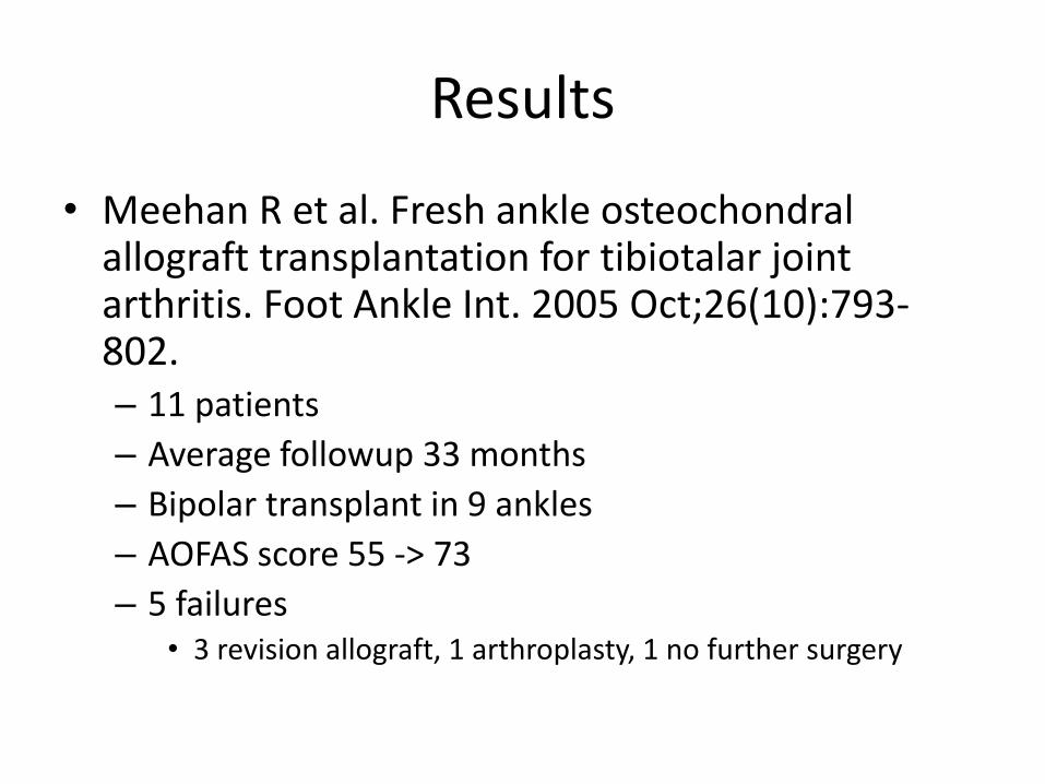
• Meehan R et al. Fresh ankle osteochondral allograft transplantation for tibiotalar joint arthritis. Foot Ankle Int. 2005 Oct;26(10):793-802. – 11 patients – Average followup 33 months – Bipolar transplant in 9 ankles – AOFAS score 55 -> 73 – 5 failures
• 3 revision allograft, 1 arthroplasty, 1 no further surgery
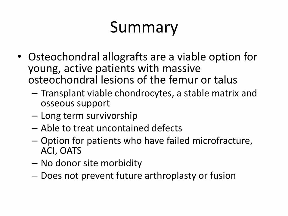
Summary • Osteochondral allografts are a viable option for
young, active patients with massive osteochondral lesions of the femur or talus – Transplant viable chondrocytes, a stable matrix and
osseous support – Long term survivorship – Able to treat uncontained defects – Option for patients who have failed microfracture,
ACI, OATS – No donor site morbidity – Does not prevent future arthroplasty or fusion
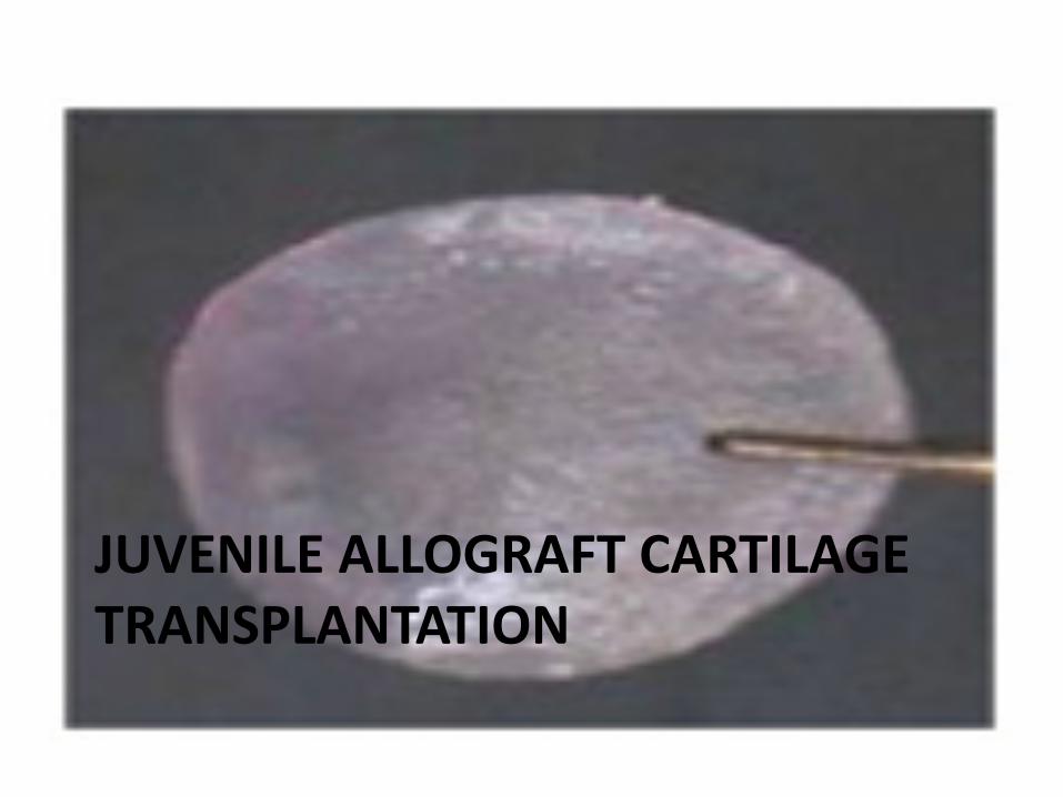
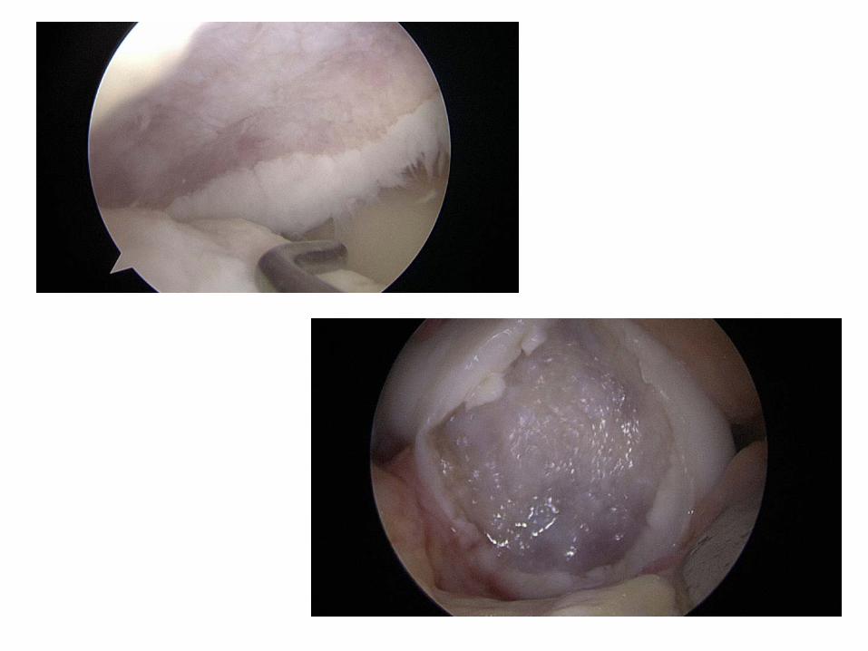
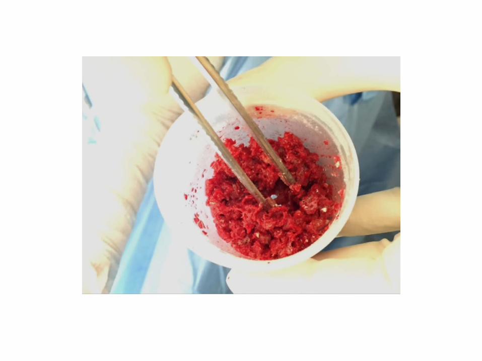
JUVENILE ALLOGRAFT CARTILAGE TRANSPLANTATION
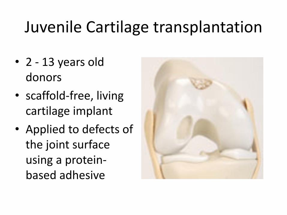
Juvenile Cartilage transplantation
• 2 - 13 years old donors
• scaffold-free, living cartilage implant
• Applied to defects of the joint surface using a protein-based adhesive
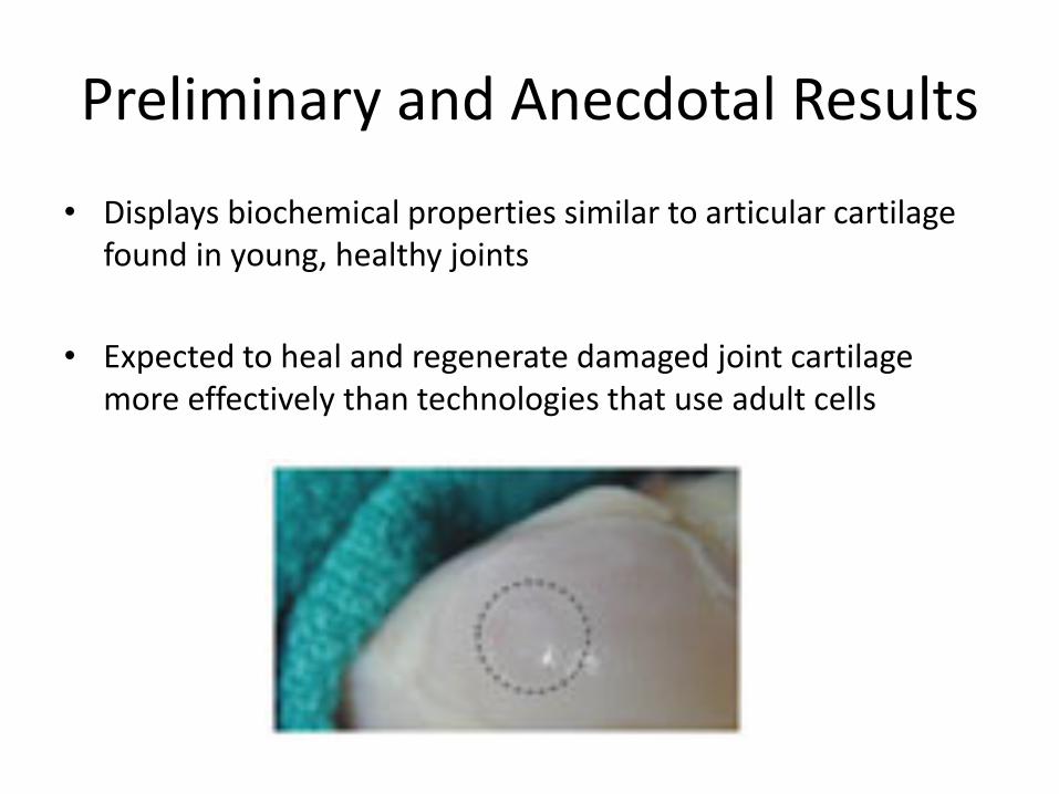
Preliminary and Anecdotal Results • Displays biochemical properties similar to articular cartilage
found in young, healthy joints
• Expected to heal and regenerate damaged joint cartilage more effectively than technologies that use adult cells
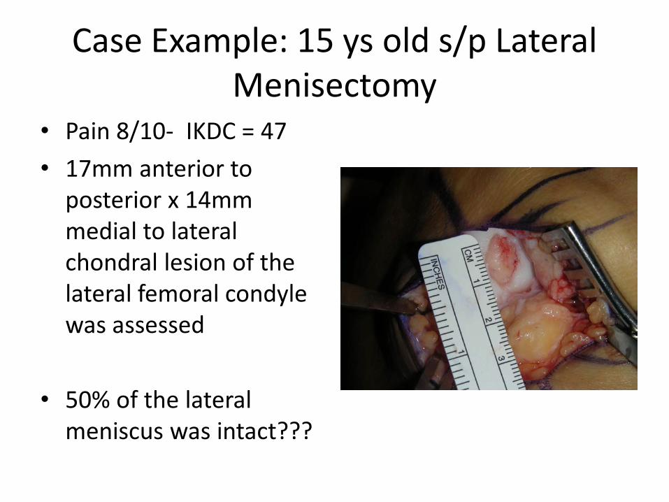
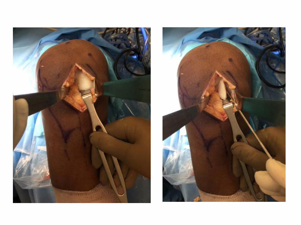
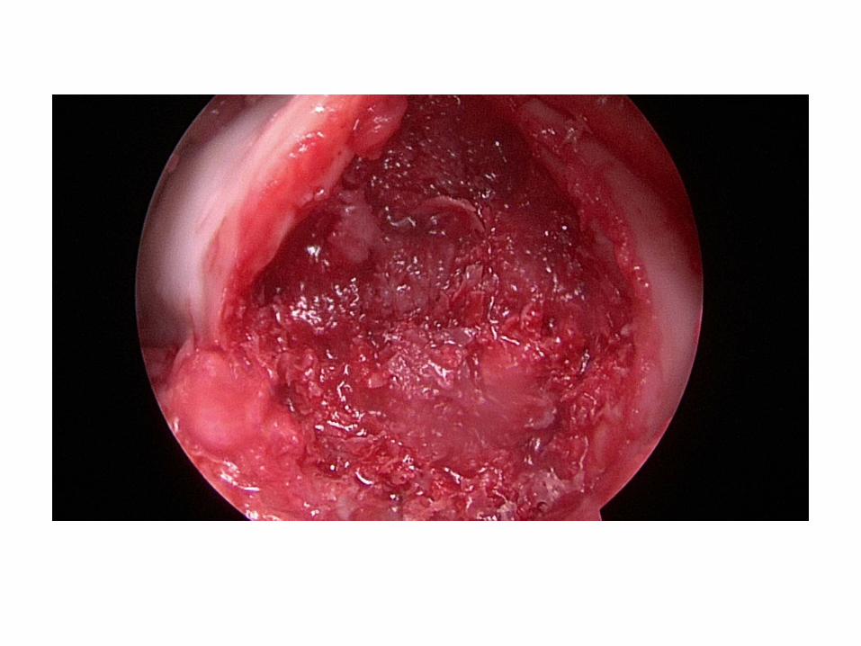
Case Example: 15 ys old s/p Lateral Menisectomy
• Pain 8/10- IKDC = 47 • 17mm anterior to
posterior x 14mm medial to lateral chondral lesion of the lateral femoral condyle was assessed
• 50% of the lateral meniscus was intact???
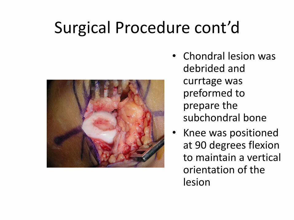
Surgical Procedure cont’d • Chondral lesion was
debrided and currtage was preformed to prepare the subchondral bone
• Knee was positioned at 90 degrees flexion to maintain a vertical orientation of the lesion
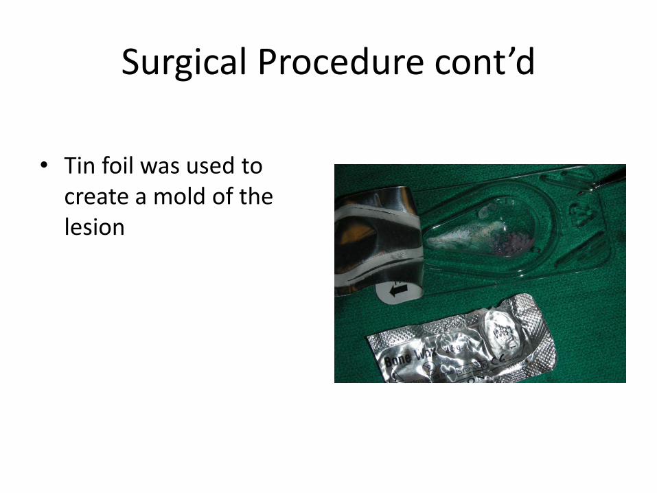
Surgical Procedure cont’d
• Tin foil was used to create a mold of the lesion
Surgical Procedure cont’d
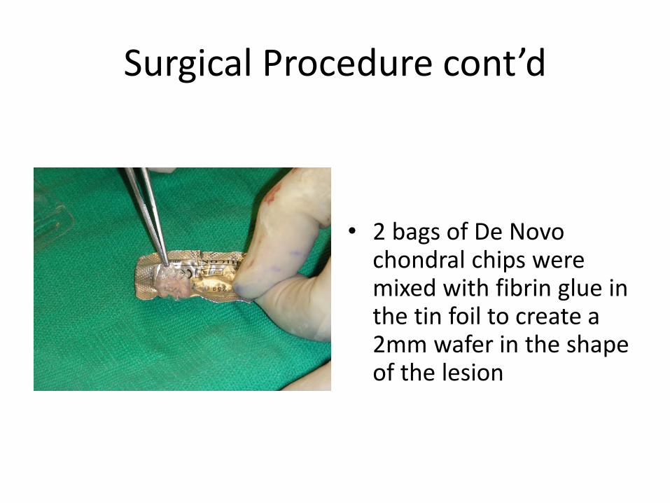
• 2 bags of De Novo chondral chips were mixed with fibrin glue in the tin foil to create a 2mm wafer in the shape of the lesion
Surgical Procedure cont’d
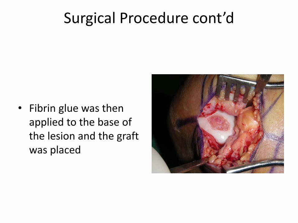
• Fibrin glue was then applied to the base of the lesion and the graft was placed
Follow-up
• At 6 weeks – Presented in post op brace on crutches. – Attending physical therapy 2 times a week – Pain 1/10 – Able to perform SLR – Progress weight bearing, full weight bearing in 1-2
weeks – Discontinue post op brace as tolerated
Follow-up
• At 18 months. – Presented weight bearing with no assistive devices – Full range of motion – Pain 0/10 – Reports no problem with knee – Continue with moderate impact activities. – Back to Soccer?
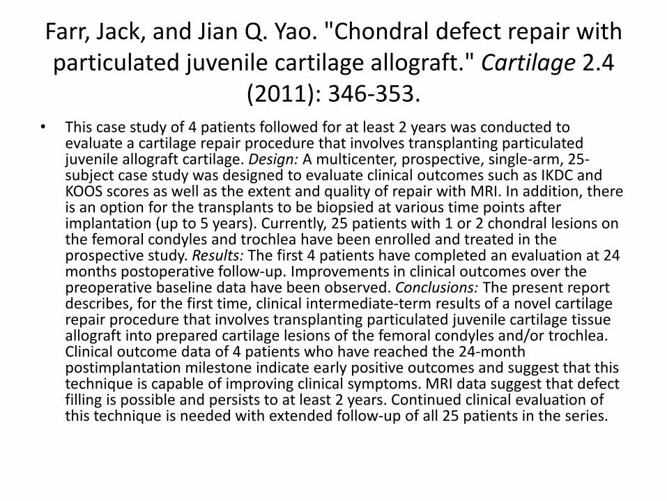
Farr, Jack, and Jian Q. Yao. "Chondral defect repair with particulated juvenile cartilage allograft." Cartilage 2.4
(2011): 346-353. • This case study of 4 patients followed for at least 2 years was conducted to
evaluate a cartilage repair procedure that involves transplanting particulated juvenile allograft cartilage. Design: A multicenter, prospective, single-arm, 25-subject case study was designed to evaluate clinical outcomes such as IKDC and KOOS scores as well as the extent and quality of repair with MRI. In addition, there is an option for the transplants to be biopsied at various time points after implantation (up to 5 years). Currently, 25 patients with 1 or 2 chondral lesions on the femoral condyles and trochlea have been enrolled and treated in the prospective study. Results: The first 4 patients have completed an evaluation at 24 months postoperative follow-up. Improvements in clinical outcomes over the preoperative baseline data have been observed. Conclusions: The present report describes, for the first time, clinical intermediate-term results of a novel cartilage repair procedure that involves transplanting particulated juvenile cartilage tissue allograft into prepared cartilage lesions of the femoral condyles and/or trochlea. Clinical outcome data of 4 patients who have reached the 24-month postimplantation milestone indicate early positive outcomes and suggest that this technique is capable of improving clinical symptoms. MRI data suggest that defect filling is possible and persists to at least 2 years. Continued clinical evaluation of this technique is needed with extended follow-up of all 25 patients in the series.

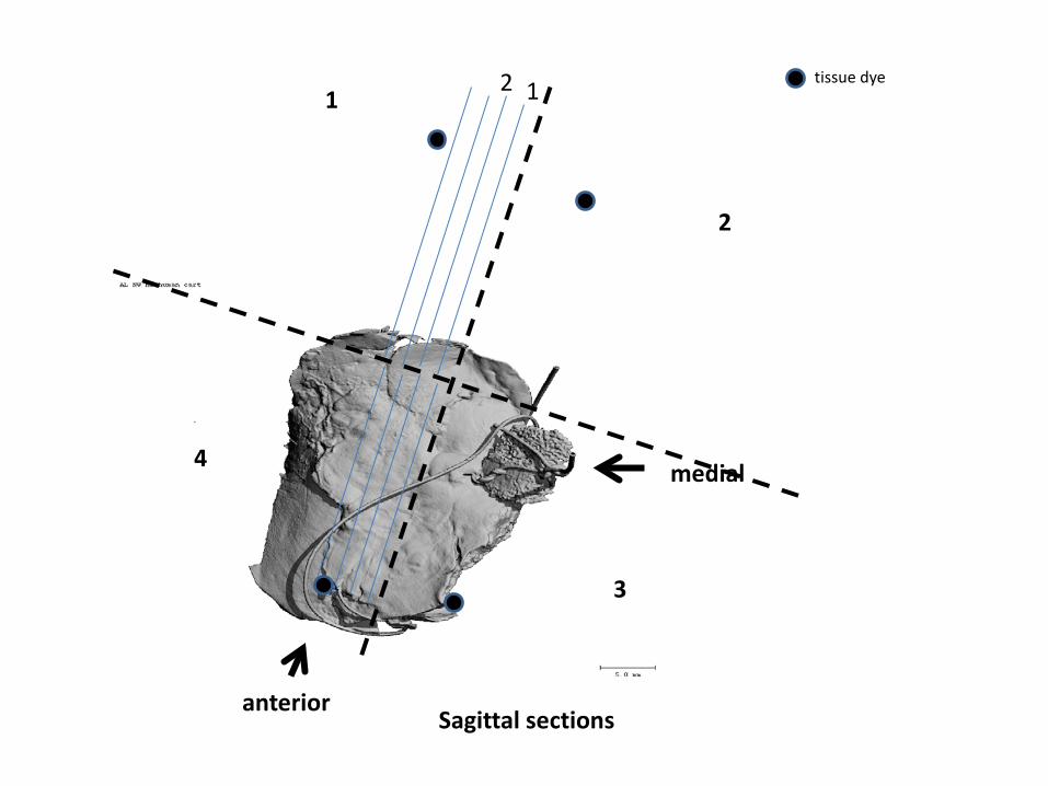
Stevens, Hazel Y., Blake E. Shockley, Nick J. Willett, Angela SP Lin, Yazdan Raji,
Robert E. Guldberg, and Sameh A. Labib. "Particulated Juvenile Articular Cartilage Implantation in the Knee A 3-Year EPIC-µCT and Histological
Examination." Cartilage (2013): 1947603513515483.
• UKA in a 44 yrs old 3 years s/p HTO and Denovo
• OATS done at 1.5 years to salvage the repair.
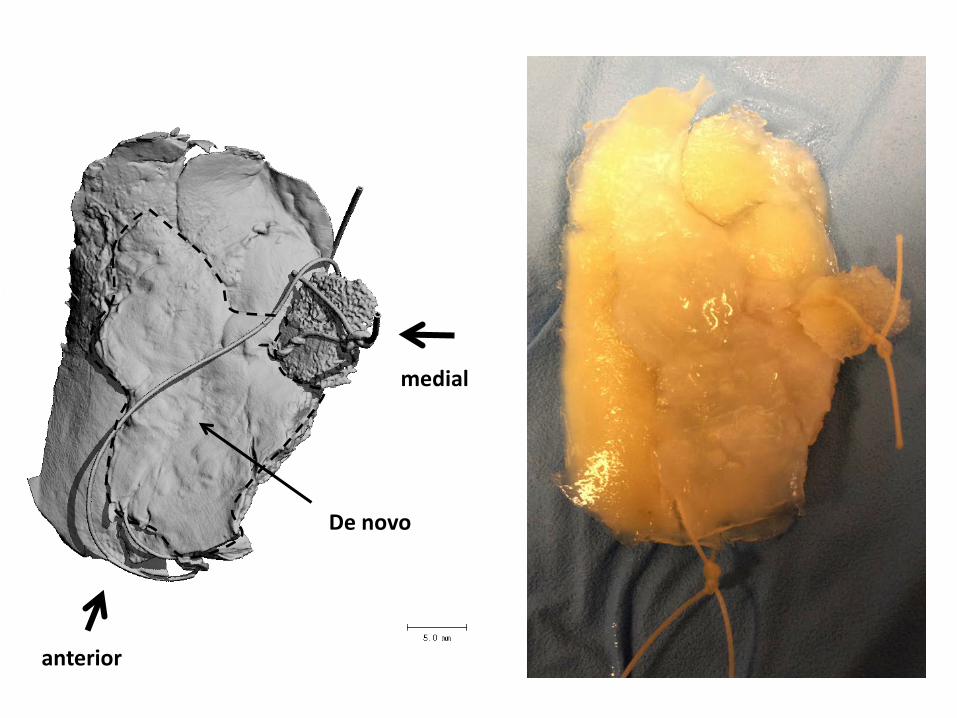
medial
anterior
De novo
medial
anterior Sagittal sections
1
2
3
4
1 2 tissue dye
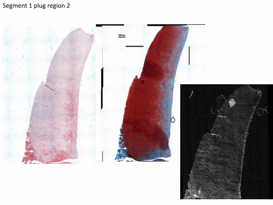
Segment 1 plug region 2
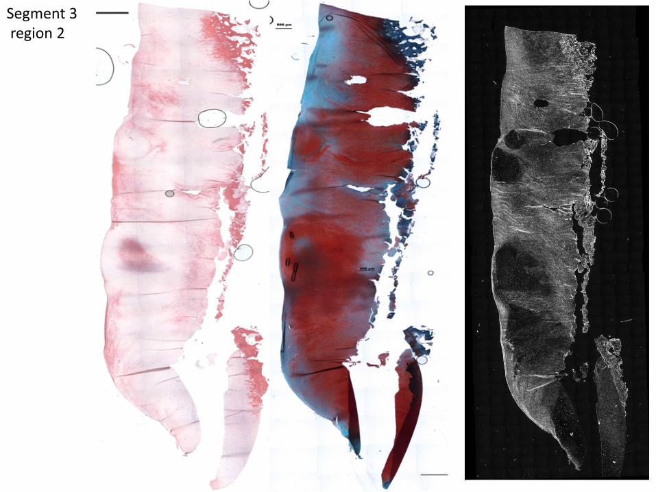
Segment 3 region 2
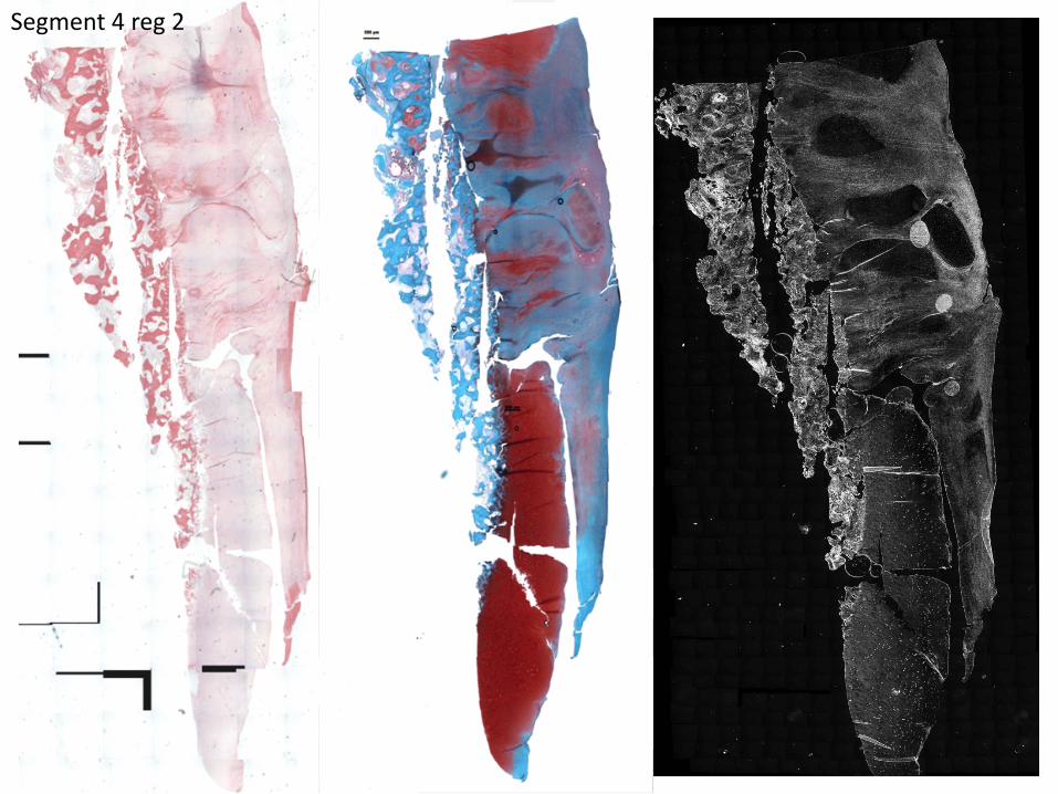
Segment 4 reg 2
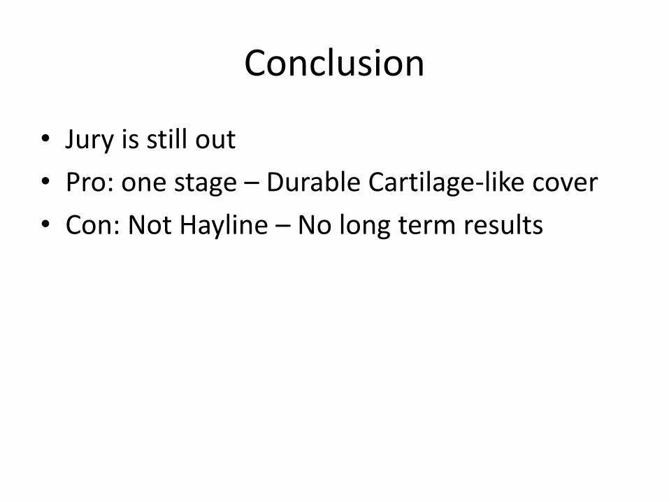
Conclusion
• Jury is still out • Pro: one stage – Durable Cartilage-like cover • Con: Not Hayline – No long term results
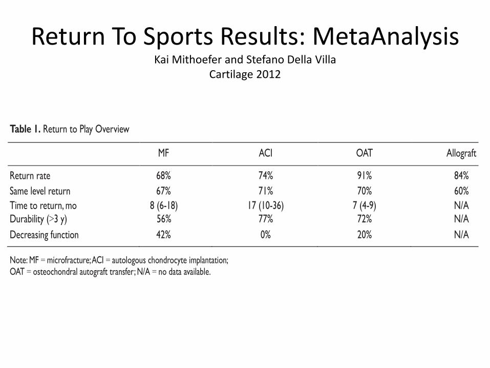
Return To Sports Results: MetaAnalysis Kai Mithoefer and Stefano Della Villa
Cartilage 2012
Table 1. Return to Play Overview MF ACI OAT Allograft Return rate 68% 74% 91% 84% Same level return 67% 71% 70% 60% Time to return, mo Durability (>3 y)
8 (6-18) 56%
17 (10-36) 77%
7 (4-9) 72%
N/A N/A
Decreasing function 42% 0% 20% N/A
Note: MF = microfracture; ACI = autologous chondrocyte implantation; OAT = osteochondral autograft transfer; N/A = no data available.
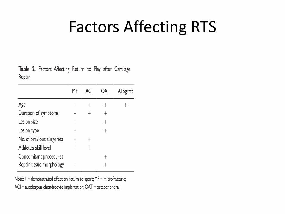
Factors Affecting RTS
Table 2. Factors Affecting Return to Play after Cartilage Repair
MF ACI OAT Allograft Age + + + + Duration of symptoms + + + Lesion size + + Lesion type + + No. of previous surgeries + + Athlete’s skill level + + Concomitant procedures Repair tissue morphology
+
+ +
Note: + = demonstrated effect on return to sport; MF = microfracture; ACI = autologous chondrocyte implantation; OAT = osteochondral
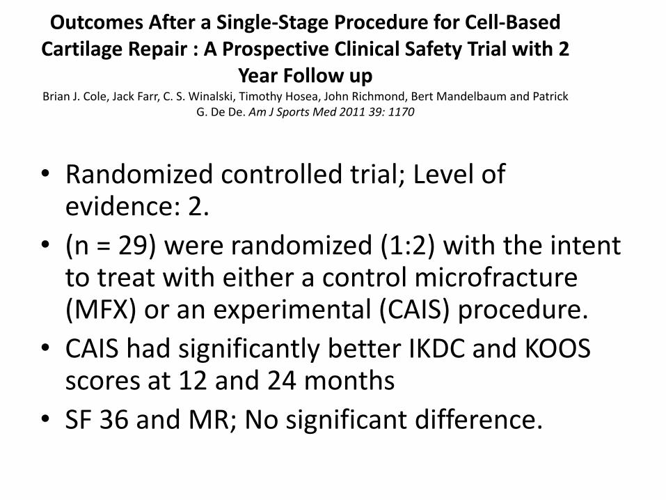
Outcomes After a Single-Stage Procedure for Cell-Based Cartilage Repair : A Prospective Clinical Safety Trial with 2
Year Follow up Brian J. Cole, Jack Farr, C. S. Winalski, Timothy Hosea, John Richmond, Bert Mandelbaum and Patrick
G. De De. Am J Sports Med 2011 39: 1170
• Randomized controlled trial; Level of
evidence: 2. • (n = 29) were randomized (1:2) with the intent
to treat with either a control microfracture (MFX) or an experimental (CAIS) procedure.
• CAIS had significantly better IKDC and KOOS scores at 12 and 24 months
• SF 36 and MR; No significant difference.
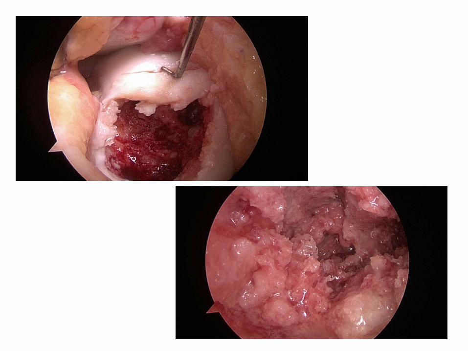
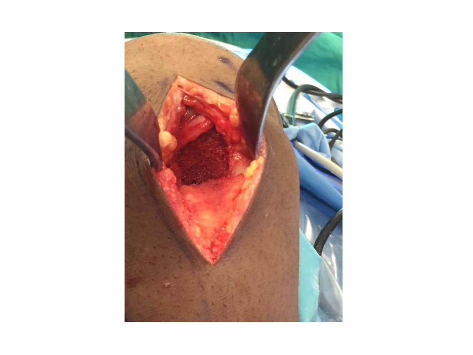
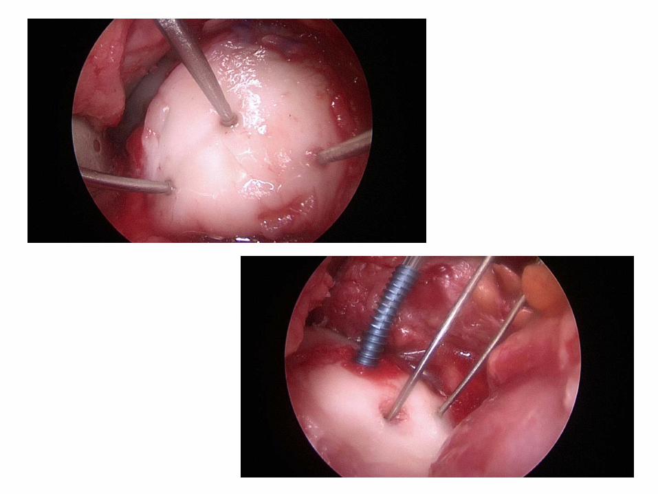
Large Unstable Osteochondral Defect Involving the Medial Femoral
Condyle MULTIPLE TECHNIQUES- Biologic
ORIF
History • 18 y/o male • Right knee pain and swelling for several months • No acute or previous injury • Playing basketball on 8/28/15 • Pain significantly increased, superior and medial to
patella, intermittent • Developed joint effusion and a limp • Not able to fully flex • Sensation of something moving around in his knee • Went to ED, plain films show effusion, sent home with
knee immobilizer
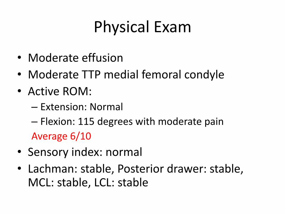
Physical Exam
• Moderate effusion • Moderate TTP medial femoral condyle • Active ROM: – Extension: Normal – Flexion: 115 degrees with moderate pain Average 6/10
• Sensory index: normal • Lachman: stable, Posterior drawer: stable,
MCL: stable, LCL: stable
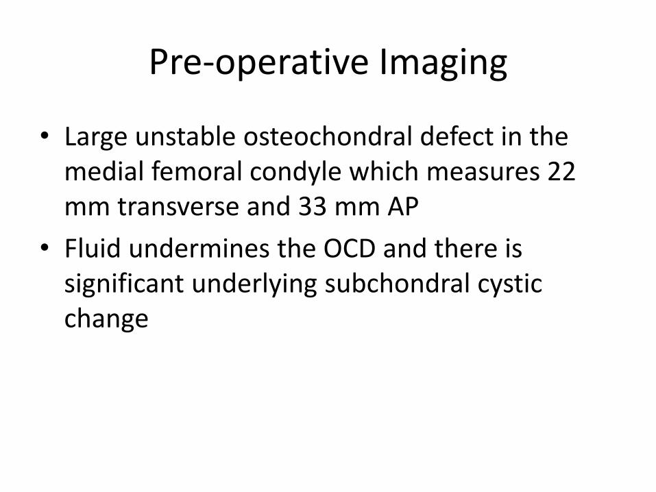
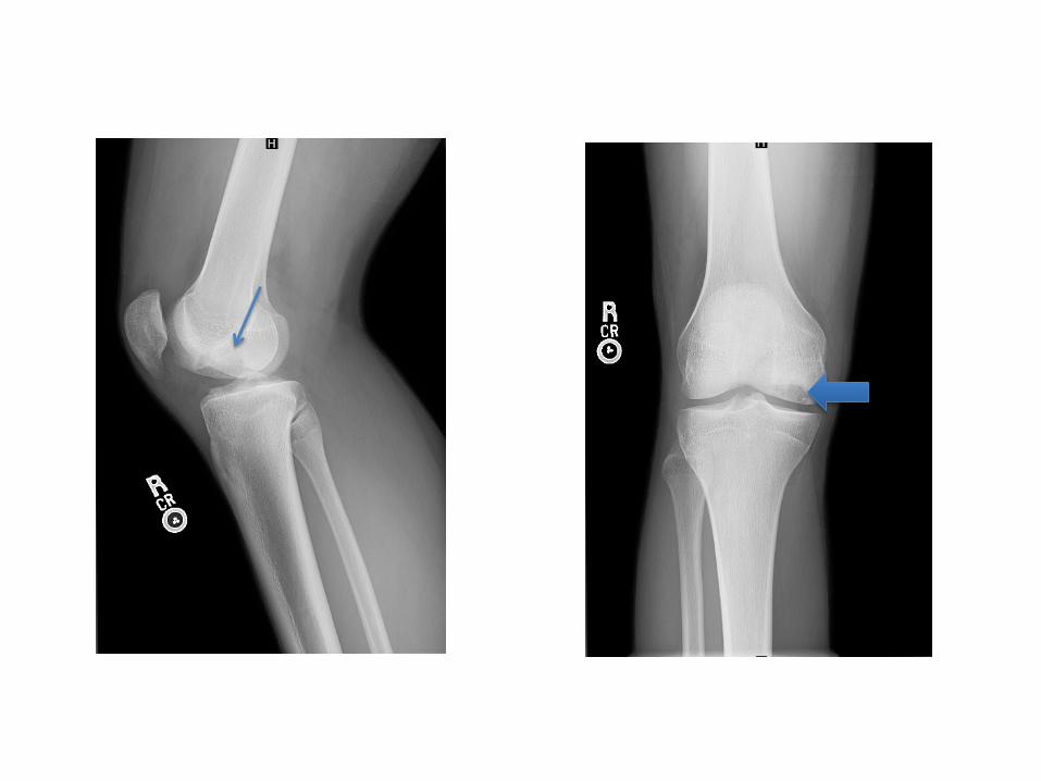
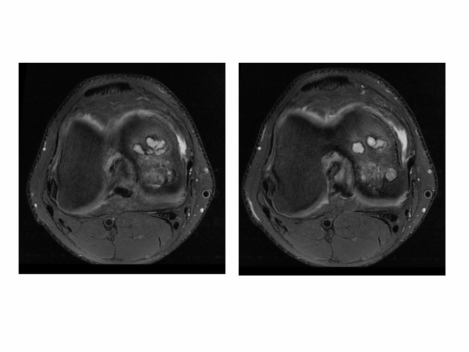
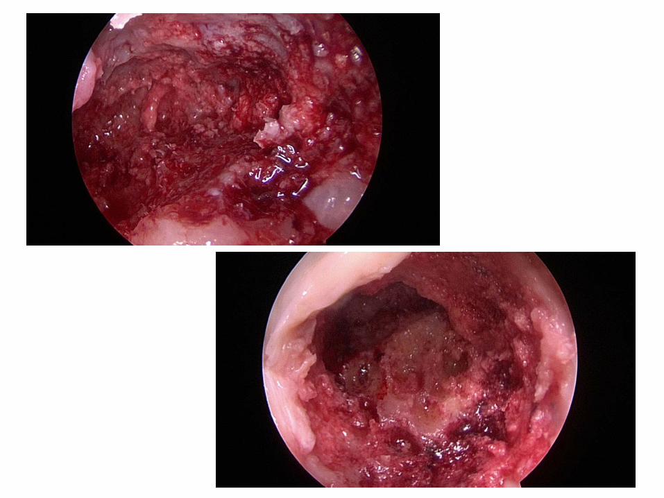
Pre-operative Imaging
• Large unstable osteochondral defect in the medial femoral condyle which measures 22 mm transverse and 33 mm AP
• Fluid undermines the OCD and there is significant underlying subchondral cystic change
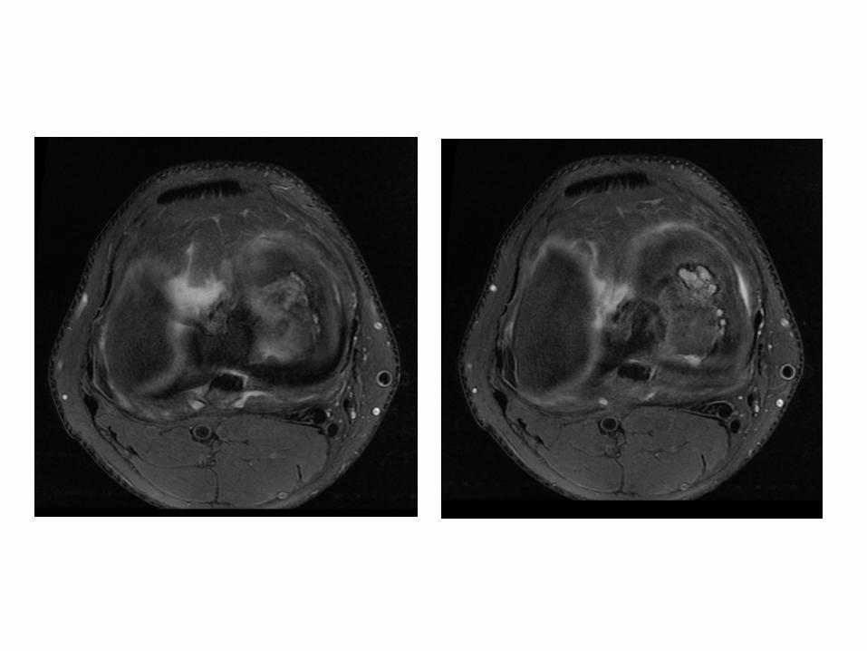
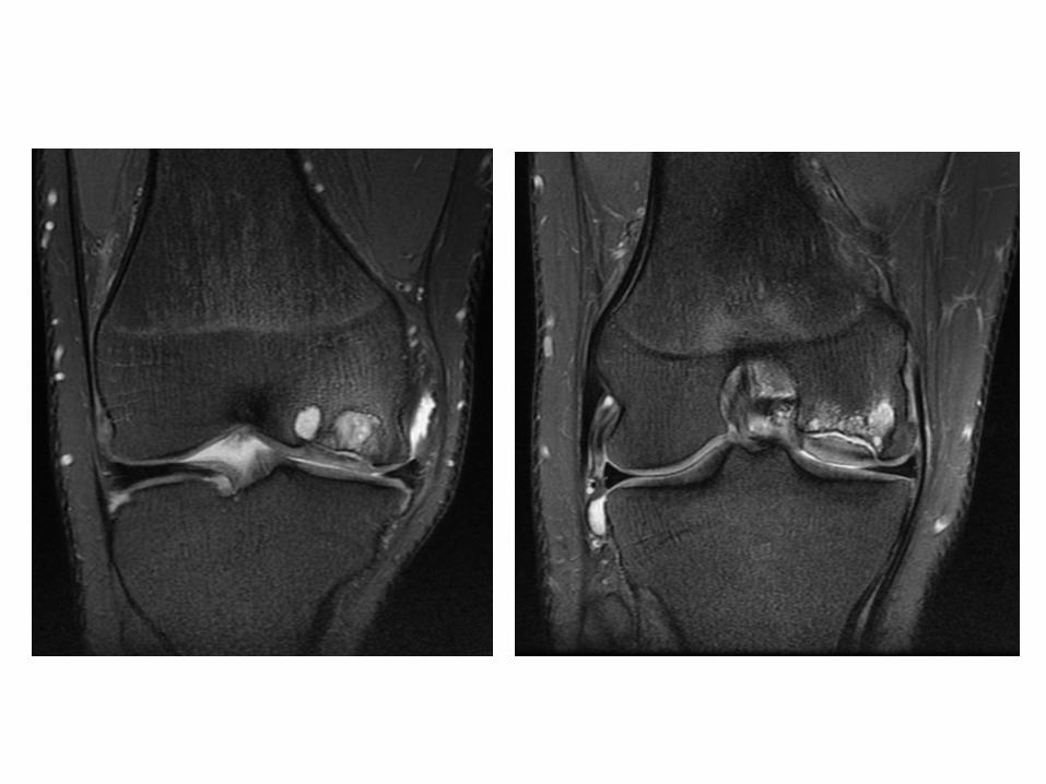
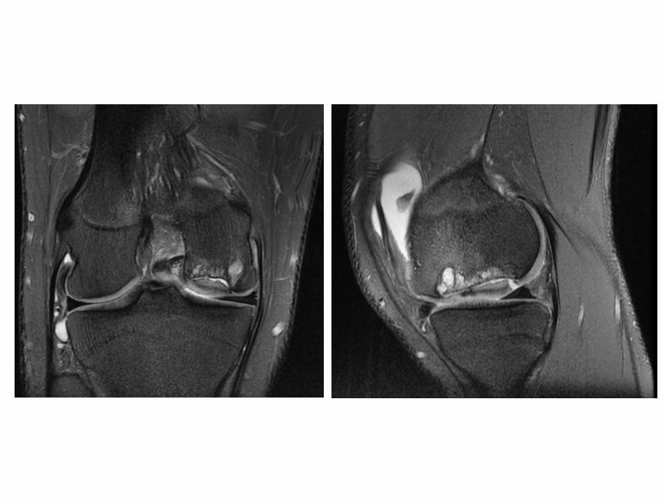
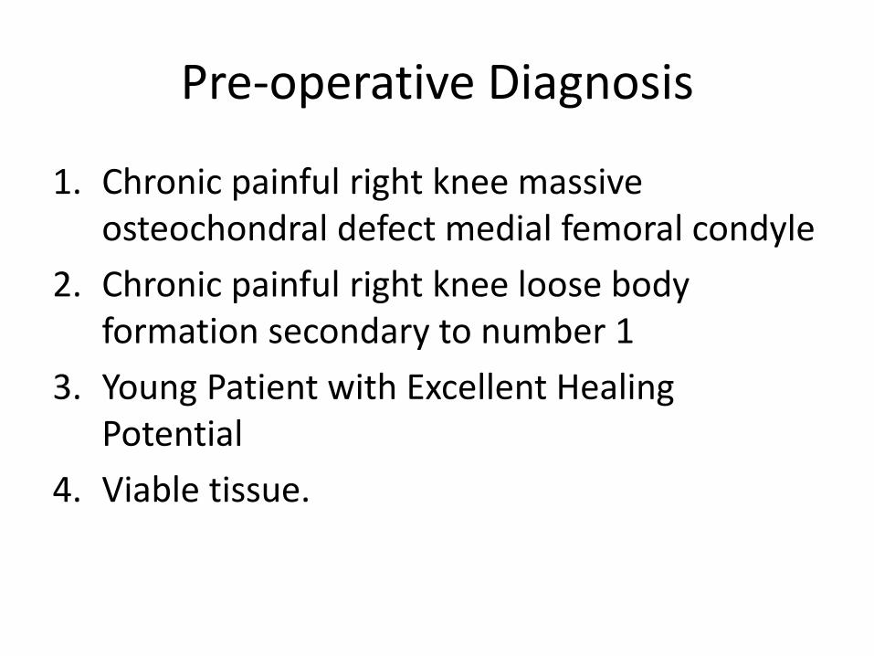
Pre-operative Diagnosis
1. Chronic painful right knee massive osteochondral defect medial femoral condyle
2. Chronic painful right knee loose body formation secondary to number 1
3. Young Patient with Excellent Healing Potential
4. Viable tissue.
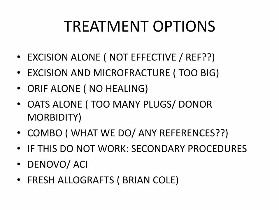
TREATMENT OPTIONS
• EXCISION ALONE ( NOT EFFECTIVE / REF??) • EXCISION AND MICROFRACTURE ( TOO BIG) • ORIF ALONE ( NO HEALING) • OATS ALONE ( TOO MANY PLUGS/ DONOR
MORBIDITY) • COMBO ( WHAT WE DO/ ANY REFERENCES??) • IF THIS DO NOT WORK: SECONDARY PROCEDURES • DENOVO/ ACI • FRESH ALLOGRAFTS ( BRIAN COLE)
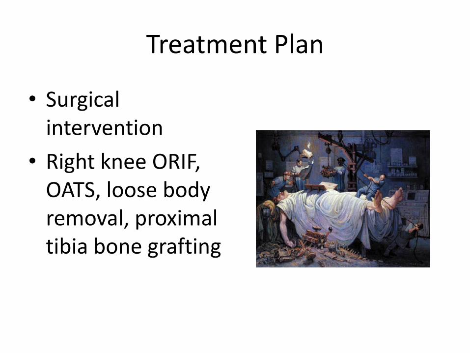
Treatment Plan
• Surgical intervention • Right knee ORIF,
OATS, loose body removal, proximal tibia bone grafting
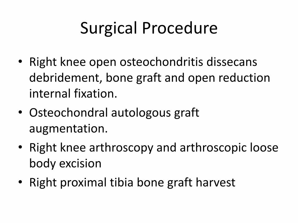
Surgical Procedure
• Right knee open osteochondritis dissecans debridement, bone graft and open reduction internal fixation.
• Osteochondral autologous graft augmentation.
• Right knee arthroscopy and arthroscopic loose body excision
• Right proximal tibia bone graft harvest
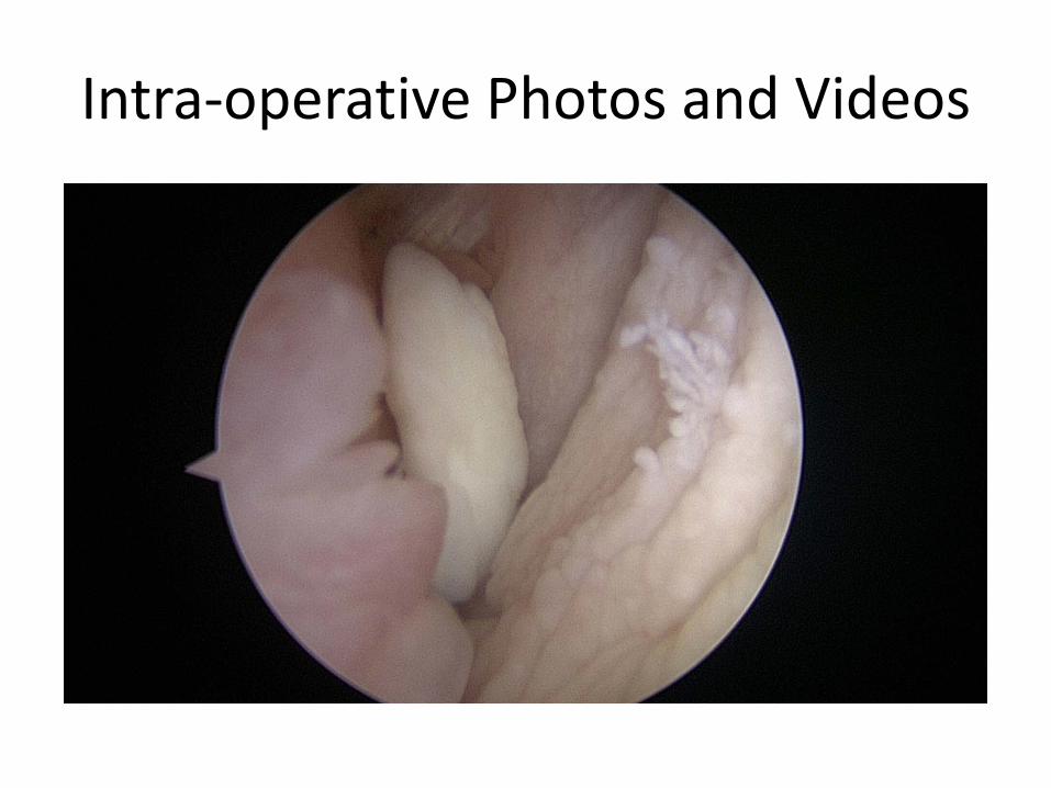
Intra-operative Photos and Videos


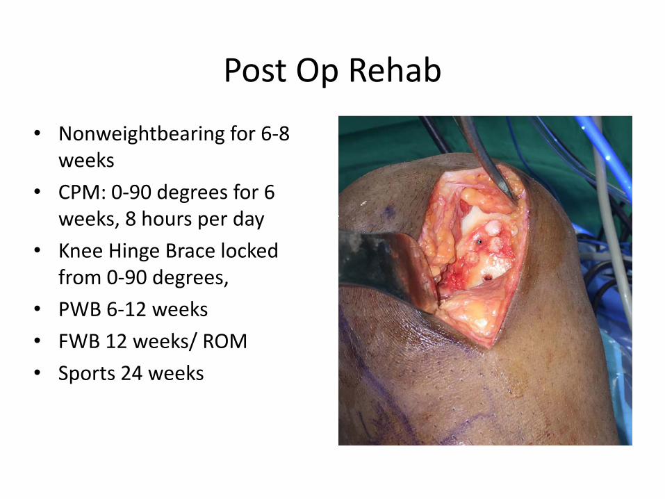
Post Op Rehab • Nonweightbearing for 6-8
weeks • CPM: 0-90 degrees for 6
weeks, 8 hours per day • Knee Hinge Brace locked
from 0-90 degrees, • PWB 6-12 weeks • FWB 12 weeks/ ROM • Sports 24 weeks
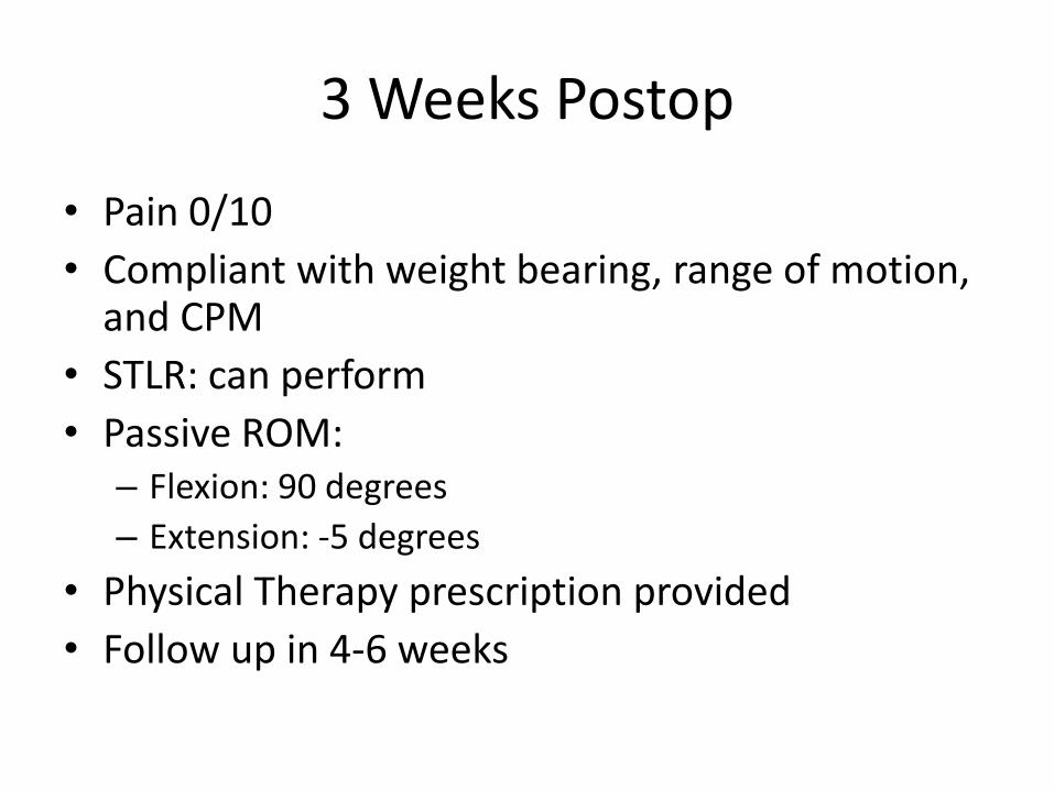
3 Weeks Postop
• Pain 0/10 • Compliant with weight bearing, range of motion,
and CPM • STLR: can perform • Passive ROM: – Flexion: 90 degrees – Extension: -5 degrees
• Physical Therapy prescription provided • Follow up in 4-6 weeks
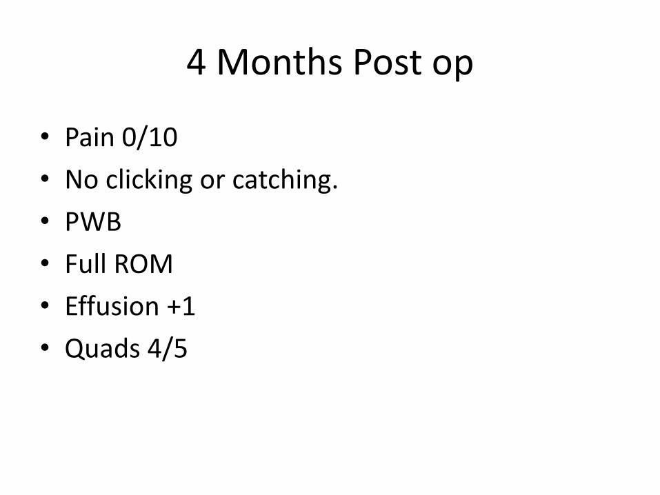
4 Months Post op
• Pain 0/10 • No clicking or catching. • PWB • Full ROM • Effusion +1 • Quads 4/5
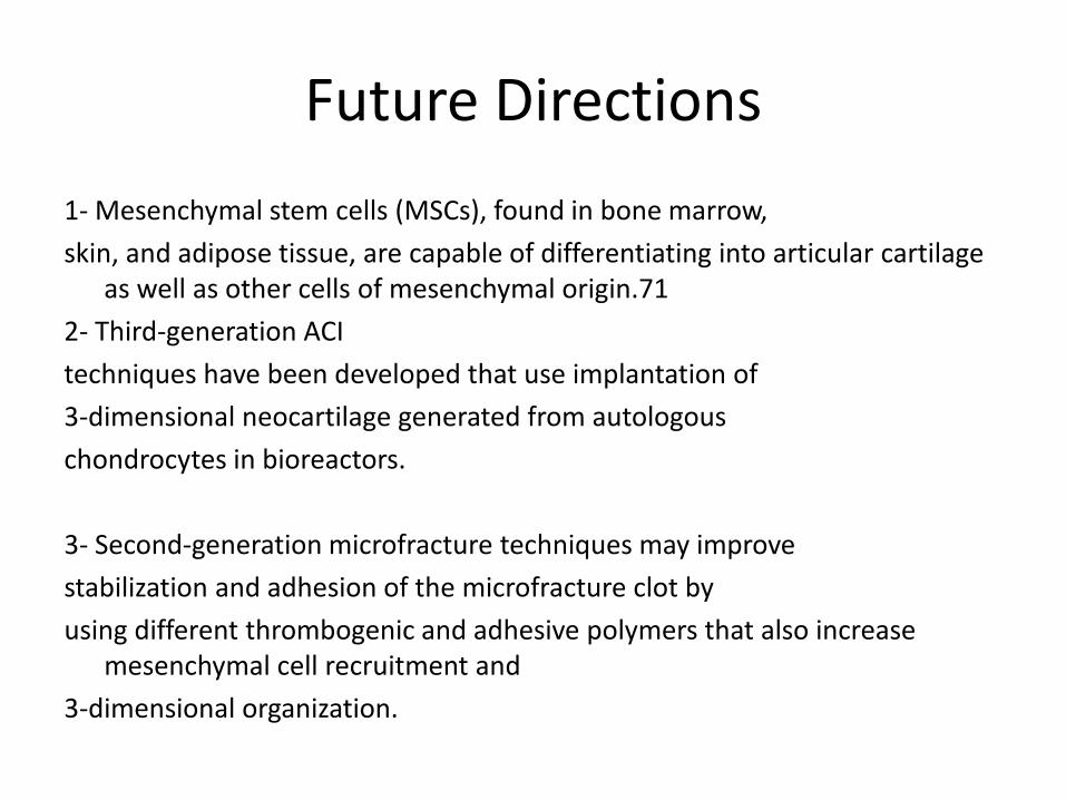
Future Directions 1- Mesenchymal stem cells (MSCs), found in bone marrow, skin, and adipose tissue, are capable of differentiating into articular cartilage
as well as other cells of mesenchymal origin.71 2- Third-generation ACI techniques have been developed that use implantation of 3-dimensional neocartilage generated from autologous chondrocytes in bioreactors. 3- Second-generation microfracture techniques may improve stabilization and adhesion of the microfracture clot by using different thrombogenic and adhesive polymers that also increase
mesenchymal cell recruitment and 3-dimensional organization.
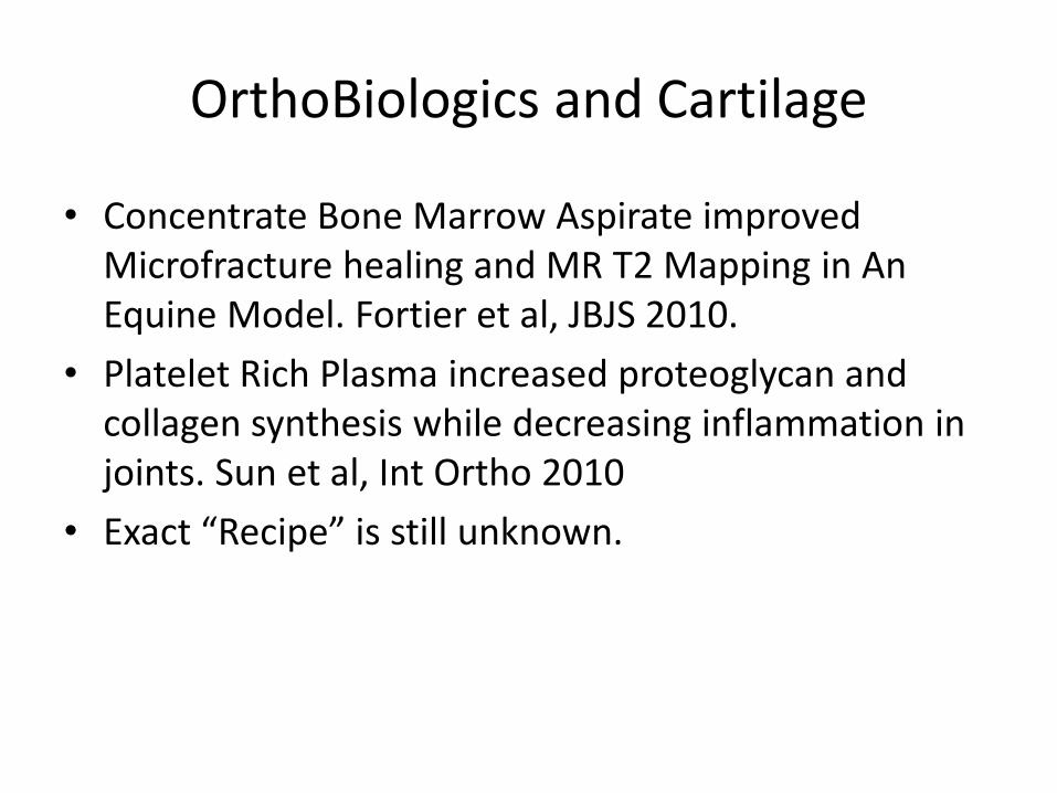
OrthoBiologics and Cartilage
• Concentrate Bone Marrow Aspirate improved Microfracture healing and MR T2 Mapping in An Equine Model. Fortier et al, JBJS 2010.
• Platelet Rich Plasma increased proteoglycan and collagen synthesis while decreasing inflammation in joints. Sun et al, Int Ortho 2010
• Exact “Recipe” is still unknown.
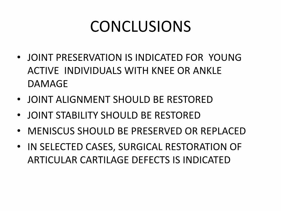
CONCLUSIONS
• JOINT PRESERVATION IS INDICATED FOR YOUNG ACTIVE INDIVIDUALS WITH KNEE OR ANKLE DAMAGE
• JOINT ALIGNMENT SHOULD BE RESTORED • JOINT STABILITY SHOULD BE RESTORED • MENISCUS SHOULD BE PRESERVED OR REPLACED • IN SELECTED CASES, SURGICAL RESTORATION OF
ARTICULAR CARTILAGE DEFECTS IS INDICATED
CONCLUSIONS • MICROFRACTURE IS FIRST LINE TREATMENT FOR SMALL
CARTILAGE LESIONS. LARGE LESIONS DON NOT DO AS WELL. • BIOLOGIC AND NON BIOLOGIC OPTIONS ARE AVAILABLE AND
IS TAILORED TO EACH PATIENT • IN THE SHORT TERM, CARTILAGE REPAIR WAS SHOWN TO
DECREASE PAIN AND IMPROVE JOINT FUNCTION. • IT REMAINS TO BE SEEN IF CARTILAGE RESTORATION WILL
DELAY OR PREVENT THE ONSET OF POST-TRAUMATIC OSTEOARTHRITIS.
• TO-DATE, CARTILAGE REPAIR IS NOT INDICATED FOR THE
TREATMENT OF ADVANCED OSTEOARTHRITIS.
THANK YOU www.drsamlabib.com


![Bioactive Scaffolds for Regeneration of Cartilage and … · role in reducing cartilage degeneration and decreasing chondrocyte apoptosis on the osteoarthritis therapy [21, 22]. Recent](https://img.pdfslide.us/doc/110x75/5f0e7a4e7e708231d43f706e/bioactive-scaffolds-for-regeneration-of-cartilage-and-role-in-reducing-cartilage.jpg)