Embed Size (px)
Citation preview
Control of Protein Function
In the cell, precise regulation of protein function is essential to avoid chaos. This chapterdescribes the most important molecular mechanisms by which protein function is regulatedin cells. These range from control of a protein’s location and lifetime within the cell to thebinding of regulatory molecules and covalent modifications such as phosphorylation thatrapidly switch protein activity on or off. Also covered here are the nucleotide-drivenswitches in conformation that underlie the action of motor proteins and that regulatemany signal transduction pathways.
3
3-0 Overview: Mechanisms of Regulation
3-1 Protein Interaction Domains
3-2 Regulation by Location
3-3 Control by pH and Redox Environment
3-4 Effector Ligands: Competitive Binding and Cooperativity
3-5 Effector Ligands: Conformational Change and Allostery
3-6 Protein Switches Based on Nucleotide Hydrolysis
3-7 GTPase Switches: Small Signaling G Proteins
3-8 GTPase Switches: Signal Relay by Heterotrimeric GTPases
3-9 GTPase Switches: Protein Synthesis
3-10 Motor Protein Switches
3-11 Regulation by Degradation
3-12 Control of Protein Function by Phosphorylation
3-13 Regulation of Signaling Protein Kinases: Activation Mechanism
3-14 Regulation of Signaling Protein Kinases: Cdk Activation
3-15 Two-Component Signaling Systems in Bacteria
3-16 Control by Proteolysis: Activation of Precursors
3-17 Protein Splicing: Autoproteolysis by Inteins
3-18 Glycosylation
3-19 Protein Targeting by Lipid Modifications
3-20 Methylation, N-acetylation, Sumoylation and Nitrosylation
Protein function in living cells is precisely regulated
A typical bacterial cell contains a total of about 250,000 protein molecules (comprising different amounts of each of several thousand different gene products), which are packed intoa volume so small that it has been estimated that, on average, they are separated from oneanother by a distance that would contain only a few molecules of water. It is likely thateukaryotic cells are at least as densely packed. In this crowded environment, precise regulationof protein function is essential to avoid chaos. Regulation of protein function in vivo tends tooccur by many different mechanisms, which fall into several general classes. Protein functioncan be controlled by localization of the gene product and/or the species it interacts with, bythe covalent or noncovalent binding of effector molecules, and by the amount and lifetime ofthe active protein.
Proteins can be targeted to specific compartments and complexes
Not all proteins are absolutely specific, and many also have more than one function.Consequently, it is often undesirable to have such proteins distributed everywhere in the cell,where they may carry out unwanted reactions. A simple way to regulate their activity is toensure that the protein is only present in its active form in the specific compartment where itis needed, or when bound in a complex with other macromolecules that participate in itsfunction. There are many ways in which specific localization can be achieved. Proteins can betargeted to cellular compartments by so-called signal sequences that are an intrinsic part of theencoded amino-acid sequence, or by attachment of, for example, a lipid tail that inserts intomembranes. They can be directed to a complex of interacting proteins by a structural interactiondomain that recognizes some covalent modification such as phosphorylation on another protein.Localization is a dynamic process and a given protein may be targeted to different compartmentsat different stages of the cell cycle: many transcription factors, for example, cycle between thenucleus and the cytosol in response to extracellular signals. When the protein is not in thelocation where it is needed, very often it is maintained in an inactive conformation.
Protein activity can be regulated by binding of an effector and by covalent modification
Protein activity can also be controlled by the binding of effector molecules, which often workby inducing conformational changes that produce inactive or active forms of the protein.Effectors may be as small as a proton or as large as another macromolecule. Effectors may bindnoncovalently or may modify the covalent structure of the protein, reversibly or irreversibly.Effectors that regulate activity by binding to the active site usually take the form of inhibitorsthat compete with the substrate for binding. Often, the product of an enzyme reaction can actas such a competitive inhibitor, allowing the enzyme to regulate itself when too much productmight be made. Ligands, including reaction products, may also bind to sites remote from theactive site and in so doing either activate or inhibit a protein. Proteins regulated in this waytend to be oligomeric and allosteric. Allosteric proteins have multiple ligand-binding sites andthese show cooperativity of binding: in positive cooperativity the first ligand molecule to bind isbound weakly, but its binding alters the conformation of the protein in such a way that bindingof the second and subsequent ligand molecules is promoted. Cooperativity may also be negative:the first ligand binding weakens and thereby effectively inhibits subsequent binding to theother sites. Metabolic pathways often employ allosteric effectors as part of a feedback controlmechanism: the end product of the pathway acts as an allosteric inhibitor of one of the earlier
3-0 Overview: Mechanisms of Regulation
Chapter 3 Control of Protein Function86 ©2004 New Science Press Ltd
Definitions
competitive inhibitor: a species that competes withsubstrate for binding to the active site of an enzymeand thus inhibits catalytic activity.
effector: a species that binds to a protein and modifiesits activity. Effectors may be as small as a proton or aslarge as a membrane and may act by covalent binding,noncovalent binding, or covalent modification.
References
Goodsell, D.S.: Inside a living cell. Trends Biochem. Sci.1991, 16:203–206.
Hodgson, D.A. and Thomas, C.M. (eds): Signals, switches,regulons, and cascades: control of bacterial geneexpression. 61st Symposium of the Society for GeneralMicrobiology (Cambridge, Cambridge University Press,2002).
Jensen, R.B. and Shapiro, L.: Proteins on the move:dynamic protein localization in prokaryotes. Trends
Cell Biol. 2000, 10:483–488.
Kornitzer, D. and Ciechanover, A.: Modes of regulationof ubiquitin-mediated protein degradation. J. CellPhysiol. 2000, 182:1–11.
Perutz, M.F.: Mechanisms of cooperativity andallosteric regulation in proteins. Q. Rev. Biophys. 1989,22:139–237.
Sato,T.K. et al.: Location, location, location: membranetargeting directed by PX domains. Science 2001,294:1881–1885.
enzymes in the pathway, so when too much of this product is synthesized it feeds back andshuts off one of the enzymes that help make it, as we shall see later in this chapter.
Binding of effector molecules can be covalent or can lead to covalent changes in a protein. Themost common form of post-translational covalent modification is reversible phosphorylationon the hydroxyl group of the side chains of serine, threonine or tyrosine residues, but manyother modifications are known, including side-chain methylation, covalent attachment ofcarbohydrates and lipids, amino-terminal acetylation and limited proteolytic cleavage, inwhich proteases cut the polypeptide chain in one or more places. Modifications such asphosphorylation or proteolytic cleavage may either activate or inactivate the protein.
Signal amplification is an essential feature in the control of cell function and covalent modification of proteins is the way such amplification is usually achieved. Often, an extra-cellular stimulus is of short duration and involves only a very low concentration, or a smallchange in concentration, of a hormone or regulatory molecule. Yet the cellular responsemust not only be very rapid; in many cases it must be massive, including a change in activitiesof many enzymes and alteration in the transcription of many genes. Covalent modificationof a protein provides a simple mechanism by which a regulatory signal can produce a verylarge output. A single molecule of an enzyme whose catalytic activity is turned on by acovalent modification can process many thousands of substrate molecules. And if that substrateis another enzyme, the amplification is further magnified. When covalent modification ofone enzyme causes it to become active so that it can, in turn, covalently modify and activateanother enzyme and so on, a regulatory cascade is set up that leads to enormous, rapidchanges in the final output. Blood clotting is an example of such an amplification cascadebased on proteolysis.
Protein activity may be regulated by protein quantity and lifetime
The activity of a protein can also be regulated by controlling its amount and lifetime in thecell. This control may be exercised at several places in the flow of information from gene toprotein. At its simplest, the amount of protein can be set by the level of transcription, whichin turn can be controlled by, for example, the strength of the promoter or the action of atranscription factor, which may be a repressor or activator. The level of mRNA may also beadjusted after transcription by varying the rate of RNA degradation. At the level of the protein,quantities are controlled by the lifetime of the molecule, which is determined by its rate ofdegradation. The rate of turnover varies considerably from protein to protein; there are severalspecific mechanisms for targeting protein molecules to degradative machinery in the cell,including covalent attachment of the small protein ubiquitin.
A single protein may be subject to many regulatory influences
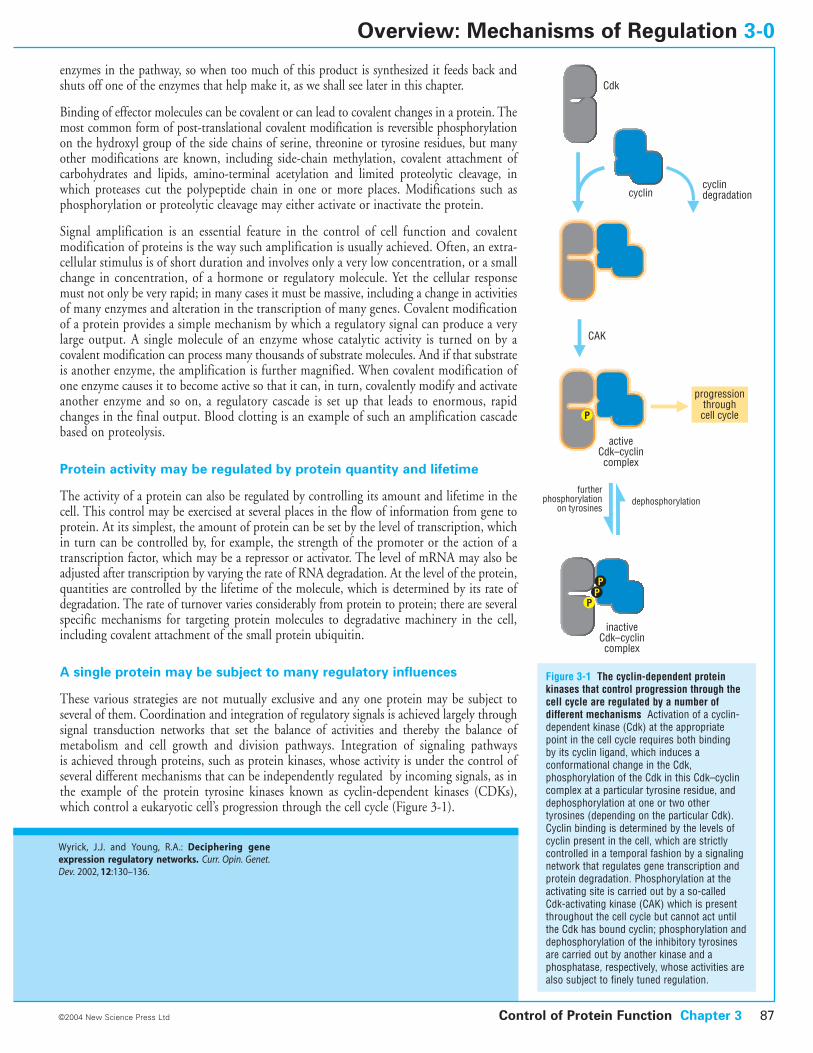
These various strategies are not mutually exclusive and any one protein may be subject to several of them. Coordination and integration of regulatory signals is achieved largely throughsignal transduction networks that set the balance of activities and thereby the balance ofmetabolism and cell growth and division pathways. Integration of signaling pathways is achieved through proteins, such as protein kinases, whose activity is under the control ofseveral different mechanisms that can be independently regulated by incoming signals, as inthe example of the protein tyrosine kinases known as cyclin-dependent kinases (CDKs),which control a eukaryotic cell’s progression through the cell cycle (Figure 3-1).
Control of Protein Function Chapter 3 87©2004 New Science Press Ltd
Overview: Mechanisms of Regulation 3-0
Wyrick, J.J. and Young, R.A.: Deciphering geneexpression regulatory networks. Curr. Opin. Genet.Dev. 2002, 12:130–136.
Figure 3-1 The cyclin-dependent proteinkinases that control progression through thecell cycle are regulated by a number ofdifferent mechanisms Activation of a cyclin-dependent kinase (Cdk) at the appropriatepoint in the cell cycle requires both binding by its cyclin ligand, which induces aconformational change in the Cdk,phosphorylation of the Cdk in this Cdk–cyclincomplex at a particular tyrosine residue, anddephosphorylation at one or two othertyrosines (depending on the particular Cdk).Cyclin binding is determined by the levels ofcyclin present in the cell, which are strictlycontrolled in a temporal fashion by a signalingnetwork that regulates gene transcription andprotein degradation. Phosphorylation at theactivating site is carried out by a so-calledCdk-activating kinase (CAK) which is presentthroughout the cell cycle but cannot act untilthe Cdk has bound cyclin; phosphorylation anddephosphorylation of the inhibitory tyrosinesare carried out by another kinase and aphosphatase, respectively, whose activities arealso subject to finely tuned regulation.
CAK
cyclincyclindegradation
Cdk
activeCdk–cyclincomplex
furtherphosphorylation
on tyrosinesdephosphorylation
inactiveCdk–cyclincomplex
progressionthroughcell cycle
The flow of information within the cell is regulated and integrated bythe combinatorial use of small protein domains that recognize specificligands
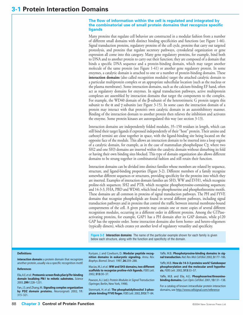
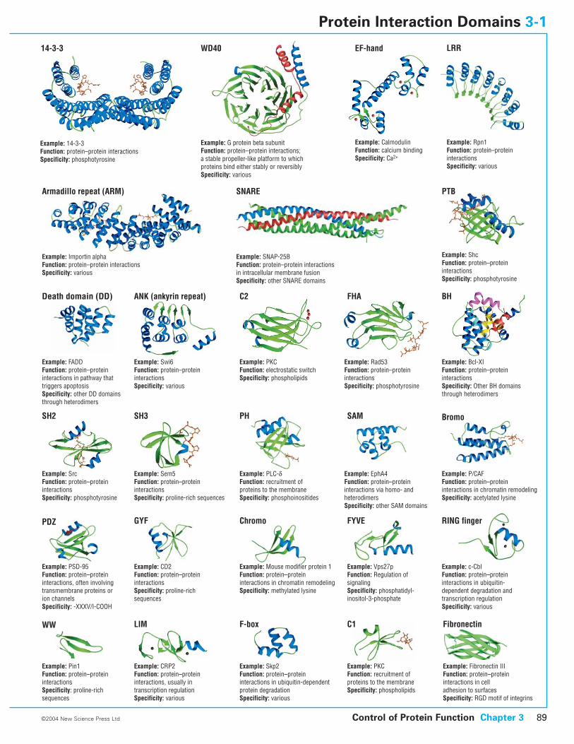
Many proteins that regulate cell behavior are constructed in a modular fashion from a numberof different small domains with distinct binding specificities and functions (see Figure 1-46).Signal transduction proteins, regulatory proteins of the cell cycle, proteins that carry out targetedproteolysis, and proteins that regulate secretory pathways, cytoskeletal organization or geneexpression all come into this category. Many gene regulatory proteins, for example, bind bothto DNA and to another protein to carry out their function; they are composed of a domain thatbinds a specific DNA sequence and a protein-binding domain, which may target anothermolecule of the same protein (see Figure 1-41) or another gene regulatory protein. In someenzymes, a catalytic domain is attached to one or a number of protein-binding domains. Theseinteraction domains (also called recognition modules) target the attached catalytic domain toa particular multiprotein complex or an appropriate subcellular location (such as the nucleus orthe plasma membrane). Some interaction domains, such as the calcium-binding EF hand, oftenact as regulatory domains for enzymes. In signal transduction pathways, active multiproteincomplexes are assembled by interaction domains that target the components to the complex.For example, the WD40 domain of the b-subunit of the heterotrimeric G protein targets thissubunit to the a and g subunits (see Figure 3-15). In some cases the interaction domain of aprotein may interact with that protein’s own catalytic domain in an autoinhibitory manner.Binding of the interaction domain to another protein then relieves the inhibition and activatesthe enzyme. Some protein kinases are autoregulated this way (see section 3-13).
Interaction domains are independently folded modules, 35–150 residues in length, which canstill bind their target ligands if expressed independently of their “host” protein. Their amino andcarboxyl termini are close together in space, with the ligand-binding site being located on theopposite face of the module. This allows an interaction domain to be inserted into a loop regionof a catalytic domain, for example, as in the case of mammalian phospholipase Cg, where twoSH2 and one SH3 domains are inserted within the catalytic domain without disturbing its foldor having their own binding sites blocked. This type of domain organization also allows differentdomains to be strung together in combinatorial fashion and still retain their function.
Interaction domains can be divided into distinct families whose members are related by sequence,structure, and ligand-binding properties (Figure 3-2). Different members of a family recognizesomewhat different sequences or structures, providing specificity for the proteins into which theyare inserted. Examples of interaction domain families are SH3, WW and EVH1, which recognizeproline-rich sequences; SH2 and PTB, which recognize phosphotyrosine-containing sequences;and 14-3-3, FHA, PBD and WD40, which bind to phosphoserine and phosphothreonine motifs.These domains are all common in proteins of signal transduction pathways. The PH and FYVEdomains that recognize phospholipids are found in several different pathways, including signaltransduction pathways and in proteins that control the traffic between internal membrane-boundcompartments of the cell. A given protein may contain one or more copies of several differentrecognition modules, occurring in a different order in different proteins. Among the GTPase-activating proteins, for example, GAP1 has a PH domain after its GAP domain, while p120GAP has the opposite order. Some interaction domains also form homo- and hetero-oligomers(typically dimers), which creates yet another level of regulatory versatility and specificity.
3-1 Protein Interaction Domains
Figure 3-2 Interaction domains The name of the particular example shown for each family is givenbelow each structure, along with the function and specificity of the domain.
Chapter 3 Control of Protein Function88 ©2004 New Science Press Ltd
Definitions
interaction domain: a protein domain that recognizesanother protein, usually via a specific recognition motif.
References
Elia,A.E.et al.: Proteomic screen finds pSer/pThr-bindingdomain localizing Plk1 to mitotic substrates. Science2003, 299:1228–1231.
Fan, J.S. and Zhang, M.: Signaling complex organizationby PDZ domain proteins. Neurosignals 2002, 11:315–321.
Kuriyan, J. and Cowburn, D.: Modular peptide recog-nition domains in eukaryotic signaling. Annu. Rev.Biophys. Biomol. Struct. 1997, 26:259–288.
Macias, M.J. et al.: WW and SH3 domains, two differentscaffolds to recognize proline-rich ligands. FEBS Lett.2002, 513:30–37.
Pawson, A.J. (ed.): Protein Modules in Signal Transduction(Springer, Berlin, New York, 1998).
Stenmark, H. et al.: The phosphatidylinositol 3-phos-phate-binding FYVE finger. FEBS Lett. 2002, 513:77–84.
Yaffe, M.B.: Phosphotyrosine-binding domains in sig-nal transduction. Nat. Rev. Mol. Cell Biol. 2002, 3:177–186.
Yaffe, M.B.: How do 14-3-3 proteins work? Gatekeeperphosphorylation and the molecular anvil hypothe-sis. FEBS Lett. 2002, 513:53–57.
Yaffe, M.B. and Elia, A.E.: Phosphoserine/threonine-binding domains. Curr. Opin. Cell Biol. 2001, 13:131–138.
For a catalog of known intracellular protein interactiondomains, see http://www.cellsignal.com/reference
Control of Protein Function Chapter 3 89©2004 New Science Press Ltd
14-3-3
Example: 14-3-3 Function: protein–protein interactions Specificity: phosphotyrosine
WD40
Example: G protein beta subunitFunction: protein–protein interactions;a stable propeller-like platform to which proteins bind either stably or reversiblySpecificity: various
EF-hand
Example: Calmodulin Function: calcium bindingSpecificity: Ca2+
Armadillo repeat (ARM)
Example: Importin alphaFunction: protein–protein interactionsSpecificity: various
SNARE
Example: SNAP-25B Function: protein–protein interactionsin intracellular membrane fusionSpecificity: other SNARE domains
PTB
Example: ShcFunction: protein–protein interactionsSpecificity: phosphotyrosine
Death domain (DD)
Example: FADDFunction: protein–protein interactions in pathway thattriggers apoptosisSpecificity: other DD domains through heterodimers
ANK (ankyrin repeat)
Example: Swi6Function: protein–protein interactionsSpecificity: various
C2
Example: PKCFunction: electrostatic switchSpecificity: phospholipids
FHA
Example: Rad53Function: protein–protein interactionsSpecificity: phosphotyrosine
BH
Example: Bcl-XIFunction: protein–proteininteractionsSpecificity: Other BH domains through heterodimers
SH2
Example: SrcFunction: protein–protein interactionsSpecificity: phosphotyrosine
SH3
Example: Sem5Function: protein–proteininteractionsSpecificity: proline-rich sequences
PH
Example: PLC-dFunction: recruitment of proteins to the membraneSpecificity: phosphoinositides
SAM
Example: EphA4Function: protein–protein interactions via homo- and heterodimersSpecificity: other SAM domains
Bromo
Example: P/CAFFunction: protein–protein interactions in chromatin remodelingSpecificity: acetylated lysine
PDZ
Example: PSD-95Function: protein–proteininteractions, often involvingtransmembrane proteins orion channelsSpecificity: -XXXV/I-COOH
GYF
Example: CD2Function: protein–proteininteractionsSpecificity: proline-richsequences
Chromo
Example: Mouse modifier protein 1Function: protein–protein interactions in chromatin remodelingSpecificity: methylated lysine
FYVE
Example: Vps27pFunction: Regulation of signalingSpecificity: phosphatidyl-inositol-3-phosphate
Example: c-CblFunction: protein–proteininteractions in ubiquitin-dependent degradation and transcription regulationSpecificity: various
Example: Pin1Function: protein–proteininteractionsSpecificity: proline-richsequences
Example: CRP2Function: protein–proteininteractions, usually intranscription regulationSpecificity: various
Example: Skp2Function: protein–protein interactions in ubiquitin-dependent protein degradationSpecificity: various
WW LIM F-box C1
Example: PKCFunction: recruitment ofproteins to the membraneSpecificity: phospholipids
Example: Fibronectin IIIFunction: protein–protein interactions in cell adhesion to surfacesSpecificity: RGD motif of integrins
Example: Rpn1Function: protein–protein interactionsSpecificity: various
RING finger
Fibronectin
LRR
Protein Interaction Domains 3-1
Protein function in the cell is context-dependent
Many cellular processes involve the interaction of two or more macromolecules: signaltransduction pathways are a good example. But when one estimates the number of gene productsapparently involved in such pathways, it frequently appears that there are too few differentproteins to account for all the different specific interactions that must be made. Put anotherway, the genomes of higher organisms seem to contain too few genes to fulfill all the cellularfunctions required. The logical conclusion is that many proteins participate in more than onecellular process. But if chaos is not to result from all these activities occurring simultaneously,both temporal and spatial control over a protein’s activity must be exercised. Temporal controlcan be achieved partly by regulating gene expression and protein lifetime. However, it isincreasingly clear that spatial context, the precise location within the cell at which a geneproduct exercises its biochemical function, is a major mechanism for regulating function.
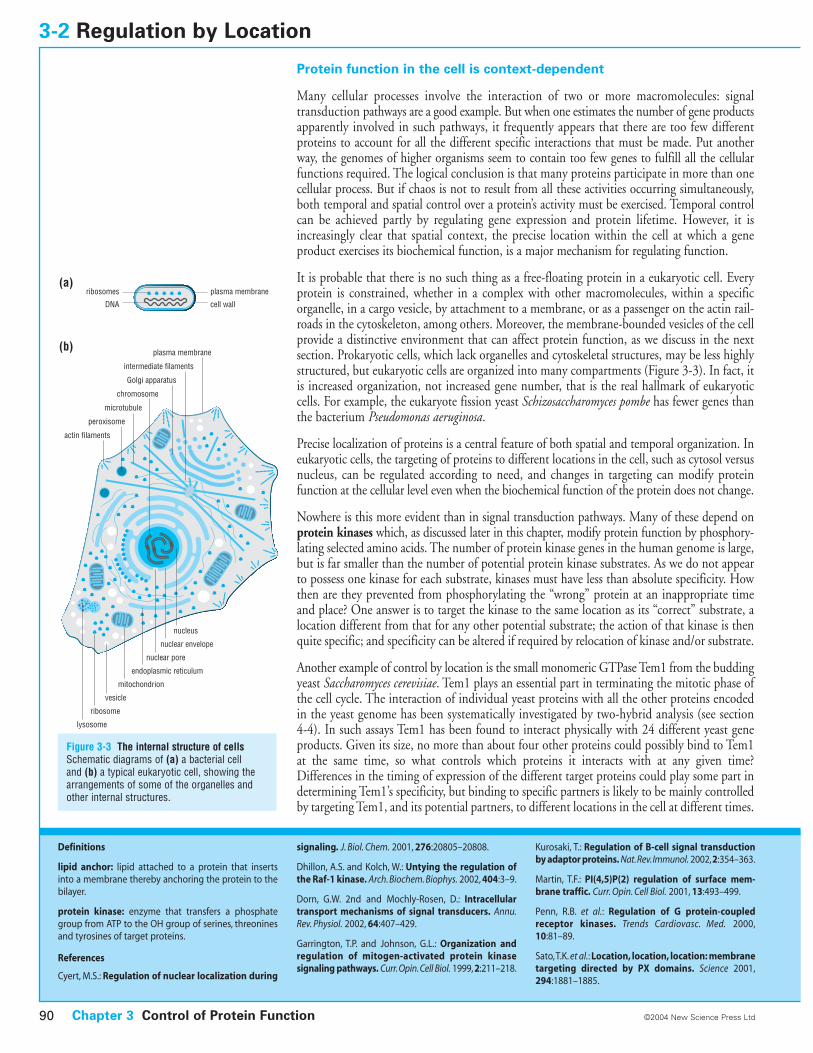
It is probable that there is no such thing as a free-floating protein in a eukaryotic cell. Everyprotein is constrained, whether in a complex with other macromolecules, within a specificorganelle, in a cargo vesicle, by attachment to a membrane, or as a passenger on the actin rail-roads in the cytoskeleton, among others. Moreover, the membrane-bounded vesicles of the cellprovide a distinctive environment that can affect protein function, as we discuss in the nextsection. Prokaryotic cells, which lack organelles and cytoskeletal structures, may be less highlystructured, but eukaryotic cells are organized into many compartments (Figure 3-3). In fact, itis increased organization, not increased gene number, that is the real hallmark of eukaryoticcells. For example, the eukaryote fission yeast Schizosaccharomyces pombe has fewer genes thanthe bacterium Pseudomonas aeruginosa.
Precise localization of proteins is a central feature of both spatial and temporal organization. Ineukaryotic cells, the targeting of proteins to different locations in the cell, such as cytosol versusnucleus, can be regulated according to need, and changes in targeting can modify proteinfunction at the cellular level even when the biochemical function of the protein does not change.
Nowhere is this more evident than in signal transduction pathways. Many of these depend onprotein kinases which, as discussed later in this chapter, modify protein function by phosphory-lating selected amino acids. The number of protein kinase genes in the human genome is large,but is far smaller than the number of potential protein kinase substrates. As we do not appearto possess one kinase for each substrate, kinases must have less than absolute specificity. Howthen are they prevented from phosphorylating the “wrong” protein at an inappropriate timeand place? One answer is to target the kinase to the same location as its “correct” substrate, alocation different from that for any other potential substrate; the action of that kinase is thenquite specific; and specificity can be altered if required by relocation of kinase and/or substrate.
Another example of control by location is the small monomeric GTPase Tem1 from the buddingyeast Saccharomyces cerevisiae. Tem1 plays an essential part in terminating the mitotic phase ofthe cell cycle. The interaction of individual yeast proteins with all the other proteins encodedin the yeast genome has been systematically investigated by two-hybrid analysis (see section4-4). In such assays Tem1 has been found to interact physically with 24 different yeast geneproducts. Given its size, no more than about four other proteins could possibly bind to Tem1at the same time, so what controls which proteins it interacts with at any given time?Differences in the timing of expression of the different target proteins could play some part indetermining Tem1’s specificity, but binding to specific partners is likely to be mainly controlledby targeting Tem1, and its potential partners, to different locations in the cell at different times.
3-2 Regulation by Location
Chapter 3 Control of Protein Function90 ©2004 New Science Press Ltd
Definitions
lipid anchor: lipid attached to a protein that insertsinto a membrane thereby anchoring the protein to thebilayer.
protein kinase: enzyme that transfers a phosphategroup from ATP to the OH group of serines, threoninesand tyrosines of target proteins.
References
Cyert, M.S.: Regulation of nuclear localization during
signaling. J. Biol. Chem. 2001, 276:20805–20808.
Dhillon, A.S. and Kolch, W.: Untying the regulation ofthe Raf-1 kinase. Arch. Biochem. Biophys. 2002, 404:3–9.
Dorn, G.W. 2nd and Mochly-Rosen, D.: Intracellulartransport mechanisms of signal transducers. Annu.Rev. Physiol. 2002, 64:407–429.
Garrington, T.P. and Johnson, G.L.: Organization andregulation of mitogen-activated protein kinase signaling pathways. Curr.Opin.Cell Biol. 1999, 2:211–218.
Kurosaki, T.: Regulation of B-cell signal transductionby adaptor proteins. Nat.Rev.Immunol. 2002,2:354–363.
Martin, T.F.: PI(4,5)P(2) regulation of surface mem-brane traffic. Curr. Opin. Cell Biol. 2001, 13:493–499.
Penn, R.B. et al.: Regulation of G protein-coupledreceptor kinases. Trends Cardiovasc. Med. 2000,10:81–89.
Sato,T.K. et al.: Location, location, location: membranetargeting directed by PX domains. Science 2001,294:1881–1885.
(a)
(b)
microtubule
chromosome
nuclear pore
nuclear envelope
vesicle
lysosome
mitochondrion
endoplasmic reticulum
nucleus
plasma membrane
intermediate filaments
Golgi apparatus
ribosome
peroxisome
actin filaments
plasma membrane
cell wall
ribosomes
DNA
Figure 3-3 The internal structure of cellsSchematic diagrams of (a) a bacterial cell and (b) a typical eukaryotic cell, showing thearrangements of some of the organelles andother internal structures.
There are several ways of targeting proteins in cells
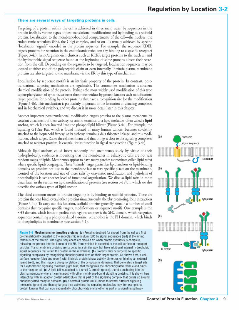
Targeting of a protein within the cell is achieved in three main ways: by sequences in theprotein itself; by various types of post-translational modification; and by binding to a scaffoldprotein. Localization to the membrane-bounded compartments of the cell—the nucleus, theendoplasmic reticulum (ER), the Golgi complex, and so on—is usually achieved by specific“localization signals” encoded in the protein sequence. For example, the sequence KDEL targets proteins for retention in the endoplasmic reticulum (by binding to a specific receptor)(Figure 3-4a); lysine/arginine-rich clusters such as KRKR target proteins to the nucleus; andthe hydrophobic signal sequence found at the beginning of some proteins directs their secre-tion from the cell. Depending on the organelle to be targeted, localization sequences may belocated at either end of the polypeptide chain or even internally. Intrinsic plasma membraneproteins are also targeted to the membrane via the ER by this type of mechanism.
Localization by sequence motifs is an intrinsic property of the protein. In contrast, post-translational targeting mechanisms are regulatable. The commonest mechanism is covalentchemical modification of the protein. Perhaps the most widely used modification of this typeis phosphorylation of tyrosine, serine or threonine residues by protein kinases; such modificationstarget proteins for binding by other proteins that have a recognition site for the modification(Figure 3-4b). This mechanism is particularly important in the formation of signaling complexesand in biochemical switches, and we discuss it in more detail later in this chapter.
Another important post-translational modification targets proteins to the plasma membrane bycovalent attachment of their carboxyl or amino terminus to a lipid molecule, often called a lipidanchor, which is then inserted into the phospholipid bilayer (Figure 3-4c). For example, thesignaling GTPase Ras, which is found mutated in many human tumors, becomes covalentlyattached to the isoprenoid farnesyl at its carboxyl terminus via a thioester linkage, and this modi-fication, which targets Ras to the cell membrane and thus brings it close to the signaling complexesattached to receptor proteins, is essential for its function in signal transduction (Figure 3-4c).
Although lipid anchors could insert randomly into membranes solely by virtue of theirhydrophobicity, evidence is mounting that the membranes in eukaryotic cells are not justrandom soups of lipids. Membranes appear to have many patches (sometimes called lipid rafts)where specific lipids congregate. These “islands” target particular lipid anchors or lipid-bindingdomains on proteins not just to the membrane but to very specific places on the membrane.Control of the location and size of these rafts by enzymatic modification and hydrolysis ofphospholipids is yet another level of functional organization. We discuss lipid rafts in moredetail later, in the section on lipid modification of proteins (see section 3-19), in which we alsodescribe the various types of lipid anchor.
The third common means of protein targeting is by binding to scaffold proteins. These areproteins that can bind several other proteins simultaneously, thereby promoting their interaction(Figure 3-4d). To carry out this function, scaffold proteins generally contain a number of smalldomains that recognize specific targets, modifications or sequence motifs. One example is theSH3 domain, which binds to proline-rich regions; another is the SH2 domain, which recognizessequences containing a phosphorylated tyrosine; yet another is the PH domain, which bindsto phospholipids in membranes (see section 3-1).
Figure 3-4 Mechanisms for targeting proteins (a) Proteins destined for export from the cell are firstco-translationally targeted to the endoplasmic reticulum (ER) by signal sequences (red) at the aminoterminus of the protein. The signal sequences are cleaved off when protein synthesis is complete,releasing the protein into the lumen of the ER, from which it is exported to the cell surface in transportvesicles. Transmembrane proteins are targeted in a similar way, but have additional internal hydrophobicsignal sequences that retain the protein in the membrane. (b) Proteins may be targeted to specificsignaling complexes by recognizing phosphorylated sites on their target protein. As shown here, a cell-surface receptor (blue and green) with intrinsic protein kinase activity dimerizes on binding an externalligand (red), and this triggers phosphorylation of the cytoplasmic domains. That generates a target sitefor a cytoplasmic signaling molecule (light blue) that recognizes the phosphorylated residue and binds to the receptor tail. (c) A lipid tail is attached to a small G protein (green), thereby anchoring it in theplasma membrane where it can interact with other membrane-bound signaling proteins. It is shown hereinteracting with an adaptor protein (dark blue) that is part of the signaling complex that builds up aroundphosphorylated receptor domains. (d) A scaffold protein (blue) binds to several different signalingmolecules (green) and thereby targets their activities: the signaling molecules may, for example, beprotein kinases that can now sequentially phosphorylate one another as part of a signaling pathway.
Control of Protein Function Chapter 3 91©2004 New Science Press Ltd
Regulation by Location 3-2
signal sequence
G protein
ribosome
ER lumen
(a)
cytoplasm
(b)
cytoplasm
(c)
(d)
P P
P
P
P P
Protein function is modulated by the environment in which the proteinoperates
All proteins are adapted to fold and function optimally in the particular environment of thecellular compartment in which they operate. The cellular aqueous solution is highly viscousand contains many components besides proteins at high concentration, including ions, freepolar organic molecules and, most important, the dissociated conjugate acid/conjugate basecomponents of water: the proton and the hydroxide ion. If these two components are presentat approximately equal concentration, as is the case in the cytosol of most cells, the solution isneutral with a pH of about 7. If protons are in excess, the solution is acidic, with a lower pH,and ionizable groups tend to be protonated. If hydroxide ions predominate, the solution isalkaline with a pH > 7 and ionizable groups will tend to be deprotonated. Distinct membrane-bounded compartments inside the cell often have a distinct internal microenvironment andthe extracellular environment represents a different aqueous environment again from that ofthe interior. We describe here some examples of the adaptations of proteins for the environmentsin which they function.
Changes in redox environment can greatly affect protein structure andfunction
The interiors of cells are for the most part reducing environments: they furnish electrons, oftenin the form of hydrogen atoms. On the other hand, outside the cell, proteins and small moleculesare typically exposed to an oxidizing environment in which electrons can be lost. The chiefeffect of this difference is that cysteine residues in proteins are usually fully reduced to –SH groups inside the cell, but are readily oxidized to disulfide S–S bridges when a protein issecreted. Cells can exploit this difference to trigger oligomerization by S–S bond formation insecreted proteins, or subunit dissociation and conformational changes when proteins areinternalized. For instance, acetylcholinesterase is synthesized as a monomer, but when secretedfrom muscle and nerve cells self-associates to form dimers and tetramers. These oligomers,which are more stable than the monomer and thus better able to survive outside the cell, arecomposed of covalently linked subunits. Each monomer has a carboxy-terminal cysteineresidue which forms an intersubunit S–S bond with the identical residue on a neighboringpolypeptide chain.
Changes in pH can drastically alter protein structure and function
Most cytosolic fluid is maintained at near neutral pH, so that neither acids nor bases predominate.However, there are specialized compartments, such as endosomal vesicles, where the pH isquite acidic. As the surfaces of soluble proteins are chiefly composed of polar side chains, manyof which are ionizable, both the net charge on a protein and the distribution of charge over thesurface can vary considerably with pH.
If ligand binding depends on electrostatic interactions (see Figure 2-23), changes in the externalpH (or ion concentration) can greatly influence binding strength by directly altering theionization states of groups that interact with the ligand or of groups on the ligand itself.Modulation of the surface charge distribution of a protein by pH changes can also affect thebiochemical function indirectly, by changing the extent of ionization of essential functionalgroups in an active site or binding site through long-range electrostatic interactions. Forinstance, endosomal proteases, which degrade internalized proteins, are only catalytically active
3-3 Control by pH and Redox Environment
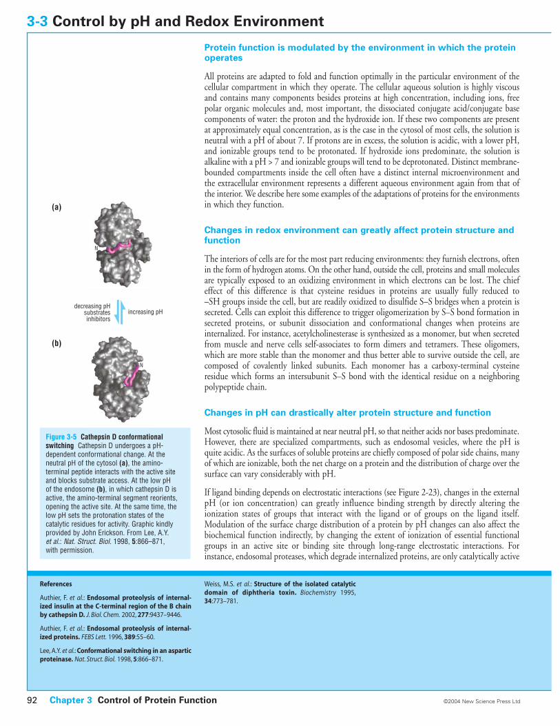
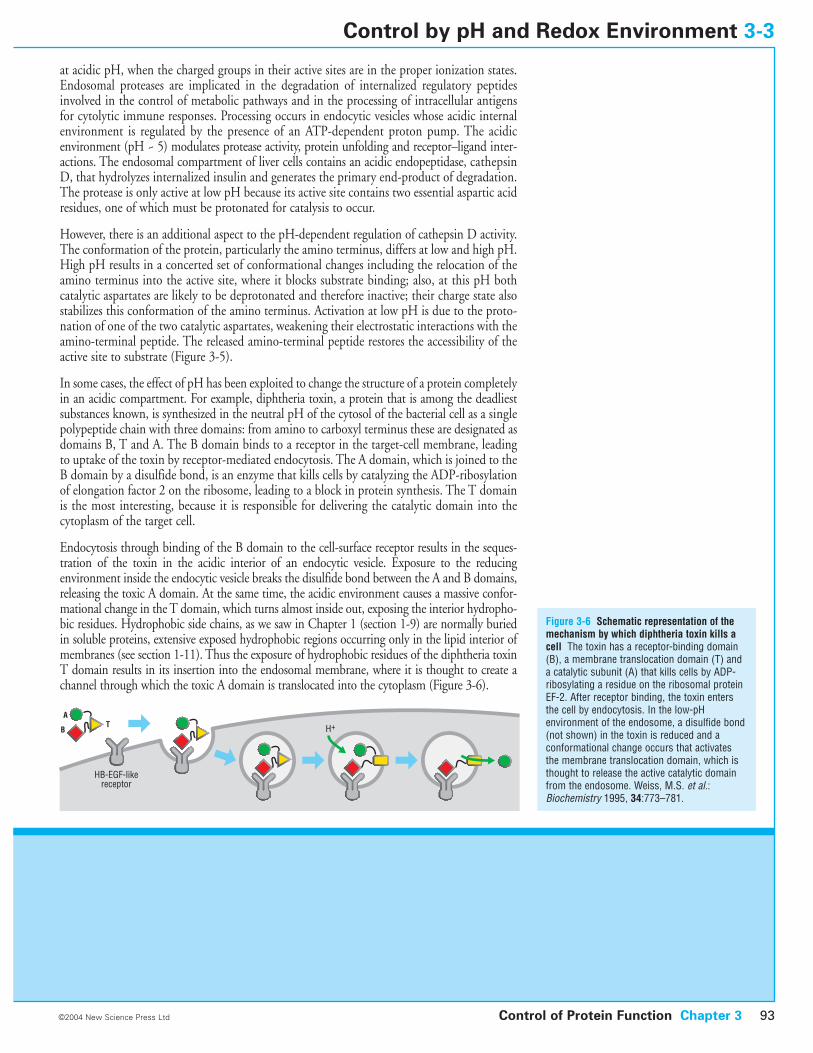
Figure 3-5 Cathepsin D conformationalswitching Cathepsin D undergoes a pH-dependent conformational change. At theneutral pH of the cytosol (a), the amino-terminal peptide interacts with the active siteand blocks substrate access. At the low pH of the endosome (b), in which cathepsin D isactive, the amino-terminal segment reorients,opening the active site. At the same time, thelow pH sets the protonation states of thecatalytic residues for activity. Graphic kindlyprovided by John Erickson. From Lee, A.Y. et al.: Nat. Struct. Biol. 1998, 5:866–871, with permission.
Chapter 3 Control of Protein Function92 ©2004 New Science Press Ltd
References
Authier, F. et al.: Endosomal proteolysis of internal-ized insulin at the C-terminal region of the B chainby cathepsin D. J. Biol. Chem. 2002, 277:9437–9446.
Authier, F. et al.: Endosomal proteolysis of internal-ized proteins. FEBS Lett. 1996, 389:55–60.
Lee, A.Y. et al.: Conformational switching in an asparticproteinase. Nat. Struct. Biol. 1998, 5:866–871.
Weiss, M.S. et al.: Structure of the isolated catalyticdomain of diphtheria toxin. Biochemistry 1995,34:773–781.
increasing pHdecreasing pH
substratesinhibitors
N
N
(a)
(b)
at acidic pH, when the charged groups in their active sites are in the proper ionization states.Endosomal proteases are implicated in the degradation of internalized regulatory peptidesinvolved in the control of metabolic pathways and in the processing of intracellular antigensfor cytolytic immune responses. Processing occurs in endocytic vesicles whose acidic internalenvironment is regulated by the presence of an ATP-dependent proton pump. The acidicenvironment (pH ~ 5) modulates protease activity, protein unfolding and receptor–ligand inter-actions. The endosomal compartment of liver cells contains an acidic endopeptidase, cathepsinD, that hydrolyzes internalized insulin and generates the primary end-product of degradation.The protease is only active at low pH because its active site contains two essential aspartic acidresidues, one of which must be protonated for catalysis to occur.
However, there is an additional aspect to the pH-dependent regulation of cathepsin D activity.The conformation of the protein, particularly the amino terminus, differs at low and high pH.High pH results in a concerted set of conformational changes including the relocation of theamino terminus into the active site, where it blocks substrate binding; also, at this pH bothcatalytic aspartates are likely to be deprotonated and therefore inactive; their charge state alsostabilizes this conformation of the amino terminus. Activation at low pH is due to the proto-nation of one of the two catalytic aspartates, weakening their electrostatic interactions with theamino-terminal peptide. The released amino-terminal peptide restores the accessibility of theactive site to substrate (Figure 3-5).
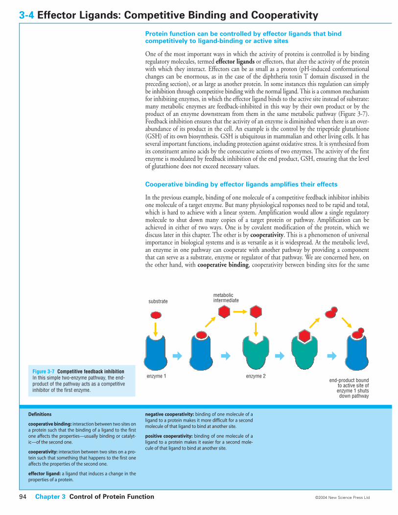
In some cases, the effect of pH has been exploited to change the structure of a protein completelyin an acidic compartment. For example, diphtheria toxin, a protein that is among the deadliestsubstances known, is synthesized in the neutral pH of the cytosol of the bacterial cell as a singlepolypeptide chain with three domains: from amino to carboxyl terminus these are designated asdomains B, T and A. The B domain binds to a receptor in the target-cell membrane, leadingto uptake of the toxin by receptor-mediated endocytosis. The A domain, which is joined to theB domain by a disulfide bond, is an enzyme that kills cells by catalyzing the ADP-ribosylationof elongation factor 2 on the ribosome, leading to a block in protein synthesis. The T domainis the most interesting, because it is responsible for delivering the catalytic domain into thecytoplasm of the target cell.
Endocytosis through binding of the B domain to the cell-surface receptor results in the seques-tration of the toxin in the acidic interior of an endocytic vesicle. Exposure to the reducing environment inside the endocytic vesicle breaks the disulfide bond between the A and B domains,releasing the toxic A domain. At the same time, the acidic environment causes a massive confor-mational change in the T domain, which turns almost inside out, exposing the interior hydropho-bic residues. Hydrophobic side chains, as we saw in Chapter 1 (section 1-9) are normally buriedin soluble proteins, extensive exposed hydrophobic regions occurring only in the lipid interior ofmembranes (see section 1-11). Thus the exposure of hydrophobic residues of the diphtheria toxinT domain results in its insertion into the endosomal membrane, where it is thought to create achannel through which the toxic A domain is translocated into the cytoplasm (Figure 3-6).
Control of Protein Function Chapter 3 93
Figure 3-6 Schematic representation of themechanism by which diphtheria toxin kills acell The toxin has a receptor-binding domain(B), a membrane translocation domain (T) anda catalytic subunit (A) that kills cells by ADP-ribosylating a residue on the ribosomal proteinEF-2. After receptor binding, the toxin entersthe cell by endocytosis. In the low-pHenvironment of the endosome, a disulfide bond(not shown) in the toxin is reduced and aconformational change occurs that activatesthe membrane translocation domain, which isthought to release the active catalytic domainfrom the endosome. Weiss, M.S. et al.:Biochemistry 1995, 34:773–781.
©2004 New Science Press Ltd
Control by pH and Redox Environment 3-3
HB-EGF-likereceptor
H+A
BT
Protein function can be controlled by effector ligands that bind competitively to ligand-binding or active sites
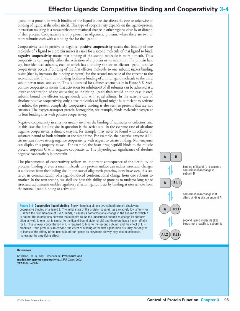
One of the most important ways in which the activity of proteins is controlled is by bindingregulatory molecules, termed effector ligands or effectors, that alter the activity of the proteinwith which they interact. Effectors can be as small as a proton (pH-induced conformationalchanges can be enormous, as in the case of the diphtheria toxin T domain discussed in the preceding section), or as large as another protein. In some instances this regulation can simplybe inhibition through competitive binding with the normal ligand. This is a common mechanismfor inhibiting enzymes, in which the effector ligand binds to the active site instead of substrate:many metabolic enzymes are feedback-inhibited in this way by their own product or by theproduct of an enzyme downstream from them in the same metabolic pathway (Figure 3-7).Feedback inhibition ensures that the activity of an enzyme is diminished when there is an over-abundance of its product in the cell. An example is the control by the tripeptide glutathione(GSH) of its own biosynthesis. GSH is ubiquitous in mammalian and other living cells. It hasseveral important functions, including protection against oxidative stress. It is synthesized fromits constituent amino acids by the consecutive actions of two enzymes. The activity of the firstenzyme is modulated by feedback inhibition of the end product, GSH, ensuring that the levelof glutathione does not exceed necessary values.
Cooperative binding by effector ligands amplifies their effects
In the previous example, binding of one molecule of a competitive feedback inhibitor inhibitsone molecule of a target enzyme. But many physiological responses need to be rapid and total,which is hard to achieve with a linear system. Amplification would allow a single regulatorymolecule to shut down many copies of a target protein or pathway. Amplification can beachieved in either of two ways. One is by covalent modification of the protein, which wediscuss later in this chapter. The other is by cooperativity. This is a phenomenon of universalimportance in biological systems and is as versatile as it is widespread. At the metabolic level,an enzyme in one pathway can cooperate with another pathway by providing a componentthat can serve as a substrate, enzyme or regulator of that pathway. We are concerned here, onthe other hand, with cooperative binding, cooperativity between binding sites for the same
3-4 Effector Ligands: Competitive Binding and Cooperativity
Figure 3-7 Competitive feedback inhibitionIn this simple two-enzyme pathway, the end-product of the pathway acts as a competitiveinhibitor of the first enzyme.
Chapter 3 Control of Protein Function94 ©2004 New Science Press Ltd
Definitions
cooperative binding: interaction between two sites ona protein such that the binding of a ligand to the firstone affects the properties—usually binding or catalyt-ic—of the second one.
cooperativity: interaction between two sites on a pro-tein such that something that happens to the first oneaffects the properties of the second one.
effector ligand: a ligand that induces a change in theproperties of a protein.
negative cooperativity: binding of one molecule of aligand to a protein makes it more difficult for a secondmolecule of that ligand to bind at another site.
positive cooperativity: binding of one molecule of aligand to a protein makes it easier for a second mole-cule of that ligand to bind at another site.
substratemetabolicintermediate
enzyme 1 enzyme 2end-product bound
to active site of enzyme 1 shuts down pathway
ligand on a protein, in which binding of the ligand at one site affects the ease or otherwise ofbinding of ligand at the other site(s). This type of cooperativity depends on the ligand–proteininteraction resulting in a measurable conformational change in other regions, close by or distant,of that protein. Cooperativity is only present in oligomeric proteins, where there are two ormore subunits each with a binding site for the ligand.
Cooperativity can be positive or negative: positive cooperativity means that binding of onemolecule of a ligand to a protein makes it easier for a second molecule of that ligand to bind;negative cooperativity means that binding of the second molecule is more difficult. Thuscooperativity can amplify either the activation of a protein or its inhibition. If a protein has,say, four identical subunits, each of which has a binding site for an effector ligand, positivecooperativity occurs if binding of the first effector molecule to one subunit makes bindingeasier (that is, increases the binding constant) for the second molecule of the effector to thesecond subunit. In turn, this binding facilitates binding of a third ligand molecule to the thirdsubunit even more, and so on. This is illustrated for a dimer schematically in Figure 3-8. Suchpositive cooperativity means that activation (or inhibition) of all subunits can be achieved at alower concentration of the activating or inhibiting ligand than would be the case if eachsubunit bound the effector independently and with equal affinity. In the extreme case ofabsolute positive cooperativity, only a few molecules of ligand might be sufficient to activateor inhibit the protein completely. Cooperative binding is also seen in proteins that are notenzymes. The oxygen-transport protein hemoglobin, for example, binds molecular oxygen atits four binding sites with positive cooperativity.
Negative cooperativity in enzymes usually involves the binding of substrates or cofactors, andin this case the binding site in question is the active site. In the extreme case of absolutenegative cooperativity, a dimeric enzyme, for example, may never be found with cofactor orsubstrate bound to both subunits at the same time. For example, the bacterial enzyme ATP-citrate lyase shows strong negative cooperativity with respect to citrate binding. Non-enzymescan display this property as well. For example, the heart drug bepridil binds to the muscleprotein troponin C with negative cooperativity. The physiological significance of absolutenegative cooperativity is uncertain.
The phenomenon of cooperativity reflects an important consequence of the flexibility ofproteins: binding of even a small molecule to a protein surface can induce structural changesat a distance from the binding site. In the case of oligomeric proteins, as we have seen, this canresult in communication of a ligand-induced conformational change from one subunit toanother. In the next section, we shall see how this ability of proteins to undergo long-rangestructural adjustments enables regulatory effector ligands to act by binding at sites remote fromthe normal ligand-binding or active site.
Control of Protein Function Chapter 3 95
Figure 3-8 Cooperative ligand binding Shown here is a simple two-subunit protein displayingcooperative binding of a ligand L. The initial state of the protein (square) has a relatively low affinity forL. When the first molecule of L (L1) binds, it causes a conformational change in the subunit to which itis bound. But interactions between the subunits cause the unoccupied subunit to change its conform-ation as well, to one that is similar to the ligand-bound state (circle) and therefore has a higher affinityfor L. Thus a lower concentration of L is required to bind to the second subunit, and the effect of L isamplified. If the protein is an enzyme, the effect of binding of the first ligand molecule may not only beto increase the affinity of the next subunit for ligand; its enzymatic activity may also be enhanced,increasing the amplifying effect.
©2004 New Science Press Ltd
Effector Ligands: Competitive Binding and Cooperativity 3-4
References
Koshland, D.E. Jr., and Hamadani, K.: Proteomics andmodels for enzyme cooperativity. J. Biol. Chem. 2002,277:46841–46844.
A
A
A
B
binding of ligand (L1) causes aconformational change insubunit B
conformational change in Balters binding site on subunit A
second ligand molecule (L2)binds more readily to subunit A
A:L2
B:L1
B:L1
B:L1
Effector molecules can cause conformational changes at distant sites
Because of the close packing of atoms in globular protein structures, even small changes inside-chain and main-chain positions at one site can be propagated through the tertiary structureof the molecule and cause conformational changes at a distant location in the protein. Indeed,the most common type of regulatory effector ligand is one that is different from the normalsubstrate or functional ligand for the protein and which binds at a site distinct from theenzyme’s catalytic site or from the site through which the protein’s function is mediated (in thecase of a non-enzyme). In hetero-oligomeric enzymes, the regulatory site is often located on adifferent subunit from the active site.
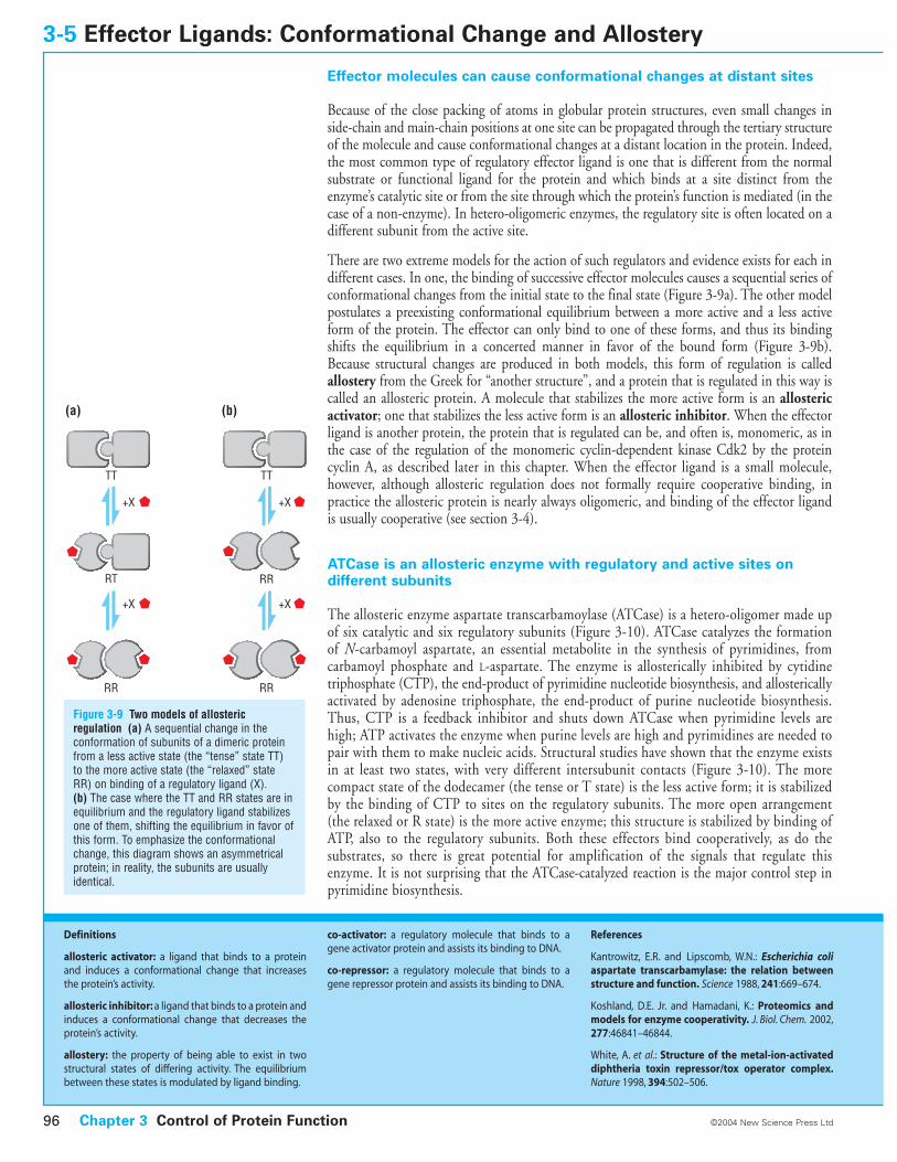
There are two extreme models for the action of such regulators and evidence exists for each indifferent cases. In one, the binding of successive effector molecules causes a sequential series ofconformational changes from the initial state to the final state (Figure 3-9a). The other modelpostulates a preexisting conformational equilibrium between a more active and a less activeform of the protein. The effector can only bind to one of these forms, and thus its bindingshifts the equilibrium in a concerted manner in favor of the bound form (Figure 3-9b).Because structural changes are produced in both models, this form of regulation is calledallostery from the Greek for “another structure”, and a protein that is regulated in this way iscalled an allosteric protein. A molecule that stabilizes the more active form is an allostericactivator; one that stabilizes the less active form is an allosteric inhibitor. When the effectorligand is another protein, the protein that is regulated can be, and often is, monomeric, as inthe case of the regulation of the monomeric cyclin-dependent kinase Cdk2 by the proteincyclin A, as described later in this chapter. When the effector ligand is a small molecule,however, although allosteric regulation does not formally require cooperative binding, inpractice the allosteric protein is nearly always oligomeric, and binding of the effector ligandis usually cooperative (see section 3-4).
ATCase is an allosteric enzyme with regulatory and active sites on different subunits
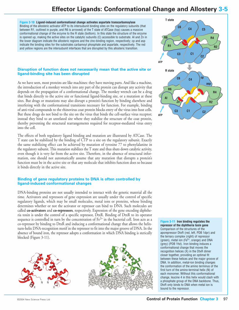
The allosteric enzyme aspartate transcarbamoylase (ATCase) is a hetero-oligomer made upof six catalytic and six regulatory subunits (Figure 3-10). ATCase catalyzes the formationof N-carbamoyl aspartate, an essential metabolite in the synthesis of pyrimidines, from carbamoyl phosphate and L-aspartate. The enzyme is allosterically inhibited by cytidinetriphosphate (CTP), the end-product of pyrimidine nucleotide biosynthesis, and allostericallyactivated by adenosine triphosphate, the end-product of purine nucleotide biosynthesis.Thus, CTP is a feedback inhibitor and shuts down ATCase when pyrimidine levels arehigh; ATP activates the enzyme when purine levels are high and pyrimidines are needed topair with them to make nucleic acids. Structural studies have shown that the enzyme existsin at least two states, with very different intersubunit contacts (Figure 3-10). The morecompact state of the dodecamer (the tense or T state) is the less active form; it is stabilizedby the binding of CTP to sites on the regulatory subunits. The more open arrangement(the relaxed or R state) is the more active enzyme; this structure is stabilized by binding ofATP, also to the regulatory subunits. Both these effectors bind cooperatively, as do the substrates, so there is great potential for amplification of the signals that regulate thisenzyme. It is not surprising that the ATCase-catalyzed reaction is the major control step inpyrimidine biosynthesis.
3-5 Effector Ligands: Conformational Change and Allostery
Chapter 3 Control of Protein Function96 ©2004 New Science Press Ltd
Definitions
allosteric activator: a ligand that binds to a proteinand induces a conformational change that increasesthe protein’s activity.
allosteric inhibitor: a ligand that binds to a protein andinduces a conformational change that decreases theprotein’s activity.
allostery: the property of being able to exist in twostructural states of differing activity. The equilibriumbetween these states is modulated by ligand binding.
co-activator: a regulatory molecule that binds to agene activator protein and assists its binding to DNA.
co-repressor: a regulatory molecule that binds to agene repressor protein and assists its binding to DNA.
References
Kantrowitz, E.R. and Lipscomb, W.N.: Escherichia coliaspartate transcarbamylase: the relation betweenstructure and function. Science 1988, 241:669–674.
Koshland, D.E. Jr. and Hamadani, K.: Proteomics andmodels for enzyme cooperativity. J. Biol. Chem. 2002,277:46841–46844.
White, A. et al.: Structure of the metal-ion-activateddiphtheria toxin repressor/tox operator complex.Nature 1998, 394:502–506.
(a) (b)
TT
RT
RR
TT
RR
RR
+X
+X
+X
+X
Figure 3-9 Two models of allostericregulation (a) A sequential change in theconformation of subunits of a dimeric proteinfrom a less active state (the “tense” state TT)to the more active state (the “relaxed” stateRR) on binding of a regulatory ligand (X). (b) The case where the TT and RR states are inequilibrium and the regulatory ligand stabilizesone of them, shifting the equilibrium in favor ofthis form. To emphasize the conformationalchange, this diagram shows an asymmetricalprotein; in reality, the subunits are usuallyidentical.
Disruption of function does not necessarily mean that the active site orligand-binding site has been disrupted
As we have seen, most proteins are like machines: they have moving parts. And like a machine,the introduction of a monkey wrench into any part of the protein can disrupt any activity thatdepends on the propagation of a conformational change. The monkey wrench can be a drugthat binds directly to the active site or functional ligand-binding site, or a mutation at thesesites. But drugs or mutations may also disrupt a protein’s function by binding elsewhere andinterfering with the conformational transitions necessary for function. For example, bindingof anti-viral compounds to the rhinovirus coat protein blocks entry of the virus into host cells.But these drugs do not bind to the site on the virus that binds the cell-surface virus receptor;instead they bind to an unrelated site where they stabilize the structure of the coat protein,thereby preventing the structural rearrangements required for receptor-mediated virus entryinto the cell.
The effects of both regulatory ligand binding and mutation are illustrated by ATCase. The T state can be stabilized by the binding of CTP to a site on the regulatory subunit. Exactlythe same stabilizing effect can be achieved by mutation of tyrosine 77 to phenylalanine inthe regulatory subunit. This mutation stabilizes the T state and thus shuts down catalytic activity,even though it is very far from the active site. Therefore, in the absence of structural infor-mation, one should not automatically assume that any mutation that disrupts a protein’sfunction must be in the active site or that any molecule that inhibits function does so becauseit binds directly in the active site.
Binding of gene regulatory proteins to DNA is often controlled by ligand-induced conformational changes
DNA-binding proteins are not usually intended to interact with the genetic material all thetime. Activators and repressors of gene expression are usually under the control of specificregulatory ligands, which may be small molecules, metal ions or proteins, whose bindingdetermines whether or not the activator or repressor can bind to DNA. Such molecules arecalled co-activators and co-repressors, respectively. Expression of the gene encoding diphthe-ria toxin is under the control of a specific repressor, DtxR. Binding of DtxR to its operatorsequence is controlled in turn by the concentration of Fe2+ in the bacterial cell. Iron acts as aco-repressor by binding to DtxR and inducing a conformational change that allows the helix-turn-helix DNA-recognition motif in the repressor to fit into the major groove of DNA. In theabsence of bound iron, the repressor adopts a conformation in which DNA binding is stericallyblocked (Figure 3-11).
Figure 3-10 Ligand-induced conformational change activates aspartate transcarbamoylaseBinding of the allosteric activator ATP to its intersubunit binding sites on the regulatory subunits (thatbetween R1, outlined in purple, and R6 is arrowed) of the T state of ATCase (top) causes a massiveconformational change of the enzyme to the R state (bottom). In this state the structure of the enzyme is opened up, making the active sites on the catalytic subunits (C) accessible to substrate. Al and Zn inthe lower diagram indicate the allosteric regions and the zinc-binding region, respectively; cp and aspindicate the binding sites for the substrates carbamoyl phosphate and aspartate, respectively. The redand yellow regions are the intersubunit interfaces that are disrupted by this allosteric transition.
Figure 3-11 Iron binding regulates therepressor of the diphtheria toxin geneComparison of the structures of theaporepressor DtxR (red, left, PDB 1dpr) andthe ternary complex (right) of repressor(green), metal ion (Fe2+, orange) and DNA (grey) (PDB 1fst). Iron binding induces aconformational change that moves therecognition helices (X) in the DtxR dimer closer together, providing an optimal fitbetween these helices and the major groove ofDNA. In addition, metal-ion binding changesthe conformation of the amino terminus of thefirst turn of the amino-terminal helix (N) ofeach monomer. Without this conformationalchange, leucine 4 in this helix would clash witha phosphate group of the DNA backbone. Thus,DtxR only binds to DNA when metal ion isbound to the repressor.
Control of Protein Function Chapter 3 97©2004 New Science Press Ltd
Effector Ligands: Conformational Change and Allostery 3-5
R6
R6
C6
T state
R state
ATP
C6
C5C4
C4
R4
R4
R2
R2
C2
C2
C1
C1
C3
C3
R1
cp
asp
AlZn
ATP
N N
X X
Conformational changes driven by nucleotide binding and hydrolysisare the basis for switching and motor properties of proteins
Not every process in a living cell runs continuously. Many of them must cycle between “on”and “off ” states in order to control cell growth and division and responses to extracellularsignals. Signal transduction pathways such as those that operate in vision and hormone-basedsignaling, vesicular transport (which is often called protein or membrane trafficking),polypeptide chain elongation during protein synthesis, and actin- and tubulin-based motorfunctions are also examples of processes that must be switched on and off under preciselydetermined circumstances. This cycling is controlled by a special set of proteins that functionas molecular switches. Although these proteins vary in structure and in the processes they control,they have a number of common features. The most important common element is theswitching mechanism itself: most of these proteins undergo conformational changes inducedby the difference between the triphosphate and diphosphate forms of a bound nucleotide. Theconformational changes are such that completely different target proteins recognize the twobound states of the switch protein, providing a simple means of altering the output of a signal.
Most protein switches are enzymes that catalyze the hydrolysis of a nucleoside triphosphate tothe diphosphate. Most often the nucleotide is guanosine triphosphate, GTP, and the switchprotein is a GTPase that hydrolyzes it to GDP. GTPase switches (also commonly called G proteins or occasionally guanine-nucleotide-binding proteins) are one major class of switchproteins; they control the on/off states of most cellular processes, including sensory perception,intracellular transport, protein synthesis and cell growth and differentiation. The second majorclass of switch proteins is composed of those ATPases that are usually associated with motorprotein complexes or transporters that move material into and out of cells and some organelles.Members of the third major class, the two-component response regulators, a group of switchesthus far found only in microbes and plants, are composed of a histidine protein kinase and asecond “response regulator” protein. They do not bind GTP or ATP in the same way as thenucleotide switch proteins discussed here, but use a covalently bound phosphate derived from thehydrolysis of ATP by the kinase to trigger a conformational change in the response regulator.
Hydrolyzable nucleotides are used to control many types of molecular switches because theenergy derived from hydrolysis of the terminal phosphate of a GTP or ATP is large enough tomake the conformational change in the switch effectively irreversible until another protein bindsto the switch, displaces the diphosphate, and allows the triphosphate to bind again. Anotherreason is that using ATP or GTP can couple the switching process to the energy state of the celland to the synthesis of DNA and RNA, both of which change the levels of nucleoside tri- anddiphosphates.
All nucleotide switch proteins have some common structural andfunctional features
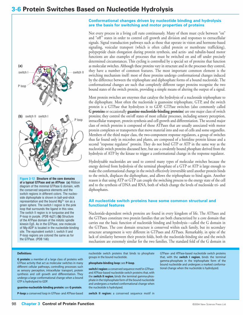
Nucleotide-dependent switch proteins are found in every kingdom of life. The ATPases andthe GTPases constitute two protein families that are both characterized by a core domain thatcarries out the basic function of nucleotide binding and hydrolysis—called the G domain inthe GTPases. The core domain structure is conserved within each family, but its secondarystructure arrangement is very different in GTPases and ATPases. Remarkably, in spite of thelack of similarity between their protein folds, both the nucleotide-binding site and the switchmechanism are extremely similar for the two families. The standard fold of the G domain in
3-6 Protein Switches Based on Nucleotide Hydrolysis
Figure 3-12 Structure of the core domains of a typical GTPase and an ATPase (a) Ribbondiagram of the minimal GTPase G domain, withthe conserved sequence elements and theswitch regions in different colors. The nucleo-side diphosphate is shown in ball-and-stickrepresentation and the bound Mg2+ ion as agreen sphere. The switch I region is the pinkloop that surrounds the ligand in this view. The switch II region is in turquoise and the P-loop in purple. (PDB 4q21) (b) Structure of the ATPase domain of the mitotic spindlekinesin Eg5. As in the GTPase, one molecule of Mg-ADP is located in the nucleotide-bindingsite. The equivalent switch I, switch II and P-loop regions are colored the same as for the GTPase. (PDB 1ii6)
Chapter 3 Control of Protein Function98 ©2004 New Science Press Ltd
Definitions
G protein: a member of a large class of proteins withGTPase activity that act as molecular switches in manydifferent cellular pathways, controlling processes suchas sensory perception, intracellular transport, proteinsynthesis and cell growth and differentiation. Theyundergo a large conformational change when a boundGTP is hydrolyzed to GDP.
guanine-nucleotide-binding protein: see G protein.
P-loop: a conserved loop in GTPase- and ATPase-based
nucleotide switch proteins that binds to phosphategroups in the bound nucleotide.
phosphate-binding loop: see P-loop.
switch I region: a conserved sequence motif in GTPase-and ATPase-based nucleotide switch proteins that, withthe switch II region, binds the terminal gamma-phos-phate in the triphosphate form of the bound nucleotideand undergoes a marked conformational change whenthe nucleotide is hydrolyzed.
switch II region: a conserved sequence motif in
GTPase- and ATPase-based nucleotide switch proteinsthat, with the switch I region, binds the terminalgamma-phosphate in the triphosphate form of thebound nucleotide and undergoes a marked conforma-tional change when the nucleotide is hydrolyzed.
switch I
switch II
switch II
switch I
P loop
P loop(b)
(a)
the GTPases consists of a mixed six-stranded beta sheet with five helices located on both sides(Figure 3-12a). There are three conserved features in nucleotide switches: the P-loop, and theswitch I and switch II sequence motifs. The P-loop or phosphate-binding loop binds thealpha- and beta-phosphates from the phosphate tail of the nucleotide. Residues from the twoswitch motifs coordinate the terminal gamma-phosphate in the triphosphate form of thebound nucleotide. A Mg2+ ion, which is complexed with the bound nucleotide, is coordinatedby the nucleotide phosphate groups and, in the triphosphate form of the switch, by residuesfrom the switch I and II regions.
These regions usually contain four to five conserved sequence elements, which are lined upalong the nucleotide-binding site. Additional important contribution to binding is made by theinteractions of the nucleotide base with a sequence that has the motif N/TKXD (where X is anyamino acid) and confers specificity for guanine. Specificity for guanine is due to the aspartateside chain in this motif, which forms a bifurcated hydrogen bond with the base, and to a main-chain interaction of an invariant alanine (from a short SAK motif ), with the guanine oxygen.
Similar conserved sequence motifs are located in equivalent spatial positions in the switchATPases, even though the core protein fold is completely different (Figure 3-12b). The adeninebase interacts with a conserved RXRP or NP motif (equivalent to the N/TKXD in theGTPases) and alpha- and beta-phosphates are bound to an equivalent P-loop with the sameconsensus sequence GXXXXGKS/T. Furthermore, both switch I and switch II sequence motifsof GTPases (DXnT and DXXG respectively) have their equivalents in ATPases: NXXSSR andDXXG, respectively.
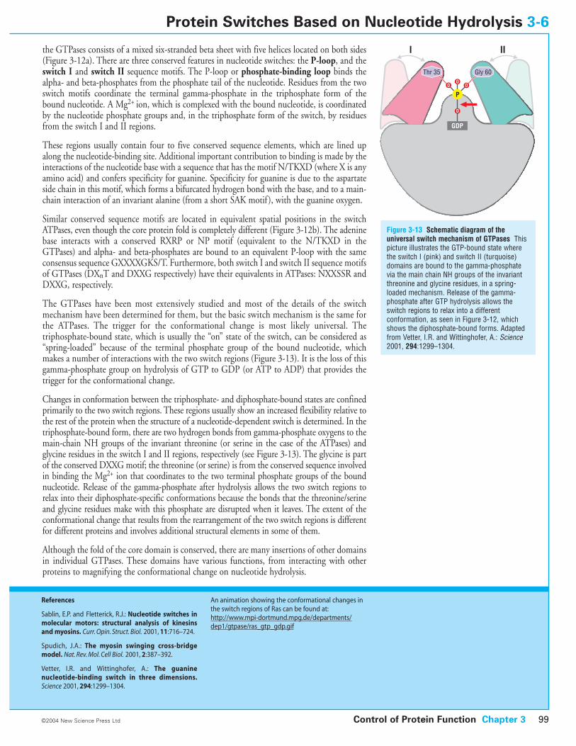
The GTPases have been most extensively studied and most of the details of the switchmechanism have been determined for them, but the basic switch mechanism is the same forthe ATPases. The trigger for the conformational change is most likely universal. Thetriphosphate-bound state, which is usually the “on” state of the switch, can be considered as“spring-loaded” because of the terminal phosphate group of the bound nucleotide, whichmakes a number of interactions with the two switch regions (Figure 3-13). It is the loss of thisgamma-phosphate group on hydrolysis of GTP to GDP (or ATP to ADP) that provides thetrigger for the conformational change.
Changes in conformation between the triphosphate- and diphosphate-bound states are confinedprimarily to the two switch regions. These regions usually show an increased flexibility relative tothe rest of the protein when the structure of a nucleotide-dependent switch is determined. In thetriphosphate-bound form, there are two hydrogen bonds from gamma-phosphate oxygens to themain-chain NH groups of the invariant threonine (or serine in the case of the ATPases) andglycine residues in the switch I and II regions, respectively (see Figure 3-13). The glycine is partof the conserved DXXG motif; the threonine (or serine) is from the conserved sequence involvedin binding the Mg2+ ion that coordinates to the two terminal phosphate groups of the boundnucleotide. Release of the gamma-phosphate after hydrolysis allows the two switch regions torelax into their diphosphate-specific conformations because the bonds that the threonine/serineand glycine residues make with this phosphate are disrupted when it leaves. The extent of theconformational change that results from the rearrangement of the two switch regions is differentfor different proteins and involves additional structural elements in some of them.
Although the fold of the core domain is conserved, there are many insertions of other domainsin individual GTPases. These domains have various functions, from interacting with otherproteins to magnifying the conformational change on nucleotide hydrolysis.
Control of Protein Function Chapter 3 99
Figure 3-13 Schematic diagram of theuniversal switch mechanism of GTPases Thispicture illustrates the GTP-bound state wherethe switch I (pink) and switch II (turquoise)domains are bound to the gamma-phosphatevia the main chain NH groups of the invariantthreonine and glycine residues, in a spring-loaded mechanism. Release of the gamma-phosphate after GTP hydrolysis allows theswitch regions to relax into a differentconformation, as seen in Figure 3-12, whichshows the diphosphate-bound forms. Adaptedfrom Vetter, I.R. and Wittinghofer, A.: Science2001, 294:1299–1304.
©2004 New Science Press Ltd
Protein Switches Based on Nucleotide Hydrolysis 3-6
References
Sablin, E.P. and Fletterick, R.J.: Nucleotide switches inmolecular motors: structural analysis of kinesinsand myosins. Curr. Opin. Struct. Biol. 2001, 11:716–724.
Spudich, J.A.: The myosin swinging cross-bridgemodel. Nat. Rev. Mol. Cell Biol. 2001, 2:387–392.
Vetter, I.R. and Wittinghofer, A.: The guaninenucleotide-binding switch in three dimensions.Science 2001, 294:1299–1304.
An animation showing the conformational changes inthe switch regions of Ras can be found at:http://www.mpi-dortmund.mpg.de/departments/dep1/gtpase/ras_gtp_gdp.gif
GDP
P
O
O
O O
Thr 35 Gly 60
I II
The switching cycle of nucleotide hydrolysis and exchange in G proteinsis modulated by the binding of other proteins
All GTPase switches operate through conformational changes induced by a change in the formof the bound nucleotide, as we saw in the previous section; but this is only the core part of theswitching mechanism. Which cellular process a particular G protein controls is determined bythe interactions it makes with other proteins in its two conformational states. We will now con-sider the rest of the switching mechanism in detail in the case of the GTPases, exemplified by thesmall monomeric GTPase Ras, whose switch function helps to control cell growth and division.
The overall switching mechanism can be viewed as an on–off cycle in which the GTP-boundstate is the “on” state and the GDP-bound state is “off ”. If we start with the switch in the offposition, the gamma-phosphate of GTP is not present and the two switch regions are in therelaxed conformations characteristic of the diphosphate-bound state of the protein (see Figure3-12a). GDP must now dissociate from the protein to allow GTP to bind. The normal rate ofdissociation is often very slow, so most G proteins have various guanine-nucleotide exchangefactors (GEFs) that bind to them and facilitate the release of GDP by inducing conformationalchanges that open up the binding site. A single G protein may be recognized by multipleGEFs, enabling one GTPase to serve as the focal point for integrating signals from differentupstream pathways.
After GDP has been released and GTP binds, the protein conformation (that is, thearrangement of the switch I and switch II regions) changes to bind the gamma-phosphate (seeFigure 3-13) and the switch is now in the on position. The altered conformation of the switchregions allows the G protein to interact with downstream effectors, activating various enzymessuch as phosphatidylinositol-3-kinase and turning on various signaling and other pathways.Several different effectors may recognize the on state of the same G protein, allowing a singleswitch to control multiple cellular processes. The switch remains on as long as GTP remainsbound to the GTPase.
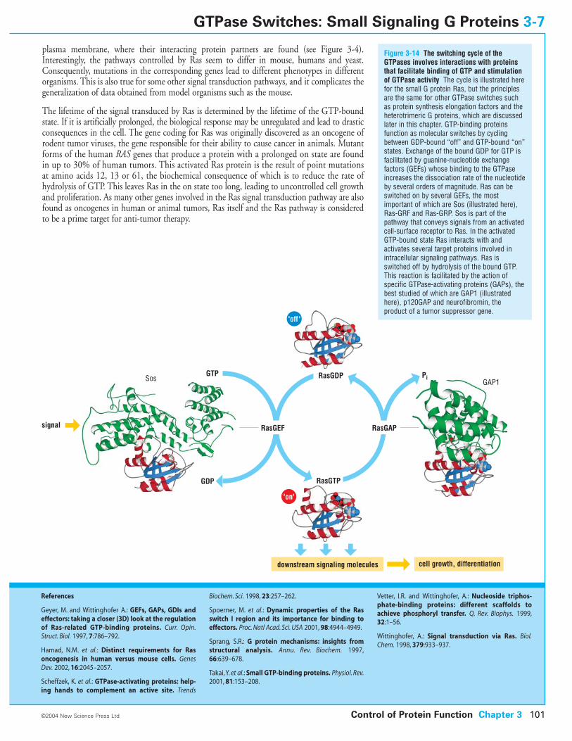
Although GTPases are enzymes and catalyze the hydrolysis of GTP to GDP, they are not veryefficient; their intrinsic rate of GTP hydrolysis is very slow. What determines the length oftime the switch remains in the on state is the activity of various GTPase-activating proteins(GAPs) that bind to the GTP-bound conformation and stimulate the catalytic activity: in thecase of Ras the binding of a GAP increases the GTPase activity by 100,000-fold (105). WhenGTP has been hydrolyzed, the switch I and switch II regions change to their relaxed conformations and the switch is back in the off state; the cycle is complete. Again, a singleG protein can interact with multiple GAPs from different upstream signaling pathways. Theon–off cycle is illustrated in Figure 3-14.
How the various GAP proteins facilitate GTP hydrolysis is not known in all cases, but at least someappear to function by stabilizing the transition state (see section 2-9). They insert an arginineside chain into the nucleotide-binding site of the GTPase to which they bind, and the positivecharge on this side chain helps to stabilize the negative charge that builds up in the transitionstate for hydrolysis of the gamma-phosphate group of GTP.
The importance of proper regulation of G-protein signaling is exemplified by the Ras family ofsmall GTPases. There are three human RAS genes, H-, N- and K-RAS, all of which code for verysimilar proteins of around 21 kDa molecular mass. They are post-translationally modified bythe covalent attachment of lipophilic groups to the carboxy-terminal end. This modification isnecessary for the Ras proteins’ biological function as switches because it targets them to the
3-7 GTPase Switches: Small Signaling G Proteins
Chapter 3 Control of Protein Function100 ©2004 New Science Press Ltd
Definitions
GTPase-activating protein (GAP): a protein that accel-erates the intrinsic GTPase activity of switch GTPases.
guanine-nucleotide exchange factor (GEF): a proteinthat facilitates exchange of GDP for GTP in switchGTPases.
plasma membrane, where their interacting protein partners are found (see Figure 3-4).Interestingly, the pathways controlled by Ras seem to differ in mouse, humans and yeast.Consequently, mutations in the corresponding genes lead to different phenotypes in differentorganisms. This is also true for some other signal transduction pathways, and it complicates thegeneralization of data obtained from model organisms such as the mouse.
The lifetime of the signal transduced by Ras is determined by the lifetime of the GTP-boundstate. If it is artificially prolonged, the biological response may be unregulated and lead to drasticconsequences in the cell. The gene coding for Ras was originally discovered as an oncogene ofrodent tumor viruses, the gene responsible for their ability to cause cancer in animals. Mutantforms of the human RAS genes that produce a protein with a prolonged on state are foundin up to 30% of human tumors. This activated Ras protein is the result of point mutationsat amino acids 12, 13 or 61, the biochemical consequence of which is to reduce the rate ofhydrolysis of GTP. This leaves Ras in the on state too long, leading to uncontrolled cell growthand proliferation. As many other genes involved in the Ras signal transduction pathway are alsofound as oncogenes in human or animal tumors, Ras itself and the Ras pathway is consideredto be a prime target for anti-tumor therapy.
Control of Protein Function Chapter 3 101©2004 New Science Press Ltd
GTPase Switches: Small Signaling G Proteins 3-7
References
Geyer, M. and Wittinghofer A.: GEFs, GAPs, GDIs andeffectors: taking a closer (3D) look at the regulationof Ras-related GTP-binding proteins. Curr. Opin.Struct. Biol. 1997, 7:786–792.
Hamad, N.M. et al.: Distinct requirements for Rasoncogenesis in human versus mouse cells. GenesDev. 2002, 16:2045–2057.
Scheffzek, K. et al.: GTPase-activating proteins: help-ing hands to complement an active site. Trends
Biochem. Sci. 1998, 23:257–262.
Spoerner, M. et al.: Dynamic properties of the Rasswitch I region and its importance for binding toeffectors. Proc. Natl Acad. Sci. USA 2001, 98:4944–4949.
Sprang, S.R.: G protein mechanisms: insights fromstructural analysis. Annu. Rev. Biochem. 1997,66:639–678.
Takai,Y. et al.: Small GTP-binding proteins. Physiol. Rev.2001, 81:153–208.
Vetter, I.R. and Wittinghofer, A.: Nucleoside triphos-phate-binding proteins: different scaffolds toachieve phosphoryl transfer. Q. Rev. Biophys. 1999,32:1–56.
Wittinghofer, A.: Signal transduction via Ras. Biol.Chem. 1998, 379:933–937.
RasGDP
RasGTP
cell growth, differentiationdownstream signaling molecules
GTP
GDP
signal RasGEF RasGAP
GAP1Sos Pi
'off '
'on'
Figure 3-14 The switching cycle of theGTPases involves interactions with proteinsthat facilitate binding of GTP and stimulationof GTPase activity The cycle is illustrated herefor the small G protein Ras, but the principlesare the same for other GTPase switches suchas protein synthesis elongation factors and theheterotrimeric G proteins, which are discussedlater in this chapter. GTP-binding proteinsfunction as molecular switches by cyclingbetween GDP-bound “off” and GTP-bound “on”states. Exchange of the bound GDP for GTP isfacilitated by guanine-nucleotide exchangefactors (GEFs) whose binding to the GTPaseincreases the dissociation rate of the nucleotideby several orders of magnitude. Ras can beswitched on by several GEFs, the mostimportant of which are Sos (illustrated here),Ras-GRF and Ras-GRP. Sos is part of thepathway that conveys signals from an activatedcell-surface receptor to Ras. In the activatedGTP-bound state Ras interacts with andactivates several target proteins involved inintracellular signaling pathways. Ras isswitched off by hydrolysis of the bound GTP.This reaction is facilitated by the action ofspecific GTPase-activating proteins (GAPs), thebest studied of which are GAP1 (illustratedhere), p120GAP and neurofibromin, theproduct of a tumor suppressor gene.
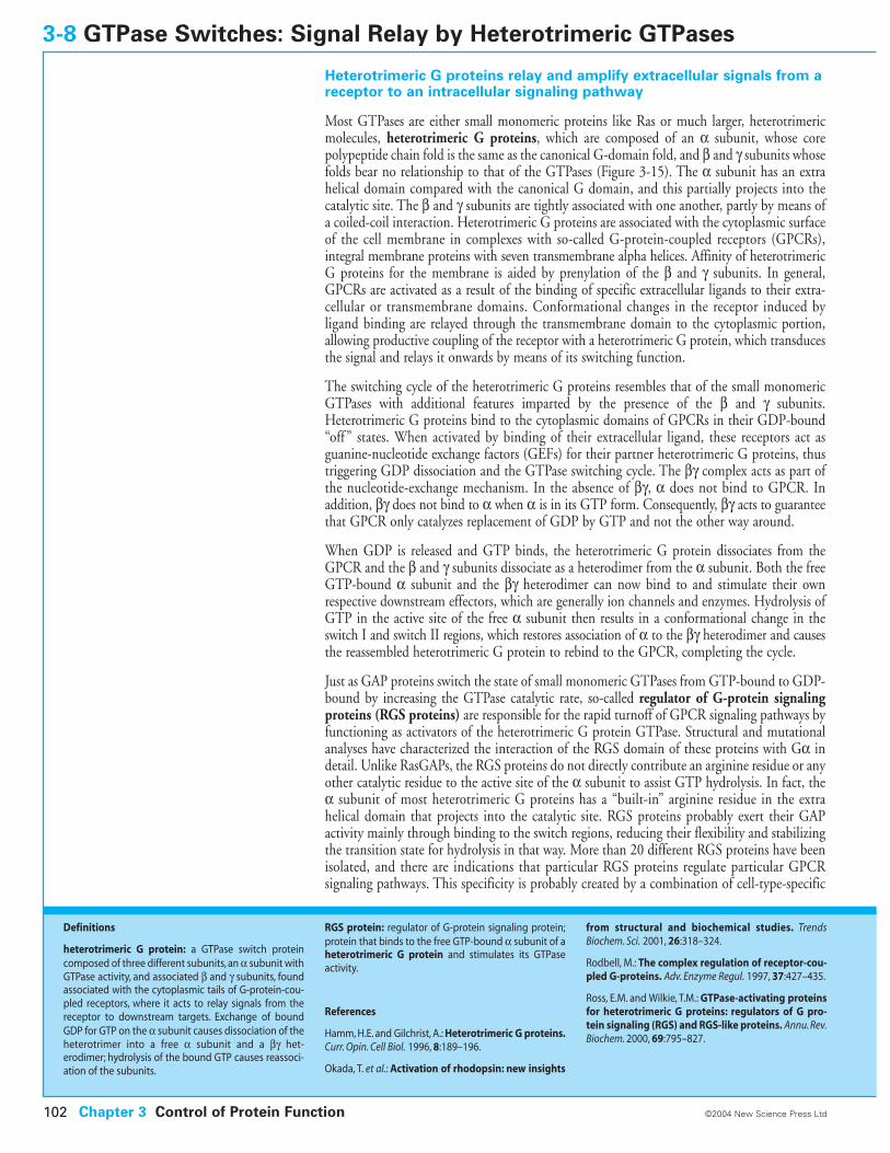
Heterotrimeric G proteins relay and amplify extracellular signals from areceptor to an intracellular signaling pathway
Most GTPases are either small monomeric proteins like Ras or much larger, heterotrimericmolecules, heterotrimeric G proteins, which are composed of an a subunit, whose corepolypeptide chain fold is the same as the canonical G-domain fold, and b and g subunits whosefolds bear no relationship to that of the GTPases (Figure 3-15). The a subunit has an extrahelical domain compared with the canonical G domain, and this partially projects into thecatalytic site. The b and g subunits are tightly associated with one another, partly by means ofa coiled-coil interaction. Heterotrimeric G proteins are associated with the cytoplasmic surfaceof the cell membrane in complexes with so-called G-protein-coupled receptors (GPCRs),integral membrane proteins with seven transmembrane alpha helices. Affinity of heterotrimericG proteins for the membrane is aided by prenylation of the b and g subunits. In general,GPCRs are activated as a result of the binding of specific extracellular ligands to their extra-cellular or transmembrane domains. Conformational changes in the receptor induced byligand binding are relayed through the transmembrane domain to the cytoplasmic portion,allowing productive coupling of the receptor with a heterotrimeric G protein, which transducesthe signal and relays it onwards by means of its switching function.
The switching cycle of the heterotrimeric G proteins resembles that of the small monomericGTPases with additional features imparted by the presence of the b and g subunits.Heterotrimeric G proteins bind to the cytoplasmic domains of GPCRs in their GDP-bound“off ” states. When activated by binding of their extracellular ligand, these receptors act asguanine-nucleotide exchange factors (GEFs) for their partner heterotrimeric G proteins, thustriggering GDP dissociation and the GTPase switching cycle. The bg complex acts as part ofthe nucleotide-exchange mechanism. In the absence of bg, a does not bind to GPCR. Inaddition, bg does not bind to a when a is in its GTP form. Consequently, bg acts to guaranteethat GPCR only catalyzes replacement of GDP by GTP and not the other way around.
When GDP is released and GTP binds, the heterotrimeric G protein dissociates from theGPCR and the b and g subunits dissociate as a heterodimer from the a subunit. Both the freeGTP-bound a subunit and the bg heterodimer can now bind to and stimulate their ownrespective downstream effectors, which are generally ion channels and enzymes. Hydrolysis ofGTP in the active site of the free a subunit then results in a conformational change in theswitch I and switch II regions, which restores association of a to the bg heterodimer and causesthe reassembled heterotrimeric G protein to rebind to the GPCR, completing the cycle.
Just as GAP proteins switch the state of small monomeric GTPases from GTP-bound to GDP-bound by increasing the GTPase catalytic rate, so-called regulator of G-protein signalingproteins (RGS proteins) are responsible for the rapid turnoff of GPCR signaling pathways byfunctioning as activators of the heterotrimeric G protein GTPase. Structural and mutationalanalyses have characterized the interaction of the RGS domain of these proteins with Ga indetail. Unlike RasGAPs, the RGS proteins do not directly contribute an arginine residue or anyother catalytic residue to the active site of the a subunit to assist GTP hydrolysis. In fact, thea subunit of most heterotrimeric G proteins has a “built-in” arginine residue in the extrahelical domain that projects into the catalytic site. RGS proteins probably exert their GAPactivity mainly through binding to the switch regions, reducing their flexibility and stabilizingthe transition state for hydrolysis in that way. More than 20 different RGS proteins have beenisolated, and there are indications that particular RGS proteins regulate particular GPCRsignaling pathways. This specificity is probably created by a combination of cell-type-specific
3-8 GTPase Switches: Signal Relay by Heterotrimeric GTPases
Chapter 3 Control of Protein Function102 ©2004 New Science Press Ltd
Definitions
heterotrimeric G protein: a GTPase switch proteincomposed of three different subunits, an a subunit withGTPase activity, and associated b and g subunits, foundassociated with the cytoplasmic tails of G-protein-cou-pled receptors, where it acts to relay signals from thereceptor to downstream targets. Exchange of boundGDP for GTP on the a subunit causes dissociation of theheterotrimer into a free a subunit and a bg het-erodimer; hydrolysis of the bound GTP causes reassoci-ation of the subunits.
RGS protein: regulator of G-protein signaling protein;protein that binds to the free GTP-bound a subunit of aheterotrimeric G protein and stimulates its GTPaseactivity.
References
Hamm, H.E. and Gilchrist, A.: Heterotrimeric G proteins.Curr. Opin. Cell Biol. 1996, 8:189–196.
Okada, T. et al.: Activation of rhodopsin: new insights
from structural and biochemical studies. TrendsBiochem. Sci. 2001, 26:318–324.
Rodbell, M.: The complex regulation of receptor-cou-pled G-proteins. Adv. Enzyme Regul. 1997, 37:427–435.
Ross, E.M. and Wilkie, T.M.: GTPase-activating proteinsfor heterotrimeric G proteins: regulators of G pro-tein signaling (RGS) and RGS-like proteins. Annu. Rev.Biochem. 2000, 69:795–827.
expression, tissue distribution, intracellular localization, post-translational modifications, anddomains other than the RGS domain that link these RGS proteins to other signaling pathways.Many heterotrimeric G proteins operate as complexes that include the RGS protein and theGPCR. This association causes rates of GTP hydrolysis and nucleotide exchange (the deactivationand activation rates, respectively, for the G protein) to become rapid and tightly coupled aslong as the ligand for the receptor is present.
GPCRs, which are characterized by seven membrane-spanning alpha helices (Figure 3-15),are the most numerous receptors in all eukaryotic genomes (1–5% of the total number ofgenes). They transduce extracellular signals as varied as light, odorants, nucleotides, nucleo-sides, peptide hormones, lipids, and proteins. Rhodopsin, the primary photoreceptor proteinin vision, is a prototypical GPCR that contains 11-cis-retinal as an intrinsic chromophore.The light-induced cis–trans isomerization of this ligand is the primary signaling event in thevisual process. It is followed by slower and incompletely defined structural rearrangementsleading to an “active state” intermediate of rhodopsin that signals via the heterotrimeric Gprotein transducin.
As well as relaying the signal onwards, heterotrimeric G proteins can also be one means bywhich it is amplified. It has been estimated that one rhodopsin molecule activated by a singlephoton of light activates around 500 molecules of transducin. Each of these activates a moleculeof its target phosphodiesterase.
There are at least eight families of GPCRs that show no sequence similarities to each otherand that have quite different ligand-binding domains and cytoplasmic domains, but whichnevertheless activate a similar set of heterotrimeric G proteins. Homo- and heterodimerizationof GPCRs seems to be the rule, and in some cases an absolute requirement, for activation. Ithas been estimated that there are about 100 “orphan” GPCRs in the human genome forwhich ligands have not yet been found. Mutations of GPCRs are responsible for a wide rangeof genetic diseases, including some hereditary forms of blindness. The importance of GPCRsin physiological processes is further illustrated by the fact that they are the targets of the majorityof therapeutic drugs and drugs of abuse.
Figure 3-15 Hypothetical model of aheterotrimeric G protein in a complex with itsG-protein-coupled receptor The GTPasesubunit is shown in blue, the b subunit, whichconsists of a series of tandem WD40 domains,in yellow, and the g subunit, which binds to theb subunit by a coiled-coil interaction, in red.The GPCR, shown here as the photoreceptorrhodopsin, is in purple embedded in aschematic of a membrane. A prenylation site onthe a subunit that targets the complex to themembrane is not shown. GDP bound to the a subunit is shown in space-fillingrepresentation. Rhodopsin is activated by thephotoisomerization of 11-cis-retinal (grey)bound to the protein. Rhodopsin has quite ashort cytoplasmic tail but many other GPCRshave large cytoplasmic domains and additionalextracellular domains to which extracellularligands bind. Because the actual structure ofthe complex of any heterotrimeric G proteinwith its receptor is not known, the preciseinteractions between the components shouldnot be inferred from this model. Adapted fromHamm, H.E. and Gilchrist, A.: Curr. Opin. CellBiol. 1996, 8:189–196.
Control of Protein Function Chapter 3 103©2004 New Science Press Ltd
GTPase Switches: Signal Relay by Heterotrimeric GTPases 3-8
membrane
�
�
�
heterotrimeric G protein
rhodopsin
EF-Tu is activated by binding to the ribosome, which thereby signals it torelease its bound tRNA
One of the central protein components in the machinery of protein synthesis is a guanine-nucleotide-dependent switch. This molecule, called elongation factor Tu or EF-Tu in prokaryotes(the analogous factor in eukaryotes is called EF-1), consists of a core domain that has thecanonical switch GTPase fold, and has two other domains that help bind a molecule of transferRNA. Although there are many different tRNA molecules required for protein synthesis, theyhave the same overall structure and all can bind to the same EF-Tu. tRNA only binds to theGTP-bound state of EF-Tu; GTP hydrolysis causes dissociation of the complex.
EF-Tu performs a key function in protein synthesis: in its GTP-bound form it escortsaminoacyl-tRNAs coming into the ribosome, where its function is to facilitate codon–anticodon interactions and check their fit. If the fit is correct, this causes conformationalchanges in the ribosome that stabilize tRNA binding and trigger GTP hydrolysis by EF-Tu.On this, the elongation factor dissociates from the tRNA and leaves the ribosome, leaving thetRNA behind to deliver its amino acid to the growing polypeptide chain (Figure 3-16). If thepairing is incorrect, however, the codon–anticodon interaction is weaker, and this permits theaminoacyl-tRNA–EF-Tu complex to dissociate before hydrolysis of GTP and release of thetRNA can occur. Thus EF-Tu increases the ratio of correct to incorrect amino acids incorporated by providing a short delay between codon–anticodon base pairing and amino-acid incorporation.
tRNA release is driven by the same nucleotide-dependent switch mechanism that operates inthe small monomeric and heterotrimeric G proteins. GTP hydrolysis causes a change in theconformations of the two switch regions, which in turn is transmitted to an alpha helix linkedto one of them. The helix is part of the interface between the GTPase domain and the othertwo domains of EF-Tu, and when it moves the entire interface rearranges, which in turn causesa change in the conformations and relative positions of all three domains. The domains, whichwere closely associated with the tRNA and with each other in the GTP-bound state, nowswing apart, one of them moving by almost 40 Å. This opening up of the protein structuredissociates the elongation factor from the bound tRNA.
Thus, in EF-Tu switching it is the correct pairing of the bound tRNA anticodon with thecodon in the mRNA on the ribosome that acts as a GAP to promote the GTPase activity. Byanalogy, one expects to find the equivalent of a GEF to promote exchange of GTP for thebound GDP that remains on EF-Tu after hydrolysis. This function is provided by anotherprotein, elongation factor Ts. When EF-Ts binds to the GDP-bound state of EF-Tu, thenucleoside diphosphate is released and GTP is able to bind. Binding of tRNA to the GTP-bound form completes the cycle (Figure 3-16). EF-Tu resembles the heterotrimeric G proteinsin that GTP hydrolysis triggers a large-scale conformational change that leads to a loss ofprotein–protein interactions, but here the interactions are among domains in the samepolypeptide chain rather than between non-identical subunits of an oligomeric protein.
The importance of EF-Tu switching in protein synthesis is underscored by the fact that dozensof antibiotics act by binding to EF-Tu and interfering with its function (although the humanhomolog EF-1 is similar in structure and function, it differs enough to be unaffected by thesemolecules). For instance, aurodox is a member of the family of kirromycin antibiotics, whichinhibit protein biosynthesis by binding to EF-Tu. When aurodox is bound, the GTP-boundconformation of EF-Tu is observed, even when GDP is bound to the nucleotide-binding site.
3-9 GTPase Switches: Protein Synthesis
Chapter 3 Control of Protein Function104 ©2004 New Science Press Ltd
References
Abel, K. et al.: An alpha to beta conformational switchin EF-Tu. Structure 1996, 4:1153–1159.
Hogg, T. et al.: Inhibitory mechanisms of antibioticstargeting elongation factor Tu. Curr. Protein Pept. Sci.2002, 3:121–131.
Nyborg, J. and Liljas, A.: Protein biosynthesis: structuralstudies of the elongation cycle. FEBS Lett. 1998,430:95–99.
Ramakrishnan,V.: Ribosome structure and the mecha-nism of translation. Cell 2002, 108:557–572.
Vogeley, L. et al.: Conformational change of elonga-tion factor Tu (EF-Tu) induced by antibiotic binding.Crystal structure of the complex between EF-Tu.GDPand aurodox. J. Biol. Chem. 2001, 276:17149–17155.
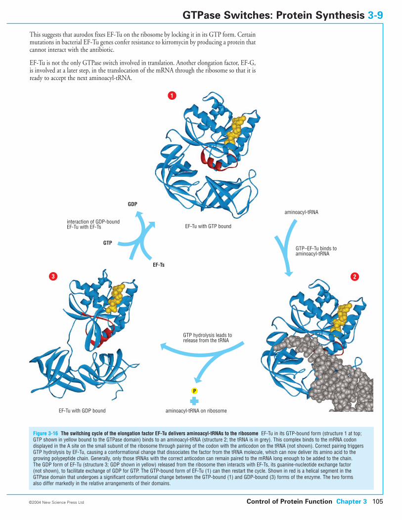
This suggests that aurodox fixes EF-Tu on the ribosome by locking it in its GTP form. Certainmutations in bacterial EF-Tu genes confer resistance to kirromycin by producing a protein thatcannot interact with the antibiotic.
EF-Tu is not the only GTPase switch involved in translation. Another elongation factor, EF-G,is involved at a later step, in the translocation of the mRNA through the ribosome so that it isready to accept the next aminoacyl-tRNA.
Control of Protein Function Chapter 3 105©2004 New Science Press Ltd
GTPase Switches: Protein Synthesis 3-9
Figure 3-16 The switching cycle of the elongation factor EF-Tu delivers aminoacyl-tRNAs to the ribosome EF-Tu in its GTP-bound form (structure 1 at top;GTP shown in yellow bound to the GTPase domain) binds to an aminoacyl-tRNA (structure 2; the tRNA is in grey). This complex binds to the mRNA codondisplayed in the A site on the small subunit of the ribosome through pairing of the codon with the anticodon on the tRNA (not shown). Correct pairing triggersGTP hydrolysis by EF-Tu, causing a conformational change that dissociates the factor from the tRNA molecule, which can now deliver its amino acid to thegrowing polypeptide chain. Generally, only those tRNAs with the correct anticodon can remain paired to the mRNA long enough to be added to the chain. The GDP form of EF-Tu (structure 3; GDP shown in yellow) released from the ribosome then interacts with EF-Ts, its guanine-nucleotide exchange factor (not shown), to facilitate exchange of GDP for GTP. The GTP-bound form of EF-Tu (1) can then restart the cycle. Shown in red is a helical segment in the GTPase domain that undergoes a significant conformational change between the GTP-bound (1) and GDP-bound (3) forms of the enzyme. The two forms also differ markedly in the relative arrangements of their domains.
aminoacyl-tRNA
EF-Ts
GTP
GDP
EF-Tu with GTP boundinteraction of GDP-boundEF-Tu with EF-Ts
GTP–EF-Tu binds toaminoacyl-tRNA
GTP hydrolysis leads torelease from the tRNA
EF-Tu with GDP bound aminoacyl-tRNA on ribosome
P
1
23
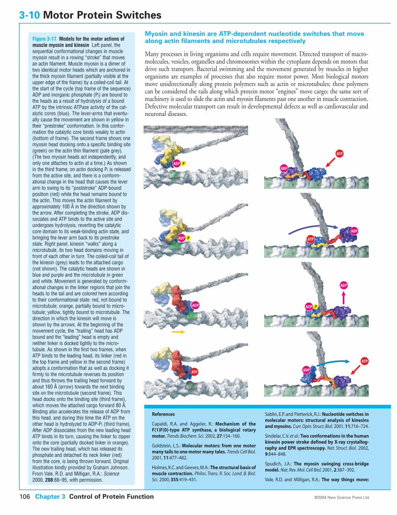
Myosin and kinesin are ATP-dependent nucleotide switches that movealong actin filaments and microtubules respectively
Many processes in living organisms and cells require movement. Directed transport of macro-molecules, vesicles, organelles and chromosomes within the cytoplasm depends on motors thatdrive such transport. Bacterial swimming and the movement generated by muscles in higherorganisms are examples of processes that also require motor power. Most biological motorsmove unidirectionally along protein polymers such as actin or microtubules; these polymerscan be considered the rails along which protein motor “engines” move cargo; the same sort ofmachinery is used to slide the actin and myosin filaments past one another in muscle contraction.Defective molecular transport can result in developmental defects as well as cardiovascular andneuronal diseases.
3-10 Motor Protein Switches
Chapter 3 Control of Protein Function106 ©2004 New Science Press Ltd
References
Capaldi, R.A. and Aggeler, R.: Mechanism of theF(1)F(0)-type ATP synthase, a biological rotarymotor. Trends Biochem. Sci. 2002, 27:154–160.
Goldstein, L.S.: Molecular motors: from one motormany tails to one motor many tales. Trends Cell Biol.2001, 11:477–482.
Holmes, K.C. and Geeves, M.A.: The structural basis ofmuscle contraction. Philos. Trans. R. Soc. Lond. B. Biol.Sci. 2000, 355:419–431.
Sablin, E.P. and Fletterick, R.J.: Nucleotide switches inmolecular motors: structural analysis of kinesinsand myosins. Curr. Opin. Struct. Biol. 2001, 11:716–724.
Sindelar, C.V. et al.: Two conformations in the humankinesin power stroke defined by X-ray crystallog-raphy and EPR spectroscopy. Nat. Struct. Biol. 2002,9:844–848.
Spudich, J.A.: The myosin swinging cross-bridgemodel. Nat. Rev. Mol. Cell Biol. 2001, 2:387–392.
Vale, R.D. and Milligan, R.A.: The way things move:
Figure 3-17 Models for the motor actions ofmuscle myosin and kinesin Left panel, thesequential conformational changes in musclemyosin result in a rowing “stroke” that movesan actin filament. Muscle myosin is a dimer oftwo identical motor heads which are anchored inthe thick myosin filament (partially visible at theupper edge of the frame) by a coiled-coil tail. Atthe start of the cycle (top frame of the sequence)ADP and inorganic phosphate (Pi) are bound tothe heads as a result of hydrolysis of a boundATP by the intrinsic ATPase activity of the cat-alytic cores (blue). The lever-arms that eventu-ally cause the movement are shown in yellow intheir “prestroke” conformation. In this confor-mation the catalytic core binds weakly to actin(bottom of frame). The second frame shows onemyosin head docking onto a specific binding site(green) on the actin thin filament (pale grey).(The two myosin heads act independently, andonly one attaches to actin at a time.) As shownin the third frame, on actin docking Pi is releasedfrom the active site, and there is a conform-ational change in the head that causes the leverarm to swing to its “poststroke” ADP-boundposition (red) while the head remains bound tothe actin. This moves the actin filament byapproximately 100 Å in the direction shown bythe arrow. After completing the stroke, ADP dis-sociates and ATP binds to the active site andundergoes hydrolysis, reverting the catalyticcore domain to its weak-binding actin state, andbringing the lever arm back to its prestrokestate. Right panel, kinesin “walks” along amicrotubule, its two head domains moving infront of each other in turn. The coiled-coil tail ofthe kinesin (grey) leads to the attached cargo(not shown). The catalytic heads are shown inblue and purple and the microtubule in greenand white. Movement is generated by conform-ational changes in the linker regions that join theheads to the tail and are colored here accordingto their conformational state: red, not bound tomicrotubule; orange, partially bound to micro-tubule; yellow, tightly bound to microtubule. Thedirection in which the kinesin will move isshown by the arrows. At the beginning of themovement cycle, the “trailing” head has ADPbound and the “leading” head is empty and neither linker is docked tightly to the micro-tubule. As shown in the first two frames, whenATP binds to the leading head, its linker (red inthe top frame and yellow in the second frame)adopts a conformation that as well as docking itfirmly to the microtubule reverses its positionand thus throws the trailing head forward byabout 160 Å (arrow) towards the next bindingsite on the microtubule (second frame). Thishead docks onto the binding site (third frame),which moves the attached cargo forward 80 Å.Binding also accelerates the release of ADP fromthis head, and during this time the ATP on theother head is hydrolyzed to ADP-Pi (third frame).After ADP dissociates from the new leading headATP binds in its turn, causing the linker to zipperonto the core (partially docked linker in orange).The new trailing head, which has released itsphosphate and detached its neck linker (red)from the core, is being thrown forward. Originalillustration kindly provided by Graham Johnson.From Vale, R.D. and Milligan, R.A.: Science2000, 288:88–95, with permission.
ADP
ADP
ADP
ADP
P
ADP P
ADP P
ADP
ATP
ATP
ATP
ATP
ADP
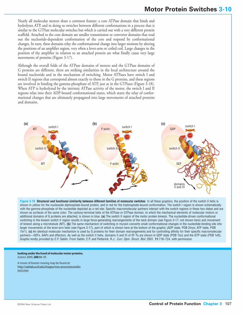
Nearly all molecular motors share a common feature: a core ATPase domain that binds andhydrolyzes ATP, and in doing so switches between different conformations in a process that issimilar to the GTPase molecular switches but which is carried out with a very different proteinscaffold. Attached to the core domain are smaller transmission or converter domains that readout the nucleotide-dependent conformation of the core and respond by conformationalchanges. In turn, these domains relay the conformational change into larger motions by alteringthe positions of an amplifier region, very often a lever-arm or coiled coil. Large changes in theposition of the amplifier in relation to an attached protein are what finally cause very largemovements of proteins (Figure 3-17).
Although the overall folds of the ATPase domains of motors and the GTPase domains of G proteins are different, there are striking similarities in the local architecture around thebound nucleotide and in the mechanism of switching. Motor ATPases have switch I andswitch II regions that correspond almost exactly to those in the G proteins, and these regionsare involved in binding the gamma-phosphate of ATP, just as in the GTPases (Figure 3-18).When ATP is hydrolyzed by the intrinsic ATPase activity of the motor, the switch I and IIregions relax into their ADP-bound conformational states, which starts the relay of confor-mational changes that are ultimately propagated into large movements of attached proteinsand domains.
Control of Protein Function Chapter 3 107
Figure 3-18 Structural and functional similarity between different families of molecular switches In all these graphics, the position of the switch II helix isshown in yellow for the nucleoside diphosphate-bound protein, and in red for the triphosphate-bound conformation. The switch I region is shown schematicallywith the gamma-phosphate of the nucleotide depicted as a red star. Specific macromolecular partners interact with the switch regions in these two states and areshown as surfaces of the same color. The carboxy-terminal helix of the ATPase or GTPase domain, to which the mechanical elements of molecular motors oradditional domains of G proteins are attached, is shown in blue. (a) The switch II region of the motor protein kinesin. The nucleotide-driven conformationalswitching in the kinesin switch II region results in large force-generating rearrangements of the neck domain (see Figure 3-17; not shown here) and movementof kinesin along a microtubule (MT). (b) The same mechanism of switching in myosin converts small conformational changes in the nucleotide-binding site intolarger movements of the lever-arm helix (see Figure 3-17), part of which is shown here at the bottom of the graphic (ADP state, PDB 2mys; ATP state, PDB1br1). (c) An identical molecular mechanism is used by G proteins for their domain rearrangements and for controlling affinity for their specific macromolecularpartners—GEFs, GAPs and effectors. As well as the switch II helix, domains II and III of EF-Tu are shown in GDP state (PDB 1tui) and the GTP state (PDB 1eft).Graphic kindly provided by E.P. Sablin. From Sablin, E.P. and Fletterick, R.J.: Curr. Opin. Struct. Biol. 2001, 11:716–724, with permission.
©2004 New Science Press Ltd
Motor Protein Switches 3-10
looking under the hood of molecular motor proteins.Science 2000, 288:88–95.
A movie of kinesin moving may be found at:http://valelab.ucsf.edu/images/mov-procmotconvkinrev5.mov
switch I switch I switch IMT F-actin GAP
GEFF-actinMT
switch IIswitch II switch II
domainsII and III
(a) (b) (c)
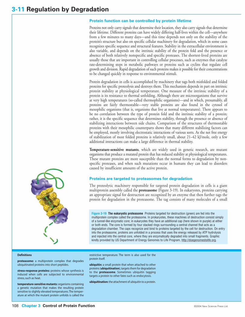
Protein function can be controlled by protein lifetime
Proteins not only carry signals that determine their location, they also carry signals that determinetheir lifetime. Different proteins can have widely differing half-lives within the cell—anywherefrom a few minutes to many days—and this time depends not only on the stability of theprotein’s structure but also on specific cellular machinery for degradation, which in some casesrecognizes specific sequence and structural features. Stability in the extracellular environment isalso variable, and depends on the intrinsic stability of the protein fold and the presence orabsence of both relatively nonspecific and specific proteases. The shortest-lived proteins areusually those that are important in controlling cellular processes, such as enzymes that catalyzerate-determining steps in metabolic pathways or proteins such as cyclins that regulate cellgrowth and division. Rapid degradation of such proteins makes it possible for their concentrationsto be changed quickly in response to environmental stimuli.
Protein degradation in cells is accomplished by machinery that tags both misfolded and foldedproteins for specific proteolysis and destroys them. This mechanism depends in part on intrinsicprotein stability at physiological temperature. One measure of the intrinsic stability of aprotein is its resistance to thermal unfolding. Although there are microorganisms that surviveat very high temperatures (so-called thermophilic organisms)—and in which, presumably, allproteins are fairly thermostable—very stable proteins are also found in the cytosol ofmesophilic organisms (that is, organisms that live at normal temperatures). There appears tobe no correlation between the type of protein fold and the intrinsic stability of a protein;rather, it is the specific sequence that determines stability, through the presence or absence ofstabilizing interactions between side chains. Comparison of the structures of thermostableproteins with their mesophilic counterparts shows that many different stabilizing factors canbe employed, mostly involving electrostatic interactions of various sorts. As the net free energyof stabilization of most folded proteins is relatively small, about 21–42 kJ/mole, only a fewadditional interactions can make a large difference in thermal stability.
Temperature-sensitive mutants, which are widely used in genetic research, are mutantorganisms that produce a mutated protein that has reduced stability at physiological temperatures.These mutant proteins are more susceptible than the normal forms to degradation by non-specific proteases, and when such mutations occur in humans they can lead to disorderscaused by insufficient amounts of the active protein.
Proteins are targeted to proteasomes for degradation
The proteolytic machinery responsible for targeted protein degradation in cells is a giantmultiprotein assembly called the proteasome (Figure 3-19). In eukaryotes, proteins carryingan appropriate signal for destruction are recognized by an enzyme that then further tags theprotein for degradation in the proteasome. The tag consists of many molecules of a small
3-11 Regulation by Degradation
Figure 3-19 The eukaryotic proteasome Proteins targeted for destruction (green) are fed into themultiprotein complex called the proteasome. In prokaryotes, these machines of destruction consist simplyof a tunnel-like enzymatic core; in eukaryotes they have an additional cap (here shown in purple) at eitheror both ends. The core is formed by four stacked rings surrounding a central channel that acts as adegradation chamber. The caps recognize and bind to proteins targeted by the cell for destruction. On entryinto the proteasome, proteins are unfolded in a process that uses the energy released by ATP hydrolysisand injected into the central core, where they are enzymatically degraded into small fragments. Graphickindly provided by US Department of Energy Genomes to Life Program, http://doegenomestolife.org.
Chapter 3 Control of Protein Function108 ©2004 New Science Press Ltd
Definitions
proteasome: a multiprotein complex that degradesubiquitinated proteins into short peptides.
stress-response proteins: proteins whose synthesis isinduced when cells are subjected to environmentalstress, such as heat.
temperature-sensitive mutants: organisms containinga genetic mutation that makes the resulting proteinsensitive to slightly elevated temperatures.The temper-ature at which the mutant protein unfolds is called the
restrictive temperature. The term is also used for theprotein itself.
ubiquitin: a small protein that when attached to otherproteins (ubiquitination), targets them for degradationto the proteasome. Sometimes ubiquitin tagging targets a protein to other fates such as endocytosis.
ubiquitination: the attachment of ubiquitin to a protein.
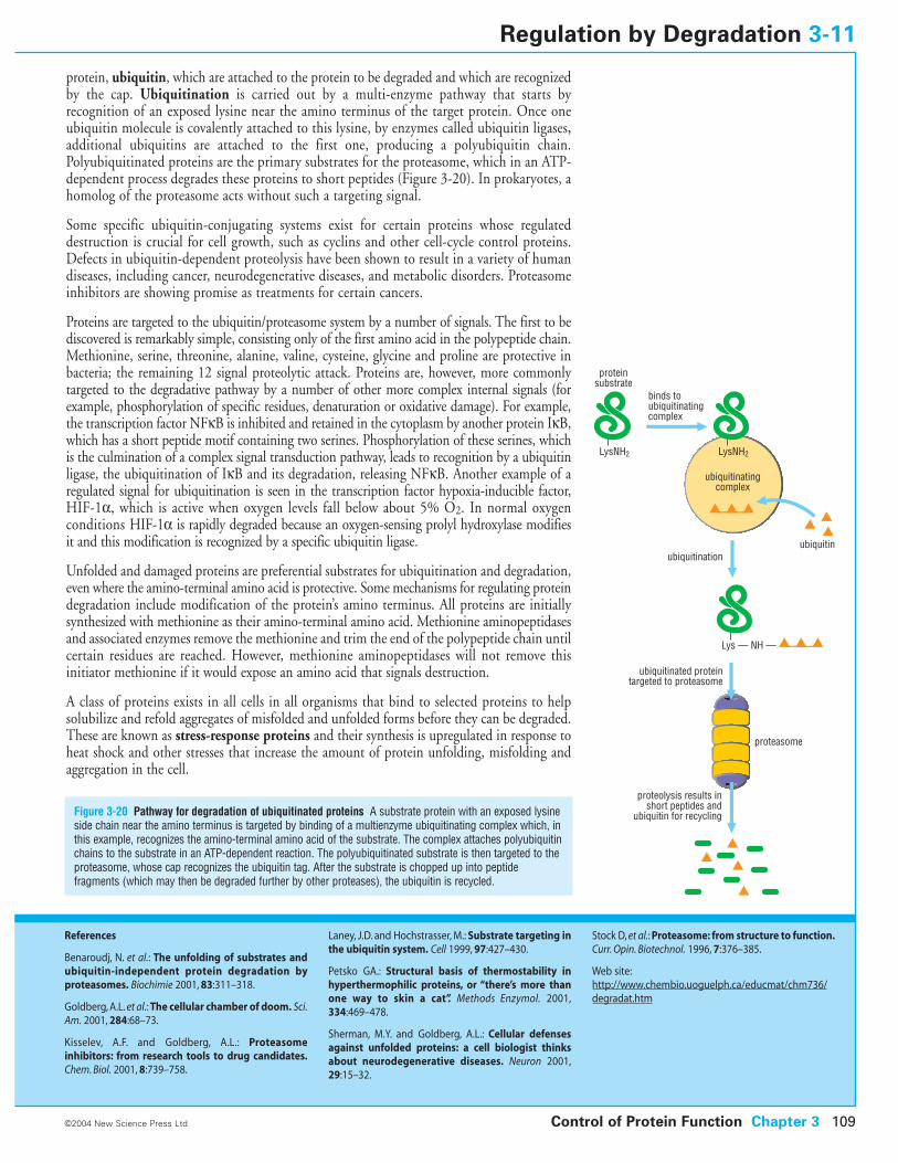
protein, ubiquitin, which are attached to the protein to be degraded and which are recognizedby the cap. Ubiquitination is carried out by a multi-enzyme pathway that starts by recognition of an exposed lysine near the amino terminus of the target protein. Once oneubiquitin molecule is covalently attached to this lysine, by enzymes called ubiquitin ligases,additional ubiquitins are attached to the first one, producing a polyubiquitin chain.Polyubiquitinated proteins are the primary substrates for the proteasome, which in an ATP-dependent process degrades these proteins to short peptides (Figure 3-20). In prokaryotes, ahomolog of the proteasome acts without such a targeting signal.
Some specific ubiquitin-conjugating systems exist for certain proteins whose regulateddestruction is crucial for cell growth, such as cyclins and other cell-cycle control proteins.Defects in ubiquitin-dependent proteolysis have been shown to result in a variety of humandiseases, including cancer, neurodegenerative diseases, and metabolic disorders. Proteasomeinhibitors are showing promise as treatments for certain cancers.
Proteins are targeted to the ubiquitin/proteasome system by a number of signals. The first to bediscovered is remarkably simple, consisting only of the first amino acid in the polypeptide chain.Methionine, serine, threonine, alanine, valine, cysteine, glycine and proline are protective inbacteria; the remaining 12 signal proteolytic attack. Proteins are, however, more commonlytargeted to the degradative pathway by a number of other more complex internal signals (forexample, phosphorylation of specific residues, denaturation or oxidative damage). For example,the transcription factor NFkB is inhibited and retained in the cytoplasm by another protein IkB,which has a short peptide motif containing two serines. Phosphorylation of these serines, whichis the culmination of a complex signal transduction pathway, leads to recognition by a ubiquitinligase, the ubiquitination of IkB and its degradation, releasing NFkB. Another example of aregulated signal for ubiquitination is seen in the transcription factor hypoxia-inducible factor,HIF-1a, which is active when oxygen levels fall below about 5% O2. In normal oxygenconditions HIF-1a is rapidly degraded because an oxygen-sensing prolyl hydroxylase modifiesit and this modification is recognized by a specific ubiquitin ligase.
Unfolded and damaged proteins are preferential substrates for ubiquitination and degradation,even where the amino-terminal amino acid is protective. Some mechanisms for regulating proteindegradation include modification of the protein’s amino terminus. All proteins are initiallysynthesized with methionine as their amino-terminal amino acid. Methionine aminopeptidasesand associated enzymes remove the methionine and trim the end of the polypeptide chain untilcertain residues are reached. However, methionine aminopeptidases will not remove thisinitiator methionine if it would expose an amino acid that signals destruction.
A class of proteins exists in all cells in all organisms that bind to selected proteins to helpsolubilize and refold aggregates of misfolded and unfolded forms before they can be degraded.These are known as stress-response proteins and their synthesis is upregulated in response toheat shock and other stresses that increase the amount of protein unfolding, misfolding andaggregation in the cell.
Control of Protein Function Chapter 3 109
Figure 3-20 Pathway for degradation of ubiquitinated proteins A substrate protein with an exposed lysineside chain near the amino terminus is targeted by binding of a multienzyme ubiquitinating complex which, inthis example, recognizes the amino-terminal amino acid of the substrate. The complex attaches polyubiquitinchains to the substrate in an ATP-dependent reaction. The polyubiquitinated substrate is then targeted to theproteasome, whose cap recognizes the ubiquitin tag. After the substrate is chopped up into peptidefragments (which may then be degraded further by other proteases), the ubiquitin is recycled.
©2004 New Science Press Ltd
Regulation by Degradation 3-11
References
Benaroudj, N. et al.: The unfolding of substrates andubiquitin-independent protein degradation by proteasomes. Biochimie 2001, 83:311–318.
Goldberg, A.L. et al.: The cellular chamber of doom. Sci.Am. 2001, 284:68–73.
Kisselev, A.F. and Goldberg, A.L.: Proteasomeinhibitors: from research tools to drug candidates.Chem. Biol. 2001, 8:739–758.
Laney, J.D. and Hochstrasser, M.: Substrate targeting inthe ubiquitin system. Cell 1999, 97:427–430.
Petsko GA.: Structural basis of thermostability inhyperthermophilic proteins, or “there’s more thanone way to skin a cat”. Methods Enzymol. 2001,334:469–478.
Sherman, M.Y. and Goldberg, A.L.: Cellular defensesagainst unfolded proteins: a cell biologist thinksabout neurodegenerative diseases. Neuron 2001,29:15–32.
Stock D, et al.: Proteasome: from structure to function.Curr. Opin. Biotechnol. 1996, 7:376–385.
Web site:http://www.chembio.uoguelph.ca/educmat/chm736/degradat.htm
binds toubiquitinatingcomplex
ubiquitinatingcomplex
ubiquitinationubiquitin
ubiquitinated proteintargeted to proteasome
proteolysis results in short peptides and
ubiquitin for recycling
proteinsubstrate
proteasome
LysNH2LysNH2
Lys — NH —
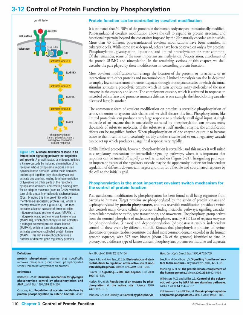
Protein function can be controlled by covalent modification
It is estimated that 50–90% of the proteins in the human body are post-translationally modified.Post-translational covalent modification allows the cell to expand its protein structural andfunctional repertoire beyond the constraints imposed by the 20 naturally encoded amino acids.More than 40 different post-translational covalent modifications have been identified ineukaryotic cells. While some are widespread, others have been observed on only a few proteins.Phosphorylation, glycosylation, lipidation, and limited proteolysis are the most common. Of the remainder, some of the most important are methylation, N-acetylation, attachment ofthe protein SUMO and nitrosylation. In the remaining sections of this chapter, we shalldescribe the part played by these modifications in controlling protein function.
Most covalent modifications can change the location of the protein, or its activity, or itsinteractions with other proteins and macromolecules. Limited proteolysis can also be deployedto amplify low-concentration or transient signals, through proteolytic cascades in which the initialstimulus activates a proteolytic enzyme which in turn activates many molecules of the nextenzyme in the cascade, and so on. The complement cascade, which is activated in response tomicrobial cell surfaces and promotes immune defenses, is one example; the blood clotting cascade,discussed later, is another.
The commonest form of covalent modification on proteins is reversible phosphorylation ofserine, threonine or tyrosine side chains and we shall discuss this first. Phosphorylation, likelimited proteolysis, can produce a very large response to a relatively small signal input. A singlemolecule of an enzyme that is catalytically activated by phosphorylation can process manythousands of substrate molecules. If the substrate is itself another enzyme, the amplificationeffects can be magnified further. When phosphorylation of one enzyme causes it to becomeactive so that it can, in turn, covalently modify another enzyme and so on, a regulatory cascadecan be set up which produces a large final response very rapidly.
Unlike limited proteolysis, however, phosphorylation is reversible, and this makes it well suitedas a regulatory mechanism for intracellular signaling pathways, where it is important thatresponses can be turned off rapidly as well as turned on (Figure 3-21). In signaling pathways,an important feature of the regulatory cascade may be the opportunity it offers for independentregulation of different downstream targets and thus for a flexible and coordinated response bythe cell to the initial signal.
Phosphorylation is the most important covalent switch mechanism forthe control of protein function
Post-translational modification by phosphorylation has been found in all living organisms frombacteria to humans. Target proteins are phosphorylated by the action of protein kinases anddephosphorylated by protein phosphatases, and this reversible modification provides a switchthat controls many diverse cellular processes including metabolic pathways, signaling cascades,intracellular membrane traffic, gene transcription, and movement. The phosphoryl group derivesfrom the terminal phosphate of nucleoside triphosphates, usually ATP. Use of separate enzymesfor phosphorylation (kinases) and dephosphorylation (phosphatases) enables independentcontrol of these events by different stimuli. Kinases that phosphorylate proteins on serine,threonine or tyrosine residues constitute the third most common domain encoded in the humangenome sequence, with 575 such kinases (about 2% of the genome) identified to date. Inprokaryotes, a different type of kinase domain phosphorylates proteins on histidine and aspartate
3-12 Control of Protein Function by Phosphorylation
Chapter 3 Control of Protein Function110 ©2004 New Science Press Ltd
Definitions
protein phosphatase: enzyme that specificallyremoves phosphate groups from phosphorylated serines, threonines or tyrosines on proteins.
References
Barford, D. et al.: Structural mechanism for glycogenphosphorylase control by phosphorylation andAMP. J. Mol. Biol. 1991, 218:233–260.
Cozzone, A.J.: Regulation of acetate metabolism byprotein phosphorylation in enteric bacteria. Annu.
Rev. Microbiol. 1998, 52:127–164.
Dean, A.M. and Koshland, D.E. Jr.: Electrostatic and stericcontributions to regulation at the active site of isoci-trate dehydrogenase. Science 1990, 249:1044–1046.
Hunter, T.: Signaling—2000 and beyond. Cell 2000,100:113–127.
Hurley, J.H. et al.: Regulation of an enzyme by phos-phorylation at the active site. Science 1990,249:1012–1016.
Johnson, L.N. and O’Reilly, M.: Control by phosphoryla-
tion. Curr. Opin. Struct. Biol. 1996, 6:762–769.
Lee, M. and Goodbourn, S.: Signalling from the cell sur-face to the nucleus. Essays Biochem. 2001, 37:71–85.
Manning, G. et al.: The protein kinase complement ofthe human genome. Science 2002, 298:1912–1934.
Wilkinson, M.G. and Millar, J.B.: Control of the eukary-otic cell cycle by MAP kinase signaling pathways.FASEB J. 2000, 14:2147–2157.
Zolnierowicz, S. and Bollen, M.: Protein phosphorylationand protein phosphatases. EMBO J. 2000, 19:483–488.
P
PP
P P
PP
P
PP
receptor
Ras
Grb2 Sos
MAPKKK
MAPKK
MAPK
cytoplasm
cell surface
activates kinase 1
activates kinase 2
activates kinase 3
phosphorylation of transcriptional activatorsleading to coordinated
cellular response
growth factor
Figure 3-21 A kinase activation cascade in anintracellular signaling pathway that regulatescell growth A growth factor, or mitogen, initiatesa kinase cascade by inducing dimerization of itsreceptor, whose cytoplasmic regions containtyrosine kinase domains. When these domainsare brought together they phosphorylate andactivate one another, leading to phosphorylationof tyrosines on other parts of the receptor’scytoplasmic domains, and creating binding sitesfor an adaptor molecule (such as Grb2), which inturn binds a guanine-nucleotide exchange factor(Sos), bringing this into proximity with themembrane-associated G protein Ras, which isthereby activated (see Figure 3-14). Ras thenactivates a kinase cascade of three so-calledmitogen-activated protein kinases (MAPKs): amitogen-activated protein kinase kinase kinase(MAPKKK), which phosphorylates and activates a mitogen-activated protein kinase kinase(MAPKK), which in turn phosphorylates andactivates a mitogen-activated protein kinase(MAPK). This last kinase phosphorylates anumber of different gene regulatory proteins.
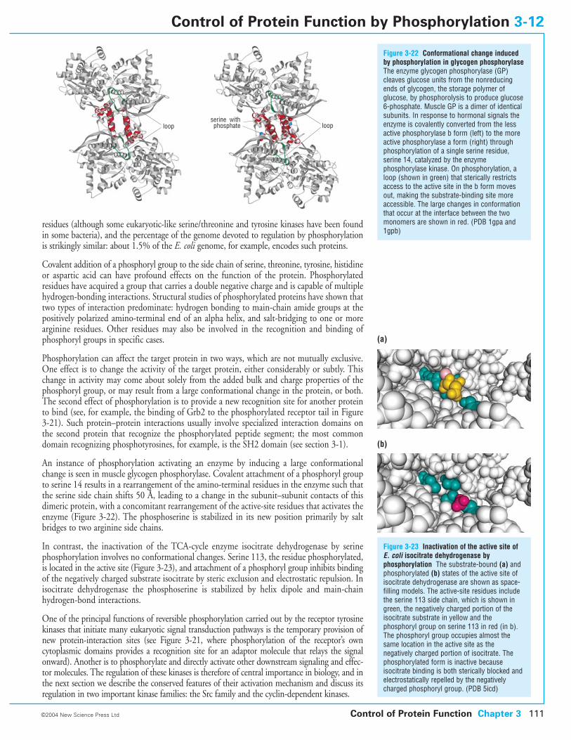
residues (although some eukaryotic-like serine/threonine and tyrosine kinases have been foundin some bacteria), and the percentage of the genome devoted to regulation by phosphorylationis strikingly similar: about 1.5% of the E. coli genome, for example, encodes such proteins.
Covalent addition of a phosphoryl group to the side chain of serine, threonine, tyrosine, histidineor aspartic acid can have profound effects on the function of the protein. Phosphorylatedresidues have acquired a group that carries a double negative charge and is capable of multiplehydrogen-bonding interactions. Structural studies of phosphorylated proteins have shown thattwo types of interaction predominate: hydrogen bonding to main-chain amide groups at thepositively polarized amino-terminal end of an alpha helix, and salt-bridging to one or morearginine residues. Other residues may also be involved in the recognition and binding ofphosphoryl groups in specific cases.
Phosphorylation can affect the target protein in two ways, which are not mutually exclusive.One effect is to change the activity of the target protein, either considerably or subtly. Thischange in activity may come about solely from the added bulk and charge properties of thephosphoryl group, or may result from a large conformational change in the protein, or both.The second effect of phosphorylation is to provide a new recognition site for another proteinto bind (see, for example, the binding of Grb2 to the phosphorylated receptor tail in Figure3-21). Such protein–protein interactions usually involve specialized interaction domains onthe second protein that recognize the phosphorylated peptide segment; the most commondomain recognizing phosphotyrosines, for example, is the SH2 domain (see section 3-1).
An instance of phosphorylation activating an enzyme by inducing a large conformationalchange is seen in muscle glycogen phosphorylase. Covalent attachment of a phosphoryl groupto serine 14 results in a rearrangement of the amino-terminal residues in the enzyme such thatthe serine side chain shifts 50 Å, leading to a change in the subunit–subunit contacts of thisdimeric protein, with a concomitant rearrangement of the active-site residues that activates theenzyme (Figure 3-22). The phosphoserine is stabilized in its new position primarily by saltbridges to two arginine side chains.
In contrast, the inactivation of the TCA-cycle enzyme isocitrate dehydrogenase by serinephosphorylation involves no conformational changes. Serine 113, the residue phosphorylated,is located in the active site (Figure 3-23), and attachment of a phosphoryl group inhibits bindingof the negatively charged substrate isocitrate by steric exclusion and electrostatic repulsion. Inisocitrate dehydrogenase the phosphoserine is stabilized by helix dipole and main-chainhydrogen-bond interactions.
One of the principal functions of reversible phosphorylation carried out by the receptor tyrosinekinases that initiate many eukaryotic signal transduction pathways is the temporary provision ofnew protein-interaction sites (see Figure 3-21, where phosphorylation of the receptor’s own cytoplasmic domains provides a recognition site for an adaptor molecule that relays the signalonward). Another is to phosphorylate and directly activate other downstream signaling and effec-tor molecules. The regulation of these kinases is therefore of central importance in biology, and inthe next section we describe the conserved features of their activation mechanism and discuss itsregulation in two important kinase families: the Src family and the cyclin-dependent kinases.
Figure 3-22 Conformational change inducedby phosphorylation in glycogen phosphorylaseThe enzyme glycogen phosphorylase (GP)cleaves glucose units from the nonreducingends of glycogen, the storage polymer ofglucose, by phosphorolysis to produce glucose6-phosphate. Muscle GP is a dimer of identicalsubunits. In response to hormonal signals theenzyme is covalently converted from the lessactive phosphorylase b form (left) to the moreactive phosphorylase a form (right) throughphosphorylation of a single serine residue,serine 14, catalyzed by the enzymephosphorylase kinase. On phosphorylation, aloop (shown in green) that sterically restrictsaccess to the active site in the b form movesout, making the substrate-binding site moreaccessible. The large changes in conformationthat occur at the interface between the twomonomers are shown in red. (PDB 1gpa and1gpb)
Figure 3-23 Inactivation of the active site ofE. coli isocitrate dehydrogenase byphosphorylation The substrate-bound (a) andphosphorylated (b) states of the active site ofisocitrate dehydrogenase are shown as space-filling models. The active-site residues includethe serine 113 side chain, which is shown ingreen, the negatively charged portion of theisocitrate substrate in yellow and thephosphoryl group on serine 113 in red (in b).The phosphoryl group occupies almost thesame location in the active site as thenegatively charged portion of isocitrate. Thephosphorylated form is inactive becauseisocitrate binding is both sterically blocked andelectrostatically repelled by the negativelycharged phosphoryl group. (PDB 5icd)
Control of Protein Function Chapter 3 111©2004 New Science Press Ltd
Control of Protein Function by Phosphorylation 3-12
(a)
(b)
loop loopserine withphosphate
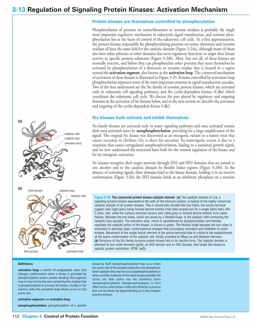
Protein kinases are themselves controlled by phosphorylation
Phosphorylation of proteins on serine/threonine or tyrosine residues is probably the singlemost important regulatory mechanism in eukaryotic signal transduction, and tyrosine phos-phorylation lies at the heart of control of the eukaryotic cell cycle. To a first approximation,the protein kinases responsible for phosphorylating proteins on serine, threonine and tyrosineresidues all have the same fold for the catalytic domain (Figure 3-24a), although many of themalso have other subunits or other domains that serve regulatory functions or target their kinaseactivity to specific protein substrates (Figure 3-24b). Most, but not all, of these kinases arenormally inactive, and before they can phosphorylate other proteins they must themselves beactivated by phosphorylation of a threonine or tyrosine residue that is located in a regiontermed the activation segment, also known as the activation loop. The conserved mechanismof activation of these kinases is illustrated in Figure 3-25. Kinases controlled by activation-loopphosphorylation represent some of the most important enzymes in signal transduction cascades.Two of the best understood are the Src family of tyrosine protein kinases, which are activatedearly in eukaryotic cell signaling pathways, and the cyclin-dependent kinases (Cdks) whichcoordinate the eukaryotic cell cycle. We discuss the part played by regulatory and targetingdomains in the activation of Src kinases below, and in the next section we describe the activationand targeting of the cyclin-dependent kinase Cdk2.
Src kinases both activate and inhibit themselves
Src-family kinases are activated early in many signaling pathways and once activated sustaintheir own activated states by autophosphorylation, providing for a large amplification of thesignal. The original Src kinase was discovered as an oncogenic variant in a tumor virus thatcauses sarcomas in chickens (Src is short for sarcoma). Its tumorigenic action is due to a mutation that causes unregulated autophosphorylation, leading to a sustained growth signal,and we now understand the structural basis both for the normal regulation of the kinase andfor its oncogenic activation.
Src kinases recognize their target proteins through SH2 and SH3 domains that are joined toone another and to the catalytic domain by flexible linker regions (Figure 3-24b). In theabsence of activating signals, these domains bind to the kinase domain, holding it in an inactiveconformation (Figure 3-26): the SH2 domain binds to an inhibitory phosphate on a tyrosine
3-13 Regulation of Signaling Protein Kinases: Activation Mechanism
Figure 3-24 The conserved protein kinase catalytic domain (a) The catalytic domain of Lck, asignaling tyrosine kinase expressed in the cells of the immune system, is typical of the highly conservedcatalytic domain of all protein kinases. This is structurally divided into two lobes, the amino-terminal(upper) lobe (light grey) being formed almost entirely from beta strands but for a single alpha helix (theC helix, red), while the carboxy-terminal (lower) lobe (dark grey) is formed almost entirely from alphahelices. Between the two lobes, which are joined by a flexible hinge, is the catalytic cleft containing thecatalytic loop (purple). The activation loop, which is repositioned by phosphorylation and therebyregulates the catalytic action of the kinase, is shown in green. The flexible hinge between the two lobes isimportant in allowing major conformational changes that accompany activation and inhibition in somekinases. Movement of the single helical element in the amino-terminal lobe is critical to the establishmentof the active conformation of the catalytic site. Kindly provided by Ming Lei and Stephen Harrison. (b) Structure of the Src-family tyrosine protein kinase Hck in its inactive form. The catalytic domain isattached to two small domains (gold), an SH3 domain and an SH2 domain, that target this kinase tospecific protein substrates. (PDB 1ad5)
Chapter 3 Control of Protein Function112 ©2004 New Science Press Ltd
Definitions
activation loop: a stretch of polypeptide chain thatchanges conformation when a kinase is activated byphosphorylation and/or protein binding. This segmentmay or may not be the one containing the residue thatis phosphorylated to activate the kinase. Usually, in theinactive state, the activation loop blocks access to theactive site.
activation segment: see activation loop.
autophosphorylation: phosphorylation of a protein
kinase by itself. Autophosphorylation may occur whenthe active site of the protein molecule to be phosphory-lated catalyzes this reaction (cis autophosphorylation) orwhen another molecule of the same kinase provides theactive site that carries out the chemistry (transautophosphorylation). Autophosphorylation in transoften occurs when kinase molecules dimerize, a processthat can be driven by ligand binding as in the receptortyrosine kinases.
catalytic loop
catalytic cleft
activation loop
(a)
(b)
activation loop
catalytic loop
catalytic cleft
SH3 domain
SH2 domain
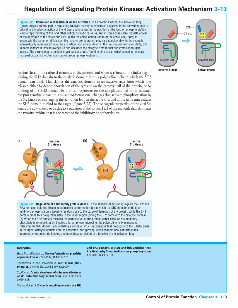
residue close to the carboxyl terminus of the protein, and when it is bound, the linker regionjoining the SH2 domain to the catalytic domain forms a polyproline helix to which the SH3domain can bind. This clamps the catalytic domain in an inactive state from which it isreleased either by dephosphorylation of the tyrosine on the carboxyl tail of the protein, or bybinding of the SH2 domain by a phosphotyrosine on the cytoplasmic tail of an activatedreceptor tyrosine kinase: this causes conformational changes that activate phosphorylation bythe Src kinase by rearranging the activation loop in the active site, and at the same time releasesthe SH3 domain to bind to the target (Figure 3-26). The oncogenic properties of the viral Srckinase are now known to be due to a mutation of the carboxyl tail of the molecule that eliminatesthe tyrosine residue that is the target of the inhibitory phosphorylation.
Control of Protein Function Chapter 3 113
Figure 3-25 Conserved mechanism of kinase activation In all protein kinases, the activation loop(green) plays a central part in regulating catalytic activity. A conserved aspartate in the activation loop iscritical to the catalytic action of the kinase, and changes in the position of the loop on phosphorylationlead to repositioning of this and other critical catalytic residues, and in some cases also regulate accessof the substrate to the active-site cleft. While the active configuration of the active site (right) isessentially the same for all kinases, the inactive configuration may vary considerably. In the exampleschematically represented here, the activation loop swings down in the inactive conformation (left), butin some kinases it instead swings up and occludes the catalytic cleft so that substrate cannot gainaccess. The purple loop is the conserved catalytic loop, found in all kinases, which contains residuesthat participate in the chemical step of protein phosphorylation.
©2004 New Science Press Ltd
Regulation of Signaling Protein Kinases: Activation Mechanism 3-13
References
Huse, M. and Kuriyan, J.: The conformational plasticityof protein kinases. Cell 2002, 109:275–282.
Theodosiou, A. and Ashworth, A.: MAP kinase phos-phatases. Genome Biol. 2002, 3:reviews3009.
Xu, W. et al.: Crystal structures of c-Src reveal featuresof its autoinhibitory mechanism. Mol. Cell 1999,3:629–638.
Young, M.A. et al.: Dynamic coupling between the SH2
and SH3 domains of c-Src and Hck underlies theirinactivation by C-terminal tyrosine phosphorylation.Cell 2001, 105:115–126.
inactive kinase active kinase
C helix
ATP
activation loopcatalytic loop
P PP
SH3
SH2
P
inactiveSrc kinase
activeSrc kinase
P PP
P PP
SH3
SH2P
(a) (b)
Figure 3-26 Regulation of a Src-family protein kinase In the absence of activating signals the SH2 andSH3 domains hold the kinase in an inactive conformation (a) in which the SH2 domain binds to aninhibitory phosphate on a tyrosine residue close to the carboxyl terminus of the protein, while the SH3domain binds to a polyproline helix in the linker region joining the SH2 domain to the catalytic domain.(b) When the SH2 domain releases the carboxyl tail of the protein, either because the inhibitoryphosphate is removed, or on binding a target phosphotyrosine, the polyproline helix rearranges,releasing the SH3 domain, and initiating a series of structural changes that propagate to the C helix (red)in the upper catalytic domain and the activation loop (green), which assume new conformationsappropriate for substrate binding and autophosphorylation of a tyrosine in the activation loop.
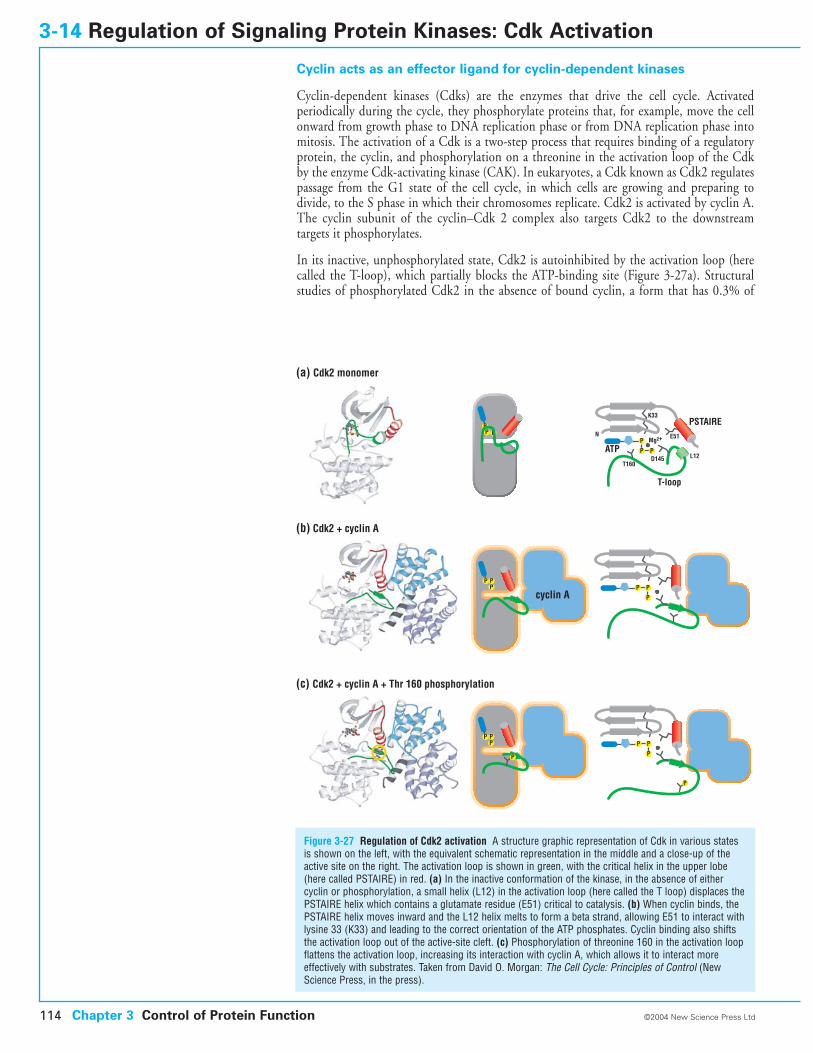
Cyclin acts as an effector ligand for cyclin-dependent kinases
Cyclin-dependent kinases (Cdks) are the enzymes that drive the cell cycle. Activated periodically during the cycle, they phosphorylate proteins that, for example, move the cellonward from growth phase to DNA replication phase or from DNA replication phase intomitosis. The activation of a Cdk is a two-step process that requires binding of a regulatoryprotein, the cyclin, and phosphorylation on a threonine in the activation loop of the Cdkby the enzyme Cdk-activating kinase (CAK). In eukaryotes, a Cdk known as Cdk2 regulatespassage from the G1 state of the cell cycle, in which cells are growing and preparing todivide, to the S phase in which their chromosomes replicate. Cdk2 is activated by cyclin A.The cyclin subunit of the cyclin–Cdk 2 complex also targets Cdk2 to the downstream targets it phosphorylates.
In its inactive, unphosphorylated state, Cdk2 is autoinhibited by the activation loop (herecalled the T-loop), which partially blocks the ATP-binding site (Figure 3-27a). Structuralstudies of phosphorylated Cdk2 in the absence of bound cyclin, a form that has 0.3% of
3-14 Regulation of Signaling Protein Kinases: Cdk Activation
Figure 3-27 Regulation of Cdk2 activation A structure graphic representation of Cdk in various statesis shown on the left, with the equivalent schematic representation in the middle and a close-up of theactive site on the right. The activation loop is shown in green, with the critical helix in the upper lobe(here called PSTAIRE) in red. (a) In the inactive conformation of the kinase, in the absence of eithercyclin or phosphorylation, a small helix (L12) in the activation loop (here called the T loop) displaces thePSTAIRE helix which contains a glutamate residue (E51) critical to catalysis. (b) When cyclin binds, thePSTAIRE helix moves inward and the L12 helix melts to form a beta strand, allowing E51 to interact withlysine 33 (K33) and leading to the correct orientation of the ATP phosphates. Cyclin binding also shiftsthe activation loop out of the active-site cleft. (c) Phosphorylation of threonine 160 in the activation loopflattens the activation loop, increasing its interaction with cyclin A, which allows it to interact moreeffectively with substrates. Taken from David O. Morgan: The Cell Cycle: Principles of Control (NewScience Press, in the press).
Chapter 3 Control of Protein Function114 ©2004 New Science Press Ltd
P PP
P
PPP
P
P
P
P
P
P
P
P
PP
PSTAIRE
L12
E51
K33
D145T160
T-loop
N
ATPMg2+
P PP
cyclin A
(a) Cdk2 monomer
(b) Cdk2 + cyclin A
(c) Cdk2 + cyclin A + Thr 160 phosphorylation
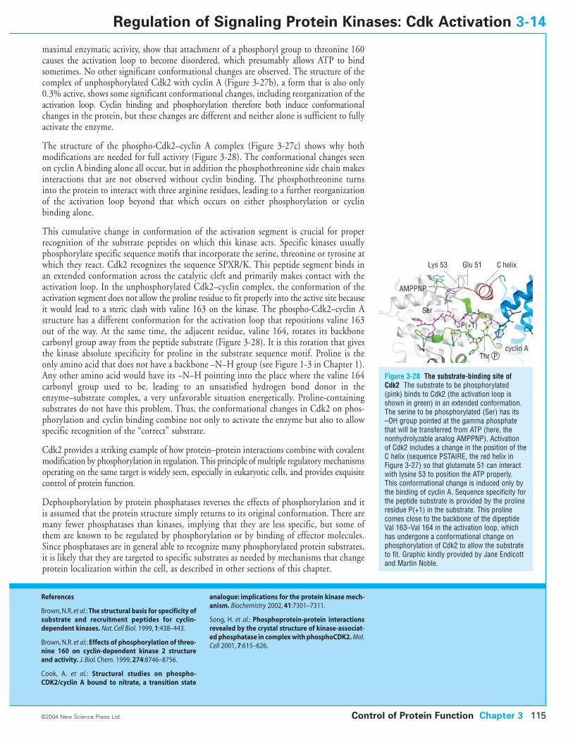
maximal enzymatic activity, show that attachment of a phosphoryl group to threonine 160causes the activation loop to become disordered, which presumably allows ATP to bindsometimes. No other significant conformational changes are observed. The structure of thecomplex of unphosphorylated Cdk2 with cyclin A (Figure 3-27b), a form that is also only0.3% active, shows some significant conformational changes, including reorganization of theactivation loop. Cyclin binding and phosphorylation therefore both induce conformationalchanges in the protein, but these changes are different and neither alone is sufficient to fullyactivate the enzyme.
The structure of the phospho-Cdk2–cyclin A complex (Figure 3-27c) shows why bothmodifications are needed for full activity (Figure 3-28). The conformational changes seenon cyclin A binding alone all occur, but in addition the phosphothreonine side chain makesinteractions that are not observed without cyclin binding. The phosphothreonine turnsinto the protein to interact with three arginine residues, leading to a further reorganizationof the activation loop beyond that which occurs on either phosphorylation or cyclin binding alone.
This cumulative change in conformation of the activation segment is crucial for properrecognition of the substrate peptides on which this kinase acts. Specific kinases usuallyphosphorylate specific sequence motifs that incorporate the serine, threonine or tyrosine atwhich they react. Cdk2 recognizes the sequence SPXR/K. This peptide segment binds inan extended conformation across the catalytic cleft and primarily makes contact with theactivation loop. In the unphosphorylated Cdk2–cyclin complex, the conformation of theactivation segment does not allow the proline residue to fit properly into the active site becauseit would lead to a steric clash with valine 163 on the kinase. The phospho-Cdk2–cyclin Astructure has a different conformation for the activation loop that repositions valine 163out of the way. At the same time, the adjacent residue, valine 164, rotates its backbone carbonyl group away from the peptide substrate (Figure 3-28). It is this rotation that givesthe kinase absolute specificity for proline in the substrate sequence motif. Proline is theonly amino acid that does not have a backbone –N–H group (see Figure 1-3 in Chapter 1).Any other amino acid would have its –N–H pointing into the place where the valine 164carbonyl group used to be, leading to an unsatisfied hydrogen bond donor in theenzyme–substrate complex, a very unfavorable situation energetically. Proline-containingsubstrates do not have this problem. Thus, the conformational changes in Cdk2 on phos-phorylation and cyclin binding combine not only to activate the enzyme but also to allowspecific recognition of the “correct” substrate.
Cdk2 provides a striking example of how protein–protein interactions combine with covalentmodification by phosphorylation in regulation. This principle of multiple regulatory mechanismsoperating on the same target is widely seen, especially in eukaryotic cells, and provides exquisitecontrol of protein function.
Dephosphorylation by protein phosphatases reverses the effects of phosphorylation and itis assumed that the protein structure simply returns to its original conformation. There aremany fewer phosphatases than kinases, implying that they are less specific, but some ofthem are known to be regulated by phosphorylation or by binding of effector molecules.Since phosphatases are in general able to recognize many phosphorylated protein substrates,it is likely that they are targeted to specific substrates as needed by mechanisms that changeprotein localization within the cell, as described in other sections of this chapter.
Control of Protein Function Chapter 3 115
Figure 3-28 The substrate-binding site ofCdk2 The substrate to be phosphorylated(pink) binds to Cdk2 (the activation loop isshown in green) in an extended conformation.The serine to be phosphorylated (Ser) has its–OH group pointed at the gamma phosphatethat will be transferred from ATP (here, thenonhydrolyzable analog AMPPNP). Activationof Cdk2 includes a change in the position of theC helix (sequence PSTAIRE, the red helix inFigure 3-27) so that glutamate 51 can interactwith lysine 53 to position the ATP properly.This conformational change is induced only bythe binding of cyclin A. Sequence specificity forthe peptide substrate is provided by the prolineresidue P(+1) in the substrate. This prolinecomes close to the backbone of the dipeptideVal 163–Val 164 in the activation loop, whichhas undergone a conformational change onphosphorylation of Cdk2 to allow the substrateto fit. Graphic kindly provided by Jane Endicottand Martin Noble.
©2004 New Science Press Ltd
Regulation of Signaling Protein Kinases: Cdk Activation 3-14
References
Brown, N.R. et al.: The structural basis for specificity ofsubstrate and recruitment peptides for cyclin-dependent kinases. Nat. Cell Biol. 1999, 1:438–443.
Brown, N.R. et al.: Effects of phosphorylation of threo-nine 160 on cyclin-dependent kinase 2 structureand activity. J. Biol. Chem. 1999, 274:8746–8756.
Cook, A. et al.: Structural studies on phospho-CDK2/cyclin A bound to nitrate, a transition state
analogue: implications for the protein kinase mech-anism. Biochemistry 2002, 41:7301–7311.
Song, H. et al.: Phosphoprotein-protein interactionsrevealed by the crystal structure of kinase-associat-ed phosphatase in complex with phosphoCDK2. Mol.Cell 2001, 7:615–626.
Lys 53
AMPPNP
Glu 51 C helix
Ser
P(+1)
Thr Pcyclin A
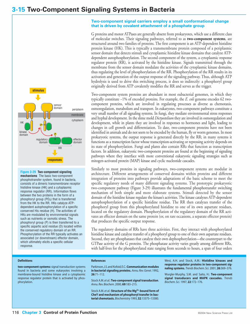
Two-component signal carriers employ a small conformational changethat is driven by covalent attachment of a phosphate group
G proteins and motor ATPases are generally absent from prokaryotes, which use a different classof molecular switches. Their signaling pathways, referred to as two-component systems, arestructured around two families of proteins. The first component is an ATP-dependent histidineprotein kinase (HK). This is typically a transmembrane protein composed of a periplasmicsensor domain that detects stimuli and cytoplasmic histidine kinase domains that catalyze ATP-dependent autophosphorylation. The second component of the system, a cytoplasmic responseregulator protein (RR), is activated by the histidine kinase. Signals transmitted through themembrane from the sensor domain modulate the activities of the cytoplasmic kinase domains,thus regulating the level of phosphorylation of the RR. Phosphorylation of the RR results in itsactivation and generation of the output response of the signaling pathway. Thus, although ATPhydrolysis is used to drive this switching process, it does so indirectly: a phosphoryl grouporiginally derived from ATP covalently modifies the RR and serves as the trigger.
Two-component system proteins are abundant in most eubacterial genomes, in which theytypically constitute ~1% of encoded proteins. For example, the E. coli genome encodes 62 two-component proteins, which are involved in regulating processes as diverse as chemotaxis,osmoregulation, metabolism and transport. In eukaryotes, two-component pathways constitute avery small number of all signaling systems. In fungi, they mediate environmental stress responsesand hyphal development. In the slime mold Dictyostelium they are involved in osmoregulation anddevelopment, while in plants they are involved in responses to hormones and light, leading tochanges in cell growth and differentiation. To date, two-component proteins have not beenidentified in animals and do not seem to be encoded by the human, fly or worm genomes. In mostprokaryotic systems, the output response is generated directly by the RR; in many systems itfunctions as a transcription factor whose transcription-activating or repressing activity depends onits state of phosphorylation. Fungi and plants also contain RRs that function as transcriptionfactors. In addition, eukaryotic two-component proteins are found at the beginning of signalingpathways where they interface with more conventional eukaryotic signaling strategies such asmitogen-activated protein (MAP) kinase and cyclic nucleotide cascades.
Similarly to most proteins in signaling pathways, two-component systems are modular inarchitecture. Different arrangements of conserved domains within proteins and differentintegration of proteins into pathways provide adaptations of the basic scheme to meet thespecific regulatory needs of many different signaling systems. The prototypic prokaryotictwo-component pathway (Figure 3-29) illustrates the fundamental phosphotransfer switchingmechanism of both simple and more elaborate systems. Stimuli detected by the sensordomain of the histidine kinase regulate the kinase’s activities. The kinase catalyzes ATP-dependentautophosphorylation of a specific histidine residue. The RR then catalyzes transfer of thephosphoryl group from this phosphorylated histidine to one of its own aspartate residues,located on the regulatory domain. Phosphorylation of the regulatory domain of the RR acti-vates an effector domain on the same protein (or, on rare occasions, a separate effector protein)that produces the specific output response.
The regulatory domains of RRs have three activities. First, they interact with phosphorylatedhistidine kinase and catalyze transfer of a phosphoryl group to one of their own aspartate residues.Second, they are phosphatases that catalyze their own dephosphorylation—the counterpart to theGTPase activity of the G proteins. The phosphatase activity varies greatly among different RRs,with half-lives for the phosphorylated state ranging from seconds to hours, a span of four orders
3-15 Two-Component Signaling Systems in Bacteria
Figure 3-29 Two-component signalingmechanisms The basic two-componentphosphotransfer system, found in bacteria,consists of a dimeric transmembrane receptorhistidine kinase (HK) and a cytoplasmicresponse regulator (RR). Information flowsbetween the two proteins in the form of aphosphoryl group (PO3) that is transferredfrom the HK to the RR. HKs catalyze ATP-dependent autophosphorylation of a specificconserved His residue (H). The activities ofHKs are modulated by environmental signalssuch as nutrients or osmotic stress. Thephosphoryl group (P) is then transferred to aspecific aspartic acid residue (D) located withinthe conserved regulatory domain of an RR.Phosphorylation of the RR typically activates anassociated (or downstream) effector domain,which ultimately elicits a specific cellularresponse.
Chapter 3 Control of Protein Function116 ©2004 New Science Press Ltd
Definitions
two-component systems: signal transduction systemsfound in bacteria and some eukaryotes involving amembrane-bound histidine kinase and a cytoplasmicresponse regulator protein that is activated by phos-phorylation.
References
Parkinson,J.S.and Kofoid,E.C.:Communication modulesin bacterial signaling proteins. Annu. Rev. Genet. 1992,26:71–112.
Stock A.M. et al.: Two-component signal transduction.Annu. Rev. Biochem. 2000, 69:183–215.
Stock A.M. et al.: Structure of the Mg2+-bound form ofCheY and mechanism of phosphoryl transfer in bac-terial chemotaxis. Biochemistry 1993, 32:13375–13380.
West, A.H. and Stock, A.M.: Histidine kinases andresponse regulator proteins in two-component sig-naling systems. Trends Biochem. Sci. 2001, 26:369–376.
Wurgler-Murphy, S.M. and Saito, H.: Two-componentsignal transducers and MAPK cascades. TrendsBiochem. Sci. 1997, 22:172–176.
P
P
periplasm
membrane
cytoplasm
effectordomain
sensordomain
kinasedomain
regulatorydomain
HK
RR
stimulus
responses
H
D
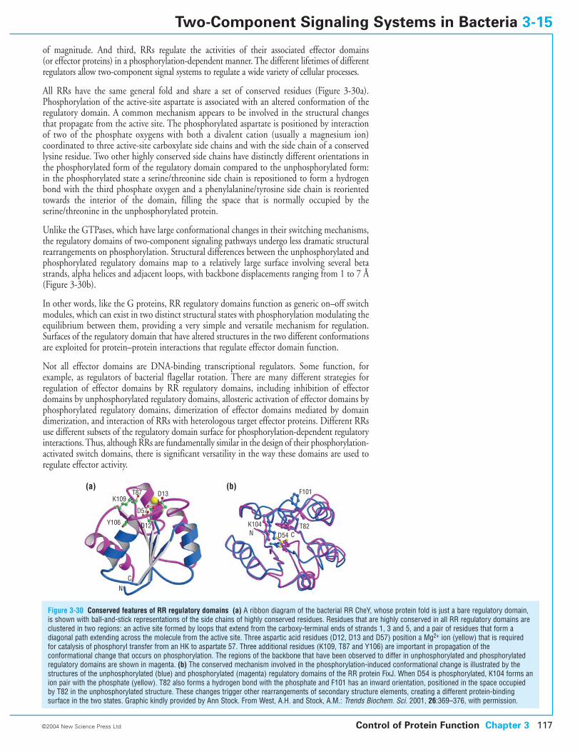
of magnitude. And third, RRs regulate the activities of their associated effector domains (or effector proteins) in a phosphorylation-dependent manner. The different lifetimes of differentregulators allow two-component signal systems to regulate a wide variety of cellular processes.
All RRs have the same general fold and share a set of conserved residues (Figure 3-30a).Phosphorylation of the active-site aspartate is associated with an altered conformation of theregulatory domain. A common mechanism appears to be involved in the structural changesthat propagate from the active site. The phosphorylated aspartate is positioned by interactionof two of the phosphate oxygens with both a divalent cation (usually a magnesium ion)coordinated to three active-site carboxylate side chains and with the side chain of a conservedlysine residue. Two other highly conserved side chains have distinctly different orientations inthe phosphorylated form of the regulatory domain compared to the unphosphorylated form:in the phosphorylated state a serine/threonine side chain is repositioned to form a hydrogenbond with the third phosphate oxygen and a phenylalanine/tyrosine side chain is reorientedtowards the interior of the domain, filling the space that is normally occupied by theserine/threonine in the unphosphorylated protein.
Unlike the GTPases, which have large conformational changes in their switching mechanisms,the regulatory domains of two-component signaling pathways undergo less dramatic structuralrearrangements on phosphorylation. Structural differences between the unphosphorylated andphosphorylated regulatory domains map to a relatively large surface involving several betastrands, alpha helices and adjacent loops, with backbone displacements ranging from 1 to 7 Å(Figure 3-30b).
In other words, like the G proteins, RR regulatory domains function as generic on–off switchmodules, which can exist in two distinct structural states with phosphorylation modulating theequilibrium between them, providing a very simple and versatile mechanism for regulation.Surfaces of the regulatory domain that have altered structures in the two different conformationsare exploited for protein–protein interactions that regulate effector domain function.
Not all effector domains are DNA-binding transcriptional regulators. Some function, forexample, as regulators of bacterial flagellar rotation. There are many different strategies for regulation of effector domains by RR regulatory domains, including inhibition of effectordomains by unphosphorylated regulatory domains, allosteric activation of effector domains byphosphorylated regulatory domains, dimerization of effector domains mediated by domaindimerization, and interaction of RRs with heterologous target effector proteins. Different RRsuse different subsets of the regulatory domain surface for phosphorylation-dependent regulatoryinteractions. Thus, although RRs are fundamentally similar in the design of their phosphorylation-activated switch domains, there is significant versatility in the way these domains are used toregulate effector activity.
Control of Protein Function Chapter 3 117©2004 New Science Press Ltd
Two-Component Signaling Systems in Bacteria 3-15
(a) (b)K109
T87 D13
D57
D12Y106
NC
N D54 CT82
F101
K104
Figure 3-30 Conserved features of RR regulatory domains (a) A ribbon diagram of the bacterial RR CheY, whose protein fold is just a bare regulatory domain,is shown with ball-and-stick representations of the side chains of highly conserved residues. Residues that are highly conserved in all RR regulatory domains areclustered in two regions: an active site formed by loops that extend from the carboxy-terminal ends of strands 1, 3 and 5, and a pair of residues that form adiagonal path extending across the molecule from the active site. Three aspartic acid residues (D12, D13 and D57) position a Mg2+ ion (yellow) that is requiredfor catalysis of phosphoryl transfer from an HK to aspartate 57. Three additional residues (K109, T87 and Y106) are important in propagation of theconformational change that occurs on phosphorylation. The regions of the backbone that have been observed to differ in unphosphorylated and phosphorylatedregulatory domains are shown in magenta. (b) The conserved mechanism involved in the phosphorylation-induced conformational change is illustrated by thestructures of the unphosphorylated (blue) and phosphorylated (magenta) regulatory domains of the RR protein FixJ. When D54 is phosphorylated, K104 forms anion pair with the phosphate (yellow). T82 also forms a hydrogen bond with the phosphate and F101 has an inward orientation, positioned in the space occupiedby T82 in the unphosphorylated structure. These changes trigger other rearrangements of secondary structure elements, creating a different protein-bindingsurface in the two states. Graphic kindly provided by Ann Stock. From West, A.H. and Stock, A.M.: Trends Biochem. Sci. 2001, 26:369–376, with permission.
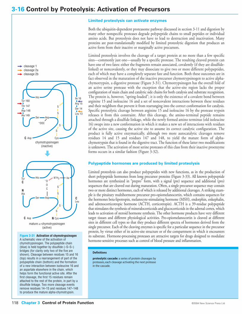
Limited proteolysis can activate enzymes
Both the ubiquitin-dependent proteasome pathway discussed in section 3-11 and digestion bymany other nonspecific proteases degrade polypeptide chains to small peptides or individualamino acids. But proteolysis does not have to lead to destruction and inactivation. Manyproteins are post-translationally modified by limited proteolytic digestion that produces anactive form from their inactive or marginally active precursors.
Limited proteolysis involves the cleavage of a target protein at no more than a few specificsites—commonly just one—usually by a specific protease. The resulting cleaved protein canhave one of two fates: either the fragments remain associated, covalently (if they are disulfide-linked) or noncovalently, or they may dissociate to give two or more different polypeptides,each of which may have a completely separate fate and function. Both these outcomes are infact observed in the maturation of the inactive precursor chymotrypsinogen to active alpha-chymotrypsin, a digestive protease (Figure 3-31). Chymotrypsinogen has the overall fold ofan active serine protease with the exception that the active-site region lacks the proper configuration of main chain and catalytic side chains for both catalysis and substrate recognition.The protein is, however, “spring-loaded”; it is only the existence of a covalent bond betweenarginine 15 and isoleucine 16 and a set of noncovalent interactions between these residuesand their neighbors that prevent it from rearranging into the correct conformation for catalysis.Specific proteolytic cleavage between arginine 15 and isoleucine 16 by the protease trypsinreleases it from this constraint. After this cleavage, the amino-terminal peptide remainsattached through a disulfide linkage, while the newly formed amino terminus (old isoleucine16) swaps into a new conformation in which it makes a new set of interactions with residuesof the active site, causing the active site to assume its correct catalytic configuration. Theproduct is fully active enzymatically, although two more autocatalytic cleavages removeresidues 14 and 15 and residues 147 and 148, to yield the mature form of alpha-chymotrypsin that is found in the digestive tract. The function of these latter two modificationsis unknown. The activation of most serine proteases of this class from their inactive proenzymeforms occurs in a similar fashion (Figure 3-32).
Polypeptide hormones are produced by limited proteolysis
Limited proteolysis can also produce polypeptides with new functions, as in the production ofshort polypeptide hormones from long precursor proteins (Figure 3-33). All known polypeptidehormones are synthesized in “prepro” form, with a signal (pre) sequence and additional (pro)sequences that are cleaved out during maturation. Often, a single precursor sequence may containtwo or more distinct hormones, each of which is released by additional cleavages. A striking exam-ple is the pituitary multihormone precursor pro-opiomelanocortin, which contains sequences forthe hormones beta-lipotropin, melanocyte-stimulating hormone (MSH), endorphin, enkephalin,and adrenocorticotropic hormone (ACTH, corticotropin). ACTH is a 39-residue polypeptidethat stimulates the synthesis of mineralocorticoids and glucocorticoids in the adrenal cortex, whichleads to activation of steroid hormone synthesis. The other hormone products have very differenttarget tissues and different physiological activities. Pro-opiomelanocortin is cleaved at differentsites in different cell types so that they produce different spectra of hormones derived from thesingle precursor. Each of the cleaving enzymes is specific for a particular sequence in the precursorprotein, by virtue either of its active-site structure or of the compartment in which it encountersits substrate. Hormone-processing proteases are attractive targets for drugs designed to modulatehormone-sensitive processes such as control of blood pressure and inflammation.
3-16 Control by Proteolysis: Activation of Precursors
Chapter 3 Control of Protein Function118 ©2004 New Science Press Ltd
Definitions
proteolytic cascade: a series of protein cleavages byproteases, each cleavage activating the next proteasein the cascade.
Figure 3-31 Activation of chymotrypsinogenA schematic view of the activation ofchymotrypsinogen. The polypeptide chain(blue) is held together by disulfide (–S–S–)bridges (for clarity only two of the five areshown). Cleavage between residues 15 and 16(top) results in a rearrangement of part of thepolypeptide chain (bottom) and the formationof a new interaction between isoleucine 16 andan aspartate elsewhere in the chain, whichhelps form the functional active site. After thefirst cleavage, the first 15 residues remainattached to the rest of the protein, in part by adisulfide linkage. Two more cleavage eventsremove residues 14–15 and residues 147–148to produce the mature alpha-chymotrypsin.
S
S
Tyr 160Ile 16
Arg 15
Ser 14
Leu 13
Thr 147
Asp 148
Ala 149Ser
His
Asp
–OOC
Cys
S
S
chymotrypsinogen(inactive)
cleavage 1cleavage 2acleavage 2b
Tyr
Ala
IleN+H3
LeuSer
His
Asp
–OOC
Cys
S
S
S
S
mature �-chymotrypsinogen(active)
Asp
AspC
C
C
N
N
C
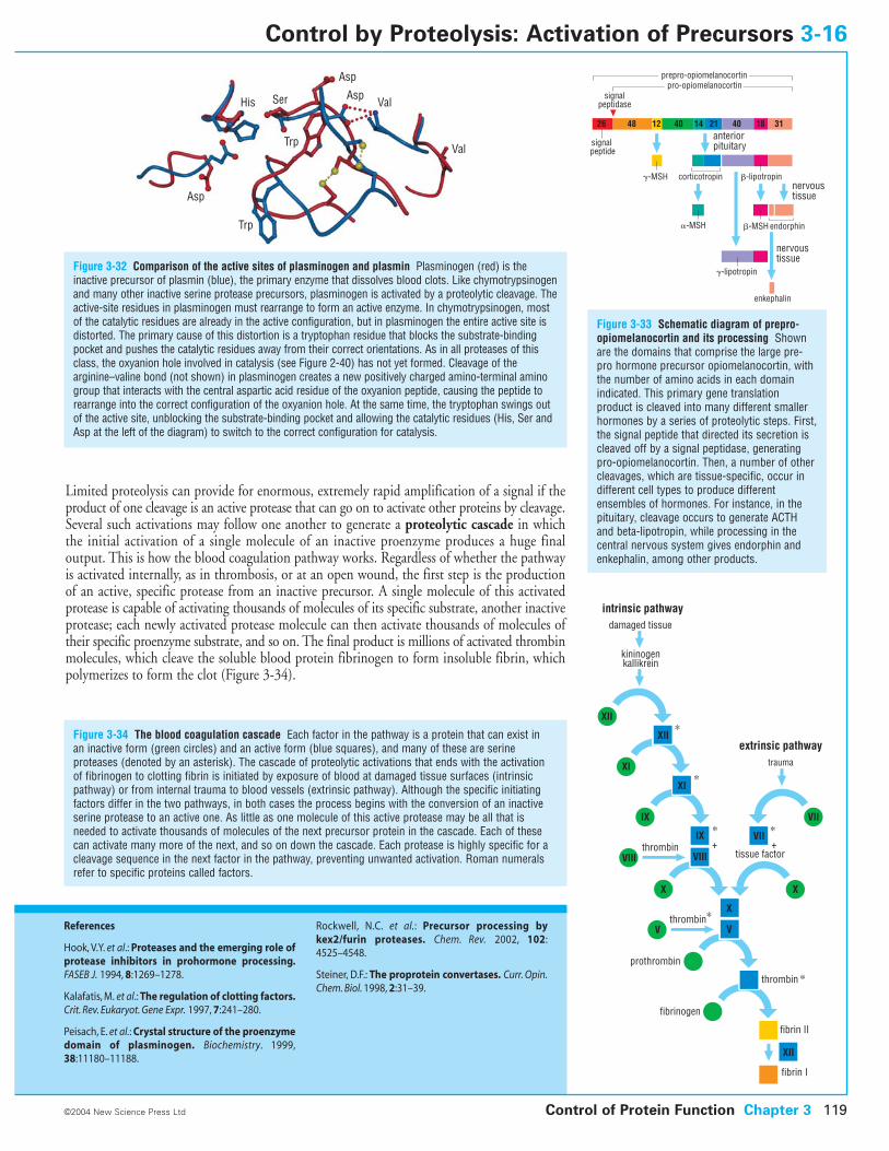
Limited proteolysis can provide for enormous, extremely rapid amplification of a signal if theproduct of one cleavage is an active protease that can go on to activate other proteins by cleavage.Several such activations may follow one another to generate a proteolytic cascade in whichthe initial activation of a single molecule of an inactive proenzyme produces a huge finaloutput. This is how the blood coagulation pathway works. Regardless of whether the pathwayis activated internally, as in thrombosis, or at an open wound, the first step is the productionof an active, specific protease from an inactive precursor. A single molecule of this activatedprotease is capable of activating thousands of molecules of its specific substrate, another inactiveprotease; each newly activated protease molecule can then activate thousands of molecules oftheir specific proenzyme substrate, and so on. The final product is millions of activated thrombinmolecules, which cleave the soluble blood protein fibrinogen to form insoluble fibrin, whichpolymerizes to form the clot (Figure 3-34).
Figure 3-32 Comparison of the active sites of plasminogen and plasmin Plasminogen (red) is theinactive precursor of plasmin (blue), the primary enzyme that dissolves blood clots. Like chymotrypsinogenand many other inactive serine protease precursors, plasminogen is activated by a proteolytic cleavage. Theactive-site residues in plasminogen must rearrange to form an active enzyme. In chymotrypsinogen, mostof the catalytic residues are already in the active configuration, but in plasminogen the entire active site isdistorted. The primary cause of this distortion is a tryptophan residue that blocks the substrate-bindingpocket and pushes the catalytic residues away from their correct orientations. As in all proteases of thisclass, the oxyanion hole involved in catalysis (see Figure 2-40) has not yet formed. Cleavage of thearginine–valine bond (not shown) in plasminogen creates a new positively charged amino-terminal aminogroup that interacts with the central aspartic acid residue of the oxyanion peptide, causing the peptide torearrange into the correct configuration of the oxyanion hole. At the same time, the tryptophan swings outof the active site, unblocking the substrate-binding pocket and allowing the catalytic residues (His, Ser andAsp at the left of the diagram) to switch to the correct configuration for catalysis.
Figure 3-33 Schematic diagram of prepro-opiomelanocortin and its processing Shownare the domains that comprise the large pre-pro hormone precursor opiomelanocortin, withthe number of amino acids in each domainindicated. This primary gene translationproduct is cleaved into many different smallerhormones by a series of proteolytic steps. First,the signal peptide that directed its secretion iscleaved off by a signal peptidase, generatingpro-opiomelanocortin. Then, a number of othercleavages, which are tissue-specific, occur indifferent cell types to produce differentensembles of hormones. For instance, in thepituitary, cleavage occurs to generate ACTHand beta-lipotropin, while processing in thecentral nervous system gives endorphin andenkephalin, among other products.
Control of Protein Function Chapter 3 119©2004 New Science Press Ltd
Control by Proteolysis: Activation of Precursors 3-16
References
Hook, V.Y. et al.: Proteases and the emerging role ofprotease inhibitors in prohormone processing.FASEB J. 1994, 8:1269–1278.
Kalafatis, M. et al.: The regulation of clotting factors.Crit. Rev. Eukaryot. Gene Expr. 1997, 7:241–280.
Peisach, E. et al.: Crystal structure of the proenzymedomain of plasminogen. Biochemistry. 1999,38:11180–11188.
Rockwell, N.C. et al.: Precursor processing bykex2/furin proteases. Chem. Rev. 2002, 102:4525–4548.
Steiner, D.F.: The proprotein convertases. Curr. Opin.Chem. Biol. 1998, 2:31–39.
Figure 3-34 The blood coagulation cascade Each factor in the pathway is a protein that can exist in an inactive form (green circles) and an active form (blue squares), and many of these are serineproteases (denoted by an asterisk). The cascade of proteolytic activations that ends with the activation of fibrinogen to clotting fibrin is initiated by exposure of blood at damaged tissue surfaces (intrinsicpathway) or from internal trauma to blood vessels (extrinsic pathway). Although the specific initiatingfactors differ in the two pathways, in both cases the process begins with the conversion of an inactiveserine protease to an active one. As little as one molecule of this active protease may be all that isneeded to activate thousands of molecules of the next precursor protein in the cascade. Each of thesecan activate many more of the next, and so on down the cascade. Each protease is highly specific for acleavage sequence in the next factor in the pathway, preventing unwanted activation. Roman numeralsrefer to specific proteins called factors.
His
Asp
Asp
Asp
Trp
Trp
Ser Val
Val
enkephalin
�-lipotropin
�-lipotropincorticotropin
�-MSH
�-MSH
�-MSH endorphin
signal peptide
signalpeptidase
nervoustissue
anteriorpituitary
nervoustissue
prepro-opiomelanocortinpro-opiomelanocortin
26 48 12 40 4014 21 18 31
fibrin II
fibrinogen
X
XII
XII
kininogenkallikrein
fibrin I
trauma
extrinsic pathway
intrinsic pathway
thrombintissue factor
+
∗
∗
∗ ∗
∗
∗
+
damaged tissue
XI
XI
IX
IX
VIIIVIII
thrombin
thrombin
prothrombin
V
VII
VII
X
V
X
XII
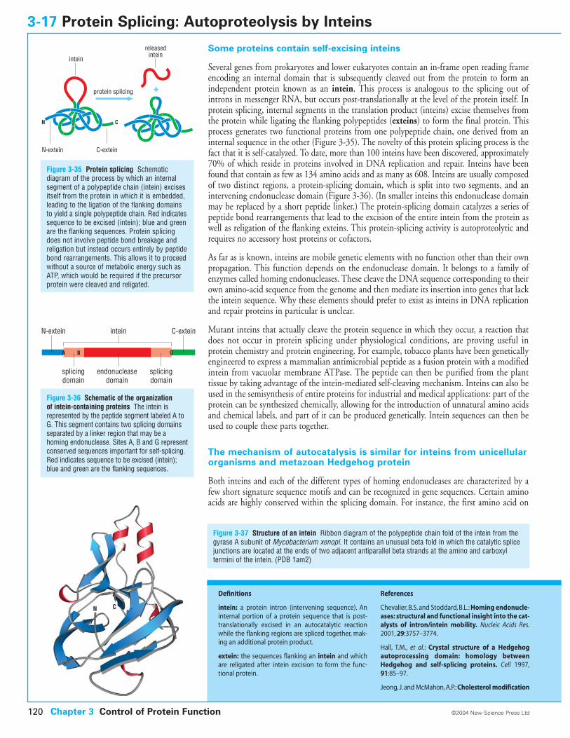
Some proteins contain self-excising inteins
Several genes from prokaryotes and lower eukaryotes contain an in-frame open reading frameencoding an internal domain that is subsequently cleaved out from the protein to form anindependent protein known as an intein. This process is analogous to the splicing out ofintrons in messenger RNA, but occurs post-translationally at the level of the protein itself. Inprotein splicing, internal segments in the translation product (inteins) excise themselves fromthe protein while ligating the flanking polypeptides (exteins) to form the final protein. Thisprocess generates two functional proteins from one polypeptide chain, one derived from aninternal sequence in the other (Figure 3-35). The novelty of this protein splicing process is thefact that it is self-catalyzed. To date, more than 100 inteins have been discovered, approximately70% of which reside in proteins involved in DNA replication and repair. Inteins have beenfound that contain as few as 134 amino acids and as many as 608. Inteins are usually composedof two distinct regions, a protein-splicing domain, which is split into two segments, and anintervening endonuclease domain (Figure 3-36). (In smaller inteins this endonuclease domainmay be replaced by a short peptide linker.) The protein-splicing domain catalyzes a series ofpeptide bond rearrangements that lead to the excision of the entire intein from the protein aswell as religation of the flanking exteins. This protein-splicing activity is autoproteolytic andrequires no accessory host proteins or cofactors.
As far as is known, inteins are mobile genetic elements with no function other than their ownpropagation. This function depends on the endonuclease domain. It belongs to a family ofenzymes called homing endonucleases. These cleave the DNA sequence corresponding to theirown amino-acid sequence from the genome and then mediate its insertion into genes that lackthe intein sequence. Why these elements should prefer to exist as inteins in DNA replicationand repair proteins in particular is unclear.
Mutant inteins that actually cleave the protein sequence in which they occur, a reaction thatdoes not occur in protein splicing under physiological conditions, are proving useful inprotein chemistry and protein engineering. For example, tobacco plants have been geneticallyengineered to express a mammalian antimicrobial peptide as a fusion protein with a modifiedintein from vacuolar membrane ATPase. The peptide can then be purified from the planttissue by taking advantage of the intein-mediated self-cleaving mechanism. Inteins can also beused in the semisynthesis of entire proteins for industrial and medical applications: part of theprotein can be synthesized chemically, allowing for the introduction of unnatural amino acidsand chemical labels, and part of it can be produced genetically. Intein sequences can then beused to couple these parts together.
The mechanism of autocatalysis is similar for inteins from unicellularorganisms and metazoan Hedgehog protein
Both inteins and each of the different types of homing endonucleases are characterized by afew short signature sequence motifs and can be recognized in gene sequences. Certain aminoacids are highly conserved within the splicing domain. For instance, the first amino acid on
3-17 Protein Splicing: Autoproteolysis by Inteins
Figure 3-35 Protein splicing Schematicdiagram of the process by which an internalsegment of a polypeptide chain (intein) excisesitself from the protein in which it is embedded,leading to the ligation of the flanking domainsto yield a single polypeptide chain. Red indicatessequence to be excised (intein); blue and greenare the flanking sequences. Protein splicingdoes not involve peptide bond breakage andreligation but instead occurs entirely by peptidebond rearrangements. This allows it to proceedwithout a source of metabolic energy such asATP, which would be required if the precursorprotein were cleaved and religated.
Chapter 3 Control of Protein Function120 ©2004 New Science Press Ltd
Definitions
intein: a protein intron (intervening sequence). Aninternal portion of a protein sequence that is post-translationally excised in an autocatalytic reactionwhile the flanking regions are spliced together, mak-ing an additional protein product.
extein: the sequences flanking an intein and whichare religated after intein excision to form the func-tional protein.
References
Chevalier, B.S. and Stoddard, B.L.: Homing endonucle-ases: structural and functional insight into the cat-alysts of intron/intein mobility. Nucleic Acids Res.2001, 29:3757–3774.
Hall, T.M., et al.: Crystal structure of a Hedgehogautoprocessing domain: homology betweenHedgehog and self-splicing proteins. Cell 1997,91:85–97.
Jeong, J. and McMahon, A.P.: Cholesterol modification
Figure 3-36 Schematic of the organization of intein-containing proteins The intein isrepresented by the peptide segment labeled A toG. This segment contains two splicing domainsseparated by a linker region that may be ahoming endonuclease. Sites A, B and G representconserved sequences important for self-splicing.Red indicates sequence to be excised (intein);blue and green are the flanking sequences.
Figure 3-37 Structure of an intein Ribbon diagram of the polypeptide chain fold of the intein from thegyrase A subunit of Mycobacterium xenopi. It contains an unusual beta fold in which the catalytic splicejunctions are located at the ends of two adjacent antiparallel beta strands at the amino and carboxyltermini of the intein. (PDB 1am2)
N-extein
splicingdomain
splicingdomain
endonucleasedomain
C-exteinintein
A GB
protein splicing
N C
N-extein C-extein
intein
releasedintein
CN
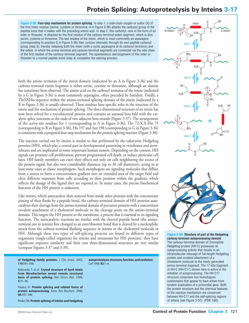
both the amino terminus of the intein domain (indicated by an A in Figure 3-36) and thecarboxy-terminal extein fragment is either serine, cysteine or threonine, although an alaninehas sometimes been observed. The amino acid on the carboxyl terminus of the intein (indicatedby a G in Figure 3-36) is most commonly asparagine, often preceded by histidine. Finally, aThrXXHis sequence within the amino-terminal splicing domain of the intein (indicated by aB in Figure 3-36) is usually observed. These residues have specific roles in the structure of theintein and the mechanism of protein splicing. The three-dimensional structure of an intein hasnow been solved for a mycobacterial protein and contains an unusual beta fold with the cat-alytic splice junctions at the ends of two adjacent beta strands (Figure 3-37). The arrangementof the active-site residues Ser 1 (corresponding to A in Figure 3-36), Thr 72-X-X-His 75(corresponding to B in Figure 3-36), His 197 and Asn 198 (corresponding to G in Figure 3-36)is consistent with a proposed four-step mechanism for the protein splicing reaction (Figure 3-38).
The reaction carried out by inteins is similar to that performed by the eukaryotic Hedgehogproteins (HH), which play a central part in developmental patterning in vertebrates and inver-tebrates and are implicated in some important human tumors. Depending on the context, HHsignals can promote cell proliferation, prevent programmed cell death, or induce particular cellfates. HH family members can exert their effects not only on cells neighboring the source ofthe protein signal, but also over considerable distances (up to 30 cell diameters), acting in atleast some cases as classic morphogens. Such morphogens are signaling molecules that diffusefrom a source to form a concentration gradient over an extended area of the target field andelicit different responses from cells according to their position within the gradient, whichreflects the dosage of the ligand they are exposed to. In many cases, the precise biochemicalfunction of the HH protein is unknown.
Like inteins, which autocatalyze their removal from inside other proteins with the concomitantjoining of their flanks by a peptide bond, the carboxy-terminal domain of HH proteins auto-catalyzes their cleavage from the amino-terminal domain of precursor proteins with a concomitantcovalent attachment of a cholesterol molecule to the cleavage point on the amino-terminaldomain. This targets the HH protein to the membrane, a process that is essential to its signalingfunction. The autocatalytic reactions are similar, with the cleaved peptide bond (the amino-terminal one in inteins) first changed to an ester/thioester bond that is cleaved by a nucleophilicattack from the carboxy-terminal flanking sequence in inteins or the cholesterol molecule inHH. Although these two types of self-splicing proteins are found in different types oforganisms (single-celled organisms for inteins and metazoans for HH proteins), they havesignificant sequence similarity and their core three-dimensional structures are very similar(compare Figures 3-37 and 3-39).
Figure 3-38 Four-step mechanism for protein splicing In step 1, a side-chain oxygen or sulfur (X) ofthe first intein residue (serine, cysteine or threonine; A in Figure 3-36) attacks the carbonyl group of thepeptide bond that it makes with the preceding amino acid. In step 2, this carbonyl, now in the form of anester or thioester, is attacked by the first residue of the carboxy-terminal extein segment, which is alsoserine, cysteine or threonine. The last residue of the intein, which is most commonly an asparagine(corresponding to position G in Figure 3-36) then cyclizes internally through its own peptide carbonylgroup (step 3), thereby releasing both the intein (with a cyclic asparagine at its carboxyl terminus) andthe extein, in which the amino-terminal and carboxy-terminal segments are connected via the side chainof the first residue of the carboxy-terminal segment. The spontaneous rearrangement of this ester orthioester to a normal peptide bond (step 4) completes the splicing process.
Control of Protein Function Chapter 3 121©2004 New Science Press Ltd
Protein Splicing: Autoproteolysis by Inteins 3-17
of Hedgehog family proteins. J. Clin. Invest. 2002,110:591–596.
Klabunde, T. et al.: Crystal structure of GyrA inteinfrom Mycobacterium xenopi reveals structuralbasis of protein splicing. Nat. Struct. Biol. 1998,5:31–36.
Paulus, H.: Protein splicing and related forms ofprotein autoprocessing. Annu. Rev. Biochem. 2000,69:447–496.
Perler, F.B.: Protein splicing of inteins and hedgehog
autoproteolysis: structure, function, and evolution.Cell 1998, 92:1–4.
N-extein C-extein
intein
step 1
step 2
XH
XH
O
O
O
H2NNH
HN
O
NH2
X
O
X
step 3
step 4
XH
O
O
H2NNH
NH
NH2
XHX
O
O
O
H2N NH
NH2
HXO
HXH2N
OO NH
Figure 3-39 Structure of part of the Hedgehogcarboxy-terminal autoprocessing domain The carboxy-terminal domain of DrosophilaHedgehog protein (HH-C) possesses anautoprocessing activity that results in anintramolecular cleavage of full-length Hedgehogprotein and covalent attachment of acholesterol molecule to the newly generatedamino-terminal fragment. The 17 kDa fragmentof HH-C (HH-C17) shown here is active in theinitiation of autoprocessing. The HH-C17structure comprises two homologoussubdomains that appear to have arisen fromtandem duplication of a primordial gene. Boththe protein structure and the chemical featuresof the reaction mechanism are conservedbetween HH-C17 and the self-splicing regionsof inteins (see Figure 3-37). (PDB 1at0)
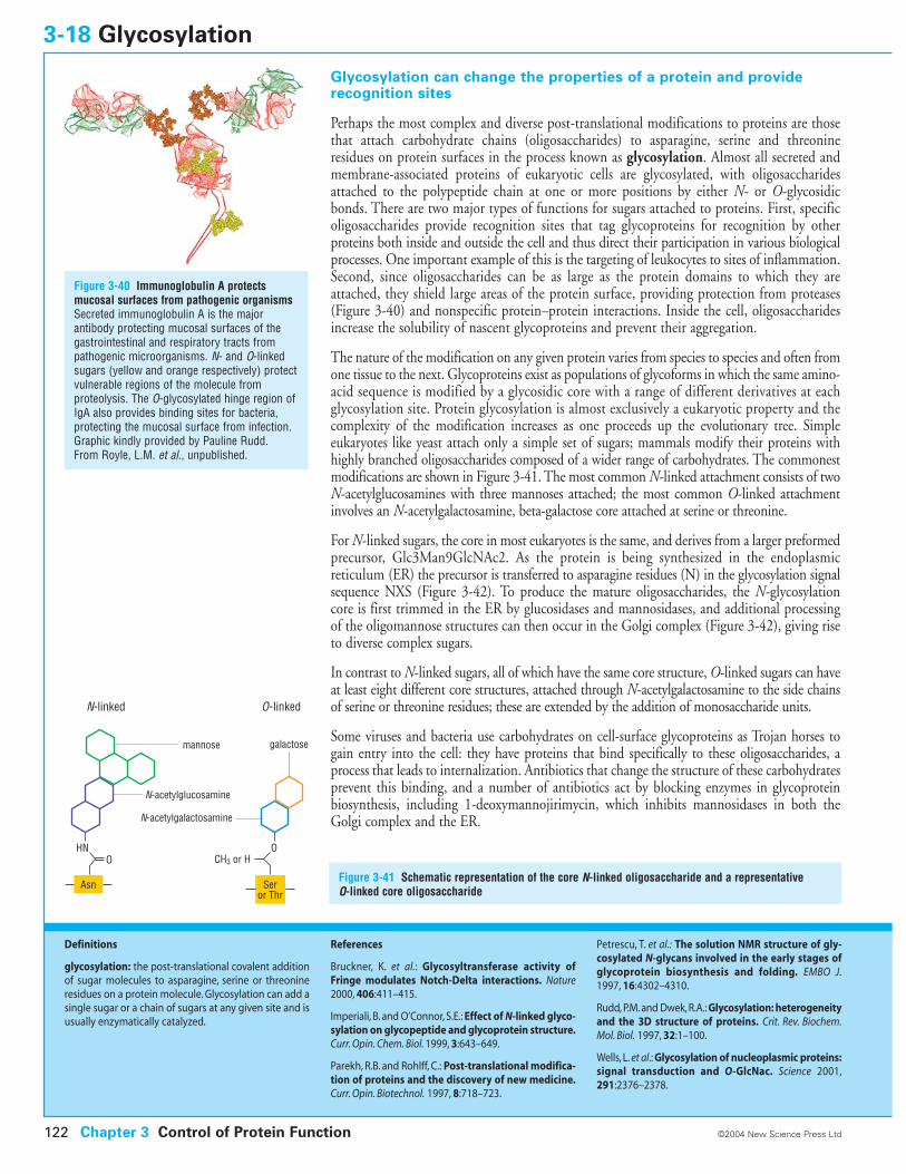
Glycosylation can change the properties of a protein and providerecognition sites
Perhaps the most complex and diverse post-translational modifications to proteins are thosethat attach carbohydrate chains (oligosaccharides) to asparagine, serine and threonineresidues on protein surfaces in the process known as glycosylation. Almost all secreted andmembrane-associated proteins of eukaryotic cells are glycosylated, with oligosaccharidesattached to the polypeptide chain at one or more positions by either N- or O-glycosidicbonds. There are two major types of functions for sugars attached to proteins. First, specificoligosaccharides provide recognition sites that tag glycoproteins for recognition by other proteins both inside and outside the cell and thus direct their participation in various biologicalprocesses. One important example of this is the targeting of leukocytes to sites of inflammation.Second, since oligosaccharides can be as large as the protein domains to which they areattached, they shield large areas of the protein surface, providing protection from proteases(Figure 3-40) and nonspecific protein–protein interactions. Inside the cell, oligosaccharidesincrease the solubility of nascent glycoproteins and prevent their aggregation.
The nature of the modification on any given protein varies from species to species and often fromone tissue to the next. Glycoproteins exist as populations of glycoforms in which the same amino-acid sequence is modified by a glycosidic core with a range of different derivatives at each glycosylation site. Protein glycosylation is almost exclusively a eukaryotic property and the complexity of the modification increases as one proceeds up the evolutionary tree. Simpleeukaryotes like yeast attach only a simple set of sugars; mammals modify their proteins withhighly branched oligosaccharides composed of a wider range of carbohydrates. The commonestmodifications are shown in Figure 3-41. The most common N-linked attachment consists of twoN-acetylglucosamines with three mannoses attached; the most common O-linked attachmentinvolves an N-acetylgalactosamine, beta-galactose core attached at serine or threonine.
For N-linked sugars, the core in most eukaryotes is the same, and derives from a larger preformedprecursor, Glc3Man9GlcNAc2. As the protein is being synthesized in the endoplasmicreticulum (ER) the precursor is transferred to asparagine residues (N) in the glycosylation signalsequence NXS (Figure 3-42). To produce the mature oligosaccharides, the N-glycosylationcore is first trimmed in the ER by glucosidases and mannosidases, and additional processingof the oligomannose structures can then occur in the Golgi complex (Figure 3-42), giving riseto diverse complex sugars.
In contrast to N-linked sugars, all of which have the same core structure, O-linked sugars can haveat least eight different core structures, attached through N-acetylgalactosamine to the side chainsof serine or threonine residues; these are extended by the addition of monosaccharide units.
Some viruses and bacteria use carbohydrates on cell-surface glycoproteins as Trojan horses togain entry into the cell: they have proteins that bind specifically to these oligosaccharides, aprocess that leads to internalization. Antibiotics that change the structure of these carbohydratesprevent this binding, and a number of antibiotics act by blocking enzymes in glycoproteinbiosynthesis, including 1-deoxymannojirimycin, which inhibits mannosidases in both theGolgi complex and the ER.
3-18 Glycosylation
Figure 3-40 Immunoglobulin A protectsmucosal surfaces from pathogenic organismsSecreted immunoglobulin A is the majorantibody protecting mucosal surfaces of thegastrointestinal and respiratory tracts frompathogenic microorganisms. N- and O-linkedsugars (yellow and orange respectively) protectvulnerable regions of the molecule fromproteolysis. The O-glycosylated hinge region ofIgA also provides binding sites for bacteria,protecting the mucosal surface from infection.Graphic kindly provided by Pauline Rudd. From Royle, L.M. et al., unpublished.
Figure 3-41 Schematic representation of the core N-linked oligosaccharide and a representative O-linked core oligosaccharide
Chapter 3 Control of Protein Function122 ©2004 New Science Press Ltd
Definitions
glycosylation: the post-translational covalent additionof sugar molecules to asparagine, serine or threonineresidues on a protein molecule. Glycosylation can add asingle sugar or a chain of sugars at any given site and isusually enzymatically catalyzed.
References
Bruckner, K. et al.: Glycosyltransferase activity ofFringe modulates Notch-Delta interactions. Nature2000, 406:411–415.
Imperiali, B. and O’Connor, S.E.: Effect of N-linked glyco-sylation on glycopeptide and glycoprotein structure.Curr. Opin. Chem. Biol. 1999, 3:643–649.
Parekh, R.B. and Rohlff, C.: Post-translational modifica-tion of proteins and the discovery of new medicine.Curr. Opin. Biotechnol. 1997, 8:718–723.
Petrescu, T. et al.: The solution NMR structure of gly-cosylated N-glycans involved in the early stages ofglycoprotein biosynthesis and folding. EMBO J.1997, 16:4302–4310.
Rudd, P.M. and Dwek, R.A.: Glycosylation: heterogeneityand the 3D structure of proteins. Crit. Rev. Biochem.Mol. Biol. 1997, 32:1–100.
Wells, L. et al.: Glycosylation of nucleoplasmic proteins:signal transduction and O-GlcNac. Science 2001,291:2376–2378.
HNCH3 or H
Asn Seror Thr
OO
mannose
N-linked O-linked
N-acetylglucosamine
N-acetylgalactosamine
galactose
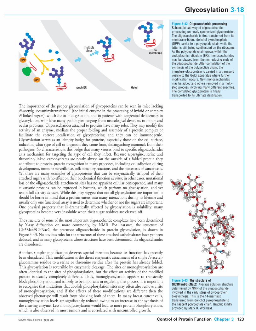
The importance of the proper glycosylation of glycoproteins can be seen in mice lackingN-acetylglucosaminyltransferase I (the initial enzyme in the processing of hybrid or complexN-linked sugars), which die at mid-gestation, and in patients with congenital deficiencies inglycosylation, who have many pathologies ranging from neurological disorders to motor andocular problems. Oligosaccharides attached to proteins have many roles. They may modify theactivity of an enzyme, mediate the proper folding and assembly of a protein complex orfacilitate the correct localization of glycoproteins; and they can be immunogenic.Glycosylation serves as an identity badge for proteins, especially those on the cell surface,indicating what type of cell or organism they come from, distinguishing mammals from theirpathogens. So characteristic is this badge that many viruses bind to specific oligosaccharidesas a mechanism for targeting the type of cell they infect. Because asparagine, serine and threonine-linked carbohydrates are nearly always on the outside of a folded protein theycontribute to protein–protein recognition in many processes, including cell adhesion duringdevelopment, immune surveillance, inflammatory reactions, and the metastasis of cancer cells.Yet there are many examples of glycoproteins that can be enzymatically stripped of theirattached sugars with no effect on their biochemical function in vitro; in other cases, mutationalloss of the oligosaccharide attachment sites has no apparent cellular consequence, and manyeukaryotic proteins can be expressed in bacteria, which perform no glycosylation, and yetretain full activity in vitro. While this may suggest that not all glycosylations are important, itshould be borne in mind that a protein enters into many interactions during its lifetime andusually only one functional assay is used to determine whether or not the sugars are important.One physical property that is dramatically affected by glycosylation is solubility: many glycoproteins become very insoluble when their sugar residues are cleaved off.
The structures of some of the most important oligosaccharide complexes have been determinedby X-ray diffraction or, more commonly, by NMR. For instance, the structure ofGlc3Man9GlcNac2, the precursor oligosaccharide in protein glycosylation, is shown inFigure 3-43. No obvious rules for the structures of these attached carbohydrates have yet beendeduced, and in many glycoproteins whose structures have been determined, the oligosaccharidesare disordered.
Another, simpler modification deserves special mention because its function has recentlybeen elucidated. This modification is the direct enzymatic attachment of a single N-acetyl-glucosamine residue to a serine or threonine residue after the protein has already folded.This glycosylation is reversible by enzymatic cleavage. The sites of monoglycosylation areoften identical to the sites of phosphorylation, but the effect on activity of the modifiedprotein is usually completely different. Thus, monoglycosylation appears to transientlyblock phosphorylation, and is likely to be important in regulating that process. It is importantto recognize that mutations that abolish phosphorylation sites may often also remove a siteof monoglycosylation, and if the effects of these modifications are different then theobserved phenotype will result from blocking both of them. In many breast cancer cells,monoglycosylation levels are significantly reduced owing to an increase in the synthesis ofthe cleaving enzyme. Less monoglycosylation would lead to more protein phosphorylation,which is also observed in most tumors and is correlated with uncontrolled growth.
Figure 3-42 Oligosaccharide processingSchematic pathway of oligosaccharideprocessing on newly synthesized glycoproteins,The oligosaccharide is first transferred from itsmembrane-bound dolichol pyrophosphate(DPP) carrier to a polypeptide chain while thelatter is still being synthesized on the ribosome.As the polypeptide chain grows within theendoplasmic reticulum (ER), monosaccharidesmay be cleaved from the nonreducing ends ofthe oligosaccharide. After completion of thesynthesis of the polypeptide chain, theimmature glycoprotein is carried in a transportvesicle to the Golgi apparatus where furthermodification occurs. New monosaccharidesmay be added and others removed in a multi-step process involving many different enzymes.The completed glycoprotein is finallytransported to its ultimate destination.
Control of Protein Function Chapter 3 123©2004 New Science Press Ltd
Glycosylation 3-18
DPP
DPP
rough ER Golgi
tomembrane
multiplesteps
Figure 3-43 The structure ofGlc3Man9GlcNac2 Average solution structuredetermined by NMR of the oligosaccharideinvolved in the early stage of glycoproteinbiosynthesis. This is the 14-mer firsttransferred from dolichol pyrophosphate to the nascent polypeptide chain. Graphic kindlyprovided by Mark R. Wormald.
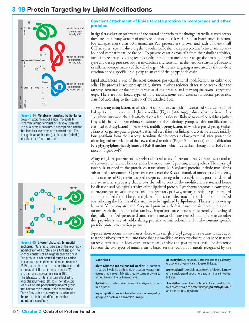
Covalent attachment of lipids targets proteins to membranes and otherproteins
In signal transduction pathways and the control of protein traffic through intracellular membranesthere are often many variants of one type of protein, each with a similar biochemical function.For example, more than 50 mammalian Rab proteins are known, and each of these smallGTPases plays a part in directing the vesicular traffic that transports proteins between membrane-bounded compartments of the cell. To prevent chaotic cross-talk from their similar activities,each of these proteins is targeted to specific intracellular membranes at specific times in the cellcycle and during processes such as metabolism and secretion, as the need for switching functionsin different compartments of the cell changes. Membrane targeting is mediated by the covalentattachment of a specific lipid group to an end of the polypeptide chain.
Lipid attachment is one of the most common post-translational modifications in eukaryoticcells. The process is sequence-specific, always involves residues either at or near either thecarboxyl terminus or the amino terminus of the protein, and may require several enzymaticsteps. There are four broad types of lipid modifications with distinct functional properties,classified according to the identity of the attached lipid.
These are: myristoylation, in which a 14-carbon fatty-acid chain is attached via a stable amidelinkage to an amino-terminal glycine residue (Figure 3-44, top); palmitoylation, in which a16-carbon fatty-acid chain is attached via a labile thioester linkage to cysteine residues (otherfatty-acid chains can sometimes substitute for the palmitoyl group, so this modification isoften called S-acylation) (Figure 3-44, middle); prenylation, in which a prenyl group (eithera farnesyl or geranylgeranyl group) is attached via a thioether linkage to a cysteine residue initiallyfour positions from the carboxyl terminus that becomes carboxy-terminal after proteolytictrimming and methylation of the new carboxyl terminus (Figure 3-44, bottom); and modificationby a glycosylphosphatidylinositol (GPI) anchor, which is attached through a carbohydratemoiety (Figure 3-45).
N-myristoylated proteins include select alpha subunits of heterotrimeric G proteins, a numberof non-receptor tyrosine kinases, and a few monomeric G proteins, among others. The myristoylmoiety is attached to the protein co-translationally. S-acylated proteins include most alphasubunits of heterotrimeric G proteins, members of the Ras superfamily of monomeric G proteins,and a number of G-protein-coupled receptors, among others. S-acylation is post-translationaland reversible, a property that allows the cell to control the modification state, and hencelocalization and biological activity, of the lipidated protein. Lymphoma proprotein convertase,an enzyme that activates proproteins in the secretory pathway, occurs in both the palmitoylatedand unmodified form. The palmitoylated form is degraded much faster than the unmodifiedone, allowing the lifetime of this enzyme to be regulated by lipidation. There is some overlapbetween N-myristoylated and S-acylated proteins such that many contain both lipid modifi-cations. Such dual modification can have important consequences, most notably targeting ofthe dually modified species to distinct membrane subdomains termed lipid rafts or to caveolae;this provides a way of sublocalizing proteins to microdomains that also contain specificprotein–protein interaction partners.
S-prenylation occurs in two classes, those with a single prenyl group on a cysteine residue at ornear the carboxyl terminus, and those that are modified on two cysteine residues at or near thecarboxyl terminus. In both cases, attachment is stable and post-translational. The differencebetween the two types of attachment is based on the recognition motifs recognized by the
3-19 Protein Targeting by Lipid Modifications
Chapter 3 Control of Protein Function124 ©2004 New Science Press Ltd
Definitions
glycosylphosphatidylinositol anchor: a complexstructure involving both lipids and carbohydrate mol-ecules that is reversibly attached to some proteins totarget them to the cell membrane.
lipidation: covalent attachment of a fatty-acid groupto a protein.
myristoylation: irreversible attachment of a myristoylgroup to a protein via an amide linkage.
palmitoylation: reversible attachment of a palmitoylgroup to a protein via a thioester linkage.
prenylation: irreversible attachment of either a farnesylor geranylgeranyl group to a protein via a thioetherlinkage.
S-acylation: reversible attachment of a fatty-acid groupto a protein via a thioester linkage; palmitoylation isan example of S-acylation.
Figure 3-45 Glycosylphosphatidylinositolanchoring Schematic diagram of the reversiblemodification of a protein by a GPI anchor. Theanchor consists of an oligosaccharide chain.The protein is connected through an amidelinkage to a phosphoethanolamine molecule (E-P) that is attached to a core tetrasaccharidecomposed of three mannose sugars (M) and a single glucosamine sugar (G). The tetrasaccharide is in turn attached tophosphatidylinositol (I). It is the fatty-acidresidues of this phosphatidylinositol group that anchor the protein to the membrane. These fatty acids may vary somewhat with the protein being modified, providingmembrane specificity.
cytosol
mem
brane
protein anchoredto membraneby fatty acid
protein anchoredto membraneby fatty acid
protein anchoredto membrane
by prenyl groupCH2 S C
C
O
CH3
H
O
C
O
N
H
OHO
S C
C
N
O
C
N H
CH2
CH2 H
H
protein
P
PM
MM
G
I
P EM
MM
G
I
PI-PLC
E
P
Figure 3-44 Membrane targeting by lipidationCovalent attachment of a lipid molecule toeither the amino-terminal or carboxy-terminalend of a protein provides a hydrophobic anchorthat localizes the protein to a membrane. Thelinkage is an amide (top), a thioester (middle)or a thioether (bottom) bond.
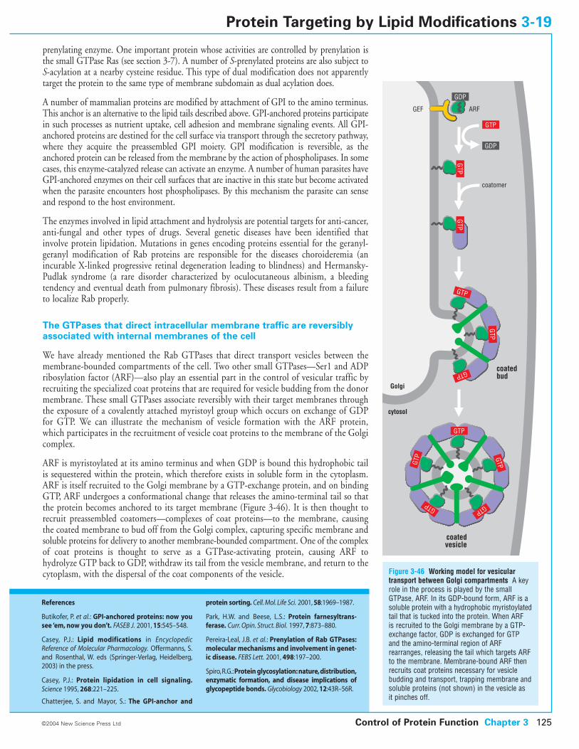
prenylating enzyme. One important protein whose activities are controlled by prenylation isthe small GTPase Ras (see section 3-7). A number of S-prenylated proteins are also subject toS-acylation at a nearby cysteine residue. This type of dual modification does not apparentlytarget the protein to the same type of membrane subdomain as dual acylation does.
A number of mammalian proteins are modified by attachment of GPI to the amino terminus.This anchor is an alternative to the lipid tails described above. GPI-anchored proteins participatein such processes as nutrient uptake, cell adhesion and membrane signaling events. All GPI-anchored proteins are destined for the cell surface via transport through the secretory pathway,where they acquire the preassembled GPI moiety. GPI modification is reversible, as theanchored protein can be released from the membrane by the action of phospholipases. In somecases, this enzyme-catalyzed release can activate an enzyme. A number of human parasites haveGPI-anchored enzymes on their cell surfaces that are inactive in this state but become activatedwhen the parasite encounters host phospholipases. By this mechanism the parasite can senseand respond to the host environment.
The enzymes involved in lipid attachment and hydrolysis are potential targets for anti-cancer,anti-fungal and other types of drugs. Several genetic diseases have been identified thatinvolve protein lipidation. Mutations in genes encoding proteins essential for the geranyl-geranyl modification of Rab proteins are responsible for the diseases choroideremia (anincurable X-linked progressive retinal degeneration leading to blindness) and Hermansky-Pudlak syndrome (a rare disorder characterized by oculocutaneous albinism, a bleeding tendency and eventual death from pulmonary fibrosis). These diseases result from a failureto localize Rab properly.
The GTPases that direct intracellular membrane traffic are reversiblyassociated with internal membranes of the cell
We have already mentioned the Rab GTPases that direct transport vesicles between themembrane-bounded compartments of the cell. Two other small GTPases—Ser1 and ADPribosylation factor (ARF)—also play an essential part in the control of vesicular traffic byrecruiting the specialized coat proteins that are required for vesicle budding from the donormembrane. These small GTPases associate reversibly with their target membranes throughthe exposure of a covalently attached myristoyl group which occurs on exchange of GDPfor GTP. We can illustrate the mechanism of vesicle formation with the ARF protein,which participates in the recruitment of vesicle coat proteins to the membrane of the Golgicomplex.
ARF is myristoylated at its amino terminus and when GDP is bound this hydrophobic tailis sequestered within the protein, which therefore exists in soluble form in the cytoplasm.ARF is itself recruited to the Golgi membrane by a GTP-exchange protein, and on bindingGTP, ARF undergoes a conformational change that releases the amino-terminal tail so thatthe protein becomes anchored to its target membrane (Figure 3-46). It is then thought torecruit preassembled coatomers—complexes of coat proteins—to the membrane, causingthe coated membrane to bud off from the Golgi complex, capturing specific membrane andsoluble proteins for delivery to another membrane-bounded compartment. One of the complexof coat proteins is thought to serve as a GTPase-activating protein, causing ARF tohydrolyze GTP back to GDP, withdraw its tail from the vesicle membrane, and return to thecytoplasm, with the dispersal of the coat components of the vesicle.
Control of Protein Function Chapter 3 125©2004 New Science Press Ltd
Protein Targeting by Lipid Modifications 3-19
References
Butikofer, P. et al.: GPI-anchored proteins: now yousee ‘em, now you don’t. FASEB J. 2001, 15:545–548.
Casey, P.J.: Lipid modifications in EncyclopedicReference of Molecular Pharmacology. Offermanns, S.and Rosenthal, W. eds (Springer-Verlag, Heidelberg,2003) in the press.
Casey, P.J.: Protein lipidation in cell signaling.Science 1995, 268:221–225.
Chatterjee, S. and Mayor, S.: The GPI-anchor and
protein sorting. Cell. Mol. Life Sci. 2001, 58:1969–1987.
Park, H.W. and Beese, L.S.: Protein farnesyltrans-ferase. Curr. Opin. Struct. Biol. 1997, 7:873–880.
Pereira-Leal, J.B. et al.: Prenylation of Rab GTPases:molecular mechanisms and involvement in genet-ic disease. FEBS Lett. 2001, 498:197–200.
Spiro,R.G.:Protein glycosylation:nature,distribution,enzymatic formation, and disease implications ofglycopeptide bonds. Glycobiology 2002, 12:43R–56R.
Figure 3-46 Working model for vesiculartransport between Golgi compartments A keyrole in the process is played by the smallGTPase, ARF. In its GDP-bound form, ARF is asoluble protein with a hydrophobic myristoylatedtail that is tucked into the protein. When ARF is recruited to the Golgi membrane by a GTP-exchange factor, GDP is exchanged for GTPand the amino-terminal region of ARFrearranges, releasing the tail which targets ARFto the membrane. Membrane-bound ARF thenrecruits coat proteins necessary for vesiclebudding and transport, trapping membrane andsoluble proteins (not shown) in the vesicle as it pinches off.
Golgi
cytosol
coatedvesicle
GTP
GTP
GTP GTP
GTP
GTP
GTP
GTP
GDP
GDP
GTPGTP
GTP
coatomer
ARFGEF
coatedbud
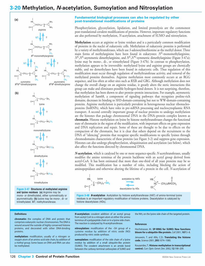
Fundamental biological processes can also be regulated by other post-translational modifications of proteins
Phosphorylation, glycosylation, lipidation, and limited proteolysis are the commonest post-translational covalent modifications of proteins. However, important regulatory functionsare also performed by methylation, N-acetylation, attachment of SUMO and nitrosylation.
Methylation occurs at arginine or lysine residues and is a particularly common modificationof proteins in the nuclei of eukaryotic cells. Methylation of eukaryotic proteins is performedby a variety of methyltransferases, which use S-adenosylmethionine as the methyl donor. Threemain forms of methylarginine have been found in eukaryotes: NG-monomethylarginine,NG,NG-asymmetric dimethylarginine, and NG,N¢G-symmetric dimethylarginine (Figure 3-47a);lysine may be mono-, di-, or trimethylated (Figure 3-47b). In contrast to phosphorylation,methylation appears to be irreversible: methylated lysine and arginine groups are chemicallystable, and no demethylases have been found in eukaryotic cells. Thus regulation of thismodification must occur through regulation of methyltransferase activity, and removal of themethylated proteins themselves. Arginine methylation most commonly occurs at an RGGsequence, and less often at other sites such as RXR and GRG. Although methylation does notchange the overall charge on an arginine residue, it greatly alters the steric interactions thisgroup can make and eliminates possible hydrogen-bond donors. It is not surprising, therefore,that methylation has been shown to alter protein–protein interactions. For example, asymmetricmethylation of Sam68, a component of signaling pathways that recognizes proline-richdomains, decreases its binding to SH3-domain-containing but not to WW-domain-containingproteins. Arginine methylation is particularly prevalent in heterogeneous nuclear ribonucleo-proteins (hnRNPs), which have roles in pre-mRNA processing and nucleocytoplasmic RNAtransport. A second centrally important group of nuclear proteins to undergo methylationare the histones that package chromosomal DNA in the DNA–protein complex known aschromatin. Histone methylation on lysine by histone methyltransferases changes the functionalstate of chromatin in the region of the modification, with important effects on gene expressionand DNA replication and repair. Some of these are thought to be due to effects on thecompaction of the chromatin, but it is clear that others depend on the recruitment to theDNA of “silencing” proteins that recognize specific modifications to specific lysines throughchromodomains characteristic of these proteins (see Figure 3-2) and suppress gene expression.Histones can also undergo phosphorylation, ubiquitination and acetylation (see below), whichalso affect the functions directed by chromosomal DNA.
N-acetylation, which is catalyzed by one or more sequence-specific N-acetyltransferases, usuallymodifies the amino terminus of the protein backbone with an acetyl group derived fromacetyl-CoA. It has been estimated that more than one-third of all yeast proteins may be somodified. This modification has a number of roles, including blocking the action ofaminopeptidases and otherwise altering the lifetime of a protein in the cell. N-acetylation of
3-20 Methylation, N-acetylation, Sumoylation and Nitrosylation
Figure 3-47 Structures of methylated arginineand lysine residues (a) Arginine may bemono- or dimethylated, either symmetrically orasymmetrically; (b) lysine may be mono-, di- ortrimethylated. MT, methyltransferase.
Figure 3-48 N-acetylation Acetylation by histone acetyltransferase (HAT) of amino-terminal lysineresidues is an important regulatory modification of histone proteins. Deacetylation is catalyzed byhistone deacetylases (HDs).
Chapter 3 Control of Protein Function126 ©2004 New Science Press Ltd
Definitions
chromatin: the complex of DNA and protein that comprises eukaryotic nuclear chromosomes.The DNA iswound around the outside of highly conserved histoneproteins, and decorated with other DNA-binding proteins.
methylation: modification, usually of a nitrogen or oxygen atom of an amino-acid side chain, by addition ofa methyl group. Some bases on DNA and RNA can alsobe methylated.
N-acetylation: covalent addition of an acetyl groupfrom acetyl-CoA to a nitrogen atom at either the aminoterminus of a polypeptide chain or in a lysine side chain.The reaction is catalyzed by N-acetyltransferase.
nitrosylation: modification of the –SH group of a cysteine residue by addition of nitric oxide (NO) produced by nitric oxide synthase.
sumoylation: modification of the side chain of a lysineresidue by addition of a small ubiquitin-like protein(SUMO). The covalent attachment is an amide bondbetween the carboxy-terminal carboxylate of SUMO and
the NH2 on the lysine side chain of the targeted protein.
References
Hochstrasser, M.: SP-RING for SUMO: New functionsbloom for a ubiquitin-like protein. Cell 2001, 107:5–8.
Jenuwein, T. and Allis, C.D.: Translating the histonecode. Science 2001, 293:1074–1080.
Kouzarides, T.: Histone methylation in transcriptionalcontrol. Curr. Opin. Genet. Dev. 2002, 12:198–209.
(CH2)4NHCCH3HCOC
HN�
acetylation by HATslysine
deacetylation by HDs
(CH2)4
+HN
CH
NH3CO
O
HNCH(CH2)3
CH(CH2)3
(CH2)3
(CH2)4
(CH2)4
COCN
H
NH2
NH2
+
+
+
NH2+
arginine
HN
COCN
H NH
NG-monomethylarginine
HNCHCO
NH
symmetricNGN'G-dimethylarginine
HNCHCO
CNH
CH3
NH+CNH
CH3
CH3
NH2+
N
CH3
CH3
asymmetricNGNG-dimethylarginine
MT ?
HNCH
NH3
NH2CH3
COlysine
monomethyllysine
HNCHCO
(CH2)4
+NH(CH3)2
(CH2)4
+N(CH3)3
MT ?
dimethyllysine
HNCHCO
MT ?
trimethyllysine
HNCHCO
MT ?
MT ? MT ?
(a)
(b)
(CH2)3
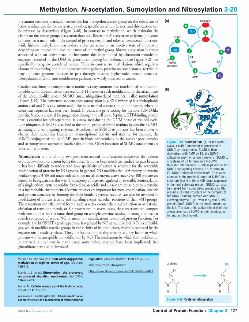
the amino terminus is usually irreversible, but the epsilon amino group on the side chain oflysine residues can also be acetylated by other, specific acetyltransferases, and this reaction canbe reversed by deacetylases (Figure 3-48). In contrast to methylation, which maintains thecharge on the amino group, acetylation does not. Reversible N-acetylation at lysine in histoneproteins has a major role in the control of gene expression and other chromosomal functions:while histone methylation may induce either an active or an inactive state of chromatin,depending on the position and the nature of the methyl group, histone acetylation is alwaysassociated with an active state of chromatin: this is promoted by chromatin-remodelingenzymes recruited to the DNA by proteins containing bromodomains (see Figure 3-2) thatspecifically recognize acetylated lysines. Thus, in contrast to methylation, which regulateschromatin by creating non-binding surfaces for regulatory proteins on core histones, acetylationmay influence genome function in part through affecting higher-order protein structure.Deregulation of chromatin modification pathways is widely observed in cancer.
Covalent attachment of one protein to another is a very common post-translational modification.In addition to ubiquitination (see section 3-11), another such modification is the attachmentof the ubiquitin-like protein SUMO (small ubiquitin-related modifier), called sumoylation(Figure 3-49). The consensus sequence for sumoylation is yKXE (where y is a hydrophobicamino acid and X is any amino acid); this is in marked contrast to ubiquitination, where noconsensus sequence has ever been found. In yeast, the gene coding for the sole SUMO-likeprotein, Smt3, is essential for progression through the cell cycle. Septin, a GTP-binding proteinthat is essential for cell separation, is sumoylated during the G2/M phase of the cell cycle.Like ubiquitin, SUMO is attached to the amino group of lysine residues by specific SUMO-activating and -conjugating enzymes. Attachment of SUMO to proteins has been shown tochange their subcellular localization, transcriptional activity and stability: for example, theSUMO conjugate of the RanGAP1 protein binds preferentially to the nuclear pore complexand so sumoylation appears to localize this protein. Other functions of SUMO attachment areuncertain at present.
Nitrosylation is one of only two post-translational modifications conserved throughoutevolution—phosphorylation being the other. Yet it has been much less studied, in part becauseit has been difficult to understand how specificity of action is achieved for the reversiblemodification of proteins by NO groups. In general, NO modifies the –SH moiety of cysteineresidues (Figure 3-50) and reacts with transition metals in enzyme active sites. Over 100 proteins areknown to be regulated in this way. The majority of these are regulated by reversible S-nitrosylationof a single critical cysteine residue flanked by an acidic and a basic amino acid or by a cysteinein a hydrophobic environment. Cysteine residues are important for metal coordination, catalysisand protein structure by forming disulfide bonds. Cysteine residues can also be involved inmodulation of protein activity and signaling events via other reactions of their –SH groups.These reactions can take several forms, such as redox events (chemical reduction or oxidation),chelation of transition metals, or S-nitrosylation. In several cases, these reactions can competewith one another for the same thiol group on a single cysteine residue, forming a molecularswitch composed of redox, NO or metal ion modifications to control protein function. Forexample, the JAK/STAT signaling pathway is regulated by NO at multiple loci. NO is a diffusiblegas, which modifies reactive groups in the vicinity of its production, which is catalyzed by theenzyme nitric oxide synthase. Thus, the localization of this enzyme is a key factor in whichproteins will be susceptible to modification by NO. The mechanism by which this modificationis reversed is unknown in many cases; some redox enzymes have been implicated; free glutathione may also be involved.
Control of Protein Function Chapter 3 127©2004 New Science Press Ltd
Methylation, N-acetylation, Sumoylation and Nitrosylation 3-20
McBride, A.E. and Silver, P. A.: State of the Arg: proteinmethylation at arginine comes of age. Cell 2001,106:5–8.
Stamler, J.S. et al.: Nitrosylation: the prototypicredox-based signaling mechanism. Cell 2001,106:675–683.
Turner, M.: Cellular memory and the histone code.Cell 2002, 111:285–291.
Workman, J.L. and Kingston, R.E.: Alteration of nucle-osome structure as a mechanism of transcriptional
regulation. Annu. Rev. Biochem. 1998, 67:545–579.
Web resource on nitrosylation:
http://www.cell.com/cgi/content/full/106/6/675/DC1
Figure 3-50 Cysteine nitrosylation
substrate
E1
E2ATP
AMP
SUMO
SUMOprecursor
Ulps
Ulps
(a)
(b)
S
S
S
S
S S
S
S
E2
E1
CH2
CH
C
O
N
H
NO
SSH
cysteine
CH2
CH
C
O
N
H
Figure 3-49 Sumoylation (a) In the SUMOcycle, a SUMO precursor is processed toSUMO by Ulp proteins. SUMO is thenderivatized with AMP by E1, the SUMO-activating enzyme, before transfer of SUMO toa cysteine of E1 to form an E1–SUMOthioester intermediate. SUMO is passed to theSUMO-conjugating enzyme, E2, to form anE2–SUMO thioester intermediate. This lattercomplex is the proximal donor of SUMO to asubstrate lysine in the yKXE target sequencein the final substrate protein. SUMO can alsobe cleaved from sumoylated proteins by Ulpproteins. (b) The structure of the complex ofthe SUMO-binding domain of a SUMO-cleaving enzyme, Ulp1, with the yeast SUMOprotein Smt3. SUMO is the small domain onthe left. The size of the active-site cleft of Ulp1allows even large SUMO–protein conjugatesto bind and be cleaved.