Embed Size (px)
Citation preview

Western Kentucky UniversityTopSCHOLAR®
Masters Theses & Specialist Projects Graduate School
12-2012
Contour Extraction of Drosophila Embryos UsingActive Contours in Scale SpaceSoujanya Siddavaram AnantaWestern Kentucky University, [email protected]
Follow this and additional works at: http://digitalcommons.wku.edu/theses
Part of the Genetics Commons, and the Theory and Algorithms Commons
This Thesis is brought to you for free and open access by TopSCHOLAR®. It has been accepted for inclusion in Masters Theses & Specialist Projects byan authorized administrator of TopSCHOLAR®. For more information, please contact [email protected].
Recommended CitationAnanta, Soujanya Siddavaram, "Contour Extraction of Drosophila Embryos Using Active Contours in Scale Space" (2012). MastersTheses & Specialist Projects. Paper 1222.http://digitalcommons.wku.edu/theses/1222


CONTOUR EXTRACTION OF DROSOPHILA EMBRYOS USING
ACTIVE CONTOURS IN SCALE SPACE
A Thesis
Presented To
The Faculty of the Department of Computer Science
Western Kentucky University
Bowling Green, Kentucky
In Partial Fulfillment
Of the Requirements for the Degree
Master of Science
By
Soujanya Siddavaram Ananta
December 2012


iii
ACKNOWLEDGMENTS
I owe my deepest gratitude to my advisor and mentor, Dr. Qi Li, for his
overwhelming encouragement, support and offered invaluable assistance. Dr. Qi Li
has exceptionally inspired and enriched my growth as both a student and person. His
supervision and guidance is unlike anything I have ever experienced. This thesis
would not have been possible without the dedication of Dr. Qi Li.
I gratefully acknowledge Dr. Guangming Xing, Dr. Huanjing Wang and Dr.
Qi Li for their supervision and precious time invested to read and provide correction to
this thesis. I am thankful that in the midst of their busy schedules, they accepted to be
members of my reading committee.
I would also like to thank those closest to me, whose presence helped make the
completion of my thesis work possible. These are Venkata Aditya Korada
(Knowledgeable expert in MATLAB who has helped me many times when I needed the
most) and Anoop Rao Paidipally. Anoop, thanks for all the patience and suggestions.
This thesis would not have been possible without your guidance and encouragement.
Most of all, I would like to thank my family, and especially my parents, for their absolute
confidence in me.
Soujanya Siddavaram Ananta

iv
CONTENTS LIST OF FIGURES vi ABSTRACT vii
1 INTRODUCTION 1
2 EXISTING METHODS FOR CONTOUR EXTRACTION 4
2.1 Edge detection . . . . . . . . . . . . . . . . . . . . . . . . . . . . . . . . 4
2.1.1 Edge detection methods . . . . . . . . . . . . . . . . . . . . . . . 5
2.1.2 Comparison between edge detection methods . . . . . . . . . . . . 6
2.1.3 Summary and limitations of edge detection . . . . . . . . . . . . . 7
2.2 Contour extraction . . . . . . . . . . . . . . . . . . . . . . . . . . . . . . 9
2.2.1 Advantages of the active contour over edge detection . . . . . . . . 13
3 PROPOSED FRAMEWORK 14
3.1 Outline of the framework . . . . . . . . . . . . . . . . . . . . . . . . . . . 15
3.2 Active contour segmentation . . . . . . . . . . . . . . . . . . . . . . . . . 17
3.2.1 Level-set . . . . . . . . . . . . . . . . . . . . . . . . . . . . . . . 18
3.2.2 Chan-Vese . . . . . . . . . . . . . . . . . . . . . . . . . . . . . . 20
3.2.3 Comparison between level-set and Chan-Vese segmentation . . . . 22
3.3 Distance-Normalized technique . . . . . . . . . . . . . . . . . . . . . . . . 25
3.4 Algorithm for optimal scale selection . . . . . . . . . . . . . . . . . . . . . 26
4 EXPERIMENT 28
4.1 Setup . . . . . . . . . . . . . . . . . . . . . . . . . . . . . . . . . . . . . 29
4.1.1 Data sets . . . . . . . . . . . . . . . . . . . . . . . . . . . . . . . 29
4.1.2 MATLAB . . . . . . . . . . . . . . . . . . . . . . . . . . . . . . . 29
4.1.3 Parameters . . . . . . . . . . . . . . . . . . . . . . . . . . . . . . 30

v
4.2 Distance-Normalized technique . . . . . . . . . . . . . . . . . . . . . . . . 31
4.3 Rejected case of contour extraction . . . . . . . . . . . . . . . . . . . . . . 32
4.3.1 Applying smoothness constraint . . . . . . . . . . . . . . . . . . . 33
4.4 Results obtained from the framework . . . . . . . . . . . . . . . . . . . . . 34
4.4.1 Successful case of extracted contours . . . . . . . . . . . . . . . . 34
4.4.2 Failure case of extracted contours . . . . . . . . . . . . . . . . . . 36
4.4.3 Successful case of extracted contours with energy parameter alpha . 37
5 CONCLUSION 38
5.1 Outcome . . . . . . . . . . . . . . . . . . . . . . . . . . . . . . . . . . . . 38
5.2 Future Scope . . . . . . . . . . . . . . . . . . . . . . . . . . . . . . . . . 40
BIBLIOGRAPHY 42

vi
LIST OF FIGURES
1.1 Embryo images containing variations in size, shape, orientation and ap-
pearance in addition to its neighboring context . . . . . . . . . . . . . . . . 2
2.1 Results of edge detection methods . . . . . . . . . . . . . . . . . . . . . . 8
2.2 Basic form of the active contour . . . . . . . . . . . . . . . . . . . . . . . 11
3.1
Variations of an embryonic image
.
.
.
.
.
.
.
.
.
.
.
.
.
.
.
.
.
.
.
.
.
.
14
3.2 Proposed framework for contour extraction in scale space . . . . . . . . . . 16
3.3 Contour results obtained by different scales by level-set. The result is not
satisfying because of (i) jaggy contour (ii) not smooth . . . . . . . . . . . . 19
3.4 Results are satisfying but the performance depends on scales . . . . . . . . 21
3.5 Comparison between level-set and Chan-Vese . . . . . . . . . . . . . . . . 23
4.1 Unsuccessful case of extracted contours using Distance-Normalized algo-
rithm . . . . . . . . . . . . . . . . . . . . . . . . . . . . . . . . . . . . . . 31
4.2 Rejected contours with angle less than 30 . . . . . . . . . . . . . . . . . . 32
4.3 Largest connected components: a) without, b) with smoothness constraint . 33
4.4 Successfully extracted contours with optimal scale in gaussian scale space . 35
4.5 Unsuccessful case of extracted contours with optimal scale . . . . . . . . . 36
4.6 Successfully extracted contours with optimal scale with energy parameter
alpha . . . . . . . . . . . . . . . . . . . . . . . . . . . . . . . . . . . . . . 37
5.1 Graph showing optimal scale of the images . . . . . . . . . . . . . . . . . 40

vii
CONTOUR EXTRACTION OF DROSOPHILA EMBRYOS USING
ACTIVE CONTOURS IN SCALE SPACE
Soujanya Siddavaram Ananta December 2012 44 Pages
Directed by: Dr. Qi Li, Dr. Guangming Xing and Dr. Huanjing Wang
Department Of Computer Science Western Kentucky University
Contour extraction of Drosophila embryos is an important step to build a
computational system for pattern matching of embryonic images which aids in the
discovery of genes. Automatic contour extraction of embryos is challenging due to several
image variations such as size, shape, orientation and neigh- boring embryos such as
touching and non-touching embryos. In this thesis, we introduce a framework for contour
extraction based on the connected components in the gaussian scale space of an embryonic
image. The active contour model is applied on the images to refine embryo contours.
Data cleaning methods are applied to smooth the jaggy contours caused by blurred
embryo boundaries. The scale space theory is applied to improve the performance of
the result. The active contour adjusts better to the object for finer scales. The proposed
framework contains three components. In the first component, we find the connected
components of the image. The second component is to find the largest component of the
image. Finally, we analyze the largest component across scales by selecting the optimal
scale corresponding to the largest component having largest area. The optimal scale at
which maximum area is attained is assumed to give information about the feature being
extracted. We tested the proposed framework on BDGP images, and the results achieved
promising accuracy in extracting the targeting embryo.

INTRODUCTION
Genetics provide the most powerful approach available to understand the
function of the human genes. Model organisms, such as Drosophila melanogaster, share
many genes with humans. The Drosophila embryo is one of the most well known model
organism that is widely used in biological research particularly in genetics and
developmental biology [32]. Drosophila is also used in life history evolution. A model
organism is a species that is studied to understand a biological phenomena, with the
expectation that the experiments made on the model organism will provide awareness in
the research of other organisms. The Drosophila embryo is a useful model organism for
studying many aspects of development because it is small and cheap to culture in the lab.
It also has a short life span.
Images of Drosophila embryos consist of significant gene expression patterns
[21]. Information of the gene expression is captured for understanding the development of
embryos at various stages. Analysis of similar gene expression patterns is important in
understanding the interaction of genes that generate the body plans of Drosophilas,
humans and other metazoans [17]. The recent technique used to study gene expression
patterns is in situ hybridization [25]. In situ hybridization(ISH) is a protocol used to
determine patterns of gene expressions during embryogenesis of Drosophila [30].
Comparison of expression patterns is most biologically meaningful when images
from a similar time point (developmental stage range) are compared [11]. A set of
embryonic images contain information on the spatial and temporal patterns that are
extremely useful for the study of gene-gene interaction which is a biological problem.
Spatiotemporal gene expression is the activation of genes of a particular location at a
particular time during development. Dark regions in an embryonic image, as shown in
Figure 1.1, indicate a significant gene expression pattern. Given two standardized images
1

of embryos at same development stage, the interaction strength of two genes can be
quantified by computing the similarity of expression patterns. Traditionally, Drosophila
development is divided manually into stages by qualitative visual inspection. The manual
labeling of stages has become a bottleneck with the dramatically increasing data, e.g., the
high resolution embryonic images contributed by The Berkley Drosophila Genome
Project (BDGP) [30, 1].
(a) embryo with variation in the orientation (b) image with partial embryo
(c) embryo image with proper alignment
Figure 1.1: Embryo images containing variations in size, shape, orientation and appearancein addition to its neighboring context
Biologists spend significant time in manually annotating images from large scale
experiments like BGDP [1]. Annotation includes data such as view, orientation, and stages
of development of the embryos [14]. The advanced microscopes have led to the rapidly
growing digital data in biology. In order to deal with these data sets, it is necessary to
develop automated methods for extracting and analyzing images. Embryo contour
extraction is an important step to build pixel-to-pixel correspondence between embryos of
interest so that the comparisons between embryonic images in a computation system are
biologically meaningful [21]. A comparative analysis of the images contributes to the
study of regulatory networks governing embryonic development [13, 10].
2

Several challenges are faced due to image variations such as size, shape,
orientation and context of the neighboring embryos. Size and shape variations are due to
different development stages. Another image variation is due to the context of
neighboring embryos i.e. the embryo of interest may or may not intersect neighboring
embryos. The problem of contour extraction from a blurred digital image is some sort of
pattern recognition problem.
In this thesis, we design an automated framework for contour extraction using
active contours with scale space. The scale space theory used in this thesis is contributed
by Lindeberg [23]. The scale-space theory focuses on the basic fact that the image
structures, like objects in the world, exist as meaningful entities over certain ranges of
scale [22]. In general, one cannot expect to know what scales are appropriate in describing
those image structures. The active contour adjusts better to the object for finer scales [29].
The proposed framework has three main stages. The first stage is applying the
active contour segmentation and finding the connected components of the image after
segmentation. The second step is to extract the largest connected component of the image.
The final step is to apply a criterion to decide the optimal scale to locate a targeting
embryo. The corresponding scale with the largest area is selected as the optimal scale for
a particular image. The scale σ which is a gaussian kernel for suppression of noise in our
experiment ranges from 0.5-30 in sampled space. Active contour model is described in
detail in Chapter 2.
This thesis is organized as follows: Chapter 2 gives a review of the two main
existing methods for contour extraction: edge detection and active contour, and the
advantages of the active contour over edge detection. Chapter 3 presents the proposed
framework used for contour extraction. Chapter 4 shows results of the proposed
framework and Chapter 5 provides the summary, conclusion, and the future scope of this
research.
3

EXISTING METHODS FOR CONTOUR EXTRACTION
This chapter mainly focuses on the existing methods of contour extraction. Edge
detection and the active contour models are the most popular methods used for contour
extraction for wide range of applications, such as image segmentation and motion
tracking. This chapter is organized as follows: Section 1 describes the various edge
detection methods, comparison between them and the limitations. Section 2 introduces
contour extraction, its significance and the active contour model which is the main
technique used for extraction of the contour in this thesis.
2.1 Edge detection
Edge detection is a basic rule in the field of image processing and computer
vision in the areas of feature detection and extraction. It refers to the process of
determining sharp discontinuities in an image. The image given as input is stored in a
matrix form which contains the pixels of the image. The gradient of the matrix is
calculated and if the gradient is large then the pixel is determined as an edge pixel. Edge
detection techniques may be grouped into two different categories:
1. Gradient based edge detection
• Sobel
• Prewitt
• Roberts
2. Laplacian based edge detection
• Laplacian
• Zero-cross
4
The key of edge detection is the choice of threshold. The choice of threshold
directly determines the results of edge detection. The lower the threshold, the more the
edges will be detected, and the result will be increasingly susceptible to noise and detect
edges of irrelevant features in the image. Conversely, a high threshold may miss subtle
edges, or result in fragmented edges. If the edge threshold is applied just to the gradient
magnitude image, the resulting edges in general, will be thick and some type of edge
thinning post-processing is necessary.
Edge thinning is a technique used to remove the undesired false points on the
edge of an image. This technique is applied after the image has been filtered for noise
(using median, gaussian filter, etc.). The edge operator has been applied to detect the edges
after the edges have been smoothed using an appropriate threshold value. This removes all
the undesired points and if applied carefully, results in one pixel thick edge elements.
2.1.1 Edge detection methods
Sobel Filter
The Sobel filter finds edges using the Sobel approximation to the derivative. It
returns edges at those points where the gradient of image is maximum. The operator
consists of a pair of convolution kernels.
Prewitt Filter
The Prewitt filter finds edges using the Prewitt approximation to the derivative. It
returns edges at those points where the gradient of image is maximum.
Roberts Filter
The Roberts filter finds edges using the Roberts approximation to the derivative.
It returns edges at those points where the gradient of image is maximum.
Laplacian Of Gaussian Filter
The Laplacian of Gaussian filter finds edges by looking for zero crossings after
filtering image with a Laplacian of Gaussian filter.
5

Zero Cross Filter
The Zero-cross filter finds edges by looking for zero crossings after filtering
image with a filter you specify.
Canny Method
The Canny method finds edges by looking for local maxima of the gradient of
image. The gradient is calculated using the derivative of a gaussian filter. The method uses
two thresholds to detect strong and weak edges, and includes the weak edges in the
output, only if they are connected to strong edges. This method is therefore more likely to
detect true weak edges.
2.1.2 Comparison between edge detection methods
Gradient based algorithms such as Sobel and Prewitt are sensitive to noise. The
Sobel operator consists of a pair of 3*3 convolution kernels. One kernel is the other
rotated by 90◦. Using the kernel the absolute magnitude of the gradient is found. The
Roberts operator consists of a pair of 2*2 convolution kernels. The absolute value of the
gradient is computed. Prewitt operator is similar to the Sobel operator as illustrated in
Figure 2.1. They are good at detecting vertical and horizontal edges in images.
The Laplacian is a 2-D measure of the 2nd spatial derivative of an image. The
Laplacian is often applied to an image that has first been smoothed with something
approximating a gaussian smoothing filter in order to reduce its sensitivity to noise.
Because these kernels are approximating a second derivative measurement on the image,
they are very sensitive to noise. To counter this, the image is often gaussian smoothed
before applying the Laplacian filter. This pre-processing step reduces the high frequency
noise components prior to the differentiation step.
6

The Canny edge detector is the optimal edge detection method. Canny followed
a list of criteria to improve the previous edge detection methods [7].
1. Low error rate - It is important not to miss the edges that occur in the image and
there should be no responses to non-edges.
2. Edge points should be well localized - The difference between the edge pixels and
the actual edge should be minimum.
3. Only one response to a single edge - The previous methods does not eliminate the
possibility of multiple responses to an edge.
The Canny edge detector first smoothes the image to eliminate the noise and
then finds the image gradient. The pixels that are less than the maximum gradient are
suppressed and the gradient is further reduced by hysteresis. Hysteresis is used to track
along the remaining pixels that have not been suppressed. Hysteresis uses two thresholds
which allows more flexibility than a single threshold approach. If the magnitude is below
the first threshold, it is made as non edge. If the magnitude is above the high threshold, it
is made as an edge. Canny edge detection algorithm has a number of adjustable
parameters, which have an impact on the effectiveness of the algorithm and the
computation time.
1. Size of the gaussian filter
2. Threshold values
Figure 2.1 shows results of the various edge detection methods.
2.1.3 Summary and limitations of edge detection
Gradient based algorithms such as Sobel, Prewitt and Roberts are sensitive to
noise and the size of kernel filter and coefficients are fixed and cannot be adapted to a
7

(a) Sobel (b) Prewitt (c) Roberts
(d) Laplacian (e) Zero-cross (f) Canny
Figure 2.1: Results of edge detection methods
given image. An adaptive edge detection algorithm is necessary to provide a robust
solution that is adaptable to varying noise levels. The performance of the Canny algorithm
depends heavily on the adjustable parameters, sigma, the standard deviation for the
gaussian filter, and two threshold values (’T1’ and ’T2’). The parameters can be adapted to
different environments. This implies more blurring, necessary for noisy images, as well as
detecting larger edges. The user can tailor the algorithm by adjusting these parameters to
adapt to different environments.
Based on comparison shown in Figure 2.1, we observe that Canny algorithm
gives the optimal result as the parameters can be adaptive to different environments. The
maximum number of edges are detected using Canny edge detector. Most of the edges are
obtained by varying the parameters using Canny edge detector whereas in Sobel, Prewitt
and Roberts not all edges are detected because of fixed parameters. These algorithms are
not able to detect the edges while removing the noise whereas Canny does.
Canny’s edge detection algorithm is computationally more expensive compared
to Sobel, Prewitt and Roberts operator. However, the Canny’s edge detection algorithm
performs better than all these operators under almost all scenarios.
8

Limitations of edge detection
1. Edges of a neighboring embryo are also detected. Note that an embryonic image
may contain a neighboring embryo, in addition to a targeting embryo.
2. All image points that have the maximum gradient are treated as edges. The gradient
can be maximum inside the embryo and that point is also treated as an edge where
as in reality it is not.
2.2 Contour extraction
In contrast to edge detection, contour extraction is a higher-level formulation
that aims to extract the boundary shape of an object of interest in an image. The image
processing stage is necessary in order to provide a standardized image database for gene
expression pattern comparison [27]. A lot of research is being done in these areas and
many approaches were reported so far. Contour extraction can be generally classified into
two categories:
1. Region-based [6, 4]
2. Edge-based [6, 24]
Edge-based techniques rely on discontinuities in image values between distinct
regions and the goal is to accurately distinguish the boundary separating these regions.
Edge-based models consist of evolving a contour in homogeneous areas, and locally
stopping it when it reaches high image gradients [28]. They have several advantages like
local coherence and robustness to region inhomogeneities, but they also have important
drawbacks that make them inefficient on noisy images or when the contour initialization is
not completely inside or outside the region to segment. Region-based searches for
equality inside a sub-region, based on a desired property, e.g. intensity, color, and texture.
Region-based techniques depend on common patterns in intensity values within a cluster
9

of neighboring pixels [6]. These models take account of the whole region and are thus
more robust to noise and to initialization than edge-based models. Due to minimization of
global energy, region-based active contours do not have any limitation placing the initial
contour. Region-based active contour can detect interior boundaries regardless of the
placement of the initial contour.
Contour extraction is an important step for feature detection. Once the contour is
extracted, different characteristics of the contour will be examined and used as features
which will later be used in pattern matching of the images. The region of interest is
extracted and used to compare gene expression patterns of the image at different stages of
development. An embryo image taken during embryogenesis usually contains multiple
embryos with region of interest at the center of the image. The neighboring embryo may
be touching or non touching.
The two main approaches of active contour based on mathematical
implementation are: Snakes and level-sets. Snakes explicitly shift predefined contour
points based on an energy minimization technique, while level-set approaches move
contours completely as a particular level of a function.
Snakes or the active contours, are used extensively in computer vision and image
processing applications, particularly to locate object boundaries. The active contour is an
important model for defining an object outline from a noisy image using techniques of
curve revolution [5]. Snakes are curves that can deform within the image plane and
capture a desired feature. Methods to evolve these contours were introduced to computer
vision by Kass, Witkin and Terzoupolus [15]. This model is an energy minimizing spline
guided by external forces and influenced by image forces that pull it towards features such
as edges [16]. The active contour model is more consistent than edge detection.
The active contours can be classified into two broad categories: parametric
active contours [15, 34] and geometric active contours [19]. The parametric active
contours represent contours explicitly as parameterized curves where as the geometric
10

contours represent contours implicitly as level-sets of two dimensional functions.
Geometric active contours are classified into two categories: models based on boundary
functionals and models based on area functionals. It is a flexible curve which will
dynamically adapt to the required edges in the image. The active contour consists of a set
of controlled points or contour points as shown in Figure 2.2.
Figure 2.2: Basic form of the active contour
The initial contour must be specified. The contour will then be attracted to features in the
image extracted by internal energy creating an attractor image. The energy function is a
weighted combination of internal and external forces. Snake is defined by :
1. a set of contour points
2. an external energy term
3. an internal energy term
An initialized contour is considered. The active contour is used to refine the
initialized contour of an embryo of interest. It dynamically moves towards the region of
interest by minimizing its energy iteratively. Minimization of energy from high to low is
an aspect of optimization. We need to shrink or expand the active contour depending on
the image which is known as external energy. There are several aspects of defining energy
11
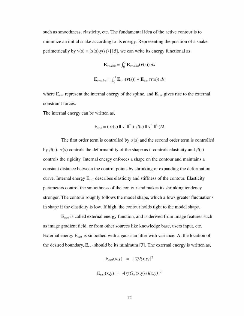
such as smoothness, elasticity, etc. The fundamental idea of the active contour is to
minimize an initial snake according to its energy. Representing the position of a snake
perimetrically by v(s) = (x(s),y(s)) [15], we can write its energy functional as
Esnake =∫ 1
0Esnake(v(s)) ds
Esnake =∫ 1
0Eint(v(s)) + Eext(v(s)) ds
where Eint represent the internal energy of the spline, and Eext gives rise to the external
constraint forces.
The internal energy can be written as,
Eint = ( α(s) || v′ ||2 + β(s) || v′′ ||2 )/2
The first order term is controlled by α(s) and the second order term is controlled
by β(s). α(s) controls the deformability of the shape as it controls elasticity and β(s)
controls the rigidity. Internal energy enforces a shape on the contour and maintains a
constant distance between the control points by shrinking or expanding the deformation
curve. Internal energy Eint describes elasticity and stiffness of the contour. Elasticity
parameters control the smoothness of the contour and makes its shrinking tendency
stronger. The contour roughly follows the model shape, which allows greater fluctuations
in shape if the elasticity is low. If high, the contour holds tight to the model shape.
Eext is called external energy function, and is derived from image features such
as image gradient field, or from other sources like knowledge base, users input, etc.
External energy Eext is smoothed with a gaussian filter with variance. At the location of
the desired boundary, Eext should be its minimum [3]. The external energy is written as,
Eext(x,y) = -|5I(x,y) |2
Eext(x,y) = -|5Gσ(x,y)∗I(x,y) |2
12

where I(x,y) is the gray scale image, Gσ(x,y) is a two dimensional gaussian kernel that
convolute with the image I(x,y). 5 is the gradient operator. The effect of using gaussian
kernel convolute with image I(x,y) is to smooth the image, and to blur the image
boundaries. The external forces are responsible for putting the snake near the desired local
minimum. The active contour has the total energy E.
During the process of contour deformation, the curvature based internal force
maintains the contour smoothness, while the gradient-based external force attracts the
contour to the desired boundaries in the image. The deformation finally stops when the
snake reaches a local energy minimum. The active contours have become an invaluable
base tool for segmentation and object recognition.
2.2.1 Advantages of the active contour over edge detection
1. Active contours are self-adaptive in search of minimal energy state.
2. The external and internal energy forces can be used to manipulate the active contour.
3. They can be made sensitive to image scale by incorporating gaussian smoothing in
the image energy function. Gaussian smoothing is used to suppress the noise in the
digital images.
The only drawback of active contour model is the existence of local minima in
the active contour energy. The main difficulty with parametric active contour algorithm is
that the initial contour must be close to the true boundary or else it will likely converge
with the wrong result [34]. The active contour segmentation may also result in multiple
components and also the contour results are sensitive to scales. The largest of the
components is expected to be the target contour.
13

PROPOSED FRAMEWORK
This chapter focuses on the proposed framework for contour extraction. Section
1 describes the outline of the framework. Section 2 addresses the active contour
segmentation and the active contour models used for the experiments and the comparison
between them. Section 3 illustrates the Distance-Normalized technique. Section 4
provides the details of the optimal scale selection criterion.
Information of the gene expression is captured for understanding development of
the embryo at various stages. In order to discover the interaction of genes automatically,
we build contour point-to-point correspondence between embryos of interest. The first step
is to extract the contour of the target embryo automatically. Embryo contour extraction is
an important step to build pixel-to-pixel correspondence between embryos of interest so
that the comparisons between embryonic images in a computation system are biologically
meaningful. Contour extraction is very challenging due to image variations. Variations
include size, shape, noise, orientation, partial embryos. Figure 3.1 shows the variations in
an embryonic image.
(a) image with partial touching embryo (b) image with partial non-touching embryo
(c) embryo with variation in the orientation
Figure 3.1: Variations of an embryonic image
Edge detection results in lot of noise after segmentation. All the points having
14

the gradient maximum are treated as edges. The gradient can be maximum inside as well
as outside the embryo. Therefore more advanced technique called the active contour is
used to eliminate the limitations of edge detection technique. The active contour model
provides an efficient way for image segmentation in which the boundaries of the object are
detected. Due to serious image variations, the existing techniques failed to obtain desirable
results. The main criteria of the research work is largest of the largest, i.e., largest area of
the resultant largest component of the image obtained after segmentation. The criterion is
to determine an automatic procedure to determine the optimal scale for a specific image.
3.1 Outline of the framework
In this section, we proposed an approach to remove contour pixels of
neighboring embryos. We assumed that the embryo of interest dominates the neighboring
context in an image. Furthermore, we categorized the neighboring context into
overlapping and non-overlapping context. It is expected that the first case is easy to handle
than the second case due to the overlapping region.
The data set is passed through the active contour model. The main scale is
considered to be σ which represents the gaussian kernel. σ was used to suppress the noise
in the digital image. σ gives the standard deviation of the gaussian low pass filter.
Convolution of the image with the gaussian filter is performed and then the convoluted
image is passed through the active contour model. The resulting images might have jaggy
contours as well as multiple connected components. Jaggy contours are eliminated by
applying the smoothness constraint. We applied the proposed criterion to extract the
largest component which is expected to be the target embryo. The proposed framework is
divided into three components. The first component is to find the number of connected
components of the resulting images of the active contour model. A connected component
is defined as a connected region whose pixels belong to the same class in a segmented
image. The second component is to find the largest component among all the connected
15
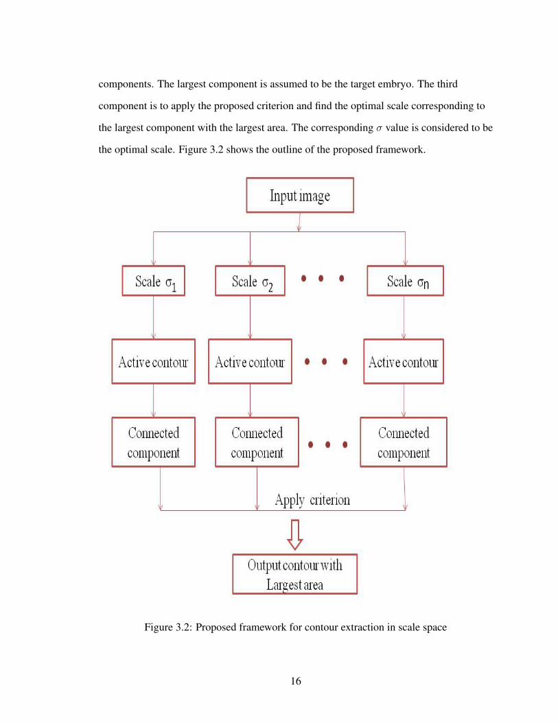
components. The largest component is assumed to be the target embryo. The third
component is to apply the proposed criterion and find the optimal scale corresponding to
the largest component with the largest area. The corresponding σ value is considered to be
the optimal scale. Figure 3.2 shows the outline of the proposed framework.
Figure 3.2: Proposed framework for contour extraction in scale space
16

3.2 Active contour segmentation
As described in the last chapter, the active contour model is a methodology
based on the use of deformable contours, which adapt its border to the diverse shapes of
the objects in the images [31]. In some methods based on this methodology, the results are
very conditioned by the selection of the initial position of the contour. The active contour
methods provide an effective way for segmentation, in which boundaries of the objects are
detected by evolving curves.
This technique is more advanced compared to edge detection. This method is
applied to refine the contour of target embryo in an image based on an initialized contour.
The active contours writhe and move under the influence of external and internal forces,
toward a feature of interest in an image, usually an edge. The active contours are most
often used with images to find and describe regions of interest [18].
We consider an initialized contour and input image. We deform the set of points
to match with the initialized contour called the active contour so that it converges with the
real contour. It is mainly based on optimization of energy. The minimization of the energy
associated with the current contour is a sum of an internal and external energy. The energy
functional which is minimized is a weighted combination of internal and external forces.
Low energy implies that the contour is more stable.
E = α1 Eint + α2 Eext
Level-set and Chan-Vese are the two active contour models used for our
experiments. The main difficulty in the active contour model is the choice of the initial
contour and value of the parameters. The only drawback of the active contour model is the
existence of local minima in the active contour energy. The existence of local minima in
its functional energy makes the initial contour critical to extract meaningful objects lying
in images.
17
3.2.1 Level-set
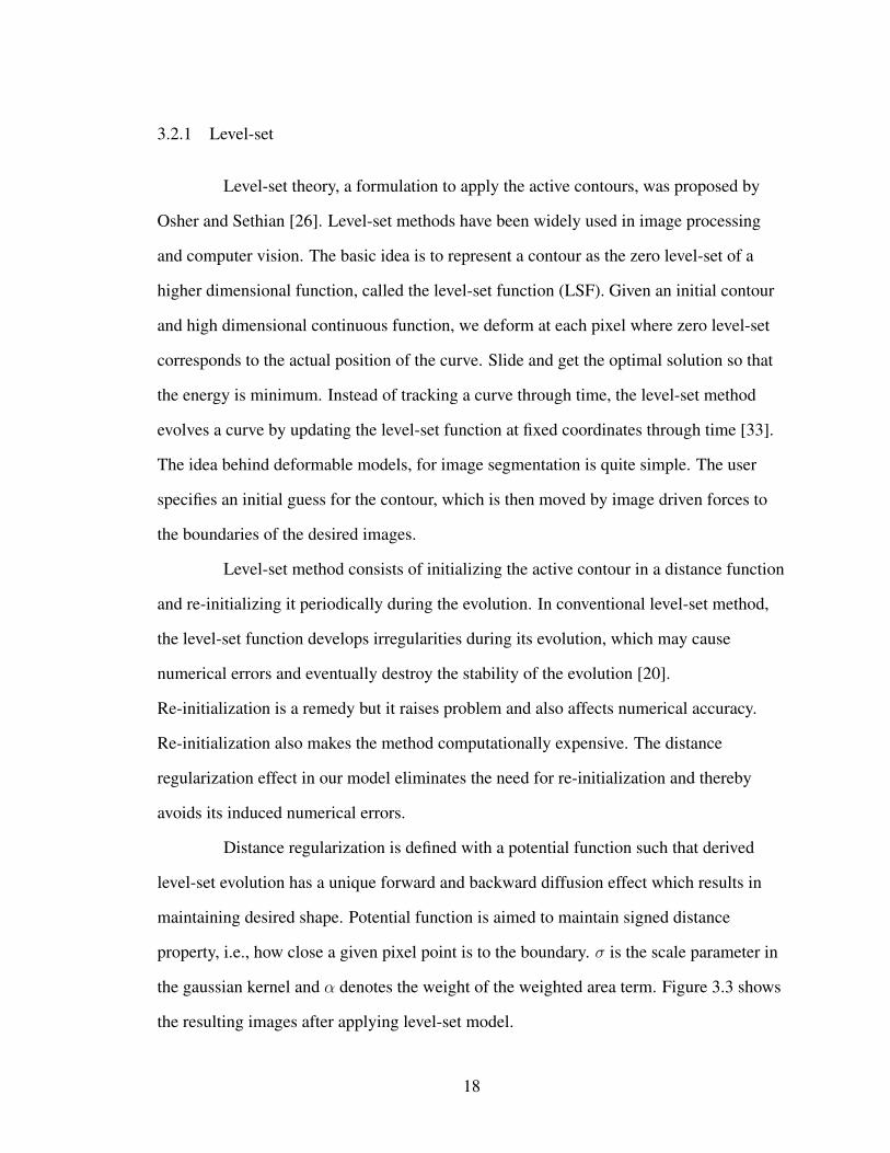
Level-set theory, a formulation to apply the active contours, was proposed by
Osher and Sethian [26]. Level-set methods have been widely used in image processing
and computer vision. The basic idea is to represent a contour as the zero level-set of a
higher dimensional function, called the level-set function (LSF). Given an initial contour
and high dimensional continuous function, we deform at each pixel where zero level-set
corresponds to the actual position of the curve. Slide and get the optimal solution so that
the energy is minimum. Instead of tracking a curve through time, the level-set method
evolves a curve by updating the level-set function at fixed coordinates through time [33].
The idea behind deformable models, for image segmentation is quite simple. The user
specifies an initial guess for the contour, which is then moved by image driven forces to
the boundaries of the desired images.
Level-set method consists of initializing the active contour in a distance function
and re-initializing it periodically during the evolution. In conventional level-set method,
the level-set function develops irregularities during its evolution, which may cause
numerical errors and eventually destroy the stability of the evolution [20].
Re-initialization is a remedy but it raises problem and also affects numerical accuracy.
Re-initialization also makes the method computationally expensive. The distance
regularization effect in our model eliminates the need for re-initialization and thereby
avoids its induced numerical errors.
Distance regularization is defined with a potential function such that derived
level-set evolution has a unique forward and backward diffusion effect which results in
maintaining desired shape. Potential function is aimed to maintain signed distance
property, i.e., how close a given pixel point is to the boundary. σ is the scale parameter in
the gaussian kernel and α denotes the weight of the weighted area term. Figure 3.3 shows
the resulting images after applying level-set model.
18

(a) σ=0.5 (b) σ=1.5 (c) σ=5
Figure 3.3: Contour results obtained by different scales by level-set. The result is notsatisfying because of (i) jaggy contour (ii) not smooth
19

3.2.2 Chan-Vese
Chan-Vese model implements level-set as well as the active contour. Chan and
Vese proposed multi-phase active contour model [9, 8], which increases the amount of
subsets that the active contours can locate simultaneously. In this segmentation technique,
we pass external energy factor α as a parameter to the function. The energy in this
technique is defined in terms of segmentation. This is a nice way to segment images
whose foregrounds and backgrounds are statistically different and homogeneous [9].
There are some objects whose boundaries are not well defined through the
gradient. The main idea of Chan and Vese model is to consider the information inside the
regions not only at their boundaries. Chan and Vese introduce a new active contour model,
called ”without edges”. Region based active contour models, e.g Chan and Vese [9] are
equivalent to boundary based active contour models. Boundary based methods handle
changes in topology and provide robust stopping terms to detect the goal contours. Most
Region-based models consists of two parts: the regularity, which determines the smooth
form of the contours, and the energy minimization part, which determines for equality of a
preferred feature within a subset. Region-based active contours divides an image into
several sub-regions. These regions exist either inside or outside the contour. The active
contour model proposed by Chan and Vese is very robust to initialization and gives good
results when there is a difference between the foreground and background means.
σ and α are the two main parameters used in the Chan-Vese model to minimize
the energy function. α value represents the weight of the external energy factor. The
higher the value of α, the smoother is the image, which is one of the aspect of defining
energy. σ is the scale parameter in the gaussian kernel. Smoothing of the image is done
using gaussian convolution. A gaussian filter smoothes an image by calculating weighted
averages in a filter box.
20
(a) σ=0.5 (b) σ=5 (c) σ=7
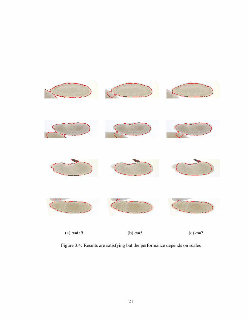
Figure 3.4: Results are satisfying but the performance depends on scales
21

The Figures 3.3 and 3.4 demonstrate that the contour results are sensitive to
scales. The contour results are different for different parameters. These figures
demonstrates the motivation of this research. We now compare the two methods, level-set
and Chan-Vese to see which method yields better results and then apply the proposed
criteria to that method.
3.2.3 Comparison between level-set and Chan-Vese segmentation
We compare the resultant output images of the level-set and Chan-Vese
segmentation. There are two main comparisons to be done:
1. Comparison of output images by considering different scale values in each of the
models.
2. Comparison between the level-set and Chan -Vese methods.
The contour is influenced by internal and external forces varying its shape
adaptively. Chan-Vese yields the better results than level-set. Chan-Vese segmentation
results in more smoothness which is one of the aspect of defining external energy. The
level-set model is the edge-based active contour model where as Chan-Vese is region
based active contour model. Edge based active contour models evolve the contour towards
one way, inside or outside. Therefore, an initial contour must be placed completely inside
or outside the region of interest. Edge-based active contours may remove the blurry
boundaries, and they are sensitive to noise as edge-based segmentation does. Figure 3.5
shows the comparison between the resulting images of level-set and Chan-Vese
segmentation.
22

(a) Level-set (b) Chan-Vese
Figure 3.5: Comparison between level-set and Chan-Vese
23

The performance of the Chan-Vese model is good but depends on scale. This is
the main motivation of this research. We need to find the scale which yields the best
result. All the current segmentation techniques iterate till the energy is minimized. The
main concern is that there is no guarantee that these techniques take global minimal
energy under consideration. The sub-optimal energy might be considered as the minimal
energy which is not the optimal solution.
The next step is to do some kind of pre-processing before segmentation. The
initial pre-processing technique that has been proposed is to calculate the weight
(normalized distance) of each pixel and multiply it with the original image pixel value and
then perform the segmentation.
24

3.3 Distance-Normalized technique
A pre-processing technique for contour extraction which considers an additional
energy factor is proposed. The idea is to consider the normalized distance of a pixel to the
center of the embryo. This pre-processing technique is applied to the active contour model
to make the resultant image more smooth. The additional energy factor that we consider is
the normalized distance at the position of the pixel under consideration.
Algorithm Distance-Normalized
1. For each image in the data set
2. Compute the longest distance from the pixel at the center to the pixel at the corner
of the image
3. Compute the distance of the pixel under consideration to the corner pixel
4. Compute ratio of the distance of the current pixel to the largest distance
5. Multiply the weight with the RGB value of the current pixel
6. The RGB values are combined to get the true color image
This pre-processing technique was applied to obtain the results after contour
extraction with more accuracy. This technique is expected to remove the neighboring
embryos at least to some extent.
25

3.4 Algorithm for optimal scale selection
The proposed criteria extracts the largest component of the image after
segmentation. The main aim is to design automatic procedure by selecting the largest area
of the largest component of all scales and determine the optimal scale for a specified
image. The contour extracted by Chan-Vese method highly depends on scale. From the
Figure 3.4, we observe that the results obtained from the Chan-Vese segmentation are
satisfying but the performance depends on scale. Different kinds of image structures give
rise to contours at different scales.
The connected components of the images obtained from the active contour
segmentation are found. As the scale increases, the number of connected components
decrease. The image is processed repeatedly by modifying the σ value. σ is the gaussian
kernel used in this framework. Gaussian smoothing is low-pass filter. Filter blurs
everything that is smaller than the filter. It suppresses high-frequency detail (noise, but
also edges), while preserving the low-frequency parts of the image (i.e. those that don’t
vary so much). We focus on analyzing largest connected components across scales. Our
criterion is to select the optimal scale by taking into account the resultant contour which
has maximum area i.e. total number of pixels compared to the resultant contours of other
scales, and its corresponding scale.
26

Algorithm Selection of parameter based on largest of the largest criteria
1. For each image in the data set
2. For each of the n scales
3. Perform Active contour segmentation
4. For every 15 pixels
5. Compute average
6. Average value is replaced for 15 pixels // for smoothing of the image
7. Find the largest component of the image
8. Compute the area for the largest component
27

EXPERIMENT
This chapter mainly focuses on the experiments done on the proposed
framework. Section 1 describes the main setup required i.e. about the data sets, the tool
used in implementing the framework and the parameters considered. Section 2 shows the
results of initial pre-processing technique proposed. Section 3 describes the case of the
rejected contours and the criterion applied. It also addresses the smoothness constraint
applied in the framework. Section 4 displays results of the framework obtained by
comparing different scales.
We will test the proposed framework on BDGP (Berkley Drosophila Genome
Project) images [1]. These images were captured using advanced microscopes for the
determination of the gene expression patterns of Drosophila embryos in different
development stages. Each image is a high-resolution spatial representation of an embryo
that might be neighbored by other embryos. The algorithm makes use of the active
contour model which is also called as snake. The active contour is a frame work which
defines an image outline from a noisy image. The active contours do not solve the entire
problem in finding target contours in images. They depend on information in image data
or higher level image understanding process. When snake is placed near the object
contour, it moves dynamically towards object contour by minimizing its energy iteratively.
28

4.1 Setup
In this section, we describe the setup of proposed framework. Firstly, we focus
on the data set images obtained from BDGP. We then address the tool used for the
development environment. Finally, we talk about the parameters used in the framework.
4.1.1 Data sets
The BDGP gene disruption project consists of a large collection of Drosophila
strains where each strain contains a single genetically engineered P transposable element
inserted in a defined genomic region. The resulting images of this project are in very large
number. Sophisticated tools are needed to annotate this amount of data. BDGP aims to
provide common data sets to be shared among various research groups as a stable basis for
the evaluation and comparison of different methods for the analysis of human DNA
sequences [1]. These data sets are used by algorithms which are aimed towards gene
finding and the identification of regulatory sequence. We use high throughput images to
determine patterns of gene expression during embryogenesis.
4.1.2 MATLAB
We use MATLAB to implement the proposed framework. MATLAB is a high
level language and user friendly interactive environment for numerical computation,
visualization and programming. The language tools and built-in functions in MATLAB
enable the user to explore multiple approaches and reach a solution at a faster pace.
MATLAB can be used for wide range of applications including image processing and
computational biology. Image processing toolbox provides set of standard algorithms and
graphical tools for image processing, analysis, visualization and algorithm development.
Feature detection, noise reduction, image segmentation can be performed using MATLAB
[2]. Graphic tools allow us to explore diverse set of images, create contours, extraction of
29

a contour and manipulate region of interest. Edge-detection algorithms identify object
boundaries in an image. Image segmentation algorithms determine region boundaries in
an image.
4.1.3 Parameters
The parameters we focus are α which is the external energy factor and σ which
defines the gaussian kernel. α is the weight of the smoothing term where as gaussian
kernel is used for suppression of noise. A gaussian filter is created and then convoluted
with the image before passing it to the active contour model. The gaussian smoothing
operator is a 2D convolution operator that is used to blur images and suppress the noise in
the image [12]. Gaussian filters are effective low pass filters from the perspective of both
the spatial and frequency domains with standard deviation σ. An edge is a local feature in
an image, and a smoothing operation that gives more significance to pixels farther away
will distort its features. Properties that make the gaussian smoothing effective are:
1. Gaussian low pass filter is rotationally symmetric, i.e., the amount of smoothing
performed by the filter in all directions will be same.
2. The gaussian function has a single lobe, i.e., each image pixel is replaced with a
weighted average of neighboring pixels resulting in smoothing. The weight given to
a neighbor decreases monotonically with distance from the central pixel, giving
most weight to central pixels.
3. The relationship between σ and degree of smoothing is a larger sigma implies wider
gaussian filter and great smoothing.
4. Implementation of large gaussian filter can be done efficiently.
30

4.2 Distance-Normalized technique
This technique is applied before performing the segmentation. The resultant
image after performing distance normalization is passed to the Chan-Vese method. This
technique is expected to smoothen the image and remove the neighboring context to some
extent. The output obtained was not as expected. The extracted contour is not the same as
the region of interest. Figure 4.1 shows the resulting images after applying the
Distance-Normalized technique and performing contour extraction.
Figure 4.1: Unsuccessful case of extracted contours using Distance-Normalized algorithm
31
4.3 Rejected case of contour extraction
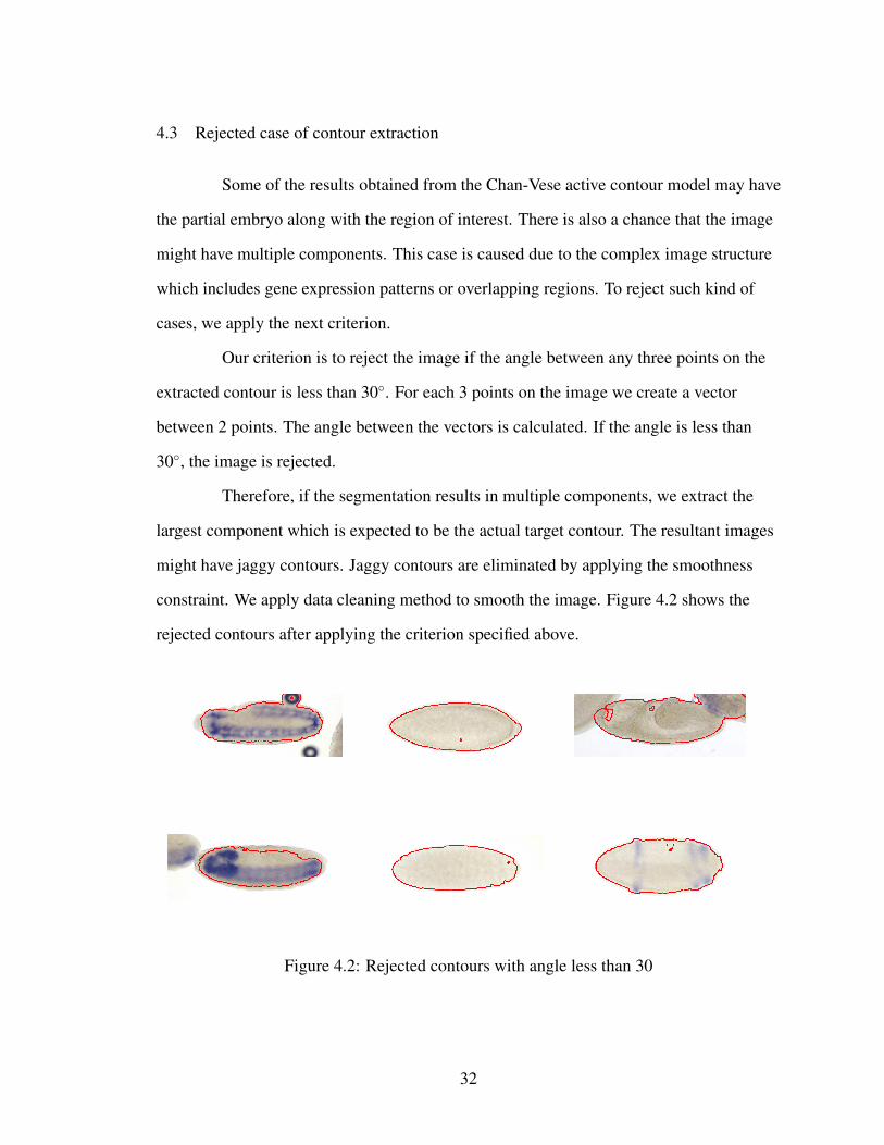
Some of the results obtained from the Chan-Vese active contour model may have
the partial embryo along with the region of interest. There is also a chance that the image
might have multiple components. This case is caused due to the complex image structure
which includes gene expression patterns or overlapping regions. To reject such kind of
cases, we apply the next criterion.
Our criterion is to reject the image if the angle between any three points on the
extracted contour is less than 30◦. For each 3 points on the image we create a vector
between 2 points. The angle between the vectors is calculated. If the angle is less than
30◦, the image is rejected.
Therefore, if the segmentation results in multiple components, we extract the
largest component which is expected to be the actual target contour. The resultant images
might have jaggy contours. Jaggy contours are eliminated by applying the smoothness
constraint. We apply data cleaning method to smooth the image. Figure 4.2 shows the
rejected contours after applying the criterion specified above.
Figure 4.2: Rejected contours with angle less than 30
32
4.3.1 Applying smoothness constraint
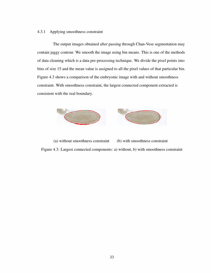
The output images obtained after passing through Chan-Vese segmentation may
contain jaggy contour. We smooth the image using bin means. This is one of the methods
of data cleaning which is a data pre-processing technique. We divide the pixel points into
bins of size 15 and the mean value is assigned to all the pixel values of that particular bin.
Figure 4.3 shows a comparison of the embryonic image with and without smoothness
constraint. With smoothness constraint, the largest connected component extracted is
consistent with the real boundary.
(a) without smoothness constraint (b) with smoothness constraint
Figure 4.3: Largest connected components: a) without, b) with smoothness constraint
33

4.4 Results obtained from the framework
We apply the proposed framework in the gaussian scale space with σ ranging
from 0.5 to 30. We give an experimental analysis of the proposed framework on the
BGDP embryonic images. We then present the successful results achieved by the
criterion. We present the images along with its optimal scale at which the image has the
largest area. We will also present an analysis of a failure case to illustrate the limitation of
the proposed framework.
4.4.1 Successful case of extracted contours
The characteristic scale of a local image pattern is the scale parameter at which
the gaussian kernel provides a local maximum. At finer scales, greater detail is
represented. For a specified image, the scale at which the area is largest for the largest
component is determined as the optimal scale. We present a set of successful results
achieved by the criteria used in the framework. The optimal scale for each image is
displayed. The scale in our experiment ranges from 0.5 to 30. Figure 4.4 shows that the
largest component of the embryo extracted based on the gaussian scale space.
34

σ = 0.5 σ = 0.5 σ = 27
σ = 5 σ = 1 σ = 11
σ = 20 σ = 15 σ = 7
σ = 3 σ = 5 σ = 10
σ = 5 σ = 7 σ = 6
Figure 4.4: Successfully extracted contours with optimal scale in gaussian scale space
35

4.4.2 Failure case of extracted contours
Figure 4.5 presents failure case of the proposed method which is caused by the
occurrence of various gene expression patterns and overlapping neighboring context of the
embryo. This case illustrates the main limitation of the proposed framework. The future
goal of this research may be to extract the actual contour even in the occurrence of the
complex image structure caused by a number of gene expression regions. With the
increment of the scale, i.e., the gaussian kernel σ, neighboring objects tend to diffuse or
merge into the region of interest. Thus, the largest component may contain not only the
region of interest but also non-region of interest objects.
σ = 2 σ = 9 σ = 2
σ = 7 σ = 1 σ = 2
Figure 4.5: Unsuccessful case of extracted contours with optimal scale
36

4.4.3 Successful case of extracted contours with energy parameter alpha
Chan-Vese method has two main parameters α and σ. α is the weight of the
smoothing term i.e. the external energy factor. Higher α implies more smoothing. σ is the
gaussian kernel which is used for suppression of the noise. The external energy factor α
does not have any significance in terms of the scale space in contour extraction. The scale
α ranges from 0.5 to 1.3. Figure 4.6 shows the resulting images after extraction of largest
component by modifying the external energy factor.
α = 0.5 α = 0.8 α = 1
α = 0.8 α = 0.5 α = 0.8
α = 0.8 α = 0.8 α = 1.3
Figure 4.6: Successfully extracted contours with optimal scale with energy parameter alpha
37

CONCLUSION
5.1 Outcome
The main outcome of this thesis includes developing a framework for contour
extraction of drosophila embryonic images. All our experiments are tested on BDGP
(Berkley Drosophila Genome Project) [1] images. BDGP has large amounts of data sets.
In order to deal with these data sets, we developed an automated model for extracting and
analyzing images. Contour extraction is an important step in comparing the images at
various development stages.
We first applied the edge detection methods on the BDGP data set. The edge
detection methods are one of the existing methods for segmentation of images. Edge
detection works well only in the images with sharp intensity transitions and relatively low
noise. Canny edge detector gives the best results among all the edge detection methods. It
consists of complex computations and is time consuming. Canny edge detection is
computationally more expensive.
We then applied the active contour methods which are more advanced than edge
detection. Snakes have proven to be useful in many applications such as image
processing, etc. The active contour models integrated with the external and internal forces
pulling the contours to the exact boundaries have shown their powerful abilities in object
segmentation. Snakes and level-set are two important models of the active contours. We
applied level-set method and Chan-Vese method on the data set. Comparison of the two
methods was done and we concluded that the Chan-Vese method provides decent
accuracy.
Parameters of these methods play a major role in the segmentation of the image
by minimizing the energy. We observed that performance of the active contour model
depends on the scales. The scale in our experiments refers to the gaussian kernel.
38

Convolution of image is done using the kernel and the Chan-Vese model is applied on the
convoluted image. To improve the performance, the first proposed pre-processing
technique is distance normalization. Applying this technique before segmentation did not
prove to be useful. The resulting images after segmentation were not accurate. Hence, this
method was discarded.
Few results of the Chan-Vese segmentation are obtained with the partial embryo
along with the region of interest. There are also few cases where the resulting image
contains multiple components. These cases can be eliminated by implementing the
heuristic, or the angle between the vectors that should be greater than 30◦. Any image
with an angle between the vector that is less than 30◦ is eliminated. Our proposed
framework extracts the largest component among all the connected components, which is
expected to be the actual target contour. The jaggy contours may be eliminated by
applying the smoothness constraint. We smoothed the image by applying bin means to
handle jaggy contour. Bin means is a data cleaning technique.
We proposed a contour extraction framework for Drosophila embryonic images.
Given a high resolution image, we applied the Chan-Vese active contour model. The
active contour model is applied to refine the initialized contour. The proposed framework
calculates the connected components of the image after passing through the active contour
model. The largest component with the largest area is extracted. This process is repeated
by modifying the scale values. The scale at which the maximum area is attained is
assumed to give information about the feature being extracted. Each image might have a
different scale at which the area is maximum for the largest component. The σ value
corresponding to the largest component is considered to be optimal scale. The active
contour adjusts better to the finer scales. We present a set of successful results achieved by
the best criterion. The scale σ in our experiment ranged 0.5-30 in sampled space, which is,
[0.5, 1, 3, 5, 7, 10, 11, 15, 20, 27]. Some of the images with inner and outer contour had
different band width concentration. Confusion of contour points was caused during
39

convergence. In the proposed method, the failure case was caused by the occurrence of
various gene-expression patterns and overlapping neighboring context of the embryo.
5.2 Future scope
Experimental results show that the proposed criterion can achieve results with
promising accuracy. One of the limitations of the framework is the processing time. The
processing time is quite long as we are working with the original size of the image to
preserve the details of the image. If the image is resized, there is a possibility of losing a
few details of the image. The accuracy of the result is governed by the convergence
criteria, used in energy minimization. To make the results more accurate, tighter
convergence criteria is required. Hence, this results in longer computation time. The
second limitation is the larger scale space. Each image might have a different optimal
scale. If we plot a graph taking image as x axis and scale as y axis, we obtain a scattered
graph. Figure 5.1 gives a picture of the graph.
Figure 5.1: Graph showing optimal scale of the images
40

To address this problem, a classification model may be developed based on the
scale space. The scale space can be divided into a range of values and each range of
values can be classified as a particular class. This classification might give us some
information about range of scales of the images having single embryo, partially touching
embryo and non touching partial embryos. In this way, we might come to the conclusion
that if the image is having single embryo, then it might belong to a particular class, and we
get the information of the range of optimal scales. Similarly, we might come to a
conclusion with touching and non touching embryos.
41

42
BIBLIOGRAPHY
[1] Bdgp: Berkeley drosophila genome project. http://www.fruitfly.org/.
[2] Mathworks. http://www.mathworks.com/.
[3] M. Airouche, L. Bentabet, and M. Zelmat. Image segmentation using active contour
model and level set method applied to detect oil spills.
[4] Vikram V. Appia and Anthony J. Yezzi. Active geodesics: Region-based active con-
tour segmentation with a global edge-based constraint. In ICCV, pages 1975–1980,
2011.
[5] Serdar Kemal Balci and Burak Acar. Active contours: A brief review.
[6] D. Baswaraj, Dr. A. Govardhan, and Dr. P. Premchand. Active contours and image
segmentation: The current state of the art. 2012.
[7] John Canny. A computational approach to edge detection. IEEE Trans. Pattern Anal.
Mach. Intell., 8(6):679–698, 1986.
[8] Tony Chan and Luminita Vese. An active contour model without edges. In Int. Conf.
Scale-Space Theories in Computer Vision, pages 141–151, 1999.
[9] Tony F. Chan and Luminita A. Vese. Active contours without edges. IEEE Transac-
tions on Image Processing, 10(2):266–277, 2001.
[10] Erwin Frise, Ann S Hammonds, and Susan E Celniker. Systematic image-driven
analysis of the spatial drosophila embryonic expression landscape, 2010.
[11] Ye J, Chen J, Li Q, and Kumar S. Classification of drosophila embryonic devel-
opmental stage range based on gene expression pattern images. Computer System
Bioinformatics Conference, 2006.
[12] Ramesh Jain, Rangachar Kasturi, and Brian G. Schunck. Machine Vision. McGraw-
Hill, Inc., 1995.
[13] Shuiwang Ji, Ying-Xin Li, Zhi-Hua Zhou, Sudhir Kumar, and Jieping Ye. A bag-of-
words approach for drosophila gene expression pattern annotation. BMC Bioinfor-
matics, 10, 2009.
[14] Shuiwang Ji, Liang Sun, Rong Jin, Sudhir Kumar, and Jieping Ye. Automated anno-
tation of drosophila gene expression patterns using a controlled vocabulary. Bioinfor-
matics, 24(17):1881–1888, 2008.
[15] Michael Kass, Andrew P. Witkin, and Demetri Terzopoulos. Snakes: Active contour
models. International Journal of Computer Vision, 1(4):321–331, 1988.

[16] Satyanad Kichenassamy, Arun Kumar, Peter Olver, Allen Tannenbaum, andAnthony Yezzi Jr. Gradient flows and geometric active contour models, 1994.
[17] S. Kumar, K. Jayaraman, S. Panchanathan, R. Gurunathan, A. Marti-Subirana, andS.J. Newfeld. Best: A novel computational approach for comparing gene expressionpatterns from early stages of drosophila melanogaster development. Genetics,16(4):2037–2047, 2002.
[18] Florence Kussener. Active contour: a parallel genetic algorithm approach.International conference on swarm intelligence.
[19] Bing Li and Scott T. Acton. Active contour external force using vector fieldconvolution for image segmentation. IEEE Transactions on Image Processing,16(8):2096–2106, 2007.
[20] Chunming Li, Chenyang Xu, Changfeng Gui, and Martin D. Fox. Distanceregularized level set evolution and its application to image segmentation. IEEETransactions on Image Processing, 19(12):3243–3254, 2010.
[21] Qi Li and Chandra Kambhamettu. Contour extraction of drosophila embryos.IEEE/ACM Trans. Comput. Biology Bioinform., pages 1509–1521, 2011.
[22] Tony Lindeberg. Edge detection and ridge detection with automatic scale selection.In CVPR, pages 465–470, 1996.
[23] Tony Lindeberg. Feature detection with automatic scale selection. InternationalJournal of Computer Vision, 30(2):79–116, 1998.
[24] ChengEn Lu, Longin Jan Latecki, and Guangxi Zhu. Contour extraction usingparticle filters. In ISVC (2), pages 192–201, 2008.
[25] Daniel L. Mace, Nicole Varnado, Weiping Zhang, Erwin Frise, and Uwe Ohler.Extraction and comparison of gene expression patterns from 2d rna in situhybridization images. Bioinformatics, 26(6):761–769, 2010.
[26] S. Osher and J. Sethian. Fronts propagating with curvature dependent speed:Algorithms based on hamilton-jacobi formulations. Journal of ComputationlPhysics, pages 12–49, 1988.
[27] Jia-Yu Pan, André G. R. Balan, Eric P. Xing, Agma J. M. Traina, and ChristosFaloutsos. Automatic mining of fruit fly embryo images. In KDD, pages 693–698,2006.
[28] Jérome Piovano and Théodore Papadopoulo. Local statistic based regionsegmentation with automatic scale selection. In ECCV (2), pages 486–499, 2008.
[29] Julia A. Schnabel and Simon R. Arridge. Active contour models for shapedescription using multiscale differential invariants. In British Machine VisionConference 1995, 1995.
43

[30] P. Tomancak et al. Systematic determination of patterns of gene expression duringdrosophila embryogenesis. Genome Biol, 3(12):0081–0088, 2002.
[31] Sandro Vega-Pons, José Luís Gil Rodríguez, and Oscar Luis Vera. Active contouralgorithm for texture segmentation using a texture feature set. In ICPR, pages 1–4,2008.
[32] Wikipedia. Drosophila. http://en.wikipedia.org/wiki/Drosophila.
[33] Chenyang Xu, Dzung L. Pham, and Jerry L. Prince. Image segmentation usingdeformable models.
[34] Chenyang Xu and Jerry L. Prince. Snakes, shapes, and gradient vector flow. IEEETransactions on Image Processing, 7(3):359–369, 1998.
44


![The development of motor coordination in Drosophila embryos · (AEL)]. Dechorionated embryos were placed on slides and covered with a layer of halocarbon oil to prevent dehydration](https://img.pdfslide.us/doc/110x75/606a9de2f26f932f510c46a6/the-development-of-motor-coordination-in-drosophila-embryos-ael-dechorionated.jpg)