Embed Size (px)
Citation preview
Thomas Jefferson UniversityJefferson Digital Commons
Wills Eye Institute Papers Wills Eye Institute
2-4-2016
Consecutive bilateral decompression retinopathyafter mitomycin C trabeculectomy: a case report.Ana Raquel Marcos FigueiredoOphthalmology Department, Centro Hospital Do Porto, EPE - Hospital de Santo António, Largo Professor Abel Salazar, Porto,Portugal
Isabel Coutinho SampaioOphthalmology Department, Centro Hospital Do Porto, EPE - Hospital de Santo António, Largo Professor Abel Salazar, Porto,Portugal
Maria João Fernandes Dos Santos MenéresOphthalmology Department, Centro Hospital Do Porto, EPE - Hospital de Santo António, Largo Professor Abel Salazar, Porto,Portugal
George L. SpaethWills Eye Hospital, Jefferson Medical College, 840 Walnut Street, Philadelphia, PA, United States, [email protected]
Let us know how access to this document benefits youFollow this and additional works at: http://jdc.jefferson.edu/willsfp
Part of the Ophthalmology Commons
This Article is brought to you for free and open access by the Jefferson Digital Commons. The Jefferson Digital Commons is a service of ThomasJefferson University's Center for Teaching and Learning (CTL). The Commons is a showcase for Jefferson books and journals, peer-reviewed scholarlypublications, unique historical collections from the University archives, and teaching tools. The Jefferson Digital Commons allows researchers andinterested readers anywhere in the world to learn about and keep up to date with Jefferson scholarship. This article has been accepted for inclusion inWills Eye Institute Papers by an authorized administrator of the Jefferson Digital Commons. For more information, please contact:[email protected].
Recommended CitationFigueiredo, Ana Raquel Marcos; Sampaio, Isabel Coutinho; Menéres, Maria João Fernandes DosSantos; and Spaeth, George L., "Consecutive bilateral decompression retinopathy after mitomycin Ctrabeculectomy: a case report." (2016). Wills Eye Institute Papers. Paper 55.http://jdc.jefferson.edu/willsfp/55
CASE REPORT Open Access
Consecutive bilateral decompressionretinopathy after mitomycin Ctrabeculectomy: a case reportAna Raquel Marcos Figueiredo1*, Isabel Coutinho Sampaio1, Maria João Fernandes dos Santos Menéres1
and George L. Spaeth2
Abstract
Background: After a successful trabeculectomy, a sudden intraocular pressure decrease may alter the intracranial tointraocular pressure ratio and cause decompression retinopathy. Frequent Valsalva maneuvers may also play a role in itspathogenesis. This condition may manifest as multiple retinal hemorrhages, edema of the optic disc, macular edema, or asudden decrease in visual acuity postoperatively. Outcomes for patients are usually good, with spontaneous resolutionoccurring within a matter of weeks. It has been rarely reported in the literature as a bilateral condition.
Case presentation: We present a case of consecutive bilateral decompression retinopathy in a 54-year-old severelyobese Caucasian woman (body mass index 37 kg/m2) with open angle glaucoma and a poor history of medicaltherapeutic compliance, who chose surgical treatment based on her inability to consistently use ocular drops. Our patientunderwent a trabeculectomy with mitomycin C in both eyes, with surgeries taking place 3 months apart. After the firstsurgery, 2 weeks postoperatively, she complained of decreased visual acuity. Examination of her right eye fundus revealedmultiple retinal hemorrhages and disc edema. There was a similar pattern in her left eye, this time including maculopathy.Her visual acuity and fundoscopic changes resolved spontaneously over a period of a month in both cases. Currently, ourpatient has well-controlled bilateral intraocular pressure, ranging between 14 and 16 mmHg, without hypotensivemedication.
Conclusions: Decompression retinopathy is a potential complication after glaucoma surgery, but has rarely been describedas a bilateral consecutive condition. A comprehensive approach could help to anticipate its occurrence and manage it.
Keywords: Decompression retinopathy, Macular edema, Open angle glaucoma, Retinal hemorrhages, Trabeculectomy
BackgroundOcular decompression retinopathy was first described byFechtner et al. in 1992 [1] as a complication of theabrupt iatrogenic lowering of intraocular pressure (IOP)after glaucoma filtering surgery. During intraocular sur-gery, entry into the eye allows the IOP to fall andequalize with the atmospheric pressure. If this change issudden and large, it can induce hemodynamic changesthat may result in bilateral decompression retinopathy.Immediately following surgery, the clinical situation ischaracterized by the appearance of diffuse retinal
hemorrhages and edema of the optic disc in associationwith decreased visual acuity [2]. The most commonoptic nerve findings described are peripapillary and opticnerve head hemorrhages. Retinal manifestations mainlycomprise intraretinal hemorrhages (92 % of retinal hem-orrhages) and, less commonly, macular edema (3 %) orserous macular detachment (5 %) [13].Outcomes are generally good for affected patients,
with spontaneous resolution occurring within a fewweeks. Over the years, the word “ocular” has beendropped, and “ocular decompression retinopathy” is nowdescribed in the literature as “decompression retinop-athy” (DR). Besides a large drop in IOP after surgery,many of the cases presented in the literature have dem-onstrated a significant increase in IOP over a relatively
* Correspondence: [email protected] Department - Centro Hospital do Porto, EPE – Hospital deSanto António, Largo Professor Abel Salazar, 4099-001 Porto, PortugalFull list of author information is available at the end of the article
© 2016 Figueiredo et al. Open Access This article is distributed under the terms of the Creative Commons Attribution 4.0International License (http://creativecommons.org/licenses/by/4.0/), which permits unrestricted use, distribution, andreproduction in any medium, provided you give appropriate credit to the original author(s) and the source, provide a link tothe Creative Commons license, and indicate if changes were made. The Creative Commons Public Domain Dedication waiver(http://creativecommons.org/publicdomain/zero/1.0/) applies to the data made available in this article, unless otherwise stated.
Figueiredo et al. Journal of Medical Case Reports (2016) 10:32 DOI 10.1186/s13256-016-0814-x

short period of time pre-surgery, or large variations inIOP with spikes followed by significant drops in pressure[3]. These and other factors are believed to be related tothe etiopathogenesis of DR [4]. Even the use of mitomy-cin C has been questioned as a potential adjuvant, andother hypotheses remain open [5].There are few published cases of DR. Although Fechtner
et al. described it as a complication following trabeculect-omy (which represents half of total cases described), caseshave been reported in other situations, such as neodym-ium: yttrium–aluminum–garnet iridotomies [6], anteriorchamber paracentesis [7], pars plana vitrectomies [8],orbital decompression, and drainage implant inser-tions [9, 10]. In 2006, Bui et al. [11] were the first to de-scribe maculopathy as an additional characteristic of DR.Our presented case concerns an extremely rare entity inophthalmology, particularly with respect to our patient’spost trabeculectomy status, and represents an event rarelybefore described in the literature with this course andmuch less with a bilateral consecutive presentation.
Case presentationA 54-year-old Caucasian woman with open angle glau-coma and a history of suboptimal medical therapeuticcompliance owing to an intolerance to drops was re-ferred to our Ophthalmology Department. She was se-verely obese (body mass index 37 kg/m2) and had type 2diabetes but was using insulin with good metabolic con-trol. Her best corrected visual acuity (BCVA) in botheyes was 1.0. Her IOP was 35 mmHg without the use ofmedication, though she achieved values of 18 mmHg inher right eye and 16 mmHg in her left eye with the useof tafluprost once daily. No changes were identified in
an examination of her anterior segment. The papillarycup was 0.3 in her right eye with a temporal notch andher left eye had a normal appearance.Our patient’s compliance to medical therapy continued
to be poor, because she blamed her drops for coughingand dyspnea attacks. Without the use of drops, her IOPvalues remained consistently in the 30s. After continu-ous non-compliance with several other ocular medica-tions, surgery was discussed as an alternative treatmentoption. The risks and potential complications of surgerywere explained to our patient. No signs of cornea orocular media opacity, retinal hemorrhage, macular orperipheral detachments, or other contraindications wereobserved in either eye. Our patient decided to opt forsurgery. Her right eye was operated on first with an un-eventful mitomycin C trabeculectomy (0.3 mg/ml,3 minutes).On the first postoperative day, our patient presented
with a diffuse, functioning, and non-leaking filtrationbleb, associated with a well-formed anterior chamber,and an IOP of 8 mmHg, with a normal appearance onfundoscopy. Two weeks postoperatively she complainedof decreased visual acuity; her BCVA was 20/32 and herIOP was 10 mmHg without medication. Fundoscopyexhibited multiple superficial, flame-shaped retinal hem-orrhages located centrifugally from the optic disc associ-ated with optic disc edema (Fig. 1a). There was noevidence of choroidal effusion. A fundus examination ofher left eye was unremarkable.Optical coherence tomography (OCT) revealed folding
of the macular retina associated with a small detachmentof the neurosensory retina (Fig. 1c). The angiographic pat-tern showed macular microaneurysms associated with
Fig. 1 Right eye retinography, angiography, and optical coherence tomography 2 weeks after surgery. a Right eye fundus photograph showingmultiple superficial, flame-shaped retinal hemorrhages located centrifugally from the optic disc associated with optic disc edema. b Fluoresceinangiography exhibited macular microaneurysms associated with fluoroscein diffusion, peripapillary hemorrhages, and late optic disc leakage.c Optical coherence tomography image revealing folding of the macular retina and a small detachment of the neurosensory retina
Figueiredo et al. Journal of Medical Case Reports (2016) 10:32 Page 2 of 6
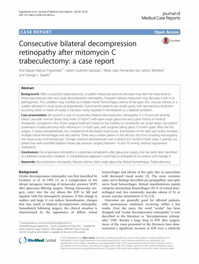
fluorescein diffusion, peripapillary hemorrhages, and lateoptic disc leakage, without ischemic areas or neovasculari-zation (Fig. 1b). One month later the overall fundoscopicchanges resolved spontaneously (Fig. 2a–d). Given thissituation, a suspected diagnosis of DR was proposed.Other possible diagnoses were retinal venous occlusion orValsalva retinopathy, but these were considered unlikelygiven the diagnostic results pattern. Subsequent follow-upvisits were satisfactory, with our patient maintaining asteady IOP of 8–14 mmHg without medication. The peri-papillary hemorrhages and optic disc edema spontan-eously recovered during the second postoperative month,and our patient’s BCVA reached 20/25.Three months later, she underwent an uncomplicated
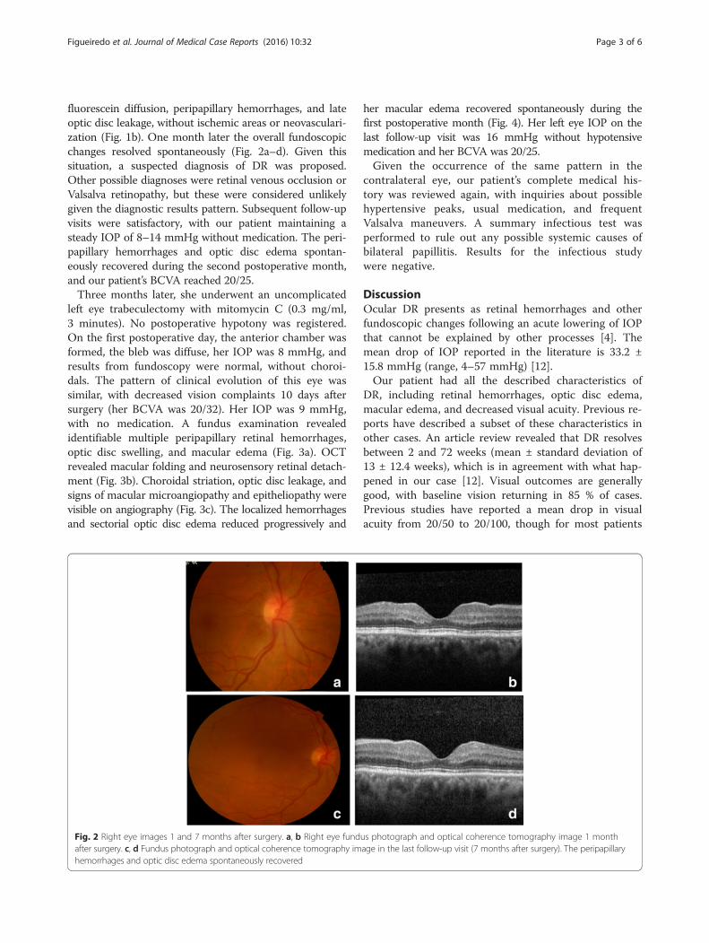
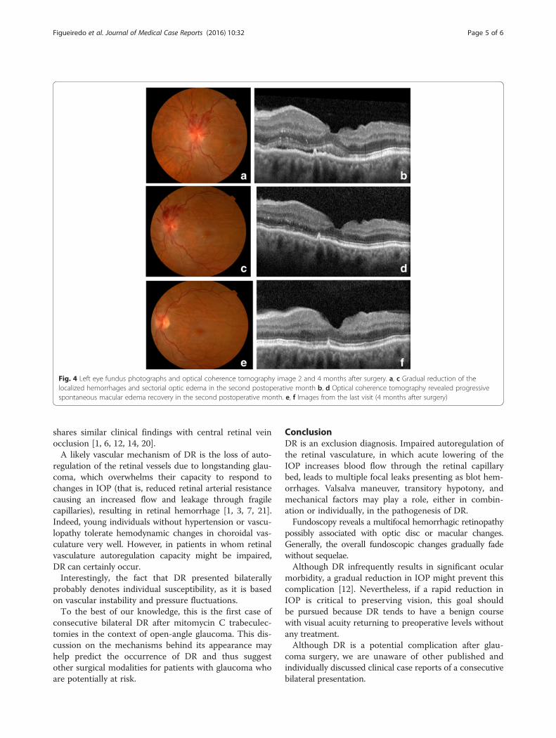
left eye trabeculectomy with mitomycin C (0.3 mg/ml,3 minutes). No postoperative hypotony was registered.On the first postoperative day, the anterior chamber wasformed, the bleb was diffuse, her IOP was 8 mmHg, andresults from fundoscopy were normal, without choroi-dals. The pattern of clinical evolution of this eye wassimilar, with decreased vision complaints 10 days aftersurgery (her BCVA was 20/32). Her IOP was 9 mmHg,with no medication. A fundus examination revealedidentifiable multiple peripapillary retinal hemorrhages,optic disc swelling, and macular edema (Fig. 3a). OCTrevealed macular folding and neurosensory retinal detach-ment (Fig. 3b). Choroidal striation, optic disc leakage, andsigns of macular microangiopathy and epitheliopathy werevisible on angiography (Fig. 3c). The localized hemorrhagesand sectorial optic disc edema reduced progressively and
her macular edema recovered spontaneously during thefirst postoperative month (Fig. 4). Her left eye IOP on thelast follow-up visit was 16 mmHg without hypotensivemedication and her BCVA was 20/25.Given the occurrence of the same pattern in the
contralateral eye, our patient’s complete medical his-tory was reviewed again, with inquiries about possiblehypertensive peaks, usual medication, and frequentValsalva maneuvers. A summary infectious test wasperformed to rule out any possible systemic causes ofbilateral papillitis. Results for the infectious studywere negative.
DiscussionOcular DR presents as retinal hemorrhages and otherfundoscopic changes following an acute lowering of IOPthat cannot be explained by other processes [4]. Themean drop of IOP reported in the literature is 33.2 ±15.8 mmHg (range, 4–57 mmHg) [12].Our patient had all the described characteristics of
DR, including retinal hemorrhages, optic disc edema,macular edema, and decreased visual acuity. Previous re-ports have described a subset of these characteristics inother cases. An article review revealed that DR resolvesbetween 2 and 72 weeks (mean ± standard deviation of13 ± 12.4 weeks), which is in agreement with what hap-pened in our case [12]. Visual outcomes are generallygood, with baseline vision returning in 85 % of cases.Previous studies have reported a mean drop in visualacuity from 20/50 to 20/100, though for most patients
Fig. 2 Right eye images 1 and 7 months after surgery. a, b Right eye fundus photograph and optical coherence tomography image 1 monthafter surgery. c, d Fundus photograph and optical coherence tomography image in the last follow-up visit (7 months after surgery). The peripapillaryhemorrhages and optic disc edema spontaneously recovered
Figueiredo et al. Journal of Medical Case Reports (2016) 10:32 Page 3 of 6
no intervention is required [12]. In our patient, herBCVA dropped from 20/20 to 20/25, which does notrepresent a significant decrease and can be considered afavorable outcome.In previously reported cases, fluorescein angiography
demonstrated normal retinal and choroidal vascular filling[12], which could help distinguish DR from other condi-tions associated with intraretinal hemorrhages. One suchcase was reported by Bui et al., where OCT scans exhib-ited macular edema and neurosensory macular detach-ment in the context of DR after trabeculectomy; however,this is not a common event in this context [11, 16, 17].All of these findings alert us to the importance of a
complete fundoscopic examination immediately aftersurgery in patients undergoing trabeculectomy or otherhypotensive techniques involving low pressure, especiallywhen pre-surgical pressure is high.The main differential diagnosis for DR involves retin-
opathy associated with Valsalva maneuver or venous oc-clusion. Our patient’s clinical characteristics (that is, herphysical constitution) could suggest the first diagnosis;however, typical ocular findings are classically describedas pre-retinal hemorrhages with predilection for themacula and possible vitreous hemorrhages. Additionally,this condition is usually simultaneously bilateral. Venousocclusion was ruled out because there was no angio-graphic evidence of venous dilatation, delayed venousfilling, or occlusive phenomena as is expected in acute
central retinal vein occlusion. Furthermore, DR appearsin patients who are typically younger, asymptomatic, andwhose fundus examinations show a full return to pre-event state. A benign idiopathic intracranial hyperten-sion pattern was also considered, given that our patientwas severely obese. This possibility was rejected, by theabsence of not only typical clinical findings (headache,nausea, and vomiting) but also optic disc edema on pre-operative fundus examination. Additionally, her intracra-nial pressure was in the high-normal range, whichmatched her biotype [18]. Although carotid dopplerultrasonography was not performed, the lack of venousdilatation, ocular pain, peripheral retinal hemorrhages,and retina and optic disc neovessels, as well as her goodvisual recovery, make the diagnosis of ocular ischemicsyndrome less likely. The diagnosis of DR was based onher typical history, retinal findings, and the exclusion ofthe other possible causes mentioned above.Currently, there is no consensus about the etiologic
mechanisms of DR, though mechanical and vascular ori-gins are likely. One of the possible mechanical explana-tions is that the decrease in IOP into the low-normalrange after trabeculectomy may alter the intracranialpressure/IOP ratio at the lamina cribrosa, resulting in itsanterior shift and expansion [1, 19]. This forward move-ment might block axonal transport, leading to compres-sion of the central retinal vein and producing optic dischemorrhages and edema. This may explain why DR
Fig. 3 Left eye retinography, optical coherence tomography (OCT), and angiography 10 days after surgery. a Fundus photograph showingmultiple peripapillary retinal hemorrhages, optic disc swelling, and macular edema. b Optical coherence tomography revealed macular edemawith neurosensory retinal detachment. c Angiography demonstrated choroidal striation, optic disc leakage, and signs of macular microangiopathyand epitheliopathy
Figueiredo et al. Journal of Medical Case Reports (2016) 10:32 Page 4 of 6
shares similar clinical findings with central retinal veinocclusion [1, 6, 12, 14, 20].A likely vascular mechanism of DR is the loss of auto-
regulation of the retinal vessels due to longstanding glau-coma, which overwhelms their capacity to respond tochanges in IOP (that is, reduced retinal arterial resistancecausing an increased flow and leakage through fragilecapillaries), resulting in retinal hemorrhage [1, 3, 7, 21].Indeed, young individuals without hypertension or vascu-lopathy tolerate hemodynamic changes in choroidal vas-culature very well. However, in patients in whom retinalvasculature autoregulation capacity might be impaired,DR can certainly occur.Interestingly, the fact that DR presented bilaterally
probably denotes individual susceptibility, as it is basedon vascular instability and pressure fluctuations.To the best of our knowledge, this is the first case of
consecutive bilateral DR after mitomycin C trabeculec-tomies in the context of open-angle glaucoma. This dis-cussion on the mechanisms behind its appearance mayhelp predict the occurrence of DR and thus suggestother surgical modalities for patients with glaucoma whoare potentially at risk.
ConclusionDR is an exclusion diagnosis. Impaired autoregulation ofthe retinal vasculature, in which acute lowering of theIOP increases blood flow through the retinal capillarybed, leads to multiple focal leaks presenting as blot hem-orrhages. Valsalva maneuver, transitory hypotony, andmechanical factors may play a role, either in combin-ation or individually, in the pathogenesis of DR.Fundoscopy reveals a multifocal hemorrhagic retinopathy
possibly associated with optic disc or macular changes.Generally, the overall fundoscopic changes gradually fadewithout sequelae.Although DR infrequently results in significant ocular
morbidity, a gradual reduction in IOP might prevent thiscomplication [12]. Nevertheless, if a rapid reduction inIOP is critical to preserving vision, this goal shouldbe pursued because DR tends to have a benign coursewith visual acuity returning to preoperative levels withoutany treatment.Although DR is a potential complication after glau-
coma surgery, we are unaware of other published andindividually discussed clinical case reports of a consecutivebilateral presentation.
Fig. 4 Left eye fundus photographs and optical coherence tomography image 2 and 4 months after surgery. a, c Gradual reduction of thelocalized hemorrhages and sectorial optic edema in the second postoperative month b, d Optical coherence tomography revealed progressivespontaneous macular edema recovery in the second postoperative month. e, f Images from the last visit (4 months after surgery)
Figueiredo et al. Journal of Medical Case Reports (2016) 10:32 Page 5 of 6

ConsentWritten informed consent was obtained from the patientfor publication of this case report and accompanyingimages. A copy of the written consent is available forreview by the Editor-in-Chief of this journal.
AbbreviationsBCVA: Best corrected visual acuity; DR: Decompression retinopathy;IOP: Intraocular pressure; OCT: Optical coherence tomography.
Competing interestsThe authors declare that they have no competing interests.
Authors’ contributionsAF operated both eyes of the patient , wrote the original article as well asselected and organized the most important images of the case. IS followedthe patient and proposed surgery. MJM analyzed and interpreted all thepatient data. GS was a major contributor in writing the manuscript.All authors read and approved the final manuscript.
AcknowledgementsFernando Emanuel Dias Correia, Neurology Department, Centro Hospital do Porto,Hospital Santo de António, Porto, Portugal FC, performed the lumbar puncture.Maria João Furtado, Ophtalmology Department, Centro Hospitalar do Porto,Hospital de Santo António, performed angiograms and was very helpful inassisting the interpretation of angiographic images.
Author details1Ophthalmology Department - Centro Hospital do Porto, EPE – Hospital deSanto António, Largo Professor Abel Salazar, 4099-001 Porto, Portugal. 2WillsEye Hospital, Jefferson Medical College, 840 Walnut Street, Philadelphia, PA19107, USA.
Received: 7 May 2015 Accepted: 14 January 2016
References1. Fechtner RD, Minckler D, Weinreb RN, Frangei G, Jampol LM. Complications
of glaucoma surgery. Ocular decompression retinopathy. Arch Ophthalmol.1992;110:965–8.
2. Juberías JR, Maquet JA, Ussa F. Decompression retinopathy withmaculopathy after trabeculectomy with mitomycin C. Arch Soc EspOftalmol. 2008;83(6):373–6.
3. Bansal A, Ramanathan US. Ocular decompression retinopathy aftertrabeculectomy with mitomycin-C for angle recession glaucoma. Indian JOphthalmol. 2009;57(2):153–4.
4. Saricaoglu MS, Kalayci D, Guven D, Karakurt A, Hasiripi H. Decompressionretinopathy and possible risk factors. Acta Ophthalmol. 2009;87(1):94–5.
5. Kozobolis VP, Kalogianni E, Katsanos A, Dardabounis D, Koukoula S, Labiris G.Ocular decompression retinopathy after deep sclerectomy with mitomycin Cin an eye with exfoliation glaucoma. Eur J Ophthalmol. 2011;21(3):324–7.
6. Landers J, Craig J. Decompression retinopathy and corneal oedemafollowing Nd:YAG laser peripheral iridotomy. Clin Exp Ophthalmol.2006;34:182e4.
7. Gupta R, Browning AC, Amoaku WM. Multiple retinal haemorrhages(decompression retinopathy) following paracentesis for macular branchartery occlusion. Eye. 2005;19:592–3.
8. Rezende F, Regis LG, Kicknger M, Alcantara S. Decompression retinopathyafter 25- gauge transconjuntival sutureless vitrectomy: report of 2 cases.Arch Ophthalmol. 2007;15:699–700.
9. Yalvac IS, Kocaoglan H, Eksioglu U, Demir N, Duman S. Decompressionretinopathy after Ahmed glaucoma valve implantation in a patient withcongenital aniridia and pseudophakia. J Cataract Refract Surg. 2004;30:1582e5.
10. Samra KA, Sieminski SF, Sarup V. Decompression retinopathy after ExPRESSshunt implantation for steroid-induced ocular hypertension: a case report.Case Rep Ophthalmol Med. 2011;21:303287.
11. Bui CM, Recchia FM, Recchia CC, Kammer JA. Optical coherencetomography findings in ocular decompression retinopathy. OphthalmicSurg Lasers Imaging. 2006;37:333–5.
12. Mukkamala SK, Patel A, Dorairaj S, McGlynn R, Sidoti PA, Weinreb RN, et al. Oculardecompression retinopathy: a review. Surv Ophthalmol. 2013;58(6):505–12.
13. Nonoyama S, Tanito M, Katsube T, Matsuoka Y, Ohira A. Decompressionretinopathy and serous retinal detachment after trabeculotomy in a patientwith systemic amyloidosis. Jpn J Ophthalmol. 2009;53(1):73–5.
14. Lai JS, Lee VY, Leung DY, Cheung TC. Decompression retinopathy followinglaser peripheral iridoplasty for acute primary angle closure. Eye. 2005;19:1345e7.
15. Wakita M, Kawaji T, Ando E, Koga T, Inatani M, Tanihara H, et al. Oculardecompression retinopathy following trabeculectomy with mitomycin Cassociated with familial amyloidotic polyneuropathy. Br J Ophthalmol.2006;90:515–6.
16. Tyagi P, Hashim A. Ocular decompression retinopathy following post-trabeculectomy suture lysis and management with triamcinolone acetonide.Int Ophthalmol. 2011;31:425–8.
17. Danias J, Rosenbaum J, Podos SM. Diffuse retinal hemorrhages (oculardecompression syndrome) after trabeculectomy with mitomycin C forneovascular glaucoma. Acta Ophthalmol Scand. 2000;78(4):468–9.
18. Kawasaki A, Purvin V. Unilateral optic disc edema following trabeculectomy.J Neuroophthalmol. 1998;18(2):121–3.
19. Lee EJ, Kim TW, Weinreb RN. Reversal of lamina cribrosa displacementand thickness after trabeculectomy in glaucoma. Ophthalmology. 2012;119(7):1359e66.
20. Grieshaber MC, Mozaffarieh M, Flammer J. What is the link between vasculardysregulation and glaucoma? Surv Ophthalmol. 2007;52 Suppl 2:S144e54.
21. Grunwald JE, Riva CE, Stone RA, Keates EU, Petrig BL. Retinal autoregulationin open-angle glaucoma. Ophthalmology. 1984;91:1690e4.
• We accept pre-submission inquiries
• Our selector tool helps you to find the most relevant journal
• We provide round the clock customer support
• Convenient online submission
• Thorough peer review
• Inclusion in PubMed and all major indexing services
• Maximum visibility for your research
Submit your manuscript atwww.biomedcentral.com/submit
Submit your next manuscript to BioMed Central and we will help you at every step:
Figueiredo et al. Journal of Medical Case Reports (2016) 10:32 Page 6 of 6


![The Guide - Diabetic Retinopathy - Vision Lossvisionloss.org.au/wp-content/uploads/2016/05/The... · the guide [diabetic retinopathy] What is Diabetic Retinopathy? Diabetic Retinopathy](https://img.pdfslide.us/doc/110x75/5e3ed00bf9c32e41ea6578a8/the-guide-diabetic-retinopathy-vision-the-guide-diabetic-retinopathy-what.jpg)