Embed Size (px)
DESCRIPTION
In this practical guide for clinicians, three experts in pediatric eye care sort out the many infectious and inflammatory causes of “red eye” in children. They provide helpful tips for determining whether conjunctivitis is bacterial, viral, or allergic, and offer guidance in selecting the appropriate treatment agent.
Citation preview
August 2010
Moderator/Chairrudolph S. Wagner, MdClinical Associate Professor of Ophthalmology and PediatricsDirector of Pediatric OphthalmologyUniversity of Medicine and Dentistry of New JerseyNew Jersey Medical SchoolNewark, New Jersey
FaCULtYPeter a. d’arienzo, MdClinical Assistant Professor in OphthalmologyNew York Medical CollegeValhalla, NY President, Manhasset Eye Physicians, PC Manhasset, NY
Mark S. dorfman, MdSenior Pediatric OphthalmologistFormer Chief of SurgeryJoe DiMaggio Children’s HospitalHollywood, Florida Past President Florida Society of Ophthalmology
www.eMPR.com
Conjunctivitis in Children: Challenges and Choices
Produced byBrought to you as an educational service by Alcon Laboratories

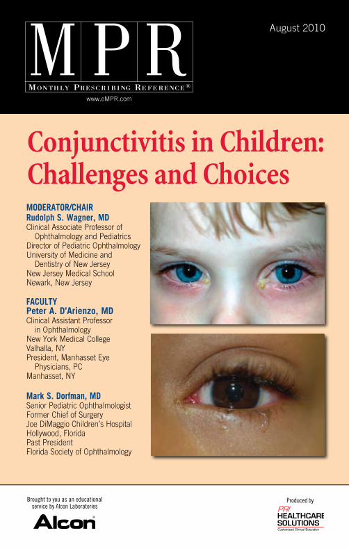
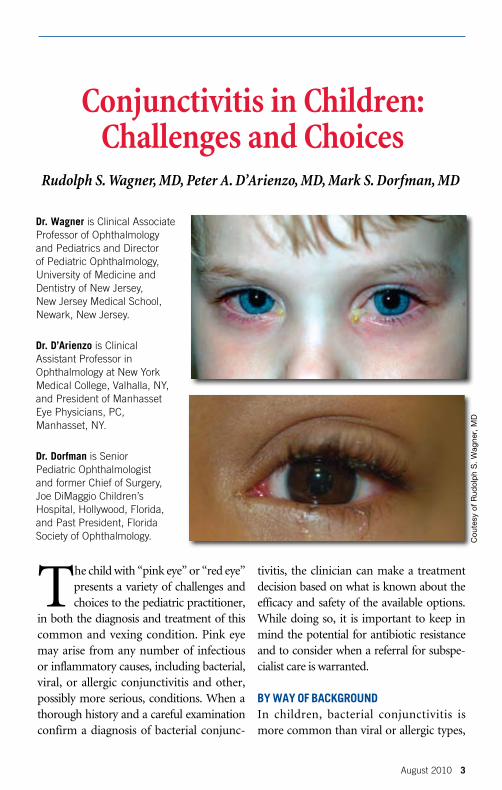
On the cover:Top photo: Bilateral purulent discharge characteristic of bacterial conjunctivitis.Bottom photo: Watery discharge typical of viral conjunctivitis.
T he child with “pink eye” or “red eye” presents a variety of challenges and choices to the pediatric practitioner,
in both the diagnosis and treatment of this common and vexing condition. Pink eye may arise from any number of infectious or inflammatory causes, including bacterial, viral, or allergic conjunctivitis and other, possibly more serious, conditions. When a thorough history and a careful examination confirm a diagnosis of bacterial conjunc-
tivitis, the clinician can make a treatment decision based on what is known about the efficacy and safety of the available options. While doing so, it is important to keep in mind the potential for antibiotic resistance and to consider when a referral for subspe-cialist care is warranted.
BY WaY oF BaCKGroUNdIn children, bacterial conjunctivitis is more common than viral or allergic types,
Conjunctivitis in Children: Challenges and Choices
August 2010 3
Rudolph S. Wagner, MD, Peter A. D’Arienzo, MD, Mark S. Dorfman, MD
Cou
tesy
of
Rud
olp
h S
. W
agne
r, M
D
dr. Wagner is Clinical Associate Professor of Ophthalmology and Pediatrics and Director of Pediatric Ophthalmology, University of Medicine and Dentistry of New Jersey, New Jersey Medical School, Newark, New Jersey.
dr. d’arienzo is Clinical Assistant Professor in Ophthalmology at New York Medical College, Valhalla, NY, and President of Manhasset Eye Physicians, PC, Manhasset, NY.
dr. dorfman is Senior Pediatric Ophthalmologist and former Chief of Surgery, Joe DiMaggio Children’s Hospital, Hollywood, Florida, and Past President, Florida Society of Ophthalmology.
4 August 2010
occurs in all geographic areas and in all races, and is seen with equal frequency among boys and girls. A landmark study among 99 children with conjunctivitis (mean age, 4.4 years) and 102 controls (mean age, 4.9 years) conducted in 1981 showed that three organisms are primarily responsible for pediatric bacterial conjunc-tivitis: Haemophilus influenzae (42% of affected children), Streptococcus pneumo-niae (12%), and adenoviruses (20%).1 In this study, only three patients were infected simultaneously with two of the pathogens. Children with adenoviral disease tended to be older than those with bacterial infection, but the age ranges overlapped consider-ably, with one quarter of those with adeno-virus infection younger than 3.5 years of age and 11% of youngsters in the bacterial group older than 8.5 years of age.1
The two primary agents of bacterial conjunctivitis have remained essentially unchanged over the years. A 1993 study in nearly 100 patients with acute con-junctivitis showed that bacterial infec-tions predominated—in 76 patients vs. 12 with viral infection—and that the most common bacterial culprits were H influenzae, S pneumoniae, and Moraxella catarrhalis, in that order.2 The children ranged in age from 4 months to 12 years. Similarly, a 2007 study in 111 children from 1 month to 18 years of age con-firmed earlier findings. Overall, 78% of patients with conjunctivitis had positive bacterial cultures; H influenzae accounted for 82% and S pneumoniae for 16%.3
In a series reported in 2010, H influenzae accounted for 68% of bacterial conjunctivitis
in 238 culture-positive patients 6 months to 17 years of age. S pneumoniae accounted for 20% of cases.4 Most conjunctivitis caused by H influenzae is untypeable, which may help explain why use of the pneumococcal and H influenzae type b (Hib) vaccines has not changed the etiology of acute conjunctivitis.3 In the 2007 and 2010 studies, Staphylococcus aureus was the third most common bacte-rial cause of conjunctivitis, accounting for 2% and 8% of cases, respectively.3,4
reCeNt oUtBreaKSHighly contagious adenovirus is a common cause of conjunctivitis outbreaks, having been reported on military bases, eye clinics, and child care centers.5 Yet several recent outbreaks serve notice that bacteria also can be the culprit and that assumptions can’t be made about which age groups will be hit hardest by which pathogen. In 2002, Dartmouth College in New Hampshire experienced an outbreak of bacterial con-junctivitis, though a viral cause initially was suspected.6 Almost 14% of the student body (698 of 5060 students) was diagnosed with conjunctivitis between January 1 and April 12; 5% of that group had repeated infections.6 Bacteria isolated from conjunc-tival swabs were identified as an atypical, unencapsulated strain of S pneumoniae (110 swabs) or H influenzae (19 swabs). One specimen grew both pathogens.6 Few large outbreaks of pneumococcal conjunc-tivitis had been reported previously.
In the Dartmouth outbreak, factors associated with developing conjunctivitis included having a roommate or other close contact with an infection, playing on a var-
Conjunctivitis in Children: Challenges and Choices
August 2010 5
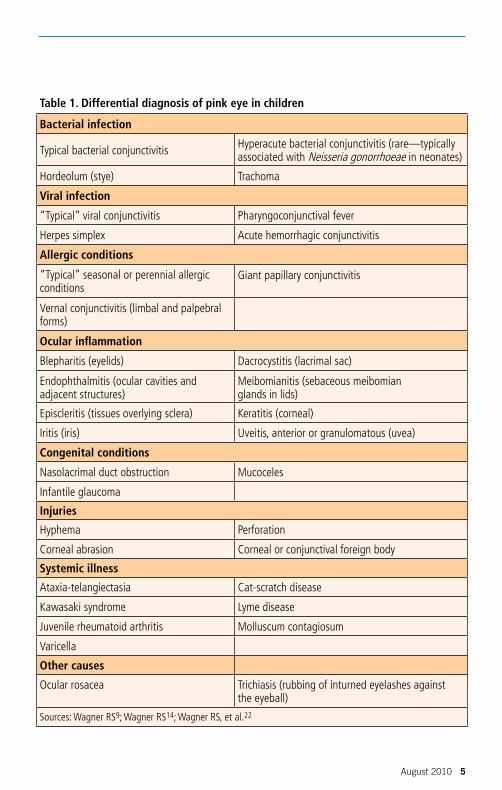
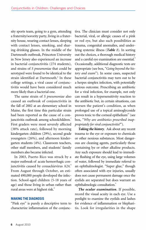
Table 1. Differential diagnosis of pink eye in children
Bacterial infection
Typical bacterial conjunctivitis Hyperacute bacterial conjunctivitis (rare—typically associated with Neisseria gonorrhoeae in neonates)
Hordeolum (stye) Trachoma
Viral infection
“Typical” viral conjunctivitis Pharyngoconjunctival fever
Herpes simplex Acute hemorrhagic conjunctivitis
Allergic conditions
“Typical” seasonal or perennial allergic conditions
Giant papillary conjunctivitis
Vernal conjunctivitis (limbal and palpebral forms)
Ocular inflammation
Blepharitis (eyelids) Dacrocystitis (lacrimal sac)
Endophthalmitis (ocular cavities and adjacent structures)
Meibomianitis (sebaceous meibomian glands in lids)
Episcleritis (tissues overlying sclera) Keratitis (corneal)
Iritis (iris) Uveitis, anterior or granulomatous (uvea)
Congenital conditions
Nasolacrimal duct obstruction Mucoceles
Infantile glaucoma
Injuries
Hyphema Perforation
Corneal abrasion Corneal or conjunctival foreign body
Systemic illness
Ataxia-telangiectasia Cat-scratch disease
Kawasaki syndrome Lyme disease
Juvenile rheumatoid arthritis Molluscum contagiosum
Varicella
Other causes
Ocular rosacea Trichiasis (rubbing of inturned eyelashes against the eyeball)
Sources: Wagner RS9; Wagner RS14; Wagner RS, et al.22
6 August 2010
sity sports team, going to a gym, attending a fraternity/sorority party, living in a frater-nity house, wearing contact lenses, sleeping with contact lenses, smoking, and shar-ing drinking glasses. In the middle of the Dartmouth outbreak, Princeton University in New Jersey also experienced an increase in bacterial conjunctivitis (274 students), and strains of S pneumoniae that could be serotyped were found to be identical to the strain identified at Dartmouth.6 In these college settings, a viral cause of conjunc-tivitis would have been considered much more likely than a bacterial one.
The same strain of S pneumoniae also caused an outbreak of conjunctivitis in the fall of 2002 at an elementary school in Maine, the first time this particular strain had been reported as the cause of a con-junctivitis outbreak among schoolchildren.7 First graders were most severely affected (38% attack rate), followed by morning kindergarten children (29%), second grade youngsters (26%), and afternoon kinder-garten students (4%). Classroom teachers, other staff members, and students’ family members also became infected.
In 2003, Puerto Rico was struck by a major outbreak of acute hemorrhagic con-junctivitis caused by coxsackievirus A24.8 From August through October, an esti-mated 490,000 people developed the infec-tion. School-aged children (5-18 years of age) and those living in urban rather than rural areas were at highest risk.8
MaKiNG the diaGNoSiS“Pink eye” is purely a descriptive term to characterize inflammation of the conjunc-
tiva. The clinician must consider not only bacterial, viral, or allergic causes of a pink or red eye, but also such possibilities as trauma, congenital anomalies, and under-lying systemic illness (Table 1). In sorting out the choices, a thorough medical history and a careful eye examination are essential.9 Occasionally, additional diagnostic tests are appropriate (see box, “Going beyond the his-tory and exam”). In some cases, suspected bacterial conjunctivitis may turn out to be a herpes simplex infection, with potentially serious outcome. Prescribing an antibiotic for a viral infection, for example, not only can result in a hypersensitivity reaction to the antibiotic but, in certain situations, can worsen the patient’s condition, as when prolonged use of a topical aminoglycoside proves toxic to the corneal epithelium10 (see box, “Why are antibiotics prescribed inap-propriately for viral conjunctivitis?”).
Taking the history. Ask about any recent trauma to the eye or exposure to chemicals or other noxious substances. Most danger-ous are cleaning agents, particularly those containing lye or other alkaline products. Any such exposure should lead to immedi-ate flushing of the eye, using large volumes of water, followed by immediate referral to an ophthalmologist. “Super glue,” though often associated with eye injuries, usually does not cause permanent damage once the eyelids are separated but does warrant an ophthalmologic consultation.9
The ocular examination. If possible, record the visual acuity in each eye. Use a penlight to examine the eyelids and lashes for evidence of inflammation or blephari-tis. Look for irregularities in the shape
Conjunctivitis in Children: Challenges and Choices
August 2010 7
of the pupil, and determine the presence of direct and consensual pupillary reac-tion. Irregular pupil size or shape suggests severe ocular trauma, as does the absence of a deep-formed anterior chamber, the presence of blood in the anterior chamber (hyphema), or visible prolapse of the iris or other uveal tissue (Table 2).9 Light sensitiv-ity suggests iritis from trauma, endogenous uveitis (as in juvenile rheumatoid arthritis), corneal abrasion, or congenital glaucoma. Other features of congenital glaucoma may include excessive tear production or a large cornea and globe in one or both eyes, or corneal haze or opacity. The finding of congenital glaucoma represents an ocular emergency, usually requiring surgery to
lower the intraocular pressure. Also check the cornea for clarity and the possibility of foreign bodies.9
Inspect the bulbar and palpebral con-junctivae for the presence of foreign bod-ies. Look for discharge or follicular reac-tion, findings that would be consistent with conjunctivitis. Be sure to evaluate the red reflex with the ophthalmoscope before examining the retina.9
If the child is unwilling or unable to open the eye so you can examine it prop-erly, try instilling a topical anesthetic agent, such as 0.5% proparacaine HCl or 0.5% tetracaine. If the instillation relieves the pain and the child opens the eye, suspect a corneal abrasion or foreign body.
GOInG BeyOnD The hISTOry AnD exAm
In certain situations, diagnostic tests or procedures are helpful for determining the cause of red eye.1 Culturing, while generally not necessary, is essential in a neonate to rule out Neisseria gonorrhoeae and Chlamydia trachomatis, infections that can result in severe ocular damage.1,2 Occasionally, it may also be advisable to culture in a young child, if symptoms per-sist despite antibiotic treatment of reasonable duration or if the history is obscure or unknown. The clinician must weigh the advantages and disadvantages of delaying treatment while awaiting culture results vs. treating empirically, guided by clinical observations.2
Cultures also may be useful for recurrent or severe purulent conjunctivitis or when the infec-tion has not responded to treatment.1 Viral cultures are not routinely used to establish a diag-nosis either, but a rapid in-office immunodiagnostic test is available for detecting adenovirus.1
Though unusual in the pediatric office setting, cytology, in the form of Gram and Giemsa stains of eye debris, also can be helpful when the infection has resisted treatment or has been overtreated.2
If the child has a unilateral red eye and the history suggests a foreign body or an abrasion, the clinician might be able to confirm the diagnosis right in the office. First apply a fluorescein strip to the eye after wetting it with an anesthetic, then visualize the cornea under cobalt-blue filtered light, using a Woods lamp or even a filter over a penlight.
references1. American Academy of Ophthalmology. Conjunctivitis, Preferred Practice Pattern. San Francisco: American Academy of Ophthalmology, 1993. http://www.aao.org/ppp. Accessed March 10, 2010.2. Wagner RS, Alcorn D, Gigliotti F, et al. Management of conjunctivitis. Part 1: diagnosis and treatment of bacterial disease. Contemp Pediatr. 2000;17(suppl): 3-14.
8 August 2010
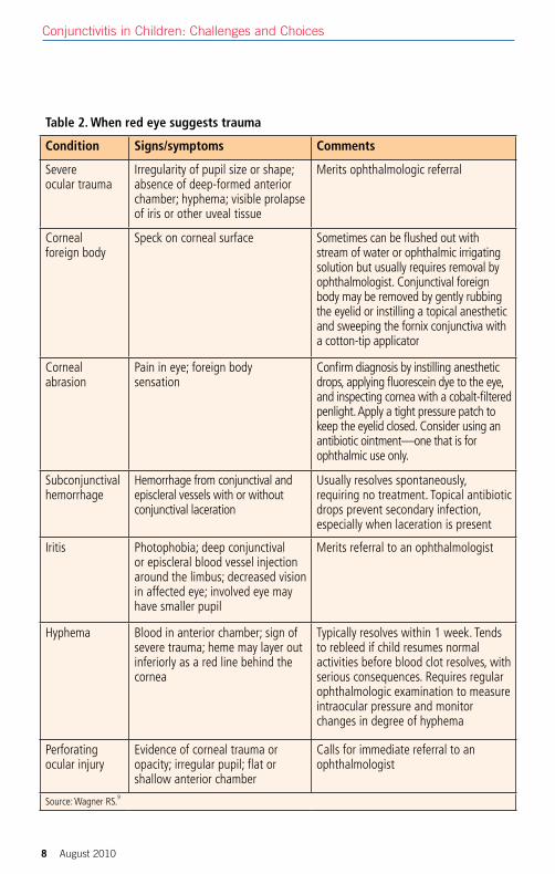
Table 2. When red eye suggests trauma
Condition Signs/symptoms Comments
Severe ocular trauma
Irregularity of pupil size or shape; absence of deep-formed anterior chamber; hyphema; visible prolapse of iris or other uveal tissue
Merits ophthalmologic referral
Corneal foreign body
Speck on corneal surface Sometimes can be flushed out with stream of water or ophthalmic irrigating solution but usually requires removal by ophthalmologist. Conjunctival foreign body may be removed by gently rubbing the eyelid or instilling a topical anesthetic and sweeping the fornix conjunctiva with a cotton-tip applicator
Corneal abrasion
Pain in eye; foreign body sensation
Confirm diagnosis by instilling anesthetic drops, applying fluorescein dye to the eye, and inspecting cornea with a cobalt-filtered penlight. Apply a tight pressure patch to keep the eyelid closed. Consider using an antibiotic ointment—one that is for ophthalmic use only.
Subconjunctival hemorrhage
Hemorrhage from conjunctival and episcleral vessels with or without conjunctival laceration
Usually resolves spontaneously, requiring no treatment. Topical antibiotic drops prevent secondary infection, especially when laceration is present
Iritis Photophobia; deep conjunctival or episcleral blood vessel injection around the limbus; decreased vision in affected eye; involved eye may have smaller pupil
Merits referral to an ophthalmologist
Hyphema Blood in anterior chamber; sign of severe trauma; heme may layer out inferiorly as a red line behind the cornea
Typically resolves within 1 week. Tends to rebleed if child resumes normal activities before blood clot resolves, with serious consequences. Requires regular ophthalmologic examination to measure intraocular pressure and monitor changes in degree of hyphema
Perforating ocular injury
Evidence of corneal trauma or opacity; irregular pupil; flat or shallow anterior chamber
Calls for immediate referral to an ophthalmologist
Source: Wagner RS.9
Conjunctivitis in Children: Challenges and Choices
August 2010 9
Although some cases of conjunctivitis start in one eye and then spread to the other, a unilateral red eye should cause the clinician to stop, pause, and consider other diagnoses. The possibilities are many, from trauma or a foreign body to herpetic infection, uveitis, and intra-ocular inflam-mation. Pain is not typically present in children with conjunctivitis; eye pain may be the result of a corneal abrasion, herpetic keratitis, or a contact lens-related ulcer, among other possible causes.
Chronicity is another diagnostic clue. Because bacterial conjunctivitis typically
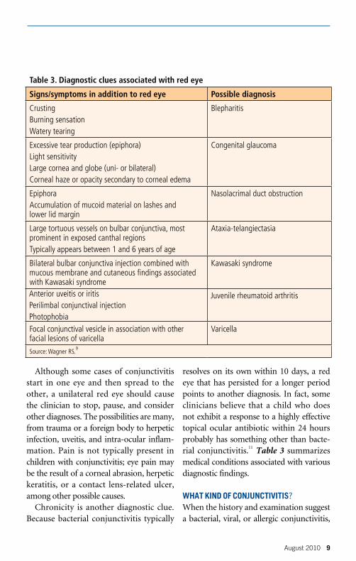
resolves on its own within 10 days, a red eye that has persisted for a longer period points to another diagnosis. In fact, some clinicians believe that a child who does not exhibit a response to a highly effective topical ocular antibiotic within 24 hours probably has something other than bacte-rial conjunctivitis.11 Table 3 summarizes medical conditions associated with various diagnostic findings.
What KiNd oF CoNJUNCtiVitiS?When the history and examination suggest a bacterial, viral, or allergic conjunctivitis,
Table 3. Diagnostic clues associated with red eye
Signs/symptoms in addition to red eye Possible diagnosis
Crusting Burning sensationWatery tearing
Blepharitis
Excessive tear production (epiphora)Light sensitivity Large cornea and globe (uni- or bilateral) Corneal haze or opacity secondary to corneal edema
Congenital glaucoma
Epiphora Accumulation of mucoid material on lashes and lower lid margin
Nasolacrimal duct obstruction
Large tortuous vessels on bulbar conjunctiva, most prominent in exposed canthal regions Typically appears between 1 and 6 years of age
Ataxia-telangiectasia
Bilateral bulbar conjunctiva injection combined with mucous membrane and cutaneous findings associated with Kawasaki syndrome
Kawasaki syndrome
Anterior uveitis or iritis Perilimbal conjunctival injection Photophobia
Juvenile rheumatoid arthritis
Focal conjunctival vesicle in association with other facial lesions of varicella
Varicella
Source: Wagner RS.9
10 August 2010
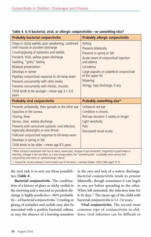
the next task is to sort out those possibili-ties (Table 4).
Bacterial conjunctivitis. The combina-tion of a history of gluey or sticky eyelids in the morning and a mucoid or purulent dis-charge is highly predictive—96% probabil-ity—of bacterial conjunctivitis.3 Crusting or gluing of eyelashes and eyelids may also be associated with a positive bacterial culture, as may the absence of a burning sensation
in the eyes and lack of a watery discharge. Bacterial conjunctivitis tends to present bilaterally, though sometimes it can begin in one eye before spreading to the other. When left untreated, the infection lasts for 8-10 days.12 The mean age of the child with bacterial conjunctivitis is 3.1-3.6 years.1
Viral conjunctivitis. The second most common type of conjunctivitis in chil-dren, viral infection can be difficult to
Table 4. Is it bacterial, viral, or allergic conjunctivitis—or something else?
Probably bacterial conjunctivitis Probably allergic conjunctivitis
Gluey or sticky eyelids upon awakening, combined with mucoid or purulent dischargeCrusting/gluing of eyelashes and eyelidsPurulent, thick, yellow-green dischargeSwelling, “gritty” feelingBilateral presentationDevelops in winterPapillary conjunctival response to slit-lamp examPresents concurrently with otitis mediaPresents concurrently with rhinitis, sinusitisChild tends to be younger—mean age 3.1-3.6 years
ItchingPresents bilaterallyPresents in spring or fallAcute onset of conjunctival injection and edemaLid edemaLarge papules on palpebral conjunctivae of the upper lidWateringStringy, ropy discharge, if any
Probably viral conjunctivitis Probably something else*
Presents unilaterally, then spreads to the other eyeOpacities in the corneaTearing, feverSerous, clear, watery dischargePresents with concurrent systemic viral infection, especially pharyngitis or sore throatFollicular conjunctival response to slit-lamp examDevelops in spring or fallChild tends to be older—mean age 8.5 years
Unilateral red eyeCondition is chronicRed eye duration 2 weeks or longerLight sensitivityPainDecreased visual acuity
*When red eye is associated with loss of vision, severe pain, changes in eye movement, irregularity in pupil shape or reactivity, changes in the red reflex, or a new droopy eyelid, the “something else” is probably more serious than conjunctivitis and merits an ophthalmologic referral.1
1. Granet DB. Ocular anatomy: Front-to-back tour of the basics. Contemp Pediatr. 2002;19(8 suppl): 8-10.
Conjunctivitis in Children: Challenges and Choices
August 2010 11
distinguish from its bacterial counter-part.3 Unlike bacterial conjunctivitis, a viral infection tends to present unilater-ally, then spread to the other eye. Viral conjunctivitis also differs from bacterial in its association with opacities in the cornea that may affect vision.
Children with viral conjunctivitis gen-erally have a concurrent systemic viral infection, most likely of the upper respi-ratory tract; the combination of con-junctivitis and sore throat suggests a viral origin. Symptoms of adenovirus conjunctivitis may persist for 2 weeks or even longer, and the infection often is accompanied by a watery discharge from the eyes. Preauricular lymph nodes are considerably more common in viral conjunctivitis but are not diagnostic. The mean age of the child with viral conjunc-tivitis is 8.5 years.1
Allergic conjunctivitis. Itching is the chief clue to allergic conjunctivitis, which is typically bilateral. Allergic conjuncti-vitis most often is seasonal, triggered by airborne pollen. Itching is intense in the spring and fall. Acute onset of conjunctival redness and edema or lid edema also are characteristic of this condition, as are large papules on the palpebral conjunctivae of the upper lid.9 The eyes may water; any discharge will be stringy or ropy in charac-ter, not green or purulent, as with bacterial conjunctivitis.
Perennial allergic conjunctivitis most often is associated with exposure to house dust mites, cat dander, air pollutants, and other allergens that are found in the environment throughout the year. The
symptoms tend to be chronic rather than seasonal in nature.13
diaGNoSeS that MUSt Not Be oVerLooKed One of the viral causes of conjunctivitis, herpes simplex infection, has potentially disastrous consequences, including cor-neal opacification and loss of vision.14 It is also important to recognize conjunctivitis-otitis media syndrome.
Herpes simplex conjunctivitis .
Distinguishing herpes simplex infection from other causes of viral conjunctivitis can be difficult. Typically, the conjunctivitis is unilateral, and the child may have vesicles on skin surrounding the eye or eyelids.14 Infection generally is accompanied by severe ocular pain or discomfort. The child may have recently had a viral illness or close con-tact with someone who did. The conjuncti-vitis may be preceded by fever, exposure to strong sunlight, or mild trauma. 14
Fluorescein staining of the cornea that
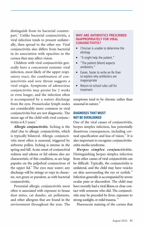
Why Are AnTIBIOTICS PreSCrIBeD InAPPrOPrIATely FOr VIrAl COnjunCTIVITIS?
• Clinicianisunabletodeterminetheetiology
• “Itmighthelpthepatient.”
• “Thepatient(Mom)expects antibiotics.”
• Easier,fastertowriteanRxthan to explain why antibiotics are inappropriate
• Return-to-schoolrulescallfor treatment
shows a dendritic or branching pattern generally points to the diagnosis of herpes simplex conjunctivitis (see case histories on pages 13 and 14). But even a penlight examination may reveal an irregularity on the corneal surface and gray or white dis-coloration in the corneal lesion. Suspicion of this condition calls for prompt referral to an ophthalmologist.14
Conjunctivitis-otitis media syndrome. Be sure to check for otitis media in any child with conjunctivitis.9,15 Irritability and a purulent discharge from the eyes are hall-marks of conjunctivitis-otitis syndrome, though the first symptoms usually are low-to-moderate grade fever, mucopuru-lent rhinorrhea, and a cough. Two or 3 days later, conjunctivitis and otitis appear; the child wakes up with eyelashes crusted together and pain in the ear.9 In the major-ity of cases the syndrome is caused by H influenzae; if left untreated, the conjuncti-vitis can progress to septicemia or menin-gitis.14 About one-quarter to one-third of children with conjunctivitis also have otitis media,1,15 and some have no ear pain.
ChooSiNG a toPiCaL aNtiBiotiCAlthough bacterial conjunctivitis usually is benign and resolves on its own within 8-10 days, the highly infectious condition brings discomfort, poses a risk of ready transmission to others, and results in school and day-care absences (see box, “When to return to school?”)11,16 Staying out of school also results in parental absence from work, with accompanying economic repercus-sions.16 The 2003 epidemic in Puerto Rico, for example, accounted for a total of 850
person-years of missed work (not including missed work for child care) and 315,000 visits to physicians’ offices, resulting in $30 million in lost worker productivity and health expenses during a 3-month period.8
Antibiotic treatment is recommended for this self-limited disease because it helps shorten the duration of illness, controls its level of contagiousness, and curbs the emergence of resistant strains by effectively eradicating the responsible pathogens.11
A number of topical antibiotic agents are available for treating bacterial conjunctivi-tis.17 It is important to remember that all agents approved by the FDA for treatment of bacterial conjunctivitis are regarded as effective in doing so. Treatment deci-sions for each individual patient should be guided by certain basic considerations, including the drug’s level of bioavailability and whether and how rapidly it eradicates the organism. Some topical agents are bac-tericidal (killing the organism) whereas others are bacteriostatic (temporarily inhibiting microorganism growth;17 see box, “Key considerations for treatment”).
Systemic treatment of bacterial con-junctivitis occasionally is warranted when symptoms are especially severe and is essential when the infection is associated with otitis media.15 Use of an antibiotic resistant to ß-lactamase is recommended for conjunctivitis-otitis syndrome, as ß-lac-tamase is common in H influenzae, the predominant etiologic agent of this highly contagious infection.9,15 The concomitant use of a topical antibiotic will allow for quick eradication of the organism from the eye and help prevent contagious spread.
12 August 2010
Conjunctivitis in Children: Challenges and Choices
August 2010 13
Macrolides. Erythromycin is active against gram-positive organisms as well as some atypical pathogens, such as Mycoplasma, Legionella, Chlamydia, and Mycobacteria. Erythromycin ointment, applied four times a day for 7 days, is well tolerated—very young children find oint-ments more comfortable than drops—and is not toxic, but it does not have good pen-etration into ocular tissue.17 Older children may not tolerate ointments as well if their vision is blurred by the treatment. In addi-tion, the ointment has been associated with hypersensitivity reactions and staphylococ-cal resistance.17,18
In 2007, the US Food and Drug Administration (FDA) approved a 1% topi-cal ophthalmic solution of azithromycin.19,20 A slightly viscous product, it has bioad-
hesive properties that tend to keep the medication on the surface of the eye lon-ger than with conventional drops, increas-ing bioavailability to the ocular surface. In addition, the agent has reduced dosing frequency compared with other treatments (one drop twice daily 8 to 12 hours apart, for the first 2 days, followed by one drop daily for the next 5 days). The product is bacteriostatic, and effectiveness is time-dependent: Skipping doses or not complet-ing the full course of therapy decreases the effectiveness of treatment and increases the likelihood that bacterial resistance will develop.19
Sulfonamides. Topical sulfa drugs, for-merly the primary medications for treat-ing bacterial conjunctivitis, have been used with less frequency in recent years. These
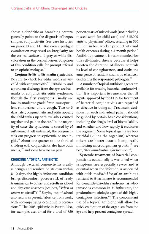
PInk eye AnD The eluSIVe DIAGnOSIS Case History #1: Matthew
The physician examines 3-year-old Matthew’s red eye and takes a history. Only the left eye is red, and the history is unclear. Think-ing Matthew probably has bacterial conjunctivitis that has not yet manifested in the right eye, he prescribes topical tobramycin and asks Matthew’s mother to report back in a few days. Three days later, Matthew’s mother calls to say that the child’s eye is still red, but still only the left one. The physician rules out trying another antibiotic. Tobramycin is bactericidal and covers the usual etiologic pathogens for conjunctivitis; also, the redness has
remained unilateral. Clearly, another office visit and consideration of another diagnosis is in order. At the visit, Matthew exhibits some light sensitivity and rubs his eye, saying something “is in it.”
The physician wets a fluorescein strip with anesthetic and applies it to the eye. He then uses a pen-light, fitted with a cobalt blue filter cap, to examine the eye. The cornea exhibits a dendritic pattern. Is this a corneal abrasion that is healing or the dendritic pattern of a herpes simplex virus? He suspects herpes simplex infection and refers Matthew to a pediatric ophthalmologist for further evaluation. The ophthalmologist confirms the diagnosis of herpes simplex with corneal involvement and prescribes a topical antiviral agent.
Cou
tesy
of R
udol
ph S
. Wag
ner,
MD
14 August 2010
agents, which are bacteriostatic, are avail-able in drop and ointment form. They have been associated with a variety of adverse effects, including allergic reactions and rare cases of Stevens-Johnson syndrome.21 The drops may sting or burn, inhibiting compliance, and the drugs are subject to bacterial resistance.22-24 An in vitro study
in 2000 showed that sulfamethoxazole had no activity against either H influenzae or S pneumoniae.24
Aminoglycosides. Gentamicin, tobramy-cin, and neomycin provide excellent cov-erage of gram-negative pathogens;17 how-ever, increasing resistance has been noted.24 In addition, aminoglycosides have been
Conjunctivitis in Children: Challenges and ChoicesConjunctivitis in Children: Challenges and Choices
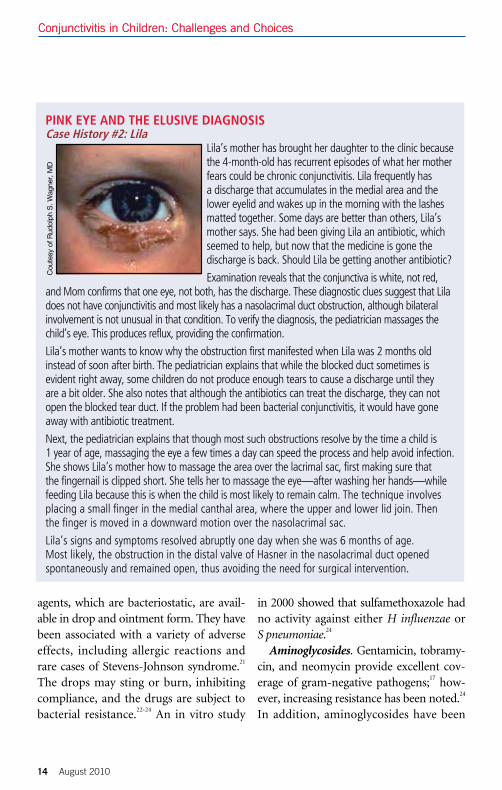
PInk eye AnD The eluSIVe DIAGnOSIS Case History #2: Lila
Lila’s mother has brought her daughter to the clinic because the 4-month-old has recurrent episodes of what her mother fears could be chronic conjunctivitis. Lila frequently has a discharge that accumulates in the medial area and the lower eyelid and wakes up in the morning with the lashes matted together. Some days are better than others, Lila’s mother says. She had been giving Lila an antibiotic, which seemed to help, but now that the medicine is gone the discharge is back. Should Lila be getting another antibiotic?
Examination reveals that the conjunctiva is white, not red, and Mom confirms that one eye, not both, has the discharge. These diagnostic clues suggest that Lila does not have conjunctivitis and most likely has a nasolacrimal duct obstruction, although bilateral involvement is not unusual in that condition. To verify the diagnosis, the pediatrician massages the child’s eye. This produces reflux, providing the confirmation.
Lila’s mother wants to know why the obstruction first manifested when Lila was 2 months old instead of soon after birth. The pediatrician explains that while the blocked duct sometimes is evident right away, some children do not produce enough tears to cause a discharge until they are a bit older. She also notes that although the antibiotics can treat the discharge, they can not open the blocked tear duct. If the problem had been bacterial conjunctivitis, it would have gone away with antibiotic treatment.
Next, the pediatrician explains that though most such obstructions resolve by the time a child is 1 year of age, massaging the eye a few times a day can speed the process and help avoid infection. She shows Lila’s mother how to massage the area over the lacrimal sac, first making sure that the fingernail is clipped short. She tells her to massage the eye—after washing her hands—while feeding Lila because this is when the child is most likely to remain calm. The technique involves placing a small finger in the medial canthal area, where the upper and lower lid join. Then the finger is moved in a downward motion over the nasolacrimal sac.
Lila’s signs and symptoms resolved abruptly one day when she was 6 months of age. Most likely, the obstruction in the distal valve of Hasner in the nasolacrimal duct opened spontaneously and remained open, thus avoiding the need for surgical intervention.
Cou
tesy
of R
udol
ph S
. Wag
ner,
MD
August 2010 15
associated with hypersensitivity and severe allergic reactions.17
Antibacterial combinations. The oph-thalmic solution of trimethoprim and poly-myxin B sulfate has activity against many gram-negative and gram-positive organ-isms, and polymyxin B is bactericidal for many gram-negative organisms.25 The agent also has been linked to increasing bacte-rial resistance, and because the solution is bacteriostatic, it may take several days to
eradicate the infection.24 The ointment poly-myxin B-neomycin has a wide range of bac-tericidal action26 but has shown increasing bacterial resistance.24 Some children develop an allergic reaction to the neomycin in the ointment, making it difficult to determine if the child’s symptoms are caused by the medication or the conjunctivitis.22
Antibiotic-corticosteroid combinations. Medications containing steroids generally have no place in treating primary conjunc-
When TO reTurn TO SChOOl?
The American Academy of Pediatrics Red Book has this to say about eye infections and school attendance: “Except when viral or bacterial conjunctivitis is accompanied by systemic signs of illness, infected children should be allowed to remain in school once any indicated therapy is implemented, unless their behavior is such that close contact with other students cannot be avoided.”1
Some schools disagree with this policy, believing it necessary to exclude students with conjunctivitis to protect other children, and the community at large, from infection. Yet some officials claim that because students with certain respiratory infections are not kept home, youngsters with conjunctivitis should not be either. Given these disparate views, it’s no wonder that state departments of health have a range of regulations about when students with conjunctivitis should be excluded from school.2 Nevertheless, in cases of acute bacterial conjunctivitis, the use of an agent that provides rapid clinical and bacterial cure is desirable for getting the child back to school as soon as possible.
Of 43 states that responded to a survey, seven allow children with conjunctivitis to remain in school, eight permit them to return once antibiotic treatment is begun, 12 allow for their return 24 hours after antibiotics first are given, 13 exclude them until the infection is “noncommunicable,” and 16 require a physician’s approval to return to school.2 This adds up to 56 responses because 17 of the states responding have multiple recommendations, often contradictory. In addition, 12 states have no official policy, though some provide guidance to school districts when asked.2 Finally, policies in only three states differentiate between viral and bacterial conjunctivitis.
As the Red Book statement suggests (six states cited Red Book as the source of their policies), children with conjunctivitis should avoid contact with others to prevent contagion. This can be difficult, especially among younger children in day care or school, who spend most of their time in groups, not sitting at desks. Nonetheless, school nurses and teachers can help prevent the spread of bacterial con-junctivitis by instituting a hand washing program and supplying supplemental alcohol cleaning gels.1
references1. American Academy of Pediatrics. Infections spread by direct contact. In: Pickering LK, ed. Red Book: 2009 Report of the Com-mittee on Infectious Diseases. 28th ed. Elk Grove Village, IL: American Academy of Pediatrics; 2009:143-145.2. Ohnsman CM. Exclusion of students with conjunctivitis from school: policies of state departments of health. J Pediatr Ophthal-mol Strabismus. 2007;44:101-105.
16 August 2010
tivitis because of the risks they pose. Topical steroids can exacerbate an underlying fun-gal or herpetic infection, resulting in irre-versible ocular damage.22 Used chronically, steroids can cause cataracts or increase intraocular pressure, heightening the risk of glaucoma.22 Also note that some topical medications for treating allergy contain ste-roids and should be used with care.
Fluoroquinolones. The 1962 introduc-tion of nalidixic acid marked the beginning of development of the quinolones, a family of synthetic broad-spectrum antibiotics that were first introduced for treatment of
systemic infection in the 1980s.27 Originally used only for treating urinary tract infec-tions, these agents are now approved for a wide variety of indications. Flumequine, the first topical fluoroquinolone, was used only briefly because of its association with ocular toxicity. Development of second- and third-generation fluoroquinolones, such as ciprofloxacin, ofloxacin, lomefloxa-cin, and levofloxacin, followed in the 1990s and beyond.27 These bactericidal agents have been used widely for treating bacterial conjunctivitis because of their broad cover-age of both gram-positive and gram-nega-
Conjunctivitis in Children: Challenges and Choices
key COnSIDerATIOnS FOr TreATmenT
In selecting among treatment options for bacterial conjunctivitis, keep in mind the following parameters: •Spectrum of activity. Because initial treatment is generally empiric, the chosen drug should
be active against the most common etiologic bacteria, especially Streptococcus pneumoniae and Haemophilus influenzae, and therefore cover both gram-positive and gram-negative organisms.
•level of availability. The agent of choice should have high bioavailability, achieved when drug content is slightly higher than the lowest concentration needed to inhibit growth of the causative bacteria and by good ocular tissue penetration.1
• rate of bacterial kill. A rapid rate of bacterial kill shortens both the duration of the infection and period of infectivity, while preventing potential complications.2
• Incidence of resistance. Also important is to choose a drug with a low incidence of resistance. Though systemic agents are far more likely to lead to bacterial resistance than topical antibiotics, resistance to the topical agents used in treating conjunctivitis is increasing.1
• Toxicity. Many practitioners avoid medications that contain neomycin because of reported hyper-sensitivity reactions. Aminoglycosides are associated with corneal toxicity. Few physicians today would prescribe chloramphenicol because of its association with aplastic anemia.3
•Comfort and convenience. Choosing an agent that isn’t associated with frequent administration or stinging or burning is particularly important for promoting adherence in children.
references1. Mah FS. Fourth-generation fluoroquinolones: new topical agents in the war on ocular bacterial infections. Curr Opin Ophthalmol. 2004;15:316-320. 2. Gigliotti F, Hendley JO, Morgan J, et al. Efficacy of topical antibiotic therapy in acute conjunctivitis in children. J Pediatr. 1984;104:623-626.3. Wagner RS, Alcorn D, Gigliotti F, et al. Management of conjunctivitis. Part 1: diagnosis and treatment of bacterial disease. Contemp Pediatr. 2000;17(suppl): 3-14.
August 2010 17
tive pathogens and their lack of toxicity. Fluoroquinolones, too, have been found
to trigger bacterial resistance.28,29 Partly for this reason, fourth-generation fluoroqui-nolones were developed. Moxifloxacin and gatifloxacin first became available in 2003; in 2009, the FDA approved another topi-cal ocular fluoroquinolone, besifloxacin. The fourth-generation fluoroquinolones have an 8-methoxy substitution on the basic quinolone molecule, which provides better coverage against gram-positive organisms, including resistant strains, than earlier-gen-eration fluoroquinolones. At the same time, coverage of gram-negative organisms is comparable to that of earlier-generation flu-oroquinolones.11 In general, fourth-genera-tion fluoroquinolones provide broad cover-age with rapid bacterial eradication. They have low toxicity and are associated with low levels of bacterial resistance. Supporting evidence comes from investigations that compare fourth-generation fluoroquinolo-nes with their predecessors, other contem-porary treatments, and one another.
aNtiBiotiC reSiStaNCeDevelopment of antibiotic resistance is thought to result from slowly decreasing levels of drug in tissue, exposing remain-ing bacteria to antibiotic levels below the concentration needed to prevent mutated strains. This mutant prevention concen-tration is defined as the range between the MIC
90 (MIC necessary to inhibit growth
of 90% of organisms) and the MIC of the least susceptible, but not resistant, mutant.30 Ocular therapy is considered less likely than systemic therapy to result
in resistance because eye drops deliver a significant amount of drug to the affected surface. In addition, ocular infections involve far fewer organisms than systemic infections and are treated for a shorter time, which also makes development of resistance less likely.
Fluoroquinolones inhibit development of resistance by binding with bacterial nuclear enzymes, disrupting their activity. Early-generation fluoroquinolones ham-per bacterial DNA synthesis by inhibit-ing DNA gyrase, an enzyme required for DNA replication, in gram-negative organ-isms. In gram-positive organisms, the drugs interrupt the enzymatic pathway by inhibiting topoisomerase IV. Because fourth-generation fluoroquinolones attack both DNA gyrase and topoisomerase IV in gram-positive organisms, two spontaneous mutations of bacterial DNA, rather than one, are required to trigger gram-positive bacterial resistance to these agents.33
A recent investigation explored the ques-tion of whether use of fourth-generation fluoroquinolones leads to bacterial resis-tance.31 Given that instillation of topical ocular fluoroquinolones results in drug overflowing to skin around the eyes and other nearby sites, the authors set out to determine if diluted drug at these sites selects for bacteria with decreased fluoro-quinolone susceptibility. They took swabs of the eyes, cheeks, nostrils, and throats of 105 children from 8 months to 12 years of age with bacterial conjunctivitis as well as 57 healthy control subjects. Children with conjunctivitis received topical moxifloxacin three times a day for 7 days. Investigators
18 August 2010
again took swabs after the treatment period and 5 weeks later and looked for changes in the susceptibility of S aureus, S pneumo-niae, and H influenzae in the eye, nose, and throat to 20 different antibiotics. The three organisms’ susceptibility to moxifloxacin did not change in the eye or other body sites, and the drug’s MIC did not increase. Also significant, even before treatment the children with conjunctivitis were found to have some drug-resistant bacteria—principally species that colonize the throat. Investigators therefore concluded that the resistance probably was attributable to sys-temic, not topical, antibiotics.31
reFereNCeS1. Gigliotti F, Williams WT, Hayden FG, et al. Etiology of acute conjunctivitis in children. J Pediatr. 1981;98:531-536.
2. Weiss A, Brinser JH, Nazar-Stewart V. Acute con-junctivitis in childhood. J Pediatr. 1993;122:10-14.3. Patel PB, Diaz MCG, Bennett J, et al. Clinical features of bacterial conjunctivitis in children. Acad Emerg Med. 2007; 14:1-5.4. Meltzer JA, Kunkov S, Crain EF. Identifying chil-dren at low risk for bacterial conjunctivitis. Arch Pediatr Adolesc Med. 2010;164:263-267.5. Chomel JJ, Szymczyszyn P, Honneger D, et al. An epidemic of adenovirus type 1 conjunctivitis. Pediatr Infect Dis J. 1989;8:884-886.6. Martin M, Turco JH, Zegans ME, et al. An out-break of conjunctivitis due to atypical Streptococcus pneumoniae. N Engl J Med. 2003;348:1112-1121.7. Centers for Disease Control and Prevention. Pneumococcal conjunctivitis at an elementary school—Maine, September 20-December 6, 2002. MMWR. 2003;52(4):64-66.8. Centers for Disease Control and Prevention. Acute hemorrhagic conjunctivitis outbreak caused by coxsackievirus A24—Puerto Rico, 2003. MMWR. 2004;53(28):632-634.9. Wagner RS. The differential diagnosis of the red eye. Contemp Pediatr. 1991;8(7):26-48.10. Stern GA, Schemmer GB, Farber RD, Gorovoy MS. Effect of topical antibiotic solutions on cor-neal epithelial wound healing. Arch Ophthalmol. 1983;101:644-647.
Conjunctivitis in Children: Challenges and Choices
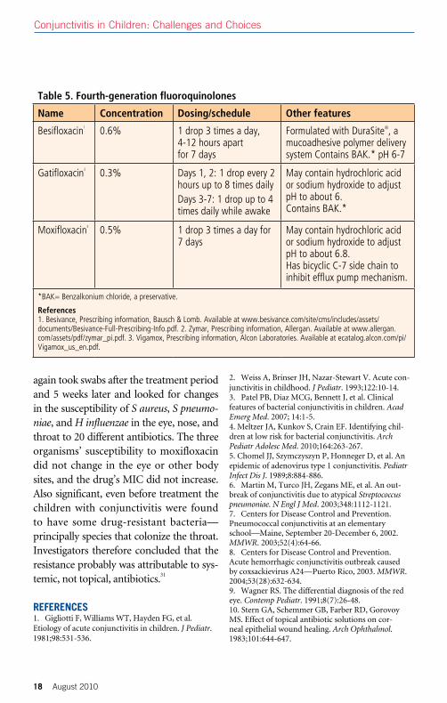
Table 5. Fourth-generation fluoroquinolones
name Concentration Dosing/schedule Other features
Besifloxacin1 0.6% 1 drop 3 times a day, 4-12 hours apart for 7 days
Formulated with DuraSite®, a mucoadhesive polymer delivery system Contains BAK.* pH 6-7
Gatifloxacin2 0.3% Days 1, 2: 1 drop every 2 hours up to 8 times dailyDays 3-7: 1 drop up to 4 times daily while awake
May contain hydrochloric acid or sodium hydroxide to adjust pH to about 6.Contains BAK.*
Moxifloxacin3 0.5% 1 drop 3 times a day for 7 days
May contain hydrochloric acid or sodium hydroxide to adjust pH to about 6.8.Has bicyclic C-7 side chain to inhibit efflux pump mechanism.
*BAK= Benzalkonium chloride, a preservative.
references1. Besivance, Prescribing information, Bausch & Lomb. Available at www.besivance.com/site/cms/includes/assets/ documents/Besivance-Full-Prescribing-Info.pdf. 2. Zymar, Prescribing information, Allergan. Available at www.allergan.com/assets/pdf/zymar_pi.pdf. 3. Vigamox, Prescribing information, Alcon Laboratories. Available at ecatalog.alcon.com/pi/Vigamox_us_en.pdf.
August 2010 19
11. Granet DB, Dorfman M, Stroman D, et al. A multicenter comparison of polymyxin B sulfate/trimethoprim ophthalmic solution and moxifloxa-cin in the speed of clinical efficacy for the treatment of bacterial conjunctivitis. J Pediatr Ophthalmol Strabismus. 2008;45:340-349.12. Gross RD. Overview: managing conjunctivitis in its varied forms. Contemp Pediatr. 2002;19(8 suppl):4-12.13. Trocme SD, Sra KK. Spectrum of ocular allergy. Curr Opin Allerg Clin Immunol. 2002;2:423-427.14. Wagner RS. Focusing on the causes of red eye. Contemp Pediatr. 1994;10(8 suppl):4-12.15. Teoh DL, Reynolds S. Diagnosis and manage-ment of pediatric conjunctivitis. Pediatr Emerg Care. 2003;19:48-55.16. Ohnsman CM. Exclusion of students with conjunctivitis from school: policies of state depart-ments of health. J Pediatr Ophthalmol Strabismus. 2007;44:101-105.17. Mah FS. New antibiotics for bacterial infections. Ophthalmol Clin North Am. 2003;16:11-27.18. Friedlaender MH. A review of the causes and treat-ment of bacterial and allergic conjunctivitis. Clin Ther. 1995; 17:800-810.19. Azasite (azithromycin ophthalmic solution) 1%. Prescribing information, Inspire Pharmaceuticals. 20. Abelson M, Protzko E, Shapiro A; 1% azithromy-cin in DuraSite Clinical Study Group. A randomized trial assessing the clinical efficacy and microbial eradi-cation of 1% azithromycin ophthalmic solution vs tobramycin in adult and pediatric subjects with bacte-rial conjunctivitis. Clin Ophthalmol. 2007;1:177-182.21. Rubin Z. Ophthalmic sulfonamide-induced Stevens-Johnson syndrome. Arch Dermatol. 1977;113:235-236.22. Wagner RS, Alcorn D, Gigliotti F, et al. Management of conjunctivitis. Part 1: diagnosis
and treatment of bacterial disease. Contemp Pediatr. 2000;17(suppl): 3-14. 23. Lichtenstein SJ. Ocular infections in children: best practices in diagnosis and treatment. Treatment options for pediatric conjunctivitis. Infect Dis Child. March 2004 (suppl). Available at www.idinchldren.com/monograph/0403/article2.asp. Accessed March 9, 2010.24. Block SL, Hedrick J, Ryler R, et al. Increasing bacterial resistance in pediatric acute conjunctivitis (1997-1998). Antimicrob Agents Chemother. 2000; 44:1650-1654.25. Polytrim Ophthalmic Solution. Prescibing infor-mation. Available at www.drugs.com/pro/polytrim.html. Accessed March 15, 2010. 26. Neosporin official FDA information, side effects and uses. Available at drugs.com/pro/Neosporin.html. Accessed March 12, 2010.27. Ball P. Quinolone generations: natural history or natural selection? J Antimicrob Chemother. 2000;46:17-24. 28. Marangon FB, Miller D, Muallem MS, et al. Ciprofloxacin and levofloxacin resistance among methicillin-sensitive Staphylococcus aureus isolates from keratitis and conjunctivitis. Am J Ophthalmol. 2004;137:453-458.29. Kowalski RP, Karenchak LM, Romanowski EG. Infectious disease: changing antibiotic susceptibility. Ophthalmol Clin North Am. 2003;16:1-9.30. Lichtenstein SJ, Wagner RS, Jamison T, et al. Speed of bacterial kill with a fluoroquinolone compared with nonfluoroquinolones: clinical implications and a review of kinetics of kill studies. Adv Ther. 2007;24:1098-1106.31. Marshall B, Cupp G, Foster K, et al. Moxifloxacin treatment of conjunctivitis: microbial effects beyond the eye. Presented at Interscience Conference on Antimicrobial Agents and Chemotherapy, September 2009; Poster C2-103.
This educational monograph is supported by a grant fromAlcon Laboratories, manufacturers of VIGAMOX® Solution
(moxifloxacin hydrochloride ophthalmic solution)

Produced byBrought to you as an educational service by Alcon Laboratories
VIG10500M