Embed Size (px)
Citation preview
Con®gurational coding, familiarity and the right hemisphereadvantage for face recognition in sheep
J.W. Peirce*, A.E. Leigh, K.M. Kendrick
Laboratory of Cognitive and Developmental Neuroscience, Babraham Institute, Babraham, Cambridge CB2 4AT, UK
Received 4 September 1998; received in revised form 7 April 1999; accepted 9 June 1999
Abstract
This study examined characteristics of visual recognition of familiar and unfamiliar faces in sheep using a 2-way
discrimination task. Of particular interest were e�ects of lateralisation and the di�erential use of internal (con®gurational) vsexternal features of the stimuli. Animals were trained in a Y-maze to identify target faces from pairs, both of which werefamiliar (same ¯ock as the subjects) or both of which were unfamiliar (di�erent ¯ock). Having been trained to identify therewarded face a series of stimuli were presented to the sheep, designed to test for the use of each visual hemi®eld in the
discriminations and the use of internal and external facial cues. The ®rst experiment showed that there was a left visual hemi®eld(LVF) advantage in the identi®cation of `hemifaces', and `mirrored hemifaces' and `chimeric' faces and that this e�ect wasstrongest with familiar faces. This represents the ®rst evidence for visual ®eld bias outside the primate literature. Results from
the second experiment showed that, whilst both familiar and unfamiliar faces could be identi®ed by the external features alone,only the familiar faces could be recognised by the internal features alone. Overall the results suggest separate recognitionmethods for socially familiar and unfamiliar faces, with the former being coded more by internal, con®gurational cues and
showing a lateral bias to the left visual ®eld. # 2000 Elsevier Science Ltd. All rights reserved.
Keywords: Asymmetry; Hemispheric lateralisation; Chimeric; Face processing; Expertise; Internal features
In humans, recognition of faces has been found todisplay a robust visual ®eld bias with faces presentedto the left of ®xation being identi®ed faster [4] andmore accurately [10,15] than those to the right. Similarresults are found for children [4,40] and commisurot-omy patients [22,33]. The left visual hemi®eld (LVF)also plays a disproportionate role in judgements ofa�ective state with chimeric faces, where half of theface is smiling and half is frowning [20,21]. It isthought that this asymmetry in perception arises fromthe neural lateralisation which has been reported byvarious human brain imaging studies such as PositronEmission Topography (PET) [34], functional MagneticResonance Imaging (fMRI) [16,29] and Magneto-Ence-
phalography (MEG) [35] in normal subjects, and by
lesion localisation in prosopagnosic patients [7 for
review]. All of these lines of research have implicated
occipitotemporal cortex in face processing with acti-
vation predominantly (although not exclusively) in the
right hemisphere.
This region may not be dedicated to face perception
per se but rather to some visual process on which face
recognition relies heavily. Diamond and Carey [8]
claim that faces are merely a category of objects which
have a common con®guration and that through experi-
ence we have learnt to identify subtle variations in this
common con®guration. They tested the hypothesis
with the use of the inversion e�ect (whereby humans
[e.g. 38,39] monkeys [28,37] and chimpanzees [26] are
particularly bad at identifying faces presented upside-
down) and found that dog-show judges demonstrate
an equivalent e�ect for dogs. Thus they suggest that
Neuropsychologia 38 (2000) 475±483
0028-3932/00/$ - see front matter # 2000 Elsevier Science Ltd. All rights reserved.
PII: S0028-3932(99 )00088 -3
www.elsevier.com/locate/neuropsychologia
* Corresponding author. Tel.: +44-1223-496364; fax: +44-1223-
496028.
E-mail address: [email protected] (J.W. Peirce).
any visual objects with a common feature con®gur-ation can show face-like e�ects if they are discrimi-nated frequently. This theory is supported byneuropsychological data which ®nds that most proso-pagnosic patients do not show agnosia purely for facesbut also for other intra-category discriminations. Forexample there are reported prosopagnosic farmers whocould no longer discriminate between their cows [1,3]or ornithologists who could no longer identify di�erentbirds [2]. There are a few cases, however, where thede®cit is found for human faces despite an intact abil-ity to identify other animal faces such as cows [5] orsheep [23].
Rhodes [31] examined the hypothesis that expertiseleads to lateralisation and con®gurational1 coding. Shemeasured the ability to recognise faces of di�erent eth-nic backgrounds (Caucasian and Chinese) in conjunc-tion with visual ®eld asymmetry and inversion (as ameasure of con®gurational coding). Whilst verifyingthat con®gurational coding was correlated to hemi®eldbias, it was not dependent on the degree of familiaritywith the ethnic background of the faces as predictedby the expertise hypothesis. It should be noted that theuse of inversion may not be a true measure of con®g-urational coding. Whilst it is frequently cited as themost likely cause for the e�ect there is evidence thatinversion of individual features could also give rise toinversion de®cits [30]. Thus, in the present study, con-®gurational coding is tested for directly with faceswhere the internal features have been masked or re-arranged rather than inverted.
In non-human primates evidence for an asymmetryin either neural activation or behavioural responseshas been mixed. Dittrich [9] used line-drawings offaces and required their macaques to indicate the facewhich was showing a certain gesture. They then pro-duced asymmetric chimeric faces and jumbled-featureversions of these stimuli to identify whether the in-ternal and external, and the left and right sides of theface were being used di�erentially. The results suggestthat the right hemi®eld was being used most (contraryto human subjects) and also that, whilst the facial out-line was the most important visual cue, the eye regionand mouth were also used.
Overman and Doty [25] used mirrored hemifaces(where one visual hemi®eld is mirrored onto the other)as stimuli and found an advantage for the left±left facein human subjects, as expected, but no equivalent biasin macaque subjects. Another method has used splitbrain macaques where presentations can be madepurely to one brain hemisphere, and this did yield anLVF advantage [14] although previous attempts to do
so by the same group had failed [12,13]. Finally, Mor-ris and Hopkins [24] found a bias in two out of threechimps (pan troglodytes) from the Savage±Rumbaughproject, performing Levy's [21] happy/sad discrimi-nation on chimeric faces. This appears to be the stron-gest evidence to date for such perceptual asymmetry ina non-human species.
At an anatomical level, the functional mapping ofthe primate brain during face recognition tasks has notbeen attempted and, at a cellular level, electrophysi-ology has shown little evidence for a right hemisphereprocessing bias. One paper even reports more face sen-sitive cells in the left hemisphere than the right [27],although this may be an artefact of the techniquewhich lends itself better to the descriptions of neuronal®ring characteristics than to the quanti®cation of cellpopulations.
Sheep have been shown to use visual cues to dis-criminate between the species or breed of faces pre-sented in a Y-maze [4] as well as identifying faces ofindividual sheep [18]. In both studies signi®cant classicinversion e�ects were found for the faces of a familiarbreed, and especially for socially familiar individuals[18]. In sheep, neither electrophysiological nor beha-vioural studies have previously attempted to identifyany asymmetry, although recent data from in-situ hy-bridisation of c-fos has shown stronger right hemi-sphere activation during a face recognition task [19].Thus, the ®rst experiment of the present paper wasdesigned to test for a visual hemi®eld bias during facerecognition. This di�ers slightly from the facial ex-pression recognition used by a number of primate [e.g.10,24] and human studies [e.g. 21] but is necessarysince sheep show very few facial expressions. Whilstthe recognition of facial identity may be a separableprocess from gesture recognition, it has also beenshown to demonstrate visual ®eld bias [e.g. 31]. Thesecond experiment tested directly for the use of con®g-urational coding in sheep face recognition. Both exper-iments compared familiar and unfamiliar individualsto test for expertise e�ects.
1. Method
1.1. Subjects
The animals used were 10 polled Dorset ewes. Thisis a hornless breed with high facial homogeneity. Theyhad been extensively trained in the Y-maze procedurewhereby they were given food reward for choosing atarget stimulus and were habituated to the maze en-vironment and the experimenters. They were keptamongst a ¯ock of 20±30 sheep. Food (grass or hay)and water was provided ad libitum throughout thestudy.
1 Some authors use the term `con®gural' rather than con®gura-
tional.
J.W. Peirce et al. / Neuropsychologia 38 (2000) 475±483476
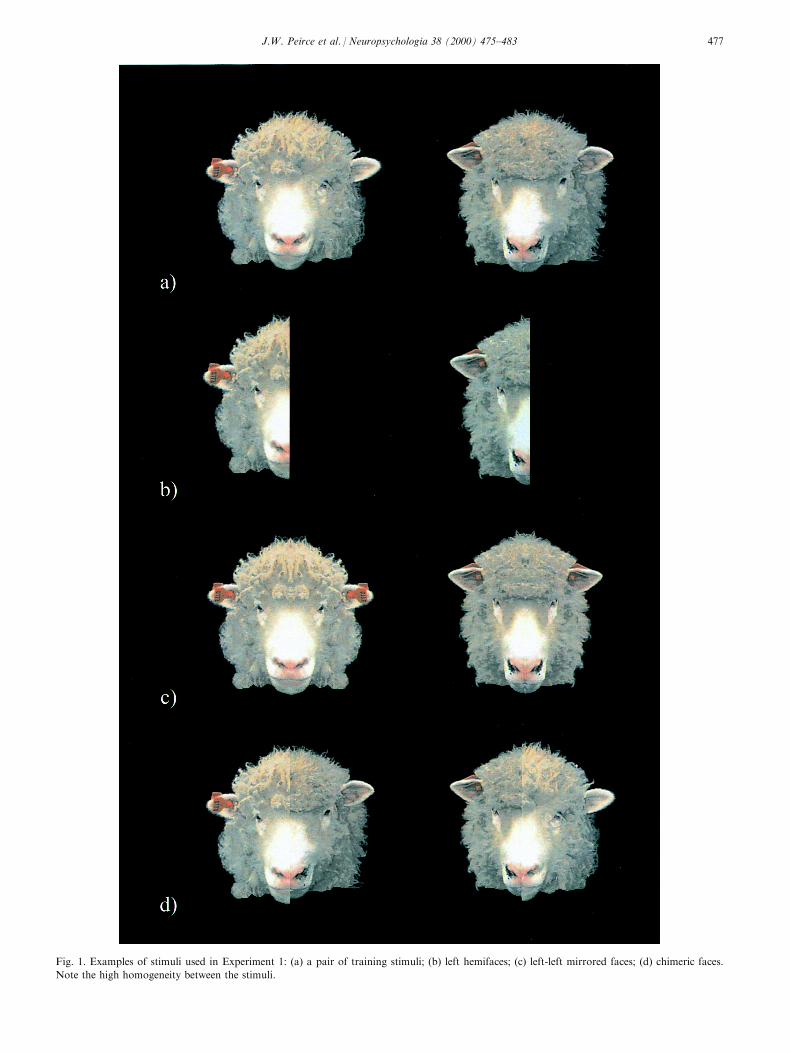
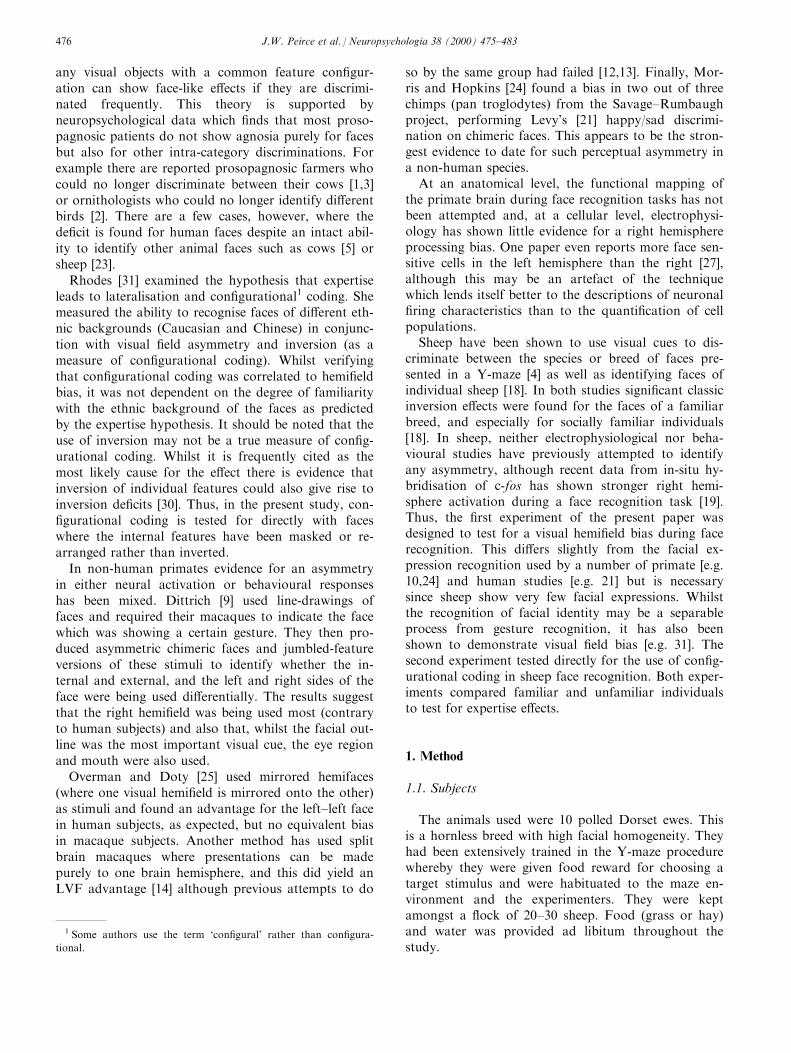
Fig. 1. Examples of stimuli used in Experiment 1: (a) a pair of training stimuli; (b) left hemifaces; (c) left-left mirrored faces; (d) chimeric faces.
Note the high homogeneity between the stimuli.
J.W. Peirce et al. / Neuropsychologia 38 (2000) 475±483 477
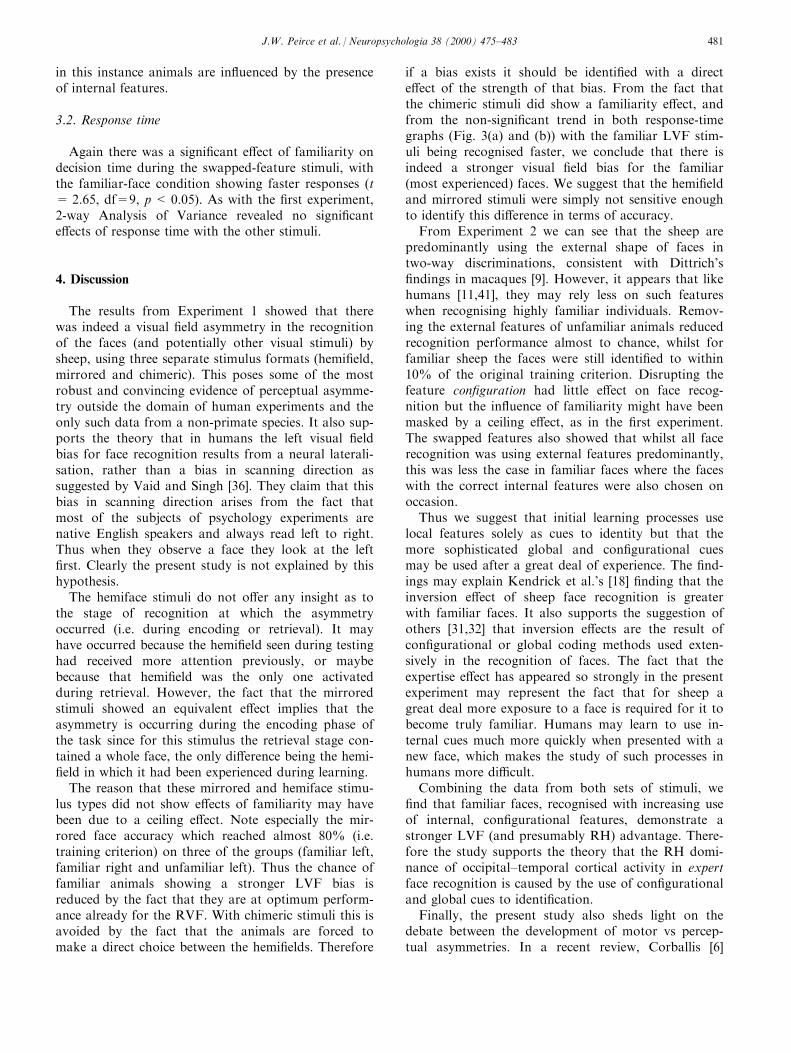
Fig. 2. Examples of stimuli used in Experiment 2: (a) internal features only; (b) external features only; (c) scrambled features; (d) swapped-feature
stimuli.
J.W. Peirce et al. / Neuropsychologia 38 (2000) 475±483478
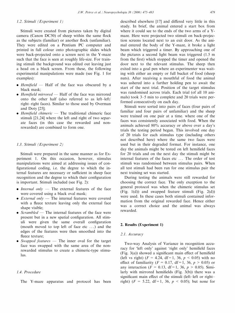
1.2. Stimuli (Experiment 1)
Stimuli were created from pictures taken by digitalcamera (Canon DC50) of sheep within the same ¯ockas the subjects (familiar) or another ¯ock (unfamiliar).They were edited on a Pentium PC computer andprinted in full colour onto photographic slides whichwere back-projected onto a screen next in the Y-mazesuch that the face is seen at roughly life-size. For train-ing stimuli the background was edited out leaving justa head on a black screen. From these, the followingexperimental manipulations were made (see Fig. 1 forexamples):
. Hemi®eld Ð Half of the face was obscured by ablack mask;
. Hemi®eld mirrored Ð Half of the face was mirroredonto the other half (also referred to as left±left/right±right faces). Similar to those used by Overmanand Doty [25];
. Hemi®eld chimeric Ð The traditional chimeric facestimuli [21,24] where the left and right of two separ-ate faces (in this case the rewarded and non-rewarded) are combined to form one.
1.3. Stimuli (Experiment 2)
Stimuli were prepared in the same manner as for Ex-periment 1. On this occasion, however, stimulusmanipulations were aimed at addressing issues of con-®gurational coding, i.e. the degree to which the in-ternal features are necessary or su�cient in sheep facerecognition and the degree to which their con®gurationis important. Stimuli included (see Fig. 2):
. Internal only Ð The external features of the facewere covered using a black oval mask;
. External only Ð The internal features were coveredwith a ¯eece texture leaving only the external faceshape visible;
. Scrambled Ð The internal features of the face werepresent but in a new spatial con®guration. All stim-uli were given the same overall con®guration(mouth moved to top left of face etc . . .) and theedges of the features were then smoothed into the¯eece texture;
. Swapped features Ð The inner oval for the targetface was swapped with the same area of the non-rewarded stimulus to create a chimeric-type stimu-lus.
1.4. Procedure
The Y-maze apparatus and protocol has been
described elsewhere [17] and di�ered very little in thisstudy. In brief, the animal entered a start box fromwhere it could see to the ends of the two arms of a Y-maze. Here were projected two stimuli on back-projec-tion screens located next to an exit door. As the ani-mal entered the body of the Y-maze, it broke a lightbeam which triggered a timer. By approaching one ofthe pictures a second light beam was triggered (1.5 mfrom the ®rst) which stopped the timer and opened thedoor next to the relevant stimulus. The sheep thenexited into a goal pen where an experimenter was wait-ing with either an empty or full bucket of food (sheepnuts). After receiving a mouthful of food the animalwas ushered into a further holding pen to await thestart of the next trial. Position of the target stimuluswas randomised across trials. Each trial (of all 10 ani-mals) took 3±5 min to complete and 20 trials were per-formed consecutively on each day.
Stimuli were sorted into pairs of faces (four pairs offamiliar and four pairs of unfamiliar) and the sheepwere trained on one pair at a time. where one of thefaces was consistently associated with food. When theanimals achieved 80% accuracy or above over a day'strials the testing period began. This involved one dayof 20 trials for each stimulus type (including othersnot described here) where the same two faces wereused but in their degraded format. For instance, oneday the animals might be tested on left hemi®eld facesfor 20 trials and on the next day the stimuli might beinternal features of the faces etc . . . The order of teststimuli was randomised between stimulus pairs. Whenall test stimuli had been run for one stimulus pair thenext training set was started.
During testing the animals were still rewarded forchoosing the correct face. The only exception to thegeneral protocol was when the chimeric stimulus set(Fig. 1(d)) and swapped feature stimuli (Fig. 2(d))were used. In these cases both stimuli contained infor-mation from the original rewarded face. Hence eitherwas a correct choice and the animal was alwaysrewarded.
2. Results (Experiment 1)
2.1. Accuracy
Two-way Analysis of Variance in recognition accu-racy for `left only' against `right only' hemi®eld faces(Fig. 3(a)) showed a signi®cant main e�ect of hemi®eld(left vs right) (F = 4.24, df=1, 36, p < 0.05) with noe�ect of familiarity (F = 0.17, df=1, 36, p > 0.05) orany interaction (F = 0.13, df=1, 36, p > 0.05). Simi-larly with mirrored hemi®elds (Fig. 3(b)) there was asigni®cant main e�ect of the stimuli (left±left or right±right) (F = 5.22, df=1, 36, p < 0.05); but none for
J.W. Peirce et al. / Neuropsychologia 38 (2000) 475±483 479

familiarity (F = 2.06, df=1, 36, p > 0.05); or any in-teraction (F=0.93, df=1, 36, p>0.05).
In the choice task where hemi®eld chimeric stimuliwere used, an e�ect of familiarity was observed (Fig.3(c)). Here, the binomial test showed a signi®cant LVFbias across the population (choice=61%, n = 800, p< 0.05) and for 5 out of 10 individual animals discri-minating familiar faces. In the non-familiar conditionhowever, no signi®cant e�ects were seen for individualsor the population. This di�erence was con®rmed by apaired-means t-test between the familiar and unfami-liar conditions (t=2.26, df=9, p<0.05).
2.2. Response time
Two-way Analysis of Variance yielded no signi®cantmain e�ects or interactions for the response times tohemiface or mirrored stimuli. This is probably becausethe measure of the response time depends not only onthe ability of the animal to recognise the face but alsoon factors such as motivation. These vary greatly
between individuals but also from day to day withinsubjects, depending on conditions such as temperature.
A paired-means t-test did reveal a signi®cant di�er-ence between the response times of sheep choosingbetween chimeric faces (Fig. 3(c)), with responsesbeing faster in the familiar face condition (t = 6.38,df=9, p<0.001).
3. Results (Experiment 2)
3.1. Accuracy
Two-way Analysis of Variance (stimulus type Xfamiliarity) for accuracy showed a main e�ect ofstimulus type (F = 22.62, df=1, 36, p < 0.001) withthe removal of external features (to make `internalonly' faces) being most damaging to discriminations inboth levels of familiarity (Fig. 4(a)). There was nomain e�ect of familiarity although there was a signi®-cant interaction between that and stimulus type (F =7.40, df=1, 36, p<0.01) suggesting that familiar faceswere less a�ected by external feature removal. Therealso appears to be a trend for the scrambled featurestimuli to impair the identi®cation of familiar faces butnot to have any e�ect on unfamiliars.
For the chimeric-type `swapped features' stimuli, theface containing the external features of the rewardedface was chosen more often for each individual sheepon familiar and unfamiliar faces ( p<0.05 in binomialtest over 80 trials). However comparing the e�ect forfamiliar and unfamiliars we ®nd that the e�ect is lessfor familiars (t=2.40, df=9, p<0.05) suggesting that
Fig. 4. Results from Experiment 2: (a) accuracy and response times
over 800 trials discriminating between the `external only', `scrambled'
and `internal only' stimuli; (b) choices made towards the stimulus
containing the external features of the target face and the internal
features of the distractor. All error bars show standard error of the
mean. �p<0.05, ��p<0.01 (see text for details of tests used).
Fig. 3. Results from Experiment 1: (a) accuracy of recognition and
response times of 10 sheep over 80 trials (per experimental condition)
discriminating between left and right hemifaces; (b) accuracy and re-
sponse time during recognition of left±left and right±right mirrored
faces; (c) choices made towards the `left chimeric face' i.e. that which
had the left of the target face combined with the right of the distrac-
tor stimulus. All error bars show standard error of the mean across
subjects. �p<0.05, ��p<0.01 (see text for details of tests used).
J.W. Peirce et al. / Neuropsychologia 38 (2000) 475±483480
in this instance animals are in¯uenced by the presenceof internal features.
3.2. Response time
Again there was a signi®cant e�ect of familiarity ondecision time during the swapped-feature stimuli, withthe familiar-face condition showing faster responses (t= 2.65, df=9, p< 0.05). As with the ®rst experiment,2-way Analysis of Variance revealed no signi®cante�ects of response time with the other stimuli.
4. Discussion
The results from Experiment 1 showed that therewas indeed a visual ®eld asymmetry in the recognitionof the faces (and potentially other visual stimuli) bysheep, using three separate stimulus formats (hemi®eld,mirrored and chimeric). This poses some of the mostrobust and convincing evidence of perceptual asymme-try outside the domain of human experiments and theonly such data from a non-primate species. It also sup-ports the theory that in humans the left visual ®eldbias for face recognition results from a neural laterali-sation, rather than a bias in scanning direction assuggested by Vaid and Singh [36]. They claim that thisbias in scanning direction arises from the fact thatmost of the subjects of psychology experiments arenative English speakers and always read left to right.Thus when they observe a face they look at the left®rst. Clearly the present study is not explained by thishypothesis.
The hemiface stimuli do not o�er any insight as tothe stage of recognition at which the asymmetryoccurred (i.e. during encoding or retrieval). It mayhave occurred because the hemi®eld seen during testinghad received more attention previously, or maybebecause that hemi®eld was the only one activatedduring retrieval. However, the fact that the mirroredstimuli showed an equivalent e�ect implies that theasymmetry is occurring during the encoding phase ofthe task since for this stimulus the retrieval stage con-tained a whole face, the only di�erence being the hemi-®eld in which it had been experienced during learning.
The reason that these mirrored and hemiface stimu-lus types did not show e�ects of familiarity may havebeen due to a ceiling e�ect. Note especially the mir-rored face accuracy which reached almost 80% (i.e.training criterion) on three of the groups (familiar left,familiar right and unfamiliar left). Thus the chance offamiliar animals showing a stronger LVF bias isreduced by the fact that they are at optimum perform-ance already for the RVF. With chimeric stimuli this isavoided by the fact that the animals are forced tomake a direct choice between the hemi®elds. Therefore
if a bias exists it should be identi®ed with a directe�ect of the strength of that bias. From the fact thatthe chimeric stimuli did show a familiarity e�ect, andfrom the non-signi®cant trend in both response-timegraphs (Fig. 3(a) and (b)) with the familiar LVF stim-uli being recognised faster, we conclude that there isindeed a stronger visual ®eld bias for the familiar(most experienced) faces. We suggest that the hemi®eldand mirrored stimuli were simply not sensitive enoughto identify this di�erence in terms of accuracy.
From Experiment 2 we can see that the sheep arepredominantly using the external shape of faces intwo-way discriminations, consistent with Dittrich's®ndings in macaques [9]. However, it appears that likehumans [11,41], they may rely less on such featureswhen recognising highly familiar individuals. Remov-ing the external features of unfamiliar animals reducedrecognition performance almost to chance, whilst forfamiliar sheep the faces were still identi®ed to within10% of the original training criterion. Disrupting thefeature con®guration had little e�ect on face recog-nition but the in¯uence of familiarity might have beenmasked by a ceiling e�ect, as in the ®rst experiment.The swapped features also showed that whilst all facerecognition was using external features predominantly,this was less the case in familiar faces where the faceswith the correct internal features were also chosen onoccasion.
Thus we suggest that initial learning processes uselocal features solely as cues to identity but that themore sophisticated global and con®gurational cuesmay be used after a great deal of experience. The ®nd-ings may explain Kendrick et al.'s [18] ®nding that theinversion e�ect of sheep face recognition is greaterwith familiar faces. It also supports the suggestion ofothers [31,32] that inversion e�ects are the result ofcon®gurational or global coding methods used exten-sively in the recognition of faces. The fact that theexpertise e�ect has appeared so strongly in the presentexperiment may represent the fact that for sheep agreat deal more exposure to a face is required for it tobecome truly familiar. Humans may learn to use in-ternal cues much more quickly when presented with anew face, which makes the study of such processes inhumans more di�cult.
Combining the data from both sets of stimuli, we®nd that familiar faces, recognised with increasing useof internal, con®gurational features, demonstrate astronger LVF (and presumably RH) advantage. There-fore the study supports the theory that the RH domi-nance of occipital±temporal cortical activity in expertface recognition is caused by the use of con®gurationaland global cues to identi®cation.
Finally, the present study also sheds light on thedebate between the development of motor vs percep-tual asymmetries. In a recent review, Corballis [6]
J.W. Peirce et al. / Neuropsychologia 38 (2000) 475±483 481
claims that the advantage of motor asymmetry (interms of handedness) and the left hemisphere localis-ation of language function has driven other forms ofneural lateralisation such as the perceptual asymmetrydescribed here. The present study describes an animalwith no apparent need for motor asymmetry, and cer-tainly no language, for which a perceptual asymmetryis manifest. For these animals the need to quicklyidentify cohorts appears to be the only task complexenough to require hemispheric specialisation.
The model may give us a method by which we canexamine more closely the relationship between the cer-ebral and perceptual asymmetries, and may also enableus to study the development of hemispheric lateralisa-tion in general.
References
[1] Assal G, Favre C, Anderes JP. [Nonrecognition of familiar ani-
mals by a farmer. Zooagnosia or prosopagnosia for animals].
Revue Neurologie 1984;140:580±4.
[2] Bornstein B. Prosopagnosia. In: Halpern L, editor. Problems of
dynamic neurology. Jerusalem: Hadassah Medical School, 1963.
[3] Bornstein B, Sroka H, Munitz H. Prosopagnosia with animal
face agnosia. Cortex 1969;5:164±9.
[4] Broman M. Reaction-time di�erences between the left and right
hemispheres for face and letter discrimination in children and
adults. Cortex 1978;14:578±91.
[5] Bruyer R, Laterre C, Seron X, Feyereisen P, Strypstein E,
Pierrard E, et al. A case of prosopagnosia with some preserved
covert remembrance of familiar faces. Brain and Cognition
1983;2:257±84.
[6] Corballis MC. Cerebral asymmetry: motoring on. Trends in
Cognitive Sciences 1998;2:152±7.
[7] De Renzi E, Perani D, Carlesimo GA, Silveri MC, Fazio F.
Prosopagnosia can be associated with damage con®ned to the
right-hemisphere Ð an MRI and PET study and a review of
the literature. Neuropsychologia 1994;32:893±902.
[8] Diamond R, Carey S. Why faces are and are not special Ð an
e�ect of expertise. Journal of Experimental Psychology Ð
General 1986;115:107±17.
[9] Dittrich W. Representation of faces in longtailed macaques
(Macaca fascicularis). Ethology 1990;85:265±78.
[10] Ellis HD, Shepherd JW. Recognition of upright and inverted
faces presented in the left and right visual ®elds. Cortex
1975;11:3±7.
[11] Ellis HD, Shepherd JW, Davies GM. Identi®cation of familiar
and unfamiliar faces from internal and external features: some
implications for theories of face recognition. Perception
1979;8:431±9.
[12] Hamilton CR. An assessment of hemispheric specialization in
monkeys. Annals of the New York Academy of Science
1977;299:222±32.
[13] Hamilton CR. Lateralization for orientation in split-brain mon-
keys. Behavioural Brain Research 1983;10:399±403.
[14] Hamilton CR, Vermeire BA. Complementary hemispheric-
specialization in monkeys. Science 1988;242:1691±4.
[15] Hilliard RD. Hemispheric laterality e�ects on a facial recog-
nition task in normal subjects. Cortex 1973;9:246±58.
[16] Kanwisher N, Chun MM, McDermott J, Ledden PJ.
Functional imaging of human visual recognition. Cognitive
Brain Research 1996;5:55±67.
[17] Kendrick KM, Atkins K, Hinton MR, Broad KD, Fabrenys C,
Keverne B. Facial and vocal discrimination in sheep. Animal
Behaviour 1995;49:1665±76.
[18] Kendrick KM, Atkins K, Hinton MR, Heavens P, Keverne B.
Are faces special for sheep Ð evidence from facial and object
discrimination-learning tests showing e�ects of inversion and
social familiarity. Behavioural Processes 1996;38:19±35.
[19] Kendrick KM, Mimmack ML, da Costa APC, Broad KD. C-
fos mapping of neural substrates involved in face discrimination
by sheep. Society for Neuroscience (Abstract) , 1998.
[20] Levine SC, Levy J. Perceptual asymmetry for chimeric faces
across the life span. Brain and Cognition 1986;5:291±306.
[21] Levy J, Heller W, Banich MT, Burton LA. Asymmetry of per-
ception in free viewing of chimeric faces. Brain and Cognition
1983;2:404±19.
[22] Levy J, Trevarthen C, Sperry RW. Perception of bilateral chi-
meric ®gures following hemispheric disconnection. Brain
1972;95:61±78.
[23] McNeil JE, Warrington EK. Prosopagnosia Ð a face-speci®c
disorder. Quarterly Journal Of Experimental Psychology
Section A Ð Human Experimental Psychology 1993;46:1±10.
[24] Morris RD, Hopkins WD. Perception of human chimeric faces
by chimpanzees Ð evidence for a right-hemisphere advantage.
Brain and Cognition 1993;21:111±22.
[25] Overman WH, Doty RW. Hemispheric specialization displayed
by man but not macaques for analysis of faces.
Neuropsychologia 1982;20:113±28.
[26] Parr LA, Dove T, Hopkins WD. Why faces may be special: evi-
dence of the inversion e�ect in chimpanzees. Journal Of
Cognitive Neuroscience 1998;10:615±22.
[27] Perrett DI, Mistlin AJ, Chitty AJ, Smith PJ, Potter DD,
Broennimann R, et al. Specialized face processing and hemi-
spheric-asymmetry in man and monkey Ð evidence from single
unit and reaction-time studies. Behavioural Brain Research
1988;29:245±58.
[28] Phelps MT, Roberts WA. Memory for pictures of upright and
inverted primate faces in humans (Homo sapiens), Squirrel
monkeys (Saimiri sciureus), and pigeons (Columba livia).
Journal of Comparative Psychology 1994;108:114±25.
[29] Puce A, Allison T, Gore JC, McCarthy G. Face-sensitive
regions in human extrastriate cortex studied by functional MRI.
Journal of Neurophysiology 1995;74:1192±9.
[30] Rakover SS, Teucher B. Facial inversion e�ects: parts and
whole relationship. Perception & Psychophysics 1997;59:752±61.
[31] Rhodes G. Con®gural coding, expertise, and the right hemi-
sphere advantage for face recognition. Brain and Cognition
1993;22:19±41.
[32] Rock I. The perception of disoriented ®gures. Scienti®c
American 1974;230:78±85.
[33] Sergent J, Bindra D. Di�erential hemispheric processing of
faces Ð methodological considerations and reinterpretation.
Psychological Bulletin 1981;89:541±54.
[34] Sergent J, Signoret JL. Functional and anatomical decompo-
sition of face processing Ð evidence from prosopagnosia and
PET study of normal subjects. Philosophical Transactions of
The Royal Society of London Series B Ð Biological Sciences
1992;335:55±62.
[35] Swithenby SJ, Brautigam S, Bailey AJ, Hari R, Tesche CD.
Processing of faces in the human brain: a multi-task MEG
study. International Journal of Psychophysiology 1997;25:61.
[36] Vaid J, Singh M. Asymmetries in the perception of facial a�ect
Ð is there an in¯uence of reading habits. Neuropsychologia
1989;27:1277±87.
[37] Wright AA, Roberts WA. Monkey and human face perception:
inversion e�ects for human faces but not for monkey faces or
scenes. Journal of Cognitive Neuroscience 1996;8:278±90.
J.W. Peirce et al. / Neuropsychologia 38 (2000) 475±483482

[38] Yin RK. Looking at upside-down faces. Journal of
Experimental Psychology 1969;81:141±5.
[39] Yin RK. Face recognition by brain-injured patients: a dissoci-
able ability? Neuropsychologia 1970;8:395±402.
[40] Young AW, Ellis HD. An experimental investigation of devel-
opmental di�erences in ability to recognise faces presented to
the left and right cerebral hemispheres. Neuropsychologia
1976;14:495±8.
[41] Young AW, Hay DC, Mcweeny KH, Flude BM, Ellis AW.
Matching familiar and unfamiliar faces on internal and external
features. Perception 1985;14:737±46.
J.W. Peirce et al. / Neuropsychologia 38 (2000) 475±483 483