Embed Size (px)
Citation preview
1C H A P T E R
698
Congenital Scoliosis
Suken A. ShahKit Song
INTRODUCTION
Congenital scoliosis is defined as a lateral deviation of the spine associated with one of a broad range of congenital vertebral malformations (CVMs) that can form during in utero devel-opment. It is distinct from other spinal deviations in which malformations do not occur and can present as an isolated spine anomaly or be associated with a large number of visceral organ and syndromic abnormalities. In Smith’s “Recognizable Patterns of Human Malformation,” over 40 syndromes have CVMs listed as one of the presenting features (1).
The malformations are always present at birth, but the development of the scoliosis may occur over time. They can occur in any part of the spinal column and are believed to be a result of the disruption of the process of somatogenesis that occurs between the 5th and 8th weeks of gestation (2–8). These malformations are not unique to humans as several other species develop such abnormalities in response to terato-genic exposures or from genetic predispositions (9–14).
Of great importance is the high incidence of associated abnormalities in other organ systems that can lead to significant adverse impacts upon the health and well-being of affected indi-viduals. In addition to this is the realization that management of the developing deformity is often extremely challenging and, due to the variation of presentation, highly individualized. This chapter focuses upon children with congenital vertebral anom-alies. We do not discuss infantile scoliosis or other conditions where there may be deformity without malformations.
INCIDENCE/PREVALENCE
The true incidence and prevalence of congenial vertebral malformations is unknown. Many individuals without vis-ible deformity are undoubtedly not found, and many series intermix patients with syndromic or other neuromuscular conditions. Large population studies utilizing screening chest x-rays for tuberculosis suggested a thoracic spine inci-dence of 0.5 to 1:1,000 live births (15), with more recent ultrasound screening studies finding an incidence of 0.1 to 0.3:1,000 live births with many of these children having other complex birth defects (16, 17). In general, the inci-dence should be considered to be rare, with kyphotic CVMs even rarer.
MECHANISM OF SPINE FORMATION
It is useful to review the evolving knowledge and understand-ing of spine formation and development as it may relate to the formation of CVMs. Much of this knowledge has been gained from the observation of human embryonic develop-ment (18) and study of murine models of spine formation. During gastrulation, four identified Hox gene clusters contain-ing 39 genes (Hox A, B, C, and D) are believed to determine positional information along the rostrocaudal axis of devel-oping vertebrates (11, 18, 19). They are expressed in cells of the developing mesoderm and ectoderm that later form the somites, which in turn form the vertebrae, ribs, and muscles. Vertebrae are derived from the paraxial mesoderm that forms from the superficial epiblast cells growing into the primitive streak during gastrulation forming the paraxial mesoderm. These develop into the segmental units of the somite, which then subdivide into ventral sclerotome (vertebral precursor) and dorsal dermatomyotome (muscle, skin, rib precursors) units (3, 18, 20). Induction of the sclerotome is signaled by
SurgicAl Procedure indexHeMiVerTeBrA exciSion . . . . . . . . . . . . . . . . . . . . . . . . 713VePTr (VerTicAl exPAndABle ProSTHeTic
TiTAniuM riB) . . . . . . . . . . . . . . . . . . . . . . . . . . . . . . . . 733
18
Weinstein_Chap18.indd 698 9/27/2013 8:41:52 PM
cHAPTer 18 | congeniTAl ScolioSiS 699
the notochord and the floor plate of the neural tube. This involves the Sonic Hedgehog protein (21). Subsequent fusion of the ventral and dorsal sclerotomes then forms vertebrae (3, 18, 20). It is believed that a molecular segmentation clock that is mediated through the Notch signaling pathways
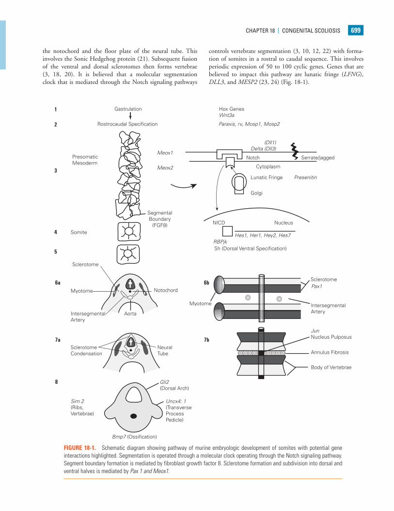
controls vertebrate segmentation (3, 10, 12, 22) with forma-tion of somites in a rostral to caudal sequence. This involves periodic expression of 50 to 100 cyclic genes. Genes that are believed to impact this pathway are lunatic fringe (LFNG), DLL3, and MESP2 (23, 24) (Fig. 18-1).
Gastrulation
Rostrocaudal Specification
Hox GenesWnt3a
PresomaticMesoderm
1
2
3
4
5
6a
7a 7b
6b
8
Somite
Sclerotome
Myotome
IntersegmentalArtery
Aorta
NeuralTube
Notochord
Myotome
SegmentalBoundary
(FGF8)
Notch Serrate/jagged
Cytoplasm
Lunatic Fringe Presenitin
Golgi
NICD
Sh (Dorsal Ventral Specification)RBPjk
Pax1
Hes1, Her1, Hey2, Hes7
Nucleus
Sclerotome
IntersegmentalArtery
Body of Vertebrae
Annulus Fibrosis
JunNucleus Pulposus
Meox1
Paraxis, rv, Mosp1, Mosp2
(Dll1)Delta (Dll3)
Meox2
SclerotomeCondensation
(Ribs,Vertebrae)
Sim 2
Bmp7 (Ossification)
Gli2(Dorsal Arch)
Uncx4: 1(TransverseProcessPedicle)
FIGURE 18-1. Schematic diagram showing pathway of murine embryologic development of somites with potential gene interactions highlighted. Segmentation is operated through a molecular clock operating through the Notch signaling pathway. Segment boundary formation is mediated by fibroblast growth factor 8. Sclerotome formation and subdivision into dorsal and ventral halves is mediated by Pax 1 and Meox1.
Weinstein_Chap18.indd 699 9/27/2013 8:41:53 PM

700 cHAPTer 18 | congeniTAl ScolioSiS
ETIOLOGY
The true etiology of the development of CVMs is unknown. The spectrum of the disorder is very large, ranging from syndromic cases such as Jarcho-Levin syndrome to isolated hemivertebrae, and a number of environmental and genetic associations have been identified, suggesting that both genetic factors and teratogenic effects from the environment play a role in the disturbances of normal spine formation.
Anoxia created by carbon monoxide exposure to mouse and chick models at a critical time and specific dose has been demonstrated to cause CVMs very similar to those found in humans (9, 25–27). Similar outcomes in humans due to expo-sure to carbon monoxide have been postulated, but not clearly established (28). Other environmental factors have been clinically associated with the development of CVM. Alcohol exposure with fetal alcohol syndrome (29), anticonvulsant medications Valproic acid and Dilantin (30–33), retinoic acid (31), hyperthermia (34), maternal insulin-dependent diabetes (35, 36), and folate deficiency (37) have all been implicated in abnormalities of the spine in humans.
Model organisms such as the chick and mouse strongly suggest that there is a genetic contribution to the development of CVM (3, 11, 12, 38). In humans, the heterogeneous clinical manifestations, variety of morphologic features, and pheno-typic presentations described as failures of formation or fail-ures of segmentation had led to the belief that the majority of cases were sporadic events (39). Recent evidence suggests that there is a strong role for genetic factors in the development of human CVMs. The genetic transmission of some cases of CVM has been shown to have a familial recurrence rate of 3% (20, 40), and an increasing number of vertebral malformation syndromes have identifiable genetic etiologies. Spondylocostal dysostosis patients have a loss of normal vertebral morphology throughout the entire spine and have been shown to have muta-tions in delta-like 1 (DLLS), mesoderm posterior 2 (MESP2), and LFNG. Mutations in these affect the Notch signaling pathway and highlight the importance of Notch in vertebral column formation in humans (3, 41, 42). Spondylothoracic dysostosis (commonly referred to as Jarcho-Levin syndrome) is a con-fusing array of CVMs, all of which have rib anomalies with fusion of all ribs. A homozygous recessive nonsense mutation in MESP2 has been identified. Alagille syndrome is an autoso-mal dominant condition with bile duct paucity, cardiac, eye, kidney, pancreas, and vertebral anomalies in 22% to 87% of affected individuals. Mutations in JAG1 and NOTCH2 have been found in these patients (2, 3). Klippel-Feil syndrome was first reported in 1894 (43) and described in 1912 (44). Most cases have been found to be sporadic in families, but autosomal dominant, recessive, and X-linked forms have been reported. There has been recent evidence that PAX1 mutation and notch pathway mutations can be found in patients with Klippel-Feil syndrome (8, 45–47). As isolated CVM are often sporadic occurrences with a given family, candidate gene analysis has been used. Using mouse–human synteny analysis, 27 eligible loci have been identified, of which 21 cause
vertebral malformations in the mouse. Six of these—PAX1, WNT3A, DLL3, SLC35A3, T(Brachyury), and TBX6—have been studied in some detail and show promise as potential loci for the formation of CVMs in humans (3, 13, 14, 46–48).
ASSOCIATED CONDITIONS
CVMs do not occur in isolation. As they develop during a critical stage of organogenesis, as many as 61% have other associated malformations (49), and the development of pro-gressive deformity may lead to secondary organ involvement. The malformations seen are in general neurologic and visceral, with most tied to the level of the CVM.
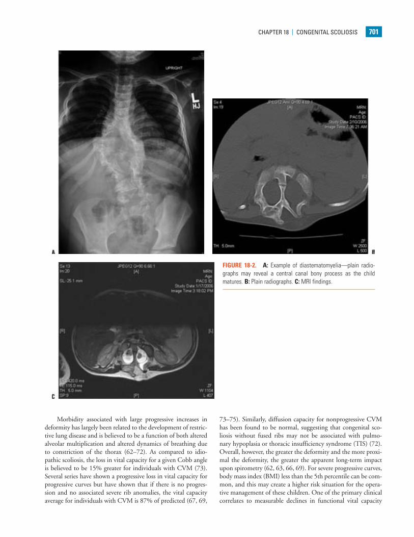
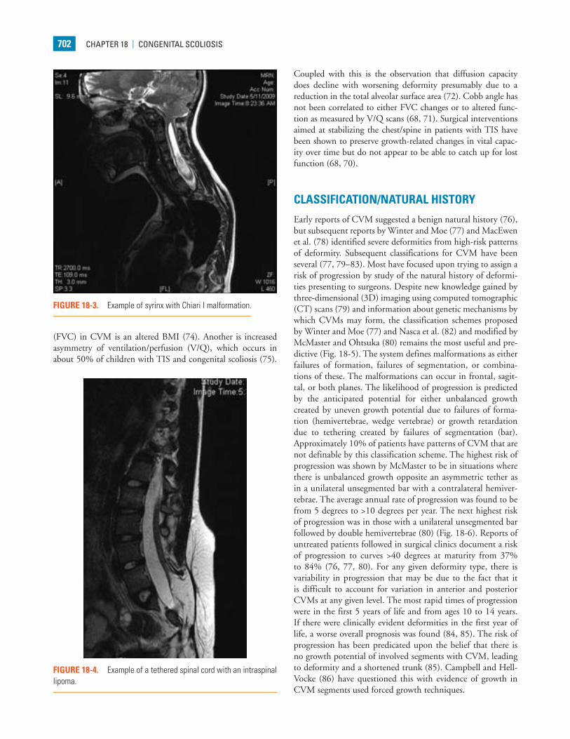
Neurologic abnormalities have been described in 18% to 38% of patients with CVM (50–54). A prospective evaluation of consecutive patients with CVM found that 38% had an intraspinal anomaly, with higher concentrations among patients with cervical and thoracic CVM, mixed patterns of segmenta-tion and formation, and congenital kyphosis (50). Half of these patients had physical findings to suggest that they had an intra-spinal lesion. Diastematomyelia was the most common finding followed by intraspinal lipoma, tethered cord, Chiari malfor-mation with or without associated syringomyelia, dermoid cyst, and epidermoid cyst (Figs. 18-2 to 18-4). For patients with an isolated hemivertebrae, there may also be a high incidence of intraspinal anomalies. Belmont found that of their 76 patients with C3VM, 29 had an isolated hemivertebrae, and of these, 8 (28%) had an MRI abnormality (55). Thirteen had abnormal clinical findings. A review of these patients and published series of similar patients suggests that an abnormal finding on the his-tory of physical examination had an accuracy of 65%, sensitivity of 59%, specificity of 87%, a negative predictive value of 72%, and a positive predictive value of 74% (45, 50, 51, 56, 57).
For patients with known congenital cardiac abnormali-ties, the incidence of scoliosis has been found to be as high as 10% (58), but most of these are normally segmented (26, 58). Farley found that only 11/48 children (23%) with congenital heart disease (CHD) who had scoliosis had congenital scolio-sis. The converse relationship of a higher incidence of CHD (baseline incidence 0.5% in the general population) in patients primarily found to have a congenital scoliosis has not been demonstrated, but it is believed that there is a higher inci-dence. Basu et al. (50) found cardiac abnormalities in 26% of patients with CVM. Two-thirds of these had knowledge of the cardiac abnormality before they presented for evaluation of their spine. Of those who did not have prior knowledge, of the cardiac defect, 2/10 (20%) did proceed to active management.
The incidence of renal anomalies in CVM has been reported as 26% to 37% (49, 50, 54, 59–61). The most com-mon anomalies have been unilateral renal agenesis, urinary duplication, ectopic kidney, reflux, and a horseshoe kidney. The incidence of renal anomalies may be higher if there is an associ-ated rib anomaly in addition to the CVM (52). In most of the series, the renal anomalies were unsuspected, leading to general recommendations to screen children for these problems.
Weinstein_Chap18.indd 700 9/27/2013 8:41:53 PM
cHAPTer 18 | congeniTAl ScolioSiS 701
FIGURE 18-2. A: Example of diastematomyelia—plain radio-graphs may reveal a central canal bony process as the child matures. B: Plain radiographs. C: MRI findings.
Morbidity associated with large progressive increases in deformity has largely been related to the development of restric-tive lung disease and is believed to be a function of both altered alveolar multiplication and altered dynamics of breathing due to constriction of the thorax (62–72). As compared to idio-pathic scoliosis, the loss in vital capacity for a given Cobb angle is believed to be 15% greater for individuals with CVM (73). Several series have shown a progressive loss in vital capacity for progressive curves but have shown that if there is no progres-sion and no associated severe rib anomalies, the vital capacity average for individuals with CVM is 87% of predicted (67, 69,
73–75). Similarly, diffusion capacity for nonprogressive CVM has been found to be normal, suggesting that congenital sco-liosis without fused ribs may not be associated with pulmo-nary hypoplasia or thoracic insufficiency syndrome (TIS) (72). Overall, however, the greater the deformity and the more proxi-mal the deformity, the greater the apparent long-term impact upon spirometry (62, 63, 66, 69). For severe progressive curves, body mass index (BMI) less than the 5th percentile can be com-mon, and this may create a higher risk situation for the opera-tive management of these children. One of the primary clinical correlates to measurable declines in functional vital capacity
Weinstein_Chap18.indd 701 9/27/2013 8:41:55 PM
702 cHAPTer 18 | congeniTAl ScolioSiS
(FVC) in CVM is an altered BMI (74). Another is increased asymmetry of ventilation/perfusion (V/Q), which occurs in about 50% of children with TIS and congenital scoliosis (75).
Coupled with this is the observation that diffusion capacity does decline with worsening deformity presumably due to a reduction in the total alveolar surface area (72). Cobb angle has not been correlated to either FVC changes or to altered func-tion as measured by V/Q scans (68, 71). Surgical interventions aimed at stabilizing the chest/spine in patients with TIS have been shown to preserve growth-related changes in vital capac-ity over time but do not appear to be able to catch up for lost function (68, 70).
CLASSIFICATION/NATURAL HISTORY
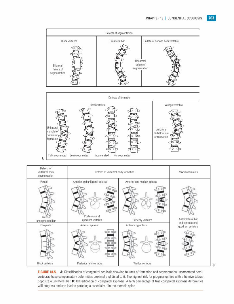
Early reports of CVM suggested a benign natural history (76), but subsequent reports by Winter and Moe (77) and MacEwen et al. (78) identified severe deformities from high-risk patterns of deformity. Subsequent classifications for CVM have been several (77, 79–83). Most have focused upon trying to assign a risk of progression by study of the natural history of deformi-ties presenting to surgeons. Despite new knowledge gained by three-dimensional (3D) imaging using computed tomographic (CT) scans (79) and information about genetic mechanisms by which CVMs may form, the classification schemes proposed by Winter and Moe (77) and Nasca et al. (82) and modified by McMaster and Ohtsuka (80) remains the most useful and pre-dictive (Fig. 18-5). The system defines malformations as either failures of formation, failures of segmentation, or combina-tions of these. The malformations can occur in frontal, sagit-tal, or both planes. The likelihood of progression is predicted by the anticipated potential for either unbalanced growth created by uneven growth potential due to failures of forma-tion (hemivertebrae, wedge vertebrae) or growth retardation due to tethering created by failures of segmentation (bar). Approximately 10% of patients have patterns of CVM that are not definable by this classification scheme. The highest risk of progression was shown by McMaster to be in situations where there is unbalanced growth opposite an asymmetric tether as in a unilateral unsegmented bar with a contralateral hemiver-tebrae. The average annual rate of progression was found to be from 5 degrees to >10 degrees per year. The next highest risk of progression was in those with a unilateral unsegmented bar followed by double hemivertebrae (80) (Fig. 18-6). Reports of untreated patients followed in surgical clinics document a risk of progression to curves >40 degrees at maturity from 37% to 84% (76, 77, 80). For any given deformity type, there is variability in progression that may be due to the fact that it is difficult to account for variation in anterior and posterior CVMs at any given level. The most rapid times of progression were in the first 5 years of life and from ages 10 to 14 years. If there were clinically evident deformities in the first year of life, a worse overall prognosis was found (84, 85). The risk of progression has been predicated upon the belief that there is no growth potential of involved segments with CVM, leading to deformity and a shortened trunk (85). Campbell and Hell-Vocke (86) have questioned this with evidence of growth in CVM segments used forced growth techniques.
FIGURE 18-3. Example of syrinx with Chiari I malformation.
FIGURE 18-4. Example of a tethered spinal cord with an intraspinal lipoma.
Weinstein_Chap18.indd 702 9/27/2013 8:41:56 PM
cHAPTer 18 | congeniTAl ScolioSiS 703
Defects of segmentation
Block vertebra
Bilateralfailure of
segmentation
Unilateralfailure of
segmentation
Unilateral bar Unilateral bar and hemivertebra
Defects of formation
Hemivertebra
Unilateralcompletefailure offormation
Fully segmented Semi-segmented Incarcerated Nonsegmented
Unilateralpartial failureof formation
Wedge vertebra
A
Defects ofvertebral-bodysegmentation
Defects of vertebral-body formation Mixed anomalies
Partial
Anteriorunsegmented bar
Block vertebra
Complete Anterior aplasia Anterior hypoplasia
Posterior hemivertebra
Anterior and unilateral aplasia
Posterolateralquadrant vertebra
Anterior and median aplasia
Butterfly vertebra
Wedge vertebra
Anterolateral barand contralateralquadrant vertebra
B
FIGURE 18-5. A: Classification of congenital scoliosis showing failures of formation and segmentation. Incarcerated hemi-vertebrae have compensatory deformities proximal and distal to it. The highest risk for progression lies with a hemivertebrae opposite a unilateral bar. B: Classification of congenital kyphosis. A high percentage of true congenital kyphosis deformities will progress and can lead to paraplegia especially if in the thoracic spine.
Weinstein_Chap18.indd 703 9/27/2013 8:41:58 PM

704 cHAPTer 18 | congeniTAl ScolioSiS
The clinical impact of progressive deformities associated with CVM has focused historically on respiratory consequences and more recently upon quality-of-life measures. There is no clear association that has been established between classifica-tions of CVM and mortality or morbidity. Patients with severe progressive deformities have been demonstrated to have severe restrictive lung disease and significant morbidity with early death (62–64, 85, 86). They have also been found to have a sig-nificantly diminished quality of life as measured by the Child Health Questionnaire in the domains of physical limitations and caregiver burdens, but not in psychosocial domains (87). Surgical intervention for these deformities has not yet been shown to alter these natural histories. It appears that pulmo-nary function decline can be stabilized with surgical treatment in some cases, but improvement in depressed vital capacity has not been observed (68, 70). Similarly, diminished quality of life does not appear to be improved after intervention (88).
EVALUATION OF PATIENTS WITH CVM
The wide range of presentations and the high incidence of vis-ceral anomalies that can be present in patients with CVMs man-date that all evaluations include a careful history and physical examination. Presentations will range from severely involved children with obvious visceral and structural anomalies to those with nonprogressive and asymptomatic malformations.
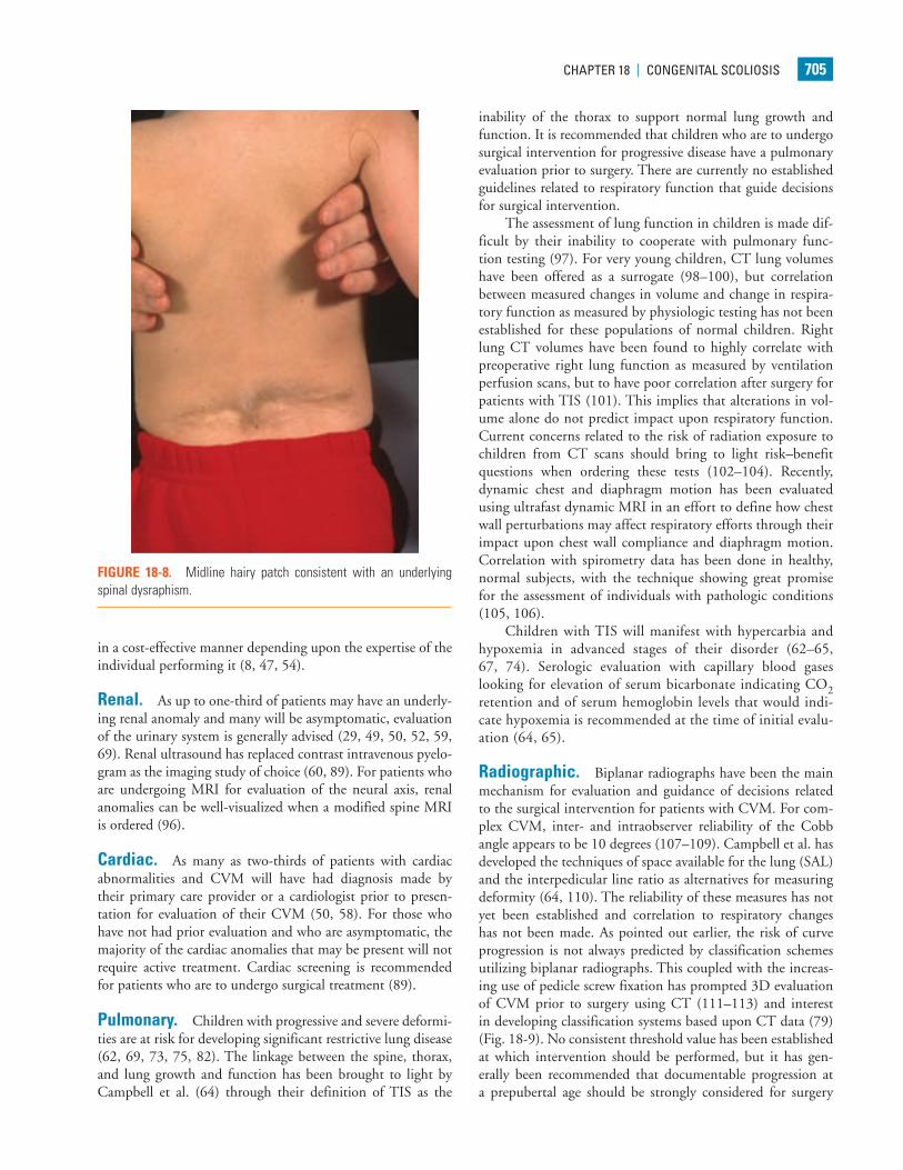
Neurologic. Many relatively uninvolved children will be referred due to incidental findings on imaging studies obtained for other reasons. Growth charts should be reviewed as chil-dren with CVMs may have altered growth velocities and can present with disproportionate growth retardation. Delays in developmental milestones such as walking and running and potty training can be important signs of an underlying spi-nal dysraphism as can symptoms of back pain in very young children. Physical findings of posterior midline trunk hairy patches, large nevi, or hemangiomas, atrophy of extremities,
a cavus foot (Figs. 18-7 and 18-8), or any neurologic abnor-malities (weakness, balance difficulties, sensory, hyperreflexia, asymmetric abdominal reflexes) can indicate an underlying spinal dysraphism in 50% to 80% of cases (19, 50, 51). It is widely recommended that an MRI of the neural axis be obtained prior to surgical intervention for CVM (45, 50–53, 56, 78, 89–91). Less clear is whether there is benefit to the routine ordering of spine MRI scans for asymptomatic chil-dren with CVM who are not to undergo surgery. The posi-tive and negative predictive value of physical examination and clinical history is 74% and 72% respectively for determination of MRI abnormalities (45, 50). As many younger children will require an anesthetic for a spine MRI, in asymptomatic non-progressive deformities in children, observation to an older age is recommended. Intrauterine MRI has been tested for defin-ing neural axis lesions in CVM. It has been found to be better than intrauterine ultrasound, but its overall accuracy and role in managing CVM are as yet unknown (92–95). For neonates with a suspected abnormality of the neural axis, ultrasound has been shown to be an effective screening tool and can be used
FIGURE 18-6. Risk of progression of types of CVM as compiled by McMaster. Highest rates of progression were in unilateral unsegmented bar and contralateral hemivertebrae.
FIGURE 18-7. Cavus foot. High incidence of neurologic abnormali-ties associated with this finding.
Weinstein_Chap18.indd 704 9/27/2013 8:42:01 PM
cHAPTer 18 | congeniTAl ScolioSiS 705
in a cost-effective manner depending upon the expertise of the individual performing it (8, 47, 54).
Renal. As up to one-third of patients may have an underly-ing renal anomaly and many will be asymptomatic, evaluation of the urinary system is generally advised (29, 49, 50, 52, 59, 69). Renal ultrasound has replaced contrast intravenous pyelo-gram as the imaging study of choice (60, 89). For patients who are undergoing MRI for evaluation of the neural axis, renal anomalies can be well-visualized when a modified spine MRI is ordered (96).
Cardiac. As many as two-thirds of patients with cardiac abnormalities and CVM will have had diagnosis made by their primary care provider or a cardiologist prior to presen-tation for evaluation of their CVM (50, 58). For those who have not had prior evaluation and who are asymptomatic, the majority of the cardiac anomalies that may be present will not require active treatment. Cardiac screening is recommended for patients who are to undergo surgical treatment (89).
Pulmonary. Children with progressive and severe deformi-ties are at risk for developing significant restrictive lung disease (62, 69, 73, 75, 82). The linkage between the spine, thorax, and lung growth and function has been brought to light by Campbell et al. (64) through their definition of TIS as the
inability of the thorax to support normal lung growth and function. It is recommended that children who are to undergo surgical intervention for progressive disease have a pulmonary evaluation prior to surgery. There are currently no established guidelines related to respiratory function that guide decisions for surgical intervention.
The assessment of lung function in children is made dif-ficult by their inability to cooperate with pulmonary func-tion testing (97). For very young children, CT lung volumes have been offered as a surrogate (98–100), but correlation between measured changes in volume and change in respira-tory function as measured by physiologic testing has not been established for these populations of normal children. Right lung CT volumes have been found to highly correlate with preoperative right lung function as measured by ventilation perfusion scans, but to have poor correlation after surgery for patients with TIS (101). This implies that alterations in vol-ume alone do not predict impact upon respiratory function. Current concerns related to the risk of radiation exposure to children from CT scans should bring to light risk–benefit questions when ordering these tests (102–104). Recently, dynamic chest and diaphragm motion has been evaluated using ultrafast dynamic MRI in an effort to define how chest wall perturbations may affect respiratory efforts through their impact upon chest wall compliance and diaphragm motion. Correlation with spirometry data has been done in healthy, normal subjects, with the technique showing great promise for the assessment of individuals with pathologic conditions (105, 106).
Children with TIS will manifest with hypercarbia and hypoxemia in advanced stages of their disorder (62–65, 67, 74). Serologic evaluation with capillary blood gases looking for elevation of serum bicarbonate indicating CO2 retention and of serum hemoglobin levels that would indi-cate hypoxemia is recommended at the time of initial evalu-ation (64, 65).
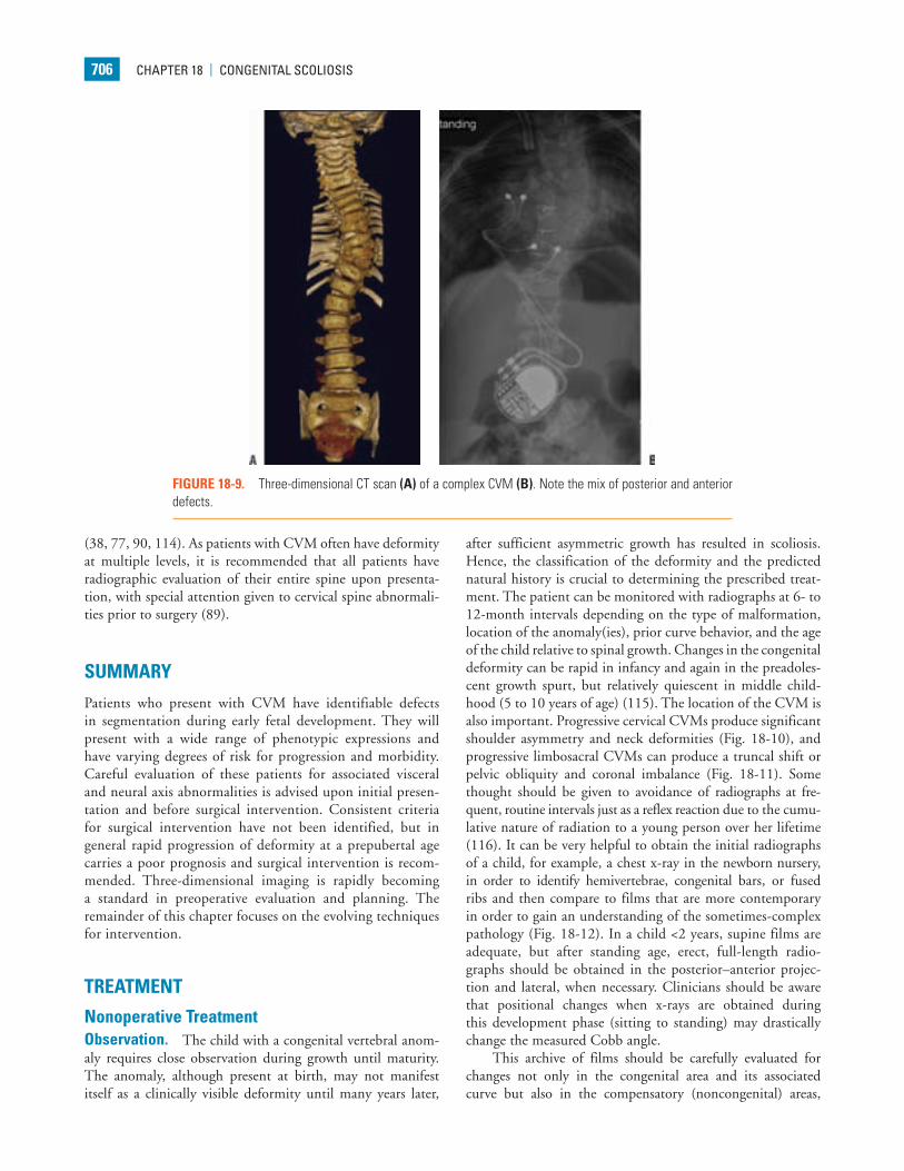
Radiographic. Biplanar radiographs have been the main mechanism for evaluation and guidance of decisions related to the surgical intervention for patients with CVM. For com-plex CVM, inter- and intraobserver reliability of the Cobb angle appears to be 10 degrees (107–109). Campbell et al. has developed the techniques of space available for the lung (SAL) and the interpedicular line ratio as alternatives for measuring deformity (64, 110). The reliability of these measures has not yet been established and correlation to respiratory changes has not been made. As pointed out earlier, the risk of curve progression is not always predicted by classification schemes utilizing biplanar radiographs. This coupled with the increas-ing use of pedicle screw fixation has prompted 3D evaluation of CVM prior to surgery using CT (111–113) and interest in developing classification systems based upon CT data (79) (Fig. 18-9). No consistent threshold value has been established at which intervention should be performed, but it has gen-erally been recommended that documentable progression at a prepubertal age should be strongly considered for surgery
FIGURE 18-8. Midline hairy patch consistent with an underlying spinal dysraphism.
Weinstein_Chap18.indd 705 9/27/2013 8:42:03 PM
706 cHAPTer 18 | congeniTAl ScolioSiS
(38, 77, 90, 114). As patients with CVM often have deformity at multiple levels, it is recommended that all patients have radiographic evaluation of their entire spine upon presenta-tion, with special attention given to cervical spine abnormali-ties prior to surgery (89).
SUMMARY
Patients who present with CVM have identifiable defects in segmentation during early fetal development. They will present with a wide range of phenotypic expressions and have varying degrees of risk for progression and morbidity. Careful evaluation of these patients for associated visceral and neural axis abnormalities is advised upon initial presen-tation and before surgical intervention. Consistent criteria for surgical intervention have not been identified, but in general rapid progression of deformity at a prepubertal age carries a poor prognosis and surgical intervention is recom-mended. Three-dimensional imaging is rapidly becoming a standard in preoperative evaluation and planning. The remainder of this chapter focuses on the evolving techniques for intervention.
TREATMENT
Nonoperative TreatmentObservation. The child with a congenital vertebral anom-aly requires close observation during growth until maturity. The anomaly, although present at birth, may not manifest itself as a clinically visible deformity until many years later,
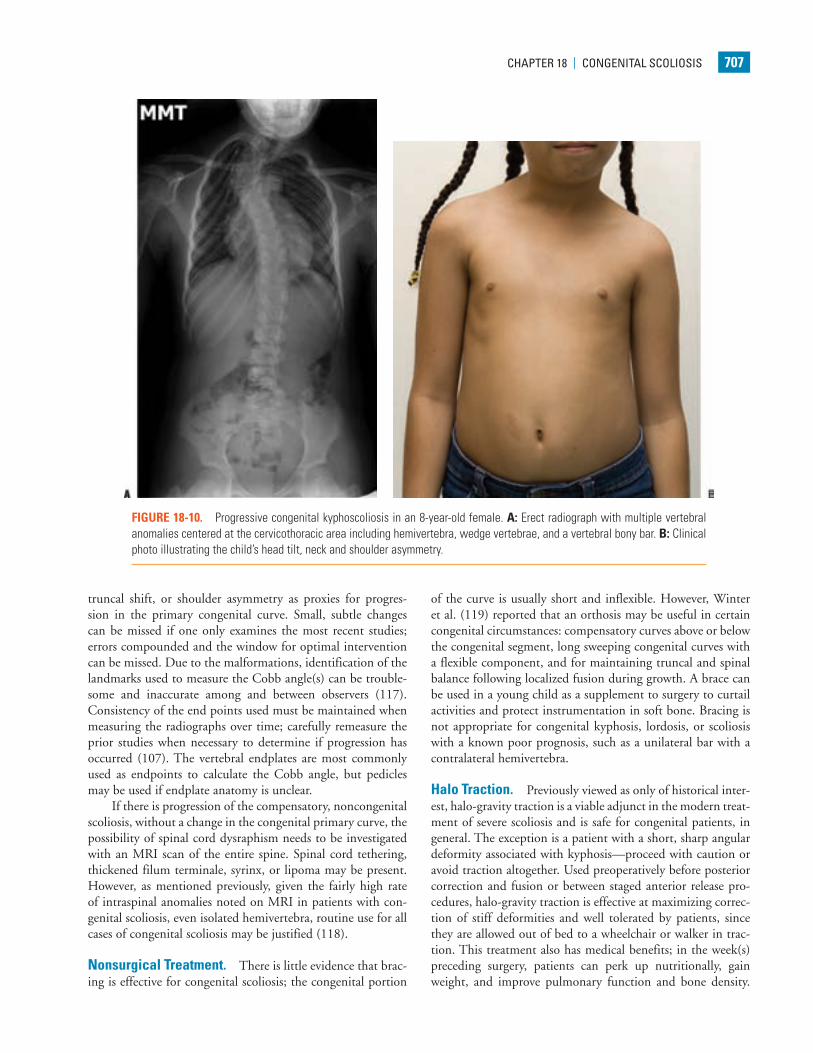
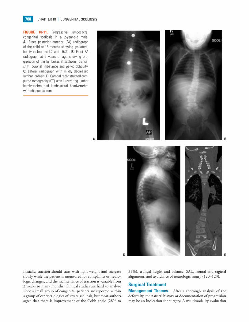
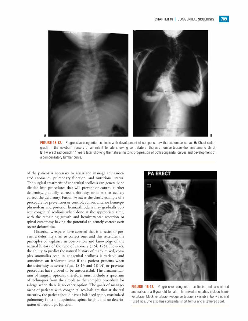
after sufficient asymmetric growth has resulted in scoliosis. Hence, the classification of the deformity and the predicted natural history is crucial to determining the prescribed treat-ment. The patient can be monitored with radiographs at 6- to 12-month intervals depending on the type of malformation, location of the anomaly(ies), prior curve behavior, and the age of the child relative to spinal growth. Changes in the congenital deformity can be rapid in infancy and again in the preadoles-cent growth spurt, but relatively quiescent in middle child-hood (5 to 10 years of age) (115). The location of the CVM is also important. Progressive cervical CVMs produce significant shoulder asymmetry and neck deformities (Fig. 18-10), and progressive limbosacral CVMs can produce a truncal shift or pelvic obliquity and coronal imbalance (Fig. 18-11). Some thought should be given to avoidance of radiographs at fre-quent, routine intervals just as a reflex reaction due to the cumu-lative nature of radiation to a young person over her lifetime (116). It can be very helpful to obtain the initial radiographs of a child, for example, a chest x-ray in the newborn nursery, in order to identify hemivertebrae, congenital bars, or fused ribs and then compare to films that are more contemporary in order to gain an understanding of the sometimes-complex pathology (Fig. 18-12). In a child <2 years, supine films are adequate, but after standing age, erect, full-length radio-graphs should be obtained in the posterior–anterior projec-tion and lateral, when necessary. Clinicians should be aware that positional changes when x-rays are obtained during this development phase (sitting to standing) may drastically change the measured Cobb angle.
This archive of films should be carefully evaluated for changes not only in the congenital area and its associated curve but also in the compensatory (noncongenital) areas,
FIGURE 18-9. Three-dimensional CT scan (A) of a complex CVM (B). Note the mix of posterior and anterior defects.
Weinstein_Chap18.indd 706 9/27/2013 8:42:05 PM
cHAPTer 18 | congeniTAl ScolioSiS 707
truncal shift, or shoulder asymmetry as proxies for progres-sion in the primary congenital curve. Small, subtle changes can be missed if one only examines the most recent studies; errors compounded and the window for optimal intervention can be missed. Due to the malformations, identification of the landmarks used to measure the Cobb angle(s) can be trouble-some and inaccurate among and between observers (117). Consistency of the end points used must be maintained when measuring the radiographs over time; carefully remeasure the prior studies when necessary to determine if progression has occurred (107). The vertebral endplates are most commonly used as endpoints to calculate the Cobb angle, but pedicles may be used if endplate anatomy is unclear.
If there is progression of the compensatory, noncongenital scoliosis, without a change in the congenital primary curve, the possibility of spinal cord dysraphism needs to be investigated with an MRI scan of the entire spine. Spinal cord tethering, thickened filum terminale, syrinx, or lipoma may be present. However, as mentioned previously, given the fairly high rate of intraspinal anomalies noted on MRI in patients with con-genital scoliosis, even isolated hemivertebra, routine use for all cases of congenital scoliosis may be justified (118).
Nonsurgical Treatment. There is little evidence that brac-ing is effective for congenital scoliosis; the congenital portion
of the curve is usually short and inflexible. However, Winter et al. (119) reported that an orthosis may be useful in certain congenital circumstances: compensatory curves above or below the congenital segment, long sweeping congenital curves with a flexible component, and for maintaining truncal and spinal balance following localized fusion during growth. A brace can be used in a young child as a supplement to surgery to curtail activities and protect instrumentation in soft bone. Bracing is not appropriate for congenital kyphosis, lordosis, or scoliosis with a known poor prognosis, such as a unilateral bar with a contralateral hemivertebra.
Halo Traction. Previously viewed as only of historical inter-est, halo-gravity traction is a viable adjunct in the modern treat-ment of severe scoliosis and is safe for congenital patients, in general. The exception is a patient with a short, sharp angular deformity associated with kyphosis—proceed with caution or avoid traction altogether. Used preoperatively before posterior correction and fusion or between staged anterior release pro-cedures, halo-gravity traction is effective at maximizing correc-tion of stiff deformities and well tolerated by patients, since they are allowed out of bed to a wheelchair or walker in trac-tion. This treatment also has medical benefits; in the week(s) preceding surgery, patients can perk up nutritionally, gain weight, and improve pulmonary function and bone density.
FIGURE 18-10. Progressive congenital kyphoscoliosis in an 8-year-old female. A: Erect radiograph with multiple vertebral anomalies centered at the cervicothoracic area including hemivertebra, wedge vertebrae, and a vertebral bony bar. B: Clinical photo illustrating the child’s head tilt, neck and shoulder asymmetry.
Weinstein_Chap18.indd 707 9/27/2013 8:42:07 PM
708 cHAPTer 18 | congeniTAl ScolioSiS
FIGURE 18-11. Progressive lumbosacral congenital scoliosis in a 2-year-old male. A: Erect posterior–anterior (PA) radiograph of the child at 18 months showing ipsilateral hemivertebrae at L2 and L5/S1. B: Erect PA radiograph at 2 years of age showing pro-gression of the lumbosacral scoliosis, truncal shift, coronal imbalance and pelvic obliquity. C: Lateral radiograph with mildly decreased lumbar lordosis. D: Coronal reconstructed com-puted tomography (CT) scan illustrating lumbar hemivertebra and lumbosacral hemivertebra with oblique sacrum.
Initially, traction should start with light weight and increase slowly while the patient is monitored for complaints or neuro-logic changes, and the maintenance of traction is variable from 2 weeks to many months. Clinical studies are hard to analyze since a small group of congenital patients are reported within a group of other etiologies of severe scoliosis, but most authors agree that there is improvement of the Cobb angle (28% to
35%), truncal height and balance, SAL, frontal and sagittal alignment, and avoidance of neurologic injury (120–123).
Surgical TreatmentManagement Themes. After a thorough analysis of the deformity, the natural history or documentation of progression may be an indication for surgery. A multimodality evaluation
Weinstein_Chap18.indd 708 9/27/2013 8:42:09 PM
cHAPTer 18 | congeniTAl ScolioSiS 709
FIGURE 18-12. Progressive congenital scoliosis with development of compensatory thoracolumbar curve. A: Chest radio-graph in the newborn nursery of an infant female showing contralateral thoracic hemivertebrae (hemimetameric shift). B: PA erect radiograph 14 years later showing the natural history: progression of both congenital curves and development of a compensatory lumbar curve.
of the patient is necessary to assess and manage any associ-ated anomalies, pulmonary function, and nutritional status. The surgical treatment of congenital scoliosis can generally be divided into procedures that will prevent or control further deformity, gradually correct deformity, or ones that acutely correct the deformity. Fusion in situ is the classic example of a procedure for prevention or control; convex anterior hemiepi-physiodesis and posterior hemiarthrodesis may gradually cor-rect congenital scoliosis when done at the appropriate time, with the remaining growth and hemivertebrae resection or spinal osteotomy having the potential to acutely correct even severe deformities.
Historically, experts have asserted that it is easier to pre-vent a deformity than to correct one, and this reiterates the principles of vigilance in observation and knowledge of the natural history of the type of anomaly (124, 125). However, the ability to predict the natural history of many mixed, com-plex anomalies seen in congenital scoliosis is variable and sometimes an irrelevant issue if the patient presents when the deformity is severe (Figs. 18-13 and 18-14) or previous procedures have proved to be unsuccessful. The armamentar-ium of surgical options, therefore, must include a spectrum of techniques from the simple to the complex procedure for salvage when there is no other option. The goals of manage-ment of patients with congenital scoliosis are that at skeletal maturity, the patient should have a balanced spine, maximized pulmonary function, optimized spinal height, and no deterio-ration of neurologic function.
FIGURE 18-13. Progressive congenital scoliosis and associated anomalies in a 9-year-old female. The mixed anomalies include hemi-vertebrae, block vertebrae, wedge vertebrae, a vertebral bony bar, and fused ribs. She also has congenital short femur and a tethered cord.
Weinstein_Chap18.indd 709 9/27/2013 8:42:10 PM
710 cHAPTer 18 | congeniTAl ScolioSiS
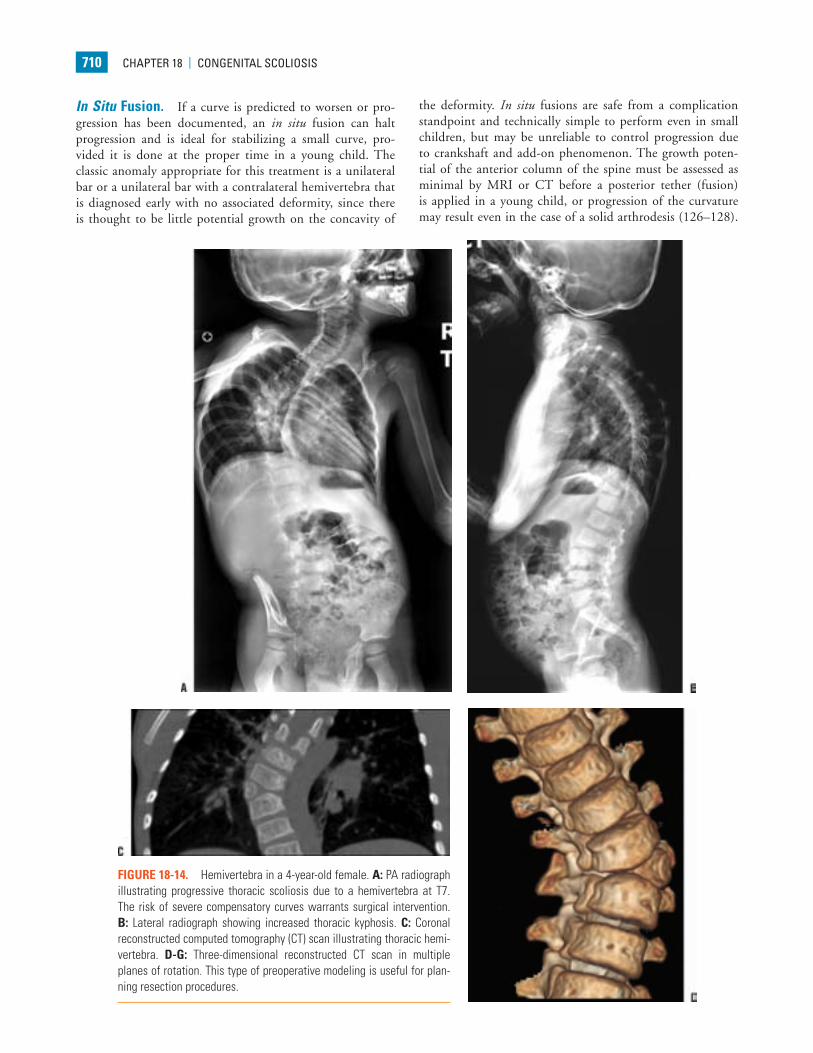
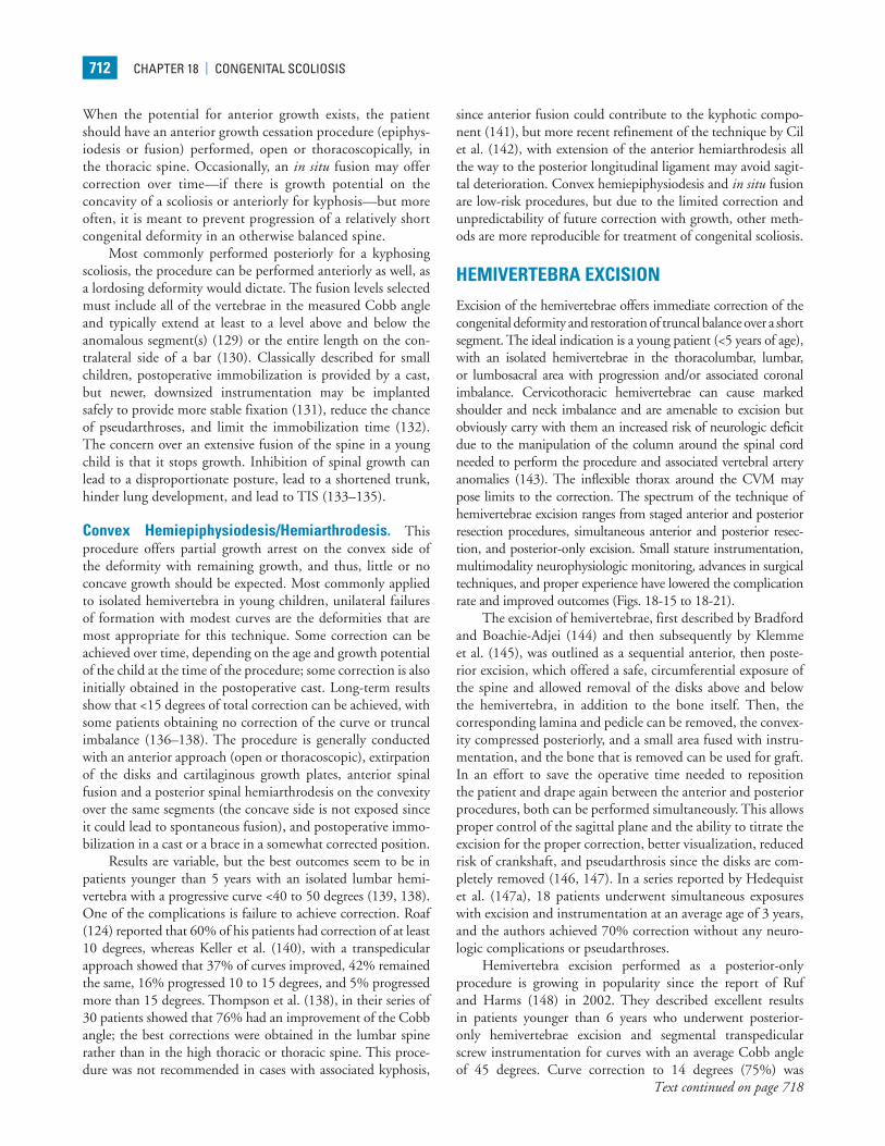
FIGURE 18-14. Hemivertebra in a 4-year-old female. A: PA radiograph illustrating progressive thoracic scoliosis due to a hemivertebra at T7. The risk of severe compensatory curves warrants surgical intervention. B: Lateral radiograph showing increased thoracic kyphosis. C: Coronal reconstructed computed tomography (CT) scan illustrating thoracic hemi-vertebra. D-G: Three-dimensional reconstructed CT scan in multiple planes of rotation. This type of preoperative modeling is useful for plan-ning resection procedures.
In Situ Fusion. If a curve is predicted to worsen or pro-gression has been documented, an in situ fusion can halt progression and is ideal for stabilizing a small curve, pro-vided it is done at the proper time in a young child. The classic anomaly appropriate for this treatment is a unilateral bar or a unilateral bar with a contralateral hemivertebra that is diagnosed early with no associated deformity, since there is thought to be little potential growth on the concavity of
the deformity. In situ fusions are safe from a complication standpoint and technically simple to perform even in small children, but may be unreliable to control progression due to crankshaft and add-on phenomenon. The growth poten-tial of the anterior column of the spine must be assessed as minimal by MRI or CT before a posterior tether (fusion) is applied in a young child, or progression of the curvature may result even in the case of a solid arthrodesis (126–128).
Weinstein_Chap18.indd 710 9/27/2013 8:42:13 PM

cHAPTer 18 | congeniTAl ScolioSiS 711
FIGURE 18-14. (Continued )
Weinstein_Chap18.indd 711 9/27/2013 8:42:17 PM
712 cHAPTer 18 | congeniTAl ScolioSiS
When the potential for anterior growth exists, the patient should have an anterior growth cessation procedure (epiphys-iodesis or fusion) performed, open or thoracoscopically, in the thoracic spine. Occasionally, an in situ fusion may offer correction over time—if there is growth potential on the concavity of a scoliosis or anteriorly for kyphosis—but more often, it is meant to prevent progression of a relatively short congenital deformity in an otherwise balanced spine.
Most commonly performed posteriorly for a kyphosing scoliosis, the procedure can be performed anteriorly as well, as a lordosing deformity would dictate. The fusion levels selected must include all of the vertebrae in the measured Cobb angle and typically extend at least to a level above and below the anomalous segment(s) (129) or the entire length on the con-tralateral side of a bar (130). Classically described for small children, postoperative immobilization is provided by a cast, but newer, downsized instrumentation may be implanted safely to provide more stable fixation (131), reduce the chance of pseudarthroses, and limit the immobilization time (132). The concern over an extensive fusion of the spine in a young child is that it stops growth. Inhibition of spinal growth can lead to a disproportionate posture, lead to a shortened trunk, hinder lung development, and lead to TIS (133–135).
Convex Hemiepiphysiodesis/Hemiarthrodesis. This procedure offers partial growth arrest on the convex side of the deformity with remaining growth, and thus, little or no concave growth should be expected. Most commonly applied to isolated hemivertebra in young children, unilateral failures of formation with modest curves are the deformities that are most appropriate for this technique. Some correction can be achieved over time, depending on the age and growth potential of the child at the time of the procedure; some correction is also initially obtained in the postoperative cast. Long-term results show that <15 degrees of total correction can be achieved, with some patients obtaining no correction of the curve or truncal imbalance (136–138). The procedure is generally conducted with an anterior approach (open or thoracoscopic), extirpation of the disks and cartilaginous growth plates, anterior spinal fusion and a posterior spinal hemiarthrodesis on the convexity over the same segments (the concave side is not exposed since it could lead to spontaneous fusion), and postoperative immo-bilization in a cast or a brace in a somewhat corrected position.
Results are variable, but the best outcomes seem to be in patients younger than 5 years with an isolated lumbar hemi-vertebra with a progressive curve <40 to 50 degrees (139, 138). One of the complications is failure to achieve correction. Roaf (124) reported that 60% of his patients had correction of at least 10 degrees, whereas Keller et al. (140), with a transpedicular approach showed that 37% of curves improved, 42% remained the same, 16% progressed 10 to 15 degrees, and 5% progressed more than 15 degrees. Thompson et al. (138), in their series of 30 patients showed that 76% had an improvement of the Cobb angle; the best corrections were obtained in the lumbar spine rather than in the high thoracic or thoracic spine. This proce-dure was not recommended in cases with associated kyphosis,
since anterior fusion could contribute to the kyphotic compo-nent (141), but more recent refinement of the technique by Cil et al. (142), with extension of the anterior hemiarthrodesis all the way to the posterior longitudinal ligament may avoid sagit-tal deterioration. Convex hemiepiphysiodesis and in situ fusion are low-risk procedures, but due to the limited correction and unpredictability of future correction with growth, other meth-ods are more reproducible for treatment of congenital scoliosis.
HEMIVERTEBRA ExCISION
Excision of the hemivertebrae offers immediate correction of the congenital deformity and restoration of truncal balance over a short segment. The ideal indication is a young patient (<5 years of age), with an isolated hemivertebrae in the thoracolumbar, lumbar, or lumbosacral area with progression and/or associated coronal imbalance. Cervicothoracic hemivertebrae can cause marked shoulder and neck imbalance and are amenable to excision but obviously carry with them an increased risk of neurologic deficit due to the manipulation of the column around the spinal cord needed to perform the procedure and associated vertebral artery anomalies (143). The inflexible thorax around the CVM may pose limits to the correction. The spectrum of the technique of hemivertebrae excision ranges from staged anterior and posterior resection procedures, simultaneous anterior and posterior resec-tion, and posterior-only excision. Small stature instrumentation, multimodality neurophysiologic monitoring, advances in surgical techniques, and proper experience have lowered the complication rate and improved outcomes (Figs. 18-15 to 18-21).
The excision of hemivertebrae, first described by Bradford and Boachie-Adjei (144) and then subsequently by Klemme et al. (145), was outlined as a sequential anterior, then poste-rior excision, which offered a safe, circumferential exposure of the spine and allowed removal of the disks above and below the hemivertebra, in addition to the bone itself. Then, the corresponding lamina and pedicle can be removed, the convex-ity compressed posteriorly, and a small area fused with instru-mentation, and the bone that is removed can be used for graft. In an effort to save the operative time needed to reposition the patient and drape again between the anterior and posterior procedures, both can be performed simultaneously. This allows proper control of the sagittal plane and the ability to titrate the excision for the proper correction, better visualization, reduced risk of crankshaft, and pseudarthrosis since the disks are com-pletely removed (146, 147). In a series reported by Hedequist et al. (147a), 18 patients underwent simultaneous exposures with excision and instrumentation at an average age of 3 years, and the authors achieved 70% correction without any neuro-logic complications or pseudarthroses.
Hemivertebra excision performed as a posterior-only procedure is growing in popularity since the report of Ruf and Harms (148) in 2002. They described excellent results in patients younger than 6 years who underwent posterior-only hemivertebrae excision and segmental transpedicular screw instrumentation for curves with an average Cobb angle of 45 degrees. Curve correction to 14 degrees (75%) was
Text continued on page 718
Weinstein_Chap18.indd 712 9/27/2013 8:42:17 PM
cHAPTer 18 | congeniTAl ScolioSiS 713
OSTEOPERIOSTEAL FLAP
RIB HEADS EXCISED
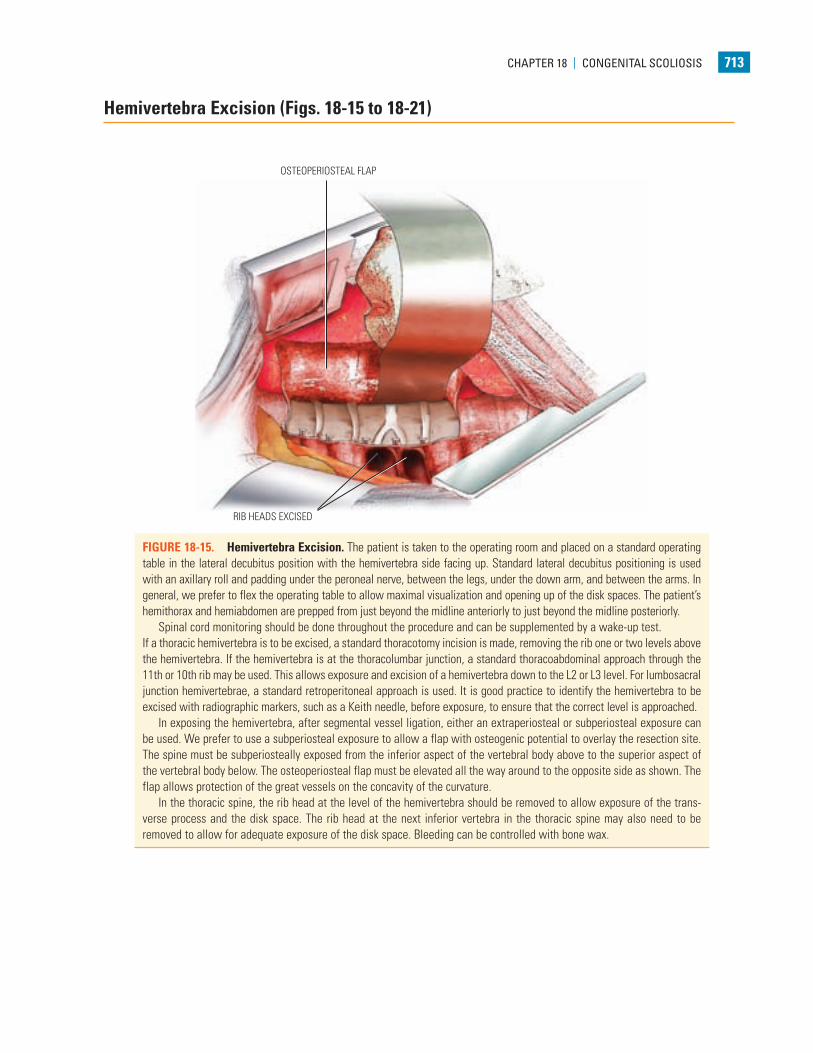
FIGURE 18-15. Hemivertebra Excision. The patient is taken to the operating room and placed on a standard operating table in the lateral decubitus position with the hemivertebra side facing up. Standard lateral decubitus positioning is used with an axillary roll and padding under the peroneal nerve, between the legs, under the down arm, and between the arms. In general, we prefer to flex the operating table to allow maximal visualization and opening up of the disk spaces. The patient’s hemithorax and hemiabdomen are prepped from just beyond the midline anteriorly to just beyond the midline posteriorly.
Spinal cord monitoring should be done throughout the procedure and can be supplemented by a wake-up test.If a thoracic hemivertebra is to be excised, a standard thoracotomy incision is made, removing the rib one or two levels above the hemivertebra. If the hemivertebra is at the thoracolumbar junction, a standard thoracoabdominal approach through the 11th or 10th rib may be used. This allows exposure and excision of a hemivertebra down to the L2 or L3 level. For lumbosacral junction hemivertebrae, a standard retroperitoneal approach is used. It is good practice to identify the hemivertebra to be excised with radiographic markers, such as a Keith needle, before exposure, to ensure that the correct level is approached.
In exposing the hemivertebra, after segmental vessel ligation, either an extraperiosteal or subperiosteal exposure can be used. We prefer to use a subperiosteal exposure to allow a flap with osteogenic potential to overlay the resection site. The spine must be subperiosteally exposed from the inferior aspect of the vertebral body above to the superior aspect of the vertebral body below. The osteoperiosteal flap must be elevated all the way around to the opposite side as shown. The flap allows protection of the great vessels on the concavity of the curvature.
In the thoracic spine, the rib head at the level of the hemivertebra should be removed to allow exposure of the trans-verse process and the disk space. The rib head at the next inferior vertebra in the thoracic spine may also need to be removed to allow for adequate exposure of the disk space. Bleeding can be controlled with bone wax.
Hemivertebra Excision (Figs. 18-15 to 18-21)
Weinstein_Chap18.indd 713 9/27/2013 8:42:19 PM
POSTERIOR ANNULUS
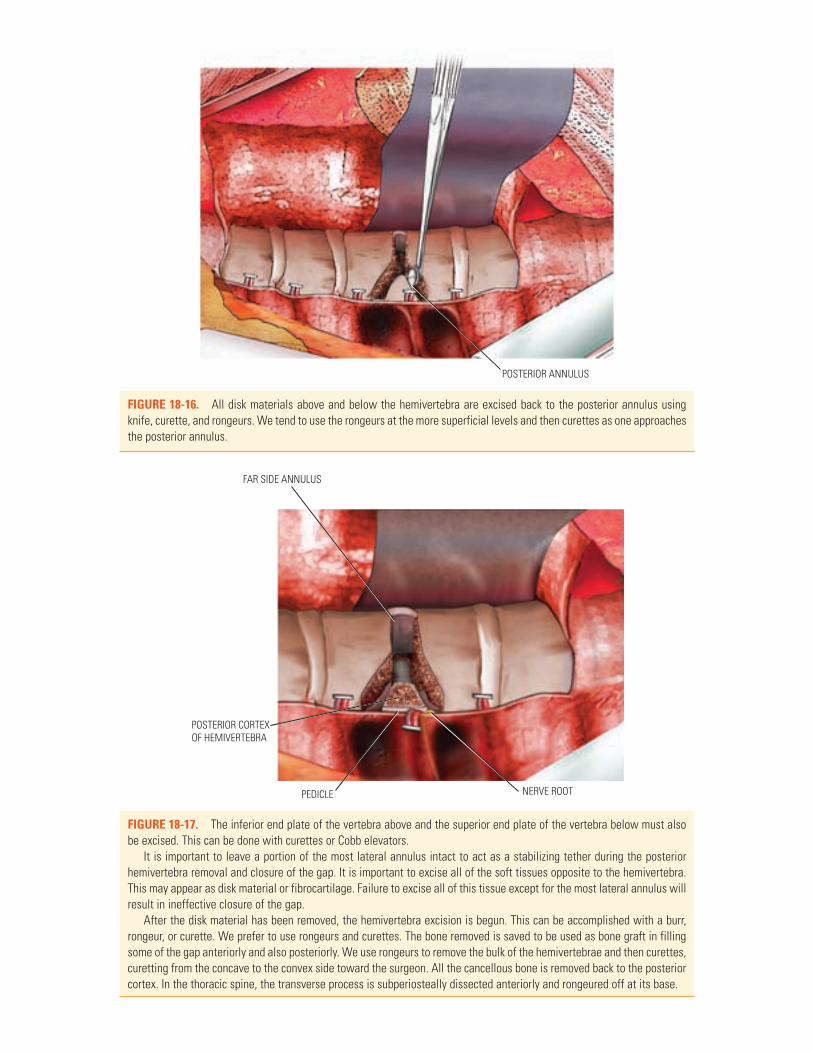
FIGURE 18-16. All disk materials above and below the hemivertebra are excised back to the posterior annulus using knife, curette, and rongeurs. We tend to use the rongeurs at the more superficial levels and then curettes as one approaches the posterior annulus.
FAR SIDE ANNULUS
POSTERIOR CORTEXOF HEMIVERTEBRA
PEDICLE NERVE ROOT
FIGURE 18-17. The inferior end plate of the vertebra above and the superior end plate of the vertebra below must also be excised. This can be done with curettes or Cobb elevators.
It is important to leave a portion of the most lateral annulus intact to act as a stabilizing tether during the posterior hemivertebra removal and closure of the gap. It is important to excise all of the soft tissues opposite to the hemivertebra. This may appear as disk material or fibrocartilage. Failure to excise all of this tissue except for the most lateral annulus will result in ineffective closure of the gap.
After the disk material has been removed, the hemivertebra excision is begun. This can be accomplished with a burr, rongeur, or curette. We prefer to use rongeurs and curettes. The bone removed is saved to be used as bone graft in filling some of the gap anteriorly and also posteriorly. We use rongeurs to remove the bulk of the hemivertebrae and then curettes, curetting from the concave to the convex side toward the surgeon. All the cancellous bone is removed back to the posterior cortex. In the thoracic spine, the transverse process is subperiosteally dissected anteriorly and rongeured off at its base.
Weinstein_Chap18.indd 714 9/27/2013 8:42:24 PM

cHAPTer 18 | congeniTAl ScolioSiS 715
FAR SIDE ANNULUS
FIGURE 18-18. Final removal of the posterior vertebral cortex can be done in one of several ways. We prefer to use a diamond-tipped dental burr to enter the spinal canal in the midline. After a small hole is made, a small Harper-Kerrison rongeur may be used to remove the entire posterior cortex of the hemivertebra.
CUT EDGE OF PEDICLE
FIGURE 18-19. After the cortical bone is removed from the apex of the hemivertebra all the way back to the pedicle, the annular tissues superiorly and inferiorly can also be removed. The neural foramina both above and below the hemivertebra must be opened completely and the nerve roots visualized. Any bleeding that is encountered should be addressed with bipolar cautery or packing with thrombin-soaked Gelfoam.
The final structure removed anteriorly is the pedicle of the hemivertebra. The pedicle can be removed in several ways. It can be gradually nibbled away with narrow-nosed rongeurs or, as we prefer, the central portion drilled until decancellated followed by the outer walls being removed with either rongeurs or curettage toward the pedicle center.
The pedicle bone should be nibbled away as far posteriorly as possible.The dura is covered with a Gelfoam or Oxycel pad or fat graft, and then bone chips from the excised hemivertebra are
loosely placed into the space. The osteoperiosteal flap is then sutured back down loosely, and in the chest the pleura is repaired. The wound is then closed in the standard fashion.
The second portion of the procedure can be done in the lateral decubitus position.However, we prefer to roll the patient prone and transfer the patient to a spine frame (e.g., Jackson table, Relton-Hall
frame). In some children, we prefer to use two laminectomy rolls (made of rolled bath blankets or foam), one under the chest and one under the iliac crests, leaving the abdomen free.
Weinstein_Chap18.indd 715 9/27/2013 8:42:28 PM

716 cHAPTer 18 | congeniTAl ScolioSiS
LIGAMENTUM FLAVUM
DURA
FIGURE 18-20. In the posterior approach, the spine is exposed from a standard midline exposure overlying the area of the hemiver-tebra. A radiograph is taken to determine the appropriate level for excision. The spine is then exposed subperiosteally. The liga-mentum flavum above and below the hemi-lamina is carefully incised and removed with the Kerrison rongeur. Any bleeding is controlled by bipolar cautery or packing with thrombin-soaked Gelfoam. After the ligamentum flavum has been removed, the hemilamina is gradually removed from the midline to its lateral aspect. We gen-erally do this with standard narrow-nosed rongeurs or Harper-Kerrison rongeurs. It is much easier to remove the posterior arch from the posterior approach when more of the pedicle has been removed by the ante-rior procedure.
Weinstein_Chap18.indd 716 9/27/2013 8:42:30 PM
cHAPTer 18 | congeniTAl ScolioSiS 717
TRANSVERSE PROCESS
PEDICLE
SMALL ELEVATOR
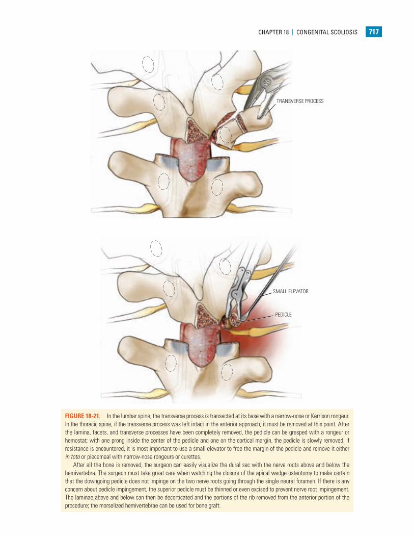
FIGURE 18-21. In the lumbar spine, the transverse process is transected at its base with a narrow-nose or Kerrison rongeur. In the thoracic spine, if the transverse process was left intact in the anterior approach, it must be removed at this point. After the lamina, facets, and transverse processes have been completely removed, the pedicle can be grasped with a rongeur or hemostat; with one prong inside the center of the pedicle and one on the cortical margin, the pedicle is slowly removed. If resistance is encountered, it is most important to use a small elevator to free the margin of the pedicle and remove it either in toto or piecemeal with narrow-nose rongeurs or curettes.
After all the bone is removed, the surgeon can easily visualize the dural sac with the nerve roots above and below the hemivertebra. The surgeon must take great care when watching the closure of the apical wedge osteotomy to make certain that the downgoing pedicle does not impinge on the two nerve roots going through the single neural foramen. If there is any concern about pedicle impingement, the superior pedicle must be thinned or even excised to prevent nerve root impingement. The laminae above and below can then be decorticated and the portions of the rib removed from the anterior portion of the procedure; the morselized hemivertebrae can be used for bone graft.
Weinstein_Chap18.indd 717 9/27/2013 8:42:33 PM
718 cHAPTer 18 | congeniTAl ScolioSiS
FIGURE 18-21. (Continued ) When the hemivertebra to be excised is at the lumbosacral junction, a separate incision may be necessary to expose the iliac crest and obtain cortical and cancellous bone.
In very young patients, internal fixation is generally not possible, and the defect is closed and maintained in that position by casting. In toddlers and older children, small compression devices may be applied using the rod and hook configuration. Another alternative in older patients is the use of pedicle screws in the pedicle above and below the hemivertebra, with a small rod connecting the two screw heads. If no internal fixation is used, after wound closure, the patient must be turned carefully onto a pediatric spica table and a body spica cast that includes both legs down to the ankles applied. The cast should extend proximally up to the clavicle line. The patient’s torso should be bent into the convexity of the curvature to maintain correction. Radiographs should be obtained in the operating room to ensure that the wedge is closed and that adequate correction has been obtained. It is also important to make certain that pressure is applied to the apex of the deformity posteriorly while the plaster is drying, to prevent the spine from drifting into kypho-sis. If adequate correction has not been obtained, the cast can be wedged while the patient is still under anesthesia. If internal fixation is used, the surgeon will decide whether casting is necessary. If the surgeon believes that the internal fixation was very stable at surgery, the patient may be placed either in a cast or a thoracolumbar spinal orthosis after surgery, with a single-leg extension, depending on the surgeon’s preference. In our opinion, external immobilization is important regardless of the stability of the internal fixation.
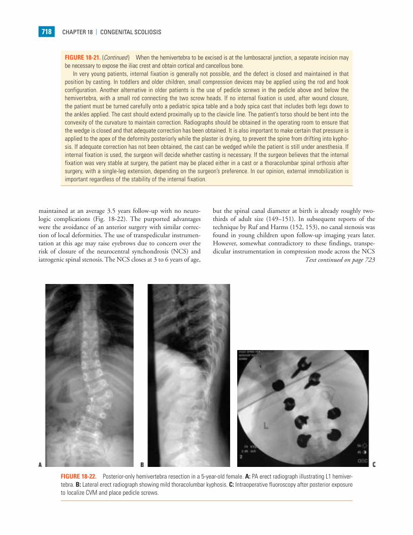
FIGURE 18-22. Posterior-only hemivertebra resection in a 5-year-old female. A: PA erect radiograph illustrating L1 hemiver-tebra. B: Lateral erect radiograph showing mild thoracolumbar kyphosis. C: Intraoperative fluoroscopy after posterior exposure to localize CVM and place pedicle screws.
maintained at an average 3.5 years follow-up with no neuro-logic complications (Fig. 18-22). The purported advantages were the avoidance of an anterior surgery with similar correc-tion of local deformities. The use of transpedicular instrumen-tation at this age may raise eyebrows due to concern over the risk of closure of the neurocentral synchondrosis (NCS) and iatrogenic spinal stenosis. The NCS closes at 3 to 6 years of age,
but the spinal canal diameter at birth is already roughly two-thirds of adult size (149–151). In subsequent reports of the technique by Ruf and Harms (152, 153), no canal stenosis was found in young children upon follow-up imaging years later. However, somewhat contradictory to these findings, transpe-dicular instrumentation in compression mode across the NCS
Text continued on page 723
Weinstein_Chap18.indd 718 9/27/2013 8:42:33 PM
cHAPTer 18 | congeniTAl ScolioSiS 719
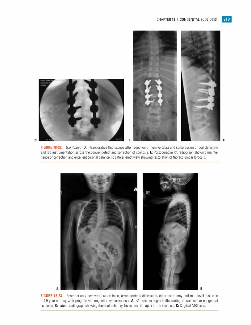
FIGURE 18-22. (Continued ) D: Intraoperative fluoroscopy after resection of hemivertebra and compression of pedicle screw and rod instrumentation across the convex defect and correction of scoliosis. E: Postoperative PA radiograph showing mainte-nance of correction and excellent coronal balance. F: Lateral erect view showing restoration of thoracolumbar lordosis.
FIGURE 18-23. Posterior-only hemivertebra excision, asymmetric pedicle subtraction osteotomy and multilevel fusion in a 4.5-year-old boy with progressive congenital kyphoscoliosis. A: PA erect radiograph illustrating thoracolumbar congenital scoliosis. B: Lateral radiograph showing thoracolumbar kyphosis near the apex of the scoliosis. C: Sagittal MRI scan.
Weinstein_Chap18.indd 719 9/27/2013 8:42:35 PM

720 cHAPTer 18 | congeniTAl ScolioSiS
FIGURE 18-23. (Continued ) D-G: Three-dimensional reconstructed CT scan in multiple planes of rotation. This type of pre-operative modeling is useful for planning resection procedures in complex deformities since it identifies anatomy, confirms relationships between and anterior and posterior elements and visualizes the sagittal plane with great clarity. H: Intraoperative photograph after subperiosteal exposure of the levels to be treated. The posterior elements and landmarks can be correlated with the CT scan images to confirm the level of resection.
Weinstein_Chap18.indd 720 9/27/2013 8:42:43 PM
cHAPTer 18 | congeniTAl ScolioSiS 721
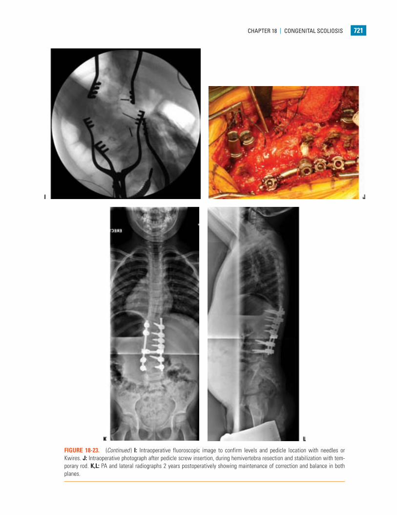
FIGURE 18-23. (Continued ) I: Intraoperative fluoroscopic image to confirm levels and pedicle location with needles or Kwires. J: Intraoperative photograph after pedicle screw insertion, during hemivertebra resection and stabilization with tem-porary rod. K,L: PA and lateral radiographs 2 years postoperatively showing maintenance of correction and balance in both planes.
Weinstein_Chap18.indd 721 9/27/2013 8:42:46 PM
722 cHAPTer 18 | congeniTAl ScolioSiS
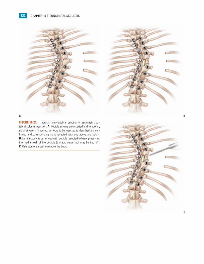
FIGURE 18-24. Thoracic hemivertebra resection or asymmetric ver-tebral column resection. A: Pedicle screws are inserted and temporary stabilizing rod is secured. Vertebra to be resected is identified and con-firmed and corresponding rib is resected with one above and below. B: Laminectomy is performed with pedicle resected to base, preserving the medial wall of the pedicle (thoracic nerve root may be tied off). C: Osteotome is used to remove the body.
Weinstein_Chap18.indd 722 9/27/2013 8:42:48 PM
cHAPTer 18 | congeniTAl ScolioSiS 723
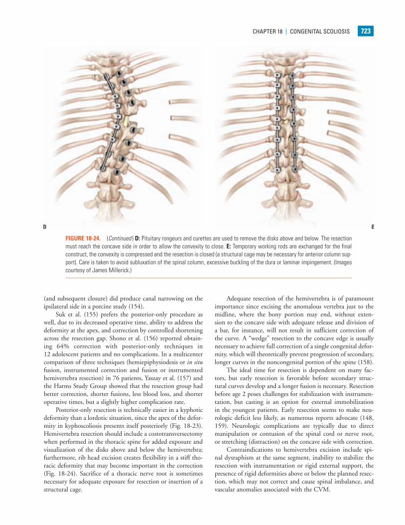
FIGURE 18-24. (Continued ) D: Pituitary rongeurs and curettes are used to remove the disks above and below. The resection must reach the concave side in order to allow the convexity to close. E: Temporary working rods are exchanged for the final construct, the convexity is compressed and the resection is closed (a structural cage may be necessary for anterior column sup-port). Care is taken to avoid subluxation of the spinal column, excessive buckling of the dura or laminar impingement. (Images courtesy of James Millerick.)
(and subsequent closure) did produce canal narrowing on the ipsilateral side in a porcine study (154).
Suk et al. (155) prefers the posterior-only procedure as well, due to its decreased operative time, ability to address the deformity at the apex, and correction by controlled shortening across the resection gap. Shono et al. (156) reported obtain-ing 64% correction with posterior-only techniques in 12 adolescent patients and no complications. In a multicenter comparison of three techniques (hemiepiphysiodesis or in situ fusion, instrumented correction and fusion or instrumented hemivertebra resection) in 76 patients, Yaszay et al. (157) and the Harms Study Group showed that the resection group had better correction, shorter fusions, less blood loss, and shorter operative times, but a slightly higher complication rate.
Posterior-only resection is technically easier in a kyphotic deformity than a lordotic situation, since the apex of the defor-mity in kyphoscoliosis presents itself posteriorly (Fig. 18-23). Hemivertebra resection should include a costotransversectomy when performed in the thoracic spine for added exposure and visualization of the disks above and below the hemivertebra; furthermore, rib head excision creates flexibility in a stiff tho-racic deformity that may become important in the correction (Fig. 18-24). Sacrifice of a thoracic nerve root is sometimes necessary for adequate exposure for resection or insertion of a structural cage.
Adequate resection of the hemivertebra is of paramount importance since excising the anomalous vertebra just to the midline, where the bony portion may end, without exten-sion to the concave side with adequate release and division of a bar, for instance, will not result in sufficient correction of the curve. A “wedge” resection to the concave edge is usually necessary to achieve full correction of a single congenital defor-mity, which will theoretically prevent progression of secondary, longer curves in the noncongenital portion of the spine (158).
The ideal time for resection is dependent on many fac-tors, but early resection is favorable before secondary struc-tural curves develop and a longer fusion is necessary. Resection before age 2 poses challenges for stabilization with instrumen-tation, but casting is an option for external immobilization in the youngest patients. Early resection seems to make neu-rologic deficit less likely, as numerous reports advocate (148, 159). Neurologic complications are typically due to direct manipulation or contusion of the spinal cord or nerve root, or stretching (distraction) on the concave side with correction.
Contraindications to hemivertebra excision include spi-nal dysraphism at the same segment, inability to stabilize the resection with instrumentation or rigid external support, the presence of rigid deformities above or below the planned resec-tion, which may not correct and cause spinal imbalance, and vascular anomalies associated with the CVM.
Weinstein_Chap18.indd 723 9/27/2013 8:42:50 PM

724 cHAPTer 18 | congeniTAl ScolioSiS
Fusion with Correction and Instrumentation. This pro-cedure is commonly performed when correction of multiple segments in the congenital deformity and or secondary curves is desired and the principle of balancing the spine is most important. It should be noted that correction of the congenital part of the curve carries with it significant difficulty compared to idiopathic scoliosis due to the stiffness of the deformity and risk of neurologic deficit, but with meticulous technique and proper monitoring, safe correction is possible. Standing x-rays will define the stable zones, truncal decompensation, congenital curves, and compensatory ones. With the use of bending and/or traction x-rays, we can define the flexibility of these respective deformities and then begin to formulate a plan to rebalance the spine. Choosing levels for instrumentation can be difficult, especially in scenarios when there are multiple noncontiguous anomalies (160). The entire curve must be included, but areas above or below the congenital curve may not behave in the same manner as idiopathic scoliosis. Rigid unbalanced curves may cause shoulder imbalance or truncal decompensation and an unhappy patient. Traction x-rays may be helpful in determining how much correction will still keep reasonable balance.
The use of instrumentation allows correction and stabi-lization until the arthrodesis heals, and the use of titanium instrumentation allows further imaging of the spinal cord, if needed. Proper preoperative planning of the levels to be included, anatomical challenges, and instrumentation needs are paramount. The surgeon should choose a versatile sys-tem with a robust inventory of proper size implants to fit the patient and take into account the biomechanics of different rod diameters and materials when planning the correction, especially in small children.
In segmented congenital deformities, facetectomy, liga-mentum flavum resection, posterior osteotomies (or resec-tions), and anterior discectomies are useful strategies to make a long, severe, rigid curve more flexible. Patients with significant growth remaining may be candidates for anterior discectomies and fusion to obtain better rotational correction and decrease the risk of crankshaft.
The correction of severe deformities is a challenging endeavor fraught with increased neurologic risk. Osteotomies of congenital fusions or bars will aid in correction, but inherent in this arena are complications of bleeding, cord or nerve root manipulation, and frequently inadequate pedicles to obtain segmental anchorage. Whenever possible, the osteotomy should shorten the spine and rely on compression correction rather than lengthening with distraction. In the most rigid, deformed, fused, or unsegmented areas, when there is no other option, vertebral column resection (VCR) is a technique to obtain profound correction over a short segment (see below).
Osteotomy of Prior Fusion or Revision Surgery. Occasi-onally, patients may present with a history of previous treat-ment such as an in situ fusion, and with subsequent growth and/or progression of the deformity, they have severe, rigid curves with pain, truncal decompensation, pelvic obliquity,
or neurologic deficit. Reconstructive surgery is indicated for these patients and may be achieved by anterior or posterior osteotomies of the previous fusion including VCR to rebalance the spine and instrumentation to restore spinal imbalance and provide the best chance for stable arthrodesis (Fig. 18-25). The basic principles are similar to the treatment of large deformi-ties mentioned above, in which resection is effective for focal correction, anterior release and/or posterior-based osteotomies are used to correct the spine or induce flexibility over longer segments, and rebalancing the spine and fusion is performed with segmental instrumentation.
Leatherman (160a) is credited with introducing a two-stage corrective procedure for severe congenital deformities in 1969. He recognized that a posterior fusion alone in a rap-idly progressing deformity may not prevent further progres-sion, particularly in cases with unilateral bars. Furthermore, he taught the principle that in order to avoid the danger of traction paraplegia (which had resulted from Harrington dis-traction instrumentation around that time), the spine must be shortened as well as straightened. As such, a two-stage resec-tion of the vertebral column was described by Leatherman and Dickson (129) in 60 patients with congenital spinal defor-mities for whom a two-stage corrective procedure provided excellent results. The first stage was anterior resection of the vertebral body, and the second stage was posterior resection, fusion of the curve, and instrumentation. In patients with con-genital scoliosis and a mean age of 11 years (2+3 years to 16+8 years), an average correction of 47% was achieved, with no significant complications including paresis. These results are especially impressive considering that nonsegmental compres-sion and distraction instrumentation was used and surgeries were performed without contemporary monitoring.
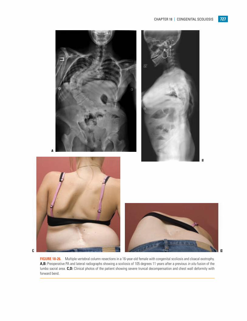
For the rigid, focal deformities, in either a primary or revi-sion situation, where there is no other option, VCR can obtain dramatic correction over a short segment, but even the expe-rienced surgeon should be prepared for excessive bleeding and assume the substantial risk of neurologic deficit. Multimodality intraoperative neurologic monitoring is mandatory since more than a quarter will have intraoperative neurologic events, and transcranial motor-evoked potentials are the most sensitive for prediction of postoperative neurologic deficit and allow prompt action intraoperatively to reduce the risk of perma-nent injury (161). Adequate laminectomy for visualization, stabilization with temporary working rods, undercutting the ends of the resection, and anterior structural grafting with a cage (to avoid shortening) may avert spinal subluxation and cord impingement. In a prospective, multicenter study of 147 patients who underwent VCRs for pediatric spinal deformity, the overall postoperative neurologic deficit rate was 13% (0.7% permanent), most of them at the spinal cord level, and com-mon risk factors included kyphosis, congenital abnormalities, and revision surgery (162). The overall radiographic correction rate was excellent for kyphoscoliosis (51%) and for congenital deformity (46%), and Scoliosis Research Society (SRS) scores were significantly improved in the self-image, satisfaction domains, and total scores (163) (Fig. 18-26).
Weinstein_Chap18.indd 724 9/27/2013 8:42:50 PM
cHAPTer 18 | congeniTAl ScolioSiS 725
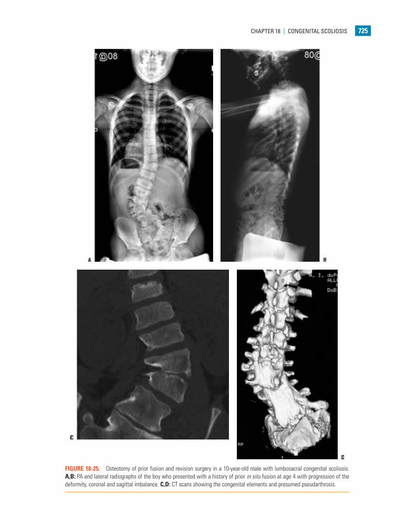
FIGURE 18-25. Osteotomy of prior fusion and revision surgery in a 10-year-old male with lumbosacral congenital scoliosis. A,B: PA and lateral radiographs of the boy who presented with a history of prior in situ fusion at age 4 with progression of the deformity, coronal and sagittal imbalance. C,D: CT scans showing the congenital elements and presumed pseudarthrosis.
Weinstein_Chap18.indd 725 9/27/2013 8:42:52 PM
726 cHAPTer 18 | congeniTAl ScolioSiS
Current segmental instrumentation has allowed earlier mobilization of the patient and improved correction, but has not decreased the technical complexity of the resection operation or the possible complications. In an effort to reduce the operative time and morbidity of staged or simultane-ous anterior/posterior procedures, single posterior resections were devised. Suk et al.’s (164) series of posterior VCRs in an adult group of 38 congenital scoliosis patients described 63% correction of the scoliosis, but described major complications including paralysis, root injuries, fixation failures, infections, and hemopneumothoraces. Lenke’s report of posterior VCR procedures for severe spinal deformities in 43 patients (het-erogeneous group of adults and children) described excellent results with no permanent neurologic deficits in this challeng-ing group of patients. He reiterated the need for spinal cord monitoring to prevent neurologic deficits since 18% patients lost intraoperative motor-evoked responses and promptly returned to baseline with surgical intervention (165).
The ability to treat severe deformities through an all-pos-terior vertebral resection has obviated the need for a circum-ferential approach in both primary and revision surgery except in special situations of lordotic deformities. Lenke et al. (166) reported on 35 consecutive pediatric patients who underwent posterior VCR of one to three levels; the average OR time was 460 minutes and average estimated blood loss (EBL) was 691 mL. Twelve patients had congenital scoliosis and had an aver-age correction of 24 degrees (60%).
Considerable experience and skill are necessary to achieve results as described above and a stepwise progression with Ponte or Smith Peterson osteotomies, pedicle subtrac-tion osteotomies, hemivertebra resection, and subsequently VCR is a reasonable approach. Careful preoperative plan-ning, localization, and identification of complex anatomy are paramount to proper decision making for osteotomy and execution of the planned procedure. The authors have found 3D CT scans and even scaled models of the spine
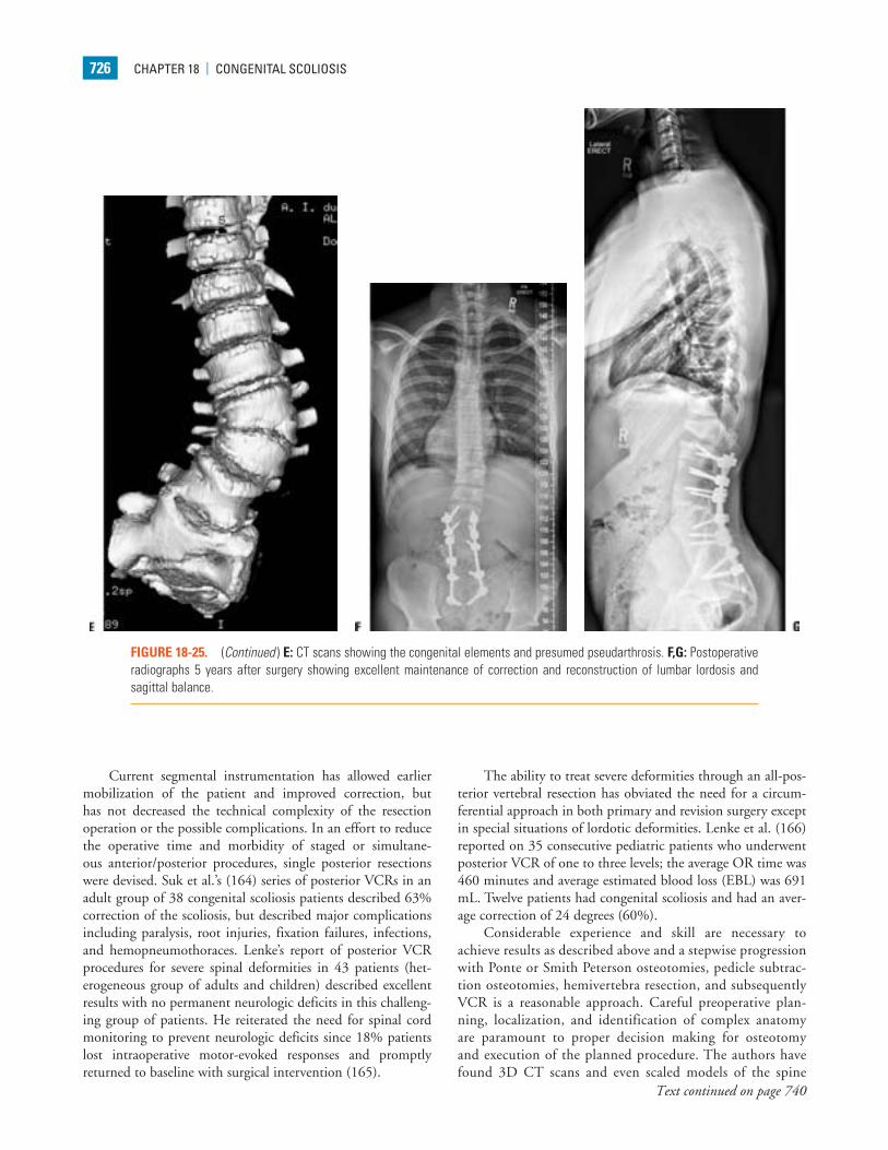
FIGURE 18-25. (Continued ) E: CT scans showing the congenital elements and presumed pseudarthrosis. F,G: Postoperative radiographs 5 years after surgery showing excellent maintenance of correction and reconstruction of lumbar lordosis and sagittal balance.
Text continued on page 740
Weinstein_Chap18.indd 726 9/27/2013 8:42:56 PM
cHAPTer 18 | congeniTAl ScolioSiS 727
FIGURE 18-26. Multiple vertebral column resections in a 16-year-old female with congenital scoliosis and cloacal exstrophy. A,B: Preoperative PA and lateral radiographs showing a scoliosis of 105 degrees 11 years after a previous in situ fusion of the lumbo sacral area. C,D: Clinical photos of the patient showing severe truncal decompensation and chest wall deformity with forward bend.
Weinstein_Chap18.indd 727 9/27/2013 8:42:59 PM
728 cHAPTer 18 | congeniTAl ScolioSiS
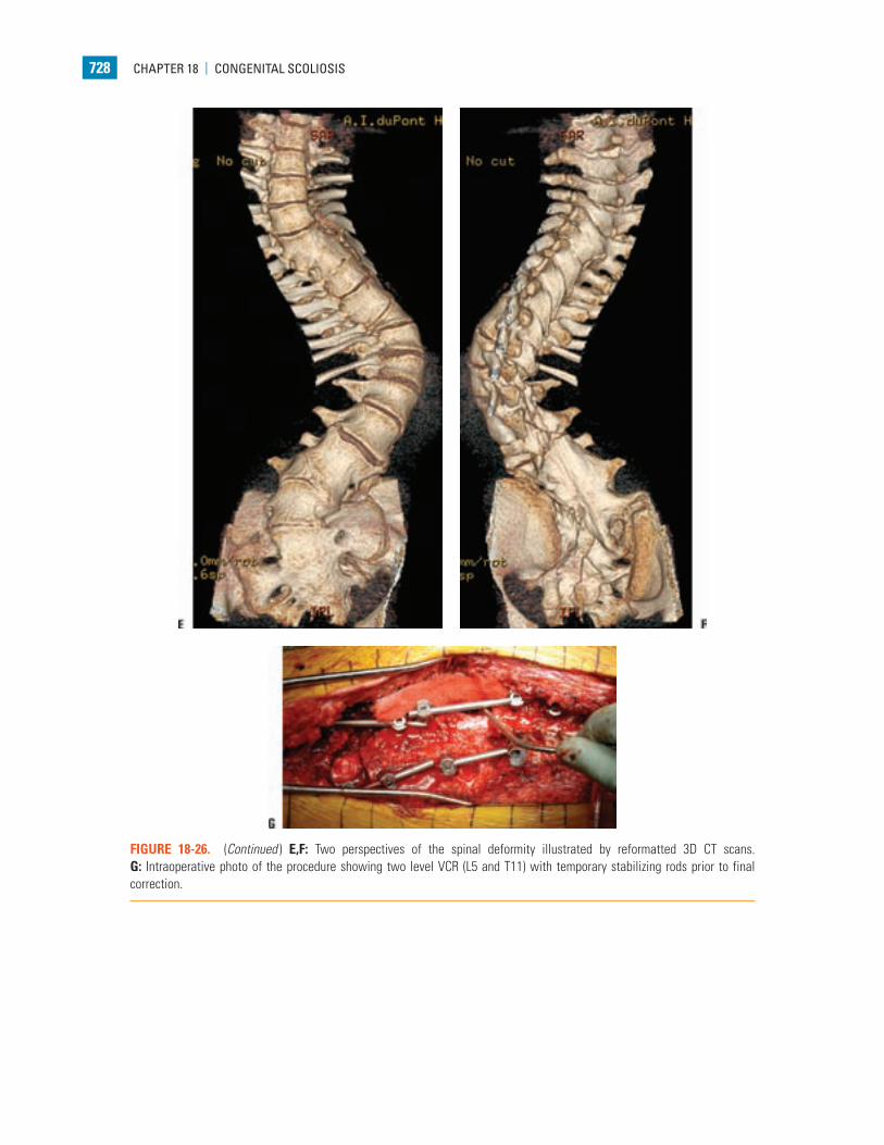
FIGURE 18-26. (Continued ) E,F: Two perspectives of the spinal deformity illustrated by reformatted 3D CT scans. G: Intraoperative photo of the procedure showing two level VCR (L5 and T11) with temporary stabilizing rods prior to final correction.
Weinstein_Chap18.indd 728 9/27/2013 8:43:06 PM

FIGURE 18-27. Growth rods in the management of congenital scoliosis in a 6-year-old female with vertebral-anus-cardio-vascular-trachea-esophagus-renal-limb-bud (VACTERL) syndrome. A: Erect PA radiograph showing a progressive scoliosis of 82 degrees and significant chest wall deformity. B: Lateral erect radiograph; note proximal thoracic kyphosis.
FIGURE 18-26. (Continued ) H,I: PA and lateral radiographs 2 years after surgery.
Weinstein_Chap18.indd 729 9/27/2013 8:43:08 PM
730 cHAPTer 18 | congeniTAl ScolioSiS
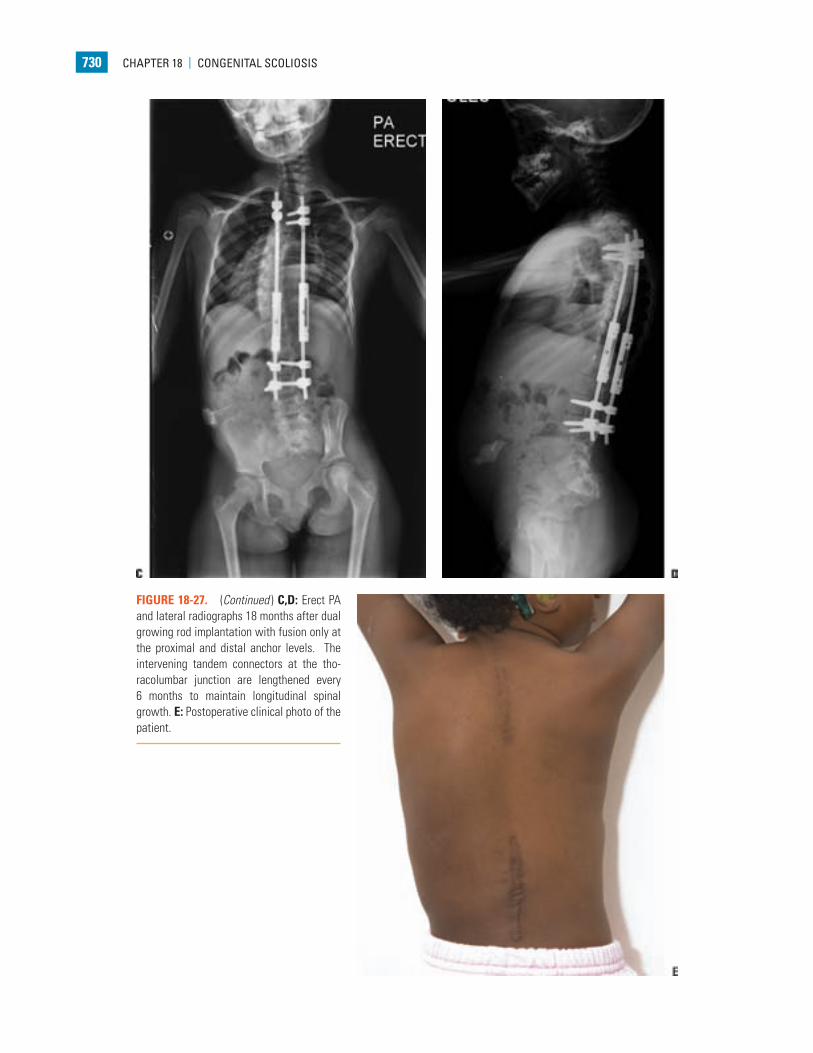
FIGURE 18-27. (Continued ) C,D: Erect PA and lateral radiographs 18 months after dual growing rod implantation with fusion only at the proximal and distal anchor levels. The intervening tandem connectors at the tho-racolumbar junction are lengthened every 6 months to maintain longitudinal spinal growth. E: Postoperative clinical photo of the patient.
Weinstein_Chap18.indd 730 9/27/2013 8:43:11 PM
cHAPTer 18 | congeniTAl ScolioSiS 731
constructed from these scans to be very useful in resection and osteotomy procedures. Not infrequently, important information can be gleaned from this step in planning. The risk of substantial bleeding in any osteotomy is to be expected, and the risk of neurologic deficit (at the spinal cord level or nerve root compromise) is considerable. The surgeon’s most trusted team should be present, and commu-nication should be open and clear to prevent an intraopera-tive complication.
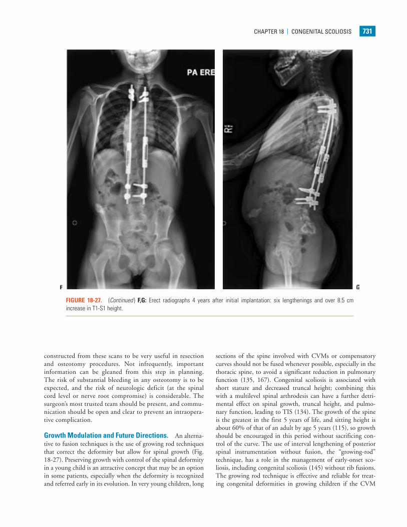
Growth Modulation and Future Directions. An alterna-tive to fusion techniques is the use of growing rod techniques that correct the deformity but allow for spinal growth (Fig. 18-27). Preserving growth with control of the spinal deformity in a young child is an attractive concept that may be an option in some patients, especially when the deformity is recognized and referred early in its evolution. In very young children, long
sections of the spine involved with CVMs or compensatory curves should not be fused whenever possible, especially in the thoracic spine, to avoid a significant reduction in pulmonary function (135, 167). Congenital scoliosis is associated with short stature and decreased truncal height; combining this with a multilevel spinal arthrodesis can have a further detri-mental effect on spinal growth, truncal height, and pulmo-nary function, leading to TIS (134). The growth of the spine is the greatest in the first 5 years of life, and sitting height is about 60% of that of an adult by age 5 years (115), so growth should be encouraged in this period without sacrificing con-trol of the curve. The use of interval lengthening of posterior spinal instrumentation without fusion, the “growing-rod” technique, has a role in the management of early-onset sco-liosis, including congenital scoliosis (145) without rib fusions. The growing rod technique is effective and reliable for treat-ing congenital deformities in growing children if the CVM
FIGURE 18-27. (Continued ) F,G: Erect radiographs 4 years after initial implantation: six lengthenings and over 8.5 cm increase in T1-S1 height.
Weinstein_Chap18.indd 731 9/27/2013 8:43:13 PM

732 cHAPTer 18 | congeniTAl ScolioSiS
segments are somewhat flexible, the anomalous segment is too large for resection, or other definitive treatment or is accompa-nied by a long, structural compensatory curve. The most com-monly used dual rod systems have been described by Akbarnia and Thompson with moderate success; modest gains in spi-nal length of 3 to 7 cm (1.2 cm per year); improvement in SAL; and an acceptable complication rate of superficial infec-tions, dislodged implants, and broken rods (168, 138). The technique involves anchorage at the proximal and distal sta-ble zones with hooks or screws after subperiosteal dissection; fusion is desirable in these small areas away from the apex for anchor stability. Rods are then tunneled submuscularly joined by tandem connectors placed at the thoracolumbar junction, where the interval distraction is performed for lengthening the spine. An apical fusion, excision, or osteotomy for the con-genital portion of the curve can be performed and then grow-ing rods implanted to lengthen the spine and accommodate growth in the compensatory curve(s) (169). Apical fusions in patients with single-rod growing systems do create challenges such as increased force to lengthen and smaller gains (170, 171) as well as crankshaft, but with dual rods these difficulties may be obviated. The vertical expandable prosthetic titanium rib (VEPTR) device is indicated for use in patients with con-genital scoliosis and fused ribs to address thoracic hypoplasia and prevent TIS when a large amount of growth remains (172) (see below). Unfortunately, both of these approaches require a scheduled operative lengthening recommended at 6-month intervals and ultimately are treated with a spinal fusion in most cases when no further lengthening can be achieved or the accelerative phase of growth is over. An alternative is a growth guidance system (Shilla) that corrects the scoliotic apex of the curve with a limited fusion and allows for continued guided growth at the proximal and distal portions of the spine, avoid-ing the need for routine operative lengthenings. Clinical data are limited, but McCarthy et al. (173) caprine animal model showed an average of 48 mm of growth at the ends of the rods near the sliding screws over 6 months. There were no implant failures, but minor wear debris was observed.
VEPTR (VERTICAL ExPANDABLE PROSTHETIC TITANIUM RIB)
The VEPTR was developed by Campbell (174) for the man-agement of TIS. This heterogeneous group of conditions is defined as those in which the function of the thorax is insuf-ficient to support normal growth and development. These conditions may be naturally occurring or can be iatrogenic. All forms of early onset scoliosis, many skeletal dysplasias, and congenital or acquired chest wall deformities including early spinal fusions are included. VEPTR devices currently are used under a humanitarian device exemption status by the FDA after undergoing an IDE trial in the early 2000s. This status requires institutions using the device to have an active insti-tutional review board approval and restricts its use to neuro-
muscular scoliosis conditions, congenital scoliosis, constricted hemithorax conditions such as fused ribs or costal dysplasias, and acquired severe chest wall deficiencies.
Historically, it was believed that growth of vertebral segments within a region of congenital malformations did not occur. As such, treatments focused on short-segment fusions and convex hemiepiphysiodesis that would include segments outside of the malformed segment in an effort to gain compensatory correction. The notion that guided or forced growth management of congenital scoliosis deformi-ties could not be done in congenital abnormalities was chal-lenged by Campbell et al. (175) with their use of rib-based systems using the VEPTR (Fig. 18-28). In an uncontrolled and nonselective group of patients with congenial scoliosis with and without rib fusions, application of rib–to-rib– or rib-to-spine–based distraction devices with an expansion tho-racoplasty (174) led to elongation of the concave side of the deformity by an average of 8 mm/year and of the convex side by 8.3 mm/year over mean follow-up of 4 years. Stabilization of the spinal deformity and secondary overall increase in lung volumes has been reported (175), leading many to believe that the combination of congenital spinal malformations and rib fusions is the ideal situation under which to use VEPTR devices (176–182).
The technique of VEPTR for congenital scoliosis and fused ribs has been described in detail by Campbell et al. (174, 175). The key components are an opening wedge thora-costomy through the fused rib segments, a lateral rib-to-rib–based strut, and a medial rib-to-rib– or rib-to-spine–based hybrid construct (Fig. 18-28A–D). In the absence of fused ribs, the role of thoracostomy has increasingly come into question due to concerns about altered chest wall compliance with the scarring created by the procedure. Rib anchors are recommended to be just lateral to the rib heads and avoid-ance of anchors above the 2nd rib is recommended due to the risk of brachial plexus injuries by proximal migration of the anchors (176, 183). Subperiosteal dissection of the ribs is done for placement of the rib cradles, and with the introduc-tion of VEPTR II, multiple anchor points and ribs can now be used. Distal spine anchors are designed to be via laminar hooks. The off-label use of pedicle screw anchors has been introduced with recommendations to have at least two distal anchors to decrease the incidence of screw migration through the pedicles. We have found that use of a ventriculoperitoneal shunt passer and chest tube facilitates submuscular passage of the hybrid rod for cases that will have a distal spine anchor. No standard exists for the frequency of expansions. Borrowing from the experience of “growing rod” systems, recommenda-tions have been for intervals of 6 months to optimize spinal growth (178, 184).
The incidence of complications related to the use of VEPTR implants has paralleled that of other growth pres-ervation systems. Anchor migration, implant failure, wound infections, and loss of control of the deformity have all been reported (185–187).
Weinstein_Chap18.indd 732 9/27/2013 8:43:13 PM
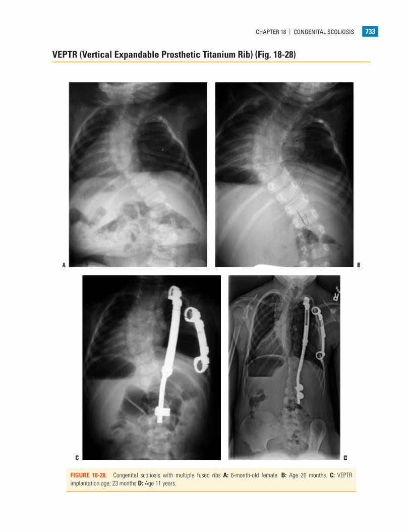
cHAPTer 18 | congeniTAl ScolioSiS 733
FIGURE 18-28. Congenital scoliosis with multiple fused ribs A: 6-month-old female. B: Age 20 months. C: VEPTR implantation age; 23 months D: Age 11 years.
VEPTR (Vertical Expandable Prosthetic Titanium Rib) (Fig. 18-28)
Weinstein_Chap18.indd 733 9/27/2013 8:43:16 PM
734 cHAPTer 18 | congeniTAl ScolioSiS
CONCLUSIONS
The treatment of congenital scoliosis is among the most interesting to the surgeon treating spinal deformities and focuses on early diagnosis and surgical treatment before large curves develop. The authors cannot emphasize enough the importance of the observational period of the treatment of patients with congenital scoliosis, namely giving thought to the evolution of the deformity with growth and acting at the appropriate time if surgery is necessary. Vertebral anomalies with a progressive natural history need to be managed aggres-sively. Outcomes are generally better with simple procedures with a favorable complication profile. In situ fusion or hemiep-iphysiodesis are viable options if there is little to no deformity present, but the potential for progression is high. For moderate deformities, correction with instrumentation and arthrodesis will provide satisfactory results. Young children with an iso-lated hemivertebra are excellent candidates for resection if pro-gression is noted; this keeps the area of arthrodesis small and avoids the development of compensatory curves. Extensive spinal arthrodesis should be avoided, especially before age 5, and growing rods can be used in children with long segmented areas of the spine to modulate growth and control the curve. TIS and congenital scoliosis with fused ribs seems to be best managed by the VEPTR device and expansion thoracostomy. For severe, rigid deformities in older children, osteotomies or resections to rebalance the spine with attention to the more structural compensatory curves may be needed. All too often, in a referral practice, we have seen missed opportunities in which a simpler procedure could have been performed, saving the patient and family from a much larger, complicated proce-dure and protracted recovery.
REFERENCES
1. Jones K, Smith DW. Smith’s recognizable patterns of human malformation, Edited 857. Philadelphia, PA: Saunders, 1997.
2. Erol B, Tracy MR, Dormans JP, et al. Congenital scoliosis and vertebral malformations: characterization of segmental defects for genetic analysis. J Pediatr Orthop 2004;24(6):674–682.
3. Giampietro PF, et al. Progress in the understanding of the genetic etiol-ogy of vertebral segmentation disorders in humans. Ann N Y Acad Sci 2009;1151:38–67.
4. Hedequist D, Emans J. Congenital scoliosis: a review and update. J Pediatr Orthop 2007;27(1):106–116.
5. Hensigner RN. Congenital scoliosis: etiology and associations. Spine (Phila PA 1976) 2009;34:1745–1750.
6. Tanaka T, Uhthoff HK. Significance of resegmentation in the pathogene-sis of vertebral body malformation. Acta Orthop Scand 1981;52:331–338.
7. Tanaka T, Uhthoff HK. The pathogenesis of congenital vertebral malfor-mations: a study based on observations made in 11 human embryos and fetuses. Acta Orthop Scand 1981;52:413–425.
8. Tracy MR, Dormans JP, Kusumi K. Klippel-Feil syndrome: clinical features and current understanding of etiology. Clin Orthop Relat Res 2004;424:183–190.
9. Alexander PG, Tuan RS. Carbon monoxide-induced axial skeletal dys-morphogenesis in the chick embryo. Birth Defects Res A Clin Mol Teratol 2003;67(4):219–230.
10. Dequeant M, Pourquie O. Segmental patterning of the vertebrate embry-onic axis. Nat Rev Genet 2008;9:370–382.
11. Kessel M, Gruss P. Murine developmental control genes. Science 1990;249:374–379.
12. Palmeirium I, Henrique D, Ish-Horowicz D, et al. Avian hairy gene expression identifies a molecular clock linked to vertebrate segmentation and somitogenesis. Cell 1997;91:639–648.
13. Shinkai Y, Tjuji T, Kawamoto Y, et al. New mutant mouse with skeletal deformities caused by mutation in delta like 3 (DLL3) gene. Exp Anim 2004;53:129–136.
14. Thomsen B, et al. A missense mutation in the bovine SLC35A3 gene, encoding a UDP-N-acetylglucosamine transporter, causes complex verte-bral malformation. Genome Res 2006;16:97–105.
15. Shands AR, Eisberg HB. The incidence of scoliosis in the state of Delaware; a study of 50,000 minifilms of the chest made during a survey for tuberculosis. J Bone Joint Surg Am 1955;37:1243–1249.
16. Forrester MB, Merz RD. Descriptive epidemiology of hemivertebrae, Hawaii, 1986–2002. Congenit Anom (Kyoto) 2006;46(4):172–176.
17. Goldstein I, Makhoul IR, Weissman A, et al. Hemivertebra: prenatal diagnosis, incidence and characteristics. Fetal Diagn Ther 2005;20(2): 121–126.
18. Ganey TM, Ogden JA. Development and maturation of the axial skeleton. In: Weinstein SL, ed. The pediatric spine. Philadelphia, PA: Lippincott Williams & Wilkins, 2001:3–54.
19. Mark M, Rijli FM, Chambon P. Homeobox genes in embryogenesis and pathogenesis. Pediatr Res 1997;42:421–429.
20. Giampietro PF, et al. Congenital and idiopathic scoliosis: clinical and genetic aspects. Clin Med Res 2003;1(2):125–136.
21. Fan C, Porter JA, Chiang C, et al. Long-range sclerotome induction by sonic hedgehog: direct role of the amino-terminal cleavage product and modulation by the cyclic AMP signaling pathway. Cell 1995;81: 457–465.
22. Conlon RQ, Reaume AG, Rossant J. Notch 1 is required for the coordi-nated segmentation of somites. Development 1995;121:1533–1545.
23. Pourquie O. Building the spine: the vertebrate segmentation clock. Cold Spring Harb Symp Quant Biol 2007;72:445–449.
24. Pourquie O, Kusumi K. When body segmentation goes wrong. Clin Genet 2001;60:409–416.
25. Farley FA, Loder RT, Nolan BT, et al. Mouse model for thoracic congeni-tal scoliosis. J Pediatr Orthop 2001;21(4):537–540.
26. Farley FA, Phillips WA, Herzenberg JE, et al. Natural history of scoliosis in congenital heart disease. J Pediatr Orthop 1991;11(1):42–47.
27. Loder RT, Hernandez MJ, Lerner AL, et al. The induction of congeni-tal spinal deformities in mice by maternal carbon monoxide exposure. J Pediatr Orthop 2000;20(5):662–666.
28. Norman CA, Halton DM. Is carbon monoxide a workplace teratogen? A review and evaluation of the literature. Am Occup Hyg 1990;34:335–347.
29. Tredwell SJ, Smith DF, Macleod PJ, et al. Cervical spine anomalies in fetal alcohol syndrome. Spine (Phila Pa 1976) 1982;7:331–334.
30. Bantz EW. Valproic acid and congenital malformations. A case report. Clin Pediatr (Phila) 1984;23:352–353.
31. Giavini E, Menegola E. Gene-teratogen interactions in chemically induced congenital malformations. Biol Neonate 2004;85:73–81.
32. Hanold KC. Teratogenic potential of valproic acid. J Obstet Gynecol Neonatal Nurs 1986;15:111–116.
33. Wide K, Winbladh B, Kallen B. Major malformations in infants exposed to antiepileptic drugs in utero, with emphasis on carbamazepine and val-proic acid: a nation-wide, population-based register study. Acta Paediatr 2004;93(2):174–176.
34. Edwards MF. Hyperthermia as a teratogen: a review of experimental stud-ies and their clinical significance. Teratog Carcinog Mutagen 1986;6:563–582.
35. Ewart-Toland A, Yankowitz J, Winder A, et al. Oculoauriculovertebral abnormalities in children of diabetic mothers. Am J Med Genet 2000;90:303–309.
36. Lowy C, Beard RW, Goldschmidt J. Congenital malformations in babies of diabetic mothers. Diabet Med 1986;3(5):458–462.
Weinstein_Chap18.indd 734 9/27/2013 8:43:16 PM

cHAPTer 18 | congeniTAl ScolioSiS 735
37. De Wals P, et al. Reduction in neural-tube defects after folic acid fortifica-tion in Canada. N Engl J Med 2007;357:135–142.
38. Huppert SS, Le A, Schroeter EH, et al. Embryonic lethality in mice homo-zygous for a processing-deficient allele of Notch-1. Nature 2000;405: 966–970.
39. McMaster MJ. Congenital scoliosis caused by a unilateral failure of verte-bral segmentation with contralateral hemivertebrae. Spine (Phila Pa 1976) 1998;23(9):998–1005.
40. Wynne-Davies R. Congenital vertebral anomalies: aetiology and relation-ship to spina bifida cystica. J Med Genet 1975;12:280–288.
41. Bulman MP, et al. Mutations in the human delta homologue, DLL3, cause axial skeletal defects in spondylocostal dysostosis. Nat Genet 2000; 24:438–441.
42. Giampietro PF, et al. DLL3 as a candidate gene for vertebral malforma-tions. Am J Med Genet A 2006;140(22):2447–2453.
43. Hutchinson J. Deformity of shoulder girdle. Br Med J 1894;1:634. 44. Klippel M, Feil A. Un cas d’absence des vertebres cervicales. Nouv Icon
Salpet 1912;25:223. 45. Clarke RA, Singh S, McKenzie H, et al. Familial Klippel-Feil syn-
drome and paracentric inversion inv(8)(q22.2q23.3). Am J Hum Genet 1995;57:1364–1370.
46. McGaughran JA, Oates D, Donnai D, et al. Mutations in PAX1 may be associated with Klippel-Feil syndrome. Eur J Hum Genet 2003;11: 468–474.
47. Thomsen MB, Schneider U, Weber M, et al. Scoliosis and congential anomalies associated with Klippel-Feil syndrome types I-III. Spine (Phila Pa 1976) 1997;22(4):396–401.
48. Auleha A, Wehrle C, Brand-Sberi B, et al. Wnt3a plays a major role in the segmentation clock controlling somitogenesis. Dev Cell 2003;4(3): 407–418.
49. Beals RK, Robbins JR, Rolfe B. Anomalies associated with vertebral mal-formations. Spine (Phila Pa 1976) 1993;18(10):1329–1332.
50. Basu PS, Elsebaie H, Noordeen MH. Congenital spinal deformity: a comprehensive assessment at presentation. Spine (Phila Pa 1976) 2002;27(20):2255–2259.
51. Bradford DS, Heithoff KB, Cohen M. Intraspinal abnormalities and con-genital spine deformities: a radiographic and MRI study. J Pediatr Orthop 1991;11:36–41.
52. McMaster MJ. Occult intraspinal anomalies and congenital scoliosis. J Bone Joint Surg Am 1984;66(4):588–601.
53. Prahinski JR, Polly DW, McHale KA, et al. Occult intraspinal anomalies in congenital scoliosis. J Pediatr Orthop 2000;20(1):59–63.
54. Tori JA, Dickson JH. Association of congenital anomalies of the spine and kidneys. Clin Orthop Relat Res 1980;148:259–262.
55. Belmont PJ, Kuko TR, Taylor KF, et al. Intraspinal anomalies associated with isolated congenital hemivertebra: the role of routine magnetic reso-nance imaging. J Bone Joint Surg Am 2004;86-A:1704–1710.
56. Cardoso M, Keating RF. Neurosurgical management of spinal dysra-phism and neurogenic scoliosis. Spine (Phila Pa 1976) 2009;34:1775– 1782.
57. Fribourg D, Delgado E. Occult spinal cord abnormalities in children referred for orthopedic complaints. Am J Orthop 2004;33(1):18–25.
58. Reckles LN, Peterson HA, Weidman WH, et al. The association of scoliosis and congenital heart defects. J Bone Joint Surg Am 57(4): 449–55:1975.
59. MacEwen GD, Winter RB, Hardy JH, et al. Evaluation of kidney anoma-lies in congenital scoliosis. Clin Orthop Relat Res 1972;434:4–7.
60. Rai AS, Taylor TK, Smith GH, et al. Congenital abnormalities of the urogenital tract in association with congenital vertebral malformations. J Bone Joint Surg Br 2002;84(6):891–895.
61. Vitko RJ, Cass AS, Winter RB. Anomalies of the genitourinary tract associated with congenital scoliosis and congenital kyphosis. J Urol 108: 655–659.
62. Berend N, Marlin GE. Arrest of alveolar multiplication in kyphoscoliosis. Pathology 1979;11:485–491.
63. Bergofsky EH. Respiratory failure in disorders of the thoracic cage. Am Rev Respir Dis 1979;119:643–669.
64. Campbell RM Jr, et al. The characteristics of thoracic insufficiency syn-drome associated with fused ribs and congenital scoliosis. J Bone Joint Surg Am 2003;85-A(3):399–408.
65. Caubet JF, et al. Increased hemoglobin levels in patients with early onset scoliosis. Spine (Phila Pa 1976) 2009;34:2534–2536.
66. Davies G, Reid L. Effect of scoliosis on growth of alveoli and pulmonary arteries and on right ventricle. Arch Dis Child 1971;46:623–632.
67. Day GA, Upadhyay SS, Ho EK, et al. Pulmonary functions in congenital scoliosis. Spine(Phila Pa 1976) 1994;19(9):1027–1031.
68. Mayer OH, Redding G. Early changes in pulmonary function after verti-cal expandable prosthetic titanium rib insertion in children with thoracic insufficiency syndrome. J Pediatr Orthop 2009;29:35–38.
69. McMaster MJ, Glasby MA, Singh H, et al. Lung function in congenital kyphosis and kyphoscoliosis. J Spinal Disord Tech 2007;20(3):203–208.
70. Motoyama EK, Deeney VF, Fine GF, et al. Effects on lung function of multiple expansion thoracoplasty in children with thoracic insufficiency syndrome: a longitudinal study. Spine(Phila Pa 1976) 2006;31(3): 284–290.
71. Redding G, Song K, Inscore S, et al. Lung function asymmetry in chil-dren with congenital and infantile scoliosis. Spine J 2008;8(4):639–644.
72. Siegler D, Zorab PA. The influence of lung volume on gas transfer in scoliosis. Br J Dis Chest 1982;76:44–50.
73. Owange-Iraka JW, Harrison A, Warner JO. Lung function in congenital and idiopathic scoliosis. Eur J Pediatr 1984;142:198–200.
74. Bowen RE, Scaduto AA, Banuelos S. Decreased body mass index and restrictive lung disease in congenital thoracic scoliosis. J Pediatr Orthop 2008;28(6):665–668.
75. Muirhead A, Conner AN. The assessment of lung function in children with scoliosis. J Bone Joint Surg Br 1985;67(5):699–702.
76. Kuhns JG, Hormell RS. Management of congenital scoliosis; review of one hundred seventy cases. AMA Arch Surg 1952;65:250–263.
77. Winter RB, Moe JH. Congential scoliosis a study of 234 patients treated and untreated. Part I: natural history. J Bone Joint Surg Am 1968;50-A:1–47.
78. MacEwen GD, Conway JJ, Miller WT. Congential scoliosis with a unilat-eral bar. Radiology 1968;90:711–715.
79. Kawakami N, Tsuji T, Imagama S, et al. Classification of congential sco-liosis and kyphosis: a new approach to the three-dimensional classification for progressive vertebral anomalies requiring operative treatment. Spine (Phila Pa 1976) 2009;34:1756–1765.
80. McMaster MJ, Ohtsuka K. The natural history of congenital scoliosis. J Bone Joint Surg Am 1982;64:1128–1147.
81. McMaster MJ, Singh H. Natural history of congenital kyphosis and kyphoscoliosis. A study of one hundred and twelve patients. J Bone Joint Surg Am 1999;81(10):1367–83.
82. Nasca RJ, Stilling FH III, Stell HH. Progression of congenital scoliosis due to hemivertebrae and hemivertebrae with bars. J Bone Joint Surg Am 1975;57(4):456–466.
83. Winter RB, Moe JH, Wang JF. Congenital kyphosis: its natural history and treatment as observed in a study of one hundred thirty patients. J Bone Joint Surg Am 1973;55:223–256.
84. Batra S, Ahuja S. Congenital scoliosis: management and future directions. Acta Orthop Belg 2008;74(2):147–160.
85. Goldberg CJ, Moore DP, Fogarty EE, et al. The natural history of early onset scoliosis. Stud Health Technol Inform 2002;91:68–70.
86. Campbell RM Jr, Hell-Vocke AK. Growth of the thoracic spine in con-genital scoliosis after expansion thoracoplasty. J Bone Joint Surg Am 2003;85-A(3):409–420.
87. Pehrsson K, Larsson S, Oden A, et al. Long-term follow-up of patients with untreated scoliosis. A study of mortality, causes of death and symp-toms. Spine (Phila Pa 1976) 1992;17:1091–1096.
88. Vitale MG, et al. Health-related quality of life in children with thoracic insufficiency syndrome. J Pediatr Orthop 2008;28:239–243.
89. Chan G, Dormans JP. Update on congenital spinal deformities: preopera-tive evaluation. Spine (Phila Pa 1976) 2009;34:1766–1774.
90. McMaster MJ, Singh H. The surgical management of congenital kyphosis and kyphoscoliosis. Spine (Phila Pa 1976) 2001;26(19):2146–2154; dis-cussion 2155.
Weinstein_Chap18.indd 735 9/27/2013 8:43:16 PM
736 cHAPTer 18 | congeniTAl ScolioSiS
91. Suh SW, Sarwark JF, Vora A, et al. Evaluating congenital spine deformi-ties for intraspinal anomalies with magnetic resonance imaging. J Pediatr Orthop 2001;21(4):525–531.
92. von Koch CS, Orit AG, Goldstein RB, et al. Fetal magnetic resonance imaging enhances detection of spinal cord anomalies in patients with sonographically detected bony anomalies of the spine. J Ultrasound Med 2005;24:781–789.
93. Lowe LH, Johanek AJ, Moore CW. Sonography of the neonatal spine: part 2, spinal disorders. AJR Am J Roentgenol 2007;188:739–744.
94. Lowe LH, Johanek AJ, Moore CW. Sonography of the neonatal spine: part I, normal anatomy, imaging pitfalls, and variations that may simu-late disorders. AJR Am J Roentgenol 2007;188:733–738.
95. Medina LS. Spinal dysraphism: categorizing risk to optimize imaging. Pediatr Radiol 2009;39(Suppl 2):S242–S246.
96. Riccio AI, Guille JT, Grissom L, et al. Magnetic resonance imaging of renal abnormalities in patients with congential osseous anomalies of the spine. J Bone Joint Surg Am 2007;89:2456–2459.
97. Beydon N. et al. An official American thoracic society/European respira-tory society statement: pulmonary function testing in preschool chil-dren. Am J Respir Crit Care Med 2007;175:1304–1345.
98. De Jong PA, Long FR, Wong JC, et al. Computed tomographic esti-mation of lung dimensions throughout the growth period. Eur Respir J 2006;27:261–267.
99. Emans JB, Caubet JF, Ordonez CL, et al. The treatment of spine and chest wall deformities with fused ribs by expansion thoracostomy and insertion of vertical expandable prosthetic titanium rib: growth of tho-racic spine and improvement of lung volumes. Spine (Phila Pa 1976) 2005;30(Suppl 17):S58–S68.
100. Gollogly S, Smith JT, White SK, et al. The volume of lung parenchyma as a function of age: a review of 1050 normal CT scans of the chest with three-dimensional volumetric reconstruction of the pulmonary system. Spine (Phila Pa 1976) 2004;29:2061–2066.
101. Redding G, Song KM, Swanson J, et al. V/Q asymmetry changes follow-ing treatment of TIS. In: Proceedings of the 44th Annual Meeting of the Scoliosis Research Society. San Antonio, TX, 2009.
102. O’Daniel JC, Stevens DM, Cody DD. Reducing radiation exposure from survey CT scans. AJR Am J Roentgenol 2005;185:509–515.
103. Paterson A, Frush DP. Dose reduction in paediatric MDCT: general principles. Clin Radiol 2007;62:507–517.
104. Paterson A, Frush DP, Donnelly LF. Helical CT of the body: are settings adjusted for pediatric patients? AJR Am J Roentgenol 2001;176:297–301.
105. Swift AJ, Woodhaouse N, Fichele S, et al. Rapid lung volumetry using ultrafast dynamic magnetic resonance imaging during forced vital capac-ity maneuver: correlation with spirometry. Invest Radiol 2007;42:37–41.
106. Voorhees A, An J, Berger KI, et al. Magnetic resonance imaging-based spirometry for regional assessment of pulmonary function. Magn Reson Med 2005;54:1146–1154.
107. Facanha-Filho FAM, Winter RB, Lonstein JE, et al. Measurement accu-racy in congenital scoliosis. J Bone Joint Surg Am 2001;83(1):42–45.
108. Heckman JD, et al. Statistical differences: consider the methodology. J Bone Joint Surg Am 2002;84:1078.
109. Loder RT, Urguhart A, Steen H, et al. Variability in cobb angle mea-surements in children with congenital scoliosis. J Bone Joint Surg Br 1995;77:768–770.
110. Campbell RM Jr, et al. The effect of opening wedge thoracostomy on thoracic insufficiency syndrome associated with fused ribs and congeni-tal scoliosis. J Bone Joint Surg Am 2004;86-A(8):1659–1674.
111. Hedequist DJ, Emans JB. The correlation of preoperative three-dimensional computed tomography reconstructions with operative findings in congenital scoliosis. Spine (Phila Pa 1976) 2003;28(22):2531–2534; discussion 1.
112. Newton PO, Hahn GW, Fricka KB, et al. Utility of three-dimensional and multiplanar reformatted computed tomography for evaluation of pediatric congenital spine abnormalities. Spine (Phila Pa 1976) 2002;27(8):844–850.
113. Hedequist DJ. Surgical treatment of congenital scoliosis. Orthop Clin North Am 2007;38(4): 497–509, vi.
114. Marks DS, Qaimkhani SA. The natural history of congenital scoliosis and kyphosis. Spine (Phila Pa 1976) 2009;34(17):1751–1755.
115. Dimeglio A. Growth of the spine before age 5 years. J Pediatr Orthop B 1993;1(2):102–107.
116. Doody MM, Lonstein JE, Stovall M, et al. Breast cancer mortality after diagnostic radiography: findings from the U.S. Scoliosis Cohort Study. Spine (Phila Pa 1976) 2000;25(16):2052–2063.
117. Loder RT, Urquhart A, Steen H, et al. Variability in Cobb angle mea-surements in children with congenital scoliosis. J Bone Joint Surg Br 1995;77(5):768–770.
118. Prahinski JR, Polly DW Jr, McHale KA, et al. Occult intraspinal anoma-lies in congenital scoliosis. J Pediatr Orthop 2000;20(1):59–63.
119. Winter RB, Moe JH, MacEwen GD, et al. The Milwaukee brace in the nonoperative treatment of congenital scoliosis. Spine 1976;1:85–96.
120. Sink EL, Karol LA, Sanders J, et al. Efficacy of perioperative halo-gravity traction in the treatment of severe scoliosis in children. J Pediatr Orthop 2001;21(4):519–524.
121. Rinella A, Lenke L, Whitaker C, et al. Perioperative halo-gravity traction in the treatment of severe scoliosis and kyphosis. Spine (Phila Pa 1976) 2005;30(4):475–482.
122. Sponseller PD, Takenaga RK, Newton P, et al. The use of traction in the treatment of severe spinal deformity. Spine (Phila Pa 1976) 2008;33(21):2305–2309.
123. Watanabe K, Lenke LG, Bridwell KH, et al. Efficacy of periopera-tive halo-gravity traction for treatment of severe scoliosis. J Orthop Sci 2010;15(6):720–730.
124. Roaf R. The treatment of progressive scoliosis by unilateral growth-arrest. J Bone Joint Surg Br 1963;45:637–651.
125. Hall JE, Herndon WA, Levine CR. Surgical treatment of congenital sco-liosis with or without Harrington instrumentation. J Bone Joint Surg Am 1981;63:608–619.
126. Dubousset J, Herring JA, Shufflebarger H. The crankshaft phenom-enon. J Pediatr Orthop 1989;9(5):541–550.
127. Terek RM, Wehner J, Lubicky JP. Crankshaft phenomenon in congenital scoliosis: a preliminary report. J Pediatr Orthop 1991;11(4):527–532.
128. Kesling KL, Lonstein JE, Denis F, et al. The crankshaft phenomenon after posterior spinal arthrodesis for congenital scoliosis: a review of 54 patients. Spine (Phila Pa 1976) 2003;28(3):267–271.
129. Leatherman KD, Dickson RA. Two-stage corrective surgery for congeni-tal deformities of the spine. J Bone Joint Surg Br 1979;61:324–328.
130. McMaster M. Congenital scoliosis caused by unilateral failure of ver-tebral segmentation with contralateral hemivertebrae. Spine (Phila Pa 1976) 1998;23:998–1005.
131. Hedequist DJ, Hall JE, Emans JB. The safety and efficacy of spinal instrumentation in children with congenital spine deformities. Spine (Phila Pa 1976) 2004;29(18):2081–2086.
132. Kim YJ, Otsuka NY, Flynn JM, et al. Surgical treatment of congenital kyphosis. Spine (Phila Pa 1976) 2001;26(20):2251–2257.
133. Thompson GH, Akbarnia BA, Kostial P, et al. Comparison of single and dual growing rod techniques followed through definitive surgery: a preliminary study. Spine (Phila Pa 1976) 2005;30(18):2039–2044.
134. Campbell RM Jr, Smith MD, Mayes TC, et al. The characteristics of thoracic insufficiency syndrome associated with fused ribs and congeni-tal scoliosis. J Bone Joint Surg Am 2003;85-A:399–408.
135. Karol LA, Johnston C, Mladenov K, et al. Pulmonary function follow-ing early thoracic fusion in non-neuromuscular scoliosis. J Bone Joint Surg Am 2008;90(6):1272–1281.
136. Winter R. Convex anterior and posterior hemiarthrodesis and hemiepiphyseodesis in young children with progressive congenital sco-liosis. J Pediatr Orthop 1981;1(4):361–366.
138. Thompson AG, Marks DS, Sayampanathan SR, et al. Long-term results of combined anterior and posterior convex epiphysiodesis for congenital scoliosis due to hemivertebrae. Spine (Phila Pa 1976) 1995;20(12):1380–1385.
139. Marks DS, Sayampanathan SR, Thompson AG, et al. Long-term results of convex epiphysiodesis for congenital scoliosis. Eur Spine J 1995;4(5):296–301.
Weinstein_Chap18.indd 736 9/27/2013 8:43:16 PM
cHAPTer 18 | congeniTAl ScolioSiS 737
140. Keller PM, Lindseth RE, DeRosa GP. Progressive congenital scoliosis treatment using a transpedicular anterior and posterior convex hemiep-iphysiodesis and hemiarthrodesis: a preliminary report. Spine (Phila Pa 1976) 1994;19(17):1933–1939.
141. Andrew T, Piggott H. Growth arrest for progressive scoliosis: combined anterior and posterior fusion of the convexity. J Bone Joint Surg Br 1985;67:193–197.
142. Cil A, Yazici M, Alanay A, et al. The course of sagittal plane abnor-mality in the patients with congenital scoliosis managed with convex growth arrest. Spine (Phila Pa 1976) 2004;29:547–552.
143. Ruf M, Jensen R, Harms J. Hemivertebra resection in the cervical spine. Spine (Phila Pa 1976) 2005;30(4):380–385.
144. Bradford DS, Boachie-Adjei O. One-stage anterior and posterior hemi-vertebral resection and arthrodesis or congenital scoliosis. J Bone Joint Surg Am 1990;72:536–540.
145. Klemme WR, Denis F, Winter RB, et al. Spinal instrumentation with-out fusion for progressive scoliosis in young children. J Pediatr Orthop 1997;17(6);734–742.
146. Bollini G, Docquier PL, Viehweger E. Lumbar hemivertebra resection. J Bone Joint Surg Am 2006;88:1043–1052.
147. Lazar RD, Hall JE. Simultaneous anterior and posterior hemivertebra excision. Clin Orthop Relat Res 1999;364:76–84.
147a. Hedequist DJ, Hall JE, Emans JB. Hemivertebra excision in children via simultaneous anterior and posterior exposures. J Pediatr Orthop 2005;25:60–63.
148. Ruf M, Harms J. Hemivertebra resection by a posterior approach: innovative operative technique and first results. Spine (Phila Pa 1976) 2002;27, 1116–1123.
149. Maat GJ, Matricali B, van Persijn van Meerten EL. Postnatal develop-ment and structure of the neurocentral junction. Its relevance for spinal surgery. Spine (Phila Pa 1976) 1996;21(6):661–666.
150. Wang JC, Nuccion SL, Feighan JE. Growth and development of the pediatric cervical spine documented radiographically. J Bone Joint Surg Am 2001;83-A:1212–1218.
151. Jeffrey JE, Campbell DM, Golden MH, et al. Antenatal factors in the development of the lumbar vertebral canal: a magnetic resonance imag-ing study. Spine (Phila Pa 1976) 2003;28(13):1418–1423.
152. Ruf M, Harms J. Pedicle screws in 1- and 2-year-old children: tech-nique, complications and effect on further growth. Spine (Phila Pa 1976) 2002;27:E460–E466.
153. Ruf M, Harms J. Posterior hemivertebra resection with transpedicular instrumentation: early correction in children aged 1 to 6 years. Spine (Phila Pa 1976) 2003;28(18):2132–2138.
154. Cil A, Yazici M, Daglioglu K, et al. The effect of pedicle screw place-ment with or without application of compression across the neuro-central cartilage on the morphology of the spinal canal and pedicle in immature pigs. Spine (Phila Pa 1976) 2005;30(11):1287–1293.
155. Suk S-I, Chung ER, Kim JH, et al. Posterior vertebral column resection for severe rigid scoliosis. Spine 2005;30:1682–1687.
156. Shono Y, Abumi K, Kaneda K. One-stage posterior hemivertebra resec-tion and correction using segmental posterior instrumentation. Spine 2001;26(7):752–7.
157. Yaszay B, O’Brien M, Shufflebarger HL, et al. The efficacy of hemi-vertebra resection for congenital scoliosis: a multicenter retrospective comparison of three surgical techniques. Spine 2011;36(24):2052-2060.
158. Ruf M, Jensen R, Letko L, et al. Hemivertebra resection and osteotomies in congenital spinal deformity. Spine 2009;34(17):1791–1799.
159. Klemme WR, Polly DW Jr, Orchowski JR. Hemivertebral exci-sion for congenital scoliosis in very young children. J Pediatr Orthop 2001;21(6):761–764.
160. Hedden D. Management themes in congenital scoliosis. J Bone Joint Surg Am 2007;89-A(Suppl 1):72–78.
160a. Leatherman K. Resection of vertebral bodies. J Bone Joint Surg Am 1969;51-A:206.
161. Sucato DJ, Shah SA, Lenke LG, et al. Prompt response to critical spinal cord monitoring changes during vertebral column resection results in a low incidence of permanent neurologic deficit. Proceedings of the 45th Annual Meeting of the Scoliosis Research Society. Kyoto, Japan, 2010:109.
162. Shah SA, Sucato DJ, Newton, PO, et al. Perioperative neurologic events from a multicenter consecutive series of pediatric vertebral col-umn resection: nature, frequency and outcomes. Proceedings of the 17th International Meeting on Advanced Spine Techniques. Toronto, Ontario, Canada, 2010:97.
163. Lenke LG, Newton PO, Sucato DJ, et al. Complications following 147 consecutive vertebral column resections for severe pediatric spinal deformity: a multicenter analysis. Spine 2012 Jul 20. [Epub ahead of print]
164. Suk S-I, Chung ER, Lee SM, et al. Posterior vertebral column resection in fixed lumbosacral deformity. Spine 2005;30(23):E703–E710.
165. Lenke LG, Sides BA, Koester LA, et al. Vertebral column resection for the treatment of severe spinal deformity. Clin Orthop Relat Res 2010;468(3): 687–699.
166. Lenke LG, O’Leary PT, Bridwell KH, et al. Posterior vertebral column resection for severe pediatric deformity: minimum two-year follow up of thirty-five consecutive patients. Spine 2009;34(20):2213–2221.
167. Canavese F, Dimeglio A, Volpatti D, et al. Dorsal arthrodesis of thoracic spine and effects on thorax growth in prepubertal New Zealand white rabbits. Spine 2007;32:E443–E450.
168. Akbarnia BA, Marks DS, Boachie-Adjei O, et al. Dual growing rod tech-nique for the treatment of progressive early-onset scoliosis: a multicenter study. Spine 2005;30:S46–S57.
169. Blakemore LC, Scoles PV, Poe-Kochert C, et al. Submuscular Isola rod with or without limited apical fusion in the management of severe spinal deformities in young children: preliminary report. Spine 2001;26:2044–2048.
170. Noordeen HM, Shah SA, Elsebaie HB, et al. In vivo distraction force and length measurements of growing rods: which factors influence on the ability to lengthen? Spine 2011;36(26):2299-303.
171. Farooq N, Garrido E, Altaf F, et al. Minimizing complications with single submuscular growing rods: a review of technique and results on 88 patients with minimum two-year follow-up. Spine 2010;35(25):252–258.
172. Yazici M, Emans J. Fusionless instrumentation systems for congeni-tal scoliosis: expandable spinal rods and vertical expandable prosthetic titanium rib in the management of congenital spine deformities in the growing child. Spine 2009;34(17):1800–1807.
173. McCarthy RM, Sucato D, Turner JL, et al. Shilla growing rods in a cap-rine animal model: a pilot study. Clin Orthop Relat Res 2010;468(3):705–710.
174. Campbell RM Jr, Smith MD, Hell-Vocke AK. Expansion thoracoplasty: the surgical technique of opening-wedge thoracostomy. Surgical tech-nique. J Bone Joint Surg Am 2004;86-A(Suppl 1):51–64.
175. Campbell RM Jr, Hell-Vocke AK. Growth of the thoracic spine in congenital scoliosis after expansion thoracoplasty. J Bone Joint Surg Am 2003;85-A(3):409–420.
176. Nassr A, Larson AN, Crane B, et al. Iatrogenic thoracic outlet syn-drome secondary to vertical expandable prosthetic titanium rib expan-sion thoracoplasty: pathogenesis and strategies for prevention/treatment. J Pediatr Orthop 2009;29(1):31–34.
177. Ramirez N, Flynn JM, Serrano JA, et al. The Vertical Expandable Prosthetic Titanium Rib in the treatment of spinal deformity due to pro-gressive early onset scoliosis. J Pediatr Orthop B 2009;18(4):197–203.
178. Thompson GH, Akbarnia BA, Campbell RM Jr. Growing rod tech-niques in early-onset scoliosis. J Pediatr Orthop 2007;27(3):354–361.
179. Samy MA, Al Zayed ZS, Shaheen MF. The effect of a vertical expandable prosthetic titanium rib on shoulder balance in patients with congenital scoliosis. J Child Orthop 2009.
180. Smith JT. The use of growth-sparing instrumentation in pediatric spinal deformity. Orthop Clin North Am 2007;38(4):547–552, vii.
181. Smith JT, Jerman J, Stringham J, et al. Does expansion thoracoplasty improve the volume of the convex lung in a windswept thorax? J Pediatr Orthop 2009;29(8):944–947.
182. Yazici M, Emans J. Fusionless instrumentation systems for congeni-tal scoliosis: expandable spinal rods and vertical expandable prosthetic titanium rib in the management of congenital spine deformities in the growing child. Spine (Phila Pa 1976) 2009;34(17):1800–1807.
Weinstein_Chap18.indd 737 9/27/2013 8:43:16 PM
738 cHAPTer 18 | congeniTAl ScolioSiS
183. Skaggs DL, Choi PD, Rice C, et al. Efficacy of intraoperative neuro-logic monitoring in surgery involving a vertical expandable prosthetic titanium rib for early-onset spinal deformity. J Bone Joint Surg Am 2009;91(7):1657–1663.
184. Akbarnia BA, Marks DS, Boachie-Adjei O, et al. Dual growing rod tech-nique for the treatment of progressive early-onset scoliosis: a multicenter study. Spine 2005;30(17 Suppl):S46–S57.
185. Campbell RM Jr, et al. The effect of opening wedge thoracostomy on thoracic insufficiency syndrome associated with fused ribs and congenital scoliosis. J Bone Joint Surg Am 2004;86-A(8):1659–1674.
186. Emans JB, Caubet JF, Ordonez CL, et al. The treatment of spine and chest wall deformities with fused ribs by expansion thoracostomy and insertion of vertical expandable prosthetic titanium rib: growth of tho-racic spine and improvement of lung volumes. Spine (Phila Pa 1976) 2005;30(17 Suppl):S58–S68.
187. Shah SC, Birknes JK, Sagoo S, et al. Vertical Expandable Prosthetic Titanium Rib (VEPTR): indications, technique, and management review. Surg Technol Int 2009;18:223–229.
Weinstein_Chap18.indd 738 9/27/2013 8:43:16 PM