Embed Size (px)
Citation preview
* CHAPTER 7
Congenitaldyserythropoietic anaemias
Hermann Heimpel, Achille Iolascon
IRON2009_CAP.7(178-201):EBMT2008 4-12-2009 16:17 Pagina 178
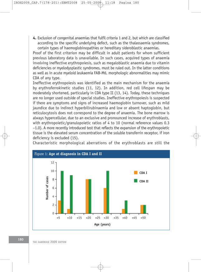
1. Definition and classificationThe congenital dyserythropoietic anaemias (CDAs, ICD-10 D64.4) comprise a groupof rare hereditary disorders that are characterised by ineffective erythropoiesis asthe predominant mechanism of anaemia and by distinct morphological abnormalitiesof erythroblasts in the bone marrow. The term was first used by Crookston et al.(1) (for cases later classified as CDA II) and by Wendt and Heimpel (2) (for caseslater classified as CDA I), but a few reports of similar cases were publishedpreviously (3-5). The working classification initially proposed by Heimpel andWendt (6), including as type III the families with autosomal dominant inheritancedescribed by others (7, 8), is still used in clinical practice. There are, however, familiesthat fulfil the general definition of the CDAs, but do not conform to any of the threeclassical types (9, 5, 10) (Table 1).In the majority of CDAs, inheritance is autosomal recessive, and due to the smallnumber of offspring in most European families, single cases in one family are therule rather than the exception. Together with the rarity of the disorder and the needto obtain bone marrow specimens for diagnosis, this explains why correct diagnosisis often delayed (Figure 1), particularly in mild cases, even when anaemia and/orhyperbilirubinaemia have been evident for many years.In general, the diagnosis of the CDAs requires the presence of all of the fourfollowing criteria:1. Evidence of congenital anaemia/jaundice or a positive family history2. Evidence of ineffective erythropoiesis3. Typical morphological appearance of bone marrow erythroblasts
DISORDERS OF ERYTHROPOIESIS, ERYTHROCYTES AND IRON METABOLISM179
CHAPTER 7 • Congenital dyserythropoietic anaemias
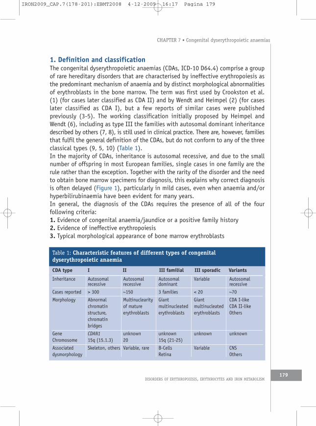
Inheritance
Cases reported
Morphology
GeneChromosome
Associateddysmorphology
Autosomalrecessive
> 300
Abnormalchromatinstructure,chromatinbridges
CDAN115q (15.1.3)
Skeleton, others
Autosomalrecessive
~150
Multinuclearityof matureerythroblasts
unknown20
Variable, rare
Autosomaldominant
3 families
Giantmultinucleatederythroblasts
unknown15q (21-25)
B-CellsRetina
Variable
< 20
Giantmultinucleatederythroblasts
unknown
Variable
Autosomalrecessive
~70
CDA I-likeCDA II-likeOthers
unknown
CNSOthers
Table 1: Characteristic features of different types of congenitaldyserythropoietic anaemia
CDA type I II III familial III sporadic Variants
IRON2009_CAP.7(178-201):EBMT2008 4-12-2009 16:17 Pagina 179
4. Exclusion of congenital anaemias that fulfil criteria 1 and 2, but which are classifiedaccording to the specific underlying defect, such as the thalassaemia syndromes,certain types of haemoglobinopathies or hereditary sideroblastic anaemias.
Proof of the first criterion may be difficult in adult patients for whom sufficientprevious laboratory data is unavailable. In such cases, acquired types of anaemiainvolving ineffective erythropoiesis, such as megaloblastic anaemia due to vitamindeficiencies or myelodysplastic syndromes, must be ruled out. In the latter conditionsas well as in acute myeloid leukaemia FAB-M6, morphologic abnormalities may mimicCDA of any type.Ineffective erythropoiesis was identified as the main mechanism for the anaemiaby erythroferrokinetic studies (11, 12). In addition, red cell lifespan may bemoderately shortened, particularly in CDA type II (13, 14). Today, these techniquesare no longer used outside of special studies. Ineffective erythropoiesis is suspectedif there are symptoms and signs of increased haemoglobin turnover, such as mildjaundice due to indirect hyperbilirubinaemia and low or absent haptoglobin, butreticulocytosis does not correspond to the degree of anaemia. The bone marrow isalways hypercellular, due to an exclusive and pronounced increase of erythroblasts,with erythropoietic/granulopoietic ratios of 4 to 10 (normal reference values 0.3–1.0). A more recently introduced test that reflects the expansion of the erythropoietictissue is the elevated serum concentration of the soluble transferrin receptor, if irondeficiency is excluded (15).Characteristic morphological aberrations of the erythroblasts are still the
THE HANDBOOK 2009 EDITION180
Figure 1: Age at diagnosis in CDA I and II
12
10
8
6
4
2
0
CDA I
CDA II
<5 <10 <15 <20 <25 <30 <35 <40 <45 <50
Age (years)
Num
berof
cases
IRON2009_CAP.7(178-201):EBMT2008 25-05-2009 11:18 Pagina 180
cornerstone of the diagnosis, and if they are present in the majority of cells inan anaemia which is definitely congenital they can be regarded as specific for thediagnosis. They are also the first step in determination of CDA type. Recognitionis much easier in smears of aspirated bone marrow than in histology specimens,and morphological analysis of both peripheral blood and appropriate bone marrowsmears is required for diagnosis of any case of CDA. In addition to panoptic staining,the specimen should be stained for non-haem iron to exclude congenitalsideroblastic anaemia with CDA-like morphological aberrations in a minority of cells,and also to estimate tissue iron stores. The number of sideroblasts may beincreased in patients with increased iron stores, but ringed sideroblasts arepresent only in exceptional cases (16).CDAs are very rare disorders. Because even haematologists only occasionally see suchpatients, specialised sources of information are needed. A European network(http://www.enerca.org) provides information on specialist centres and givesaccess to new publications for both physicians and patients. A variety of microscopicviews can be seen at http://bildatlas.onkodin.de/bildatlas/content/e1352/e1775/e1872/e2500/e2501/index_ger.html.All types of CDA share a high incidence of splenomegaly, cholelithiasis and ironoverload (5, 17, 18). As in other forms of anaemia with ineffective erythropoiesis, thisis due to up-regulation of iron absorption (19), mediated by hepcidin. Extramedullaryhaematopoiesis presenting as paravertebral masses may be observed in all types of CDA.
2. CDA I (MIM 224120)
2.1 Epidemiology and clinical presentationCDA I is less frequent than CDA II, with 89 cases from 82 families collated in theGerman registry of CDAs and/or identified from published case reports together with70 additional cases in a large Bedouin tribe described by Tamary et al. fromIsrael (20, 21). At least for Europe, this probably reflects true differences ofprevalence, since there is no evidence that the ascertainment rate of cases of thetwo types is different. Most families have been detected among Western Europeansand Arabs, but single cases have also been reported from the USA, India, Japan,Australia, New Zealand, Polynesia and China. The degree of anaemia is variable notonly between families, but for unexplained reasons (modifier genes) may also bedifferent among siblings (22). Most patients have life-long anaemia withhaemoglobin concentration between 7 and 11 g/mL. Occasionally, there are severecases requiring transfusion in-utero (23) or immediately after birth and regular bloodtransfusions during childhood and adolescence (24, 25). At the other extreme there
DISORDERS OF ERYTHROPOIESIS, ERYTHROCYTES AND IRON METABOLISM
CHAPTER 7 • Congenital dyserythropoietic anaemias
181
IRON2009_CAP.7(178-201):EBMT2008 4-12-2009 16:17 Pagina 181
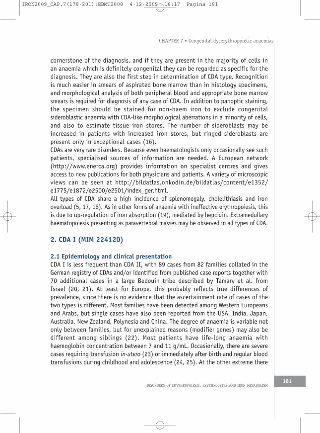
are patients with only borderline low haemoglobin but distinct macrocytosis. Theanaemia is usually macrocytic with MCV between 100 and 120 fl, but may benormocytic in childhood (22).As in any case of chronic unclassified anaemia, careful inspection of the peripheralblood smear is the first step in verifying the suspected diagnosis. There is distinctanisocytosis and poikilocytosis. Large poikilocytes and elliptocytes are reminiscentof changes seen in megaloblastic anaemias, which is the most frequent erroneousdiagnosis made before the correct diagnosis is recognised. Basophilic stippledcells are always present, and Cabot rings may be seen in some cases, even beforesplenectomy. Except for the occasional presence of late erythroblasts, the distributionand shape of nucleated cells in the peripheral blood are normal.Light microscopy of bone marrow erythroblasts in bone marrow smears is the nextstep toward confirmation of the diagnosis. Detailed illustrated descriptions can befound in previous publications, (5, 26). 30 to 60% of early and late polychromaticerythroblasts show characteristic and bizarre abnormalities of nuclear shape and sizeand of chromatin structure, but proerythroblasts and immature basophilicerythroblasts usually appear normal. In less severely affected cells, chromatinstrands are coarser than normal and interrupted by irregularly shaped translucentareas. In more severely affected erythroblasts the chromatin structure is lost andthe nucleus contains weakly stained material that is not sharply delineated fromthe surrounding cytoplasm. There are large polyploid cells, and a minority of cellsshow bi- or multinuclearity. In contrast to CDA II, the nuclei of binucleated cellsare of different size and shape. A hallmark of CDA I is the presence of incompletelydivided cells with thin chromatin bridges between the pairs of erythroblasts, whichmay also be seen between two nuclei in a single cell (Figure 2). With the exceptionof a few cases of erythroleukaemia, such bridges are virtually specific for CDA I, butsince they may be present in less than 3% of cells, at least 500 consecutive cellsshould be examined when searching for this abnormality. The cytoplasm of manycells shows Howell-Jolly bodies and/or intensive and irregular basophilic stippling.These changes are sometimes called megaloblastic, but in contrast to megaloblasticanaemia the characteristic loose, fine chromatin structure of erythroblast nuclei islacking, as are giant granulopoietic cells and hyperlobulation of megakaryocytes.Experts who have seen cases of CDA I before are easily able to make the diagnosisby light microscopy, but electron microscopy shows particularly specific alterations(26-28). They are absent in very early erythroblasts but become distinct asmaturation progresses. The heterochromatin is denser than normal and formssharply delineated clumps with small translucent vacuoles, giving rise to themetaphor of “Swiss cheese appearance” (29), and cytoplasm may penetrate throughwidened pores of the nuclear envelope (Figure 2).
THE HANDBOOK 2009 EDITION182
IRON2009_CAP.7(178-201):EBMT2008 4-12-2009 16:17 Pagina 182
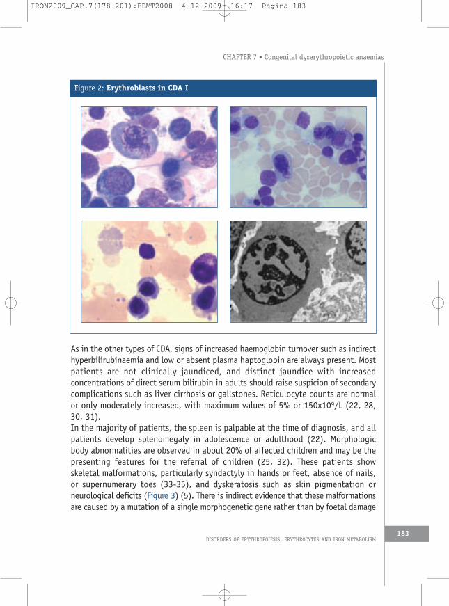
As in the other types of CDA, signs of increased haemoglobin turnover such as indirecthyperbilirubinaemia and low or absent plasma haptoglobin are always present. Mostpatients are not clinically jaundiced, and distinct jaundice with increasedconcentrations of direct serum bilirubin in adults should raise suspicion of secondarycomplications such as liver cirrhosis or gallstones. Reticulocyte counts are normalor only moderately increased, with maximum values of 5% or 150x109/L (22, 28,30, 31).In the majority of patients, the spleen is palpable at the time of diagnosis, and allpatients develop splenomegaly in adolescence or adulthood (22). Morphologicbody abnormalities are observed in about 20% of affected children and may be thepresenting features for the referral of children (25, 32). These patients showskeletal malformations, particularly syndactyly in hands or feet, absence of nails,or supernumerary toes (33-35), and dyskeratosis such as skin pigmentation orneurological deficits (Figure 3) (5). There is indirect evidence that these malformationsare caused by a mutation of a single morphogenetic gene rather than by foetal damage
DISORDERS OF ERYTHROPOIESIS, ERYTHROCYTES AND IRON METABOLISM
CHAPTER 7 • Congenital dyserythropoietic anaemias
183
Figure 2: Erythroblasts in CDA I
IRON2009_CAP.7(178-201):EBMT2008 4-12-2009 16:17 Pagina 183
(36). Short stature may be the result of pituitary failure due to unrecognisedsecondary haemochromatosis (37).
2.2 PathophysiologyThe ineffective erythropoiesis corresponds to abnormalities of the cell cycledistribution. Most mononucleated polychromatic cells – those stages that show themost distinct morphological abnormalities – have DNA contents between 2c and 4c,but many become arrested during their progress through the S-Phase as shown bythe absence of H3-thymidine uptake in vitro. Exceptionally large mononuclear as wellas binucleated cells or those connected by chromatin bridges reach polyploid DNAvalues up to 8c (29, 38, 39). Erythroblasts showing chromatin abnormalities by EMalso show an arrest of protein synthesis, and intramedullary necrobiosis of interphaseerythroblasts is more likely than apoptosis. The relationship between mutations inthe CDAN1 gene (the gene responsible for CDA I) and ineffective erythropoiesis isnot yet understood. Studies are in progress to define the role of the codanin-1 proteinin normal erythropoiesis, but it is known that codanin-1 is localised to nuclearheterochromatin in interphase cells and is expressed in the S-Phase (40).
2.3 GeneticsThe gene responsible for CDA I (CDAN1 gene) was mapped to the long arm ofchromosome 15 between 15q15.1q15.3 by homozygosity mapping in four Bedouinfamilies with a high degree of consanguinity (20) and could be assigned to a 0.5cM interval (41). Similar results were reported in six patients from Europe and theNear East (42). The CDAN1 gene was cloned and found to contain 28 exons spanning
THE HANDBOOK 2009 EDITION184
Figure 3: Syndactyly of toes in a case of CDA I (courtesy of J. Goede, Zurich)
IRON2009_CAP.7(178-201):EBMT2008 4-12-2009 16:17 Pagina 184
15 kb and encoding a protein named codanin-1. A founder mutation Arg1042Trpwas identified in all Bedouin patients. From studies in unrelated patients ofEuropean, Bedouin, North-American and Asian origin, altogether 36 different pointmutations, distributed over 13 exons have been detected (22, 43-45). The majorityof mutations (70%) are located on the 3’ half of the gene. In another series of 51CDA I cases, in 15 patients [29%, originating in Israel (5), Germany (6), and England(4)], only one mutation was identified, although splice site mutations were notexcluded. In 6 of the 51 case with the definite phenotype of CDA I, no mutationwas found (H. Tamary personal communication), suggesting either a promoterdefect or a mutation in another gene (44). As is often the cases in orphan disorders,molecular study was the clue to understanding the role of a number of proteins inerythropoiesis, including the codanin in CDA I that is localised to the nuclearheterochromatin and upregulated during S phase by E2F1, the main regulator of G1/Stransition of the cell cycle (40, 43).
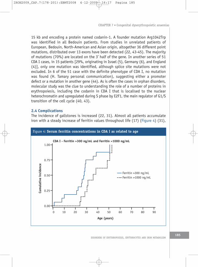
2.4 ComplicationsThe incidence of gallstones is increased (22, 31). Almost all patients accumulateiron with a steady increase of ferritin values throughout life (17) (Figure 4) (31),
DISORDERS OF ERYTHROPOIESIS, ERYTHROCYTES AND IRON METABOLISM
CHAPTER 7 • Congenital dyserythropoietic anaemias
185
Figure 4: Serum ferritin concentrations in CDA I as related to age
CDA I - Ferritin >300 ng/mL and Ferritin >1000 ng/mL
0 10 20 30 40 50 60 70 80 90
Age (years)
1.00
0.75
0.50
0.25
0.00
Cumulativeincide
nce
Ferritin >300 ng/mLFerritin >1000 ng/mL
IRON2009_CAP.7(178-201):EBMT2008 4-12-2009 16:17 Pagina 185
independently of the presence of any HFE gene mutations (46). In severe cases, ironoverload becomes apparent in childhood (37, 47). Patients with organ damage anddeath from secondary haemachromatosis were observed before ferritin levels weresystematically monitored and iron depletion therapy introduced (22).
3. CDA II (MIM 224120)
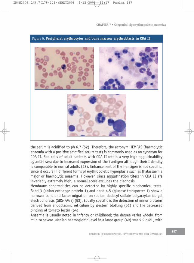
3.1 Epidemiology and clinical presentationCDA II is the less uncommon form of CDA. The geographic distribution of affectedpatients suggests a higher frequency of the gene in Italy and in the Mediterraneancountries as compared to central and northern Europe. At present it is difficult toassess whether this is due to a clustered distribution (48) or to greater diagnosticawareness.The International Registry on CDA II (49) includes epidemiology, clinical findingsand molecular studies. Through April 2008, 76 Italian and 46 non-Italian patientsfrom 60 and 36 families, respectively, were registered (A. Iolascon, unpublished).To these patients we must add 49 patients from the German CDA registry (14), aswell as more than 50 further cases published as case reports (3).The regional distribution of the Italian patients demonstrated clustering in SouthernItaly, and this phenomenon suggests a founder effect. However, molecular studiesby means of microsatellites, localised where the gene was mapped, failed todemonstrate the existence of a common haplotype (48).The main clinical findings are normocytic anaemia, jaundice and variable splenomegaly(14, 49). These features are also present in hereditary spherocytosis (HS) and it ispossible to confuse these two conditions, especially because osmotic fragilitytests give similar results in both. The most useful pointer to diagnosis of CDA II isan inadequate reticulocyte count for the degree of anaemia. The red cell distributionwidths for cell volume (RDW, anisocytosis) and for haemoglobin concentration (HDW,anisochromia) are also helpful in distinguishing HS from CDA II. Characteristicallythe RDW is increased in CDA II and the HDW is increased in HS, resulting in anRDW/HDW ratio that is significantly greater in CDA than HS (50).CDA II is associated with a well-defined morphological phenotype: bi- ormultinucleated late precursors (6, 49) (Figure 5) and flat vesicles of variablelength visualised by electron microscopy (5, 51). Peripheral blood smears showdistinct aniso-poikolocytosis with basophilic stippled red cells and a few (occasionallybinucleated) mature erythroblasts. The red cells express an antigen that binds toa natural cold-reacting Ig-M antibody present in the serum of 40% to 60% of healthyindividuals. Antibody binding can be demonstrated by agglutination or by lysis when
THE HANDBOOK 2009 EDITION186
IRON2009_CAP.7(178-201):EBMT2008 4-12-2009 16:17 Pagina 186
the serum is acidified to ph 6.7 (52). Therefore, the acronym HEMPAS (haemolyticanaemia with a positive acidified serum test) is commonly used as an synonym forCDA II. Red cells of adult patients with CDA II retain a very high agglutinabilityby anti-i sera due to increased expression of the i antigen although their I densityis comparable to normal adults (52). Enhancement of the i-antigen is not specific,since it occurs in different forms of erythropoietic hyperplasia such as thalassaemiamajor or haemolytic anaemia. However, since agglutination titers in CDA II areinvariably extremely high, a normal score excludes the diagnosis.Membrane abnormalities can be detected by highly specific biochemical tests.Band 3 (anion exchange protein 1) and band 4.5 (glucose transporter 1) show anarrower band and faster migration on sodium dodecyl sulfate-polyacrylamide gelelectrophoresis (SDS-PAGE) (53). Equally specific is the detection of minor proteinsderived from endoplasmic reticulum by Western blotting (51) and the decreasedbinding of tomato lectin (54).Anaemia is usually noted in infancy or childhood; the degree varies widely, frommild to severe. Median haemoglobin level in a large group (49) was 9.9 g/dL, with
DISORDERS OF ERYTHROPOIESIS, ERYTHROCYTES AND IRON METABOLISM
CHAPTER 7 • Congenital dyserythropoietic anaemias
187
Figure 5: Peripheral erythrocytes and bone marrow erythroblasts in CDA II
IRON2009_CAP.7(178-201):EBMT2008 4-12-2009 16:17 Pagina 187
a range from 6.0 to 12.7. In the majority in adult patients untransfused levels of8 to 11 g/dL are observed (14).Few patients require regular transfusions, and as in CDA I transfusion may only beneeded in infancy. In a number of specific reported cases transfusion dependencewas generated by the interaction with another red blood cell defect. Three of thesesubjects were heterozygotes for CDA II and beta-thalassaemia (Iolascon, unpublisheddata) or a G-6PDH variant (55).
3.2 PathophysiologyThe life-long anaemia results from the combination of ineffective erythropoiesis andmoderate peripheral haemolysis.The principal biochemical feature is the hypoglycosylation of certain proteins,including transferrin and band 3. It appears that in CDA II a genetic factor blocksthe glycosylation of glycoprotein acceptors and shifts polylactosamines to lipidacceptors. The reduced glycosylation is associated with abnormal function of band3 in CDA II patients. Analysis of red cells from CDA II patients demonstrated anarrower- than-usual band 3 with slightly faster migration on SDS-PAGE, while analysisof anion transport (inhibition of sulphate flux by H2-DIDS) showed decreasedactivity of the anion transport for the band 3 molecule. Furthermore the CDA IIerythrocytes were found to contain higher amounts of aggregate band 3 than controlerythrocytes (56). Aggregated band 3 has been reported to bind naturally occurringantibodies, possibly mediating the phagocytic removal of red blood cells. These resultssuggested that the haemolysis found in CDA II patients may be ascribed toclusterings of band 3, leading to IgG binding and phagocytosis, rather than tosecondary modification of the erythrocytic cytoskeletal structure.
3.3 GeneticsThe results of structural analysis of CDA II band 3 carbohydrates suggesteddisruption of the biosynthesis involving the N-acetylglucosaminyltransferase II (GnT-II) and alpha-mannosidase II (MANII) steps. However, linkage analysis in patientsfrom Southern Italy excluded these candidate genes (57), and a genome-widesearch yielded conclusive evidence for linkage of CDA II to microsatellite markerson the long arm of chromosome 20 (20q11.2). A maximum two-point lod score of5.4 at q=0.00 with the marker D20S863 was obtained (58). Mapping of the geneshows that there is genetic heterogeneity in this condition.
3.4 ComplicationsGallstone formation, apparently related to the increased haemoglobin turnover, is
THE HANDBOOK 2009 EDITION188
IRON2009_CAP.7(178-201):EBMT2008 4-12-2009 16:17 Pagina 188
the most prevalent complication (14, 49). A significant correlation has beenobserved between the UGT1A (TA)7/(TA)7 genotype, i.e., Gilbert’s syndrome, andthe increased rate of gallstones in CDA II patients. The effects of Gilbert’s syndromeare clearly visible when CDA II patients from the same families but with differentUGT1A genotypes are compared (59).Secondary haemochromatosis is the most important long-term complication. As inCDA I, iron overload is not dependent on (albeit enhanced by) transfusions. It maybe alleviated by ongoing iron loss, such as menstrual bleeding or pregnancies.Haemochromatosis can lead to organ damage if not recognised and properly treated(14, 19, 60).
4. CDA III (MIM 105600)CDA III was first described in 1962 under the name of Hereditary BenignErythroreticulosis (8) or “Västerbotten anomaly” in members of a large family livingin Northern Sweden, and designated as type III after types I and II were classified(6). At present, the fifth generation of this family is being investigated, and mostdata on CDA III have been described by the investigators from Umea, Sweden (61).Clinical presentation is similar to that of type I and II patients, but anaemia isnever severe and transfusions are not required. In contrast to other types, thereis no clinically relevant iron overload. In addition to ineffective erythropoiesis,there is intravascular haemolysis, as demonstrated by haemosiderinuria andabsence of serum haptoglobin (62). The most marked anomaly in the bone marrowis the presence of giant multinucleated erythroblasts resembling the gianterythroblasts seen in the early phase of the erythroid aplasia initiated by parvovirusB19. Of great interest are additional features, such as abnormalities of the retinawith angioid streaks and macular degeneration, and a high incidence of monoclonalgammopathy with or without multiple myeloma. The responsible gene has beenmapped to a locus close to the CDAN1 gene on a 4.5 cM interval between 15q21and 15q25 (63). The genetic changes associated with the haemopoietic andocular abnormalities are unknown. There are two more families with similarhaemopoietic changes and dominant inheritance living in North and SouthAmerica, but only a few details are known, and it is not clear whether they sharethe same genetic basis.Non-familial CDA III is the rarest type of CDA, with fewer than 20 well-documentedcases. They are probably due to other genetic lesions (61). We have observed CDAIII-like giant erythroblasts in multiple myeloma (unpublished), raising doubts asto whether reported patients in whom CDA III was detected on examination formalignant lymphoma (64, 65) had true congenital anaemia.
DISORDERS OF ERYTHROPOIESIS, ERYTHROCYTES AND IRON METABOLISM
CHAPTER 7 • Congenital dyserythropoietic anaemias
189
IRON2009_CAP.7(178-201):EBMT2008 4-12-2009 16:17 Pagina 189
5. CDA variantsBefore and after the core group of CDAs was recognised, familial and sporadic casesof congenital anaemia were reported that fulfilled the four general criteria of theCDAs but could not be attributed to one of the three groups (5, 9, 66). These variantsform an extremely heterogenous group, and failure to attribute such cases toeither to one of the three types or to any other defined congenital anaemia mayresult from incomplete diagnostic workup. However, in many reported patients otherknown disorders were carefully excluded by extensive and repeated examination,including serological, biochemical and morphological analysis. As in CDA I and II,the mode of inheritance is generally, though not always, autosomal recessive butnothing is known about the genes that may be involved. A preliminary classificationbased on a proposal by Wickramasinghe (5) and cases in the German CDA Registrydefines the following groups:a. CDA Type IV (67, 68) is described as having typical morphological features of CDAII but with a negative acidified serum test. Some reports were later reclassified whenretesting with more sera gave positive results. However, other authors (68, 69) haveobserved families whose acid serum tests are consistently negative, and in none ofthem were any of the other tests for recognising the membrane abnormality foundto be positive. Patients with CDA type IV have a severe clinical course and requireregular transfusions. Hydrops foetalis has been reported in five cases (70, 71).b. CDA with prominent erythroblastosis after splenectomy (72-75): Clinical featuresand morphology resemble CDA II, but acidified serum tests are consistently negative.Up to 50 x109 mature erythroblasts/L have been seen for many years followingsplenectomy.c. CDA with intraerythroblastic inclusions as described by Wickramasinghe (5).d. Congenital ineffective erythropoiesis and erythroid hyperplasia and absence oferythroid dysplasia: Such cases have been published under terms such as shunthyperbilirubinaemia or idiopathic dyserythropoietic jaundice (5). One such patientwho was enrolled in the German CDA Registry was followed over 20 years anddeveloped iron overload.e. CDA with thrombocytopenia (76). One patient had distinct extramedullaryhaematopoiesis in liver and spleen, and although the authors used the term “CDA” todescribe him, this case could also be grouped within the chronic congenital bone marrowfailure syndromes. Recently a GATA I mutation (G208R) was detected in this family (C.Kratz, unpublished, as previously described in congenital thrombocytopenia (77, 78).
6. Therapy of CDAsThe approach to treatment depends on age, type of CDA, severity of expression and
THE HANDBOOK 2009 EDITION190
IRON2009_CAP.7(178-201):EBMT2008 4-12-2009 16:17 Pagina 190
comorbidity. Most patients with CDA have only mild or moderate anaemia. Transfusionscontribute to iron overload, and this risk has to be individually weighed against thefailure to thrive in infants and children with severe anaemia, and against the riskof damage to the mother and the foetus in pregnancy. About 50% and 10% ofneonates with CDA I (21) and CDA II (49), respectively, need at least one transfusion,and some remain transfusion-dependent during childhood. In most but not alladolescents and adults, the need for transfusions is limited to aplastic crises,pregnancy, periods of severe infections or major operations. Seven cases oftransfusion dependence were reported to the International Registry of CDA II(unpublished). Two were characterised by coinheritance with heterozygous beta-thalassaemia. One patient with CDA II and beta-thalassaemia failed to benefit fromsplenectomy, whereas this procedure had a favourable effect in his brother, who lackedthe beta-thalassaemia trait. This suggests that interaction between these twoconditions was responsible for a more severe clinical picture, and it may be that excessof globin alpha chains with precipitation within CDA II erythroid cells increasesineffective erythropoiesis. Because of the pronounced erythroid hyperplasia,supportive supplementation with Vitamin B12 and folic acid is frequently given, butwithout any evidence of efficacy. There is also no evidence of benefit for erythropoietinformulations (79) (own unpublished observations). Iron supplementation should bestrictly avoided due to the tendency for iron overload, but unfortunately is oftengiven before the correct diagnosis is made.Two treatments are effective in improving the chronic anaemia: interferon (IFN)-αand splenectomy.
6.1 Interferon-aIFN-α is effective in CDA type I, but there is no evidence of efficacy in other typesof CDA. Restoration of erythropoiesis was first observed as an unexpected result ina female with CDA I treated with IFN-α-2a for post-transfusional chronic viralhepatitis (80), and iron uptake returned to normal after continuation of treatmentfor nine years (81). Reported effective doses in a total of 18 patients ranged from 4to 9 million IU/week in adults and 7.8 to 12.5 million IU/m2/week in children (82),given thrice weekly or on alternate days. The same effects are achieved by pegylatedinterferon (Peg-IFN)-α-2b 30 µg to 50 µg in one weekly injection (35). There is noevidence of different activity dependent on gender or the use of interferon-α-2a orα-2b. Normal haemoglobin concentration was achieved in all treated patients.Erythrokinetic studies demonstrated a striking reduction of the ineffective erythropoiesis,and electron-microscopic studies showed a reduction in nuclear structural abnormalities(81, 83). When IFN therapy was stopped, haemoglobin levels returned to previous
DISORDERS OF ERYTHROPOIESIS, ERYTHROCYTES AND IRON METABOLISM
CHAPTER 7 • Congenital dyserythropoietic anaemias
191
IRON2009_CAP.7(178-201):EBMT2008 4-12-2009 16:17 Pagina 191
values. A dose below 4 to 9 million IU of IFN or 50 µg Peg-IFN per week is probablysufficient for maintenance in adults. The pathophysiological basis of the beneficialeffect of IFN in CDA I is not understood. One study on cell lines treated with IFN-αestablished which genes were up- or down-regulated by this drug but no clearexplanation was found for its mode of action (84).
6.2 SplenectomySplenectomy leads to a moderate but sustained increase in haemoglobin concentrationand decrease of serum bilirubin levels in CDA II (14, 49). Red cell survival normalises(13, 85), demonstrating that, as in hereditary spherocytosis, abnormal CDA IIerythrocytes may survive normally in an asplenic individual. Splenectomy does notprevent further iron loading, even in patients whose haemoglobin concentrationsbecome nearly normal (14). This may be explained by the observation that ironloading is more closely correlated to the expansion of the erythroid marrow thanto the anaemia itself, which in CDA II is determined by ineffective erythropoiesisas well as shortened red cell survival.The main benefit of splenectomy is abrogation of transfusion requirements andincrease of the haemoglobin concentration in severe cases. In other patients, it isadvisable to follow the same guidelines as for splenectomy in mild cases ofhereditary spherocytosis (86). Splenectomy is not recommended in CDA I (22), andindividual decisions have to be made in CDA variants with transfusion dependencyand an enlarged spleen.
6.3 CholecystectomyCholecystectomy is often indicated in patients with all types of CDA, and decisionmaking should follow the normal practice for cholelithiasis (87). Morbidity andmortality following cholecystectomy are expected to be lower in the pediatric age group.
6.4 Management of iron overloadThe main problem encountered by patients after the first years of life is that of ironoverload, which is not related to transfusion requirement. It has been known sinceearly observations of CDA that patients with CDA I or II (as well as those with varianttypes) are at risk of iron overload in the same way patients with other chronic statesof ineffective erythropoiesis. This has been confirmed by many case reports (3). Ironaccumulates steadily throughout life, with kinetics similar to those in patients withuntreated hereditary haemochromatosis (Figures 4 and 6). There is, however,distinct variability among individuals, which is not explained by HFE genepolymorphism (49). Even in patients with mild or moderate anaemia, ferritin levels
THE HANDBOOK 2009 EDITION192
IRON2009_CAP.7(178-201):EBMT2008 4-12-2009 16:17 Pagina 192
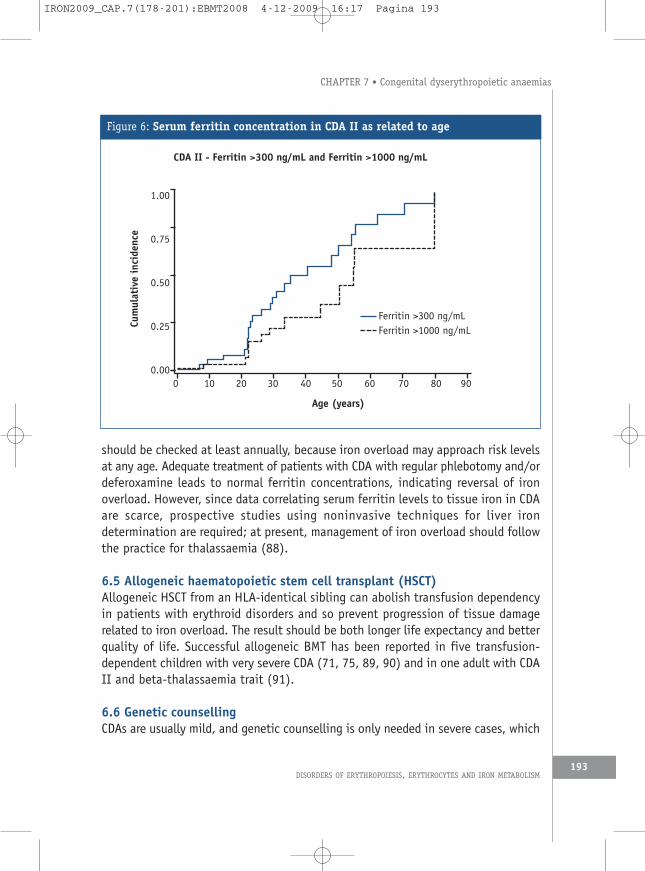
should be checked at least annually, because iron overload may approach risk levelsat any age. Adequate treatment of patients with CDA with regular phlebotomy and/ordeferoxamine leads to normal ferritin concentrations, indicating reversal of ironoverload. However, since data correlating serum ferritin levels to tissue iron in CDAare scarce, prospective studies using noninvasive techniques for liver irondetermination are required; at present, management of iron overload should followthe practice for thalassaemia (88).
6.5 Allogeneic haematopoietic stem cell transplant (HSCT)Allogeneic HSCT from an HLA-identical sibling can abolish transfusion dependencyin patients with erythroid disorders and so prevent progression of tissue damagerelated to iron overload. The result should be both longer life expectancy and betterquality of life. Successful allogeneic BMT has been reported in five transfusion-dependent children with very severe CDA (71, 75, 89, 90) and in one adult with CDAII and beta-thalassaemia trait (91).
6.6 Genetic counsellingCDAs are usually mild, and genetic counselling is only needed in severe cases, which
DISORDERS OF ERYTHROPOIESIS, ERYTHROCYTES AND IRON METABOLISM
CHAPTER 7 • Congenital dyserythropoietic anaemias
193
Figure 6: Serum ferritin concentration in CDA II as related to age
CDA II - Ferritin >300 ng/mL and Ferritin >1000 ng/mL
0 10 20 30 40 50 60 70 80 90
Age (years)
1.00
0.75
0.50
0.25
0.00
Cumulativeincide
nce
Ferritin >300 ng/mLFerritin >1000 ng/mL
IRON2009_CAP.7(178-201):EBMT2008 4-12-2009 16:17 Pagina 193
are the least understood and may depend on the presence of coexistent abnormalities.Counselling has to be based on the pattern of inheritance, since there are no evidence-based methods for early antenatal diagnosis.
7. Summary and conclusionsThe CDAs are a heterogeneous group of hereditary disorders, both at clinical andgenetic levels. Mapping and cloning have shown that these conditions havedifferent molecular mechanisms that induce disturbances of cell maturation and celldivision during erythropoiesis. In general the features of dyserythropoiesis, interms of ineffective erythropoiesis, should be demonstrated by a number of differentcriteria: bone marrow evaluation by light microscopy, ultrastructural features,assessment of red cell production and destruction, and studies of iron metabolism.Certainly the main assessment method, and one which is easy to perform, is lightmicroscopy. Biochemical (such as in CDA II) or molecular methods (such as in CDAI) are required for exact classification. Effective modalities of therapy includegeneral measures, such as iron depletion, and specific measures such as IFN-α inCDA I and splenectomy in CDA II. In the future, microarray and proteomic studiesmay prove useful in defining further genes involved in these conditions andpossibly new drugs will become available.
References1. Crookston JH, Godwin TF, Wightmann KJR et al.Congenital dyserythropoietic anemia. Abstr
XIth Congress Internat Soc Hematol, Sydney 1966.2. Wendt F, Heimpel H. Kongenitale dyserythropoetische Anämie bei einem zweieiigen
Zwillingspaar. Med Klinik 1967; 62: 172-177.3. Heimpel H. Congenital dyserythropoietic anemias: Epidemiology, clinical significance and
progress in understanding their pathogenesis. Ann Hematol 2004; 83: 613-621.4. Delaunay J, Iolascon A. The congenital dyserythropoietic anaemias. Baillieres Best
Pract Res Clin Haematol 1999 12: 691-705.5. Wickramasinghe SN. Congenital dyserythropoietic anaemias: Clinical features,
haematological morphology and new biochemical data. Blood Rev 1998; 12: 178-200.6. Heimpel H, Wendt F. Congenital dyserythropoietic anemia with karyorrhexis and
multinuclearity of erythroblasts. Helv Med Acta 1968; 34: 103-115.7. Wolff JA, Von Hofe H. Familial erythroid multinuclearity. Blood 1951; 6: 1274-1283.8. Bergström I, Jacobsson L. Hereditary benign erythroreticulosis. Blood 1962; 19: 296-
303.9. Boogaerts MA, Verwilghen RL. Variants of congenital dyserythropoietic anaemia: An
Update. Haematologia (Budap.)1982; 15: 211-219.10. Wickramasinghe SN, Wood WG. Advances in the understanding of the congenital
dyserythropoietic anaemias. Br J Haematol 2005; 131: 431-446.
THE HANDBOOK 2009 EDITION194
IRON2009_CAP.7(178-201):EBMT2008 4-12-2009 16:17 Pagina 194
11. Heimpel H, Wendt F, Klemm D et al. Kongenitale dyserythropoietische Anämie. Dtsch ArchKlin Med 1968; 215: 174-194.
12. Faille A, Najean Y, Dresch C. Cinétique de l’érythropoièse dans 14 cas “d’érythropoièseinefficace” avec anomalies morphologiques des érythroblastes et polynucléarité. NouvRev Fr Hematol 1972; 12: 631-652.
13. Barosi G, Cazzola M, Stefannelli M, Ascari E. Studies of ineffective erythropoiesis andperipheral haemolysis in congenital dyserythropoietic anaemia type II. Br J Haematol1979; 43: 243-250.
14. Heimpel H, Anselstetter V, Chrobak L et al. Congenital dyserythropoietic anemia typeII: Epidemiology, clinical appearance, and prognosis based on long-term observation.Blood 2003; 102: 4576-4581.
15. Cazzola M, Beguin Y, Bergamaschi G et al. Soluble transferrin receptor as a potentialdeterminant of iron loading in congenital anaemias due to ineffective erythropoiesis.Br J Haematol 1999; 106: 752-755.
16. Soysal T, Akun E, Ozaras R et al. Congenital dyserythropoietic anemia type I withringed sideroblasts. Haematologia (Budap.) 2000; 30: 45-49.
17. Heimpel H. Dyserythropoiese und dyserythropoietische Anämien. Schweiz Med Wochenschr1975; 105: 1562-1568.
18. Iolascon A, D’Agostaro G, Perrotta S et al. Congenital dyserythropoietic anemia type II:Molecular basis and clinical aspects. Haematologica 1996; 81: 543-559.
19. Cazzola M, Barosi G, Bergamaschi G et al. Iron loading in congenital dyserythropoieticanaemias and congenital sideroblastic anaemias. Br J Haematol 1983; 54: 649-654.
20. Tamary H, Shalmon L, Shalev H et al. Lokalisation of the gene for congenitaldyserythropoietic anemia type I to chromosome 15q15.1.3 [abstract]. Blood 1996; 88:144.
21. Shalev H, Kapelushnik J, Moser A et al. A comprehensive study of the neonatalmanifestations of congenital dyserythropoietic anemia type I. J Pediatr Hematol Oncol2004; 26: 746-748.
22. Heimpel H, Schwarz K, Ebnöther M et al. Congenital dyserythropoietic anemia type I (CDAI): Molecular genetics, clinical appearance and prognosis based on long-term observation.Blood 2006; 107: 334-340.
23. Parez N, Dommergues M, Zupan V et al. Severe congenital dyserythropoietic anaemia typeI: Prenatal management, transfusion support and alpha-interferon therapy. Br J Haematol2000; 110: 420-423.
24. Madero L, Munoz A, Fernandez Fuertes I et al. Anemia diseritropoyetica tipo I depresentaciòn en el periodo neonatal. Sangre Barc 1987; 32: 495-501.
25. Bader-Meunier B, Leverger G, Tchernia G et al. Clinical and Laboratory Manifestationsof Congenital Dyserythropoietic Anemia Type I in a Cohort of French Children. J PediatrHematol Oncol 2005; 27: 416-419.
26. Heimpel H, Forteza-Vila J, Queisser W, Spiertz E. Electron and light microscopic studyof the erythropoiesis of patients with congenital dyserythropoietic anemia. Blood 1971;37: 299-310.
27. Breton-Gorius J, Daniel M, Clauvel JP, Dreyfus B. Anomalies ultrastructurales des
DISORDERS OF ERYTHROPOIESIS, ERYTHROCYTES AND IRON METABOLISM
CHAPTER 7 • Congenital dyserythropoietic anaemias
195
IRON2009_CAP.7(178-201):EBMT2008 4-12-2009 16:17 Pagina 195
érythroblastes et des érythrocytes dans six cas de dysérythropoièse congénitale. NouvRev Fr Hematol 1973; 13: 23-50.
28. Wickramasinghe SN. Congenital dyerythropoietic anemia (CDA) type I: Survey of casesin the UK and response to alpha interferon. Int J Hematol 1996; 64 (suppl 1): S22.
29. Wickramasinghe SN, Pippard MJ. Studies of erythroblast function in congenitaldyserythropoietic anaemia, type I: Evidence of impaired DNA, RNA, and protein synthesisand unbalanced globin chain synthesis in ultrastructurally abnormal cells. J Clin Pathol1986; 39: 881-890.
30. Heimpel H. Congenital dyserythropoietic anemia type I: Clinical and experimentalaspects. In: Porter R, Fitzsimons DW, eds. Congenital disorders of erythropoiesis. CibaFoundation Symposium 37 (new series). Amsterdam: Elsevier 1976: 135-149.
31. Shalev H, Kapleushnik Y, Haeskelzon L et al. Clinical and laboratory manifestations ofcongenital dyserythropoietic anemia type I in young adults. Eur J Haematol 2002; 68:170-174.
32. Sabry MA, Zaki M, al Awadi SA et al. Non-haematological traits associated withcongenital dyserythropoietic anaemia type 1: A new entity emerging. Clin Dysmorphol1997; 6: 205-212.
33. Holmberg L, Jansson L, Rausing A, Henriksson P. Type I congenital dyserythropoieticanaemia with myelopoietic abnormalities and hand malformations. Scand J Haematol1978; 21: 72-79.
34. Gasser C. Congenital-dyserythropoietic-malformation syndrome [abstract]. 6th meetingof the International Society of Hematology, Athens, 1981.
35. Goede JS, Benz R, Fehr J et al. Congenital dyserythropoietic anemia type I with boneabnormalities, mutations of the CDAN I gene, and significant responsiveness to alpha-interferon therapy. Ann Hematol 2006; 85: 591-595.
36. Le Merrer M, Girot R, Parent P et al. Acral dysostosis dyserythropoiesis syndrome. Eur JPediatr 1995; 154: 384-388.
37. Facon T, Mannessier L, Lepelly P et al. Congenital dyserythropoetic anemia type I. Reporton monocygotic twins with associated hemochromatosis and short stature. Blut 1990;61: 248-251.
38. Queisser W, Spiertz E, Jost E, Heimpel H. Proliferation disturbances of erythroblasts incongenital dyserythropoietic anemia type I and II. Acta Haematol 1971; 45: 65-76.
39. Tamary H, Shalev H, Luria D et al. Clinical features and studies of erythropoiesis in IsraeliBedouins with congenital dyserythropoietic anemia type I. Blood 1996; 87: 1763-1770.
40. Noy-Lotan S, Dgany O, Lahmi R et al. Codanin-1, the protein encoded by the gene mutatedin congenital dyserythropoietic anemia type I (CDAN1), is cell cycle regulated.Haematologica 2009; in print.
41. Tamary H, Shalmon L, Shalev H et al. Localization of the gene for congenitaldyserythropoietic anemia type I to a <1-cM interval on chromosome 15q15.1-15.3. AmJ Hum Genet 1998; 62: 1062-1069.
42. Hodges VM, Molloy GY, Wickramasinghe SN. Genetic heterogeneity of congenital
THE HANDBOOK 2009 EDITION196
IRON2009_CAP.7(178-201):EBMT2008 4-12-2009 16:17 Pagina 196
dyserythropoietic anemia type I [letter]. Blood 1999; 94: 1139-1140.43. Dgany O, Avidan N, Delaunay J et al. Congenital dyserythropoietic anemia type I is caused
by mutations in codanin-1. Am J Hum Genet 2002; 71: 1467-1474.44. Ahmed MR, Chehal A, Zahed L et al. Linkage and mutational analysis of the CDAN1 gene
reveals genetic heterogeneity in congenital dyserythropoietic anemia type I. Blood 2006;107: 4968-4969.
45. Ru YX, Zhu XF, Yan WW et al. Congenital dyserythropoietic anemia in a Chinese familywith a mutation of the CDAN1-gene. Ann Hematol 2008; 87: 751-754.
46. Wickramasinghe SN, Thein SL, Srichairatanakool S, Porter JB. Determinants of ironstatus and bilirubin levels in congenital dyserythropoietic anaemia type I. Br J Haematol1999; 107: 522-525.
47. Smithson WA, Perrault J. Use of subcutaneous deferoxamine in a child withhemochromatosis associated with congenital dyserythropoietic anemia, type I. Mayo Clinproc 1982; 57: 322-325.
48. Iolascon A, Servedio V, Carbone R et al. Geographic distribution of CDA-II: Did afounder effect operate in Southern Italy? Haematologica 2000; 85: 470-474.
49. Iolascon A, Delaunay J, Wickramasinghe SN et al. Natural history of congenitaldyserythropoietic anemia type II. Blood 2001; 98: 1258-1260.
50. Danise P, Amendola G, Nobili B et al. Flow-cytometric analysis of erythrocytes andreticulocytes in congenital dyserythropoietic anaemia type II (CDA II): Value indifferential diagnosis with hereditary spherocytosis. Clin Lab Haematol 2001; 23: 7-13.
51. Alloisio N, Texier P, Denoroy L et al. The cisternae decorating the red blood cellmembrane in congenital dyserythropoietic anemia (type II) originate from the endoplasmicreticulum. Blood 1996; 87: 4433-4439.
52. Crookston JH, Crookston MC, Burnie KL et al. Hereditary erythroblastic multinuclearityassociated with a positive acidified-serum test: A type of congenital dyserythropoieticanemia. Br J Haematol 1969; 17: 11-26.
53. Anselstetter V, Horstmann K, Heimpel H. Congenital dyserythropoetic anaemia, types Iand II: Aberrant pattern of erythrocyte membrane proteins in CDA II, as revealed by two-dimensional polyacrylamide gel electrophoresis. Br J Haematol 1977; 35: 209-215.
54. Denecke J, Kranz C, Nimtz M et al. Characterization of the N-glycosylation phenotypeof erythrocyte membrane proteins in congenital dyserythropoietic anemia type II (CDAII/HEMPAS). Glycoconj J 2008; 25: 375-382.
55. Gangarossa S, Romano V, Miraglia del Giudice E et al. Congenital dyserythropoieticanemia type II associated with G6PD Seattle in a Sicilian child. Acta Haematol 1995; 93:36-39.
56. De Franceschi L, Turrini F, del Giudice EM et al. Decreased band 3 anion transport activityand band 3 clusterization in congenital dyserythropoietic anemia type II. Exp Haematol1998; 26: 869-873.
57. Iolascon A, Miraglia del Giudice E, Perrotta S et al. Exclusion of three candidate genesas determinants of congenital dyserythropoietic anemia type II (CDA-II). Blood 1997;90: 4197-4200.
DISORDERS OF ERYTHROPOIESIS, ERYTHROCYTES AND IRON METABOLISM
CHAPTER 7 • Congenital dyserythropoietic anaemias
197
IRON2009_CAP.7(178-201):EBMT2008 4-12-2009 16:17 Pagina 197
58. Gasparini P, Miraglia del Giudice E, Delaunay J et al. Localization of the congenitaldyserythropoietic anemia II locus to chromosome 20q11.2 by genome wide search. AmJ Hum Genet 1997; 61: 1112-1116.
59. Perrotta S, del Giudice EM, Carbone R et al. Gilbert’s syndrome accounts for thephenotypic variability of congenital dyserythropoietic anemia type II (CDA-II). J Pediatr2000; 136: 556-559.
60. Hovinga JA, Solenthaler M, Dufour JF. Congenital dyserythropoietic anaemia type II(HEMPAS) and haemochromatosis: A report of two cases. Eur J Gastroenterol Hepatol2003; 15: 1141-1147.
61. Sandstroem H, Wahlin A. Congenital dyserythropoietic anemia type III. Haematologica2000; 85: 753-757.
62. Sandstroem H, Wahlin A, Eriksson M et al. Intravascular haemolysis and increasedprevalence of myeloma and monoclonal gammopathy in congenital dyserythropoieticanaemia, type III. Eur J Haematol 1994; 52: 42-46.
63. Lind L, Sandstroem H, Wahlin A et al. Localization of the gene for congenitaldyserythropoietic anemia type III, CDAN3, to chromosome 15q21-q25. Hum Mol Genet1995; 4: 109-112.
64. Byrnes RK, Dhru R, Brady AM et al. Congenital dyserythropoietic anemia in treatedHodgkin’s disease. Hum Pathol 1980; 11: 485-486.
65. McCluggage WG, Hull D, Mayne E et al. Malignant-Lymphoma in CongenitalDyserythropoietic Anemia Type-III. J Clin Pathol 1996; 49: 599-602.
66. David G, VanDorpe A. Aberrant congenital dyserythropoietic anemias. In: Lewis SM,Verwilghen RL, editors. Dyserythropoiesis. London: Academic Press; 1977: 92-102.
67. McBride JA, Wilson WE, Baillie N. Congenital dyserythropoietic anaemia - type IV(Abstr.). Blood 1971; 38: 837.
68. Benjamin JT, Rosse WF, Dalldorf FG, Mc Millan CW. Congenital dyserythropoetic anemia- type IV. J Pediatr 1975; 87: 210-216.
69. Eldor A, Matzner Y, Kahane I et al. Aberrant congenital dyserythropoietic anemia withnegative acidified serum tests and features of thalassemia in a Kurdish family. Isr J MedSci 1978; 14: 1138-1143.
70. Carter C, Darbyshire PJ, Wickramasinghe SN. A congenital dyserythropoietic anaemia variantpresenting as hydrops foetalis. Br J Haematol 1989; 72: 289-290.
71. Remacha AF, Badell I, Pujol-Moix N et al. Hydrops fetalis-associated congenitaldyserythropoietic anemia treated with intrauterine transfusions and bone marrowtransplantation. Blood 2002; 100: 356-358.
72. Bethlenfalvay NC, Hadnagy GS, Heimpel H. Unclassified type of congenitaldyserythropoietic anemia (CDA) with prominent peripheral erythoblastosis. Br J Haematol1985; 60: 541-550.
73. Bird AR, Karabus CD, Hartley PS. Type IV congenital dyserythropoietic anemia with unusualresponse to splenectomy. Am J Pediatr Hematol Oncol 1985; 7: 196-199.
74. Adams CD, Kessler JF. Circulating nucleated red blood cells following splenectomy in apatient with congenital dyserythropoietic anemia. Am J Hematol 1991; 38: 120-123.
THE HANDBOOK 2009 EDITION198
IRON2009_CAP.7(178-201):EBMT2008 4-12-2009 16:17 Pagina 198
75. Shukry-Schulz S, Lawitschka A, Matthes S et al. Congenital dyserythropoietic anemia -can peripheral stem cell transplantation be a therapeutic appoach? Report of two cases[abstract]. Hematol J 2000; 1: 44.
76. Koenig E, Osieka R, Brittinger G. Atypische kongenitale dyserythropoietische Anämie mitThrombozytopenie. Verh Dtsch Ges Inn Med 1973; 79: 490.
77. Nichols KE, Crispino JD, Poncz M et al. Familial dyserythropoietic anaemia andthrombocytopenia due to an inherited mutation in GATA1. Nat Genet 2000; 24: 266-270.
78. Kratz CP, Niemeyer CM, Karow A et al. Congenital transfusion-dependent anemia andthrombocytopenia with myelodysplasia due to a recurrent GATA1(G208R) germlinemutation. Leukemia 2008; 22: 432-434.
79. Tamary H, Shalev H, Pinsk V et al. No response to recombinant human erythropoietintherapy in patients with congenital dyserythropoietic anemia type I. Pediatr HematolOncol 1999; 16: 165-168.
80. Lavabre Bertrand T, Blanc P, Navarro R et al. Alpha-Interferon therapy for congenitaldyserythropoiesis type I. Br J Haematol 1995; 89: 929-932.
81. Lavabre-Bertrand T, Ramos J, Delfour C et al. Long-term alpha-interferon treatment iseffective on anaemia and significantly reduces iron overload in congenitaldyserythropoiesis type I. Eur J Haematol 2004; 73: 380-383.
82. Marwaha RK, Bansal D, Trehan A, Garewal G. Interferon therapy in congenitaldyserythropoietic anemia type I/II. Pediatr Hematol Oncol 2005; 22: 133-138.
83. Wickramasinghe SN. Response of CDA type I to alpha-interferon [letter]. Eur J Haematol1997; 58: 121-123.
84. Iolascon A, Volinia S, Borriello A et al. Genes transcriptionally modulated by interferonalpha2a correlate with the cytokine activity. Haematologica 2004; 89: 1046-1053.
85. Chrobak L, Spacek J. [Favorable effect of splenectomy on anemia in 3 siblings with typeII congenital dyserythropoietic anemia (HEMPAS). Ultrastructural changes in erythrocytesafter splenectomy]. Vnitr Lek 1997; 43: 635-638.
86. Marchetti M, Quaglini S, Barosi G. Prophylactic splenectomy and cholecystectomy in mildhereditary spherocytosis: Analyzing the decision in different clinical scenarios. J InternMed 1998; 244: 217-226.
87. Williams EJ, Green J, Beckingham I et al. Guidelines on the management of commonbile duct stones (CBDS). Gut 2008; 57: 1004-1021.
88. Taher A, Isma’eel H, Cappellini MD. Thalassemia intermedia: Revisited. Blood Cells MolDis 2006; 37: 12-20.
89. Ariffin WA, Karnaneedi S, Choo KE, Normah J. Congenital dyserythropoietic anaemia: Reportof three cases. J Paediatr Child Health 1996; 32: 191.
90. Ayas M, al Jefri A, Baothman A et al. Transfusion-dependent congenital dyserythropoieticanemia type I successfully treated with allogeneic stem cell transplantation. BoneMarrow Transplant 2002; 29: 681-682.
91. Iolascon A, Sabato V, De Mattia D, Locatelli F. Bone marrow transplantation in a caseof severe, type II congenital dyserythropoietic anaemia (CDA II). Bone Marrow Transplant2001; 27: 213-215.
DISORDERS OF ERYTHROPOIESIS, ERYTHROCYTES AND IRON METABOLISM
CHAPTER 7 • Congenital dyserythropoietic anaemias
199
IRON2009_CAP.7(178-201):EBMT2008 4-12-2009 16:17 Pagina 199
Multiple Choice Questionnaire
To find the correct answer, go to http://www.esh.org/iron-handbook2009answers.htm
1. Which one of the following criteria is not compatiblewith the diagnosis of CDA:a) Evidence of congenital anaemia/jaundice or a positive
family history . . . . . . . . . . . . . . . . . . . . . . . . . . . . . . . . . . . . . . . . . . . . . . . . . . . . . . . . . . . . . . . . . .
b) Evidence of ineffective erythropoiesis . . . . . . . . . . . . . . . . . . . . . . . . . . . . . . . . . . . . . . .
c) Typical morphological appearance of bone marrow erythroblasts . . . . . . . . . .
d) MCV reduced to less than 70 fl . . . . . . . . . . . . . . . . . . . . . . . . . . . . . . . . . . . . . . . . . . . . . . .
2. Which one of the following findings does not suggestthe diagnosis of CDA type I:a) Peripheral blood: anisocytosis and poikilocytosis . . . . . . . . . . . . . . . . . . . . . . . . . .
b) Peripheral blood: basophilic stippled cells . . . . . . . . . . . . . . . . . . . . . . . . . . . . . . . . . .
c) Electron microscopy: presence of double membranein mature erythroblasts . . . . . . . . . . . . . . . . . . . . . . . . . . . . . . . . . . . . . . . . . . . . . . . . . . . . . . .
d) Electron microscopy: chromatin bridges between erythroblasts . . . . . . . . . . .
3. Which is the causative gene for CDA I?a) Codanin . . . . . . . . . . . . . . . . . . . . . . . . . . . . . . . . . . . . . . . . . . . . . . . . . . . . . . . . . . . . . . . . . . . . . . . . .
b) BCRA1 . . . . . . . . . . . . . . . . . . . . . . . . . . . . . . . . . . . . . . . . . . . . . . . . . . . . . . . . . . . . . . . . . . . . . . . . . . .
c) wt1 . . . . . . . . . . . . . . . . . . . . . . . . . . . . . . . . . . . . . . . . . . . . . . . . . . . . . . . . . . . . . . . . . . . . . . . . . . . . . .
d) Spectrin alpha . . . . . . . . . . . . . . . . . . . . . . . . . . . . . . . . . . . . . . . . . . . . . . . . . . . . . . . . . . . . . . . . .
4. Which of the following tests is not useful for the diagnosisof CDA II?a) SDS-PAGE of red cell membrane proteins . . . . . . . . . . . . . . . . . . . . . . . . . . . . . . . . . . . .
b) Western-blot for RE proteins . . . . . . . . . . . . . . . . . . . . . . . . . . . . . . . . . . . . . . . . . . . . . . . . . .
c) Increased anti-i agglutinability . . . . . . . . . . . . . . . . . . . . . . . . . . . . . . . . . . . . . . . . . . . . . .
d) Low iron saturation of serum transferrin . . . . . . . . . . . . . . . . . . . . . . . . . . . . . . . . . . . .
5. Please identify the common complication(s) of CDA II:
THE HANDBOOK 2009 EDITION200
IRON2009_CAP.7(178-201):EBMT2008 4-12-2009 16:17 Pagina 200
a) Iron overload, gallstones . . . . . . . . . . . . . . . . . . . . . . . . . . . . . . . . . . . . . . . . . . . . . . . . . . . . .
b) Splenomegaly and renal failure . . . . . . . . . . . . . . . . . . . . . . . . . . . . . . . . . . . . . . . . . . . . . . .
c) Thrombocytopenia and splenomegaly . . . . . . . . . . . . . . . . . . . . . . . . . . . . . . . . . . . . . . . .
d) Symptomatic osteoporosis . . . . . . . . . . . . . . . . . . . . . . . . . . . . . . . . . . . . . . . . . . . . . . . . . . . .
DISORDERS OF ERYTHROPOIESIS, ERYTHROCYTES AND IRON METABOLISM
CHAPTER 7 • Congenital dyserythropoietic anaemias
201
IRON2009_CAP.7(178-201):EBMT2008 4-12-2009 16:17 Pagina 201