Embed Size (px)
Citation preview

Anesth Pain Med. 2016 October; 6(5):e36607.
Published online 2016 May 30.
doi: 10.5812/aapm.36607.
Research Article
Comparison of Spinal Needle Deflection in a Ballistic Gel Model
Ethan Rand,1 George Christolias,2 Christopher Visco,2 and Jaspal R. Singh3,*
1Department of Rehabilitation and Regenerative Medicine, Presbyterian Hospital, New York, The United States2Department of Rehabilitation and Regenerative Medicine, Columbia University Medical Center, Presbyterian Hospital, New York, The United States3Department of Rehabilitation and Regenerative Medicine, Weill Cornell Medicine Spine Center, Presbyterian Hospital, New York, The United States
*Corresponding author: Jaspal R. Singh, Department of Rehabilitation and Regenerative Medicine, Weill Cornell Medicine Spine Center, Presbyterian Hospital, 525 East 68thStreet, Baker 16, New York, The United States. Tel: +01-2127461500, Fax: +01-2127468303, E-mail: [email protected]
Received 2016 January 29; Revised 2016 February 27; Accepted 2016 April 08.
Abstract
Background: Percutaneous diagnostic and therapeutic procedures are commonly used in the treatment of spinal pain. The successof these procedures depends on the accuracy of needle placement, which is influenced by needle size and shape.Objectives: The purpose of this study is to examine and quantify the deviation of commonly used spinal needles based on needletip design and gauge, using a ballistic gel tissue simulant.Materials and Methods: Six needles commonly used in spinal procedures (Quincke, Short Bevel, Chiba, Tuohy, Hustead, Whitacre)were selected for use in this study. Ballistic gel samples were made in molds of two depths, 40mm and 80 mm. Each needle wasmounted in a drill press to ensure an accurate needle trajectory. Distance of deflection was recorded for each needle.Results: In comparing the mean deflection of 22 gauge needles of all types at 80 mm of depth, deflection was greatest amongbeveled needles [Short Bevel (9.96±0.77 mm), Quincke (8.89±0.17 mm), Chiba (7.71± 1.16 mm)], moderate among epidural needles[Tuohy (7.64 ± 0.16 mm) and least among the pencil-point needles [Whitacre (0.73 ± 0.34 mm)]. Increased gauge (25 g) led to asignificant increase in deflection among beveled needles. The direction of deflection was away from the bevel with Quincke, Chibaand Short Beveled needles and toward the bevel of the Tuohy and Hustead needles. Deflection of the Whitacre pencil-point needlewas minimal.Conclusions: There is clinical utility in knowing the relative deflection of various needle tips. When a procedure requires a needleto be steered around obstacles, or along non-collinear targets, the predictable and large amount of deflection obtained through useof a beveled spinal needle may prove beneficial.
Keywords: Spinal Needle, Needle Deflection, Back Pain, Ballistic Gel, Interventional Pain
1. Background
Percutaneous diagnostic and therapeutic proceduresare commonly used in the treatment of axial and radicularspinal pain. The success of these procedures depends onthe accuracy of needle placement. An assortment of nee-dle types has been developed with a variety of tip shapes.The disparate needle designs play an integral role in deter-mining the extent of needle deflection, accuracy of needletrajectory and, ultimately, in placement of the injectate atthe intended target. Needle deflection can be controlledand utilized to correct for needle misalignment or to ac-cess non-collinear targets, and is commonly known as nee-dle steering. Further understanding of the precise degreeof deflection of each needle type has the potential to aidclinicians in improving needle steering and target accu-racy by selecting the appropriate needle for their needs.
Based on limited studies of needle deflection, beveledspinal needles (e.g. Quincke) tend to deflect away from thebeveled surface and notch located on the hub and the de-
gree of deflection is related to gauge and depth of insertion(1, 2). Pencil-point needles (e.g. Whitacre) generally mini-mally deflect in the direction of the distal injection orifice(1). Epidural needles (e.g. Tuohy) tend to deflect toward thenotch on the hub and beveled surface, in the direction ofthe curved tip (3). Thus far, studies have used porcine (1,2, 4), polystyrene foam (5), and Plastisol (polyvinylchloridesuspension) (6, 7) tissue simulants.
Ballistic gel, composed of collagen derived from ani-mal tissue, has been well established as a tissue simulant,as it has a similar density and viscosity to human mus-cle tissue (8). While having similar physical properties tohuman muscle tissue, ballistic gel does not contain theheterogeneous properties of muscle (e.g. fascial planes),which can further impact needle deflection (9). The ra-tionale behind using polystyrene foam is not discussedby Ahn et al. (2002) (5). Although polystyrene has beenused as an ingredient in other tissue simulants, to ourknowledge polystyrene foam has not been validated for
Copyright © 2016, Iranian Society of Regional Anesthesia and Pain Medicine (ISRAPM). This is an open-access article distributed under the terms of the Creative CommonsAttribution-NonCommercial 4.0 International License (http://creativecommons.org/licenses/by-nc/4.0/) which permits copy and redistribute the material just innoncommercial usages, provided the original work is properly cited.

Rand E et al.
this purpose. Additionally, Plastisol is produced by addinga softener to plastic, creating a polyvinylchloride suspen-sion. The ingredients necessary to produce Plastisol areless accessible and the process is more complex when com-pared to ballistic gel (10). This study is the first experimentknown to the authors of needle deflection using a ballisticgel tissue simulant.
2. Objectives
The purpose of this study is to further examine andquantify the deviation of commonly used spinal needlesbased on needle tip design and gauge, using a ballistic geltissue simulant.
3. Materials and Methods
Six needles commonly used in spinal procedures wereselected. Three beveled spinal needles (Quincke, ShortBevel, Chiba), two epidural needles (Tuohy, Hustead), andone pencil-point needle (Whitacre) were used in this study(Becton, Dickinson and Company, Franklin Lakes, NJ). 25-gauge (Quincke, Short Bevel, Whitacre), 22-gauge (Quincke,Short Bevel, Tuohy, Whitacre, Chiba), and 18-gauge (Hus-tead) needles were tested.
3.1. Gel Preparation
Ballistic gel samples were made from an 11% by mass250 bloom Knox gel (Kraft Foods Group Inc, Northfield, IL).The mixture was made with eight ounces of gel mix and 64ounces of 40° C filtered water stirred slowly to dissolve allthe particles and to minimize the formation of air bubbles.This yielded a uniform consistency of all gel samples thatwere then poured into cylindrical aluminum molds. A to-tal of 20 gel molds were made (10 with 80 mm depth and10 with 40 mm depth) from a single batch of gel mixture.It was vital that the solution be poured into the molds ina very slow and deliberate manner to avoid frothing of thesolution and air bubbles clinging to the mold. Ballistic gelrequires a storage temperature of approximately 10° C. Theballistic gel specimens were made one day prior to the ex-periment trials. This was done because the physical prop-erties of ballistic gel tend to change within 48 hours of cur-ing, as the gel begins to dehydrate and harden.
Ballistic gel samples were made in aluminum molds oftwo depths, 40 mm and 80 mm respectively. The 40 mmmold had a volume of 82 milliliters and a radius of 26.8mm, while the 80 mm mold was composed of a 164 mL vol-ume gel. All molds were made from the same gel solution.
3.2. Drill Press Mount
The drill press (also known as a pedestal or bench drill)was used in order to ensure accurate and uniform needletrajectory into the ballistic gel sample. The set up com-prised of a base, column (pillar) and spindle with an up-right and a movable grip controllable by a handle. Handlesradiating from a central hub moved the spindle verticallyparallel to the axis of the column. The drill press used wasRYOBI Drill (One World Technologies, Inc., Anderson, SC)with five-inch excursion of the spindle (Figure 1). The an-gle of the spindle was fixed relative to the table allowingconsistent and accurate trajectory.
Figure 1. RYOBI Drill with Five-Inch Excursion of the Spindle
3.3. Needle Placement
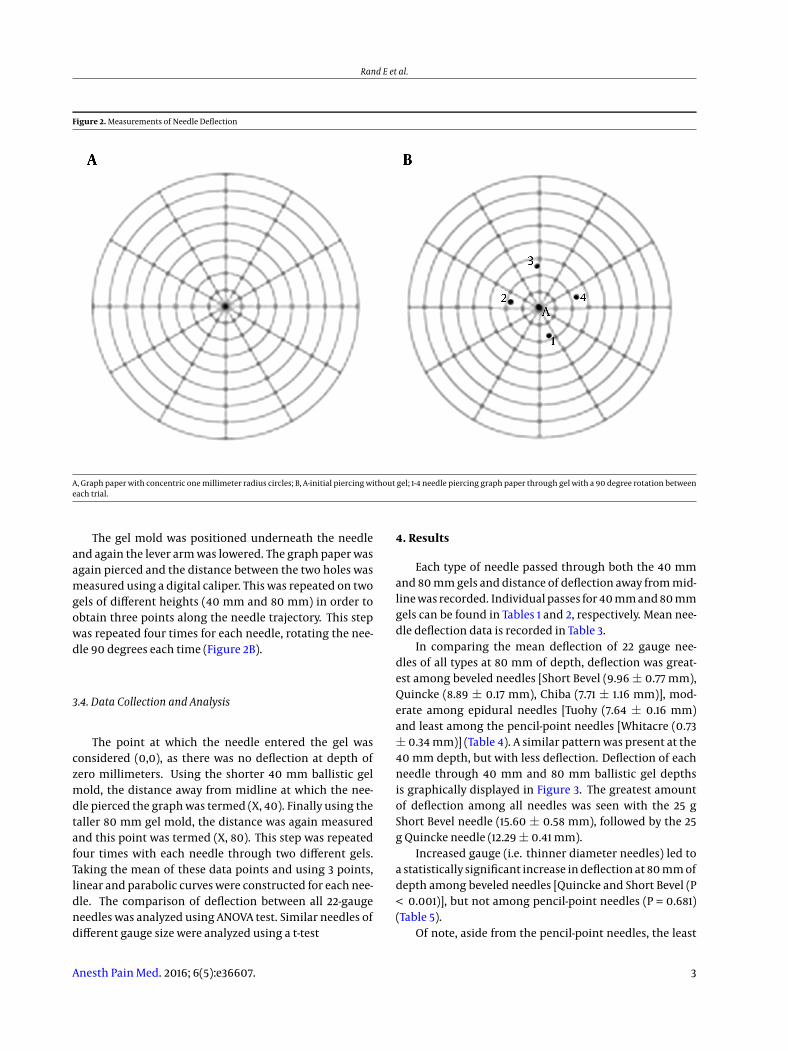
Each needle was mounted in a drill press in order to en-sure accurate needle trajectory into the ballistic gel sam-ple. A level was used to align the needle vertically to the geland after each needle was positioned, a bubble level wasused to ensure accurate vertical positioning of the needle.The needle was secured to the grip and driven down to thecenter of the graph paper, piercing it (Figure 2A).
2 Anesth Pain Med. 2016; 6(5):e36607.
Rand E et al.
Figure 2. Measurements of Needle Deflection
A, Graph paper with concentric one millimeter radius circles; B, A-initial piercing without gel; 1-4 needle piercing graph paper through gel with a 90 degree rotation betweeneach trial.
The gel mold was positioned underneath the needleand again the lever arm was lowered. The graph paper wasagain pierced and the distance between the two holes wasmeasured using a digital caliper. This was repeated on twogels of different heights (40 mm and 80 mm) in order toobtain three points along the needle trajectory. This stepwas repeated four times for each needle, rotating the nee-dle 90 degrees each time (Figure 2B).
3.4. Data Collection and Analysis
The point at which the needle entered the gel wasconsidered (0,0), as there was no deflection at depth ofzero millimeters. Using the shorter 40 mm ballistic gelmold, the distance away from midline at which the nee-dle pierced the graph was termed (X, 40). Finally using thetaller 80 mm gel mold, the distance was again measuredand this point was termed (X, 80). This step was repeatedfour times with each needle through two different gels.Taking the mean of these data points and using 3 points,linear and parabolic curves were constructed for each nee-dle. The comparison of deflection between all 22-gaugeneedles was analyzed using ANOVA test. Similar needles ofdifferent gauge size were analyzed using a t-test
4. Results
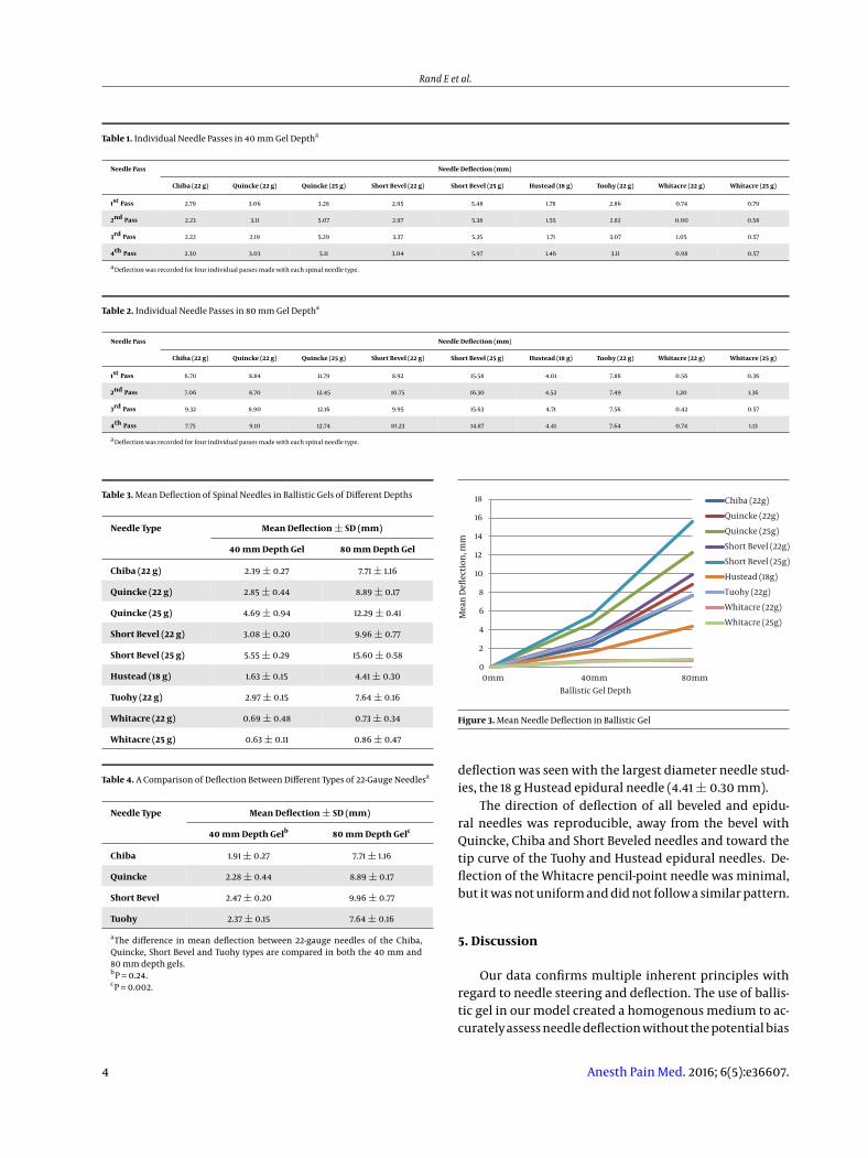
Each type of needle passed through both the 40 mmand 80 mm gels and distance of deflection away from mid-line was recorded. Individual passes for 40 mm and 80 mmgels can be found in Tables 1 and 2, respectively. Mean nee-dle deflection data is recorded in Table 3.
In comparing the mean deflection of 22 gauge nee-dles of all types at 80 mm of depth, deflection was great-est among beveled needles [Short Bevel (9.96 ± 0.77 mm),Quincke (8.89 ± 0.17 mm), Chiba (7.71 ± 1.16 mm)], mod-erate among epidural needles [Tuohy (7.64 ± 0.16 mm)and least among the pencil-point needles [Whitacre (0.73± 0.34 mm)] (Table 4). A similar pattern was present at the40 mm depth, but with less deflection. Deflection of eachneedle through 40 mm and 80 mm ballistic gel depthsis graphically displayed in Figure 3. The greatest amountof deflection among all needles was seen with the 25 gShort Bevel needle (15.60 ± 0.58 mm), followed by the 25g Quincke needle (12.29 ± 0.41 mm).
Increased gauge (i.e. thinner diameter needles) led toa statistically significant increase in deflection at 80 mm ofdepth among beveled needles [Quincke and Short Bevel (P< 0.001)], but not among pencil-point needles (P = 0.681)(Table 5).
Of note, aside from the pencil-point needles, the least
Anesth Pain Med. 2016; 6(5):e36607. 3
Rand E et al.
Table 1. Individual Needle Passes in 40 mm Gel Deptha
Needle Pass Needle Deflection (mm)
Chiba (22 g) Quincke (22 g) Quincke (25 g) Short Bevel (22 g) Short Bevel (25 g) Hustead (18 g) Tuohy (22 g) Whitacre (22 g) Whitacre (25 g)
1st Pass 2.79 3.06 3.28 2.95 5.48 1.78 2.86 0.74 0.79
2nd Pass 2.23 3.11 5.07 2.97 5.38 1.55 2.82 0.00 0.58
3rd Pass 2.22 2.19 5.29 3.37 5.35 1.71 3.07 1.05 0.57
4th Pass 2.30 3.03 5.11 3.04 5.97 1.46 3.11 0.98 0.57
a Deflection was recorded for four individual passes made with each spinal needle type.
Table 2. Individual Needle Passes in 80 mm Gel Deptha
Needle Pass Needle Deflection (mm)
Chiba (22 g) Quincke (22 g) Quincke (25 g) Short Bevel (22 g) Short Bevel (25 g) Hustead (18 g) Tuohy (22 g) Whitacre (22 g) Whitacre (25 g)
1st Pass 6.70 8.84 11.79 8.92 15.58 4.01 7.86 0.56 0.36
2nd Pass 7.06 8.70 12.45 10.75 16.30 4.52 7.49 1.20 1.36
3rd Pass 9.32 8.90 12.16 9.95 15.63 4.71 7.56 0.42 0.57
4th Pass 7.75 9.10 12.74 10.23 14.87 4.41 7.64 0.74 1.13
a Deflection was recorded for four individual passes made with each spinal needle type.
Table 3. Mean Deflection of Spinal Needles in Ballistic Gels of Different Depths
Needle Type Mean Deflection ± SD (mm)
40 mm Depth Gel 80 mm Depth Gel
Chiba (22 g) 2.39 ± 0.27 7.71 ± 1.16
Quincke (22 g) 2.85 ± 0.44 8.89 ± 0.17
Quincke (25 g) 4.69 ± 0.94 12.29 ± 0.41
Short Bevel (22 g) 3.08 ± 0.20 9.96 ± 0.77
Short Bevel (25 g) 5.55 ± 0.29 15.60 ± 0.58
Hustead (18 g) 1.63 ± 0.15 4.41 ± 0.30
Tuohy (22 g) 2.97 ± 0.15 7.64 ± 0.16
Whitacre (22 g) 0.69 ± 0.48 0.73 ± 0.34
Whitacre (25 g) 0.63 ± 0.11 0.86 ± 0.47
Table 4. A Comparison of Deflection Between Different Types of 22-Gauge Needlesa
Needle Type Mean Deflection ± SD (mm)
40 mm Depth Gelb 80 mm Depth Gelc
Chiba 1.91 ± 0.27 7.71 ± 1.16
Quincke 2.28 ± 0.44 8.89 ± 0.17
Short Bevel 2.47 ± 0.20 9.96 ± 0.77
Tuohy 2.37 ± 0.15 7.64 ± 0.16
aThe difference in mean deflection between 22-gauge needles of the Chiba,Quincke, Short Bevel and Tuohy types are compared in both the 40 mm and80 mm depth gels.bP = 0.24.cP = 0.002.
0
2
4
6
8
10
12
14
16
18
0mm 40mm 80mm
Ballistic Gel Depth
Chiba (22g)
Quincke (22g)
Quincke (25g)
Short Bevel (22g)
Short Bevel (25g)
Hustead (18g)
Tuohy (22g)
Whitacre (22g)
Whitacre (25g)
Mea
n D
eflec
tion
, mm
Figure 3. Mean Needle Deflection in Ballistic Gel
deflection was seen with the largest diameter needle stud-ies, the 18 g Hustead epidural needle (4.41 ± 0.30 mm).
The direction of deflection of all beveled and epidu-ral needles was reproducible, away from the bevel withQuincke, Chiba and Short Beveled needles and toward thetip curve of the Tuohy and Hustead epidural needles. De-flection of the Whitacre pencil-point needle was minimal,but it was not uniform and did not follow a similar pattern.
5. Discussion
Our data confirms multiple inherent principles withregard to needle steering and deflection. The use of ballis-tic gel in our model created a homogenous medium to ac-curately assess needle deflection without the potential bias
4 Anesth Pain Med. 2016; 6(5):e36607.

Rand E et al.
Table 5. A Comparison of Deflection Between Different Gauges of the Same Needle Typea
Needle Type Mean Deflection ± SD (mm)
40 mm Depth Gel P 80 mm Depth Gel P
Quincke 0.012 < 0.001
22 g 2.28 ± 0.44 8.89 ± 0.17
25 g 3.75 ± 0.94 12.29 ± 0.41
Short Bevel < 0.001 < 0.001
22 g 2.47 ± 0.20 9.96 ± 0.77
25 g 4.44 ± 0.29 15.60 ± 0.58
Whitacre 0.801 0.681
22 g 0.55 ± 0.48 0.73 ± 0.34
25 g 0.73 ± 0.34 0.86 ± 0.47
aThe difference in mean deflection between 22-gauge and 25-gauge needles of the Quincke, Short Bevel, and Whitacre types are compared in both the 40 mm and 80 mmdepth gels.
introduced by animal facial planes that may be present inporcine or bovine injection models. Collectively, beveledneedles (Quincke, Short Bevel, Chiba) behaved predictablyand consistently. Beveled needles deviate away from thenotch (and thus bevel) toward the longer end of the needle.Consistent with Newton’s third Law, is the generation of aforce by the gel medium perpendicular to the beveled sur-face of each needle. Force can be broken down into com-ponent vectors. As such, the gel medium produced a forcevector directed back along the axis of the needle, as well asanother force perpendicular to the beveled side of the nee-dle directing it toward its longer end, away from the bevelopening.
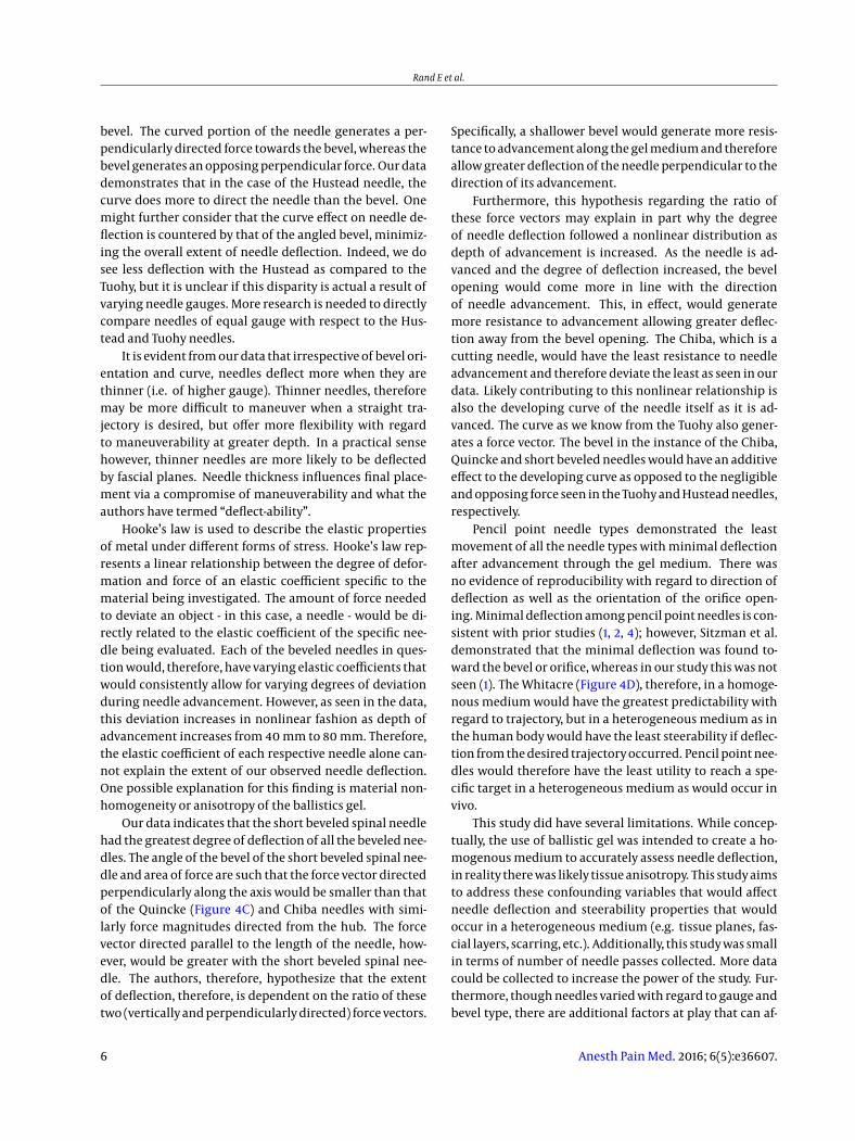
Tuohy needles in contrast behave differently whencompared to beveled needles (Figure 4A). Tuohy needleshave a curved side with the bevel opening perpendicularto the length of the needle to the side opposite the curve.Therefore, as the needle is advanced, there is little contri-bution from the bevel opening to generate a perpendicu-larly directed force vector. More so contributing to a per-pendicularly directed force vector is the curved portion ofthe needle. The force the gel medium imparts back ontothe curve as the needle is advanced generates componentvectors, with the perpendicularly directed vector compo-nent causing needle deviation away from the curve of theneedle or toward the bevel opening and notch on the hub.
The Hustead needle has some structural similarities tothe Tuohy in that there is a bevel side and curved side tothe needle (Figure 4B). It is structurally different in that thebeveled end is angled relative to the long axis of the nee-dle and therefore the curve does not extend as far as in theTuohy. Force vectors therefore are generated perpendicu-lar to the length of the needle by both the curve and the
Figure 4. Needle Types
A, Tuohy style needle; B, Hustead style needle; C, Quincke style needle; D, Whitacre-point style needle. Reprinted with permission from independent medical asso-ciates, 7301 124th Avenue, Largo, FL 33371.
Anesth Pain Med. 2016; 6(5):e36607. 5
Rand E et al.
bevel. The curved portion of the needle generates a per-pendicularly directed force towards the bevel, whereas thebevel generates an opposing perpendicular force. Our datademonstrates that in the case of the Hustead needle, thecurve does more to direct the needle than the bevel. Onemight further consider that the curve effect on needle de-flection is countered by that of the angled bevel, minimiz-ing the overall extent of needle deflection. Indeed, we dosee less deflection with the Hustead as compared to theTuohy, but it is unclear if this disparity is actual a result ofvarying needle gauges. More research is needed to directlycompare needles of equal gauge with respect to the Hus-tead and Tuohy needles.
It is evident from our data that irrespective of bevel ori-entation and curve, needles deflect more when they arethinner (i.e. of higher gauge). Thinner needles, thereforemay be more difficult to maneuver when a straight tra-jectory is desired, but offer more flexibility with regardto maneuverability at greater depth. In a practical sensehowever, thinner needles are more likely to be deflectedby fascial planes. Needle thickness influences final place-ment via a compromise of maneuverability and what theauthors have termed “deflect-ability”.
Hooke’s law is used to describe the elastic propertiesof metal under different forms of stress. Hooke’s law rep-resents a linear relationship between the degree of defor-mation and force of an elastic coefficient specific to thematerial being investigated. The amount of force neededto deviate an object - in this case, a needle - would be di-rectly related to the elastic coefficient of the specific nee-dle being evaluated. Each of the beveled needles in ques-tion would, therefore, have varying elastic coefficients thatwould consistently allow for varying degrees of deviationduring needle advancement. However, as seen in the data,this deviation increases in nonlinear fashion as depth ofadvancement increases from 40 mm to 80 mm. Therefore,the elastic coefficient of each respective needle alone can-not explain the extent of our observed needle deflection.One possible explanation for this finding is material non-homogeneity or anisotropy of the ballistics gel.
Our data indicates that the short beveled spinal needlehad the greatest degree of deflection of all the beveled nee-dles. The angle of the bevel of the short beveled spinal nee-dle and area of force are such that the force vector directedperpendicularly along the axis would be smaller than thatof the Quincke (Figure 4C) and Chiba needles with simi-larly force magnitudes directed from the hub. The forcevector directed parallel to the length of the needle, how-ever, would be greater with the short beveled spinal nee-dle. The authors, therefore, hypothesize that the extentof deflection, therefore, is dependent on the ratio of thesetwo (vertically and perpendicularly directed) force vectors.
Specifically, a shallower bevel would generate more resis-tance to advancement along the gel medium and thereforeallow greater deflection of the needle perpendicular to thedirection of its advancement.
Furthermore, this hypothesis regarding the ratio ofthese force vectors may explain in part why the degreeof needle deflection followed a nonlinear distribution asdepth of advancement is increased. As the needle is ad-vanced and the degree of deflection increased, the bevelopening would come more in line with the directionof needle advancement. This, in effect, would generatemore resistance to advancement allowing greater deflec-tion away from the bevel opening. The Chiba, which is acutting needle, would have the least resistance to needleadvancement and therefore deviate the least as seen in ourdata. Likely contributing to this nonlinear relationship isalso the developing curve of the needle itself as it is ad-vanced. The curve as we know from the Tuohy also gener-ates a force vector. The bevel in the instance of the Chiba,Quincke and short beveled needles would have an additiveeffect to the developing curve as opposed to the negligibleand opposing force seen in the Tuohy and Hustead needles,respectively.
Pencil point needle types demonstrated the leastmovement of all the needle types with minimal deflectionafter advancement through the gel medium. There wasno evidence of reproducibility with regard to direction ofdeflection as well as the orientation of the orifice open-ing. Minimal deflection among pencil point needles is con-sistent with prior studies (1, 2, 4); however, Sitzman et al.demonstrated that the minimal deflection was found to-ward the bevel or orifice, whereas in our study this was notseen (1). The Whitacre (Figure 4D), therefore, in a homoge-nous medium would have the greatest predictability withregard to trajectory, but in a heterogeneous medium as inthe human body would have the least steerability if deflec-tion from the desired trajectory occurred. Pencil point nee-dles would therefore have the least utility to reach a spe-cific target in a heterogeneous medium as would occur invivo.
This study did have several limitations. While concep-tually, the use of ballistic gel was intended to create a ho-mogenous medium to accurately assess needle deflection,in reality there was likely tissue anisotropy. This study aimsto address these confounding variables that would affectneedle deflection and steerability properties that wouldoccur in a heterogeneous medium (e.g. tissue planes, fas-cial layers, scarring, etc.). Additionally, this study was smallin terms of number of needle passes collected. More datacould be collected to increase the power of the study. Fur-thermore, though needles varied with regard to gauge andbevel type, there are additional factors at play that can af-
6 Anesth Pain Med. 2016; 6(5):e36607.

Rand E et al.
fect elastic coefficients. This may include metal hardness,wall thickness, and industrial preparatory factors that playa role in the intrinsic elastic coefficient of different needletypes irrespective of the bevel and/or curve at the end ofthe needle. In order to most accurately assess the effectof curve and bevel on needle deflection, consistent coeffi-cients of elasticity for similarly gauged needles of differenttypes are necessary.
Our findings confirm that gauge and shape of the nee-dle tip play a crucial role in determining the degree anddirection of deflection (1-4). Clinically utilizing this infor-mation, when a procedure requires a needle to be steeredaround obstacles, or along non-collinear targets, the pre-dictable and large amount of deflection obtained throughuse of a beveled spinal needle may prove beneficial. On theother hand, when minimal deflection is desired, a pencil-point or large diameter (small gauge) needle may be themost appropriate selection. These findings are also signif-icant in the case of obese patients, where the increased tis-sue depth required to reach a target successfully may causea greater degree of deflection.
Acknowledgments
The authors thank Kunal Shah and the Precision Fabri-cation Facility of Rockefeller University for technical assis-tance with equipment used in illustration 1.
References
1. Sitzman BT, Uncles DR. The effects of needle type, gauge, and tipbend on spinal needle deflection. Anesth Analg. 1996;82(2):297–301.[PubMed: 8561330].
2. Drummond GB, Scott DH. Deflection of spinal needles by the bevel.Anaesthesia. 1980;35(9):854–7. [PubMed: 7446926].
3. Baumgarten RK. Importance of the needle bevel during spinal andepidural anesthesia. Reg Anesth. 1995;20(3):234–8. [PubMed: 7547661].
4. Kopacz DJ, Allen HW. Comparison of needle deviation during re-gional anesthetic techniques in a laboratory model. Anesth Analg.1995;81(3):630–3. [PubMed: 7653834].
5. Ahn WS, Bahk JH, Lim YJ, Kim YC. The effect of introducer gauge, de-sign and bevel direction on the deflection of spinal needles. Anaes-thesia. 2002;57(10):1007–11. [PubMed: 12358959].
6. Datla NV, Konh B, Honarvar M, Podder TK, Dicker AP, Yu Y, etal. A model to predict deflection of bevel-tipped active needleadvancing in soft tissue. Med Eng Phys. 2014;36(3):285–93. doi:10.1016/j.medengphy.2013.11.006. [PubMed: 24296105].
7. Wedlick TR, Okamura AM. Characterization of pre-curved needles forsteering in tissue. Conf Proc IEEE Eng Med Biol Soc. 2009;2009:1200–3.doi: 10.1109/IEMBS.2009.5333407. [PubMed: 19963994].
8. Jussila J, Leppaniemi A, Paronen M, Kulomaki E. Ballis-tic skin simulant. Forensic Sci Int. 2005;150(1):63–71. doi:10.1016/j.forsciint.2004.06.039. [PubMed: 15837009].
9. Kwon J, Subhash G. Compressive strain rate sensitiv-ity of ballistic gelatin. J Biomech. 2010;43(3):420–5. doi:10.1016/j.jbiomech.2009.10.008. [PubMed: 19863960].
10. Misra S, Reed KB, Schafer BW, Ramesh KT, Okamura AM. Mechanicsof Flexible Needles Robotically Steered through Soft Tissue. Int J RobRes. 2010;29(13):1640–60. doi: 10.1177/0278364910369714. [PubMed:21170164].
Anesth Pain Med. 2016; 6(5):e36607. 7


![[Clement Hal] Clement, Hal - Needle 1 - Needle](https://img.pdfslide.us/doc/110x75/577cb1001a28aba7118b67ae/clement-hal-clement-hal-needle-1-needle.jpg)