Embed Size (px)
Citation preview
Commonly Missed Fractures in the Emergency Department
Taylor SittlerMS IV - UMASS
Images courtesy of Jim Wu, MD, Sanjay Shetty, MD and Mary Hochman, MD
Taylor Sittler, MS IV
Gillian Lieberman, MD
Diagnostic Errors in the ED
Due to time and bed constraints in Emergency Departments, diagnostic error is more likely than in less urgent settingsMissed fractures can comprise a large fraction of total diagnostic errorsThe most common reason for missed fracture is misreading of radiographsPeak error rate from 8pm-2am, so read carefully at night!
Emerg Med J 2001;18:263–269BMC Emerg Med. 2006 Feb 16;6:4
Taylor Sittler, MS IV
Gillian Lieberman, MD
CASE
ED evaluation for: 57F S/P FALL. Patient tripped, fell, sustained small avulsion-
type laceration to the left lateral hand, a 2cm superficial laceration to the left forehead/eyebrow. Patient complains of left elbow pain and has left lateral elbow point tenderness.
No evidence of any other trauma.
Taylor Sittler, MS IV
Gillian Lieberman, MD
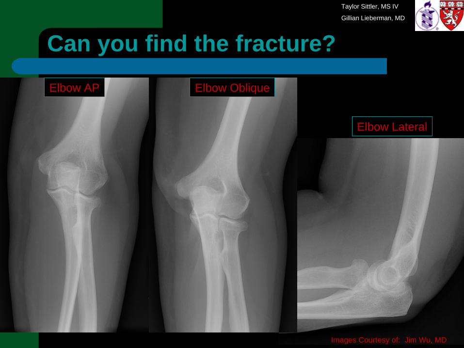
Can you find the fracture?
Images Courtesy of: Jim Wu, MD
Elbow AP Elbow Oblique
Elbow Lateral
Taylor Sittler, MS IV
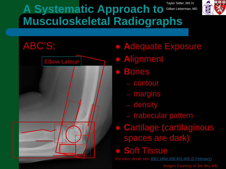
Gillian Lieberman, MDA Systematic Approach to Musculoskeletal Radiographs
Adequate ExposureAlignmentBones – contour– margins– density– trabecular pattern
Cartilage (cartilaginous spaces are dark)Soft Tissue
For more detail, see: BMJ 1994;308:401-405 (5 February)
ABC’S:
Images Courtesy of Jim Wu, MD
Elbow Lateral
Taylor Sittler, MS IV
Gillian Lieberman, MD
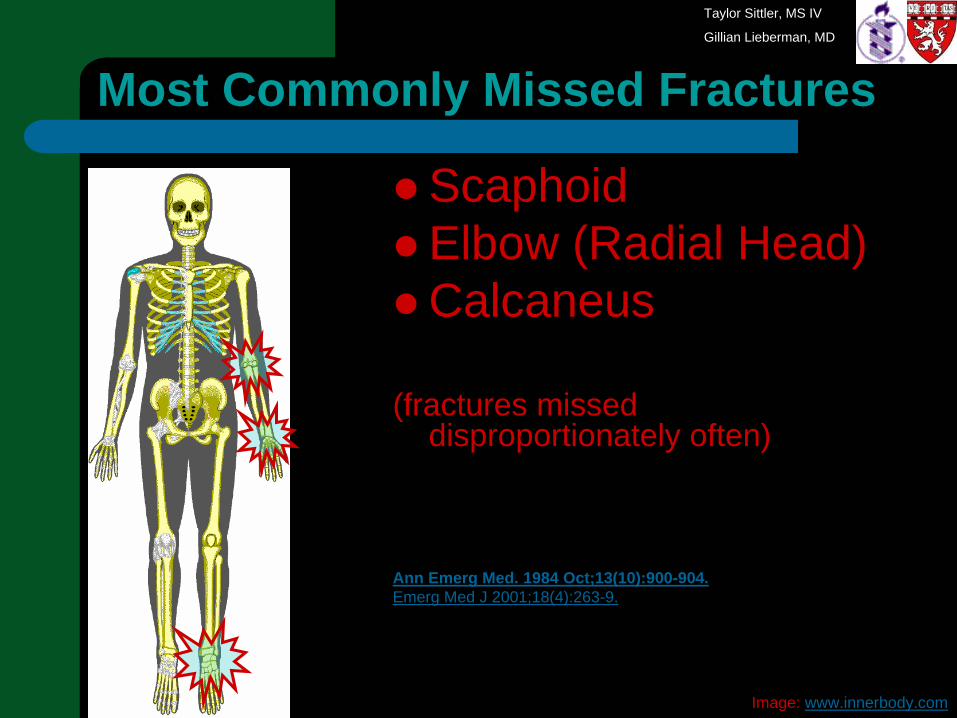
ScaphoidElbow (Radial Head)Calcaneus
(fractures missed disproportionately often)
Ann Emerg Med. 1984 Oct;13(10):900-904.Emerg Med J 2001;18(4):263-9.
Most Commonly Missed Fractures
Image: www.innerbody.com
Taylor Sittler, MS IV
Gillian Lieberman, MD
Scaphoid Fracture: Epidemiology
Annual incidence is 4.3/10,000Predominantly young malesPrimarily due to fall (classically on an outstretched hand) or post-trauma10% associated with other wrist fracture
Scand J Plast Reconstr Surg Hand Surg. 1999 Dec;33(4):423-6.
Mil Med. 2006 May;171(5):404-8.
Taylor Sittler, MS IV
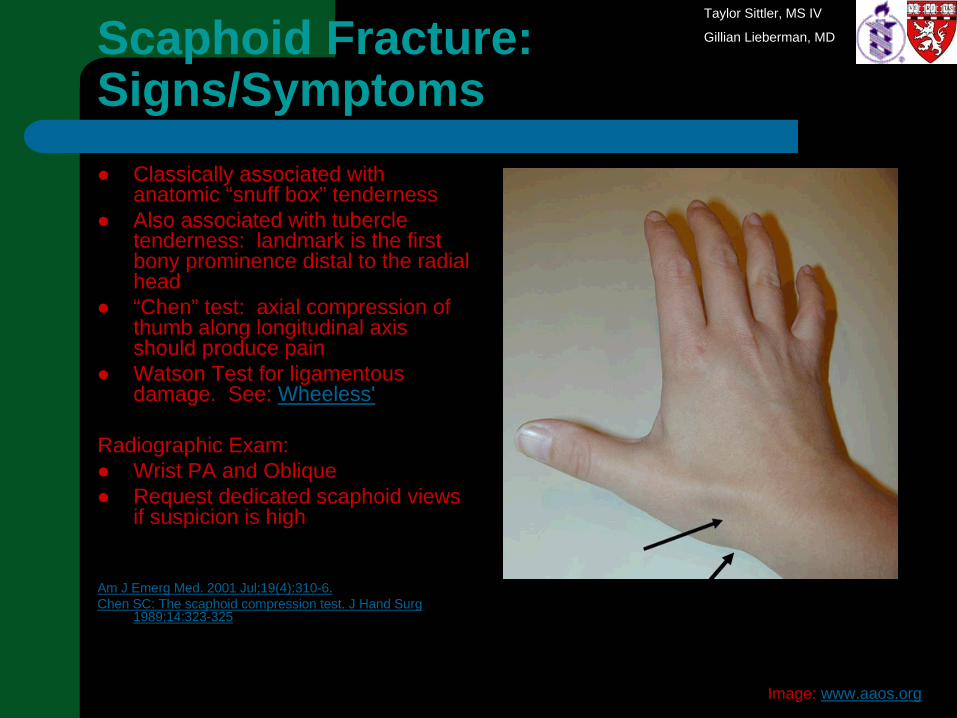
Gillian Lieberman, MDScaphoid Fracture: Signs/Symptoms
Classically associated with anatomic “snuff box” tenderness Also associated with tubercle tenderness: landmark is the first bony prominence distal to the radial head“Chen” test: axial compression of thumb along longitudinal axis should produce painWatson Test for ligamentousdamage. See: Wheeless'
Radiographic Exam:Wrist PA and ObliqueRequest dedicated scaphoid views if suspicion is high
Am J Emerg Med. 2001 Jul;19(4):310-6. Chen SC: The scaphoid compression test. J Hand Surg
1989;14:323-325
Image: www.aaos.org
Taylor Sittler, MS IV
Gillian Lieberman, MD
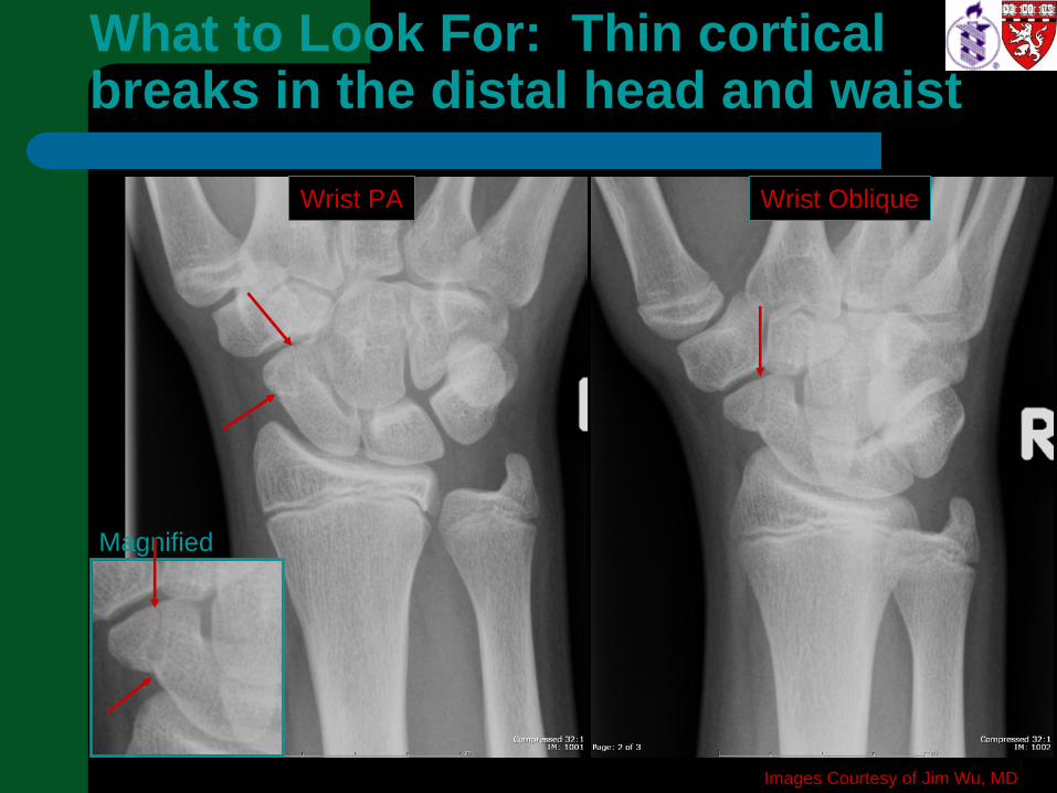
Magnified
Images Courtesy of Jim Wu, MD
Wrist PA Wrist Oblique
What to Look For: Thin cortical breaks in the distal head and waist
Taylor Sittler, MS IV
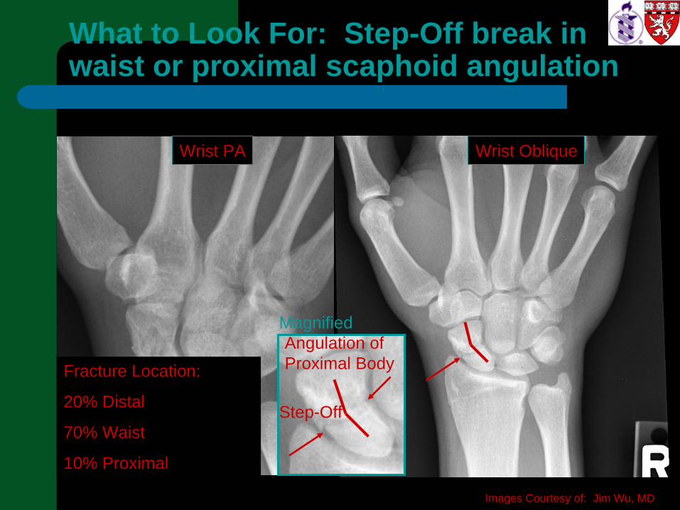
Gillian Lieberman, MDWhat to Look For: Step-Off break in waist or proximal scaphoid angulation
Magnified
Images Courtesy of: Jim Wu, MD
Step-Off
Angulation of Proximal BodyFracture Location:
20% Distal
70% Waist
10% Proximal
Wrist PA Wrist Oblique
Taylor Sittler, MS IV
Gillian Lieberman, MD
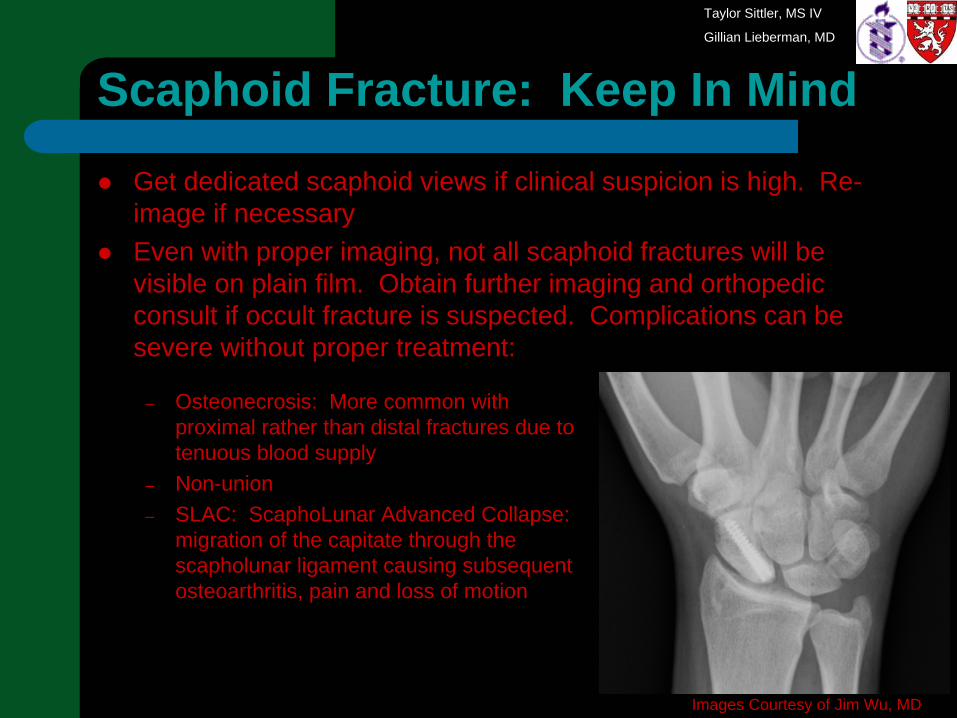
– Osteonecrosis: More common with proximal rather than distal fractures due to tenuous blood supply
– Non-union– SLAC: ScaphoLunar Advanced Collapse:
migration of the capitate through the scapholunar ligament causing subsequent osteoarthritis, pain and loss of motion
Scaphoid Fracture: Keep In Mind
Get dedicated scaphoid views if clinical suspicion is high. Re-image if necessaryEven with proper imaging, not all scaphoid fractures will be visible on plain film. Obtain further imaging and orthopedic consult if occult fracture is suspected. Complications can be severe without proper treatment:
Images Courtesy of Jim Wu, MD
Taylor Sittler, MS IV
Gillian Lieberman, MD
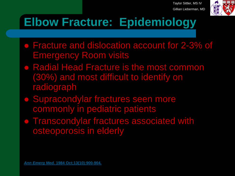
Elbow Fracture: Epidemiology
Fracture and dislocation account for 2-3% of Emergency Room visitsRadial Head Fracture is the most common (30%) and most difficult to identify on radiographSupracondylar fractures seen more commonly in pediatric patientsTranscondylar fractures associated with osteoporosis in elderly
Ann Emerg Med. 1984 Oct;13(10):900-904.
Taylor Sittler, MS IV
Gillian Lieberman, MD
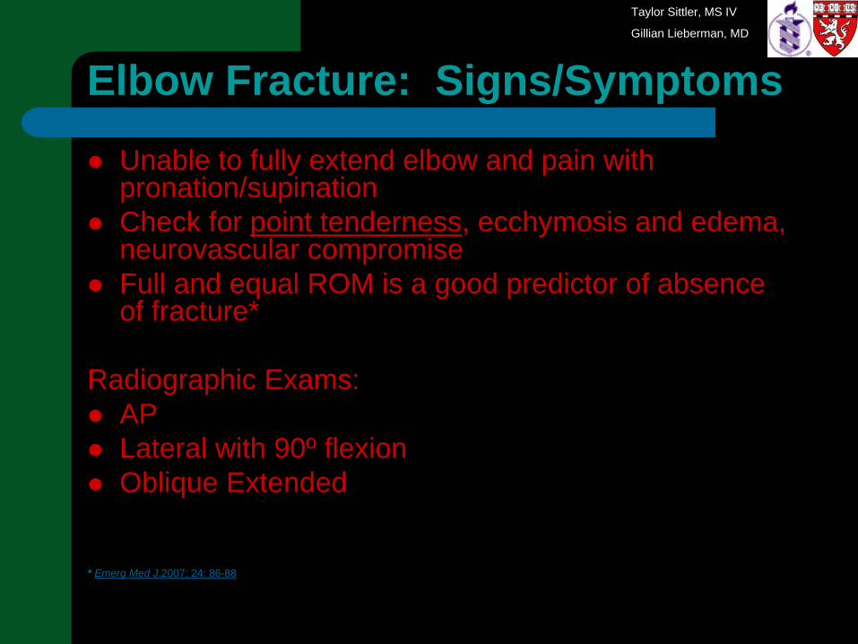
Elbow Fracture: Signs/Symptoms
Unable to fully extend elbow and pain with pronation/supinationCheck for point tenderness, ecchymosis and edema, neurovascular compromise Full and equal ROM is a good predictor of absence of fracture*
Radiographic Exams:APLateral with 90º flexionOblique Extended
* Emerg Med J.2007; 24: 86-88
Taylor Sittler, MS IV
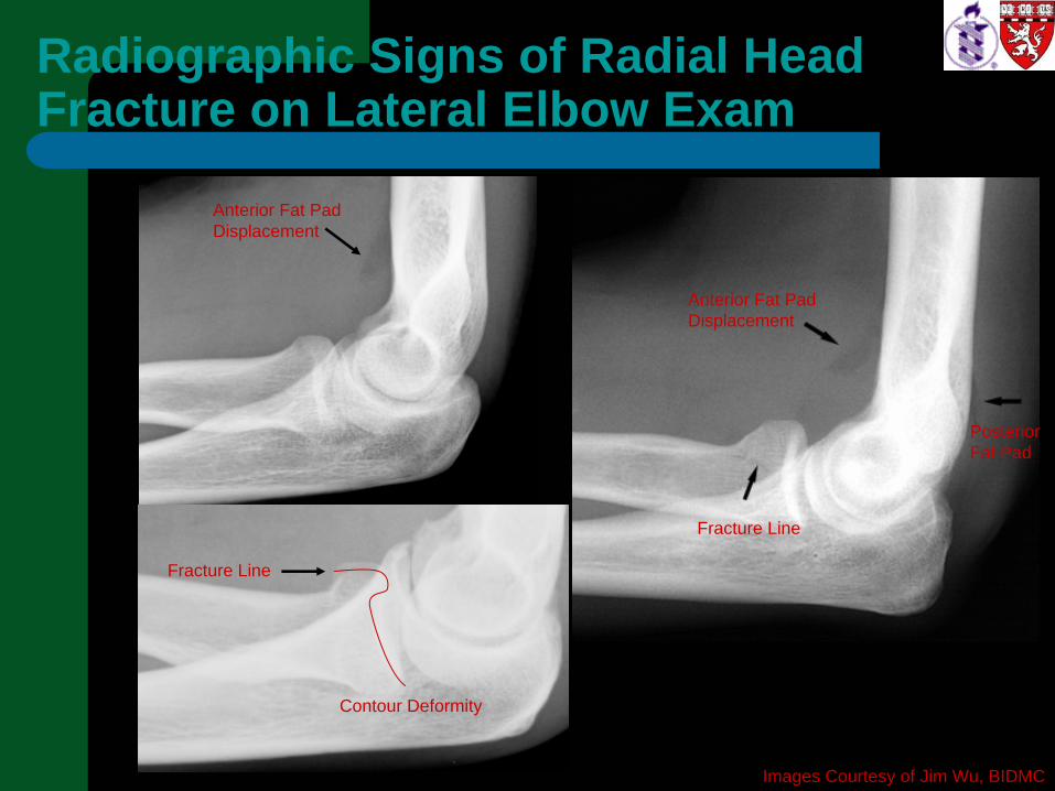
Gillian Lieberman, MDRadiographic Signs of Radial Head Fracture on Lateral Elbow Exam
Anterior Fat Pad Displacement
Posterior Fat Pad
Anterior Fat Pad Displacement
Fracture Line
Fracture Line
Contour Deformity
Images Courtesy of Jim Wu, BIDMC
Taylor Sittler, MS IV
Gillian Lieberman, MD
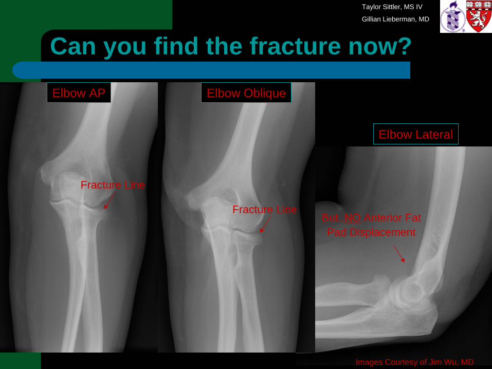
Can you find the fracture now?
Images Courtesy of Jim Wu, MD
Fracture Line
Fracture LineBut..NO Anterior Fat Pad Displacement
Elbow AP Elbow Oblique
Elbow Lateral

Taylor Sittler, MS IV
Gillian Lieberman, MD
Elbow Fracture: Keep in Mind
Ask for 3 Views: AP, oblique, lateralLook for sail sign and posterior fat padIf these signs are present but no fracture is identified, radial head fracture is likelyAdditionally, look for a fracture line, and contour deformity

Taylor Sittler, MS IV
Gillian Lieberman, MD
Calcaneal Fracture: Epidemiology
Most frequently injured bone in the foot75% are intra-articularFrequently there are associated injuriesMost patients with calcaneal fracture are men in their working years, majority are industrial workersFall from a height, motor vehicle accident is the most common mechanismLess common but frequently missed radiologically –anterior process avulsion / calcaneal stress fracture
Am J Emerg Med. 2004 Nov;22(7):607-11.Radiographics. 2005 Sep-Oct;25(5):1215-26. Clin Podiatr Med Surg. 2005 Jan;22(1):45-54.
Taylor Sittler, MS IV
Gillian Lieberman, MD
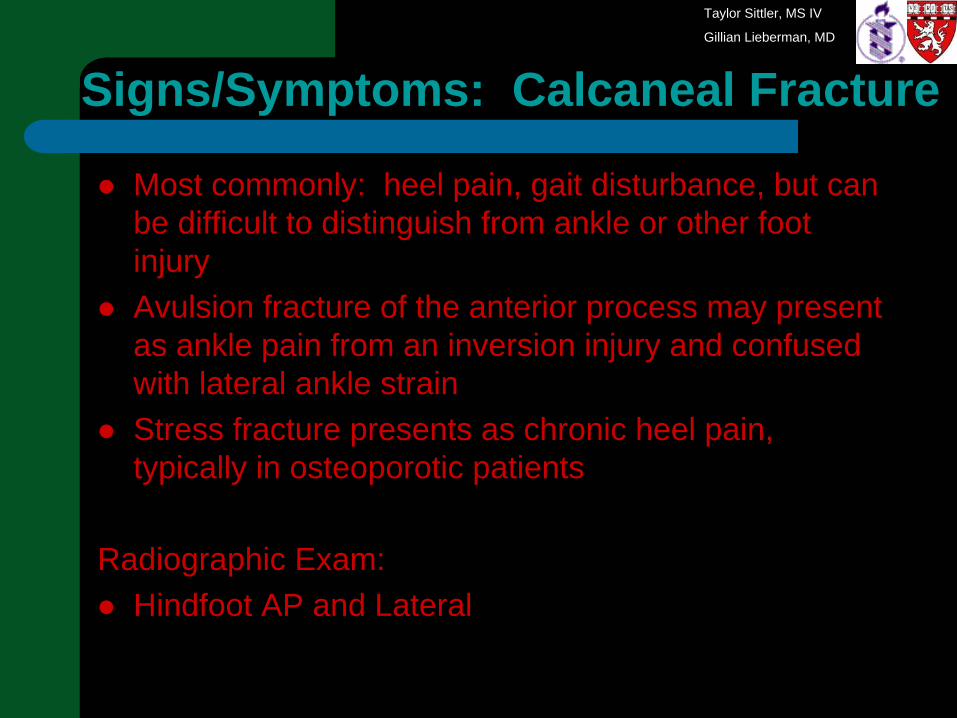
Most commonly: heel pain, gait disturbance, but can be difficult to distinguish from ankle or other foot injuryAvulsion fracture of the anterior process may present as ankle pain from an inversion injury and confused with lateral ankle strainStress fracture presents as chronic heel pain, typically in osteoporotic patients
Radiographic Exam:Hindfoot AP and Lateral
Signs/Symptoms: Calcaneal Fracture
Taylor Sittler, MS IV
Gillian Lieberman, MD
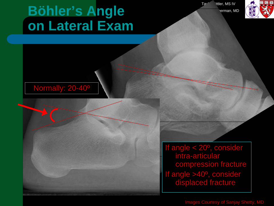
Normally: 20-40º
Böhler’s Angle on Lateral Exam
If angle < 20º, consider intra-articular compression fracture
If angle >40º, consider displaced fracture
Images Courtesy of Sanjay Shetty, MD
Taylor Sittler, MS IV
Gillian Lieberman, MD
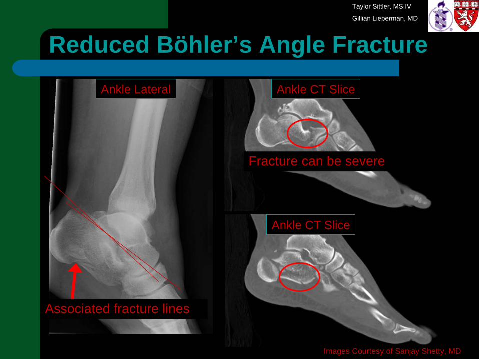
Reduced Böhler’s Angle Fracture
Associated fracture lines
Fracture can be severe
Images Courtesy of Sanjay Shetty, MD
Ankle Lateral Ankle CT Slice
Ankle CT Slice
Taylor Sittler, MS IV
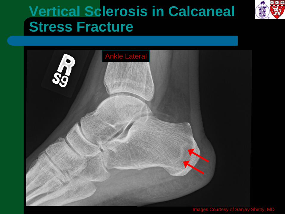
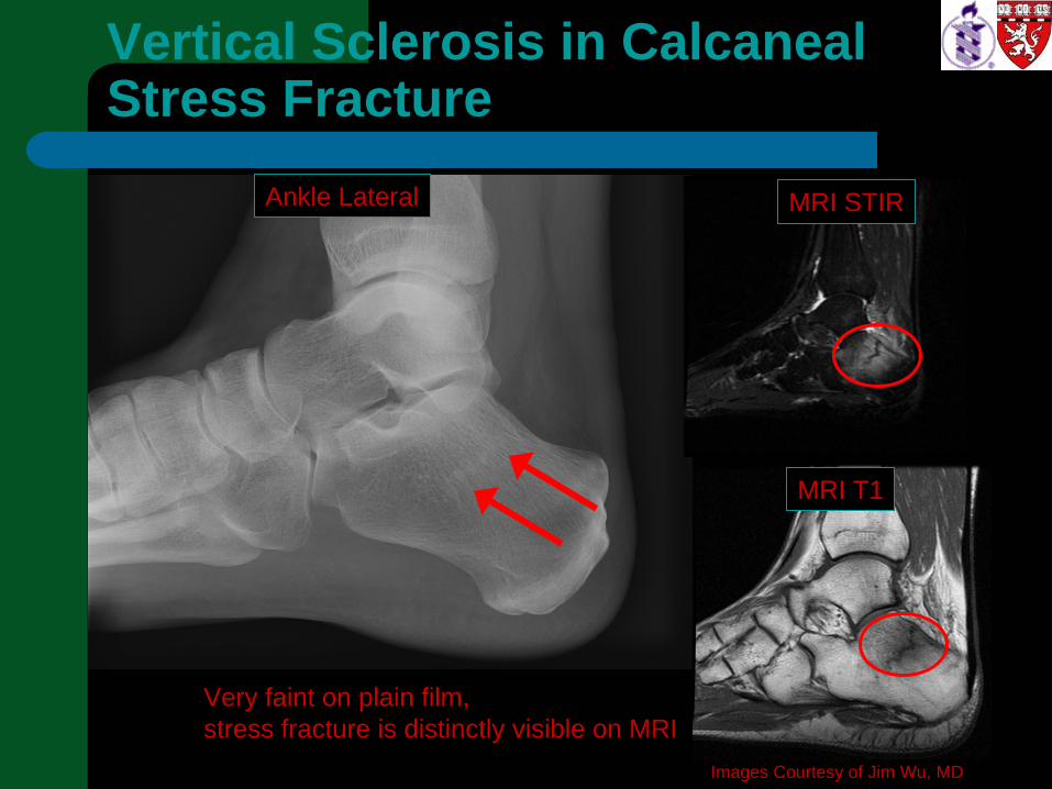
Gillian Lieberman, MDVertical Sclerosis in Calcaneal Stress Fracture
Images Courtesy of Sanjay Shetty, MD
Ankle Lateral
Taylor Sittler, MS IV
Gillian Lieberman, MDVertical Sclerosis in Calcaneal Stress Fracture
Images Courtesy of Jim Wu, MD
Ankle Lateral MRI STIR
MRI T1
Very faint on plain film, stress fracture is distinctly visible on MRI
Taylor Sittler, MS IV
Gillian Lieberman, MD
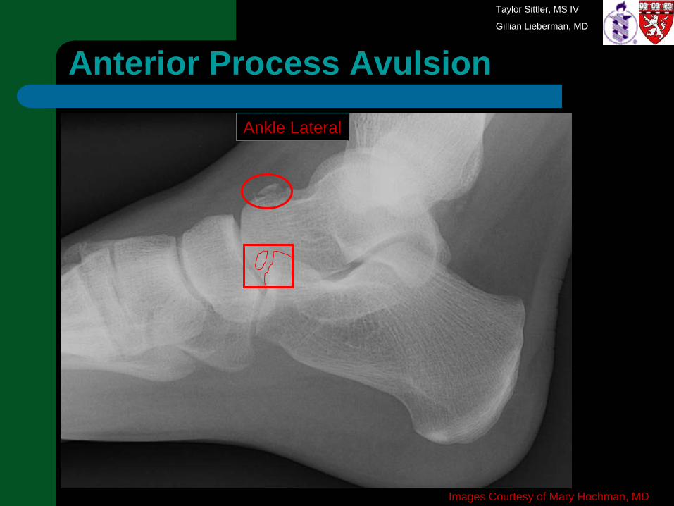
Images Courtesy of Mary Hochman, MD
Anterior Process Avulsion
Ankle Lateral
Taylor Sittler, MS IV
Gillian Lieberman, MD
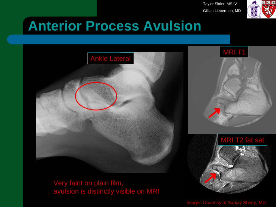
Anterior Process Avulsion
Images Courtesy of Sanjay Shetty, MD
Ankle LateralMRI T1
MRI T2 fat sat
Very faint on plain film, avulsion is distinctly visible on MRI
Taylor Sittler, MS IV
Gillian Lieberman, MD
Calcaneal Fracture: Keep In Mind
Böhler’s Angle 20°- 40°Look for fracture: follow all lucent lines carefullyConsider associated injury (and think about the other foot!)Stress Fracture: Heel pain and vertical sclerosisCheck for Anterior Process Avulsion in patients with chronic ankle/foot pain and inversion injury – CT or MRI may be required to make the diagnosis
Taylor Sittler, MS IV
Gillian Lieberman, MD
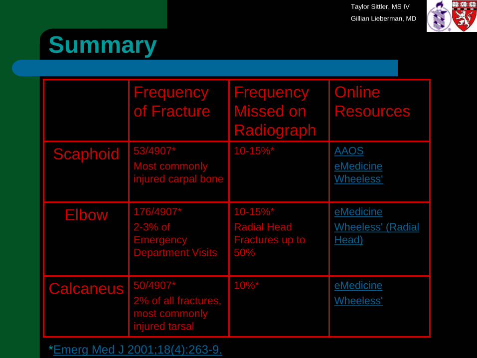
Summary
Frequency of Fracture
Frequency Missed on Radiograph
Online Resources
Scaphoid 53/4907*Most commonly injured carpal bone
10-15%* AAOSeMedicine Wheeless‘
Elbow 176/4907*2-3% of Emergency Department Visits
10-15%*Radial Head Fractures up to 50%
eMedicineWheeless' (Radial Head)
Calcaneus 50/4907*2% of all fractures, most commonly injured tarsal
10%* eMedicineWheeless'
*Emerg Med J 2001;18(4):263-9.

Taylor Sittler, MS IV
Gillian Lieberman, MD
Acknowledgements
Sanjay Shetty, MD Radiology, BIDMC
Mary Hochman, MD Radiology, BIDMC
Jim Wu, MD Radiology, BIDMC
Dan Siegal, MD Radiology, BIDMC
Gillian Lieberman, MD Radiology, BIDMC
Pamela Lepkowski Radiology, BIDMC