Embed Size (px)
Citation preview

Vertebral Osteomyelitis of the Lumbar Spine
Andrew Taliaferro, HMS III Gillian Lieberman, MD
October 2015 Andrew Taliaferro, HMS III Gillian Lieberman, MD

Agenda 1. Vertebral Anatomy and Arterial Supply 2. Vertebral Osteomyelitis
• Clinical Features • Routes of Infection and Complications • Imaging Modalities
3. Our Patient • Clinical Presentation • Imaging Findings • Differential Diagnosis • Clinical Course
4. Summary
2
Andrew Taliaferro, HMS III Gillian Lieberman, MD

1. Vertebral Anatomy and Arterial Supply
Lumbar artery
(Left) Colorado Comprehensive Spine Institute. (Right) Ratcliffe JF. The arterial anatomy of the adult human lumbar vertebral body: a microarteriographic study. J Anat. 1980;131:57-79.
3
Of note, the lumbar arteries send metaphyseal anastomoses to give arterial supply to the vertebral body, the disc, and the adjacent vertebral bodies.
Intervertebral disc
Andrew Taliaferro, HMS III Gillian Lieberman, MD

2. Vertebral Osteomyelitis: Clinical Features • Vertebral osteomyelitis is an infection of one or more
vertebrae Incidence: 2.4 cases per 100,000 people Location: Typically involves 2 adjacent vertebral bodies and the
intervertebral disc between them. oLumbar spine (58%) oThoracic Spine (30%) oCervical Spine (11%)
Presentation: Back pain (86%), Fever (35-60%), Neurologic impairment (33%)
Zimmerli W. Vertebral Osteomyelitis. N Engl J Med. 2010;362:1022-9.
4
Andrew Taliaferro, HMS III Gillian Lieberman, MD

2. Vertebral Osteomyelitis: Routes of Infection • Routes of infection: Hematogenous seeding (S. aureus, E. coli) – most common Direct inoculation during spinal procedure (S. epidermidis, P. acnes) Contiguous spread from adjacent soft tissue infection
Zimmerli W. Vertebral Osteomyelitis. N Engl J Med. 2010;362:1022-9.
5
Andrew Taliaferro, HMS III Gillian Lieberman, MD

2. Vertebral Osteomyelitis: Complications • Complications: Epidural abscess – neurosurgical emergency Psoas abscess Paraspinal abscess Extension of infection to aorta and/or IVC Vertebral body collapse
Zimmerli W. Vertebral Osteomyelitis. N Engl J Med. 2010;362:1022-9.
6
Andrew Taliaferro, HMS III Gillian Lieberman, MD

2. Vertebral Osteomyelitis: Imaging Modalities • Menu of Imaging Modalities:
• MRI • CT • Plain Film • Combined Gallium SPECT and Tc-99 Bone Scan • In-111 labeled leukocyte scan
Davis PC, Wippold FJ, Brunberg JA, et al. ACR Appropriateness Criteria on low back pain. J Am Coll Radiol. 2009;6:401-7. Modic MT, Feiglin DH, Piraino DW, et al. Vertebral osteomyelitis: assessment using MR. Radiology. 1985;157(December):157-166.
7
Andrew Taliaferro, HMS III Gillian Lieberman, MD

2. Vertebral Osteomyelitis: Imaging Modalities • MRI:
• 96% Sensitivity, 92% Specificity • First line if vertebral osteomyelitis is suspected, or if
neurologic deficits are present • IV contrast is necessary to distinguish between epidural abscess
and phlegmon Dagirmanjian A, Schils J, McHenry M, Modic MT. MR imaging of vertebral osteomyelitis revisited. Am J Roentgenol. 1996;167:1539-1543. Davis PC, Wippold FJ, Brunberg JA, et al. ACR Appropriateness Criteria on low back pain. J Am Coll Radiol. 2009;6:401-7.
8
Andrew Taliaferro, HMS III Gillian Lieberman, MD

2. Vertebral Osteomyelitis: Imaging Modalities • MRI, continued:
• T1: Decreased signal intensity in disc and adjacent vertebral bodies, and loss of endplate definition
• T2: Increased signal intensity in disc • C+: Ring enhancement suggest abscess, Homogenous enhancement
suggest phlegmon Dagirmanjian A, Schils J, McHenry M, Modic MT. MR imaging of vertebral osteomyelitis revisited. Am J Roentgenol. 1996;167:1539-1543. Modic MT, Feiglin DH, Piraino DW, et al. Vertebral osteomyelitis: assessment using MR. Radiology. 1985;157(December):157-166.
9
Andrew Taliaferro, HMS III Gillian Lieberman, MD

2. Vertebral Osteomyelitis: Imaging Modalities • Plain Film:
• Not ideal for detecting vertebral osteomyelitis • 82% Sensitivity, 57% Specificity • Radiographic findings appear 6-8 weeks after onset of symptoms • Despite this, often the first step taken in working up a patient with recurrent back pain
• Findings on Plain Film: • Disc space narrowing with end plate erosion • Vertebral body destruction • Sclerosis and new bone formation as healing progresses
Davis PC, Wippold FJ, Brunberg JA, et al. ACR Appropriateness Criteria on low back pain. J Am Coll Radiol. 2009;6:401-7. James SLJ, Davies a M. Imaging of infectious spinal disorders in children and adults. Eur J Radiol. 2006;58(December 2005):27-40. Modic MT, Feiglin DH, Piraino DW, et al. Vertebral osteomyelitis: assessment using MR. Radiology. 1985;157(December):157-166.
10
Andrew Taliaferro, HMS III Gillian Lieberman, MD

2. Vertebral Osteomyelitis: Imaging Modalities • CT:
• Useful for visualizing bony destruction if MRI is contraindicated. • Limited study data • Findings: End plate destruction, hypodense vertebral bodies
• SPECT and Tc-99 Bone Scintigraphy: • Useful if MRI is contraindicated • 90% Sensitivity, ~100% Specificity. • Findings: Increased uptake around the affected vertebral bodies
• In-111 Labeled leukocyte scan: • Low sensitivity, so rarely used.
Modic MT, Feiglin DH, Piraino DW, et al. Vertebral osteomyelitis: assessment using MR. Radiology. 1985;157(December):157-166. Palestro CJ, Kim CK, Swyer AJ, et al. Radionuclide diagnosis of vertebral osteomyelitis: indium-111-leukocyte and technetium-99m-methylene diphosphonate bone scintigraphy. J Nucl Med. 1991;32:1861-5. Eisenberg B, Powe JE, Alavi A. Cold Defects in ln-111 Labeled Leukocyte Imaging of Osteomyelitis in the Axial Skeleton. Clin Nucl Med. 1991;16:103-106.
11
Andrew Taliaferro, HMS III Gillian Lieberman, MD

3. Our Patient: Clinical Presentation • HPI: 57M with history of L3-L4 osteomyelitis (diagnosed in 8/2014)
complicated by R psoas abscess (s/p IR drainage in 3/2015) who presented in April 2015 with a 2 month history of low grade fever, recurrent back pain and progressive lower extremity weakness for 1 week.
• PMH: 8.1cm AAA s/p EVAR and resection of infected aneurysmal hematoma (in 9/2014 in the UK)
• SH: Receives medical care in the Middle East, the UK, and the USA. • Exam: Afebrile, BP 112/68, HR 108. Exam otherwise significant for a I/VI
systolic murmur at the LSB, minimal tenderness overlying the lumbar spine, 3/5 right lower extremity strength, 2/5 left lower extremity strength, decreased light touch sensation and absent DTRs in lower extremities.
• Labs: all cultures negative at the time of presentation 12
Andrew Taliaferro, HMS III Gillian Lieberman, MD
* *
PACS BIDMC
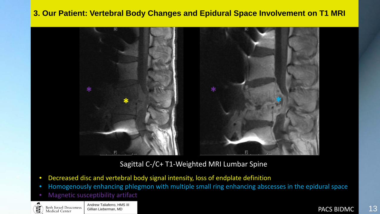
Sagittal C-/C+ T1-Weighted MRI Lumbar Spine
13
3. Our Patient: Vertebral Body Changes and Epidural Space Involvement on T1 MRI
• Decreased disc and vertebral body signal intensity, loss of endplate definition • Homogenously enhancing phlegmon with multiple small ring enhancing abscesses in the epidural space • Magnetic susceptibility artifact
* *
Andrew Taliaferro, HMS III Gillian Lieberman, MD

14
3. Our Patient: Psoas Abscess on T1 MRI
Axial C+ T1-Weighted MRI Lumbar Spine
• Ring enhancing fluid collection suggestive of psoas abscess • Magnetic susceptibility artifact
* *
Andrew Taliaferro, HMS III Gillian Lieberman, MD PACS BIDMC

15
3. Our Patient: Vertebral body changes on T2 and STIR MRI
PACS BIDMC
Sagittal T2-weighted and STIR MRI Lumbar Spine
• Increased intervertebral disc signal intensity • Magnetic susceptibility artifact
* *
Andrew Taliaferro, HMS III Gillian Lieberman, MD
16
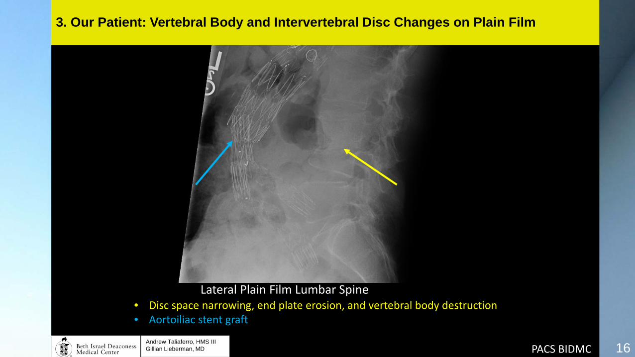
3. Our Patient: Vertebral Body and Intervertebral Disc Changes on Plain Film
PACS BIDMC
Lateral Plain Film Lumbar Spine • Disc space narrowing, end plate erosion, and vertebral body destruction • Aortoiliac stent graft
Andrew Taliaferro, HMS III Gillian Lieberman, MD

*
17
3. Our Patient: Psoas Abscess and Vertebral Body Changes on CT
*
PACS BIDMC
Coronal and Axial C- CT Lumbar Spine • Fluid collection suggestive of psoas abscess • Destructive changes of the vertebral body with end-plate destruction Andrew Taliaferro, HMS III
Gillian Lieberman, MD

3. Our Patient: Differential Diagnosis • Focal Vertebral Body Abnormality with Low T1 signal and High
T2 signal: • Infection (Osteomyelitis or Discitis, including Pott Disease) • Acute Fracture • Degenerative Joint Disease • Flow artifact from aorta or iliac arteries • Primary bone tumor • Osseous metastasis • Multiple Myeloma
Reeder MM, ed. Reeder and Felson’s Gamuts in Radiology. 4th ed. New York: Springer-Verlag New York; 2003:Gamuts M-107 and M-108.
18
Andrew Taliaferro, HMS III Gillian Lieberman, MD
3. Our Patient: Differential Diagnosis • Extradural Lesion with Abnormal Adjacent Bone:
• Infection (Osteomyelitis with epidural abscess or granuloma) • Osseous metastasis with epidural involvement • Osteoporosis with fracture and granulation tissue • Posttraumatic fracture fragment or hematoma • Spinal neoplasm • Lymphoma
Reeder MM, ed. Reeder and Felson’s Gamuts in Radiology. 4th ed. New York: Springer-Verlag New York; 2003:Gamuts M-107 and M-108.
19
Andrew Taliaferro, HMS III Gillian Lieberman, MD

3. Our Patient: Clinical Course • April 2015: Urgent decompression via L3-L4 corpectomy via
anterior approach with L2-L5 anterior fusion • Psoas abscess had increased in size since previous drainage, so
drained by IR. • Epidural culture were positive for Coxiella burnetti
• May 2015: L3-L4 laminectomy via posterior approach with L1-S1 posterior fusion
• June 2015: Readmitted for fever, found to have new L psoas abscess and new sinus tract to R psoas abscess
20
Andrew Taliaferro, HMS III Gillian Lieberman, MD

21
3. Our Patient: Post-Operative Plain Film
PACS BIDMC
Cross Table Lateral Plain Film Lumbar Spine • Posterior L1-S1 Fusion • Anterior L2-L5 Fusion • Aortoiliac stent-graft Andrew Taliaferro, HMS III
Gillian Lieberman, MD

4. Summary • Vertebral osteomyelitis most often affects adjacent vertebrae in the lumbar
spine, and results either from hematogenous seeding, direct inoculation, or contiguous spread.
• MRI spine with and without contrast is the best imaging modality for
detecting vertebral osteomyelitis and its complications, including epidural abscess (which requires urgent neurosurgical decompression).
• Findings on MRI include decreased disc and vertebral body signal
intensity as well as loss of endplate definition on T1, and increased disc signal intensity on T2. Ring enhancing fluid collections are suggestive of abscess.
22
Andrew Taliaferro, HMS III Gillian Lieberman, MD

References Dagirmanjian A, Schils J, McHenry M, Modic MT. MR imaging of vertebral osteomyelitis revisited. Am J Roentgenol. 1996;167:1539-1543. http://www.ncbi.nlm.nih.gov/pubmed/8956593. Davis PC, Wippold FJ, Brunberg JA, et al. ACR Appropriateness Criteria on low back pain. J Am Coll Radiol. 2009;6:401-7. http://www.ncbi.nlm.nih.gov/pubmed/19467485. Eisenberg B, Powe JE, Alavi A. Cold Defects in ln-111 Labeled Leukocyte Imaging of Osteomyelitis in the Axial Skeleton. Clin Nucl Med. 1991;16:103-106. http://www.ncbi.nlm.nih.gov/pubmed/1900746. James SLJ, Davies a M. Imaging of infectious spinal disorders in children and adults. Eur J Radiol. 2006;58(December 2005):27-40. http://www.ncbi.nlm.nih.gov/pubmed/16413726. Modic MT, Feiglin DH, Piraino DW, et al. Vertebral osteomyelitis: assessment using MR. Radiology. 1985;157(December):157-166. http://www.ncbi.nlm.nih.gov/pubmed/3875878. Palestro CJ, Kim CK, Swyer AJ, Vallabhajosula S, Goldsmith SJ. Radionuclide diagnosis of vertebral osteomyelitis: indium-111-leukocyte and technetium-99m-methylene diphosphonate bone scintigraphy. J Nucl Med. 1991;32:1861-5. http://www.ncbi.nlm.nih.gov/pubmed/1919723. Ratcliffe JF. The arterial anatomy of the adult human lumbar vertebral body: a microarteriographic study. J Anat. 1980;131:57-79. http://www.ncbi.nlm.nih.gov/pubmed/7440404. Reeder MM, ed. Reeder and Felson’s Gamuts in Radiology. 4th ed. New York: Springer-Verlag New York; 2003: Gamuts M-107 and M-108. Zimmerli W. Vertebral Osteomyelitis. N Engl J Med. 2010;362:1022-9. http://www.ncbi.nlm.nih.gov/pubmed/20237348.
23
Andrew Taliaferro, HMS III Gillian Lieberman, MD

Acknowledgements • Rafeeque Bhadelia, MD • Gillian Lieberman, MD • Katie Armstrong • Phil Purvis • BIDMC Image Archive Staff
24
Andrew Taliaferro, HMS III Gillian Lieberman, MD