Embed Size (px)
Citation preview

C H A P T E R 1COMMON DISEASES
ACUTE FEVER
The overall mean oral temperature for healthy adult individuals is 36.8 + 0.4ºC, with a nadir at 6 AM and a peak at 4-6 PM. A morning temperature of greater than 37.2ºC and an evening temperature of greater than 37.7ºC is often considered as fever. Fever may be continuous, intermittent or remittent. However, with frequent self-medication with antipyretics, classic patterns are not generally seen.
Diagnosis
It is important to work towards fi nding the cause of fever. A meticulous history of chronology of symptoms, any associated focal symptom(s), exposure to infectious agents and occupational history may be useful. A thorough physical examination repeated on a regular basis may provide potentially diagnostic clues such as rash, lymphadenopathy, hepatomegaly, splenomegaly, abdominal tenderness, altered sensorium, neck stiffness, lung crepts, etc. Drug fever should be considered when the cause of fever is elusive.
Diagnostic tests
A large range of diagnoses may possibly be the cause of fever. If the history and physical examination suggest that it is likely to be more than a simple URI or viral fever, investigations are indicated. The extent and focus of diagnostic work-up will depend upon the extent and pace of illness, diagnostic possibilities and the immune status of the host. If there are no clinical clues, the work-up should include a complete haemogram with ESR, smear for malarial parasite, blood culture, Widal test, urine analysis including urine culture. If the febrile illness is prolonged beyond 2 weeks, an X-ray chest is indicated even in the absence of respiratory symptoms. Any abnormal fl uid collection should be sampled. Ultrasonography is needed in some cases of acute fever such as in amoebic liver abscess.
Treatment
Routine use of antipyretics in low-grade fever is not justifi ed. This may mask important clinical indications. However, in acute febrile illnesses suggestive of viral or bacterial cause, fever should be symptomatically treated.
Chapter-01.indd 1Chapter-01.indd 1 11/9/2012 3:57:21 PM11/9/2012 3:57:21 PM

2 STANDARD TREATMENT GUIDELINES
Nonpharmacological
Hydrotherapy with tepid water, rest and plenty of oral fl uids.
Pharmacological
Non-specifi c. Tab. Paracetamol 500-1000 mg (max 4 g in 24 hours) 6-8 hourly.(Caution: Reduce dose in frail elderly, adults weighing <50 kg and those at risk of hepatotoxicity)OrTab. Ibuprofen 400-600 mg 8 hourly.Specifi c. Antibiotics/antimalarials depending upon the cause suggested by clinical and laboratory evaluation.
Outcome
In most cases of fever, patient may either recover spontaneously or a diagnosis is reached after repeated clinical evaluation and investigations. If no diagnosis is reached in up to 3 weeks, patient is said to be having fever of unknown origin (FUO) and should be managed accordingly.
Patient educationSelf-medication and over-medication should be avoided. �
Avoid injectable paracetamol/NSAIDs. �
Antibiotics should be taken only on advice of a physician. �
Avoid covering the patient having high fever with blanket, etc. �
Plenty of fl uids should be taken. Stay in cool environment. Washing/sponging of �
face and limbs should be done repeatedly.
References
1. Alterations in Body Temperature. In: Harrison’s Principles of Internal Medicine. Fauci, Braunwald, Kasper et al (eds), 18th Edition, McGraw Hill Company Inc., New York, 2012; pp 143-170.
2. Physiological Changes in Infected Patients. In: Oxford Textbook of Medicine. Warrell DA, Cox TM, Firth JD, Benz EJ Jr (eds), 4th Edition, Oxford University Press, 2003; pp 1.289-1.292.
FEVER IN CHILDREN
Fever in children is defi ned as a rectal temperature of >38°C, oral temperature of >37.5°C or an axillary temperature of >37.2°C. Fever less than 41.7°C does not cause brain damage. Only 4% of children with fever develop febrile seizure. Hyperpyrexia. Fever above 41.5°C is called hyperpyrexia and warrants aggressive antipyretic therapy because of risk of irreversible organ damage.
Chapter-01.indd 2Chapter-01.indd 2 11/9/2012 3:57:22 PM11/9/2012 3:57:22 PM
COMMON DISEASES 3
Fever of unknown origin (FUO). It is defi ned as fever of more than three weeks duration, documented fevers above 38.3°C on multiple occasions, and lack of specifi c diagnosis after 1 week of admission and investigation in a hospital setting.Nosocomial FUO. This refers to hospitalized patients receiving acute care in whom infection or fever was absent on admission but in whom a fever of 38.3°C or more occurs on several occasions. Multiple readings of more than 38.3°C in a patient with less than 500 neutrophils/mm3 are labelled as neutropenic FUO.
Treatment
Documentation of fever
Oral temperature is accurate provided no hot/cold drinks have been consumed �
in preceding 20 minutes. Axillary temperatures are least accurate and rectal thermometers are uncomfortable, especially in older children. Their use should be restricted to children < 6 months. Ear tympanic membrane thermometers are accurate refl ection of inner body temperature, are safer than mercury ones. Thermometer must be left in place for 2 minutes for rectal, 3 minutes for oral and �
5-6 minutes for recording axillary temperature.Digital thermometers may measure temperature within 2 seconds and are accurate �
but expensive. Liquid crystal strips applied to forehead for recording temperature are not accurate.
Find a cause
Try to fi nd a focus of infection by careful history and physical examination. �
Short duration fevers (less than 2 weeks) are usually due to infections. Look for any �
characteristic feature suggesting involvement of a particular system. Character of the fever (such as relapsing, Pel Ebstein, step ladder, etc.) may give a clue to the cause. Heat hyperpyrexia, dehydration fever, allergy to drug (drug fever), and haemolytic crisis are less common causes of short fevers.
There are 3 major categories of children presenting with fever; see respective sections for their management: 1. Fever due to infection without localized signs (Table 1.1). 2. Fever due to infection with localized signs (Table 1.2) 3. Fever with rash (Table 1.3)
Additional causes for fever lasting longer than 7 days (Table 1.4)
Long duration fevers lasting more than 2 weeks should be investigated for infections, �
malignancies, connective tissue disorders, autoimmune diseases and metabolic causes.Appropriate laboratory investigations such as total and differential leucocyte �
count, peripheral smear, urinalysis, serological tests, radiological investigations, and cultures of blood and body fl uids are carried out as indicated by the signs and symptoms related with fever.
Chapter-01.indd 3Chapter-01.indd 3 11/9/2012 3:57:22 PM11/9/2012 3:57:22 PM

4 STANDARD TREATMENT GUIDELINES
Children with any one of the following conditions must be seen immediately: Age <3 months old, fever >40.6°C, crying inconsolably, crying when moved/touched, diffi cult to awaken, neck is stiff, purple/red spots are present on skin, breathing is diffi cult and does not get better even after clearing of nasal passages, drooling of saliva and inability to swallow, convulsions and looks or acts very sick.Children with any one of the following should be seen as early as possible: Child is 3-6 months old (unless fever occurs within 48 hours after a DPT vaccination and has no other serious symptom), fever >40°C, burning/pain occurs during micturition, fever has been present for >24 hours and then returned, and in case of fever present for more than 72 hours.
Table 1.1. Differential diagnosis of fever without localizing signs
Diagnosis In favourMalaria (only in children exposed to malaria transmission)
• Sudden onset of fever with rigors followed by sweating• Blood fi lm positive• Rapid diagnostic test positive• Severe anaemia• Enlarged spleen
Septicaemia • Seriously ill and obviously ill with no apparent cause• Purpura, petechiae• Shock or hypothermia in severely malnourished
Typhoid • Seriously and obviously ill with no apparent cause• Abdominal tenderness• Shock • Confusion
Urinary tract infection • Costo-vertebral angle or suprapubic tenderness• Crying on passing urine• Passing urine more frequent than usual• Incontinence in previously continent child• White blood cells and/or bacteria in urine or microscopy
Table 1.2. Differential diagnosis of fever with localizing signs
Diagnosis In favourMeningitis • Fever with headache, vomiting
• Convulsions• Stiff neck• Bulging fontanelle• Meningococcal rash (petechial or purpuric)
Otitis media • Red immobile eardrum on otoscopy• Pus draining from ear• Ear pain
Mastoiditis • Tender swelling above or behind earOsteomyelitis • Local tenderness
• Refusal to move the affected limb• Refusal to bear weight on leg
Chapter-01.indd 4Chapter-01.indd 4 11/9/2012 3:57:22 PM11/9/2012 3:57:22 PM

COMMON DISEASES 5
Septic arthritis • Joint hot, tender, swollenPneumonia • Cough with fast breathing
• Lower chest wall indrawing• Fever• Coarse crackles• Nasal fl aring• Grunting
Viral upper respiratory tract infection
• Symptoms of cough/cold• No systemic upset
Table 1.3. Differential diagnosis of fever with rash
Diagnosis In favour Diagnosis In favour
Measles • Typical rash (maculopapular)• Cough, runny nose, red eyes• Recent exposure to a
measles case• No documented measles
immunization
Meningococcal infection
• Petechial or purpuric rash
• Bruising• Shock• Stiff neck (if meningitis)
Viral infections • Mild transient upset• Transient non-specifi c rash
Dengue haemorrhagic fever
• Abdominal tenderness• Skin petechiae• Bleeding from nose or
gums or GI bleed• Shock
Table 1.4. Additional differential diagnosis* of fever lasting longer than 7 days
Diagnosis In favour Diagnosis In favourAbscess • Fever with no obvious
focus of infection (deep abscess)
• Tender or fl uctuant mass• Local tenderness or pain• Specifi c signs depend
on site subphrenic, liver, psoas, retroperitoneal, lung, renal, etc.
Infective endocarditis
• Weight loss• Enlarged spleen• Anaemia• Heart murmur• Petechiae• Splinter haemorrhages in
nailbeds• Microscopic haematuria• Finger clubbing
Rheumatic fever
• Heart murmur which may change over time
• Arthritis/arthralgia• Cardiac failure• Fast pulse rate• Pericardial friction rub• Chorea• Recent known
streptococcal infection
Tuberculosis • Weight loss• Anorexia, night sweats• Cough• Enlarged liver and/or spleen• Family history of TB• Chest X-ray suggestive of
TB• Tuberculin test positive• Lymphadenopathy
Chapter-01.indd 5Chapter-01.indd 5 11/9/2012 3:57:22 PM11/9/2012 3:57:22 PM
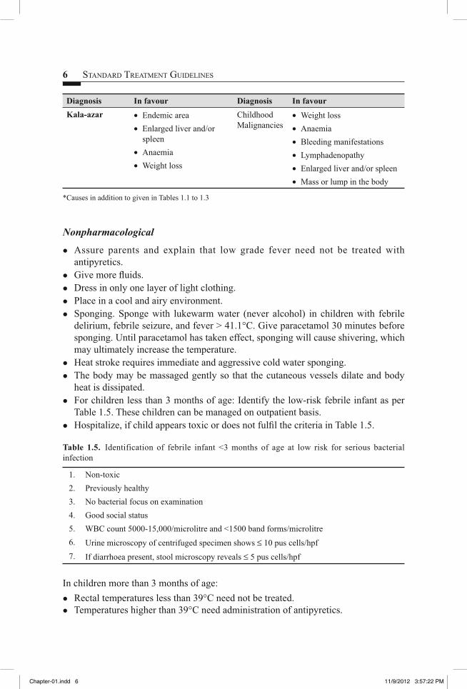
6 STANDARD TREATMENT GUIDELINES
Diagnosis In favour Diagnosis In favourKala-azar • Endemic area
• Enlarged liver and/or spleen
• Anaemia• Weight loss
Childhood Malignancies
• Weight loss• Anaemia• Bleeding manifestations• Lymphadenopathy• Enlarged liver and/or spleen• Mass or lump in the body
*Causes in addition to given in Tables 1.1 to 1.3
Nonpharmacological
Assure parents and explain that low grade fever need not be treated with �
antipyretics.Give more fl uids. �
Dress in only one layer of light clothing. �
Place in a cool and airy environment. �
Sponging. Sponge with lukewarm water (never alcohol) in children with febrile �
delirium, febrile seizure, and fever > 41.1°C. Give paracetamol 30 minutes before sponging. Until paracetamol has taken effect, sponging will cause shivering, which may ultimately increase the temperature.Heat stroke requires immediate and aggressive cold water sponging. �
The body may be massaged gently so that the cutaneous vessels dilate and body �
heat is dissipated.For children less than 3 months of age: Identify the low-risk febrile infant as per �
Table 1.5. These children can be managed on outpatient basis.Hospitalize, if child appears toxic or does not fulfi l the criteria in Table 1.5. �
Table 1.5. Identification of febrile infant <3 months of age at low risk for serious bacterial infection
1. Non-toxic 2. Previously healthy 3. No bacterial focus on examination 4. Good social status 5. WBC count 5000-15,000/microlitre and <1500 band forms/microlitre6. Urine microscopy of centrifuged specimen shows ≤ 10 pus cells/hpf 7. If diarrhoea present, stool microscopy reveals ≤ 5 pus cells/hpf
In children more than 3 months of age:Rectal temperatures less than 39°C need not be treated. �
Temperatures higher than 39°C need administration of antipyretics. �
Chapter-01.indd 6Chapter-01.indd 6 11/9/2012 3:57:22 PM11/9/2012 3:57:22 PM

COMMON DISEASES 7
Pharmacological
Tab/syr. Paracetamol 15 mg/kg/dose, dose can be repeated at 4 hourly interval (Paracetamol reduces fever by 1-2°C within 2 hours).
(Caution: IV paracetamol is NOT recommended in children with age <6 months and <5 kg weight)
OrTab/syr. Ibuprofen 10 mg/kg/dose, dose can be repeated at 8 hourly intervals.(Note: Effi cacy is similar to paracetamol. Effect lasts for 6-8 hours as compared to
4-6 hours for paracetamol).(Caution: Aspirin should NOT be used for the risk of Reye’s syndrome). Specifi c
treatment for the cause of fever should be simultaneously undertaken.
Monitoring
Close monitoring of all children, especially young febrile infants, is essential.
References
1. Fever. In: Current Paediatric Diagnosis and Treatment. Hay WW, Hayward AR, Levin MJ, Sandheimer JM (eds). 15th Edition. Lange Medical Books, New York, 2001; pp 211-212.
2. Fevers in Childhood. In: Ghai’s Essential Paediatrics. Ghai OP, Gupta P, Paul VK (eds). 6th Edition. Interprint, New Delhi, 2004; pp 201-239.
3. Facility Based IMNCI (F-IMNCI) Participants Manual. WHO, UNICEF, and Ministry of Health & Family Welfare, Government of India, 2009.
FEVER OF UNKNOWN ORIGIN (FUO)
FUO is defi ned as the presence of fever of 38.3ºC (>101ºF) or more recorded on several occasions, evolving for at least 3 weeks with no diagnosis reached even after one week of relevant and intelligent investigations. FUO is usually an uncommon presentation of common diseases. FUO are classifi ed into four main categories along with common causes in each of these categories. 1. Classic FUO—corresponds to the previous defi nition except that instead of one
week of investigations, it requires up to 3 outpatient visits or 3 days in the hospital, viz. tuberculosis, abscesses, bacterial endocarditis, visceral leishmaniasis, non-Hodgkin’s lymphoma, Hodgkin’s lymphoma, acute leukaemia, systemic lupus erythromatosis.
2. HIV-related FUO—the duration of fever is >4 weeks for inpatients or >3 days for hospitalized patients with HIV infections, viz. tuberculosis, cryptococcosis, Pneumocustis jiroveci pneumonia, bacterial pneumonia.
3. Nosocomial FUO—fever of >38.3°C on several occasions lasting for more than 72 hours, developing after admission in a hospitalized patient and remains undiagnosed after 3 days of investigation including 2 days incubation of cultures, viz. postoperative (abscess, haematoma, foreign bodies), infected prostheses, infected catheters, Clostridium diffi cle colitis, deep vein thrombosis, pulmonary embolism, drug fever.
Chapter-01.indd 7Chapter-01.indd 7 11/9/2012 3:57:22 PM11/9/2012 3:57:22 PM
8 STANDARD TREATMENT GUIDELINES
4. Neutropenic FUO—similar to the previous defi nition, except that it occurs in a patient who has neutrophil count of less than 500/mm3 or expected to fall to this level in 1-2 days, viz. Gram-negative bacterial, staphylococcal, central venous catheter infections, invasive fungal infections, dental abscesses, perianal infections, cytomegalovirus, herpes simplex virus infections.If patient does not fi t into any of the above defi nition, the patient should be referred
to a specialist for investigations and management.
SALIENT FEATURES� Prolonged unexplained fever, often with no localizing clue on history, physical
examination and basic laboratory investigations.
Diagnostic evaluation
A detailed clinical history and repeated and meticulous physical examination are valuable in providing potentially diagnostic clues (PDC) to the cause of fever in these patients. No single algorithmic approach to diagnosis can be recommended for all patients of FUO and diagnostic approach needs to be individualized.
A complete haemogram including peripheral blood smear for malarial parasite, serum biochemistry particularly liver function tests, a tuberculin test and an X-ray of chest should be done in every patient with prolonged fever. Other investigations which are often helpful include tests related to collagen vascular disease; an ultrasonography of abdomen to localize intra-abdominal foci of infections and a contrast enhanced computed tomography (CECT) of chest and abdomen in detecting mediastinal lymph nodes and parenchymal lung abnormalities not seen on conventional chest X-ray. Further, diagnostic approach should take into consideration the PDCs from the evaluation of history, results of repeated physical examination, basic investigations and any investigation done prior to this episode. If any abnormal or doubtful lesion is detected FNAC/biopsy should be obtained.
Treatment
Treatment will be based on the specifi c cause of fever. Thorough investigations generally yield a specifi c cause of fever in about 90% of patients. Sometimes evaluation may need discontinuation of all drugs being taken by the patient to rule out drug fever as the cause of FUO.
Symptomatic treatment for fever (for details see section on fever). Sponging with lukewarm water may be done, if fever produces discomfort. The emphasis in patients with classic FUO is on continued observation and examination.
(Caution: Avoid ‘shotgun’ trials. Empirical therapies consisting of therapeutic trials commonly used in patients with FUO are: Antibiotics, antitubercular treatment (ATT) and corticosteroids).
Chapter-01.indd 8Chapter-01.indd 8 11/9/2012 3:57:22 PM11/9/2012 3:57:22 PM
COMMON DISEASES 9
If on the basis of clinical evaluation and inability to reach a defi nitive diagnosis, a therapeutic trial is started, the following principles must be kept in mind:
Give only one set of trial at a given time. �
The doses of drugs and period of therapeutic trial must be adequate. �
The patient must be followed closely for response. �
The ability of glucocorticoids and NSAIDs to mask fever while permitting the spread of infection dictates that their use should be avoided unless infection has been largely ruled out.
Follow-up
In about 10% of cases, no cause may be diagnosed despite thorough evaluation. In such cases, if patient is well preserved, just a close clinical and investigative follow-up may be enough to look for any PDCs which may be evolving or appear later in the course of disease. However, if the patient is sick or is deteriorating and no diagnosis is reached, an appropriate empirical therapeutic trial is justifi ed.
Patient educationSelf-medication should be avoided. �
Antibiotics should be taken only on advice of a physician. �
Avoid covering the patient with high fever with a blanket, etc. �
Plenty of fl uids should be taken. Stay in a cool environment. Washing/sponging of �
face and limbs should be done repeatedly.
References
1. Fever of Unknown Origin. In: Harrison’s Principles of Internal Medicine. Fauci, Braunwald, Kasper et al (eds), 18th Edition, McGraw Hill Company Inc., New York, 2012; 158-164.
2. Facility Based IMNCI (F-IMNCI) Participants Manual. WHO, UNICEF, and Ministry of Health & Family Welfare, Government of India, 2009.
3. Fever of Unknown Origin. In API Textbook of Medicine. Munjal YP (ed), 9th edition, JP Brothers, India, pp 42-46.
ANAEMIA
Anaemia is defi ned as a low haemoglobin level (adult males <13 g/dl; adult females <12 g/dl; pregnant women, <11 g/dl). The common causes of anaemia in India are:
Reduced production due to defi ciency of iron, folic acid, or vitamin B � 12; or an ineffective erythropoiesis secondary to many causes (anaemia of chronic disease, secondary to infections and infl ammation, endocrinal disorders, primary bone marrow disorders like infi ltration or hypoplasia).Blood loss (which also leads to iron defi ciency). �
Increased destruction of RBCs (haemolysis due to many causes of which, a �
thalassaemia is the commonest).
Chapter-01.indd 9Chapter-01.indd 9 11/9/2012 3:57:22 PM11/9/2012 3:57:22 PM
10 STANDARD TREATMENT GUIDELINES
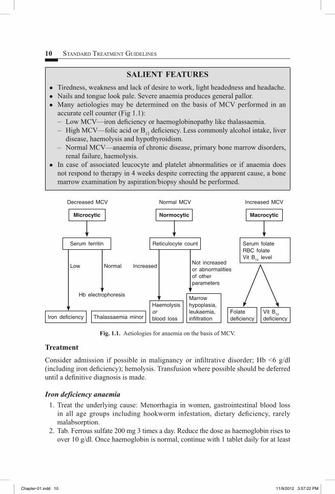
SALIENT FEATURES � Tiredness, weakness and lack of desire to work, light headedness and headache. � Nails and tongue look pale. Severe anaemia produces general pallor. � Many aetiologies may be determined on the basis of MCV performed in an
accurate cell counter (Fig 1.1): – Low MCV—iron defi ciency or haemoglobinopathy like thalassaemia. – High MCV—folic acid or B12 defi ciency. Less commonly alcohol intake, liver
disease, haemolysis and hypothyroidism. – Normal MCV—anaemia of chronic disease, primary bone marrow disorders,
renal failure, haemolysis. � In case of associated leucocyte and platelet abnormalities or if anaemia does
not respond to therapy in 4 weeks despite correcting the apparent cause, a bone marrow examination by aspiration/biopsy should be performed.
Decreased MCV
Hb electrophoresis
Low NormalNot increased or abnormalities of other parameters
Increased
Normal MCV Increased MCV
Microcytic
Serum ferritin
Iron defi ciency Thalassaemia minor
Normocytic
Reticulocyte count
Haemolysisorblood loss
Marrow hypoplasia, leukaemia, infi ltration
Folatedefi ciency
Vit B12
defi ciency
Macrocytic
Serum folateRBC folateVit B12 level
Fig. 1.1. Aetiologies for anaemia on the basis of MCV.
Treatment
Consider admission if possible in malignancy or infi ltrative disorder; Hb <6 g/dl (including iron defi ciency); hemolysis. Transfusion where possible should be deferred until a defi nitive diagnosis is made.
Iron deficiency anaemia
1. Treat the underlying cause: Menorrhagia in women, gastrointestinal blood loss in all age groups including hookworm infestation, dietary defi ciency, rarely malabsorption.
2. Tab. Ferrous sulfate 200 mg 3 times a day. Reduce the dose as haemoglobin rises to over 10 g/dl. Once haemoglobin is normal, continue with 1 tablet daily for at least
Chapter-01.indd 10Chapter-01.indd 10 11/9/2012 3:57:22 PM11/9/2012 3:57:22 PM
COMMON DISEASES 11
three months. Other preparations of iron are not superior, but they can be tried if patient does not fi nd ferrous sulfate suitable. These include ferrous fumarate and ferrous gluconate.The rate of rise of haemoglobin should be 1 g/dl per week. If this does not occur,
consider ongoing blood loss, noncompliance, and associated haemoglobinopathy like thalassaemia carrier status, malabsorption, or an incorrect diagnosis.
Parenteral iron does not lead to a faster rise in haemoglobin. It is indicated in the following situations: (i) Intolerance of oral iron, (ii) In late pregnancy to ensure that foetal stores of iron are replenished rapidly, (iii) If ongoing blood loss exceeds the capacity to absorb oral iron (like in inoperable malignancy), (iv) In noncompliant patient, (v) Malabsorption of iron. (Caution: There is danger of anaphylactoid reactions; hence facilities to manage these should be readily available).
(See also anaemia in pregnancy and anaemia in paediatric section in Chapters 15 and 19).
Folic acid deficiency
1. Treat the cause: Dietary defi ciency, increased requirement as in pregnancy and children, haemolytic anaemia.
2. Tab. Folic acid 5 mg daily. This dose is adequate even in malabsorption syndrome.
Vitamin B12
deficiency
1. Treat the cause: Dietary defi ciency in vegetarians and pernicious anaemia. Although uncommon, it is also under diagnosed due to lack of facilities.
2. Tab. Vitamin B12 500 mcg thrice in a day until recovery, then 500-1000 mcg once in a day as in haematinic tablets.
Or Inj. Vitamin B12 1000 mcg IM, one injection on alternate days for total 5 injections,
then once a week for 5 weeks, then once in 3 to 6 months will be adequate for most patients.
Note: Oral vitamin B12 is indicated only in dietary defi ciency states, and not in pernicious anaemia.
Patient educationEducate the patient about preventive measures for worm infestation. �
Inform about importance of taking adequate food with green leafy vegetables to �
meet the nutritional requirement and cooking food in iron utensils may increase iron content in the diet.Iron tablets sometimes produce stomach upset, therefore, take iron tablets �
after meals; reduce the dose of iron, if it produces stomach ache, diarrhoea or constipation.Iron should not be taken with milk or milk products; should be either taken one hour �
before or two hours after milk or milk products.Stools would turn black during oral iron therapy. �
Chapter-01.indd 11Chapter-01.indd 11 11/9/2012 3:57:22 PM11/9/2012 3:57:22 PM

12 STANDARD TREATMENT GUIDELINES
Explain that the response to iron therapy is gradual and it takes weeks or months for �
haemoglobin to become normal. Continue iron tablets for 6 months.Keep iron tablets out of the reach of children. They may swallow the tablets as �
candies causing adverse reactions including death.
Reference
1. Anaemia and Polycythaemia. In: Harrison’s Principles of Internal Medicine. Fauci, Braunwald, Kasper et al (eds), 18th Edition, McGraw Hill Company Inc., New York, 2012, pp. 448-456.
DIZZINESS AND VERTIGO
The term dizziness is used for lightheadedness, faintness, spinning, giddiness, confusion and blackouts. Dizziness is classifi ed in three categories: (1) faintness (syncope and presyncopal symptoms), (2) vertigo and (3) miscellaneous head sensation. The common causes of vertigo include benign paroxysmal positional vertigo (BPPV), vestibular neuronitis, chronic suppurative otitis media, Meniere’s disease, cervical spondylosis, drug-induced vertigo due to administration of aminoglycosides, furosemide, etc. Systemic problems such as long-standing diabetes, hypertension may also be a causative factor. Vertigo as a psychosomatic manifestation should be ruled out. If the entire list of common causes is excluded by clinical examination and investigations, the vertigo may be termed as idiopathic.
SALIENT FEATURES� Sensation of patient spinning or the environment spinning around him in a specifi c
and fi xed direction. � Spontaneous nystagmus (most important physical sign) in primary position with
eyes looking straightforward.
Important notes
Axioms for defi ning a dizzy spell as vestibular: If the patient in a signifi cant spell does not have spontaneous labyrinthine nystagmus, and also if the dizziness has been non-episodic and continuous for two or three months, then this dizziness cannot be vestibular.
Treatment
Nonpharmacological
Reassure the patient and in cases where positional vertigo cannot be ruled out, advise the patient to take complete rest with minimal movements only.
Pharmacological
Tab. Cinnarizine 25 mg three times a day till resolution of symptoms.
Chapter-01.indd 12Chapter-01.indd 12 11/9/2012 3:57:22 PM11/9/2012 3:57:22 PM
COMMON DISEASES 13
OrTab. Betahistine 8 mg three times a day.OrTab. Prochlorperazine 25 mg three times a day.The duration of drug administration depends on the disease entity as well as the
persistence of symptoms.If patient has acute, severe nausea and vomiting:Inj. Prochlorperazine 25 mg by deep IM injection stat, may be repeated after eight
hours, if required.If there is no response to medical treatment:
Refer to ENT specialist for Canthrone-Cooksey exercises. These are special �
exercises which facilitate the process of adaptation of the vestibule. Refer patients with Meniere’s disease for surgery to eliminate the offending �
labyrinth.
Patient education
Explain that the antivertigo drugs are likely to cause sedation, therefore, patient should avoid tasks requiring alertness.
Reference
1. Vertigo. In: Scott Brown’s Otolaryngology. Boothy JB (ed), 6th Edition, Vol. 3, 1997.
JAUNDICE
Jaundice is defi ned as yellow discoloration of skin, sclera and tissues caused by increased levels of circulating bilirubin. Approximately 250-350 mg of bilirubin is formed daily, mostly from the breakdown of aged RBCs (70-80%) and rest from other haem proteins in the marrow and liver. It is taken up by liver, conjugated and excreted in bile. Serum bilirubin may increase due to derangement occurring at any level:
Increased production due to excessive haemolysis, results in unconjugated �
hyperbilirubinaemia (>80% unconjugated serum bilirubin), jaundice is mild (bilirubin <10 mg%) and associated with absence of bilirubin in urine (acholuric jaundice). Impaired conjugation in hepatocellular damage (usually results in increase in �
both fractions of bilirubin due to impaired conjugation and associated decreased canalicular excretion).Impaired excretion due to intra- or extra-hepatic cholestasis, resulting in conjugated �
hyperbilirubinaemia (>50% conjugated serum bilirubin), associated with absence of urobilinogen and bile salts in urine.
Common causes of jaundice in clinical practice include acute viral hepatitis, alcoholic hepatitis, chronic hepatitis/cirrhosis, gallstones and malignancy of gallbladder/pancreas or extra-hepatic biliary system. Chronic haemolytic anaemias are less common and usually present in childhood or sometime in young adults.
Chapter-01.indd 13Chapter-01.indd 13 11/9/2012 3:57:22 PM11/9/2012 3:57:22 PM

14 STANDARD TREATMENT GUIDELINES
Approach to diagnosis of jaundice includes initial differentiation between the three types of jaundice by appropriate clinical history, examination and investigations including full blood counts, liver function tests (LFTs), viral markers, ultrasound examination of liver and biliary tract and if indicated CT scan of abdomen/ERCP.
Treatment of acute viral hepatitis is detailed below.
ACUTE VIRAL HEPATITIS
Acute viral hepatitis is caused by hepatitis virus A, E (faeco-orally transmission) or B, C (parenteral transmission).
SALIENT FEATURESClinically, the onset is with a prodromal phase (nausea, vomiting, anorexia, fever, �
dull aching pain in upper right abdomen followed by icteric phase (appearance of jaundice in 3-7 days of onset, associated with improvement in nausea and return of appetite) followed by convalescent phase, when jaundice gradually settles. The total duration of episode usually lasts for 2-6 weeks. Convalescent phase may �
be complicated by cholestatic phase, when levels of conjugated bilirubin may increase and may take several weeks to improve. Diagnosis can be confi rmed by detection of IgM antibodies to different viruses �
(A, E and B) or detection of HCV RNA.
Treatment
Nonpharmacological
During prodromal phase, adequate intake of fl uids should be maintained. Once the appetite improves, patient should be advised to take normal diet (fat restriction or giving high carbohydrate has no advantage).
Indications for hospitalization are—severe prodromal symptoms causing dehydration, presence of early signs of hepatic encephalopathy (e.g. altered sensorium, disturbed sleep pattern, fl apping tremors), decreased liver span on examination.
Pharmacological
If patient has severe nausea or vomiting. 1. Tab. Domperidone 10 mg as and when required (maximum 3 times a day). Or Tab. Mosapride 5 mg as and when required (maximum 3 times a day). Or Inj. Metoclopramide 10 mg 3 times a day IM or IV. 2. IV fl uids as required in case of uncontrolled nausea or vomiting.
Chapter-01.indd 14Chapter-01.indd 14 11/9/2012 3:57:22 PM11/9/2012 3:57:22 PM
COMMON DISEASES 15
Follow-up/monitoringRepeat LFT at weekly interval. �
Patient can resume activity, when the enzyme levels come down to less than 3-5 �
times normal.In patient with HBV infection, check for disappearance of HBsAg at 3-6 months. �
Hepatitis B and hepatitis C virus infections warrant long-term follow-up. �
Patient educationExplain the relatives to report and hospitalize the patient, if there is alteration in �
behaviour or sensorium of patient.There is no need to isolate the patient. �
Patient should avoid taking alcohol for 4-6 months after recovery. �
Spouse of the patient with acute viral hepatitis B, should use barrier method to �
prevent sexual transmission and vaccinated against hepatitis B.(See also jaundice and acute viral hepatitis in children in Chapter 19).
References
1. Acute Viral Hepatitis. In: Harrison’s Principles of Internal Medicine. Fauci, Braunwald, Kasper et al (eds), 18th Edition, McGraw Hill Company Inc., New York, 2012; pp 2537-2557.
2. Jaundice. In: Harrison’s Principles of Internal Medicine. Fauci, Braunwald, Kasper et al (eds), 18th Edition, McGraw Hill Company Inc., New York, 2012; pp 324-329.
TUBERCULOSIS AND REVISED NATIONAL TB CONTROL PROGRAMME (RNTCP)
Tuberculosis (TB) is one of the most prevalent chronic infections in our country and is responsible for high morbidity and mortality. TB is caused by Mycobacterium tuberculosis, and affl icts the lungs most commonly. In one-third or more, extra-pulmonary involvement is seen. Tubercular lymphadenopathy is the commonest form of extrapulmonary tuberculosis. All cases of TB is a notifi able disease, should be reported to the local/district/state health authorities, as it is a notifi able disease.
SALIENT FEATURES Pulmonary TB usually presents with fever, malaise, chronic cough with sputum �
production, anorexia and weight loss. Sometimes chest pain and haemoptysis may be the presenting symptoms. �
Extrapulmonary tuberculosis presents most commonly as prolonged fever and �
cervical, mediastinal or mesenteric lymphadenopathy. Abdominal tuberculosis may present as ascites, chronic abdominal pain, �
diarrhoea, recurrent subacute intestinal obstruction, etc.CNS tuberculosis presents as irritability, headache, vomiting, chronic meningitis, �
seizures or focal neurological defi cits, altered sensorium.
Chapter-01.indd 15Chapter-01.indd 15 11/9/2012 3:57:22 PM11/9/2012 3:57:22 PM

16 STANDARD TREATMENT GUIDELINES
Skeletal tuberculosis may present as Pott’s spine, tuberculous osteomyelitis, �
monoarticular arthritis. Tubercular constrictive pericarditis presents with oedema/ascites. �
Symptoms of genitourinary TB include tubovarian masses, secondary �
amenorrhoea in women, chronic epididymo-orchitis in men and painless haematuria in both the sexes. Diagnostic algorithm is given in Fig. 1.2.Defi nitive diagnosis is made only by demonstration of AFB on smear or culture of �
the sputum or bronchial secretions. Chest radiograph merely localizes the site of pathology and does not defi ne an aetiology. There are no pathognomonic radiological signs of tuberculosis. Chest X-ray is sensitive but less specifi c with higher inter- and intra-reader variation, should be used judiciously. Defi nitive diagnosis of extrapulmonary tuberculosis is made on the basis of FNAC or fi ndings of caseous granuloma with presence of AFB in the tissue, fl uid for cytology, biochemical analysis and smear examination; although ultrasonography and radiological examination of the system involved are useful investigations. CT scan is rarely necessary and is not cost and radiation effective. Chest CT scan, however, may offer an opportunity for CT guided biopsy for tissue diagnosis. Tests not recommended in diagnosis of tuberculosis are BCG test, serology (IgM, IgG, IgA antibodies against MTB antigens), PCR tests and Gene expert.Childhood tuberculosis is suspected, when an ill child has a history of chronic �
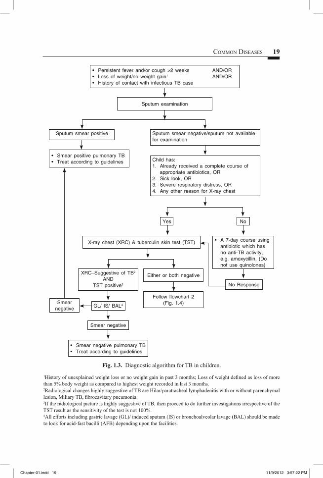
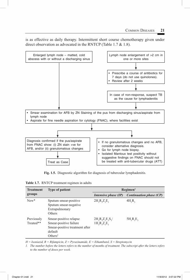
illness that includes cough and fever, weight loss or failure to thrive, an inability to return to normal health after measles or whooping cough, and history of contact with an adult case of pulmonary tuberculosis. The diagnosis of tuberculosis in children is extremely challenging due to relative inability to demonstrate AFB- the gold standard (Figs. 1.3 & 1.4).Diagnostic algorithm for TB lymphadenitis is given in Fig. 1.5. �
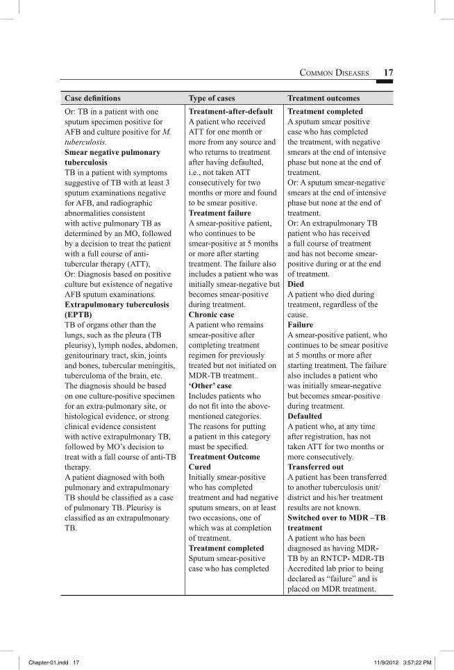
Table 1.6. Defi ning and documentation of TB
Case defi nitions Type of cases Treatment outcomesSmear positive pulmonary TB (PTB)TB in a patient with at least two initial sputum smear examinations (direct smear microscopy) positive for AFB,Or: TB in a patient with one sputum examination positive for AFB and radiographic abnormalities consistent with active pulmonary TB as determined by the treating medical offi cer (MO).
New caseA patient who has never taken treatment for TB or has taken ATT for less than 1 month.RelapseA patient declared cured of TB by a physician, but who reports back to the health service and is found to be bacteriologically positive.
CuredAn initially smear-positive patient, who has completed the treatment and has negative sputum smears on at least 2 occasions (one of which is at completion of treatment).
Chapter-01.indd 16Chapter-01.indd 16 11/9/2012 3:57:22 PM11/9/2012 3:57:22 PM
COMMON DISEASES 17
Case defi nitions Type of cases Treatment outcomesOr: TB in a patient with one sputum specimen positive for AFB and culture positive for M. tuberculosis.Smear negative pulmonary tuberculosisTB in a patient with symptoms suggestive of TB with at least 3 sputum examinations negative for AFB, and radiographic abnormalities consistent with active pulmonary TB as determined by an MO, followed by a decision to treat the patient with a full course of anti-tubercular therapy (ATT),Or: Diagnosis based on positive culture but existence of negative AFB sputum examinations.Extrapulmonary tuberculosis (EPTB)TB of organs other than the lungs, such as the pleura (TB pleurisy), lymph nodes, abdomen, genitourinary tract, skin, joints and bones, tubercular meningitis, tuberculoma of the brain, etc.The diagnosis should be based on one culture-positive specimen for an extra-pulmonary site, or histological evidence, or strong clinical evidence consistent with active extrapulmonary TB, followed by MO’s decision to treat with a full course of anti-TB therapy.A patient diagnosed with both pulmonary and extrapulmonary TB should be classifi ed as a case of pulmonary TB. Pleurisy is classifi ed as an extrapulmonary TB.
Treatment-after-defaultA patient who received ATT for one month or more from any source and who returns to treatment after having defaulted, i.e., not taken ATT consecutively for two months or more and found to be smear positive.Treatment failureA smear-positive patient, who continues to be smear-positive at 5 months or more after starting treatment. The failure also includes a patient who was initially smear-negative but becomes smear-positive during treatment.Chronic caseA patient who remains smear-positive after completing treatment regimen for previously treated but not initiated on MDR-TB treatment..‘Other’ caseIncludes patients who do not fi t into the above-mentioned categories. The reasons for putting a patient in this category must be specifi ed.Treatment OutcomeCuredInitially smear-positive who has completed treatment and had negative sputum smears, on at least two occasions, one of which was at completion of treatment.Treatment completedSputum smear-positive case who has completed
Treatment completedA sputum smear positive case who has completed the treatment, with negative smears at the end of intensive phase but none at the end of treatment.Or: A sputum smear-negative smears at the end of intensive phase but none at the end of treatment.Or: An extrapulmonary TB patient who has received a full course of treatment and has not become smear-positive during or at the end of treatment.DiedA patient who died during treatment, regardless of the cause.FailureA smear-positive patient, who continues to be smear positive at 5 months or more after starting treatment. The failure also includes a patient who was initially smear-negative but becomes smear-positive during treatment.DefaultedA patient who, at any time after registration, has not taken ATT for two months or more consecutively.Transferred outA patient has been transferred to another tuberculosis unit/district and his/her treatment results are not known.Switched over to MDR –TB treatmentA patient who has been diagnosed as having MDR-TB by an RNTCP- MDR-TB Accredited lab prior to being declared as “failure” and is placed on MDR treatment.
Chapter-01.indd 17Chapter-01.indd 17 11/9/2012 3:57:22 PM11/9/2012 3:57:22 PM
18 STANDARD TREATMENT GUIDELINES
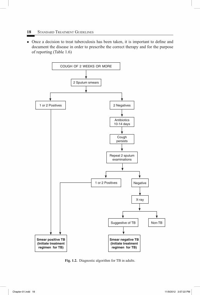
Once a decision to treat tuberculosis has been taken, it is important to defi ne and �
document the disease in order to prescribe the correct therapy and for the purpose of reporting (Table 1.6)
COUGH OF 2 WEEKS OR MORE
2 Sputum smears
1 or 2 Positives
1 or 2 Positives
Suggestive of TB
Smear negative TB (Initiate treatment regimen for TB)
Smear positive TB (Initiate treatment regimen for TB)
2 Negatives
Antibiotics 10-14 days
Cough persists
Repeat 2 sputum examinations
X-ray
Negative
Non-TB
Fig. 1.2. Diagnostic algorithm for TB in adults.
Chapter-01.indd 18Chapter-01.indd 18 11/9/2012 3:57:22 PM11/9/2012 3:57:22 PM
COMMON DISEASES 19
No
• Persistent fever and/or cough >2 weeks AND/OR• Loss of weight/no weight gain1 AND/OR• History of contact with infectious TB case
• Smear positive pulmonary TB• Treat according to guidelines
• A 7-day course using antibiotic which has no anti-TB activity, e.g. amoxycillin, (Do not use quinolones)
No Response
X-ray chest (XRC) & tuberculin skin test (TST)
Sputum examination
Sputum smear positive
XRC–Suggestive of TB2
ANDTST positive3
GL/ IS/ BAL4
Smear negative
Smear negative
Either or both negative
Follow fl owchart 2 (Fig. 1.4)
Yes
Sputum smear negative/sputum not available for examination
Child has:1. Already received a complete course of
appropriate antibiotics, OR2. Sick look, OR3. Severe respiratory distress, OR4. Any other reason for X-ray chest
• Smear negative pulmonary TB• Treat according to guidelines
Fig. 1.3. Diagnostic algorithm for TB in children.
1History of unexplained weight loss or no weight gain in past 3 months; Loss of weight defi ned as loss of more than 5% body weight as compared to highest weight recorded in last 3 months.2Radiological changes highly suggestive of TB are Hilar/paratracheal lymphadenitis with or without parenchymal lesion, Miliary TB, fi brocavitary pneumonia.3If the radiological picture is highly suggestive of TB, then proceed to do further investigations irrespective of the TST result as the sensitivity of the test is not 100%.4All efforts including gastric lavage (GL)/ induced sputum (IS) or bronchoalveolar lavage (BAL) should be made to look for acid-fast bacilli (AFB) depending upon the facilities.
Chapter-01.indd 19Chapter-01.indd 19 11/9/2012 3:57:22 PM11/9/2012 3:57:22 PM
20 STANDARD TREATMENT GUIDELINES
Treatment
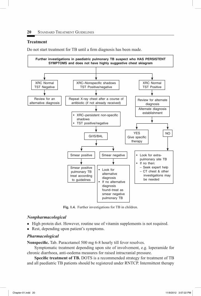
Do not start treatment for TB until a fi rm diagnosis has been made.
Further investigations in paediatric pulmonary TB suspect who HAS PERSISTENT SYMPTOMS and does not have highly suggestive chest skiagram
XRC NormalTST Negative
Review for an alternative diagnosis
YESGive specifi c
therapy
• Look for extra-pulmonary site TB
• If no then:– Seek expert help– CT chest & other
investigations may be needed
Gl/IS/BAL
Smear positive
Smear positive pulmonary TB treat according to guidelines
NO
XRC NormalTST Positive
Review for alternate diagnosis
Alternate diagnosis establishment
XRC–Nonspecifi c shadowsTST Positive/negative
Repeat X-ray chest after a course of antibiotic (if not already received)
• XRC–persistent non-specifi c shadows
• TST positive/negative
Smear negative
• Look for alternative diagnosis
• If no alternative diagnosis found–treat as smear negative pulmonary TB
Fig. 1.4. Further investigations for TB in children.
Nonpharmacological
High protein diet. However, routine use of vitamin supplements is not required. �
Rest, depending upon patient’s symptoms. �
Pharmacological
Nonspecifi c. Tab. Paracetamol 500 mg 6-8 hourly till fever resolves. Symptomatic treatment depending upon site of involvement, e.g. loperamide for
chronic diarrhoea, anti-oedema measures for raised intracranial pressure.Specifi c treatment of TB. DOTS is a recommended strategy for treatment of TB
and all paediatric TB patients should be registered under RNTCP. Intermittent therapy
Chapter-01.indd 20Chapter-01.indd 20 11/9/2012 3:57:22 PM11/9/2012 3:57:22 PM
COMMON DISEASES 21
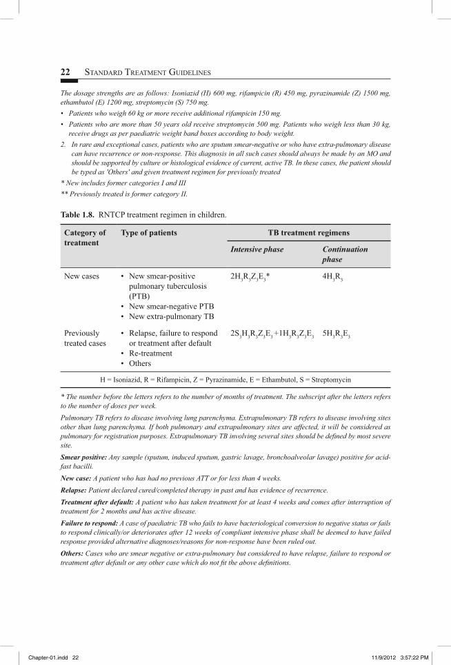
is as effective as daily therapy. Intermittent short course chemotherapy given under direct observation as advocated in the RNTCP (Table 1.7 & 1.8).
• Smear examination for AFB by ZN Staining of the pus from discharging sinus/aspirate from lymph node
• Aspirate for fi ne needle aspiration for cytology (FNAC), where facilities exist
Diagnosis confi rmed if the pus/aspirate from FNAC show: (i) ZN stain +ve for AFB, and/or (ii) granulomatous changes
• If no granulomatous changes and no AFB, consider alternative diagnosis.
• Go for lymph node biopsy.• Isolated Mantoux test positivity without
suggestive fi ndings on FNAC should not be treated with anti-tubercular drugs (ATT)Treat as Case
Enlarged lymph node – matted, cold abscess with or without a discharging sinus
Lymph node enlargement of >2 cm in one or more sites
In case of non-response, suspect TB as the cause for lymphadenitis
• Prescribe a course of antibiotics for 7 days (do not use quinolones).
• Review after 2 weeks
Fig. 1.5. Diagnostic algorithm for diagnosis of tubercular lymphadenitis.
Table 1.7. RNTCP treatment regimen in adults
Treatment groups
Type of patient Regimen1
Intensive phase (IP) Continuation phase (CP)
New* Sputum smear-positiveSputum smear-negativeExtrapulmonaryOthers
2H3R3Z3E3 4H3R3
Previously Treated**
Smear-positive relapseSmear-positive failureSmear-positive treatment after defaultOthers2
2H3R3Z3E3S3/ 1H3R3Z3E3
5H3R3E3
H = Isoniazid, R = Rifampicin, Z = Pyrazinamide, E = Ethambutol, S = Streptomycin1. The number before the letters refers to the number of months of treatment. The subscript after the letters refers
to the number of doses per week.
Chapter-01.indd 21Chapter-01.indd 21 11/9/2012 3:57:22 PM11/9/2012 3:57:22 PM
22 STANDARD TREATMENT GUIDELINES
The dosage strengths are as follows: Isoniazid (H) 600 mg, rifampicin (R) 450 mg, pyrazinamide (Z) 1500 mg, ethambutol (E) 1200 mg, streptomycin (S) 750 mg.• Patients who weigh 60 kg or more receive additional rifampicin 150 mg.• Patients who are more than 50 years old receive streptomycin 500 mg. Patients who weigh less than 30 kg,
receive drugs as per paediatric weight band boxes according to body weight.2. In rare and exceptional cases, patients who are sputum smear-negative or who have extra-pulmonary disease
can have recurrence or non-response. This diagnosis in all such cases should always be made by an MO and should be supported by culture or histological evidence of current, active TB. In these cases, the patient should be typed as 'Others' and given treatment regimen for previously treated
* New includes former categories I and III** Previously treated is former category II.
Table 1.8. RNTCP treatment regimen in children.
Category of treatment
Type of patients TB treatment regimens
Intensive phase Continuation phase
New cases • New smear-positive pulmonary tuberculosis (PTB)
• New smear-negative PTB• New extra-pulmonary TB
2H3R3Z3E3* 4H3R3
Previously treated cases
• Relapse, failure to respond or treatment after default
• Re-treatment • Others
2S3H3R3Z3E3 +1H3R3Z3E3 5H3R3E3
H = Isoniazid, R = Rifampicin, Z = Pyrazinamide, E = Ethambutol, S = Streptomycin
* The number before the letters refers to the number of months of treatment. The subscript after the letters refers to the number of doses per week.Pulmonary TB refers to disease involving lung parenchyma. Extrapulmonary TB refers to disease involving sites other than lung parenchyma. If both pulmonary and extrapulmonary sites are affected, it will be considered as pulmonary for registration purposes. Extrapulmonary TB involving several sites should be defi ned by most severe site.Smear positive: Any sample (sputum, induced sputum, gastric lavage, bronchoalveolar lavage) positive for acid-fast bacilli.New case: A patient who has had no previous ATT or for less than 4 weeks.Relapse: Patient declared cured/completed therapy in past and has evidence of recurrence.Treatment after default: A patient who has taken treatment for at least 4 weeks and comes after interruption of treatment for 2 months and has active disease.Failure to respond: A case of paediatric TB who fails to have bacteriological conversion to negative status or fails to respond clinically/or deteriorates after 12 weeks of compliant intensive phase shall be deemed to have failed response provided alternative diagnoses/reasons for non-response have been ruled out.Others: Cases who are smear negative or extra-pulmonary but considered to have relapse, failure to respond or treatment after default or any other case which do not fi t the above defi nitions.
Chapter-01.indd 22Chapter-01.indd 22 11/9/2012 3:57:22 PM11/9/2012 3:57:22 PM

COMMON DISEASES 23
In patients with TB meningitis on Category I treatment, the four drugs used during the intensive phase can either be HRZE or HRZS. The present evidence suggests that ethambutol can be used in children.
Children who show poor or no response at 8 weeks of intensive phase may be given benefi t of extension of IP for one more month. In patients with TB meningitis, spinal TB, miliary/disseminated TB and osteoarticular TB, the continuation phase shall be extended by 3 months making the total duration of treatment to a total of 9 months. A further extension may be done for 3 more months in continuation phase (making the total duration of treatment to 12 months) on a case to case basis in case of delayed response and as per the discretion of the treating physician.
Steroids should be used initially in hospitalized cases of TBM and TB pericarditis and reduced gradually over 6 to 8 weeks. In all instances before starting a child on previously treated regimen, patient should be examined by a paediatrician or TB expert, whichever available.
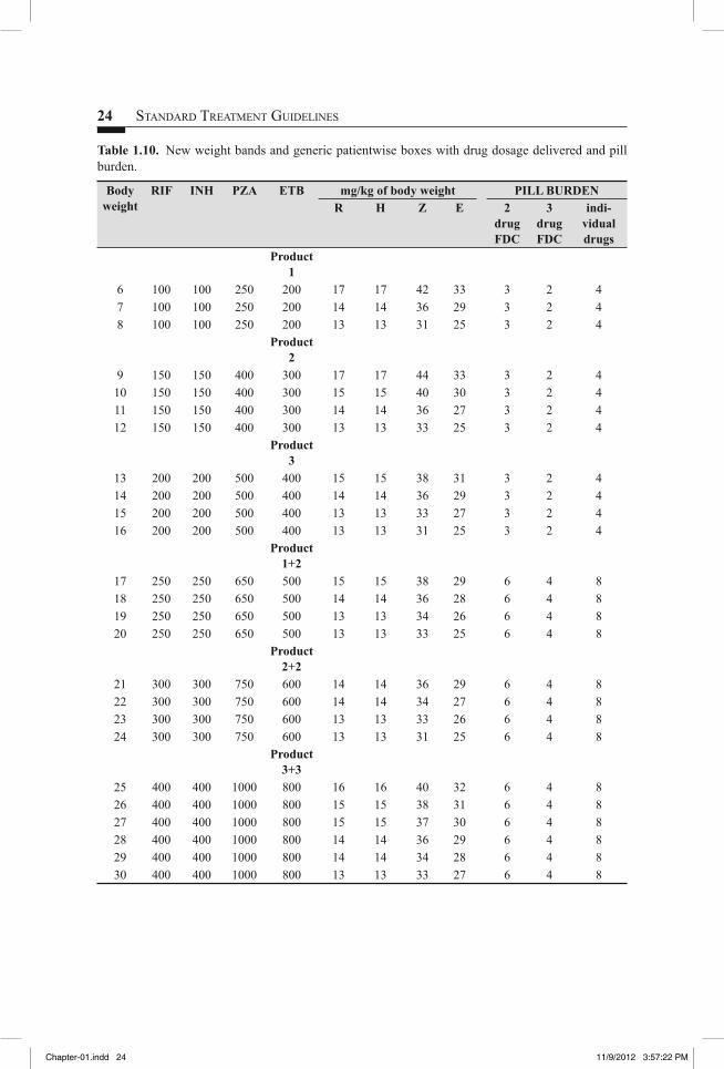
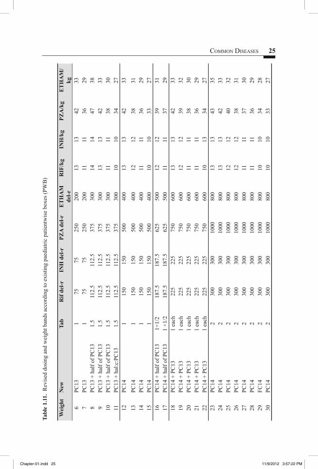
Children can tolerate much higher doses than the adults so while calculating the dose, do not round off to a lower amount of drug. As children can have signifi cant increase in body weight on treatment, the doses may be increased in proportion of increase in body weight (Table 1.9-1.11).
Table 1.9. Drug dosage charts for the anti-tuberculosis drugs
Drug Formulations Daily therapy
Thrice weekly therapy
Route, frequency
(mg/kg/day)
INH (H) Tab. 100, 300 mgSyrup 100 mg/5 ml
10 15 (12-17) Oral, once a day
Rifampicin (R) Cap 150, 300, 450, 600 mg Susp. 100 mg/5 ml
10 15(12-17) Oral, once a day Empty stomach
Pyrazinamide (Z)
Tab. 500, 750, 1000 mg, Syrup 300 mg/5 ml
25-35 35(30-40) Oral, once a day
Ethambutol (E) Tab. 200, 400, 800, 1000 mg
15 30(25-30) Oral, once a day
Streptomycin (S)
Inj. 500, 750, 1000 mg
15 15 Intramuscular, once a day
Chapter-01.indd 23Chapter-01.indd 23 11/9/2012 3:57:22 PM11/9/2012 3:57:22 PM
24 STANDARD TREATMENT GUIDELINES
Table 1.10. New weight bands and generic patientwise boxes with drug dosage delivered and pill burden.
Body weight
RIF INH PZA ETB mg/kg of body weight PILL BURDENR H Z E 2
drug FDC
3 drug FDC
indi-vidual drugs
Product 1
6 100 100 250 200 17 17 42 33 3 2 47 100 100 250 200 14 14 36 29 3 2 48 100 100 250 200 13 13 31 25 3 2 4
Product 2
9 150 150 400 300 17 17 44 33 3 2 410 150 150 400 300 15 15 40 30 3 2 411 150 150 400 300 14 14 36 27 3 2 412 150 150 400 300 13 13 33 25 3 2 4
Product 3
13 200 200 500 400 15 15 38 31 3 2 414 200 200 500 400 14 14 36 29 3 2 415 200 200 500 400 13 13 33 27 3 2 416 200 200 500 400 13 13 31 25 3 2 4
Product 1+2
17 250 250 650 500 15 15 38 29 6 4 818 250 250 650 500 14 14 36 28 6 4 819 250 250 650 500 13 13 34 26 6 4 820 250 250 650 500 13 13 33 25 6 4 8
Product 2+2
21 300 300 750 600 14 14 36 29 6 4 822 300 300 750 600 14 14 34 27 6 4 823 300 300 750 600 13 13 33 26 6 4 824 300 300 750 600 13 13 31 25 6 4 8
Product 3+3
25 400 400 1000 800 16 16 40 32 6 4 826 400 400 1000 800 15 15 38 31 6 4 827 400 400 1000 800 15 15 37 30 6 4 828 400 400 1000 800 14 14 36 29 6 4 829 400 400 1000 800 14 14 34 28 6 4 830 400 400 1000 800 13 13 33 27 6 4 8
Chapter-01.indd 24Chapter-01.indd 24 11/9/2012 3:57:22 PM11/9/2012 3:57:22 PM
COMMON DISEASES 25Ta
ble
1.11
. R
evis
ed d
osin
g an
d w
eigh
t ban
ds a
ccor
ding
to e
xist
ing
paed
iatri
c pa
tient
wis
e bo
xes (
PWB
)
Wei
ght
New
Tab
Rif
del-r
INH
del
-rPZ
A d
el-r
ET
HA
M
del-r
RIF
/kg
INH
/kg
PZA
/kg
ET
HA
M/
kg6
PC13
175
7525
020
013
1342
337
PC13
175
7525
020
011
1136
298
PC13
+ h
alf o
f PC
131.
511
2.5
112.
537
530
014
1447
389
PC13
+ h
alf o
f PC
131.
511
2.5
112.
537
530
013
1342
3310
PC13
+ h
alf o
f PC
131.
511
2.5
112.
537
530
011
1138
3011
PC13
+ h
al:c
:PC
131.
511
2.5
112.
537
530
010
1034
2712
PC14
115
015
050
040
013
1342
3313
PC14
115
015
050
040
012
1238
3114
PC14
115
015
050
040
011
1136
2915
PC14
115
015
050
040
010
1033
2716
PC14
+ h
alf o
f PC
131+
1/2
187.
518
7.5
625
500
1212
3931
17PC
14 +
hal
f of P
C13
1 +1
/218
7.5
187.
562
550
011
1137
2918
PC14
+ P
C13
1 ea
ch22
522
575
060
013
1342
3319
PC14
+ P
C13
1 ea
ch22
522
575
060
012
1239
3220
PC14
+ P
C13
1 ea
ch22
522
575
060
011
1138
3021
PC14
+ P
C13
1 ea
ch22
522
575
060
011
1136
2922
PC14
+ P
C13
1 ea
ch22
522
575
060
010
1334
2723
PC14
230
030
010
0080
013
1343
3524
PC14
230
030
010
0080
013
1342
3325
PC14
230
030
010
0080
012
1240
3226
PC14
230
030
010
0080
012
1238
3127
PC14
230
030
010
0080
011
1137
3028
PC14
230
030
010
0080
011
1136
2929
FC14
230
030
010
0080
010
1034
2830
PC14
230
030
010
0080
010
1033
27
Chapter-01.indd 25Chapter-01.indd 25 11/9/2012 3:57:22 PM11/9/2012 3:57:22 PM
26 STANDARD TREATMENT GUIDELINES
Treatment in special situations
Treatment of MDR tuberculosis (To be treated under DOTS Plus of RNTCP).Very important to prevent MDR by avoiding monotherapy/poor compliance to �
treatment.Drugs susceptibility testing should be done. If not available, treatment regimen as �
above. Patients with meningitis, bone and joint tuberculosis and miliary TB should receive minimum of 12 months of treatment.
Pregnant women. Avoid Pyrazinamide, Streptomycin. Give Isoniazid (INH), Rifampicin and Ethambutol for 2 months followed by INH and Rifampicin for 7 months. Lactating women can continue to breastfeed.
Patients with renal disease.Avoid aminoglycosides. �
Avoid Ethambutol and monitor for side effects. �
Reduce doses of INH and Pyrazinamide in cases of severe renal failure. �
Patient with hepatic disease. Avoid INH, Rifampicin and Pyrazinamide.
Patients with HIV/AIDS. All patients diagnosed as TB cases should be referred to nearest ICTC for HIV testing. ART to be given to all patients with extrapulmonary TB (stage 4) and all those with pulmonary TB (stage 3) with CD4 count <350 cells/ cu mm (for details see section on AIDS in Chapter 7).
Patients with pericardial effusion, severe pleural effusion, meningitis. Steroid (oral/injectable) to be given along with the antitubercular therapy.
In tubercular meningitis (see section on tubercular meningitis)
Tubercular pericarditis. In addition to ATT, Tab. Prednisolone 40-60 mg for 2 weeks with gradual tapering over next 4 weeks.
Pleural effusion. In addition to ATT, Tab. Prednisolone may be considered, in patients who are toxic or with large effusions.
Chemoprophylaxis
The dose of INH for chemoprophylaxis was recommended to be 10 mg/kg administered daily for 6 months. TB preventive therapy should be provided to:
All asymptomatic contacts (under 6 years of age) of a smear positive case, after �
ruling out active disease and irrespective of their BCG or nutritional status.Chemoprophylaxis is also recommended for all HIV-infected children who either �
had a known exposure to an infectious TB case or are tuberculin skin test (TST) positive (≥5 mm induration) but have no active TB disease.All tuberculin skin test (TST) positive children who are receiving immunosuppressive �
therapy (e.g. children with nephrotic syndrome, acute leukaemia, etc.).
Chapter-01.indd 26Chapter-01.indd 26 11/9/2012 3:57:22 PM11/9/2012 3:57:22 PM
COMMON DISEASES 27
A child born to mother who was diagnosed to have TB in pregnancy should receive �
prophylaxis for 6 months, provided congenital TB has been ruled out.BCG vaccination can be given at birth even if INH chemoprophylaxis is planned.
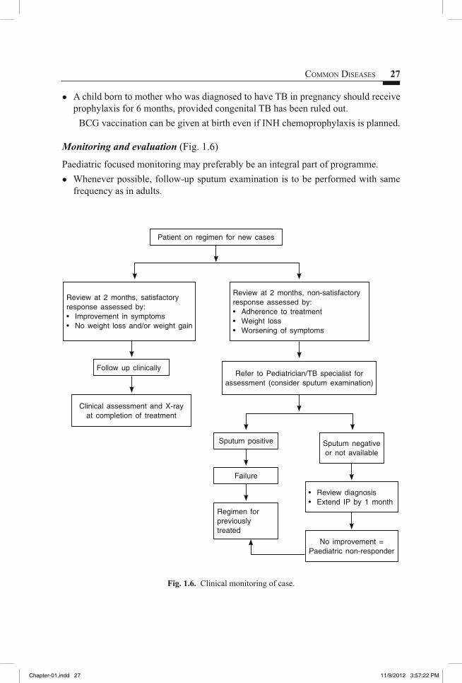
Monitoring and evaluation (Fig. 1.6)
Paediatric focused monitoring may preferably be an integral part of programme.Whenever possible, follow-up sputum examination is to be performed with same �
frequency as in adults.
Patient on regimen for new cases
Review at 2 months, non-satisfactory response assessed by:• Adherence to treatment• Weight loss• Worsening of symptoms
Sputum positive
Failure
Regimen for previously treated
Sputum negative or not available
• Review diagnosis• Extend IP by 1 month
No improvement = Paediatric non-responder
Refer to Pediatrician/TB specialist for assessment (consider sputum examination)
Clinical assessment and X-ray at completion of treatment
Follow up clinically
Review at 2 months, satisfactory response assessed by:• Improvement in symptoms• No weight loss and/or weight gain
Fig. 1.6. Clinical monitoring of case.
Chapter-01.indd 27Chapter-01.indd 27 11/9/2012 3:57:22 PM11/9/2012 3:57:22 PM

28 STANDARD TREATMENT GUIDELINES
Clinical symptomatic improvement is to be assessed at the end of intensive phase �
of treatment and at the end of treatment. Improvement should be judged by absence of fever or cough, a decrease in size of lymph node(s) and weight gain/no weight loss.Radiological improvement is to be assessed by chest X-ray examination in all smear- �
negative pulmonary TB cases at end of treatment.DOTS is the recommended strategy for treatment in adults and children. All paediatric TB patients should be registered under RNTCP. It is important to ensure completion of treatment in every case put on treatment to prevent emergence of resistance, particularly to Rifampicin. In the rare circumstances where a patient is given daily therapy, observation and completion of therapy remains as important. It is the duty of the prescriber to ensure appropriate and complete treatment in all cases. Management of patients with treatment interruption is shown in Fig. 1.7.
Treatment Interruption
Interruption of <2 months
Initial treatment of <1 month
Continue same treatment to complete all doses
Initial treatment of >1 month
Repeat smear examination
PositiveRetain original type. Start on treatment afresh with same regimen
Type TADStart on regimens
for previously treated
Type othersStart on regimens
for previously treated
Interruption of >2 months (outcome - default)
Negative
Fig. 1.7. Management of patients with treatment interruptions.
Chapter-01.indd 28Chapter-01.indd 28 11/9/2012 3:57:22 PM11/9/2012 3:57:22 PM

COMMON DISEASES 29
Recording and reporting
In addition to the existing information, especially in relation to paediatric TB patients, the treatment card should include information on:
Basis for starting treatment along with categorization. �
Documentation of clinical and radiological monitoring as described above. This �
information could be clubbed with the table for laboratory results in the present treatment card.X-rays should be retained until treatment completion, and a drawing of the X-ray �
picture with comments, entered in the remarks column.Provision to check correct categorization and drug dosages. A dosage table based on �
patient’s weight could be printed on the card to ensure correct dosage for the child.
Assessment of response to therapy
1. The short course chemotherapy as enlisted above leads to a rapid clinical response in most patients in 2-4 weeks. Inadequate combination or dosage of this can lead to emergence of resistance and should be avoided at all cost.
2. The response to therapy should be monitored by bacteriological conversion in positive cases and by other markers like clinical and/or radiological improvement in AFB negative cases at the end of 2 months of intensive phase. A bacteriological conversion in over 80% of cases after 2 months of therapy is expected. If the patient continues to excrete bacteria after 2 months, the intensive phase needs to be extended by a month, and also ensure patient compliance, as non-adherence is the most common cause for non-response. If a patient continues to be symptomatic or bacteriologically positive after an extended phase of IP, then the patient should be extensively re-evaluated and treatment failure/drug resistance should be suspected. The patient should be referred to a higher centre for further management. Remember persistence or recurrence of symptoms or radiological shadow could be due to secondary or coinfection with other organisms or due to a non-tuberculous lesion. Radiological response may lag behind bacteriologic cure and hence should not be the deciding factor for stopping of treatment. In patients with extrapulmonary tuberculosis, the response to treatment is assessed clinically.
3. All patients should have baseline LFTs; should be monitored regularly in patients at high risk of hepatitis, e.g. old patients, alcoholics, diabetics and malnourished.
4. Monitoring and management of side effects: The suggested therapy is usually well tolerated. However, some patients can develop GI intolerance, vomiting, etc. for which only symptomatic therapy is required. Commonest major side effect with suggested regime is drug-induced hepatitis. The easily recognizable symptom of high-coloured urine in jaundiced patient is masked due to discolouration of urine because of rifampicin. Suspect hepatitis, if vomiting is persistent and associated with anorexia. Clinically, icterus may be evident. In all cases of jaundice, stop treatment and refer to a higher centre for evaluation. In most patients, the drugs can be reintroduced after the hepatitis has resolved. Pyrazinamide-induced
Chapter-01.indd 29Chapter-01.indd 29 11/9/2012 3:57:22 PM11/9/2012 3:57:22 PM

30 STANDARD TREATMENT GUIDELINES
arthralgia or arthritis usually responds suitably to analgesic therapy. Drug rash and hypersensitivity is a major side effect where patient needs to be referred to a higher centre. Peripheral neuropathy due to INH is treated with oral vitamin B6. Ethambutol can cause optic neuritis particularly when used in high doses and requires omission of the drug once this side effect occurs.
5. In case of hypersensitivity reaction, discontinue all drugs, re-challenge with individual drug to determine the likely offending drug. Do not reintroduce rifampicin in patients who develop thrombocytopenia. Hyperuricaemia can occur due to pyrazinamide. Needs to be discontinued only in case of secondary gout.
Patient education
The patients should be impressed upon the necessity of complying with periodic �
follow-up sputum examination schedule as advised. In case patients experience any unusual symptoms after initiation/during treatment, �
they should be instructed to approach the medical offi cer and report the same. On their own, they should not take a decision either to stop or to continue the drugs.Smoking of tobacco adversely affects the treatment outcome and, therefore, give �
simple tips to quit smoking and refer to the smoking cessation clinic and protect from passive smoking. The environment of the patient has to be smoke free at home/offi ce and at clinic. Check smoking status of the TB patient at every interaction. Alcohol abuse: � Elicit history of addiction to alcohol and if found alcoholic, advise to strictly refrain from alcohol as it would increase the chances of patient developing hepatitis (jaundice), irregularity in drug intake and adverse treatment outcome. Rifampicin colours the urine as well as other body secretions orange-red. Patient �
must be warned about this to avoid unnecessary alarm. The patient should also be advised to take Rifampicin on an empty stomach and not to take any meals for about 1 hour afterwards for good absorption of the drug.The patient or the primary caregiver must be advised regarding the probable side �
effects and explained when to contact the treating doctor.A health functionary should preferably supervise the treatment of tuberculosis as �
far as possible. However, it is of utmost importance that the patient and the family are informed about the need to complete all the treatment for whole of the duration. They must be explained the need for prolonged therapy even after the sickness disappears (symptoms abate). Inadequate or incomplete treatment increases the chance of multidrug resistance which is diffi cult to treat.Importance of screening symptomatic contacts and children below 6 years: � Encourage patients to bring symptomatic adult contacts and all children aged six years and below for screening at health facility for early detection of cases among them and appropriate treatment. Eligible children will be administered chemoprophylaxis.Proper sputum disposal and personal hygiene (covering the mouth while coughing) �
should be explained for infectious patients.The fears of the patient and/or the caregiver regarding the disease should be �
addressed as this disease has a lot of social stigma.
Chapter-01.indd 30Chapter-01.indd 30 11/9/2012 3:57:22 PM11/9/2012 3:57:22 PM

COMMON DISEASES 31
Ethambutol is a hygroscopic drug which tends to crumble, if not properly stored, �
particularly during rainy season.
References
1. Tuberculosis. In: Harrison’s Principles of Internal Medicine. Fauci, Braunwald, Kasper et al (eds), 18th Edition, McGraw Hill Company Inc., New York, 2012; pp 1340-1359.
2. RNTCP: TB in Children. Consensus Guidelines of Paediatricians, TB experts and TB Control Programme Managers, Central TB Divisons. Directorate of Health and Family Welfare, Nirman Bhavan, New Delhi, 2012.
3. Technical Guidelines for Tuberculosis Control. Central TB Division. Directorate General of Health Services, Ministry of Health and Family Welfare, Nirman Bhawan, New Delhi, 2009.
MALARIA AND NATIONAL ANTI-MALARIA DRUG POLICY (2010)
Parasitic infection due to protozoa of genus Plasmodium transmitted by the female Anopheles mosquito. There are four plasmodia species: P. falciparum, P. vivax, P. malariae, and P. ovale.
SALIENT FEATURES� Malaria is an acute and chronic protozoan illness characterized by paroxysms
of fever, chills, sweats, fatigue, anaemia and splenomegaly. In atypical cases, classical symptoms may not manifest.
� Falciparum malaria (severe and complicated malaria) severe manifestations can develop over a short span of 12-24 hours and is associated in varying degrees with the following clinical signs:
Cerebral: Mental clouding, coma, convulsions, delirium and occasionally localizing signs. Hyperpyrexia (>40.5ºC), haemolysis, haematocrit <15% or Hb <5 g/dl, hypoglycaemia, oliguria, anuria, pulmonary oedema, macroscopic haemoglobinuria and jaundice.
� Diagnosis is made by presence of protozoa in the blood in thick and thin smear slides. Thick smear for easy detection of parasite and thin smear for identifi cation of species. Note that blood fi lms may be negative even in a severe attack because of sequestration of parasites in the deep capillaries. Rapid diagnostic kits (RDK) can be used for detection of P. falciparum where microscopy results are not obtainable within 24 hours of sample collection
Treatment of malaria 1. All fever cases suspected to be malaria should be investigated by microscopy or
RDT. 2. Patients of uncomplicated malaria can be managed at primary level but patients with
severe malaria with complications should be admitted and managed in a hospital where facilities for detailed investigations and blood transfusion exist.
Chapter-01.indd 31Chapter-01.indd 31 11/9/2012 3:57:22 PM11/9/2012 3:57:22 PM
32 STANDARD TREATMENT GUIDELINES
3. P. vivax cases should be treated with chloroquine for three days and Primaquine for 14 days. Primaquine is used to prevent relapse but is contraindicated in pregnant women, infants and individuals with G6PD defi ciency. Note: Patients should be instructed to report back in case of haematuria or high-coloured urine/cyanosis or blue coloration of lips and Primaquine should be stopped in such cases. Care should be taken in patients with anaemia.
4. P. falciparum cases should be treated with ACT (Artesunate 3 days + Sulphadoxine- Pyrimethamine 1 day). This is to be accompanied by single dose primaquine on day 2.
5. Pregnant women with uncomplicated P. falciparum should be treated as follows:1st trimester: Quinine �
2nd & 3rd trimester: ACT �
Note: Primaquine is contraindicated in pregnant woman. 6. In cases where parasitological diagnosis is not possible due to non-availability of
either timely microscopy or RDT, suspected malaria cases should be treated with full course of chloroquine, till the results of microscopy are received. Once the parasitological diagnosis is available, appropriate treatment as per the species, is to be administered.
7. Presumptive treatment with chloroquine is no more recommended. 8. Resistance should be suspected, if in spite of full treatment with no history of
vomiting, diarrhoea, patient does not respond within 72 hours, clinically and parasitologically. Such cases not responding to ACT, should be treated with oral Quinine with Tetracycline/Doxycycline. These instances should be reported to concerned District Malaria /State Malaria Offi cer/ROHFW for initiation of therapeutic effi cacy studies.
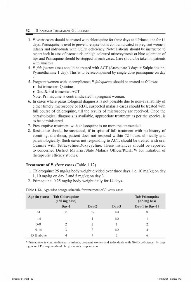
Treatment of P. vivax cases (Table 1.12) 1. Chloroquine: 25 mg/kg body weight divided over three days, i.e. 10 mg/kg on day
1, 10 mg/kg on day 2 and 5 mg/kg on day 3. 2. Primaquine: 0.25 mg/kg body weight daily for 14 days.
Table 1.12. Age-wise dosage schedule for treatment of P. vivax cases
Age (in years) Tab Chloroquine(150 mg base)
Tab Primaquine (2.5 mg base
Day-1 Day-2 Day-3 Day-1 to Day-14<1 ½ ½ 1/4 0
1-4 1 1 1/2 15-8 2 2 1 29-14 3 3 1/2 4
15 & above 4 4 2 6
* Primaquine is contraindicated in infants, pregnant women and individuals with G6PD defi ciency. 14 days regimen of Primaquine should be given under supervision
Chapter-01.indd 32Chapter-01.indd 32 11/9/2012 3:57:22 PM11/9/2012 3:57:22 PM

COMMON DISEASES 33
Treatment of uncomplicated P. falciparum cases (Table 1.13) 1. Artemisinin based combination therapy (ACT): Artesunate 4 mg/kg body weight
daily for 3 days plus Sulfadoxine (25 mg/kg body weight) -Pyrimethamine (1.25 mg/kg body weight) on fi rst day.
(Caution: ACT is not to be given in 1st trimester of pregnancy). 2. Primaquine: 0.75 mg/kg body weight on day 2: 0.75 mg/kg body weight on
day 2.Table 1.13. Age-wise dosage schedule for treatment of P. falciparum cases
Age (in years) 1st day 2nd day 3rd dayArtesunate
(50 mg)SP* Artesunate
(50 mg)Primaquine
(7.5 mgBase)
Artesunate(50 mg)
<1 ¼ ¼ ½ 0 1/21-4 1 1 1 1 15-8 2 ½ 2 2 29-14 3 2 3 4 315 & above 4 3 4 6 4
Treatment of uncomplicated P. falciparum cases in pregnancy
1st trimester: Quinine salt 10 mg/kg 3 times daily for 7 days (Caution: Quinine may induce hypoglycaemia; pregnant women should not start taking quinine on an empty stomach and should eat regularly, while on quinine treatment).
2nd and 3rd trimesters: ACT as per dosage given above.
Treatment of mixed infections (P. vivax + P. falciparum) case
All mixed infections should be treated with full course of ACT and Primaquine 0.25 mg per kg body weight daily for 14 days.
Treatment of severe malaria cases
Severe malaria is an emergency and treatment should be given as per severity and associated complications which can best be decided by the treating physician.
Inj. Artesunate: 2.4 mg/kg body weight IV or IM given on admission (time = 0h); then at 12 h and 24 h and then once a day.
(Caution: Care should be taken to dilute artesunate powder in 5% sodium bicarbonate provided in the pack)
OrInj. Artemether: 3.2 mg/kg body weight IM given on admission and then 1.6 mg/
kg body weight per day.OrInj. Arteether: 150 mg IM daily for 3 days in adults only (not recommended for
children).
Chapter-01.indd 33Chapter-01.indd 33 11/9/2012 3:57:23 PM11/9/2012 3:57:23 PM

34 STANDARD TREATMENT GUIDELINES
OrQuinine: 20 mg/kg body weight on admission (IV infusion or divided IM injection)
followed by maintenance dose of 10 mg/kg body weight 8 hourly. The infusion rate should not exceed 5 mg salt/kg body weight per hour. Loading dose of Quinine, i.e. 20 mg/kg body weight on admission may not be given, if the patient has already received quinine or if the clinician feels inappropriate). NEVER GIVE BOLUS INJECTION OF QUININE. If parenteral quinine therapy needs to be continued beyond 48 hours, reduce dose to 7 mg/kg body weight 8 hourly. Note: The parenteral treatment in severe malaria cases should be given for minimum of 24 hours once started (irrespective of the patient’s ability to tolerate oral medication earlier than 24 hours).
After parenteral artemisinin therapy, patients will receive a full course of oral ACT for 3 days.
Those patients who received parenteral Quinine therapy and can take orally should receive: Oral Quinine 10 mg/kg body weight three times a day for 7 days (including the days, when parenteral Quinine was administered) plus Doxycycline 3 mg/kg body weight once a day or Clindamycin 10 mg/kg body weight 12-hourly for 7 days (Doxycycline is contraindicated in pregnant women and children under 8 years of age; instead, clindamycin 10 mg/kg body weight 12 hourly for 7 days should be used).
Patients receiving artemisinin derivatives should get full course of oral ACT. However, Act containing mefl oquine should be avoided in cerebral malaria due to neuropsychiatric complications.
Supportive treatment
Treat fever, hypoglycaemia, electrolyte imbalance, hypotension, renal failure, anaemia, convulsions appropriately (for details see respective sections).
Chemoprophylaxis
Chemoprophylaxis should be administered only in selective groups in high P. falciparum endemic areas. Use of personal protection measures including insecticide treated bed nets (ITN) / long lasting insecticidal nets (LLIN) should be encouraged for pregnant women and other vulnerable population including travellers for longer stay. However, for longer stay of military and para-military forces in high Pf endemic areas, the practice of chemoprophylaxis should be followed wherever appropriate, e.g. troops on night patrol duty and decisions of their medical administrative authority should be followed.
Short-term chemoprophylaxis (up to 6 weeks)
Tab. Doxycycline 100 mg once daily for adults and 1.5 mg/kg once daily for children (contraindicated in children below 8 years). The drug should be started 2 days before
Chapter-01.indd 34Chapter-01.indd 34 11/9/2012 3:57:23 PM11/9/2012 3:57:23 PM
COMMON DISEASES 35
travel and continued for 4 weeks after leaving the malarious area. Note: It is not recommended for pregnant women and children less than 8 years.
Chemoprophylaxis for longer stay (more than 6 weeks)
Tab. Mefl oquine 250 mg weekly for adults and should be administered two weeks before, during and four weeks after exposure. Note: Mefl oquine is contraindicated in individuals with history of convulsions, neuropsychiatric problems and cardiac conditions. Therefore, necessary precautions should be taken and all should undergo screening before prescription of the drug.
Patient education 1. To take measures to stop mosquito breeding and protection from mosquitoes, e.g.
mosquito nets, repellents, long sleeves, long trousers, etc. 2. Fever without any other signs and symptoms should be reported to nearest health
facility. 3. Chloroquine should be given with plenty of water after food and not on empty
stomach. If chloroquine syrup is not available for children, the tablet should be crushed and given with honey or thick syrup.
4. Watch for side effects of drugs prescribed. Chloroquine may cause nausea, vomiting and diarrhoea, mild headache and skin allergy/rash.
5. If vomiting occurs within 30 minutes of chloroquine intake, repeat the dose of chloroquine.
6. Chloroquine, primaquine and sulphadoxine + pyrimethamine should not be given, if patient is suffering from G6PD defi ciency.
7. To report back if develops haematuria or high-coloured urine, cyanosis and stop primaquine immediately.
8. If no improvement after 48 hours or if condition worsens, occurrence of cerebral malaria symptoms should seek medical help immediately.
References
1. National Antimalaria Programme (NAMP) Drug Policy, Government of India, Ministry of Health and Family Welfare, Directorate General of Health Services. Directorate of National Anti Malaria Programme. Delhi, 2010.
2. Malaria (Plasmodium). In: Nelson’s Textbook of Paediatrics. Behrman RE, Kliegman RM, Jenson HB (eds), 19th Edition, Harcourt Publishers International Company, 2011; pp 1198-1207.
3. Facility Based IMNCI (F-IMNCI) Participants manual. WHO, unicef, and Ministry of Health & Family Welfare, Government of India, 2009.
DENGUEDengue is the most important emerging tropical viral disease of human beings in the world today. Aedes aegypti, a day time mosquito, is the principal vector in India and countries of South-east Asian region, mostly seen in rainy season or in months following rainy season. All cases of dengue fever should be reported to the local/district/state health authorities, as it is a notifi able disease.
Chapter-01.indd 35Chapter-01.indd 35 11/9/2012 3:57:23 PM11/9/2012 3:57:23 PM
36 STANDARD TREATMENT GUIDELINES
SALIENT FEATURES• All four dengue virus types (Den 1, 2, 3 and 4) infections may be asymptomatic
or may lead to undifferentiated fever, dengue fever (DF), or dengue haemorrhagic fever (DHF) with plasma leakage that may lead to hypovolaemic shock, dengue shock syndrome (DSS).
• Dengue fever is an acute febrile illness of 2-7 days duration with two or more of the following manifestations: Headache, retro-orbital pain, myalgia/arthralgia, rash, haemorrhagic manifestation (petechiae and positive tourniquet test) and leucopenia.
• Confi rmation of diagnosis of dengue fever is based on demonstration of IgM antibody specifi c for dengue virus. Total leucocytes count is either normal or decreased. Platelet count is less than normal.
• Dengue haemorrhagic fever (DHF), if one or more of the following are present: Positive tourniquet test, petechiae, ecchymosis or purpura, bleeding from mucosa, injection or other sites, haematemesis or melaena, thrombocytopenia (platelets 100,000/mm3 or less) and evidence of plasma leakage.
• Dengue shock syndrome (DSS). All the above criteria of DHF plus signs of circulatory failure.
Note: The tourniquet test is performed by infl ating a blood pressure cuff to a point mid-way between the systolic and diastolic pressures for 5 minutes. A test is considered positive, when 10 or more petechiae per 2.5 cm2 are observed. In DHF, the test usually gives a defi nitive positive result (i.e. >20 petechiae). The test may be negative or mildly positive during the phase of profound shock.
Treatment
DF/DHF has an unpredictable course. Most patients have a febrile phase lasting 2-7 days followed by a critical phase (2-3 days), during this phase, the patient is afebrile and is at risk of developing DHF/DSS. A patient can progress from DHF to DSS and depending on the stage of the disease when the patient reports, a mixed picture can be seen. DHF is classifi ed into four grades of severity, where grades III and IV are considered to be DSS. The presence of thrombocytopenia with concurrent haemoconcentration differentiates grades I and II DHF from DF.Grade I : Fever accompanied by non-specifi c constitutional symptoms; the only
haemorrhagic manifestation is a positive tourniquet test and/or easy bruising.
Grade II : Spontaneous bleeding in addition to the manifestations of grade I patients, usually in the form of skin or other haemorrhages.
Grade III : Circulatory failure manifested by a rapid, weak pulse and narrowing of pulse pressure or hypotension, with the presence of cold, clammy skin and restlessness.
Grade IV : Profound shock with undetectable blood pressure or pulse.
Chapter-01.indd 36Chapter-01.indd 36 11/9/2012 3:57:23 PM11/9/2012 3:57:23 PM
COMMON DISEASES 37
DF and DHF during febrile phase
Most cases of DHF grade I can be managed on outpatient basis with instructions to report immediately, if patient develops any of the following danger signals: Severe abdominal pain, passage of black stools, bleeding into the skin or from the nose or gums, sweating and cold skin.
Nonpharmacological
Rest and plenty of oral fl uids or ORS.
Pharmacological
1. Tab. Paracetamol 500 mg 6 hourly (not more than 4 times in 24 hours). (Caution: No role of antibiotics, steroids; do not give aspirin or ibuprofen as these
medicines may aggravate bleeding). 2. ORS in patients with dehydration. Follow-up daily until temperature is normal. Check haematocrit daily where
possible. Check for signs of severe illness.
Indications for hospitalization
Hospitalization for bolus intravenous fl uid therapy may be necessary where signifi cant dehydration has occurred and rapid volume expansion is needed because of reduced blood volume due to plasma leak. Signs in such cases include: Tachycardia, increased capillary refi ll time (>2 seconds), cool, mottled or pale skin, diminished peripheral pulses, changes in mental status, oliguria, sudden rise in haematocrit or continuously elevated haematocrit despite administration of fl uids, narrowing of pulse pressure (<20 mm Hg), hypotension (a late fi nding representing uncorrected shock).
Fluid management – cases without shock (pulse pressure >20 mm Hg) (Fig. 1.8) 1. In cases of severe dengue fever without shock, therapy should be initiated with
crystalloid fl uids such as 5% dextrose in normal saline 6 ml/kg/h for 1-2 h. 2. Check vital signs, urine output and haematocrit after 3 h. If there is improvement,
fluid administration can be decreased to 3 ml/kg/h for 3 h. With further improvement, continue IV therapy 3 ml/kg/h for 6-12 h and then discontinue.
3. If there is no improvement with initial fl uid therapy, increase IV therapy to 10 ml/kg over 2 h. In case of improvement, reduce fl uid volume from 10 ml to 6 ml and further to 3 ml/kg/h accordingly.
4. In cases with no improvement with 10 ml/kg/h fl uid therapy, vital signs, urine output and haematocrit should be checked. If vital signs are unstable and haematocrit is rising, fl uid therapy should be changed to colloid (Dextran 40 or plasma) 10 ml/kg for 1 hour. Cases with unstable vital signs and falling haematocrit (suggesting internal bleeding), should be given whole blood 10 ml/kg over one
Chapter-01.indd 37Chapter-01.indd 37 11/9/2012 3:57:23 PM11/9/2012 3:57:23 PM
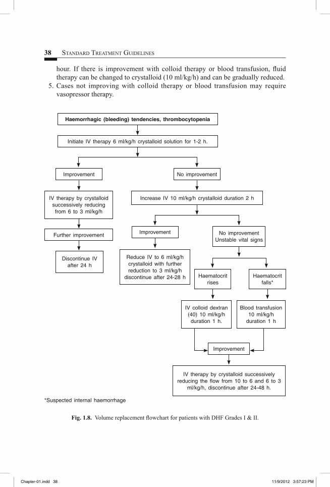
38 STANDARD TREATMENT GUIDELINES
Fig. 1.8. Volume replacement fl owchart for patients with DHF Grades I & II.
hour. If there is improvement with colloid therapy or blood transfusion, fl uid therapy can be changed to crystalloid (10 ml/kg/h) and can be gradually reduced.
5. Cases not improving with colloid therapy or blood transfusion may require vasopressor therapy.
Haemorrhagic (bleeding) tendencies, thrombocytopenia
Initiate IV therapy 6 ml/kg/h crystalloid solution for 1-2 h.
IV therapy by crystalloid successively reducing the fl ow from 10 to 6 and 6 to 3
ml/kg/h, discontinue after 24-48 h.
IV colloid dextran (40) 10 ml/kg/h duration 1 h.
Blood transfusion 10 ml/kg/h
duration 1 h
Haematocrit rises
Haematocrit falls*
IV therapy by crystalloid successively reducing from 6 to 3 ml/kg/h
Further improvement
Discontinue IV after 24 h
Increase IV 10 ml/kg/h crystalloid duration 2 h
Reduce IV to 6 ml/kg/h crystalloid with further reduction to 3 ml/kg/h
discontinue after 24-28 h
Improvement
Improvement No improvement Unstable vital signs
No improvement
Improvement
*Suspected internal haemorrhage
Chapter-01.indd 38Chapter-01.indd 38 11/9/2012 3:57:23 PM11/9/2012 3:57:23 PM
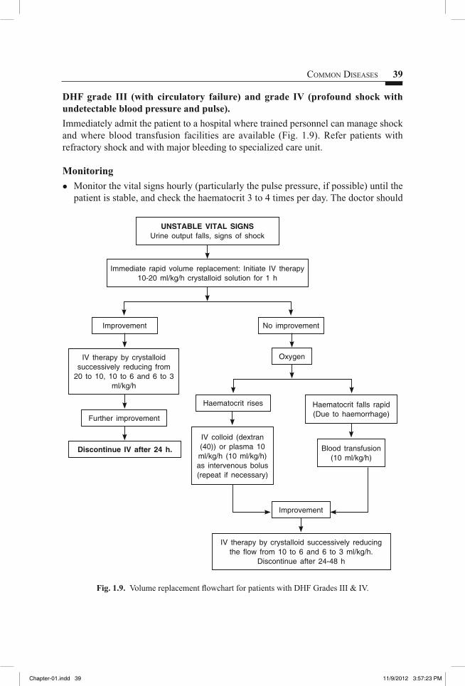
COMMON DISEASES 39
DHF grade III (with circulatory failure) and grade IV (profound shock with undetectable blood pressure and pulse). Immediately admit the patient to a hospital where trained personnel can manage shock and where blood transfusion facilities are available (Fig. 1.9). Refer patients with refractory shock and with major bleeding to specialized care unit.
MonitoringMonitor the vital signs hourly (particularly the pulse pressure, if possible) until the �
patient is stable, and check the haematocrit 3 to 4 times per day. The doctor should
Fig. 1.9. Volume replacement fl owchart for patients with DHF Grades III & IV.
UNSTABLE VITAL SIGNSUrine output falls, signs of shock
Immediate rapid volume replacement: Initiate IV therapy 10-20 ml/kg/h crystalloid solution for 1 h
IV colloid (dextran (40)) or plasma 10 ml/kg/h (10 ml/kg/h) as intervenous bolus (repeat if necessary)
Blood transfusion (10 ml/kg/h)
IV therapy by crystalloid successively reducing from
20 to 10, 10 to 6 and 6 to 3ml/kg/h
Improvement
Haematocrit rises
Further improvement
IV therapy by crystalloid successively reducing the fl ow from 10 to 6 and 6 to 3 ml/kg/h.
Discontinue after 24-48 h
Discontinue IV after 24 h.
Haematocrit falls rapid(Due to haemorrhage)
No improvement
Oxygen
Improvement
Chapter-01.indd 39Chapter-01.indd 39 11/9/2012 3:57:23 PM11/9/2012 3:57:23 PM
40 STANDARD TREATMENT GUIDELINES
review the patient at least four times per day and only prescribe intravenous fl uids for a maximum of 6 h at a time.For children without shock, nurses should check the child’s vital signs (temperature, �
pulse and blood pressure) at least four times per day and the haematocrit once daily, and a doctor should review the patient at least once daily.Check the platelet count daily, where possible, in the acute phase. �
Keep a detailed record of all fl uid intake and output. �
During convalescent phase (2-3 days) after recovery from crucial/shock stage advise rest, normal diet. Signs of recovery are stable pulse, BP and respiratory rate, normal temperature, no evidence of bleeding, return of appetite, no vomiting, good urinary output, stable haematocrit and convalescent confl uent petechial rash.
Criteria for discharging patientsAbsence of fever for at least 24 hours without the use of antipyretic agents. �
Return of appetite. �
Visible clinical improvement. �
Good urine output. �
Minimum of three days after recovery from shock. �
No respiratory distress from pleural effusion and no ascites. �
Platelet count of more than 50,000/mm � 3.Note:Improvement: Haematocrit falls, pulse rate and blood pressure stable, urine output rises.No improvement: Haematocrit or pulse rate rises, pulse pressure falls below 20 mmHg, urine output falls.Unstable vital signs: Urine output falls, signs of shock.
Patient educationSince this disease can rapidly become very serious and lead to a medical emergency, �
carefully watch for danger signs and immediately report to a doctor. Do not wait.The complications usually appear between the third and fi fth day of illness. �
Watch the patient for two days after the fever disappears. Arthralgia may continue �
longer but eventually resolves with no sequelae.Give large amounts of fl uids (water, soups, milk and juices) along with normal �
diet.All control efforts should be directed against the mosquitoes and prevent mosquito �
bites by using appropriate full sleeved clothing, repellent creams, bed nets, etc. Efforts should be intensifi ed before the transmission season (during and after the rainy season) and during epidemics.
Chapter-01.indd 40Chapter-01.indd 40 11/9/2012 3:57:23 PM11/9/2012 3:57:23 PM
COMMON DISEASES 41
References
1. Guidelines for Clinical management of Dengue fever, Dengue Haemorrhagic Fever, Dengue Shock Syndrome. National Vector Borne Disease Control Programme (NVBDCP), Government of India, 2008.
2. Facility Based IMNCI (F-IMNCI) Participants manual. WHO, UNICEF, and Ministry of Health & Family Welfare, Government of India, 2009.
CHIKUNGUNYA
Chikungunya is caused by an arbovirus and transmitted by Aedes aegypti mosquito. It resembles dengue fever, it is rarely life-threatening. After an incubation period of 4-7 days, symptoms last for 3-7 days. Severe cases of chikungunya can occur in the elderly, in the very young (newborns) and in those who are immunocompromised.
SALIENT FEATURES� Sudden onset of fl u-like symptoms including fever, chills, headache, nausea,
vomiting, severe joint pain (arthralgia) and rash. Rash may appear at the outset or several days into the illness; its development often coincides with defervescence, which takes place around day 2 or day 3 of the disease. The rash is most intense on trunk and limbs and may desquamate.
� Migratory polyarthritis usually affects the small joints. The joints of the extremities in particular become swollen and painful to touch. Although rare, the infection can result in meningoencephalitis, especially in newborns and those with pre-existing medical conditions. Pregnant women can pass the virus to their foetus. Haemorrhage is rare and all but a few patients recover within 3-5 days.
� Residual arthritis, with morning stiffness, swelling and pain on movement may persist for weeks or months after recovery.
Treatment
There is no specifi c treatment and is same as for dengue (For details see section on Dengue).
Dengue fever and chikungunya outbreaks evolve quickly, requiring emergency actions to immediately control infected mosquitoes in order to interrupt or reduce transmission and to reduce or eliminate the breeding sites of the vector mosquito, Ae. aegypti.
References
1. Guidelines for Clinical management of Dengue fever / Dengue Haemorrhagic Fever, Dengue Shock Syndrome. National Vector Borne Disease Control Programme (NVBDCP), Government of India, 2008.
2. Facility Based IMNCI (F-IMNCI) Participants manual. WHO, unicef, and Ministry of Health & Family Welfare, Government of India, 2009.
Chapter-01.indd 41Chapter-01.indd 41 11/9/2012 3:57:23 PM11/9/2012 3:57:23 PM

42 STANDARD TREATMENT GUIDELINES
TYPHOID OR ENTERIC FEVER
It is caused by Salmonella typhi and paratyphi. Salmonella typhi causes a variety of illnesses including asymptomatic carriage, gastroenteritis, enteric fever, etc.
SALIENT FEATURES� The onset of fever is typically gradual, continuous (temperature up to 40°C) with
constitutional symptoms like malaise, anorexia, lethargy, headache, constipation or diarrhoea (pea-soup stool), etc. which may be associated with abdominal pain and tenderness, hepatomegaly, splenomegaly, and/or change in mentation. Usually, the patient is sick and toxic looking with a coated tongue and has a soft splenomegaly. Typhoid fever can present atypically in young infants as an acute febrile illness with shock and hypothermia.
� Examination may reveal a toxic look with relative bradycardia and mild soft splenomegaly. Complete blood counts in most cases with typhoid fever are normal. Leucopenia or pancytopenia is seen in 10-25% cases. Diagnosis is suggested by rising titers of ‘O’ antibodies (Widal test) and confi rmed by isolation of organism in blood, bone-marrow, urine or stool. Level of 1 in 160 dilution or more is taken as positive test. Widal test may be negative in cases with fever of less than 5-7 days duration. Blood culture and sensitivity testing/ IGM.
� Complications like hepatitis, peritonitis, meningitis, pneumonitis, and myocarditis can occur, usually after the fi rst week.
Treatment
Nonpharmacological
Adequate nutrition and hydration should be maintained ensuring adequate intake either orally or with intravenous fl uids (in severely ill). In-patient treatment is recommended, if patient is very sick, not accepting orally with inadequate urine output, patient has altered sensorium/drowsiness or is having very high pyrexia particularly in the second week of illness when the risk of complications increases or if the complications have already ensued.
Pharmacological
Management of fever (see section on fever) �
Antipyretics can cause precipitous fall in temperature and even shock, in enteric �
fever. They should be used judiciously. Therefore, hydrotherapy is preferred for fever management in such patients.
Specifi c therapy. Multidrug resistance is prevalent among S. typhi. Antibiotics are recommended on the basis of available institutional culture and sensitivity pattern or epidemiological data.
Chapter-01.indd 42Chapter-01.indd 42 11/9/2012 3:57:23 PM11/9/2012 3:57:23 PM

COMMON DISEASES 43
Uncomplicated enteric fever
Tab. Ciprofl oxacin 10 mg/kg in 2 divided doses, up to a maximum of 750 mg twice a day for 10-14 days (for 1 week after the fever subsides). Oral drug should be taken about an hour after meals and not on empty stomach.
OrTab. Ofl oxacin 200-400 mg daily for 5-7 days.OrCap. Azithromycin 10-20 mg/kg (max 500 to 1000 mg/day) once daily for 5
days.Severe enteric fever (hospitalized patients).Inj. Ceftriaxone 50-60 mg/kg per day IV or IM in 2 divided doses or as a single
dose for 7-10 days (preferred in pregnant women patients, children or patients resistant to quinolones).
OrTab. Cefi xime 200-400 mg daily as single dose or 2 divided doses for 14 days.OrInj. Ciprofl oxacin 200 mg IV twice a dayIf there is no response after 5 days, alternative diagnosis should be
considered.
Enetric fever in children
Uncomplicated enteric fever.
Tab. Cefi xime 10-20 mg/kg/day in 2 divided doses for 14-21 days.OrTab. Chloramphenicol 50-75 mg/kg/day in 4 divided doses for 14-21 daysOrCap. Ampicillin 75-100 mg/kg/day in 4 divided doses for 14 daysOr Tab. Cotrimoxazole (8TMP +40SMX)/day in 2 divided doses for 14 days.Or Cap. Azithromycin 10-20 mg/kg (max 500 to 1000 mg/day) once daily for 7
days.The usual duration of antibiotic treatment is 10-14 days or at least 7 days after the
patient has become afebrile. Intravenous therapy is used during acute phase among the admitted patients. Less sick patients can be treated with oral drugs on an outpatient basis.
Severely ill hospitalized patientsInj. Ceftriaxone 75-100 mg/kg/day IV in 2 divided doses.OrInj. Cefotaxime 75-100 mg/kg/day IM or IV in 2 divided doses
Chapter-01.indd 43Chapter-01.indd 43 11/9/2012 3:57:23 PM11/9/2012 3:57:23 PM
44 STANDARD TREATMENT GUIDELINES
In multidrug resistant cases
Inj. Chloramphenicol 100 mg/kg/day IV or infusion in 4 divided doses for 14-21 days.
OrInj. Ampicillin 100 mg/kg/day IV/IM in 4 divided doses for 14 daysOrInj. Cotrimoxazole-(8TMP+40SMX)/day in 2 divided doses for 14 daysReport to the physician immediately if abdominal symptoms worsen or occurrence
of bleeding per rectum or alteration in sensorium and shock (severe typhoid with high risk of fatality).
Severe typhoid with shock or patients with enteric encephalopathy should be hospitalized and treated as above plus Inj. Dexamethasone 3 mg/kg IV fi rst dose followed by 1 mg/kg IV every 6 hourly for 8 doses.
Chronic carrier state (1-5% patients continue to excrete bacilli in stool for more than 1 year).
If nalidixic acid sensitive Tab. Ciprofl oxacin 750 mg twice a day for 28 days or Cap. Amoxicillin 100 mg/kg/d with probenecid acid 30 mg/kg/day or Tab. Cotrimoxazole 10 mg/kg/d of TMP for 4-6 weeks.
Assessment of response to therapy
The toxic look of the patient decreases and appetite starts returning in 72-96 hours �
of treatment and gradually the fever also starts responding, touches the baseline for increasing duration. The fever can take as long as 7 days to respond. Some times the patient may apparently appear to have responded whereas �
patient may be developing impending shock due to complications. So a careful clinical assessment should be done, particularly, if there is a precipitous fall in temperature.
Modification or step up therapy, if required
The patient should be monitored for complications and usual indications for �
inpatient treatment are: Myocarditis (fall in perfusion and BP, arrhythmias), altered sensorium, shock (tachycardia, cold clammy skin, diaphoresis, hypotension), perforation peritonitis (acute pain in abdomen, guarding, rigidity, hypotension, bilious vomiting). In case the patient worsens or fails to show any response to therapy in 4-7 days or �
so, as discussed above, then a change of antibiotics is suggested, preferably on the basis of the culture sensitivity report, where available.
Patient/parent educationSmall frequent feeds should continue. Give plenty of oral fl uids and compensate for �
increased fl uid loss from the body due to high grade fever. Fever usually lasts 5-7 days even after starting effective treatment in most cases. �
Frequent change of therapy should, therefore, be avoided.
Chapter-01.indd 44Chapter-01.indd 44 11/9/2012 3:57:23 PM11/9/2012 3:57:23 PM
COMMON DISEASES 45
The treatment should be completed till the patient has been afebrile for at least 7 days �
as incomplete treatment increases the risk of relapse and emergence of resistance. The caregivers of the patients should be informed about the complications as �
described above. Ciprofl oxacin and ofl oxacin are very bitter and cause severe nausea and gastritis. �
Patient should be asked to report any missed doses due to vomiting. Three types of vaccines are available to prevent this disease (see section on �
immunization for details).
References
1. Salmonellosis. In: Harrison’s Principles of Internal Medicine. Fauci, Braunwald, Kasper et al (eds), 18th Edition, McGraw Hill Company Inc., New York, 2012; pp 1274-1281.
2. IAP Task Force report: Management of Enteric Fever in Children. Indian Paediatrics 2006; 884-887.
3. Salmonella. In: Nelson’s Textbook of Paediatrics. Behrman RE, Kliegman RM, Jenson HB (eds), 19th Edition, Harcourt Publishers International Company, 2011; pp 948-961.
RHEUMATIC FEVER AND RHEUMATIC HEART DISEASE
Rheumatic fever is a multi-system infl ammatory disease that occurs as a delayed sequelae (2-6 weeks) to group A beta haemolytic streptococcal pharyngitis. It is commonly a disease of childhood between the ages of 5-15 years. The disorder is largely self-limited and resolves without sequelae but chronic and progressive damage to heart valves lead to rheumatic heart disease (RHD).
SALIENT FEATURES� Diagnosis is based on presence of two major or one major and two minor Jone’s
criteria in addition to evidence of recent streptococcal infection (raised ASO titer (>333 units for children and >250 units for adults), positive throat swab or recent scarlet fever) is necessary for diagnosis of rheumatic fever.
� Major criteria: Arthritis, carditis, subcutaneous nodules, chorea, erythema marginatum.
� Minor criteria: Fever, arthralgia, elevated acute phase reactants (ESR, CRP) and prolonged PR interval.
� RHD most frequently affects mitral and aortic valves. Isolated aortic valve involvement is rare and tricuspid and pulmonary valve involvement is unusual.
� Complications of RHD are cardiac failure and infective endocarditis.
Treatment (acute rheumatic fever)
Hospitalization is needed for moderate to severe carditis, severe arthritis or chorea.
Chapter-01.indd 45Chapter-01.indd 45 11/9/2012 3:57:23 PM11/9/2012 3:57:23 PM

46 STANDARD TREATMENT GUIDELINES
Nonpharmacological
Rest is individualized according to symptoms. For arthritis, rest for two weeks is adequate. Carditis without congestive heart failure (CHF) needs 4-6 weeks of rest. In cases of CHF, rest must be continued till the CHF is controlled.
Appropriate diet is a must for a growing child with cardiac involvement.In severe CHF, salt restriction, fl uid restriction, upright posture. Protect patient
from getting injured in chorea.
Pharmacological
1. In arthritis and mild carditis without CHF.Tab. Aspirin 6-8 mg/kg/d in 4 divided doses for 2-3 weeks, taper doses once
symptoms resolve.In children, 100 mg/kg/day for 3-5 days followed by 60-70 mg/kg/day and for older
children 50 mg/kg/day for 4 weeks to be given after meals. (Caution: Avoid gastric irritants, allow frequent feeding, medicine must not be
taken on empty stomach and monitor for tinnitus, deafness, respiratory alkalosis/acidosis).
If no response in 4 days, rule out other conditions like chronic infl ammatory, myeloproliferative disorders before switching over to steroids.
In carditis with CHF. Prednisolone: 2 mg/kg/d, maximum 80 mg/day till ESR normalizes—usually 2 weeks and taper over 2-4 weeks, reduce dose by 2.5-5 mg every 3rd day. Start aspirin 50-75 mg/kg/d simultaneously, to complete total 12 weeks.
(Caution: Monitor blood pressure and blood sugar). Duration of treatment for arthritis is 4-6 weeks and for carditis is 3-6 months.If no response to oral steroid therapy, start Inj. Methyl Prednisolone 30 mg/kg/day
for 3 days.2. For treatment of CHF (see section on cardiac failure). 3. In chorea. Mild chorea is treated with quite environment, and sedatives like oral
phenobarbitone or diazepam. If there is no response, Tab. Haloperidol 0.25-0.5 mg/kg/d in 2-3 divided doses for 2-4 weeks after clinical improvement
OrTab. Sodium valproate 15 mg/kg/day for 2-4 weeks after clinical improvementOr Tab. Carbamazepine 7-20 mg/kg/day for 2-4 weeks after clinical improvementResistant cases can be treated with plasmapheresis or pimozide. If there are laboratory features of rheumatic activity (ESR, CRP, ASO), anti-
infl ammatory drugs must also be given.4. All patients with acute rheumatic fever should be treated as if they have group A
streptococci infection whether or not the organism is actually recovered from culture:Inj. Benzathine penicillin 1.2 MU single IM after test doseIn children: 1.2 MU (>27 kg), 0.6 MU (<27 kg) IM single injection.OrOral penicillin V 500 mg twice daily for 10 days.
Chapter-01.indd 46Chapter-01.indd 46 11/9/2012 3:57:23 PM11/9/2012 3:57:23 PM

COMMON DISEASES 47
In children: 125-250 mg twice daily for 10 days.OrTab Azithromycin 12.5 mg/kg/day once daily for 5 daysOrCap Cephalexin 15-20 mg/kg/dose twice a day for 10 days.In acute rheumatic fever, observe for appearance of valvular lesions (most
common in the fi rst four weeks of disease) and in RHD for effort intolerance, signs and symptoms of CHF, echocardiographic studies of cardiac functions.
Usually joint pains disappear within 24 to 48 hours, tachycardia settles, pericardial friction rub, if present, disappears and gradually ESR comes to normal. In established cardiac lesions with CHF not controlled by medical management, patient should be referred to a higher centre for surgical intervention.
Monitoring
1. Monitor blood levels of salicylates. 2. Watch for salicylate toxicity (ototoxicity, hyperventilation and metabolic
acidosis). 3. Follow up for response to fever and decrease in acute phase reactants; to reduce
salicylates or taper steroids. 4. Echocardiography for monitoring complications of carditis. 5. Termination of anti-infl ammatory therapy may be followed by the reappearance of
clinical manifestation. Usually not treated unless clinical manifestations are severe; reinstate aspirin or steroids in such cases.
For secondary prevention (for prevention of recurrences)
Inj. Benzathine penicillin 1.2 MU (if weight >37 kg) or 0.6MU (if weight <37 kg) IM every 3 weeks
OrTab penicillin V 500 mg twice a day; in children 250 mg twice a dayOrIf patient is allergic to penicillin, Tab. Erythromycin 20 mg/kg/dose (max 500 mg)
2 times a day(Caution: Contraindicated in liver disease).
Duration of prophylaxis
Duration of secondary prevention is individualized. 1. Rheumatic fever with carditis and residual valvular involvement at least until 40
years or lifelong. 2. Rheumatic fever with carditis and no residual valvular involvement, for 10 years
or up to 25 years or whichever is later. 3. Rheumatic fever without carditis, for 5 years or until 18 years whichever is later.
Chapter-01.indd 47Chapter-01.indd 47 11/9/2012 3:57:23 PM11/9/2012 3:57:23 PM
48 STANDARD TREATMENT GUIDELINES
Patient/parent educationEarly and adequate treatment of sore throat. �
Patients with RHD should avoid contact with sore throat cases and if possible �
environmental modifi cations, e.g. avoid overcrowding.Explain consequences of rheumatic fever and ensure monitoring to rule out residual �
valvular involvement and compliance with prophylaxis.Patients with valvular involvement should report to cardiologist for evaluation and �
further management. Explain the importance of prophylaxis against rheumatic carditis as detailed �
earlier.
References
1. Rheumatic Fever. In: Nelson’s Textbook of Paediatrics. Behrman RE, Kliegman RM, Jenson HB (eds), 19th Edition, Harcourt Publishers International Company, 2011; pp 921-925.
2. Consensus guidelines on pediatric acute rheumatic fever and rheumatic heart disease. Working Group on Pediatric Acute Rheumatic Fever and Cardiology Chapter of Indian Academy of Pediatrics. Indian Pediatrics 2008; 45: 565-573.
EPILEPSY
Epilepsies are a group of disorders characterized by chronic, recurrent, paroxysmal changes in neurological function caused by abnormalities in the electrical activity of the brain. Each episode of neurologic dysfunction is called a seizure. Isolated non- recurrent seizures may occur in otherwise healthy individuals for a variety of reasons, and under these circumstances, the individual is not said to have epilepsy.
SALIENT FEATURESA seizure (convulsion) is defi ned as a paroxysmal involuntary disturbance of brain function that may manifest as an impairment or loss of consciousness, abnormal motor activity, behavioural abnormalities, sensory disturbances, or autonomic dysfunction. Some seizures are characterized by abnormal movements without loss or impairment of consciousness.� Diagnosis should be made by a description of the seizure(s) from the patient and
witnesses about frequency, symptoms during and following attacks, duration, circumstances and trigger factors, injury, tongue biting and incontinence.
� Diagnostically relevant factors are seizure types, age at onset (many epilepsies are age-specifi c), family history, past history of head injury, febrile convulsions, precipitating factors, e.g. photic stimulation, alcohol and other drug intake, EEG for evidence of generalized or focal abnormality.
� CT/MRI for evidence of structural lesion. � Consider differential diagnosis of vasovagal syncope, nonepileptic attack disorder
(pseudo seizures), migraine and breathholding spells, etc.
Chapter-01.indd 48Chapter-01.indd 48 11/9/2012 3:57:23 PM11/9/2012 3:57:23 PM
COMMON DISEASES 49
Treatment
For immediate care during seizure, see section on Status Epilepticus. Long-term treatment is required for recurrent seizures. First episode of seizures with no previous history of same or other types of seizures and where neurological and metabolic diseases are ruled out, may be kept under observation and are not treated unless parents/patients are not willing to take the risk. However, patients presenting with status epilepticus, partial seizures, Todd’s palsy, strong family history of epilepsy and with abnormal CT head and EEG have a higher risk of recurrence and can be put on long-term therapy after fi rst seizure. For alcohol withdrawal and metabolic or drug related seizures, long-term treatment is considered only if there are recurrences suggestive of epilepsy. Treatment for seizures following head injury should be initiated after fi rst seizure. However, duration of treatment depends on risk of late epilepsy. Any seizure presenting after 20 years of age should be investigated for secondary causes of seizures.
Pharmacological
Generalised tonic clonic seizures
Tab. Valproic acid 15-40 mg/kg/day in 2 divided doses increased by 200 mg at 3 days interval.
OrTab. Phenytoin 3-8 mg/kg/day in 2-3 divided doses or single night dose.OrTab. Carbamazepine 10-35 mg/kg/day (600-1800 mg 3 times a day).OrTab. Phenobarbitone 60-180 mg/day at night.In children: 5-8 mg/kg/day.
Partial seizures (simple and complex partial seizures)
Tab. Carbamazepine 10-35 mg/kg/day (600-1800 mg 3 times a day).OrTab. Valproic acid 15-40 mg/kg/day in 2 divided doses increased by 200 mg at 3
days interval.OrTab. Phenobarbitone 60-180 mg/day at night.In children: 5-8 mg/kg/day (For neonatal and febrile seizures see Chapter 19)
Patient should preferably be controlled on a single drug (monotherapy). �
Start the drug with low dose. If seizures recur, the dose can be increased after �
checking the compliance/drug levels.If seizures remain uncontrolled despite reaching maximum dose of fi rst drug, add �
another drug as above and gradually reach the maximum dose of second.If seizures are controlled by addition of second drug; always try withdrawal of fi rst �
drug after few weeks of control of seizures.
Chapter-01.indd 49Chapter-01.indd 49 11/9/2012 3:57:23 PM11/9/2012 3:57:23 PM
50 STANDARD TREATMENT GUIDELINES
Combination therapy (polytherapy or adjunctive or ‘add-on’ therapy) can be �
considered when two attempts at monotherapy with AEDs have not resulted in seizure freedom. If seizures continue despite trial with two AEDs, patient should be referred to a �
specialist for evaluation. The formulation or brand of AED should preferably not be changed (variations in �
bioavailability or different pharmacokinetic profi les may increase the potential for reduced effect or excessive side effects). Modifi ed release formulations offer ease of administration due to less frequent �
dosing and better compliance. These are costlier than regular formulations. Once daily administration of AEDs should be used with caution during pregnancy. �
Routine laboratory tests during AED therapy Complete blood count, liver enzymes and renal functions before starting AED. �
Serum calcium, alkaline phosphatase and other tests of bone metabolism every year �
for adults taking enzyme-inducing drug. Asymptomatic minor abnormalities in blood test results are not necessarily an �
indication for changes in medication. Therapeutic drug monitoring (TDM) is not routinely indicated for management of �
epilepsy. Indications are: When poor compliance is suspected; no response despite adequate dosage and compliance; drug toxicity in medicolegal cases; patient is on multiple AEDs; during pregnancy, status epilepticus, liver or kidney disease.
Frequency of follow-up People with epilepsy should maintain a seizure diary and have regular follow-up to �
ensure that the prescribed medication is taken as advised and to detect any adverse effects of AED. This will also avoid a situation in which they continue to take treatment that is ineffective or poorly tolerated. The fi rst follow-up may be undertaken at anytime within 2-4 weeks of initiation of �
treatment. Subsequent follow-ups at every 3-6 months, depending on the control of seizures and side effects. The doctor should review the seizure diary to assess effi cacy tolerability and ensure �
AED compliance. Lifestyle issues such as sleep, regular food intake, alcohol use, driving and pregnancy (if planned) should also be discussed.
If seizures are not controlled with addition of second drug, the patient should be referred to a higher centre for further evaluation and second line drugs such as Lamotrigine, Topiramate, Tiagabine and Gabapentin.
(For seizures due to granuloma see section on Neurocysticercosis)
Generalized absence, myoclonic and akinetic seizures
Sodium valproate is the drug of fi rst choice. In patients who do not achieve adequate seizure control on sodium valproate or do not tolerate, refer to a neurologist. The second choice depends on the seizure type and epilepsy syndrome.
Chapter-01.indd 50Chapter-01.indd 50 11/9/2012 3:57:23 PM11/9/2012 3:57:23 PM
COMMON DISEASES 51
Age dependent epileptic encephalopathies (ADEE)
It includes early infantile epileptic encephalopathy, infantile spasms and Lennox Gastaut syndrome (LGS) with onset within one month, 4-12 months and 1-6 years, respectively. These are diffi cult to control and generally have associated mental defects.
Infantile spasms (myoclonic jerks, hyper-arrhythmia on EEG and mental retardation) 1. Inj. ACTH 30-40 units/day. Or Tab. Prednisolone 2-4 mg/kg/day in 2-3 divided doses. 2. Syr. Sodium valproate 15-40 mg/kg/day in 2-3 divided doses. Inj. ACTH or Tab. Prednisolone is given for 2 weeks with tapering over next 2
weeks while sodium valproate is continued (after seizures are controlled) for 2-3 years.
3. Tab. Clonazepam 0.01-0.03 mg/kg in 2-3 divided doses.
Lennox-Gastaut syndrome
For control of seizures, multiple drugs may be required and treatment should be best carried out at a specialized centre. Valproic acid and clobazam should be used initially. Lamotrigine and topiramate to be added in case of continuing seizures. Avoid carbamazepine.
Discontinuing antiepileptic drug (AED) therapy
Withdrawal of AED medication can be discussed with patients suffering from idiopathic epilepsy after two years seizures free period. AEDs should be withdrawn over a period of 3-6 months or longer because abrupt withdrawal may cause status epilepticus. Withdraw one drug at a time in patients on multiple AEDs. If seizures recur during or after withdrawal, revert back to their AED dose before reduction.
Surgery for epilepsy
Refractory epilepsy in childhood can be defi ned as epilepsy which is uncontrolled despite adequate trials of three first line AEDs. However, before labelling as intractable seizures rule out errors in management and must be looked for as pseudo-intractability often results from an inadequate dose, irrational polytherapy or wrong choice of AED. Every effort should be made to keep a seizure diary and see if a specifi c AED is actually helping or in some cases worsening the seizures, e.g. carbamazepine/oxcarbazine may worsen and sometimes even induce absence/myoclonic seizures.
Refer intractable epilepsy early to a tertiary centre for appropriate evaluation (including high-end MRI using standardized epilepsy protocols, video EEG, etc.) as
Chapter-01.indd 51Chapter-01.indd 51 11/9/2012 3:57:23 PM11/9/2012 3:57:23 PM

52 STANDARD TREATMENT GUIDELINES
well as to get guidance on management options like newer AEDs, the ketogenic diet and surgery.
Patient/parent education
Important information for caregivers in case a person is found having a seizure or is unconscious after a seizure:
DO’S:
Put the person on one side and allow the fi t to be over. The fi t is usually over in �
1-2 minutes. Loosen the person’s clothes.Inform his/here relatives and/or the treating doctor in case any contact details are �
available in his/her pocket.Rush the person to the nearby hospital/medical facility in case the fi ts do not stop �
or there are several fi ts one after the other.
DONT’S:
Put anything like a spoon, piece of wood or cloth in between the teeth or in the �
mouth or a key in his hands. Put a shoe or onion in front of his nose.Forcibly stop his arms and legs from jerking. �
Give him anything to drink or eat. �
Crowd around the person having seizure. �
Most parents are initially frightened by the diagnosis of epilepsy and require support �
and accurate information. The physician should anticipate questions, including inquiries about duration of the seizure disorder, side effects of medication and convulsions, aetiology, social and academic repercussions, and parental guilt.Provide information to parents and encourage them to maintain a seizure diary and �
treat the child as normally as possible. For most children with epilepsy, restriction of physical activity is unnecessary except that the child must be attended by a responsible adult while the child is bathing and swimming.Most children with epilepsy are well controlled on medication, have normal �
intelligence, and can be expected to lead normal lives. However, these children require careful monitoring, as learning disabilities are more common in children with epilepsy than in the general population.Cooperation and understanding among the parents, physician, teacher, and child �
enhance the outlook for the patients with epilepsy.Counselling should also include fi rst aid measures to be used, if the seizure recurs. �
Patients should be instructed to avoid high-risk activities like swimming, driving, �
roof tops, fi re places, etc. for at least 6 months after the last seizure.Explain that medications should be taken exactly as prescribed. Irregular intake of �
drugs or sudden stoppage can lead to status epilepticus and will also prolong the duration of the treatment.
Chapter-01.indd 52Chapter-01.indd 52 11/9/2012 3:57:23 PM11/9/2012 3:57:23 PM
COMMON DISEASES 53
To report immediately in case of status epilepticus, if seizure frequency increases, �
develops any intermittent illness especially fever and behavioural problem.Young women on AED must consult doctor before conceiving. �
Explain possible long-term side effects of the AEDs: � Phenytoin—gingival hypertrophy, hirsutism, acne, chloasma, coarsening of facial features, sedation and ataxia. Carbamazepine—sedation, ataxia, diplopia. Valproic acid—sedation, ataxia, diplopia and hair loss. Hepatotoxicity can occur in children.Explain special precautions to be taken with AEDs: In patients on valproic acid, �
frequent liver function test to be done at beginning and fi rst six months of drug therapy.For prevention of gum hypertrophy, patient should be advised to maintain good oral �
hygiene and frequent rinsing of mouth.In patients on AEDs—explain possible risk of drug interactions especially oral �
contraceptives and antitubercular drugs.
References
1. Seizures and Epilepsy. In: Harrison’s Principles of Internal Medicine. Fauci, Braunwald, Kasper et al (eds), 18th Edition, McGraw Hill Company Inc., New York, 2012; pp 3251-3270.
2. Seizures in Childhood. In: Nelson’s Textbook of Paediatrics. Behrman RE, Kliegman RM, Jenson HB (eds), 19th Edition, Harcourt Publishers International Company, 2011; pp 2013-2039.
3. Age Dependent Epileptic Encephalopathy (ADDE). In: Essentials of Paediatric Neurology. B. Talukdar (ed), 2nd edition, New Age International (P) Ltd., 2009; pp 264-274.
4. Expert Committee on Pediatric Epilepsy, Indian Academy of Pediatrics. Guidelines for diagnosis and management of childhood epilepsy. Indian Pediatr. 2009;46:681-698.
5. Guidelines for management of Status Epilepticus in India. Indian Epilepsy Association 2008.
STATUS EPILEPTICUS (SE)
Status epilepticus (SE) is an emergency condition associated with high morbidity and mortality, if not treated early and effectively. However, about 12- 30% of adult patients fi rst present with status epilepticus as their fi rst presentation. It often occurs in patients with pre-existing epilepsy. SE can occur due to underlying metabolic disturbances, central nervous system (CNS) infections, head trauma and hypoxia.
SALIENT FEATURESContinuous seizures lasting for at least 30 minutes or two or more discrete �
seizures between which there is incomplete recovery of consciousness. Compensatory mechanisms start failing after 30 minutes resulting in hypotension, decreased cerebral blood flow, normo- or hypoglycaemia, hypoxia and hyperpyrexia.Transient or early (0-30 minutes) physiological changes are hypertension, �
increased cerebral blood flow, hyperglycaemia, hyperkalaemia and lactic acidosis.
Chapter-01.indd 53Chapter-01.indd 53 11/9/2012 3:57:23 PM11/9/2012 3:57:23 PM
54 STANDARD TREATMENT GUIDELINES
Treatment
Out-of-hospital settingChildren and young adults: Rectal diazepam 0.5 mg/kg or buccal midazolam 0.2-0.3 mg/kg.Adults Rectal diazepam 10 mg or buccal midazolam 10 mg
IV administration by local doctor or nurse (on doctor’s advice) Inj. Lorazepam 2 mg IV or Inj. Diazepam 5-10 mg IV.
General measures: Secure airway, breathing and circulation, safety and check random sugar.
Nonpharmacological
In hospital, immediately ensure adequate oxygenation by nasal cannula or mask, position patient’s head for optimal airway patency; patient should be transported in lateral position and clear the mouth from secretions/frothing.
Rule out the treatable metabolic causes which can precipitate epilepsy. Establish IV access, draw venous blood samples for glucose level, serum
chemistries, haematological studies, toxicology screens and determination of antiepileptic drug levels and EEG monitoring (if available).
Assess oxygenation with pulse oximetry or periodic arterial blood gas determinations. If hypoglycaemia is established or if blood glucose determination is not available, administer glucose, in adults, 25% Dextrose IV 50-100 ml immediately (to be preceded by 50 mg IM Thiamine, if patient is a known alcoholic). In children, the dose of glucose is 2 ml/kg of 25% glucose.
Pharmacological (adults)
1. Inj. Lorazepam 0.1 mg/kg (max 4 mg) at the rate of 2 mg/min IV over one minute (can be repeated after 10-20 min).
Or Inj. Diazepam 0.2 mg/kg (max 10 mg) at 5 mg/min IV over one minute (can be
repeated, if seizures do not stop after 5 minutes).
Second stage established GCSE (20-60 minutes)
Inj. Phenytoin 15-20 mg/kg slow infusion in saline (not more than 50 mg/min). (Caution: Phenytoin is incompatible with glucose containing solutions; purge IV
line with normal saline before administering phenytoin infusion; IM not recommended as absorption is erratic). If seizures are not controlled after 10 minutes after a loading dose of phenytoin, give additional dose of Phenytoin 5-10 mg/kg IV at the rate of 50 mg/min.
Or Inj. Fosphenytoin 15-18 mg/kg phenytoin equivalent (PE) at 150 mg PE/min(Caution: Cardiac monitoring and check BP as it can produce hypotension/
arrhythmia).
Chapter-01.indd 54Chapter-01.indd 54 11/9/2012 3:57:23 PM11/9/2012 3:57:23 PM

COMMON DISEASES 55
2. If seizures are continuing after 10 minutes of loading dose of phenytoin/fosphenytoin, give additional Phenytoin 5-10 mg/kg or 5-10 mg/kg phenytoin equivalent.
3. If seizures are continuing, Inj. Sodium valproate 25-35 mg/kg IV at the rate of 6 mg/kg/hour
Or Inj. Phenobarbitone 20 mg/kg IV at 60 mg/minutes (should be considered
where ventilator facility is available as it can cause hypotension and respiratory depression). Once seizures are controlled, commence longer term AED with one of Tab. Sodium valproate 800-1500 mg/day orally Or Tab. Phenytoin 300 mg/day orally Or Tab. Carbamazepine 400-1200 mg/day.If status persists after 60 minutes (refractory SE): Identify the precipitating or
underlying cause of SE and institute treatment accordingly and shift patient to a tertiary care hospital with ICU or emergency care unit having ventilation facility). 1. Prepare for mechanical ventilation, place EEG monitor, place arterial catheter and
central catheter. 2. Give anaesthetic dose of Inj. Midazolam 0.2 mg/kg (max 10 mg) IV bolus over 2
minutes followed by 0.1-0.4 mg/kg/h continuous infusion. Or Inj. Propofol 2.5 mg/kg IV bolus followed by 5-10 mg/kg/h. Or Inj. Thiopental 10-20 mg/kg IV over one hour followed by 0.5 -1 mg/kg/h
infusion. 3. Coma phase: Continue pharmacologic coma for 12 hour after last seizures with
EEG goal of burst suppression. 4. Weaning phase: Reduce infusion of the anaesthetic agent every 3 hours with EEG
monitoring, if there are no clinical or electrographic seizures, then wean off. If seizures recur, re-institute coma therapy with the same anaesthetic agent to which the seizures responded. Try to wean as outlined above, if there are no clinical or electrographic seizures for last 12 hours.
5. General measures: Identifi cation and treatment of medical complications including hyperthermia. Consider treating acidosis if pH 7.2 or if symptomatic in the form of cardiac conduction disturbances or haemodynamically unstable.
Status epilepticus in children (See Fig. 1.10)
Non-convulsive status epilepticus (NCSE)NCSE is less critical compared to convulsive status but requires ICU with facility for continuous EEG monitoring. General measures and investigation apply as described for GCSE. As the NCSE is more common in the elderly, non-anaesthetizing anticonvulsants may be tried.
Chapter-01.indd 55Chapter-01.indd 55 11/9/2012 3:57:23 PM11/9/2012 3:57:23 PM

56 STANDARD TREATMENT GUIDELINES
Establish ABCs: Establish IV access, draw blood for laboratory investigations IV glucose, calcium, or pyridoxine (in neonates and infants)
IV Lorazepam 0.1 mg/kgOR
IV diazepam 0.2 mg/kg followed by IV phenytoin/fosphenytoin(If no IV access, use PR diazepam 0.5 mg/kg or buccal/nasal/IM midazolam 0.2 mg/kg;
intraosseous access could be considered as a next step, if IV still not available.)
Repeat lorazepam/diazepam once more SOS (5-10 mins)
IV fosphenytoin 20 PE (phenytoin equivalent)/kg/phenytoin 20 mg/kg (30 mins) (Consider transfer to PICU facilities as child at risk of refractory status)
IV valproate (1:1 diluted NS 20-40 mg/kg over 1-5 minutes; given as continuous infusion at a rate of 5 mg/kg/h, if required.
ORIV phenobarbital 15-20 mg/kg
(Re-assess airway again; consider tracheal intubation, if the airway is compromised or the patient develops respiratory depression) (45-60 min)
Transfer to a PICU set-up is mandatory as the child has refractory SE and will need intensive monitoring in a tertiary PICU set-up.
Midazolam infusion (loading dose of 0.2 mg/kg, followed by 0.1 mg/kg/h titrate every 15 mins upwards by 0.05 mg/kg/h till control; maximum dose 2 mg/kg/h)
ORPropofol infusion/pentothal infusion
(Propofol should not be routinely recommended in view of signifi cant morbidity and mortality in children)
General anaesthesia, if above steps fail(Tertiary hospital set-up essential)
In refractory status epilepticus, needing coma producing therapies (Pentothal, etc.),EEG monitoring preferably continuous should be used, if available. It should also be used, if
coma persists despite control of convulsive status epilepticus (to exclude non-convulsive status epilepticus)
Fig. 1.10. Management of status epilepticus in children.
Maintenance AED treatment following control of status epilepticus
Along with emergency treatment of GCSE and NCSE, maintenance AED therapy should be given to prevent recurrence of seizures.
Chapter-01.indd 56Chapter-01.indd 56 11/9/2012 3:57:23 PM11/9/2012 3:57:23 PM

COMMON DISEASES 57
In patients known to have epilepsy, their usual AED should be maintained and dose adjustments may be carried depending on serum AED levels.
In patients presenting for the fi rst time as status, start AEDs to control status and then can be continued as oral maintenance therapy.
NCSE may not require long-term AEDs. When required, choose the AEd depending upon the clinical situation.
Patient educationConvulsive SE is a serious complication most often seen in patients with pre-existing �
epilepsy and is most often precipitated by missing or discontinuing medication or associated medical illness.If patient continues to convulse for more than 5 min or does not regain consciousness �
after a seizure, the patient should be hospitalized.Patient should be transported in lateral position and mouth should be cleared from �
secretions/frothing.
Reference
1. Seizures and epilepsy. In: Harrison’s Principles of Internal Medicine. Fauci, Braunwald, Kasper et al (eds), 18th Edition, McGraw Hill Company Inc., New York, 2012; pp 3251-3270.
2. Expert Committee on Pediatric Epilepsy, Indian Academy of Pediatrics. Guidelines for diagnosis and management of childhood epilepsy. Indian Pediatr. 2009;46:681-698.
3. Guidelines for management of Status Epilepticus in India. Indian Epilepsy Association 2008.
ASTHMA
A chronic infl ammatory disease characterized by increased responsiveness of the airways to a number of stimuli resulting in their narrowing which is reversible spontaneously or with treatment.
SALIENT FEATURESClassic triad of cough, wheeze, breathlessness; however, all the three may not �
be present. The patient may have tachypnoea, rhonchi and varying degree of dyspnoea. Clinical symptoms may be increased due to upper respiratory viral infections, �
exercise, exposure to smoke, dust, cold air, cold food or various allergens. Diagnosis is clinical and demonstration of reversible airway obstruction on �
pulmonary function tests (Figs. 1.11 and 1.12). Treatment of asthma depends upon the severity of the disease.
Mild acute asthma is characterized by: Cough with or without wheeze, some diffi culty in respiration but no problems of speech or feeding. Oxygen saturation of more than 95% and PEFR of more than 80% predicted.
Chapter-01.indd 57Chapter-01.indd 57 11/9/2012 3:57:23 PM11/9/2012 3:57:23 PM
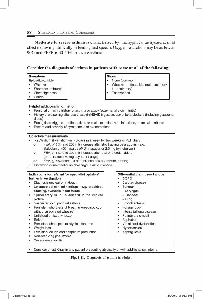
58 STANDARD TREATMENT GUIDELINES
Consider the diagnosis of asthma in patients with some or all of the following:
SymptomsEpisodic/variable• Wheeze• Shortness of breath• Chest tightness• Cough
Signs• None (common)• Wheeze – diffuse, bilateral, expiratory
(+ inspiratory)• Tachypnoea
Helpful additional information• Personal or family history of asthma or atopy (eczema, allergic rhinitis)• History of worsening after use of aspirin/NSAID ingestion, use of beta-blockers (including glaucoma
drops)• Recognised triggers – pollens, dust, animals, exercise, viral infections, chemicals, irritants• Pattern and severity of symptoms and exacerbations
Objective measurements• > 20% diurnal variation on > 3 days in a week for two weeks of PEF diary
or FEV1 >15% (and 200 ml) increase after short acting beta agonist (e.g. Salbutamol 400 mcg by pMDI + spacer or 2.5 mg by nebulizer)or FEV1 >15% (and 200 ml) increase after trial or steroid tablets (prednisolone 30 mg/day for 14 days)or FEV1 >15% decrease after six minutes of exercise/running
• Histamine or methacholine challenge in diffi cult cases
Indications for referral for specialist opinion/further investigation• Diagnosis unclear or in doubt• Unexpected clinical findings, e.g. crackles,
clubbing, cyanosis, heart failure• Spirometery or PFTs don’t fit in the clinical
picture• Suspected occupational asthma• Persistent shortness of breath (non-episodic, or
without associated wheeze)• Unilateral or fi xed wheeze• Stridor• Persistent chest pain or atypical features• Weight loss• Persistent cough and/or sputum production• Non-resolving pneumonia• Severe eosinophilia
Differential diagnoses include:• COPD• Cardiac disease• Tumour – Laryngeal – Tracheal – Lung• Bronchiectasis• Foreign body• Interstitial lung disease• Pulmonary emboli• Aspiration• Vocal cord dysfunction• Hypertension• Aspergillosis
• Consider chest X-ray in any patient presenting atypically or with additional symptoms
Fig. 1.11. Diagnosis of asthma in adults.
Moderate to severe asthma is characterized by: Tachypnoea, tachycardia, mild chest indrawing, diffi culty in feeding and speech. Oxygen saturation may be as low as 90% and PEFR is 30-60% in severe asthma.
Chapter-01.indd 58Chapter-01.indd 58 11/9/2012 3:57:23 PM11/9/2012 3:57:23 PM
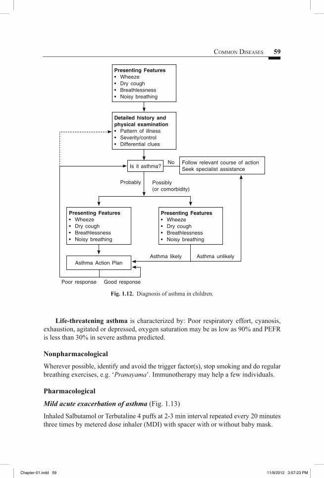
COMMON DISEASES 59
Presenting Features• Wheeze• Dry cough• Breathlessness• Noisy breathing
Presenting Features• Wheeze• Dry cough• Breathlessness• Noisy breathing
Presenting Features• Wheeze• Dry cough• Breathlessness• Noisy breathing
Asthma Action Plan
Detailed history and physical examination• Pattern of illness• Severity/control• Differential clues
Is it asthma?No
Probably Possibly(or comorbidity)
Asthma likely
Poor response Good response
Asthma unlikely
Follow relevant course of action Seek specialist assistance
Fig. 1.12. Diagnosis of asthma in children.
Life-threatening asthma is characterized by: Poor respiratory effort, cyanosis, exhaustion, agitated or depressed, oxygen saturation may be as low as 90% and PEFR is less than 30% in severe asthma predicted.
Nonpharmacological
Wherever possible, identify and avoid the trigger factor(s), stop smoking and do regular breathing exercises, e.g. ‘Pranayama’. Immunotherapy may help a few individuals.
Pharmacological
Mild acute exacerbation of asthma (Fig. 1.13)
Inhaled Salbutamol or Terbutaline 4 puffs at 2-3 min interval repeated every 20 minutes three times by metered dose inhaler (MDI) with spacer with or without baby mask.
Chapter-01.indd 59Chapter-01.indd 59 11/9/2012 3:57:23 PM11/9/2012 3:57:23 PM
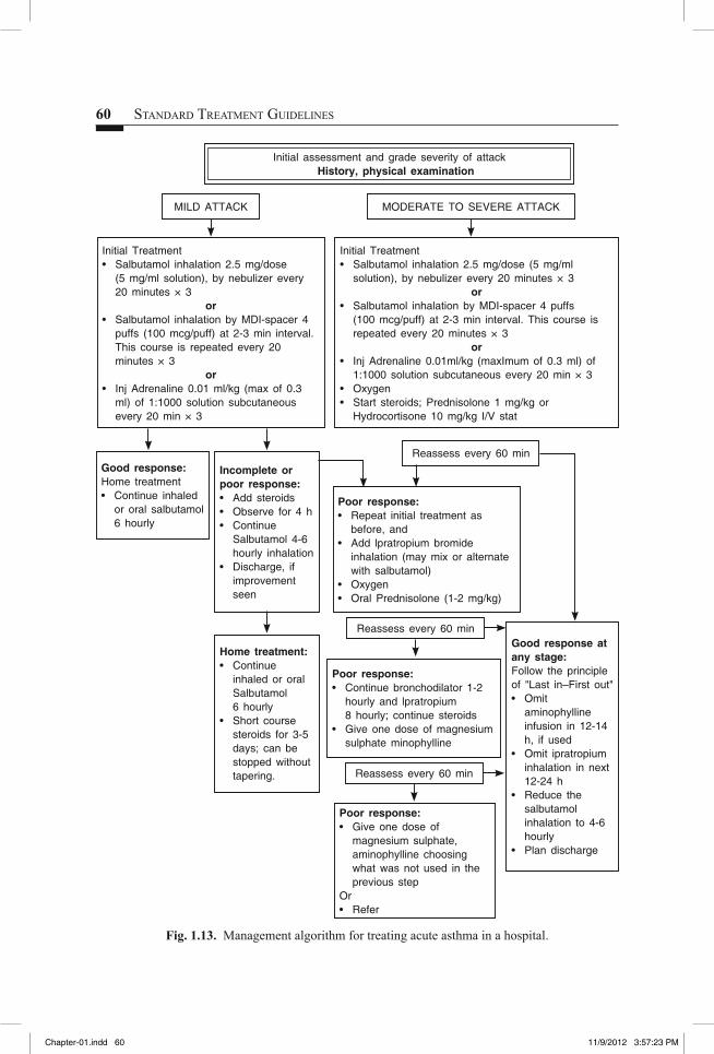
60 STANDARD TREATMENT GUIDELINES
Fig. 1.13. Management algorithm for treating acute asthma in a hospital.
Initial Treatment• Salbutamol inhalation 2.5 mg/dose
(5 mg/ml solution), by nebulizer every 20 minutes × 3
or• Salbutamol inhalation by MDI-spacer 4
puffs (100 mcg/puff) at 2-3 min interval. This course is repeated every 20 minutes × 3
or• Inj Adrenaline 0.01 ml/kg (max of 0.3
ml) of 1:1000 solution subcutaneous every 20 min × 3
Good response:Home treatment• Continue inhaled
or oral salbutamol 6 hourly
Reassess every 60 min
Reassess every 60 min
Reassess every 60 min
Incomplete or poor response:• Add steroids• Observe for 4 h • Continue
Salbutamol 4-6 hourly inhalation
• Discharge, if improvement seen
Poor response:• Repeat initial treatment as
before, and• Add lpratropium bromide
inhalation (may mix or alternate with salbutamol)
• Oxygen• Oral Prednisolone (1-2 mg/kg)
Poor response:• Continue bronchodilator 1-2
hourly and lpratropium 8 hourly; continue steroids
• Give one dose of magnesium sulphate minophylline
Poor response:• Give one dose of
magnesium sulphate, aminophylline choosing what was not used in the previous step
Or• Refer
Good response at any stage:Follow the principle of "Last in–First out"• Omit
aminophylline infusion in 12-14 h, if used
• Omit ipratropium inhalation in next 12-24 h
• Reduce the salbutamol inhalation to 4-6 hourly
• Plan discharge
Home treatment:• Continue
inhaled or oral Salbutamol 6 hourly
• Short course steroids for 3-5 days; can be stopped without tapering.
Initial Treatment• Salbutamol inhalation 2.5 mg/dose (5 mg/ml
solution), by nebulizer every 20 minutes × 3or
• Salbutamol inhalation by MDI-spacer 4 puffs (100 mcg/puff) at 2-3 min interval. This course is repeated every 20 minutes × 3
or• Inj Adrenaline 0.01ml/kg (maxImum of 0.3 ml) of
1:1000 solution subcutaneous every 20 min × 3• Oxygen• Start steroids; Prednisolone 1 mg/kg or
Hydrocortisone 10 mg/kg I/V stat
MILD ATTACK MODERATE TO SEVERE ATTACK
Initial assessment and grade severity of attack History, physical examination
Chapter-01.indd 60Chapter-01.indd 60 11/9/2012 3:57:23 PM11/9/2012 3:57:23 PM

COMMON DISEASES 61
Good response is defi ned as patient feeling well with minimal or no dyspnoea, marked improvement in heart rate, respiratory rate and little or no rhonchi on auscultation with oxygen saturation above 95%. Patients showing good response may be sent home on inhaled or oral bronchodilators for 7-10 days.
If patient is on maintenance treatment with high dose inhaled steroids or attended emergency room in last 72 hours add Tab. Prednisolone 1 mg/kg/day for 3-5 days. If patient is already on low dose maintenance inhaled steroids, to continue it. Call the patient again for reassessment after 1-2 weeks or early if symptoms are not getting controlled or worsening.
Patients not showing good response should be treated as moderate to severe acute asthma.
Moderate to severe acute asthma (Fig. 1.13)
Get an X-ray chest and rule out infection, pneumothorax or collapse of segments or lobes of lung and manage accordingly. Do arterial blood gas analysis and serum electrolytes for detection of acidosis and hypokalaemia and manage accordingly and reassess after 60 minutes.
Life-threatening asthma
1. Oxygen inhalation 4 L/min to maintain SpO2 >90%. 2. Inj. Terbutaline 10 mcg/kg subcutaneously or IV (maximum 40 mcg/day). 3. Inhaled Salbutamol/Terbutaline preferably by nebulizer (as discussed above). 4. Ipratropium Bromide 250 mcg by nebulizer with Salbutamol. 5. Inj. Hydrocortisone 10 mg/kg IV. 6. Inj. Aminophylline 5 mg/kg bolus slowly followed by 0.8-1.2 mg/kg/hour slow
infusion (If patient has received theophylline preparation in last 72 hours; reduce bolus dose to 2.5 mg/kg).
7. Inj. Magnesium sulphate 40 mg/kg in 50 ml 5% dextrose as slow infusion over 30 minutes can be considered.If no response do arterial blood gas analysis, X-ray chest and serum electrolytes.
Intubate the patient if no or poor respiratory effort, increased carbon dioxide with respiratory acidosis. Transfer to intensive care unit as early as possible.
If above therapy fails. Transfer should be arranged so that oxygen and inhalation therapy can be continued on the way.
Notes:Antibiotics are required only if there is a consolidation, high grade fever or �
polymorphonuclear leucocytosis.Mucolytics and cough syrups are not helpful. �
Sedation should be avoided in acute asthma. �
Non-sedating antihistaminics may be used, if associated allergic rhinitis is there. �
Chapter-01.indd 61Chapter-01.indd 61 11/9/2012 3:57:23 PM11/9/2012 3:57:23 PM
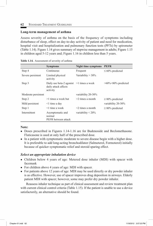
62 STANDARD TREATMENT GUIDELINES
Long-term management of asthma
Assess severity of asthma on the basis of the frequency of symptoms including disturbance of sleep, effect on day-to-day activity of patient and need for medication, hospital visit and hospitalization and pulmonary function tests (PFTs) by spirometer (Table 1.14). Figure 1.14 gives summary of stepwise management in adults; Figure 1.15 in children aged 5-12 years and; Figure 1.16 in children less than 5 years.
Table 1.14. Assessment of severity of asthma
Symptoms Night time symptoms PEFRStep 4 Continuous Frequent ≤ 60% predictedSevere persistent Limited physical
activityVariability > 30%
Step 3 Daily use beta-2 agonist daily attack affects activity
>1 times a week >60%<80% predicted
Moderate persistent variability 20-30%Step 2 >1 times a week but >2 times a month ≥ 80% predicted Mild persistent <1 time a day variability 20-30%Step 1 <1 time a week <2 times a month ≥ 80% predictedIntermittent Asymptomatic and
normal PEFR between attack
variability < 20%
Note:Doses prescribed in Figures 1.14-1.16 are for Budenoside and Beclomethasone. �
Fluticasone is used at only half of the prescribed dose. In a patient with symptomatic moderate to severe disease begin with a higher dose. �
It is preferable to add long-acting bronchodilator (Salmeterol, Formoterol) initially because of quicker symptomatic relief and steroid sparing effect.
Select an appropriate inhalation device
Children below 4 years of age: Metered dose inhaler (MDI) with spacer with �
facemaskFor children above 4 years of age: MDI with spacer. �
For patients above 12 years of age: MDI may be used directly or dry powder inhaler �
is as effective. However, use of spacer improves drug deposition in airways. Elderly patient MDI with spacer; however, some may prefer dry powder inhaler.
Reassess inhaler technique as part of clinical assessment and review treatment plan with current clinical control criteria (Table 1.15). If the patient is unable to use a device satisfactorily, an alternative should be found.
Chapter-01.indd 62Chapter-01.indd 62 11/9/2012 3:57:23 PM11/9/2012 3:57:23 PM
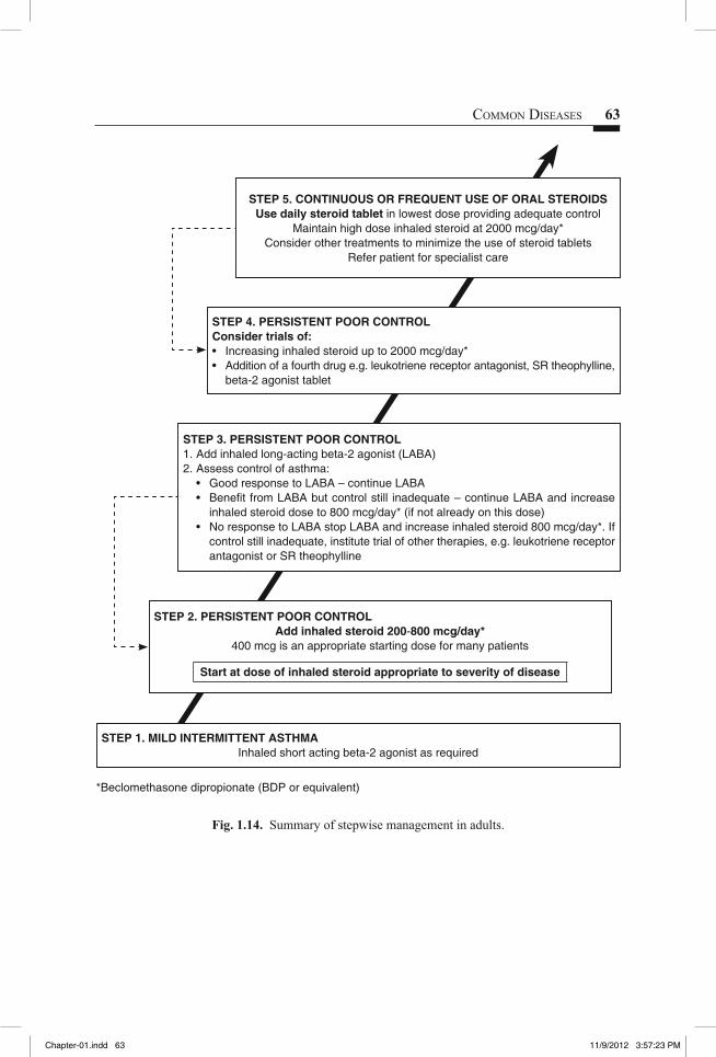
COMMON DISEASES 63
STEP 5. CONTINUOUS OR FREQUENT USE OF ORAL STEROIDSUse daily steroid tablet in lowest dose providing adequate control
Maintain high dose inhaled steroid at 2000 mcg/day*Consider other treatments to minimize the use of steroid tablets
Refer patient for specialist care
STEP 4. PERSISTENT POOR CONTROLConsider trials of: • Increasing inhaled steroid up to 2000 mcg/day*• Addition of a fourth drug e.g. leukotriene receptor antagonist, SR theophylline,
beta-2 agonist tablet
STEP 3. PERSISTENT POOR CONTROL1. Add inhaled long-acting beta-2 agonist (LABA)2. Assess control of asthma:
• Good response to LABA – continue LABA• Benefi t from LABA but control still inadequate – continue LABA and increase
inhaled steroid dose to 800 mcg/day* (if not already on this dose)• No response to LABA stop LABA and increase inhaled steroid 800 mcg/day*. If
control still inadequate, institute trial of other therapies, e.g. leukotriene receptor antagonist or SR theophylline
STEP 2. PERSISTENT POOR CONTROLAdd inhaled steroid 200-800 mcg/day*
400 mcg is an appropriate starting dose for many patients
Start at dose of inhaled steroid appropriate to severity of disease
STEP 1. MILD INTERMITTENT ASTHMAInhaled short acting beta-2 agonist as required
*Beclomethasone dipropionate (BDP or equivalent)
Fig. 1.14. Summary of stepwise management in adults.
Chapter-01.indd 63Chapter-01.indd 63 11/9/2012 3:57:23 PM11/9/2012 3:57:23 PM
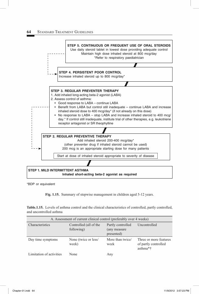
64 STANDARD TREATMENT GUIDELINES
STEP 5. CONTINUOUS OR FREQUENT USE OF ORAL STEROIDSUse daily steroid tablet in lowest dose providing adequate control
Maintain high dose inhaled steroid at 800 mcg/day*Refer to respiratory paediatrician
STEP 4. PERSISTENT POOR CONTROL Increase inhaled steroid up to 800 mcg/day*
STEP 3. REGULAR PREVENTER THERAPY1. Add inhaled long-acting beta-2 agonist (LABA)2. Assess control of asthma:
• Good response to LABA – continue LABA• Benefi t from LABA but control still inadequate – continue LABA and increase
inhaled steroid dose to 400 mcg/day* (if not already on this dose)• No response to LABA – stop LABA and increase inhaled steroid to 400 mcg/
day.* If control still inadequate, institute trial of other therapies, e.g. leukotriene receptor antagonist or SR theophylline
STEP 2. REGULAR PREVENTIVE THERAPYAdd inhaled steroid 200-400 mcg/day*
(other preventer drug if inhaled steroid cannot be used)200 mcg is an appropriate starting dose for many patients
Start at dose of inhaled steroid appropriate to severity of disease
STEP 1. MILD INTERMITTENT ASTHMAInhaled short-acting beta-2 agonist as required
*BDP or equivalent
Fig. 1.15. Summary of stepwise management in children aged 5-12 years.
Table.1.15. Levels of asthma control and the clinical characteristics of controlled, partly controlled, and uncontrolled asthma
A. Assessment of current clinical control (preferably over 4 weeks)Characteristics Controlled (all of the
following)Partly controlled (any measure presented)
Uncontrolled
Day time symptoms None (twice or less/week)
More than twice/week
Three or more features of partly controlled asthma*†
Limitation of activities None Any
Chapter-01.indd 64Chapter-01.indd 64 11/9/2012 3:57:23 PM11/9/2012 3:57:23 PM

COMMON DISEASES 65
STEP 4. PERSISTENT POOR CONTROLRefer to respiratory paediatrician
STEP 3. ADD ON THERAPYIn children aged 2-5 years consider trial of leukotriene receptor antagonistIn children under 2 years consider proceeding to step 4.
STEP 2. REGULAR PREVENTER THERAPYAdd inhaled steroid 200-400 mcg/day*!Or leukotriene receptor antagonistif inhaled steroid cannot be used
STEP 1. MILD INTERMITTENT ASTHMAInhaled short-acting beta-2 agonist as required
*BDP or equivalent! Higher nominal doses may be required if drug delivery is diffi cult
Fig. 1.16. Summary of stepwise management in children aged less than 5 years.
Nocturnal symptoms/ awaking
None Any
Need for reliever/ rescue inhaler
None (twice or less/week)
More than twice/week
Lung function (PEF or FEV1) ‡
Normal <80% predicted or personal best (if known)
B. Assessment of future risk (risk or exacerbations, instability, rapid decline in lung function, side effects)Features that are associated with increased risk of adverse events in the future include:Poor clinical control, frequent exacerbations in past year*, ever admission to critical care for asthma, low FEV1, exposure to cigarette smoke, high dose medications.
*Any exacerbation should prompt review of maintenance treatment to ensure that it is adequate.†By defi nition, an exacerbation in any week makes that an uncontrolled asthma week.‡Without administration of bronchodilator, lung function is not a reliable test for children 5 years and younger.
Chapter-01.indd 65Chapter-01.indd 65 11/9/2012 3:57:23 PM11/9/2012 3:57:23 PM
66 STANDARD TREATMENT GUIDELINES
Follow-up and modification in treatment
Call the patient every 8-12 weeks. On each visit, examine the patient; look for adverse effects of the drugs and record height and weight in children. Measure PEFR/PFTs in older children and record the assessed status of disease.
If there is no improvement or deterioration, look for possible cause such as poor compliance, wrong technique of inhalation, continued use of empty canister, inappropriate doses, infection (otitis media, sinusitis, pneumonitis), continued exposure to allergens, under assessment of illness in previous visit, allergic rhinitis and sinusitis. If no cause is found, a step up may be considered, i.e. increase in dose and frequency of medication.
Step down the medications, if control is sustained for at least 3 months and follow a gradual stepwise reduction in treatment. When deciding which drug to step down fi rst and at what rate, the severity of asthma, the side effects of treatment, the benefi cial effect achieved, and the patients’ preference should all be taken into account. Patients should be maintained at the lowest possible dose of inhaled steroid. Reduction in inhaled steroid dose should be slow as patients deteriorate at different rates.
Home treatment of acute exacerbation 1. Identify acute exacerbation by increase in cough, wheeze and breathlessness. 2. Measure PEFR (if feasible), if decreased by 15% from the baseline, administer
Salbutamol by MDI with spacer with or without facemask, one puff at a time, repeated every 2-4 minutes up to a maximum of 10-20 puffs with monitoring of symptoms.If symptoms are relieved and PEFR is increased at the end of inhalation, continue
on Salbutamol/Terbutaline every 4-6 hours and a visit to treating physician should be planned.
If there is no improvement or partial improvement or there are symptoms of life-threatening attack (severe distress, diffi culty in speech, feeding, cyanosis, exhaustion) at any time, the patient should be immediately transferred to a hospital and during transportation continue inhaled Salbutamol/Terbutaline and give a dose of prednisolone (1-2 mg/kg).
Antibiotics
Antibiotics should not be given routinely for acute asthma. Antimicrobial treatment is indicated, however, when there is persistent fever and other signs of pneumonia such as bronchial breathing. Mere presence of crackles is not an evidence of pneumonia and does not warrant antibiotics.
Patient/parent educationExplain the nature and pathogenesis of asthma in simple language. �
Emphasize that there is a wide-spectrum of severity of asthma and that most patients �
can lead active and normal life.
Chapter-01.indd 66Chapter-01.indd 66 11/9/2012 3:57:23 PM11/9/2012 3:57:23 PM

COMMON DISEASES 67
Ask to maintain a record of daily symptoms such as cough, coryza, wheeze and �
breathlessness. A record of sleep disturbances, absence from school due to illness and medication is required to keep the patient symptom free.Environmental control to avoid precipitating factors is equally important. �
Patient/parents should avoid dusting (wet mopping is preferred), when children are �
around.Avoid using carpets, stuffed toys, open bookshelves, smoking and chemical sprays �
in the house. Mosquito nets should be preferred over repellents.Food with chemicals like preservatives/colouring agents should be avoided. �
Inhalation technique: It is best to use MDI with spacers, however, if low dose �
steroids are being given then dry powder inhaler can also be used. MDI must be shaken well before inhalation. It is then attached to spacer (commercial/indigenous made from plastic bottle) and patient is asked to inhale 3-4 times slowly and deeply just when the drug is released by activation of MDI. Patient/parents must be advised to check the canister every few days dipping it in a �
tumbler full of water. An empty canister fl oats. The spacer should be cleaned monthly with detergent and dried in air. The �
mouthpiece should be wiped clean of detergent before use.
References
1. Consensus Guidelines on Management of Childhood Asthma in India. Indian Paediatrics 1999; 36: 157-165.
2. Pocket Guide for Asthma management and Prevention (for adults and children older than 5 years). Global Initiative for Asthma. 2011.
3. The British Guidelines on Management of Asthma: A National Clinical Guideline. British Thoracic Society, Scottish Intercollegiate Guidelines Network, 2012.
4. Facility Based IMNCI (F-IMNCI) Participants Manual. WHO, UNICEF, and Ministry of Health & Family Welfare, Government of India, 2009.
Chapter-01.indd 67Chapter-01.indd 67 11/9/2012 3:57:23 PM11/9/2012 3:57:23 PM