Embed Size (px)
Citation preview
Commentary -------------------------------------------------
Unraveling the Neuroanatomy of Epilepsy
Marek A. Mirski 1
Over the last 50 years, a collective attempt has been made to define the brain elements important in the generation and propagation of seizure activity throughout the brain. Seizures are of many types, and the clinical characteristics of each is dependent on the region of brain involved. The term epilepsy, for example, encompasses a wide variety of recurrent seizure disorders that have been classified in accordance to the location and extent of the seizure process within the brain. Fundamentally, seizures are of two types. Seizures may be partial (focal) in nature or they may be generalized. This distinction is appropriate for two reasons. First, the extent of cortical involvement differs between these two groups. Second, and more important, each seizure type has a neuroanatomic mechanism of expression that is fundamentally distinct.
In the examination of the origin of seizures, many analytical tools and methods have been used. Surface and depth electroencephalographic recording have provided the majority of evidence to date, although radiographic techniques such as radionuclide autoradiography, positron emission tomography, computed tomography (CT), and various magnetic resonance (MR) sequence studies have proved to be of substantial value. The greatest consideration has been given to the study of the focal epilepsies, in which structural disease is frequently present. These seizures display electroencephalographic and clinical manifestations consistent with the involvement of only a portion of the cortex and its corresponding functional systems. The spread of a focal seizure to adjacent cortical regions is presumed to be via local synaptic connections. Such appears to be the case in the classic "Jacksonian march ," a focal seizure that spreads along the motor strip to excite progressively the cortical neurons that control topographically associated limb musculature (1). Other partial seizures, such as many
temporal lobe epilepsies, are formally described as partial complex because consciousness with the environment is disturbed. The anatomy involved in this form of epilepsy is somewhat more involved than a simple partial seizure because of the recruitment of deeper brain elements that affect our conscious behavior. Most commonly, elements of the limbic brain, usually the hippocampus or amygdala and their connections, play a major role in the expression of partial complex seizures. Radiographic studies, particularly MR, have been useful in some cases of partial seizures in locating brain disease, especially hippocampal sclerosis (2-4).
As a diagnostic or investigational tool for the generalized epilepsies, radiographic imaging has not been extremely useful. These seizures encompass a completely distinct group of paroxysmal disorders. The expression of this seizure type results from an activation of the entire cerebral cortex. The seizures are exemplified by bilaterally synchronous and symmetrical epileptiform discharges on electroencephalogram and clinical behavior characterized by a loss of consciousness and generalized convulsive or paralytic motor phenomena. In the majority of instances, no diffuse or focal brain disease or generalized metabolic disturbance can be convincingly demonstrated.
In this issue of AJNR, Marmourian and Brown describe three cases of asymmetric mamillary bodies identified with MR imaging or autopsy in patients with histories of intractable epilepsy (5). One patient had clear generalized attacks resulting in staring episodes and atonic spells causing him to fall repeatedly. On MR, the patient had normal-appearing temporal lobes and absence of one mamillary body. The seizures were not well described in another patient who had gross atrophy of the right hippocampus and complete absence of the ipsilateral mamillary body. The third
1 ssistant Professor, The Johns Hopkins Medical Inst itutions, Departmen t of Anesthesiology and Critical Care Medicine, 600 North Wolfe Street, Me er 8-134. Baltimore, MD 21287-7834.
Index terms: H pothalamus; Brain , anatom ; Brain , magnetic resonance; Seizures; Brain, temporal lobe
AJNR 14: 1336-1342. No / Dec 1993 0195-6108/ 93/ 1406-1336 © American Society of Neuroradiology
1336
AJNR: 14, November / December 1993
patient suffered from partial complex seizures. By MR analysis, one hippocampus had increased signal consistent with sclerosis and the ipsilateral mamillary body was smaller than the contralateral one. In each of the above cases, the mamillary body was structurally abnormal and, in two of the three patients, the mamillary abnormality on MR was either not coincident with, or was more severe than, disease in the ipsilateral hippocampus.
Of interest in this report is the description of human diseases of the mamillary system associated with clinical seizures. That the hippocampus appears involved in two of the three patients is not surprising, especially in the patient with partial seizures. The association of mamillary body disease out of proportion to, or in the apparent absence of, any hippocampal injury is somewhat more intriguing. The evidence cited, sparse as it is, may suggest a primary role of the mamillary bodies in partial complex epilepsy, a concept not previously entertained. Of perhaps greater interest, in at least one patient the paroxysms appeared to be either primary or secondary generalized seizures. So little is known about the neuroanatomy propagating these epileptiform attacks that any human brain abnormality that can be linked to them could be an important mechanistic clue.
One valuable clue toward understanding the nature of generalized epilepsy is provided by the stereotypic electrical and behavioral expression of these seizures. These characteristics support the premise that these paroxysmal events are not the result of a chaotic, but rather of a wellorchestrated propagation of neuronal synaptic activity. Unfortunately, the rapid holocortical involvement of neuronal activity during such episodes has made it difficult to identify specific cell groups or pathways recruited early in the seizure process. Nevertheless, since the first half of this century, investigators have begun to elucidate the neuroanatomy associated with the early propagation of these seizures.
Studies during the 1940s and 1950s used depth recording and electrical stimulation to demonstrate that both facilitory and inhibitory influences on seizure activity exist in both the diencephalon (6-8) and brain stem (9-14). These findings suggested important roles for regions such as the reticular formations of the pons, mesencephalon, and thalamus. At the time, these areas were considered to be connected with the "centrencephalon," an as yet unidentified but distinct region
COMMENTARY 1337
within the brain substance thought to be a source of primary generalized paroxysmal activity. Since then it has become increasingly apparent that generalized seizures do not originate from a single structure but rather are the result of an integrated expression of facilitory and inhibitory influences between cortex and subcortical structures. The spread of electrical activity is presumed to be mediated through diffuse connections (15) . Specific subcortical synaptic paths , therefore , may serve as important links to propagate this paroxysmal activity. Within the past two decades, more well-defined areas of subcortex have indeed been linked with seizure mechanisms, including individual thalamic nuclei such as the ventral anterior (16), centromedian (17 , 18), and lateral posterior nuclei (19). More caudally, evidence suggests that the fields of Fore! in the subthalamic region (20) and the interpeduncular complex (21 , 22) have a role in seizure expression. In the midbrain , the substantia nigra has received considerable attention for its role in mediating seizures using a variety of convulsant models (23-25). Recently , a region in rat forebrain , termed "area tempesta," has been shown to be a potential trigger area for seizures (26-28).
Despite this growing number of brain sites linked to the mediation of seizures, the precise neuroanatomic pathways underlying seizure propagation have remained difficult to elucidate. The rapid and uniform brain excitation that occurs during a generalized seizure nevertheless underscores the need fo r early recruitment of major neuroanatomic paths connecting cortical and subcortical brain into the seizure process.
Clinical and experimental evidence suggest that the mamillary body and its immediate connections may be an important subcortical pathway mediating the expression of generalized seizures. Because of its location within the posterior hypothalamus, the mamillary system is placed in a unique position to act as a synaptic link between brain stem and forebrain in the propagation of paroxysmal electrical activity .
An important consideration when suggesting a link between specific neuronal structures and early seizure expression is that synaptic pathways must exist that could potentially propagate the epileptiform activity in a rapid and diffuse fashion. The mamilla ry body satisfies this criterion with afferent and efferent connections that have been well described (29-34). Primary afferents include the large subicular derived postcommissural fornix and pathways from the brain stem ventral
1338 MIRSKI
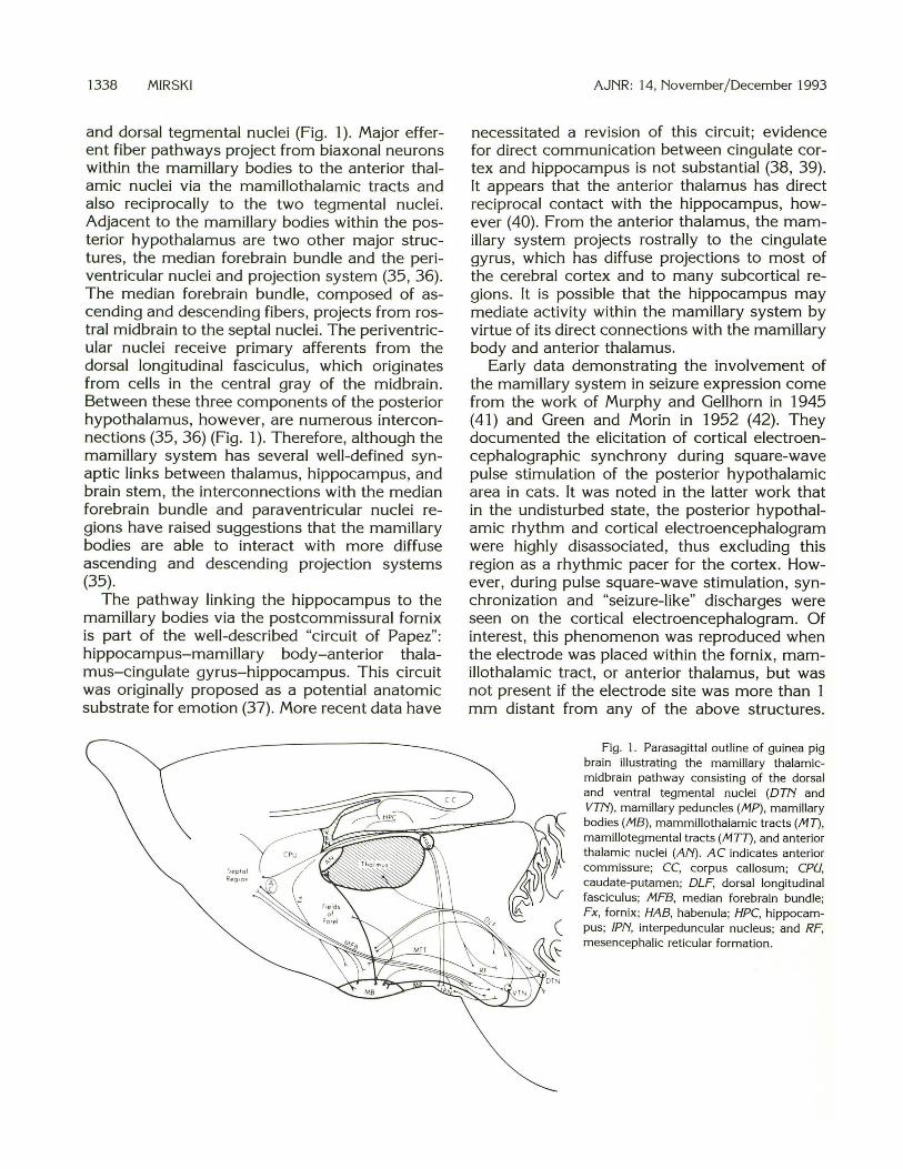
and dorsal tegmental nuclei (Fig. 1 ). Major efferent fiber pathways project from biaxonal neurons within the mamillary bodies to the anterior thalamic nuclei via the mamillothalamic tracts and also reciprocally to the two tegmental nuclei. Adjacent to the mamillary bodies within the posterior hypothalamus are two other major structures, the median forebrain bundle and the periventricular nuclei and projection system (35, 36). The median forebrain bundle, composed of ascending and descending fibers, projects from rostral midbrain to the septal nuclei. The periventricular nuclei receive primary afferents from the dorsal longitudinal fasciculus, which originates from cells in the central gray of the midbrain. Between these three components of the posterior hypothalamus, however, are numerous interconnections (35 , 36) (Fig. 1 ). Therefore, although the mamillary system has several well-defined synaptic links between thalamus, hippocampus, and brain stem, the interconnections with the median forebrain bundle and paraventricular nuclei regions have raised suggestions that the mamillary bodies are able to interact with more diffuse ascending and descending projection systems (35).
The pathway linking the hippocampus to the mamillary bodies via the postcommissural fornix is part of the well-described "circuit of Papez": hippocampus-mamillary body-anterior thalamus-cingulate gyrus-hippocampus. This circuit was originally proposed as a potential anatomic substrate for emotion (37). More recent data have
AJNR: 14, November/ December 1993
necessitated a rev1s1on of this circuit; evidence for direct communication between cingulate cortex and hippocampus is not substantial (38, 39). It appears that the anterior thalamus has direct reciprocal contact with the hippocampus, however (40). From the anterior thalamus, the mamillary system projects rostrally to the cingulate gyrus, which has diffuse projections to most of the cerebral cortex and to many subcortical regions. It is possible that the hippocampus may mediate activity within the mamillary system by virtue of its direct connections with the mamillary body and anterior thalamus.
Early data demonstrating the involvement of the mamillary system in seizure expression come from the work of Murphy and Gellhorn in 1945 (41) and Green and Morin in 1952 (42). They documented the elicitation of cortical electroencephalographic synchrony during square-wave pulse stimulation of the posterior hypothalamic area in cats. It was noted in the latter work that in the undisturbed state, the posterior hypothalamic rhythm and cortical electroencephalogram were highly disassociated, thus excluding this region as a rhythmic pacer for the cortex. However, during pulse square-wave stimulation, synchronization and "seizure-like" discharges were seen on the cortical electroencephalogram. Of interest, this phenomenon was reproduced when the electrode was placed within the fornix, mamillothalamic tract, or anterior thalamus, but was not present if the electrode site was more than 1 mm distant from any of the above structures.
Fig. 1. Parasagittal outline of guinea pig brain illustrating the mamillary thalamicmidbrain pathway consisting of the dorsal and ventral tegmental nuclei (DTN and VTN) , mamillary peduncles (/11P), mamillary bodies (/118), mammillothalamic tracts (/117), mamillotegmental tracts (MTT), and anterior thalamic nuclei (AN) . AC indicates anterior commissure; CC, corpus callosum; CPU, caudate-putamen; DLF, dorsal longitudinal fasciculus; MFB, median forebrain bundle; Fx, fornix ; HAB, habenula; HPC, hippocampus; lPN, interpeduncular nucleus; and RF, mesencephalic reticular formation .
AJNR: 14, November/ December 1993
Later Jinnai et a! (20) working in the cat model demonstrated that focal destruction of the mamillary bodies raised the seizure threshold to the chemical convulsant pentylenetetrazol. These data supported the concept that this brain region had a facilitory role in seizure expression. The anterior hypothalamus, in contrast, appeared to have an inhibitory action on pentylenetetrazol convulsant activity.
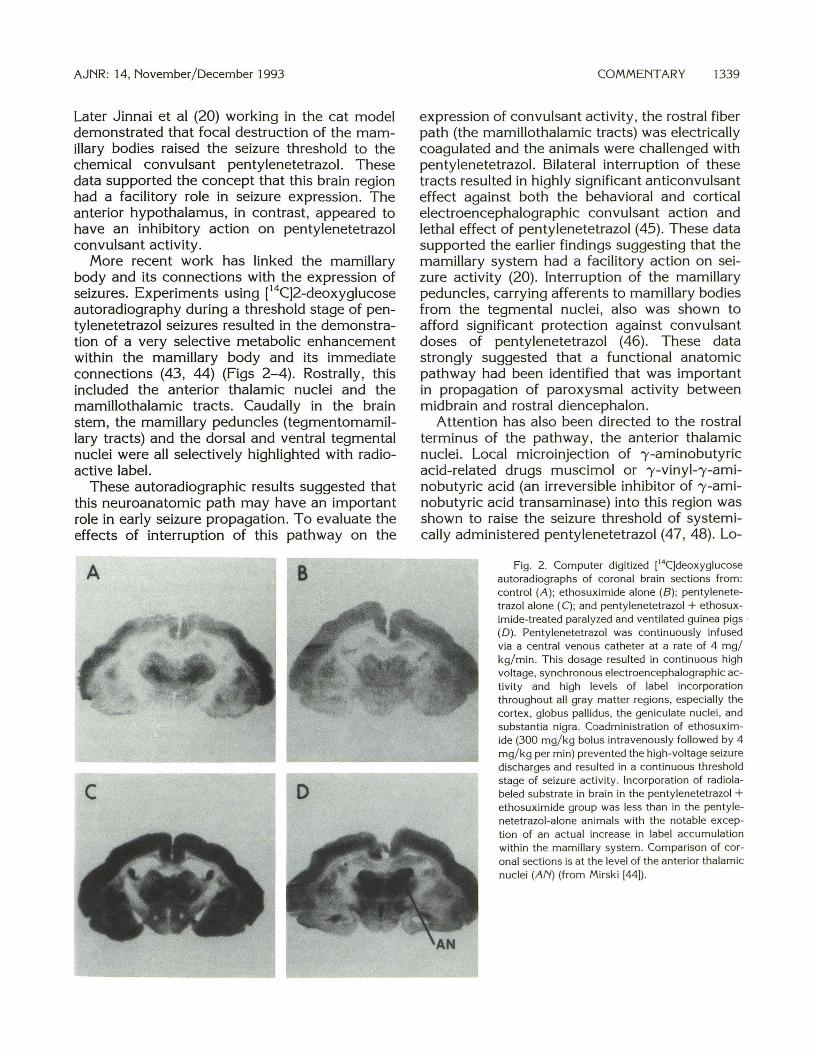
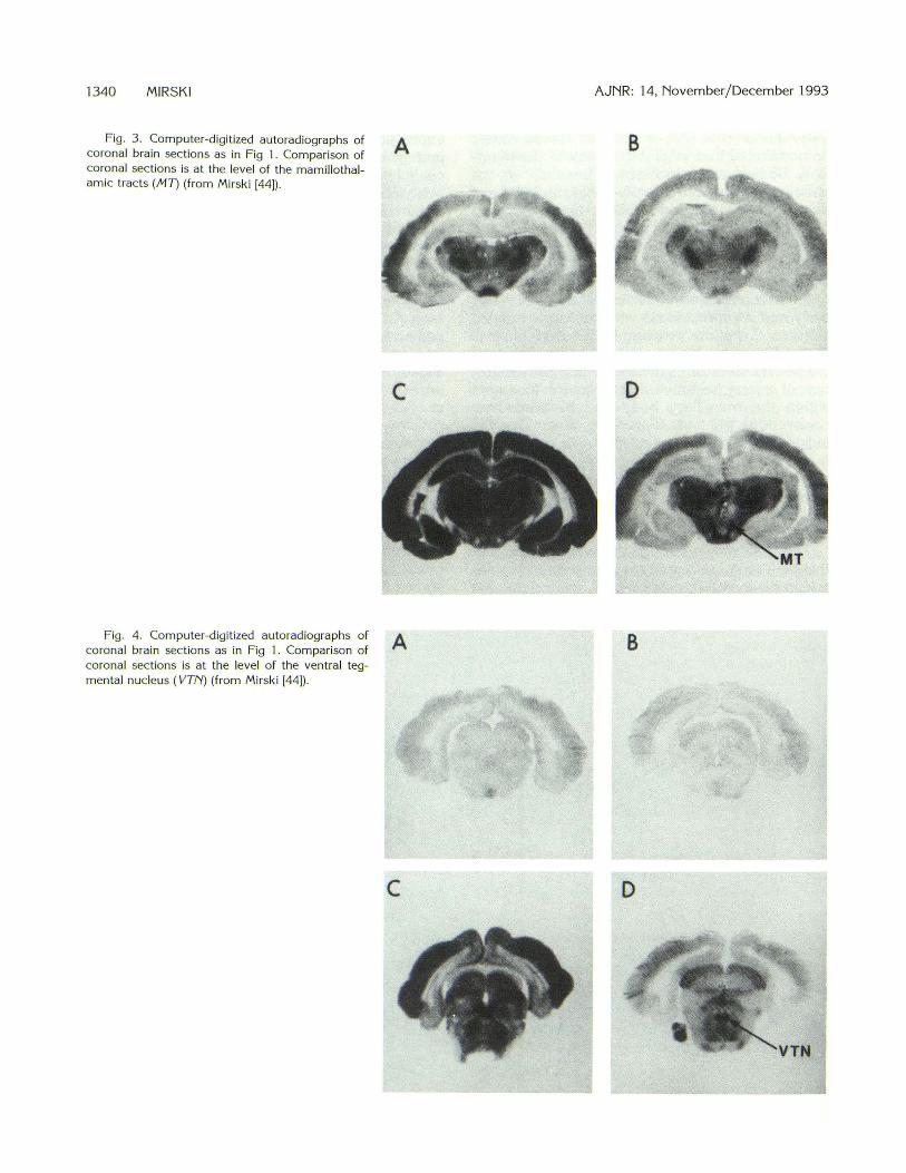
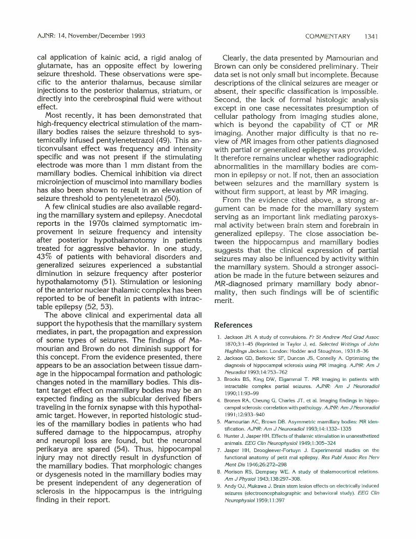
More recent work has linked the mamillary body and its connections with the expression of seizures. Experiments using [14C]2-deoxyglucose autoradiography during a threshold stage of pentylenetetrazol seizures resulted in the demonstration of a very selective metabolic enhancement within the mamillary body and its immediate connections (43, 44) (Figs 2-4). Rostrally , this included the anterior thalamic nuclei and the mamillothalamic tracts. Caudally in the brain stem, the mamillary peduncles (tegmentomamillary tracts) and the dorsal and ventral tegmental nuclei were all selectively highlighted with radioactive label.
These autoradiographic results suggested that this neuroanatomic path may have an important role in early seizure propagation. To evaluate the effects of interruption of this pathway on the
c D
COMMENTARY 1339
expression of convulsant activity , the rostral fiber path (the mamillothalamic tracts) was electrically coagulated and the animals were challenged with pentylenetetrazol. Bilateral interruption of these tracts resulted in highly significant anticonvulsant effect against both the behavioral and cortical electroencephalographic convulsant action and lethal effect of pentylenetetrazol (45) . These data supported the earlier findings suggesting that the mamillary system had a facilitory action on seizure activity (20). Interruption of the mamillary peduncles, carrying afferents to mamillary bodies from the tegmental nuclei , also was shown to afford significant protection against convulsant doses of pentylenetetrazol (46). These data strongly suggested that a functional anatomic pathway had been identified that was important in propagation of paroxysmal activity between midbrain and rostral diencephalon.
Attention has also been directed to the rostral terminus of the pathway, the anterior thalamic nuclei. Local microinjection of ')'-aminobutyric acid-related drugs muscimol or ')'-vinyl-')'-aminobutyric acid (an irreversible inhibitor of ')'-aminobutyric acid transaminase) into this region was shown to raise the seizure threshold of systemically administered pentylenetetrazol (47, 48). Lo-
Fig. 2. Computer digitized [14C]deoxyglucose autoradiographs of coronal brain sections from: control (A ); ethosuximide alone (B ); penty lenetetrazol alone (C); and pentylenetetrazol + ethosuximide-treated paralyzed and ventilated guinea pigs · (D). Pentylenetetrazol was continuously infused via a central venous catheter at a rate of 4 mg/ kg/ min. This dosage resulted in continuous high voltage, synchronous electroencephalographic activity and high levels of label incorporation throughout all gray matter regions, especially the cortex, globus pallidus, the geniculate nuclei , and substantia nigra. Coadministrat ion of ethosuximide (300 mg/ kg bolus intravenously followed by 4 mg/ k g per min) prevented the high-voltage seizure discharges and resulted in a continuous threshold stage of seizure act ivity . Incorporat ion of radio labeled substrate in brain in the penty lenetetrazol + ethosuximide group was less than in the penty le-netetrazol-alone animals with the notable exception of an actual increase in label accumulation within the mamillary system . Comparison of coronal sections is at the level of the anterior tha lamic nuclei (AN) (from Mirski [44]).
1340 MIRSKI
Fig. 3. Computer-digitized autoradiographs of coronal brain sections as in Fig 1 . Comparison of coronal sections is at the level of the mamillothalamic tracts (MT) (from Mirski [44]) .
Fig. 4. Computer-digitized autorad iographs of coronal bra in sections as in Fig 1. Comparison of coronal sections is at the level of the ventral tegmental nucleus ( VTN) (from Mirski [44]) .
A
c
A
c
AJNR: 14, November/ December 1993
B
D
B
D
AJNR: 14, November/ December 1993
cal application of kainic acid, a rigid analog of glutamate, has an opposite effect by lowering seizure threshold . These observations were specific to the anterior thalamus , because similar injections to the posterior thalamus, striatum, or directly into the cerebrospinal fluid were without effect.
Most recently , it has been demonstrated that high-frequency electrical stimulation of the mamillary bodies raises the seizure threshold to systemically infused pentylenetetrazol (49) . This anticonvulsant effect was frequency and intensity specific and was not present if the stimulating electrode was more than 1 mm distant from the mamillary bodies. Chemical inhibition via direct microinjection of muscimol into mamillary bodies has also been shown to result in an elevation of seizure threshold to pentylenetetrazol (50).
A few clinical studies are also available regarding the mamillary system and epilepsy. Anecdotal reports in the 1970s claimed symptomatic improvement in seizure frequency and intensity after posterior hypothalamotomy in patients treated for aggressive behavior. In one study, 43 % of patients with behavioral disorders and generalized seizures experienced a substantial diminution in seizure frequency after posterior hypothalamotomy (51). Stimulation or lesioning of the anterior nuclear thalamic complex has been reported to be of benefit in patients with intractable epilepsy (52, 53).
The above clinical and experimental data all support the hypothesis that the mamillary system mediates, in part, the propagation and expression of some types of seizures. The findings of Mamourian and Brown do not diminish support fo r this concept. From the evidence presented, there appears to be an association between tissue damage in the hippocampal formation and pathologic changes noted in the mamillary bodies. This distant target effect on mamillary bodies may be an expected finding as the subicular derived fibers traveling in the fornix synapse with this hypothalamic target. However, in reported histologic studies of the mamillary bodies in patients who had suffered damage to the hippocam pus, atrophy and neuropil loss are found, but the neuronal perikarya are spared (54). Thus, hippocampal injury may not directly result in dysfunction of the mamillary bodies. That morphologic changes or dysgenesis noted in the mamillary bodies may be present independent of any degeneration of sclerosis in the hippocam pus is the intriguing finding in their report.
COMMENTARY 1341
Clearly, the data presented by Mamourian and Brown can only be considered preliminary. Their data set is not only small but incomplete. Because descriptions of the clinical seizures are meager or absent, their specific classification is impossible. Second , the lack of formal histologic analysis except in one case necessitates presumption of cellular pathology from imaging studies alone , which is beyond the capability of CT or MR imaging. Another major difficulty is that no review of MR images from other patients diagnosed with partial or generalized epilepsy was provided . It therefore remains unclear whether radiographic abnormalities in the mamillary bodies are common in epilepsy or not . If not, then an association between seizures and the mamillary system is without firm support, at least by MR imaging.
From the evidence cited above , a strong argument can be made for the mamillary system serving as an important link mediating paroxysmal activity between brain stem and forebrain in generalized epilepsy. The close association between the hippocampus and mamillary bodies suggests that the clinical expression of partial seizures may also be influenced by activity within the mamillary system. Should a stronger association be made in the future between seizures and MR-diagnosed primary mamillary body abnormality, then such findings will be of scientific merit .
References
1. Jackson JH. A study of convulsions. Fr St Andrew Med Grad Assoc
1870;3:1-45 (Reprin ted in Tay lor J, ed. Selected Writings of John
Hughlings Jackson. London: Hodder and Stoughton, 1931:8-36 2. Jackson GD, Berkovic SF, Duncan JS, Connelly A. Optim izing the
diagnosis of hippocampal sclerosis using MR imaging. AJNR: Am J
Neuradiol 1993; 14:753-762 3. Brooks BS, King DW, Elgam mal T . MR imaging in patients with
intractable complex partia l seizures. AJNR: Am J Neuroradiol
1990; 11 :93- 99
4. Bronen RA , Cheung G, Charles JT, et al. Imaging f indings in hippo
campal sclerosis: correlation with pathology. AJNR: Am J Neuroradiol
199 1; 12:933- 940
5. Mamourian AC, Brown DB. Asymmetric mamillary bodies: MR iden
t ification. AJNR: Am J Neuroradio/ 1993; 14:1332-1335
6. Hunter J , Jasper HH. Effects of thalamic stim ulation in unanesthetized
animals. EEG C/in Neurophysiol 1949; 1 :305- 324 7. Jasper HH, Droogleever-Fortuyn J. Experimental studies on the
functional ana tomy of petit mal epilepsy. Res Pub/ Assoc Res Nerv
Ment Dis 1946;26:272- 298 8. Morison RS, Dempsey WE. A study of thalamocortical relations.
A m J Physio/1 943 ;138:297-308.
9. Andy OJ, Mukawa J. Brain stem lesion effects on electrically induced
seizures (electroencepha lographic and behaviora l study). EEG C/in
Neurophysio/ 1959; 11 :397
1342 MIRSKI
10. Bergman F, Costin A, Gutman J. A low threshold convulsive area
in the rabbit's mesencephalon. EEG Clin Neurophysiol 1963;15:
683-690 11 . Browning RA, Simonton RL, Turner FJ . Antagonism of experimen
tally induced tonic seizures following a lesion in the midbrain tegmen
tum. Epilepsia 1981 ;22:595-601
12. Guerrero-Figueroa R, Barros A, deBadbian Verster F, Heath RG.
Experimental "petit-mal " in kittens. Arch Neurol1963;9:297-306
13. lngvar DH. Reproduction of the 3 per second spike and wave EEG
pattern by subcortical electrical stimulation in cats. Acta Physiol
Scand 1955; 33:137-150
14. Kriendler A , Zuckermann E, Steriade M , Chimion D. Electroclinical
features of convulsions induced by stimulation of the brain stem. J
Neurophysiol1958;21 :430-436
15. Gloor P. Generalized cortico-reticular epilepsies. Some considerations
on the pathophysiology of generalized bilaterally synchronous spike
and wave discharges. Epilepsia 1968;9:249-263
16. Kusske JA, Ojemann GA, Ward AA Jr. Effects of lesions in ventral
anterior thalamus on experimental focal epilepsy. Exp Neural
1972;34:279-290
17. Miller JW, Ferrendelli JA. The central median nucleus: thalamic site
of seizure regulation. Brain Res 1990;297-300
18. Velasco F, Velasco M , Ogarrio C, Fanghanel G. Electrical stimulation
of the centromedian thalamic nucleus in the treatment of convulsive
seizures: a preliminary report. Epilepsia 1987;28:421-430
19. Van Straa ten JJ. Abolition of electrically induced cortical seizures by
stereotactic thalamic lesions. Neurology 1975;25: 141-149
20. Jinnai D, Mogami H, Mukawa J , Iwata Y, Kobayashi K. Effects of
brain-stem lesions on Metrazol-induced seizures in cats. EEG Clin
Neurophysiol 1969;27 :404-411
21. Patel S, Millan MH, Mello LM, Meldrum BS. 2-Amino-7-phosphono
heptanoic acid (2-APH) infusion into entopeduncular nucleus protects
against limbic seizures in rats. Neurosci Let 1986;64:226-230
22. Patel S, Millan MH, Meldrum BS. Decrease in excitatory transmission
within the lateral habenula and the mediodorsal thalamus protects
against limbic seizures in rats. Exp Neurol1988;101:63-74
23. Garant DS, Gale K. Lesions of the substantia nigra protect against
experimentally induced seizures. Brain Res 1983;273:156-161
24. ladarola MJ, Gale K. Cellular compartments of GABA in brain and
their relationships to anticonvulsant activity. Mol Cell Biochem
1981 ;39:305-330
25. Turski L, Cavalheiro EA, Tursk i WA, et al. Susceptibility to seizures
produced by pilocarpine in rats after microinjection of isoniazid
and -y-v iny i-GABA into the substantia nigra. Brain Res 1978;372:
294-309
26. Maggio R, Gale K. Seizures evoked from area tempestas are subject
to control by GABA and glutamate receptors in substantia nigra. Exp
Neural 1989; 105:184-188
27. Piredda S, Gale K . Evidence that the deep prepiriform cortex contains
a crucial epileptogenic site. Nature 1985;317:623-625
28. Piredda S, Gale K. Role of excitatory amino ac id transmission in the
genesis of seizures elicited from the deep prepiriform cortex. Brain
Res 1986;205-210
29. Cruce JAF. An autoradiographic study of the descending connections
of the mamillary nuclei of the rat. J Camp Neurol1 977;631-644
30. Fry FJ , Cowan WM. A study of retrograde cell degeneration in the
lateral mamillary nucleus of the cat, with special reference to the role
of axonal branching in the preservation of the cell. J Comp Neural
1972; 144:1-24
31 . Hayakawa T , Zyo K. A comparative anatomical study of the tegmen
tomamillary projections in some mammals: a horseradish peroxidase
study. Brain Res 1984;300:335-349
AJNR: 14, November/ December 1993
32. Veazey RB, Amaral DG, Cowan WM. The morphology and connections of the posterior hypotha lamus in cynomolgus monkey (macaca
fasciculars). I. Cytoarchitectonic organization . J Camp Neural
1982;207: 114-1 34 33. Veazey RB, Amaral DG, Cowan MW. The morphology and connec
tions of the posterior hypothalamus in cynomolgus monkey (macaca
fasciculars). II. Efferent connections. J Camp Neural 1982;207:
135-156 34. Swanson LW, Cowan WM. Hippocampo-hypothalamic connections:
origin in subicular cortex, not ammon 's horn. Science 1975;189:
303-304 35. Cowan WM, Guillery RW, Powell TPS. The origin of the mamillary
peduncle and other hypothalamic connections from the midbrain. J
Anat 1964;98:345-363 36. Guillaery RW. Degeneration in the hypothalamic connections of the
albino rat. J Anat 1957;91:91-115
37. Papez JW. A proposed mechanism of emotion. Arch Neural
1937;38:725-744 38. Domesick VB. Projections form the cingulate cortex in the rat. Brain
Res 1969; 12:296-300 39. Dagi TF, Poletti CE. Reformulation of the papez circuit: absence of
hippocampal influence on cingu late cortex unit activity in the primate.
Brain Res 1983;259:229-236
40. Domesick VB. Thalamic relationships of the medial cortex in the rat.
Brain Behav Evol1972;6:457-483
41. Murphy JP, Gellhorn J. Further investigations on diencephalic-cortical
relations and their significance for the problem of emotion. J Neuro
physiol1945 ;8:431-442 42. Green JD, Morin F. Hypothalamic electrical activity and hypothalamo
cortical relationships. Am J Physiol 1953; 172:175-186
43. Mirski MA, Ferrendelli JA. Individual and combined effects of con
vulsant and anticonvulsant drugs on regional brain metabolism. Soc
Neurosci Abstr 1983;9:627
44. Mirski MA, Ferrendelli JA. Selective metabolic activation of the
mamillary bodies and their connections during ethosuximide-induced
suppression of pentylenetetrazol seizures. Epilepsia 1986;51:
194-203
45. Mirski MA, Ferrendelli JA. Interruption of the mammillothalamic
tracts prevents seizures in guinea pigs. Science 1984;226:72-74
46. Mirski MA, Ferrendell i JA. Interruption of the connections of the
mamillary bodies protect against generalized pentylenetetrazol sei
zures in guinea pigs. J Neurosci 1987;7:662-670
47 . Mirski MA, Ferrendelli JA. Anterior thalamic mediation of generalized
pentylenetetrazol seizures. Brain Res 1986;399:212-223
48. Miller JW, McKeon AC, Ferrendelli JA. Functional anatomy of pen
tylenetetrazol and electroshock seizures in the rat brainstem. Ann
Neurol1987;22:6 15-621
49. Mirski MA, Fisher RA. Posterior hypothalamic stimulation inhibits
PTZ-induced seizures in rats . Epilepsia 1992;53:37
50. Mirski MA, Fisher RA. Pharmacological inhibition of posterior hypo
thalamus raises seizure threshold in rats. Epilepsia (in press)
51 . Sa no K, Sekino H, Mayanagi Y. Results of stimulation and destruction
of the posterior hypothalamus in cases with violent, aggressive, or
restless behaviors. In: Hitchcock E, Laitinen L, Vaernet K, eds.
Psychosurgery. Springfield, IL: Charles C Thomas, 1972:57-75
52. Mulen S, Vailati G, Karasick J , Mailis M . Thalamic lesions for the
control of epilepsy. Arch Neurol1 967;16:277-285
53. Sussman NM, Goldman HW, Jackel RA, et a!. Anterior thalamic
stimulation in medica lly intractable epilepsy. II . Preliminary clinical
results. Epilepsia 1988;29:677
54. Lindboe CF, Erichsen AA, Strom EH. Atrophy and sponginess of the
mamillary bodies with neuronal sparing: not only inactive wernick 's
encephalopathy. APMIS 1989;97:667- 670