Embed Size (px)
Citation preview
28054
High-Precision Mass Analysis of RI Sample for Cross-Section Measurements
S.Nakamura, Y. Shibahara1, A. Uehara
1, T. Fujii
2,
A. Kimura, B. Hales and O. Iwamoto
Japan Atomic Energy Agency 1Kyoto University Research Reactor Institute 2Osaka University
INTRODUCTION: The ImPACT project[1] aims to realize
a "large reduction of and exploitation of resources in high-level
nuclear waste by nuclear transmutation", by using an accelerator
to accomplish nuclear transmutation of long-lived fission prod-
ucts (LLFPs). To perform accelerator-based nuclear transmuta-
tion effectively, it is necessary to acquire reaction cross-section
data for radionuclides across a wide spectrum of incident particle
energies. This project targets the major LLFPs: 107
Pd, 93
Zr, 135
Cs, 126
Sn, and 79
Se, as well as incidentally the medium-lived fission
product 137
Cs. We targeted the 135
Cs because of its long half-life
(2.3×10⁶ yr[2]). However, it is impossible to obtain a pure
135Cs
sample because it is not supplied by available vendors. This is
also why the quantity of data is so scarce [3, 4]. Isotope separa-
tion of 135
Cs and 137
Cs is difficult, and therefore 135
Cs is included
as an impurity in available 137
Cs samples. The isotope abun-
dance of 135
Cs and 137
Cs in a standard 137
Cs source has been
measured by mass spectrometry. Then, the amount of 135
Cs can
be obtained from yields of 662 keV decay gamma-rays of 137
Cs
and the isotope abundance. It is also necessary to suppress con-
tamination of the mass spectrometer from radioactive 137
Cs and 135
Cs. It is necessary to perform highly precise analysis for an
extremely small amount of sample. Therefore, this research aims
to apply mass spectrometry to a very small amount of radioiso-
tope sample, and to confirm its effectiveness.
EXPERIMENTS: A standard solution of 137
Cs (370kBq)
was obtained through the Japan Radioisotope Association. An
appropriate amount of the 137
Cs solution was dispensed, and then
converted to nitrate by a chemical processing. The mass spec-
trometer TRITON was used for this analysis, which was made
by Thermo Fisher Scientific. A small amount of the chemically
processed solution, about 10Bq, was pipetted onto a Re filament
with a TaO activator, and then dried. Three filaments with ap-
plied Cs solution were prepared. The filaments were attached
onto the ion source of the mass spectrometer.
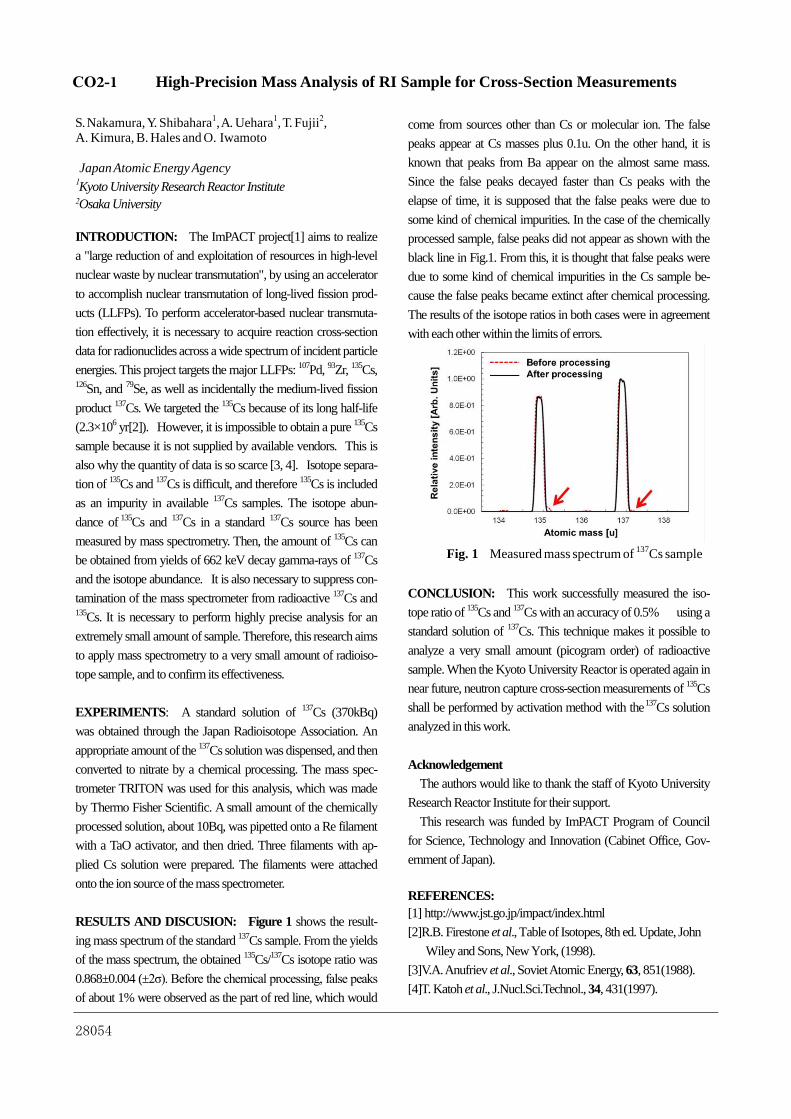
RESULTS AND DISCUSION: Figure 1 shows the result-
ing mass spectrum of the standard 137
Cs sample. From the yields
of the mass spectrum, the obtained 135
Cs/137
Cs isotope ratio was
0.868±0.004 (±2σ). Before the chemical processing, false peaks
of about 1% were observed as the part of red line, which would
come from sources other than Cs or molecular ion. The false
peaks appear at Cs masses plus 0.1u. On the other hand, it is
known that peaks from Ba appear on the almost same mass.
Since the false peaks decayed faster than Cs peaks with the
elapse of time, it is supposed that the false peaks were due to
some kind of chemical impurities. In the case of the chemically
processed sample, false peaks did not appear as shown with the
black line in Fig.1. From this, it is thought that false peaks were
due to some kind of chemical impurities in the Cs sample be-
cause the false peaks became extinct after chemical processing.
The results of the isotope ratios in both cases were in agreement
with each other within the limits of errors.
CONCLUSION: This work successfully measured the iso-
tope ratio of 135
Cs and 137
Cs with an accuracy of 0.5% using a
standard solution of 137
Cs. This technique makes it possible to
analyze a very small amount (picogram order) of radioactive
sample. When the Kyoto University Reactor is operated again in
near future, neutron capture cross-section measurements of 135
Cs
shall be performed by activation method with the 137
Cs solution
analyzed in this work.
Acknowledgement
The authors would like to thank the staff of Kyoto University
Research Reactor Institute for their support.
This research was funded by ImPACT Program of Council
for Science, Technology and Innovation (Cabinet Office, Gov-
ernment of Japan).
REFERENCES:
[1] http://www.jst.go.jp/impact/index.html
[2]R.B. Firestone et al., Table of Isotopes, 8th ed. Update, John
Wiley and Sons, New York, (1998).
[3]V.A. Anufriev et al., Soviet Atomic Energy, 63, 851(1988).
[4]T. Katoh et al., J.Nucl.Sci.Technol., 34, 431(1997).
Fig. 1 Measured mass spectrum of 137
Cs sample
CO2-1

28061
Measurement of Doppler Effect by Small Accelerator Neutron Source (1)
T. Sano, J. Hori, Y. Takahashi, K. Nakajima, H. Unesaki
Research Reactor Institute, Kyoto University
INTRODUCTION: In order to reduce TRU, the re-
search and technology development entitled as “TRU
burning fast reactor cycle using uranium-free TRU metal
fuel” have been started Japan at October 2014 [1]. The
feature of the fast reactor is high content TRU and Zr without uranium in the fuel alloy so that additional TRU
is not produced. On the other hand, uranium-free TRU
metallic fuel leads to the reduction of the Doppler reac-
tivity. Thus, the utilization of fuel alloy such as Mo and
Nb instead of Zr is considered as one of the counter-
measures [2]. As the Doppler effects depend on the mag-
nitude of self-shielding at the resonances, it is important
to verify the Doppler effects at each resonance of the fuel alloy (Mo or Nb) materials to evaluate the feasibility of
the uranium-free TRU metallic fuel. However, the dif-
ferential experiment of the Doppler effects for Mo and
Nb has not been carried out so far. Therefore, we have
initiated the measurement of the Doppler effects for Mo
sample by Time-of-flight (TOF) method with the KUR-
RI-LINAC pulsed neutron source.
EXPERIMENTS: We measured the Doppler effects of Mo sample with the KURRI-LINAC pulsed-neutron
source. In the experiment, neutron capture rates in Mo
sample at 300 K and 600 K were obtained by prompt
gamma-ray measurement with the TOF method. The Mo
sample was placed in the center of a heating device at a
distance of 10 m from the Ta target. The surface temper-
ature of the sample was observed by thermoelectric cou-
ple and controlled to be constant during the irradiation by a glass-heater. Two kinds of natural molybdenum samples
with different thickness of 0.5 mmt and 3.0 mmt were
prepared to identify the neutron self-shielding effects for
each resonance. Those were metallic plates of 2.0×2.0
cm2. The measurements with thick and thin Mo samples,
the graphite sample with 3.0 mm in thickness, and no
sample (blank run) at 300 K and 600 K were carried out.
RESULTS: The measured TOF spectra with 0.5 mmt and 3.0 mmt Mo samples at 300K are shown in Fig. 1.
The main resonances of Mo-95, Mo-96, Mo-97 and
Mo-98 were clearly observed in the energy range from 10
eV to 1 keV. For further analysis, counting statistics ob-served in TOF spectrum were improved by rebinning the
data into the JSF70 group structure [5] corresponding to
the lethargy width of 0.25. Comparison of the rebinned
TOF spectra for the 3.0 mmt sample at 300 K and 600 K
is shown in Fig. 1. The Doppler capture rate ratio defined
as the capture rate at 600 K divided by it at 300 K was
obtained for each energy group. The Doppler capture rate
ratio were 1.3 ± 0.7 % at 44.9 eV, 8.0 ± 0.7 % at 70.9 eV, 1.0 ± 0.7% at 131.4 eV and 6.5 ± 3.0% at 358.6 eV reso-
nances.
44.9 eV : 1.3±0.7%
70.9 eV : 8.0±0.7%
131.4 eV : 1.0±0.7%358.6 eV : 6.5±3.0%
This study is being performed under the contract with
the Ministry of Education, Culture, Sports, Science and
Technology of Japan (MEXT) in the framework of
MEXT’s Nuclear System Research and Development
Program.
REFERENCES: [1] K. Arie, et. al., Proc. of Global 2015, Sep. 20-24, Par-
is, France, Paper 5096, (2015).
[2] K. Arie, et. al., Proc. of ICAPP 2014, Charlotte, USA,
April 6-9, (2014).
[3] Y. Nagaya et. al., JAERI1348, Japan Atomic Energy
Research Institute, (2005).
[4] K. Shibata, et. al., J. Nucl. Sci. Technol. 48(1), 1-30
(2011).[5] M.Nakagawa, K. Tsuchihashi, JAERI 1294 (1984)
Fig. 2 the experimental TOF spectra for 3.0 mmt Mo sample at 300 K
and 600 K
Fig. 1 The measured TOF spectra with Mo samples, graphite sample and no sample
CO2-2

28062
Development of Neutron Detectors for Precise Measurement of Epi-thermal Neutrons
T. Matsumoto, A. Masuda, H. Harano, H. Tomita1, Y.
Ichinose1, K. Uema1, T. Iguchi1, K. Watanabe1, A. Uri-
tani1 and J. Hori2
National Metrology Insitute of Japan, National Institute
of Advanced Industrial Science and Technology 1Department of Engineering, Nagoya University 2Research Reactor Institute, Kyoto University
INTRODUCTION: Evaluation of neutron fluence
and neutron dose equivalent for the epi-thermal
neutron region is very important in work places
with neutron sources or nuclear fuels as well as
irradiation fields in a boron neutron capture
therapy (BNCT). It is not easy to determine pre-
cisely the neutron fluence for the epi-thermal
neutrons in an irradiation field because of large
uncertainty of reaction cross sections in the
epi-thermal region. In the present study, we
have developed neutron detectors for absolute
measurement of epi-thermal neutrons, high in-
tensity epi-thermal neutrons in a BNCT field and
an imaging detector for a hidden material.
EXPERIMENTS: A collimated neutron beam
was obtained by the photo-neutron reaction using
a water-cooled tantalum target at the KURRI
Linac [1]. We experimentally evaluated charac-
teristics of the epi-thermal neutron detector for
absolute measurement, the high intensity
epi-thermal neutron detector and the imaging
detector.
(1) Epi-thermal neutron detector for absolute
measurement
The epi-thermal neutron detector for absolute measure-
ment is composed of a 6Li6natGd10B3O9:Ce+ (LGB)
scintillation detector and two BGO scintillation
detectors. In our previous experiments, we used
an NaI(Tl) scintillation detector instead of the
BGO scintillation detectors. However, the NaI(Tl)
scintillator has large sensitivity to neutrons as a
comparison with the BGO scintillator. The 50
mm-diameter and 5-mm thick LGB scintillator
was set at the center of the beam line. The 50.8
mm-diameter and 50.8 mm thick BGO scintilla-
tors were located on both sides of the LGB scin-
tillator and out of neutron beam. When the LGB
scintillator detects neutrons by the 10B(n,γα) reac-
tion, 478 keV monoenergetic gamma rays are
produced and subsequently detected with the
BGO scintillators. Moreover, the absolute neutron
fluence is determined by measuring gamma rays
from the 10B(n,γα) reaction with the BGO scintil-
lators in setting a 1-cm thick 10B4C total absorp-
tion sample in front of the LGB scintillator.
Number of alpha particles and gamma rays pro-
duced by the 10B(n,) reaction in the LGB scin-
tillator are absolutely determined by the coinci-
dence measurements. Pulse height spectra of the
LGB and BGO scintillators from the 10B(n,) re-
action were obtained in the coincidence meas-
urements. However, the evaluation of the back-
ground due to scattered neutrons was not suffi-
cient. In future, we will improve the measure-
ment process based on the present experimental
results.
(2) High intensity epi-thermal neutron detector The high intensity epi-thermal neutron detector is important in the BNCT. The detector is simp-
ly composed of a 6Li-glass scintillator and a 7Li-glass scintillator and photomultipliers (PMTs). Current from an anode output of the PMT is de-
tected by a current integrator. The 7Li-glass scintillator is used to subtract the gamma ray contribution detected by the 6Li-glass scintillator. We experimentally evaluated the difference be-
tween the neutron detection efficiencies of the 6Li-glass and 7Li-glass scintillators using the Linac neutron source. Finally, we successfully obtained the relation between the thermal neu-
tron flux and output current using the neutron detection efficiencies in the thermal neutron standard field of the National Institute of Ad-
vanced Industrial Science and Technology. In future, we will perform the high flux neutron ir-
radiation using the KUR.
(3) Imaging detector
The imaging detector is used to search hidden materials such as nuclear materials and radioac-
tive sources. The imaging detector is composed of multi-pixel type CdTe detectors. The imaging detector detects prompt gamma rays produced by the neutron capture reaction in the hidden mate-
rials or gamma rays from a hidden radioactive source. After that, the imaging detector deter-
mines a position of the hidden material or the hidden radioactive source. Characteristics of the detector were evaluated using gamma rays from the neutron capture reactions of 10B and 197Au. We will continue the experiments in order to ver-
ify potential of the practical realization using the TOF method.
REFERENCE:
[1] K. Kobayashi et al., Annu. Rep. Res. Reac-
torinst. Kyoto Univ. 22, 142 (1989).
This work was supported by JSPS KA-
KENHI(JP16K21679,JP16K09030).
CO2-3

28076
Research and Development for Accracy Improvement of Neutron Nuclear Data on
Long-lived Radioactive Nnuclei at KURRI-Linac
J. Hori, T. Sano, Y. Takahashi and H.Yashima1
Research Reactor Institute, Kyoto University
INTRODUCTION: There are strong requests for re-
ducing the uncertainty of neutron capture cross section
data of minor actinides (MAs) to estimate the transmuta-
tion rate of those long-lived radioactive nuclei in the in-
novative reactor system. In recent years, intense pulsed
spallation neutron sources became available to remarka-
bly improve the precision of neutron TOF data. However,
there are discrepancies out of a range of tolerance be-
tween current experimental results. It is understood that
the unrecognized systematic errors make a difference. In
order to recognize and reduce the systematic errors, the
project entitled as “Research and development for Accu-
racy Improvement of neutron nuclear data on Minor AC-
tinides (AIMAC)” has been started [1]. In this project,
we aim at obtaining the resonance parameters precisely
of MAs by combining the neutron capture -ray meas-
urement to transmission neutron measurement at
KURRI-Linac. Neptunium-237 is one of the most im-
portant MAs with a long half-life. In this year, neutron
total cross section and capture cross section of 237
Np
were measured and resonance analysis was also per-
formed.
EXPERIMENTS: The total and capture cross section
measurements have been performed by the neutron
time-of-flight (TOF) method using the 46-MeV electron
linear accelerator at the Research Reactor Institute, Kyoto
University (KURRI-LINAC). Transmitted neutrons were
detected by a 6 mm thick GS20 6Li-glass scintillator.
Neptunium oxide powder of 1.13 g packed in an alumi-
num disk container of 30mm in diameter and 0.4 mm
thick wall, which was placed at a distance of 10.15 m
from the photo-neutron source. An aluminum disk con-
tainer without the neptunium oxide powder was also used
as a dummy case. The linac was operated with an elec-
tron energy of about 30 MeV, an averaged beam current
of 17 A, a repetition rate of 50 Hz, a pulse width of 0.1
s. Neutron capture gamma rays from the sample were
measured with a 4 bismuth germinate (BGO) scintilla-
tion detectors composed of 12 BGO cylindrical crystals
having 2 inch. in diameter and 2 inch. in length. A cap-
ture sample was set in the center of the detector. Three
samples of 237
Np with different thickness were used; the
activities of the samples were 26, 5.2 and 1 MBq, respec-
tively. The incident neutron flux shape was measured
with a 10
B sample.
RESULTS: Preliminary result of total cross section of 237
Np is shown in Fig. 1. The resonance analysis of the
deduced cross section values were performed with the
SAMMY code [ 2] in the energy range from 10-2
to 80 eV.
The resonance parameters of the 0.49-eV resonance were
modified to reproduce the present results. The neutron
capture yields for the samples with different thickness
were also obtained as shown in Fig. 2. The neutron
self-shielding and multiple-scattering effect in the sample
depends on the resonance parameter. Therefore, the data
were used for the cross-check of resonance parameters.
Fig. 1 Preliminary results of neutron total cross section of 237
Np
Fig. 2 Capture yields for three 237
Np samples measured by the BGO spectrometer
Present study includes the result of “Research and De-
velopment for accuracy improvement of neutron nuclear
data on minor actinides” entrusted to the Japan Atomic
Energy Agency by the Ministry of Education, Culture,
Sports, Science and Technology of Japan (MEXT).
REFERENCES:
[1]H. Harada et al., EPJ Web of Conferences 93, 06001 (2015).
[2]N. W. Larson, ORNL/TM-9179/R6, Oak Ridge National
Laboratory, 1998.
CO2-4