Embed Size (px)
Citation preview
REPORT
Mutations in CNNM4 Cause Jalili Syndrome,Consisting of Autosomal-Recessive Cone-RodDystrophy and Amelogenesis Imperfecta
David A. Parry,1,16 Alan J. Mighell,1,2,16 Walid El-Sayed,1,2 Roger C. Shore,2 Ismail K. Jalili,1
Helene Dollfus,3,4 Agnes Bloch-Zupan,5,6,7 Roman Carlos,8 Ian M. Carr,1 Louise M. Downey,9
Katharine M. Blain,10 David C. Mansfield,11 Mehdi Shahrabi,12 Mansour Heidari,13 Parissa Aref,12
Mohsen Abbasi,12 Michel Michaelides,14,15 Anthony T. Moore,14,15 Jennifer Kirkham,2
and Chris F. Inglehearn1,*
The combination of recessively inherited cone-rod dystrophy (CRD) and amelogenesis imperfecta (AI) was first reported by Jalili and
Smith in 1988 in a family subsequently linked to a locus on chromosome 2q11, and it has since been reported in a second small family.
We have identified five further ethnically diverse families cosegregating CRD and AI. Phenotypic characterization of teeth and visual
function in the published and new families reveals a consistent syndrome in all seven families, and all link or are consistent with linkage
to 2q11, confirming the existence of a genetically homogenous condition that we now propose to call Jalili syndrome. Using a posi-
tional-candidate approach, we have identified mutations in the CNNM4 gene, encoding a putative metal transporter, accounting for
the condition in all seven families. Nine mutations are described in all, three missense, three terminations, two large deletions, and
a single base insertion. We confirmed expression of Cnnm4 in the neural retina and in ameloblasts in the developing tooth, suggesting
a hitherto unknown connection between tooth biomineralization and retinal function. The identification of CNNM4 as the causative
gene for Jalili syndrome, characterized by syndromic CRD with AI, has the potential to provide new insights into the roles of metal trans-
port in visual function and biomineralization.
Cone-rod dystrophy (CRD [MIM 120970]) usually manifests
in childhood or early adulthood with predominant or equal
loss of cone compared to rod photoreceptors, reduced visual
acuity, color-visionabnormalities, photophobia, and visual-
field loss.1 Mutations in ABCA4 (MIM*601691), AIPL1 (MIM
*604392), CRX (MIM þ602225), GUCA1A (MIM *600364),
GUCY2D (MIM *600179), PITPNM3 (MIM *608921),
RIMS1 (MIM *606629), SEMA4A (MIM *6072920), RPGR
(MIM *312610), PROM1 (MIM *604365), and UNC119
(MIM *604011) have been associated with CRD, which
can be inherited in an autosomal-dominant, autosomal-
recessive, or X-linked manner (RetNet). These genes encode
proteins of diverse functions, including participants in the
phototransduction cascade and the visual cycle, structural
components of photoreceptors, and photoreceptor-specific
transcription factors.
In comparison with retinal degeneration, little is under-
stood about the genetic causes of abnormal tooth biominer-
alization (reviewed by Bailleul-Forestier and colleagues2,3).
This group of conditions is traditionally considered to
266 The American Journal of Human Genetics 84, 266–273, Februar
involve either enamel (amelogenesis imperfecta [AI] [MIM
#204700]) or dentine (dentinogenesis imperfecta [DI]
[MIM #125490] and dentine dysplasias [MIM %125400]),
the two major hard-tissue components of teeth. Nonsyn-
dromic AI has been attributed to mutations in five genes,
which fall into three groups and involve the following:
enamel-matrix proteins (AMELX4 [MIM *300391]; ENAM5
[MIM *606585]), enzymes controlling postsecretory pro-
cessing of enamel-matrix proteins (KLK46 [MIM *603767];
MMP207 [MIM *604629]), and a gene of unknown function
(FAM83H8 [MIM *611927]). Only a single gene (DSPP [MIM
*125485]) has been implicated in nonsyndromic DI.9
In addition, CRD, AI, or DI can be part of syndromes
involving multiple tissues and organs. CRD is a feature of
some forms of Bardet Biedl syndrome (MIM 209900),
Alstrom syndrome (MIM 203800), spinocerebellar ataxia
type 7 (MIM 164500), and selected syndromes charac-
terized by ectodermal abnormalities. AI and DI have
been described as part of syndromes involving bone abnor-
malities, giving insight into different forms of human
1Leeds Institute of Molecular Medicine, University of Leeds, St. James’s University Hospital, Leeds LS9 7TF, UK; 2Leeds Dental Institute, University of Leeds,
Leeds LS2 9LU, UK; 3AVENIR Inserm, Universite de Strasbourg, 67085 Strasbourg, France; 4Centre de reference pour les affections ophtalmologiques heredi-
taires (CARGO), Hopitaux Universitaires de Strasbourg, 67000 Strasbourg, France; 5Department of Paediatric Dentistry, Faculty of Dentistry, Louis Pasteur
University, 67000 Strasbourg, France; 6Reference Centre for Oral Manifestations of Rare Diseases, Service de Soins Bucco-Dentaires, Hopitaux Universitaires
de Strasbourg, 67000 Strasbourg, France; 7Institute of Genetics and Molecular and Cellular Biology, Inserm, U596, CNRS, UMR7104, 67404 Illkirch Cedex,
France; 8Centro Clinico de Cabeza y Cuello, Guatemala City 01010, Guatemala; 9Hull and East Yorkshire Eye Hospital, Hull HU3 2JZ, UK; 10Child Dental
Health, Dundee Dental Hospital, Dundee DD1 4HR, UK; 11Department of Ophthalmology, Inverclyde Royal Hospital, Greenock PA16 0XN, UK; 12Faculty
of Dentistry, Tehran University of Medical Sciences, 1417613151 Tehran, Iran; 13Department of Medical Genetics, Tehran University of Medical Sciences,
1417613151 Tehran, Iran; 14Institute of Ophthalmology, University College London, London EC1V 9EL, UK; 15Moorfields Eye Hospital, London EC1V
2PD, UK16These authors contributed equally to this work
*Correspondence: [email protected]
DOI 10.1016/j.ajhg.2009.01.009. ª2009 by The American Society of Human Genetics. All rights reserved.
y 13, 2009
biomineralization.3,10 However, there is no agreed term for
developmental abnormalities of enamel plus dentine, even
though this condition is increasingly recognized. The
abnormal dental phenotypes observed with MSX2 muta-
tions have been described as AI even though there is
evidence of dentine involvement.11
The combination of familial CRD and AI (MIM
%217080) was first reported in a large consanguineous
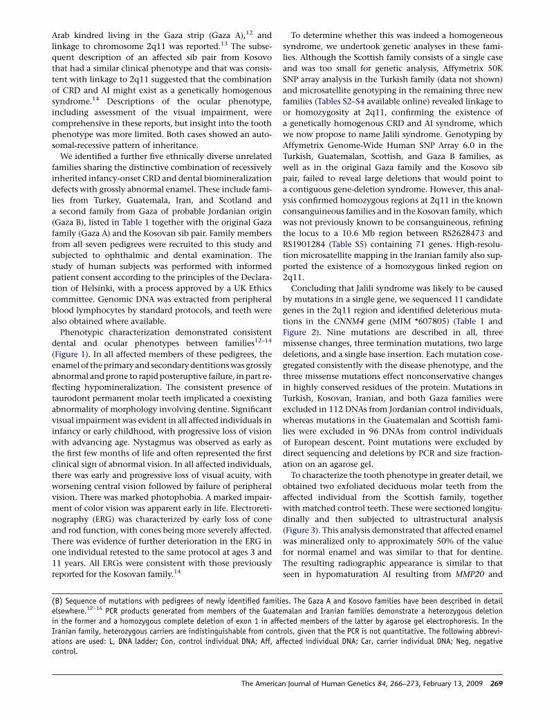
Table 1. A Summary of the CNNM4 Mutations Identified
Origin Reference Family History of Consanguinity Mutation 1 Mutation 2
Gaza A 12, 13 Yes c.599C/A; Ser200Tyr c.599C/A; Ser200Tyr
Kosovo 14 No c.1312 dupC; Leu438ProfsX9 c.1312 dupC; Leu438ProfsX9
Gaza B Yes c.1813 C/T; Arg605X c.1813 C/T; Arg605X
Guatemala No c.2149C/T; Gln717X c.62_145 del; Leu21HisfsX185
Turkey Yes c.586T/C; Ser196Pro c.586T/C; Ser196Pro
Iran Yes c.1-?_1403þ?del c.1-?_1403þ?del
Scotland No c.971T/C; Leu324Pro c.1690C/T; Gln564X
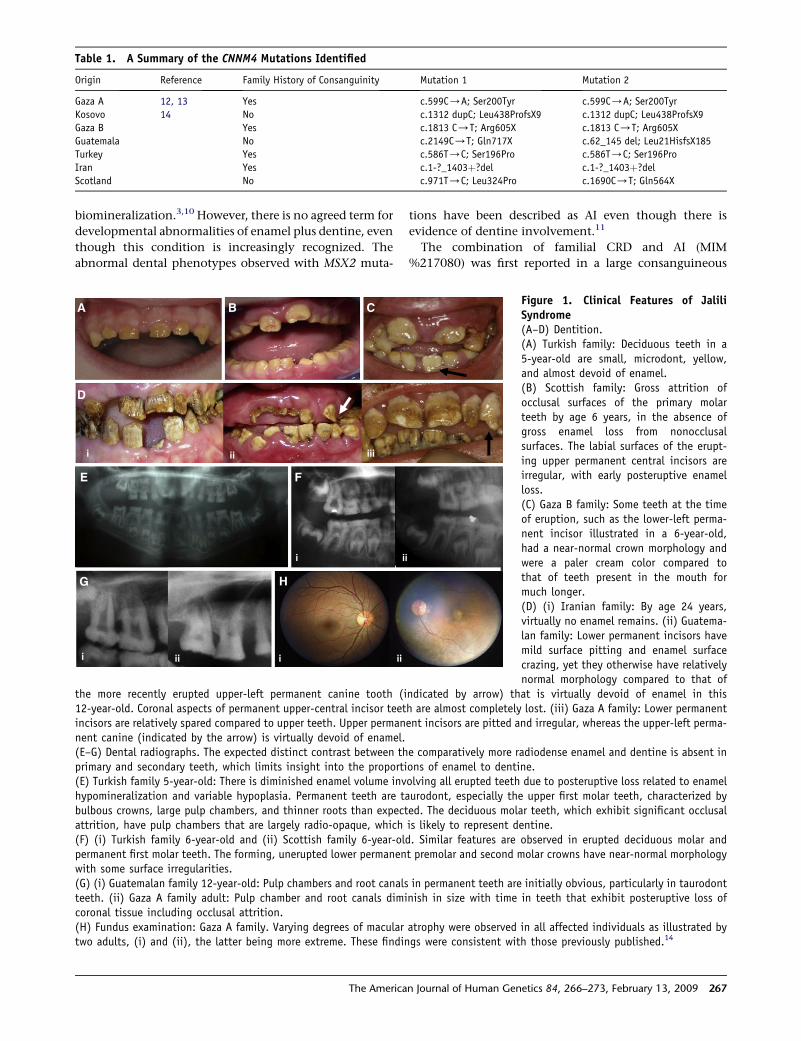
A B C
D
F
i
ii
G H
i ii iii
i iii
E
ii
Figure 1. Clinical Features of JaliliSyndrome(A–D) Dentition.(A) Turkish family: Deciduous teeth in a5-year-old are small, microdont, yellow,and almost devoid of enamel.(B) Scottish family: Gross attrition ofocclusal surfaces of the primary molarteeth by age 6 years, in the absence ofgross enamel loss from nonocclusalsurfaces. The labial surfaces of the erupt-ing upper permanent central incisors areirregular, with early posteruptive enamelloss.(C) Gaza B family: Some teeth at the timeof eruption, such as the lower-left perma-nent incisor illustrated in a 6-year-old,had a near-normal crown morphology andwere a paler cream color compared tothat of teeth present in the mouth formuch longer.(D) (i) Iranian family: By age 24 years,virtually no enamel remains. (ii) Guatema-lan family: Lower permanent incisors havemild surface pitting and enamel surfacecrazing, yet they otherwise have relativelynormal morphology compared to that of
the more recently erupted upper-left permanent canine tooth (indicated by arrow) that is virtually devoid of enamel in this12-year-old. Coronal aspects of permanent upper-central incisor teeth are almost completely lost. (iii) Gaza A family: Lower permanentincisors are relatively spared compared to upper teeth. Upper permanent incisors are pitted and irregular, whereas the upper-left perma-nent canine (indicated by the arrow) is virtually devoid of enamel.(E–G) Dental radiographs. The expected distinct contrast between the comparatively more radiodense enamel and dentine is absent inprimary and secondary teeth, which limits insight into the proportions of enamel to dentine.(E) Turkish family 5-year-old: There is diminished enamel volume involving all erupted teeth due to posteruptive loss related to enamelhypomineralization and variable hypoplasia. Permanent teeth are taurodont, especially the upper first molar teeth, characterized bybulbous crowns, large pulp chambers, and thinner roots than expected. The deciduous molar teeth, which exhibit significant occlusalattrition, have pulp chambers that are largely radio-opaque, which is likely to represent dentine.(F) (i) Turkish family 6-year-old and (ii) Scottish family 6-year-old. Similar features are observed in erupted deciduous molar andpermanent first molar teeth. The forming, unerupted lower permanent premolar and second molar crowns have near-normal morphologywith some surface irregularities.(G) (i) Guatemalan family 12-year-old: Pulp chambers and root canals in permanent teeth are initially obvious, particularly in taurodontteeth. (ii) Gaza A family adult: Pulp chamber and root canals diminish in size with time in teeth that exhibit posteruptive loss ofcoronal tissue including occlusal attrition.(H) Fundus examination: Gaza A family. Varying degrees of macular atrophy were observed in all affected individuals as illustrated bytwo adults, (i) and (ii), the latter being more extreme. These findings were consistent with those previously published.14
The American Journal of Human Genetics 84, 266–273, February 13, 2009 267
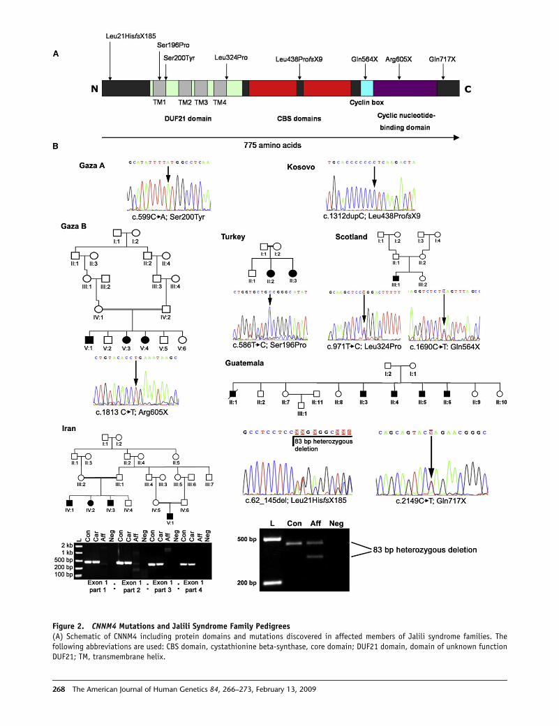
Figure 2. CNNM4 Mutations and Jalili Syndrome Family Pedigrees(A) Schematic of CNNM4 including protein domains and mutations discovered in affected members of Jalili syndrome families. Thefollowing abbreviations are used: CBS domain, cystathionine beta-synthase, core domain; DUF21 domain, domain of unknown functionDUF21; TM, transmembrane helix.
268 The American Journal of Human Genetics 84, 266–273, February 13, 2009
Arab kindred living in the Gaza strip (Gaza A),12 and
linkage to chromosome 2q11 was reported.13 The subse-
quent description of an affected sib pair from Kosovo
that had a similar clinical phenotype and that was consis-
tent with linkage to 2q11 suggested that the combination
of CRD and AI might exist as a genetically homogenous
syndrome.14 Descriptions of the ocular phenotype,
including assessment of the visual impairment, were
comprehensive in these reports, but insight into the tooth
phenotype was more limited. Both cases showed an auto-
somal-recessive pattern of inheritance.
We identified a further five ethnically diverse unrelated
families sharing the distinctive combination of recessively
inherited infancy-onset CRD and dental biomineralization
defects with grossly abnormal enamel. These include fami-
lies from Turkey, Guatemala, Iran, and Scotland and
a second family from Gaza of probable Jordanian origin
(Gaza B), listed in Table 1 together with the original Gaza
family (Gaza A) and the Kosovan sib pair. Family members
from all seven pedigrees were recruited to this study and
subjected to ophthalmic and dental examination. The
study of human subjects was performed with informed
patient consent according to the principles of the Declara-
tion of Helsinki, with a process approved by a UK Ethics
committee. Genomic DNA was extracted from peripheral
blood lymphocytes by standard protocols, and teeth were
also obtained where available.
Phenotypic characterization demonstrated consistent
dental and ocular phenotypes between families12–14
(Figure 1). In all affected members of these pedigrees, the
enamel of the primary and secondary dentitions was grossly
abnormal and prone to rapid posteruptive failure, in part re-
flecting hypomineralization. The consistent presence of
taurodont permanent molar teeth implicated a coexisting
abnormality of morphology involving dentine. Significant
visual impairment was evident in all affected individuals in
infancy or early childhood, with progressive loss of vision
with advancing age. Nystagmus was observed as early as
the first few months of life and often represented the first
clinical sign of abnormal vision. In all affected individuals,
there was early and progressive loss of visual acuity, with
worsening central vision followed by failure of peripheral
vision. There was marked photophobia. A marked impair-
ment of color vision was apparent early in life. Electroreti-
nography (ERG) was characterized by early loss of cone
and rod function, with cones being more severely affected.
There was evidence of further deterioration in the ERG in
one individual retested to the same protocol at ages 3 and
11 years. All ERGs were consistent with those previously
reported for the Kosovan family.14
The Americ
To determine whether this was indeed a homogeneous
syndrome, we undertook genetic analyses in these fami-
lies. Although the Scottish family consists of a single case
and was too small for genetic analysis, Affymetrix 50K
SNP array analysis in the Turkish family (data not shown)
and microsatellite genotyping in the remaining three new
families (Tables S2–S4 available online) revealed linkage to
or homozygosity at 2q11, confirming the existence of
a genetically homogenous CRD and AI syndrome, which
we now propose to name Jalili syndrome. Genotyping by
Affymetrix Genome-Wide Human SNP Array 6.0 in the
Turkish, Guatemalan, Scottish, and Gaza B families, as
well as in the original Gaza family and the Kosovo sib
pair, failed to reveal large deletions that would point to
a contiguous gene-deletion syndrome. However, this anal-
ysis confirmed homozygous regions at 2q11 in the known
consanguineous families and in the Kosovan family, which
was not previously known to be consanguineous, refining
the locus to a 10.6 Mb region between RS2628473 and
RS1901284 (Table S5) containing 71 genes. High-resolu-
tion microsatellite mapping in the Iranian family also sup-
ported the existence of a homozygous linked region on
2q11.
Concluding that Jalili syndrome was likely to be caused
by mutations in a single gene, we sequenced 11 candidate
genes in the 2q11 region and identified deleterious muta-
tions in the CNNM4 gene (MIM *607805) (Table 1 and
Figure 2). Nine mutations are described in all, three
missense changes, three termination mutations, two large
deletions, and a single base insertion. Each mutation cose-
gregated consistently with the disease phenotype, and the
three missense mutations effect nonconservative changes
in highly conserved residues of the protein. Mutations in
Turkish, Kosovan, Iranian, and both Gaza families were
excluded in 112 DNAs from Jordanian control individuals,
whereas mutations in the Guatemalan and Scottish fami-
lies were excluded in 96 DNAs from control individuals
of European descent. Point mutations were excluded by
direct sequencing and deletions by PCR and size fraction-
ation on an agarose gel.
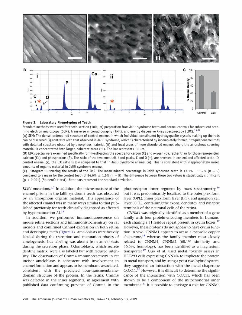
To characterize the tooth phenotype in greater detail, we
obtained two exfoliated deciduous molar teeth from the
affected individual from the Scottish family, together
with matched control teeth. These were sectioned longitu-
dinally and then subjected to ultrastructural analysis
(Figure 3). This analysis demonstrated that affected enamel
was mineralized only to approximately 50% of the value
for normal enamel and was similar to that for dentine.
The resulting radiographic appearance is similar to that
seen in hypomaturation AI resulting from MMP20 and
(B) Sequence of mutations with pedigrees of newly identified families. The Gaza A and Kosovo families have been described in detailelsewhere.12–14 PCR products generated from members of the Guatemalan and Iranian families demonstrate a heterozygous deletionin the former and a homozygous complete deletion of exon 1 in affected members of the latter by agarose gel electrophoresis. In theIranian family, heterozygous carriers are indistinguishable from controls, given that the PCR is not quantitative. The following abbrevi-ations are used: L, DNA ladder; Con, control individual DNA; Aff, affected individual DNA; Car, carrier individual DNA; Neg, negativecontrol.
an Journal of Human Genetics 84, 266–273, February 13, 2009 269
B C
A
Figure 3. Laboratory Phenotyping of TeethStandard methods were used for tooth-section (100 mm) preparation from Jalili syndrome teeth and normal controls for subsequent scan-ning electron microscopy (SEM), transverse microradiography (TMR), and energy dispersive X-ray spectroscopy (EDX).15,37
(A) SEM: The dense, ordered rod structure of control enamel in which individual constituent hydroxyapatite crystals making up the rodscan be discerned (i) contrasts with that observed in Jalili syndrome, which is characterized by incompletely formed, irregular enamel rodswith detailed structure obscured by amorphous material (ii) and focal areas of more disordered enamel where the amorphous coveringmaterial is concentrated into larger, coherent areas (iii). The bar represents 10 mm.(B) EDX spectra were examined specifically for investigating the spectra for carbon (C) and oxygen (O), rather than for those representingcalcium (Ca) and phosphorous (P). The ratio of the two most left-hand peaks, C and O (*), are reversed in control and affected teeth. Incontrol enamel (i), the C:O ratio is low compared to that in Jalili Syndrome enamel (ii). This is consistent with inappropriately raisedamounts of organic material in Jalili syndrome enamel.(C) Histogram illustrating the results of the TMR. The mean mineral percentage in Jalili syndrome teeth is 43.1% 5 1.7% (n ¼ 5)compared to a mean for the control teeth of 84.6% 5 1.5% (n ¼ 5). The difference between these two values is statistically significant(p < 0.001) (Student’s t test). Error bars represent the standard deviation.
KLK4 mutations.6,7 In addition, the microstructure of the
enamel prisms in the Jalili syndrome teeth was obscured
by an amorphous organic material. This appearance of
the affected enamel was in many ways similar to that pub-
lished previously for teeth clinically diagnosed as affected
by hypomaturation AI.15
In addition, we performed immunofluorescence on
mouse retina sections and immunohistochemistry on rat
incisors and confirmed Cnnm4 expression in both retina
and developing teeth (Figure 4). Ameloblasts were heavily
labeled during the transition and maturation phases of
amelogenesis, but labeling was absent from ameloblasts
during the secretion phase. Odontoblasts, which secrete
dentine matrix, were also labeled but with reduced inten-
sity. The observation of Cnnm4 immunoreactivity in rat
incisor ameloblasts is consistent with involvement in
enamel formation and appears to be membrane associated,
consistent with the predicted four-transmembrane-
domain structure of the protein. In the retina, Cnnm4
was detected in the inner segments, in agreement with
published data confirming presence of Cnnm4 in the
270 The American Journal of Human Genetics 84, 266–273, February
photoreceptor inner segment by mass spectrometry,16
but it was predominantly localized to the outer plexiform
layer (OPL), inner plexiform layer (IPL), and ganglion cell
layer (GCL), containing the axons, dendrites, and synaptic
terminals of the neuronal cells of the retina.
CNNM4 was originally identified as a member of a gene
family with four protein-encoding members in humans,
each sharing a 31 residue repeat present in cyclin boxes.17
However, these proteins do not appear to have cyclin func-
tion in vivo. CNNM1 appears to act as a cytosolic copper
chaperone,18 whereas the family member most closely
related to CNNM4, CNNM2 (68.1% similarity and
56.5%, homology), has been identified as a magnesium
transporter.19 Guo et al. used metal toxicity assays in
HEK293 cells expressing CNNM4 to implicate the protein
in metal transport, and by using a yeast two-hybrid system,
they suggested an interaction with the metal chaperone
COX11.20 However, it is difficult to determine the signifi-
cance of the interaction with COX11, which has been
shown to be a component of the mitochondrial inner
membrane.21 It is possible to envisage a role for CNNM4
13, 2009
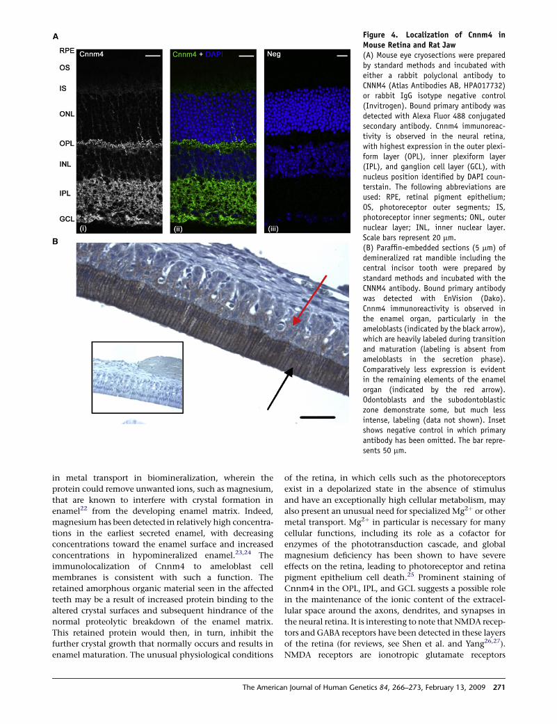
Figure 4. Localization of Cnnm4 inMouse Retina and Rat Jaw(A) Mouse eye cryosections were preparedby standard methods and incubated witheither a rabbit polyclonal antibody toCNNM4 (Atlas Antibodies AB, HPA017732)or rabbit IgG isotype negative control(Invitrogen). Bound primary antibody wasdetected with Alexa Fluor 488 conjugatedsecondary antibody. Cnnm4 immunoreac-tivity is observed in the neural retina,with highest expression in the outer plexi-form layer (OPL), inner plexiform layer(IPL), and ganglion cell layer (GCL), withnucleus position identified by DAPI coun-terstain. The following abbreviations areused: RPE, retinal pigment epithelium;OS, photoreceptor outer segments; IS,photoreceptor inner segments; ONL, outernuclear layer; INL, inner nuclear layer.Scale bars represent 20 mm.(B) Paraffin-embedded sections (5 mm) ofdemineralized rat mandible including thecentral incisor tooth were prepared bystandard methods and incubated with theCNNM4 antibody. Bound primary antibodywas detected with EnVision (Dako).Cnnm4 immunoreactivity is observed inthe enamel organ, particularly in theameloblasts (indicated by the black arrow),which are heavily labeled during transitionand maturation (labeling is absent fromameloblasts in the secretion phase).Comparatively less expression is evidentin the remaining elements of the enamelorgan (indicated by the red arrow).Odontoblasts and the subodontoblasticzone demonstrate some, but much lessintense, labeling (data not shown). Insetshows negative control in which primaryantibody has been omitted. The bar repre-sents 50 mm.
in metal transport in biomineralization, wherein the
protein could remove unwanted ions, such as magnesium,
that are known to interfere with crystal formation in
enamel22 from the developing enamel matrix. Indeed,
magnesium has been detected in relatively high concentra-
tions in the earliest secreted enamel, with decreasing
concentrations toward the enamel surface and increased
concentrations in hypomineralized enamel.23,24 The
immunolocalization of Cnnm4 to ameloblast cell
membranes is consistent with such a function. The
retained amorphous organic material seen in the affected
teeth may be a result of increased protein binding to the
altered crystal surfaces and subsequent hindrance of the
normal proteolytic breakdown of the enamel matrix.
This retained protein would then, in turn, inhibit the
further crystal growth that normally occurs and results in
enamel maturation. The unusual physiological conditions
The Americ
of the retina, in which cells such as the photoreceptors
exist in a depolarized state in the absence of stimulus
and have an exceptionally high cellular metabolism, may
also present an unusual need for specialized Mg2þ or other
metal transport. Mg2þ in particular is necessary for many
cellular functions, including its role as a cofactor for
enzymes of the phototransduction cascade, and global
magnesium deficiency has been shown to have severe
effects on the retina, leading to photoreceptor and retina
pigment epithelium cell death.25 Prominent staining of
Cnnm4 in the OPL, IPL, and GCL suggests a possible role
in the maintenance of the ionic content of the extracel-
lular space around the axons, dendrites, and synapses in
the neural retina. It is interesting to note that NMDA recep-
tors and GABA receptors have been detected in these layers
of the retina (for reviews, see Shen et al. and Yang26,27).
NMDA receptors are ionotropic glutamate receptors
an Journal of Human Genetics 84, 266–273, February 13, 2009 271
subject to voltage-dependent block by Mg2þ ions28 and are
noncompetitively inhibited by Zn2þ ions.29 Although the
functions of NMDA receptors in the retina are not entirely
clear, a defect in either Mg2þ or Zn2þ transport is likely to
lead to abnormal functioning of these receptors and their
parent cells. GABA receptors modulate neurotransmitter
release in the retina30 and are also inhibited by Zn2þ
ions.29 As such, failure of Zn2þ transport could lead to ex-
citotoxic conditions in the retina. Future work will be
required to confirm whether CNNM4 shares the magne-
sium-transport function of CNNM2 or whether it is
involved in the transport of other metal ions.
Like CNNM2, CNNM4 is predicted to have four trans-
membrane helices and contains a cyclic nucleotide-binding
domain, two cystathionine-beta-synthase (CBS) domains,
and a DUF21 domain of unknown function. CBS domains
appear to have a role in sensing the energy status of cells
by binding to ATP and are present in proteins such as CLC
chloride channels and IMPDH1, mutations in which are
a known cause of retinal degeneration.31 Furthermore,
cyclic nucleotide-binding domains are present in the cyclic
nucleotide-gated channels of photoreceptors. These chan-
nels are involved in ion transport and maintaining the
photoreceptor dark current, and they are closed by reduc-
tion in cGMP levels upon phototransduction. Several of
the protein subunits making up these channels are impli-
cated in retinal disease.32–35 On the basis of homology to
CNNM2, CNNM4 is likely to behave as a transporter rather
than a channel, and its localization in the retina as deter-
mined by immunofluorescence suggests that its role is quite
different from that of cyclic nucleotide-gated channels.
However, it will be interesting to determine whether
CNNM4 has an ion-transport function that can be regu-
lated by cyclic nucleotides in a similar manner.
The finding that mutations in CNNM4 have clinical
consequences that are limited to retinal function and
biomineralization is surprising given the wide tissue distri-
bution of the encoded protein,17,36 but it is far from unprec-
edented. To our knowledge, this is only the second gene to
be identified as a cause of AI with a purely genetic approach.
Although immunolocalization data support a role for
CNNM4 in retinal function and biomineralization, further
work will be required to uncover its function in these
tissues. This gene discovery not only suggests the intriguing
possibility of a direct link between tooth biomineralization
and retinal function, but it may also give a novel insight
into the role of metal transport in these processes.
Supplemental Data
Supplemental Data include five tables and can be found with this
article online at http://www.ajhg.org/.
Acknowledgments
We thank the Jalili syndrome families for their participation,
Michael Aldred and James Simmer for information on additional
families, Sue Keat for assistance with immunohistochemistry,
272 The American Journal of Human Genetics 84, 266–273, February
Sarah Myers for assistance with ultrastructural analyses of teeth,
and Jim Curry for advice on retinal histology. This work was
funded by the Wellcome Trust (grant numbers 073477, 082448,
and 075945), Yorkshire Eye Research (grant number 009), and
an Egyptian government scholarship to W.E.S. A.T.M. and M.M.
are supported by the National Institute for Health Research (Moor-
fields Biomedical Research Centre).
Received: November 21, 2008
Revised: December 24, 2008
Accepted: January 13, 2009
Published online: February 5, 2009
Web Resources
The URLs for data presented herein are as follows:
Online Mendelian Inheritance in Man (OMIM), http://www.ncbi.
nlm.nih.gov/Omim/
RetNet, the Retinal Information Network, http://www.sph.uth.
tmc.edu/RetNet/
References
1. Michaelides, M., Hardcastle, A.J., Hunt, D.M., and Moore, A.T.
(2006). Progressive cone and cone-rod dystrophies: Pheno-
types and underlying molecular genetic basis. Surv. Ophthal-
mol. 51, 232–258.
2. Bailleul-Forestier, I., Molla, M., Verloes, A., and Berdal, A.
(2008). The genetic basis of inherited anomalies of the teeth.
Part 1: Clinical and molecular aspects of non-syndromic
dental disorders. Eur. J. Med. Genet. 51, 273–291.
3. Bailleul-Forestier, I.,Berdal,A.,Vinckier, F.,de Ravel,T., Fryns, J.P.,
and Verloes, A. (2008). The genetic basis of inherited anomalies
of the teeth. Part 2: Syndromes with significant dental involve-
ment. Eur. J. Med. Genet. 51, 383–408.
4. Lagerstrom, M., Dahl, N., Nakahori, Y., Nakagome, Y.,
Backman, B., Landegren, U., and Pettersson, U. (1991). A dele-
tion in the amelogenin gene (AMG) causes X-linked amelo-
genesis imperfecta (AIH1). Genomics 10, 971–975.
5. Rajpar, M.H., Harley, K., Laing, C.,Davies,R.M., and Dixon, M.J.
(2001). Mutation of the gene encoding the enamel-specific
protein, enamelin, causes autosomal-dominant amelogenesis
imperfecta. Hum. Mol. Genet. 10, 1673–1677.
6. Hart, P.S., Hart, T.C., Michalec, M.D., Ryu, O.H., Simmons, D.,
Hong, S., and Wright, J.T. (2004). Mutation in kallikrein 4
causes autosomal recessive hypomaturation amelogenesis im-
perfecta. J. Med. Genet. 41, 545–549.
7. Kim, J.W., Simmer, J.P., Hart, T.C., Hart, P.S., Ramaswami, M.D.,
Bartlett, J.D., and Hu, J.C. (2005). MMP-20 mutation in auto-
somal recessive pigmented hypomaturation amelogenesis im-
perfecta. J. Med. Genet. 42, 271–275.
8. Kim, J.W., Lee, S.K., Lee, Z.H., Park, J.C., Lee, K.E., Lee, M.H.,
Park, J.T., Seo, B.M., Hu, J.C., and Simmer, J.P. (2008).
FAM83H mutations in families with autosomal-dominant
hypocalcified amelogenesis imperfecta. Am. J. Hum. Genet.
82, 489–494.
9. Zhang, X., Zhao, J., Li, C., Gao, S., Qiu, C., Liu, P., Wu, G.,
Qiang, B., Lo, W.H., and Shen, Y. (2001). DSPP mutation in
dentinogenesis imperfecta Shields type II. Nat. Genet. 27,
151–152.
10. Dong, J., Amor, D., Aldred, M.J., Gu, T., Escamilla, M., and
MacDougall, M. (2005). DLX3 mutation associated with
13, 2009
autosomal dominant amelogenesis imperfecta with tauro-
dontism. Am. J. Med. Genet. A. 133A, 138–141.
11. Suda, N., Kitahara, Y., and Ohyama, K. (2006). A case of amelo-
genesis imperfecta, cleft lip and palate and polycystic kidney
disease. Orthod. Craniofac. Res. 9, 52–56.
12. Jalili, I.K., and Smith, N.J. (1988). A progressive cone-rod
dystrophy and amelogenesis imperfecta: A new syndrome.
J. Med. Genet. 25, 738–740.
13. Downey, L.M.,Keen, T.J., Jalili, I.K., McHale, J., Aldred,M.J.,Rob-
ertson, S.P., Mighell, A., Fayle, S., Wissinger, B., and Inglehearn,
C.F. (2002). Identification of a locus on chromosome 2q11 at
which recessive amelogenesis imperfecta and cone-rod
dystrophy cosegregate. Eur. J. Hum. Genet. 10, 865–869.
14. Michaelides, M., Bloch-Zupan, A., Holder, G.E., Hunt, D.M.,
and Moore, A.T. (2004). An autosomal recessive cone-rod
dystrophy associated with amelogenesis imperfecta. J. Med.
Genet. 41, 468–473.
15. Shore, R.C., Backman, B., Brookes, S.J., Kirkham, J., Wood, S.R.,
and Robinson, C. (2002). Inheritance pattern and elemental
compositionof enamel affected by hypomaturation amelogen-
esis imperfecta. Connect. Tissue Res. 43, 466–471.
16. Liu, Q., Tan, G., Levenkova, N., Li, T., Pugh, E.N., Jr., Rux, J.J.,
Speicher, D.W., and Pierce, E.A. (2007). The proteome of the
mouse photoreceptor sensory cilium complex. Mol. Cell. Pro-
teomics 6, 1299–1317.
17. Wang, C.-Y., Shi, J.-D., Yang, P., Kumar, P.G., Li, Q.-Z., Run,
Q.-G., Su, Y.-C., Scott, H.S., Kao, K.-J., and She, J.-X. (2003).
Molecular cloning and characterization of a novel gene family
of four ancient conserved domain proteins (ACDP). Gene 306,
37–44.
18. Alderton, A., Davies, P., Illman, K., and Brown, D.R. (2007).
Ancient conserved domain protein-1 binds copper and
modifies its retention in cells. J. Neurochem. 103, 312–321.
19. Goytain, A., and Quamme, G.A. (2005). Functional character-
ization of ACDP2 (ancient conserved domain protein), a diva-
lent metal transporter. Physiol. Genomics 22, 382–389.
20. Guo, D., Ling, J., Wang, M.-H., She, J.-X., Gu, J., and Wang, C.-Y.
(2005). Physical interaction and functional coupling between
ACDP4 and the intracellular ion chaperone COX11, an impli-
cation of the role of ACDP4 in essential metal ion transport and
homeostasis. Mol. Pain 1, 15.
21. Tzagoloff, A., Capitanio, N., Nobrega, M.P., and Gatti, D.
(1990). Cytochrome oxidase assembly in yeast requires the
product of COX11, a homolog of the P. denitrificans protein
encoded by ORF3. EMBO J. 9, 2759–2764.
22. Bachra, B.N., Trautz, O.R., and Simon, S.L. (1965). Precipita-
tion of calcium carbonates and phosphates. 3. The effect of
magnesium and fluoride ions on the spontaneous precipita-
tion of calcium carbonates and phosphates. Arch. Oral Biol.
10, 731–738.
23. Robinson, C., Hallsworth, A.S., and Kirkham, J. (1984). Distri-
bution and uptake of magnesium by developing deciduous
bovine incisor enamel. Arch. Oral Biol. 29, 479–482.
The Americ
24. Jalevik, B., Odelius, H., Dietz, W., and Noren, J. (2001).
Secondary ion mass spectrometry and X-ray microanalysis of
hypomineralized enamel in human permanent first molars.
Arch. Oral Biol. 46, 239–247.
25. Gong, H., Amemiya, T., and Takaya, K. (2001). Retinal changes
in magnesium-deficient rats. Exp. Eye Res. 72, 23–32.
26. Shen, Y., Liu, X.L., and Yang, X.L. (2006). N-methyl-D-aspar-
tate receptors in the retina. Mol. Neurobiol. 34, 163–179.
27. Yang, X.-L. (2004). Characterization of receptors for gluta-
mate and GABA in retinal neurons. Prog. Neurobiol. 73,
127–150.
28. Mayer, M.L., Westbrook, G.L., and Guthrie, P.B. (1984).
Voltage-dependent block by Mg2þ of NMDA responses in
spinal cord neurones. Nature 309, 261–263.
29. Westbrook, G.L., and Mayer, M.L. (1987). Micromolar concen-
trations of Zn2þ antagonize NMDA and GABA responses of
hippocampal neurons. Nature 328, 640–643.
30. Zhang, J., Jung, C.S., and Slaughter, M.M. (1997). Serial inhib-
itory synapses in retina. Vis. Neurosci. 14, 553–563.
31. Bowne, S.J., Sullivan, L.S., Blanton, S.H., Cepko, C.L.,
Blackshaw, S., Birch, D.G., Hughbanks-Wheaton, D., Hecken-
lively, J.R., and Daiger, S.P. (2002). Mutations in the inosine
monophosphate dehydrogenase 1 gene (IMPDH1) cause the
RP10 form of autosomal dominant retinitis pigmentosa.
Hum. Mol. Genet. 11, 559–568.
32. Dryja, T.P., Finn, J.T., Peng, Y.W., McGee, T.L., Berson, E.L.,
and Yau, K.W. (1995). Mutations in the gene encoding the
alpha subunit of the rod cGMP-gated channel in autosomal
recessive retinitis pigmentosa. Proc. Natl. Acad. Sci. USA 92,
10177–10181.
33. Bareil, C., Hamel, C.P., Delague, V., Arnaud, B., Demaille, J.,
and Claustres, M. (2001). Segregation of a mutation in
CNGB1 encoding the beta-subunit of the rod cGMP-gated
channel in a family with autosomal recessive retinitis pigmen-
tosa. Hum. Genet. 108, 328–334.
34. Sundin,O.H.,Yang, J.M.,Li,Y., Zhu,D.,Hurd, J.N.,Mitchell,T.N.,
Silva, E.D., and Maumenee, I.H. (2000). Genetic basis of total
colourblindness among the Pingelapese islanders. Nat. Genet.
25, 289–293.
35. Kohl, S., Marx, T., Giddings, I., Jagle, H., Jacobson, S.G.,
Apfelstedt-Sylla, E., Zrenner, E., Sharpe, L.T., and Wissinger,
B. (1998). Total colourblindness is caused by mutations in
the gene encoding the alpha-subunit of the cone photore-
ceptor cGMP-gated cation channel. Nat. Genet. 19,
257–259.
36. Wang, C.-Y., Yang, P., Shi, J.-D., Purohit, S., Guo, D., An, H.,
Gu, J.-G., Ling, J., Dong, Z., and She, J.-X. (2004). Molecular
cloning and characterization of the mouse Acdp gene family.
BMC Genomics 5, 7.
37. Barron, M.J., Brookes, S.J., Draper, C.E., Garrod, D., Kirkham, J.,
Shore, R.C., and Dixon, M.J. (2008). The cell adhesion mole-
cule nectin-1 is critical for normal enamel formation in mice.
Hum. Mol. Genet. 17, 3509–3520.
an Journal of Human Genetics 84, 266–273, February 13, 2009 273
![[XLS] · Web viewQ:\IPAMS\Colorado\DJ_basin\baseline_emiss\DJ_basin_emission_summary_020608.xls:readme 2009 2006 2009 2006 2009 2006 2009 2006 2009 2006 1407.8375628476231 1290.1091452411449](https://img.pdfslide.us/doc/110x75/5ae8065a7f8b9acc268f812c/xls-viewqipamscoloradodjbasinbaselineemissdjbasinemissionsummary020608xlsreadme.jpg)