Embed Size (px)
Citation preview
THE JOURNAL 0 1989 by The American Society for Biochemistry and Molecular Biology,
OF BIOLOGICAL CHEMISTRY Inc.
Vol. 264, No. 23, Issue of August 15, pp. 13888-13895,1989 Printed in U. S. A.
Cloning and Sequence Analysis of the Escherichia coli metH Gene Encoding Cobalamin-dependent Methionine Synthase and Isolation of a Tryptic Fragment Containing the Cobalamin-binding Domain*
(Received for publication, October 21, 1988, and in revised form, March 22, 1989)
Ruma V. Banerjee, Nancy L. Johnston, James Kenneth Sobeski, Prasanta Datta, and Rowena G . Matthew@ From the Biophysics Research Division and the Department of Biological Chemistry, the University of Michigan, A n n Arbor, Michigan 48109
A gene encoding cobalamin-dependent methionine synthase (EC 2.1.1.13) has been isolated from a plas- mid library of Escherichia coli K-12 DNA by comple- mentation to methionine prototrophy in an E. coli strain lacking both cobalamin-dependent and -inde- pendent methionine synthase activities (RK4536: metE, metH). Maxicell expression of a series of plas- mids containing deletions in the metH structural gene was employed to map the position and orientation of the gene on the cloned DNA fragment. A 6.3-kilobase EcoRI-Sun fragment containing the gene was cloned into the sequencing vector pGEM3B for double- stranded DNA sequencing; the MetH coding region consists of 3372 nucleotides. The enzyme was purified from an overproducing strain of E. coli harboring the recombinant plasmid, in which the level of methionine synthase was elevated 30- to 40-fold over wild-type E. coli. Recombinant enzyme is a protein of 123,640 mo- lecular weight and has a turnover number of 1,450 min" in the standard assay. These values are to be compared with previously reported values of 133,000 for the molecular weight and 1,240-1,560 min" for the turnover number of the homogenous enzyme puri- fied from a wild-type strain of E. coli B (Frasca, V., Banerjee, R. V., Dunham, W. R., Sands, R. H., and Matthews, R. G . (1988) Biochemistry 27, 8458- 8465). Limited proteolysis of the native enzyme with trypsin resulted in loss of enzyme activity but retention of bound cobalamin on a peptide fragment of 28,000 molecular weight. This fragment has been shown to extend from residue 643 to residue 900 of the 1124- residue deduced amino acid sequence.
The terminal step in the de nouo biosynthesis of methionine in Escherichia coli involves a transmethylation from CH3H4folate' to homocysteine. The reaction is catalyzed by
* This work was supported in part by National Institutes of Health Grant GM24908 (to R. G. M.) and by University of Michigan Grant PRG 136251 (to P. D.). The costs of publication of this article were defrayed in part by the payment of page charges. This article must therefore be hereby marked "advertisement" in accordance with 18 U.S.C. Section 1734 solely to indicate this fact.
to the GenBankTM/EMBL Data Bank with accession numbeds) The nucleotide sequence(s1 reported in this paper has been submitted
504975. $To whom correspondence and reprint requests should be ad-
dressed: Biophysics Research Division, 2200 Bonisteel Blvd., the University of Michigan, Ann Arbor, MI 48109. Tel.: 313-764-9459.
The abbreviations used are: CHsHlfolate, 5-methyl tetrahydro- folate; AP', ampicillin-resistant; SDS, sodium dodecyl sulfate; MOPS, 4-morpholinepropanesulfonic acid; KPi, potassium phosphate buffer; TLCK, N"-p-tosyl-L-lysine chloromethyl ketone; FPLC, fast protein liquid chromatography; kb, kilobaseb).
two forms of methionine synthase: the cobalamin-dependent (MetH) and the cobalamin-independent (MetE) enzymes. The methyl donor for both enzymes is CH3H4folate, produced by the metF gene product a t a point of convergence of two major pathways, the methionine biosynthetic pathway and the folate or C1 pathway. While the metE gene product requires the triglutamate form of the folate substrate, the metH gene product can utilize either the mono- or trigluta- mate forms. During aerobic growth, formation of the active metH holoenzyme requires an exogenous supply of cobalamin as E. coli are unable to synthesize the prosthetic group aero- bically. Our laboratory has been engaged in studying the mechanism of the reaction catalyzed by the cobalamin-de- pendent methionine synthase. However, the availability of only small amounts of pure enzyme has been a limiting factor. Thus, as a first step to ensuring greater availability of the enzyme, cloning, sequencing, and expression of the cobala- min-dependent methionine synthase (EC 2.1.1.13) from E. coli K-12 were undertaken.
We report here the cloning of the metH gene by screening a plasmid library of E. coli DNA by complementation of strain RK4536 (metE-, metH-), which requires methionine for growth, to Met+ in the presence of hydroxocobalamin. From the nucleotide sequence data, together with enzyme activity assays and maxicell expression, the cloned complementing activity was positively identified as the structural metH gene.
Limited proteolysis of methionine synthase was attempted in an effort to delineate the region responsible for cobalamin binding in the primary sequence. The orange-pink color as- sociated initially with the intact protein was found to be associated with a fragment of 28,000 molecular weight after tryptic digestion of the native enzyme. N-terminal sequences of tryptic fragments confirm portions of the deduced amino acid sequence and establish the boundaries of the cobalamin- binding region.
EXPERIMENTAL PROCEDURES
Materials-Tryptone and yeast extract were supplied by Difco. Agarose, lysozyme, T4 DNA ligase, ribonuclease, and all restriction enzymes were obtained from Bethesda Research Laboratories. LOW melting Sea Plaque agarose was from FMC Bioproducts. The Gene Clean Kit from Bio 101 was employed to recover DNA from agarose gels. The Erase-A-Base and the K/RT sequencing kits were purchased from Promega. [35S]Methionine (800 Ci/mmol) and EN3HANCE were from Du Pont-New England Nuclear Research Products and u - [ ~ ~ S ] dATP (400 Ci/mmol) was from Amersham. The following items were bought from Sigma: L-amino acids, nucleotides, hydroxo-, cyano, and methylcobalamin, sodium ampicillin, D-cycloserine, dithiothreitol, phenylmethylsulfonyl fluoride, TLCK, EDTA, DEAE-Sepharose (fast flow), trypsin (treated with L-1-tosylamido-2-phenylethyl chlo- romethyl ketone), and ethidium bromide.
13888

Cobalamin-dependent Methionine Synthase 13889
Bacterial Strains and Plasmids-All bacterial strains used are derivatives of E. coli K-12 and are reported in Table I. Plasmid pUC8 has been described previously (Messing and Viera, 1982). Plasmids pGEM3B and pGEM4B were from Promega.
Media-1,B broth, LB agar, and M9 minimal media were prepared as described by Maniatis et al. (1982). Minimal MOPS was prepared as described by Neidhardt et al. (1974). MOPS minimal medium was supplemented with 0.4% glucose and thiamine. The concentrations of amino acid and purine supplements were those used in the defined rich medium described by Wanner et al. (1977). Other supplements were added at the following concentrations: ampicillin, 100 pg/ml in liquid cultures and 50 pg/ml in plates; cobalamin, 1 pM; and thiamine, 10 p ~ . Supplements to glucose minimal media for growth of RK4536 were leucine, proline, tryptophan, lysine, arginine, methionine, ade- nine, and guanine.
DNA Preparation-Plasmid DNA was prepared on a small scale as described in the Promega K/RT sequencing technical manual, while large scale purifications were done as described by Maniatis et al. (1982). DNA restriction fragments were isolated from 1% low melting agarose gels and purified by adsorption to “glass milk” as specified by the supplier of the Gene Clean kit.
Maxicell Expression-The maxicell strain CSR603 was trans- formed with nested deletions constructed from p3B6.3 and containing variable lengths of the metH-containing insert, and transformants were selected by their resistance to ampicillin. Expression of plasmid- encoded proteins in maxicells was accomplished as described by Silhavy et al. (1984), and the [35S]methionine-labeled proteins were analyzed on either O’Farrell gels (1975) according to the protocol of Blumenthal et al. (1976) or on 12% polyacrylamide gels containing SDS. The gels were fixed overnight in a solution containing 25% isopropyl alcohol, 10% acetic acid, and 1% trichloroacetic acid. Fluo- rography was done by immersing the gels in EN3HANCE with shak- ing for 30 min followed by shaking in cold water for 30 min. The gels were dried under vacuum at 60 “C for 2.5 h prior to exposure.
Generation of Nested Deletions and Nucleotide Sequence Analysis- A series of nested deletions were generated with exonuclease 111 by the procedure described by Henikoff (1984) as recommended by the supplier of the Erase-A-Base kit. The digestion was done at 35 “C, and samples were removed every 30 s. The estimated rate of digestion under these conditions was approximately 400 bases/min. Double- stranded nucleotide sequence analysis was done by the dideoxynucle- otide method of Sanger et al. (1977), using w [ ~ ~ S ] ~ A T P as recom- mended by the manufacturer of the K/RT sequencing kit. Klenow polymerase was employed for the sequencing reactions at 55 “C. Sequence of the transcribed strand was determined with the SP6 primer for p3B6.3 deletion subclones. Sequence analysis of the op- posite strand was accomplished by extending synthetic primers (17- mers) that had been synthesized on an AB1 Model 380A DNA Synthesizer at the DNA Sequencing Facility, Dept. of Biological Chemistry, the University of Michigan.
Enzyme Purification and Assay-Enzyme activity was monitored in crude extracts of recombinant bacteria containing p4B6.3 as fol- lows. Six 1-liter cultures of DH5aF’/p4B6.3 in glucose minimal medium (M9) supplemented with cobalamin (1 p ~ ) , micronutrients (containing molybdate, borate, Co2+, Cu2+, MnZ+, and Zn2+ as de- scribed by Neidhardt et al., 1974), and ampicillin were grown to an absorbance at 420 nM of 3.3. The cultures were protected from light when supplemented with methylcobalamin owing to its light sensitiv- ity. Cells were harvested by pelleting at 12,000 X g for 10 min. The
TABLE I List of strains used in this investigation
Strain Genotype Source RK4536 leuB6, proC32, trpE38,lysA23, B. Bachmann,
argHl metE70, metH156, purE42, azi6, ton-423,
Yale University
lacZ36, tsx67, tonA23, gyrA90, rspllO9, xyl-5, X-mtl-I, rpoB308, thi-1
gyrA, hsdRl7 (rk-, mk+), supE44, thi-I, X-relA1,
Laboratories
A(lacZYA-argF)Ul69 CSR603 uvrA6, recAI, rspl, X-, thr, leu F. C. Neidhardt,
pro, arg, thi-I University of
DH5aF’ &80dlacZAM15, endAI, recAI, Bethesda Research
Michigan
cells were washed once with cold 180 mM KP,, pH 7.2. The pellet (-18 g wet weight) was resuspended in 100 ml of 180 mM KPi, pH 7.2, to which 200 pl of TLCK (1 mg/ml) and 100 p1 of phenylmeth- ylsulfonyl-fluoride (20 mg/ml) were added. The cells (kept cold in an ice water bath) were immediately disrupted with a Branson Sonifier, Model 185, fitted at an output setting of 7, for 4 X 1 min with 2-min breaks during cycles to prevent overheating. The suspension was ultracentrifuged at 100,000 X g for 1 h to remove cell debris and unbroken cells. Samples were removed for enzyme activity and pro- tein assays. The orange-pink supernatant fluid was immediately loaded onto two DEAE-Sepharose columns (2.2 X 20 cm) equilibrated with 180 mM KPi. Each column was washed with 100 ml of 180 mM KPi (containing 1 p~ adenosylmethionine), and the protein was eluted with a 500-ml linear gradient from 180 to 500 mM KP, (con- taining 1 p~ adenosylmethionine). Fractions were monitored for enzyme activity by the assay that has been described previously (Frasca et al., 1988). Enzyme-containing fractions were concentrated and dialyzed overnight against 25 mM KPi and purified by FPLC on a Mono Q HR (16/10, from Pharmacia LKB Biotechnology) column as described previously (Frasca et aL, 1988). Fractions were inspected visually, and those with the pink color of methionine synthase were pooled and concentrated. One unit of methionine synthase activity catalyzed the formation of 1 pmol of methionine/min at 37 “C. The turnover number was estimated as micromoles of methionine formed (min)” (pmol of cobalamin)”. The cobalamin content of the enzyme was estimated spectrophotometrically using an c4,,, of 11,000 M-’. Protein content was measured by the Bio-Rad protein assay with bovine serum albumin as a standard. Protein solutions were concen- trated under nitrogen in an Amicon ultrafiltration cell fitted with a PM 30 membrane and concentrated enzyme solutions were desalted by continuous diafiltration in the ultrafiltration cell by washing with 4 volumes of dialysis buffer (20 mM KPi, 20% glycerol). The flow of buffer from an Amicon RC 800 reservoir was controlled by an Amicon concentration/dialysis selector (CDS-10). Small volumes of protein were concentrated in Centricon 30 microconcentrators (Amicon).
Separation of Tryptic Peptides-The specific conditions employed for the trypsin digestions are described in the legends of Figs. 6 and 7. Tryptic fragments of methionine synthase were isolated in one of two ways. FPLC separations were performed on a Superose 12 HR column (from Pharmacia). The protein was eluted isocratically with 50 mM KPi, pH 7.2, containing 500 mM KCl, and the protein fractions were concentrated and desalted in Centricon 30 microconcentrators. Alternatively, the fragments after proteolysis were separated on na- tive gels (lacking detergents) by electrophoresis at low voltage (100 V) so as to prevent the buffer from overheating. Electroelution (in an apparatus from Schleicher and Schuell) of the bands from native gels was performed overnight at 100 V in TAE buffer (40 mM Tris acetate, pH 8.2, 1 mM EDTA). The protein was then desalted on an Econo- Pac lODG column from Bio-Rad and concentrated by lyophilization in a Speed Vac.
N-terminal Sequence Determination-The N-terminal sequences of the enzyme purified from E. coli B and of the tryptic peptides from the recombinant enzyme were determined by gas phase sequence analysis at the UM Protein Sequencing Facility, University of Mich- igan, Ann Arbor, MI.
Computational Methods-Compilation of sequence overlap was accomplished with the DM sequence analysis program written by David W. Mount and Bruce Conrad, University of Arizona, Tuscon (Mount and Conrad, 1986). Codon translation and open reading frames were plotted with the same program. Codon preference and test code analyses based on the algorithms derived by Gribskov et al. (1984) and Fickett (1982), respectively, were plotted with programs in the Wisconsin Package. Protein (NBRF, Release 13) and DNA (GenBank, Release 52) databases were searched with Wordsearch in the Wisconsin GCG package (Devereaux et al., 1984). The PI of the deduced amino acid sequence was calculated by using an algorithm developed by Alex Pertsemlidis, Department of Biological Chemistry, the University of Michigan. Quantitative estimation of protein bands on analytical gels was accomplished by digital conversion of data with a video acquisition system with a CCD camera and analyzed with an interactive program written in Fortran by Shawn Williams (Biophys- ics Research Division, the University of Michigan) for a Silicon Graphics Iris 2500 T computer.
RESULTS
Cloning of the met€€ Gene-To construct a plasmid library of E. coli genome, random fragments of 5 to 10 kb pairs of

13890 Cobalamin-dependent Methionine Synthase
DNA, isolated by partial Sau3A digestion followed by sucrose density gradient centrifugation, were cloned into the BamHI site of the plasmid pUC8. The metH gene was cloned by transforming E. coli strain RK4536 (metE70, metH156) with the plasmid library and selecting for Ap’Met+ transformants on minimal medium plates containing hydroxocobalamin and other supplements but lacking methionine. Of the nine trans- formants found, seven showed a cobalamin requirement and harbored the parental markers. The plasmid (designated pMH4) from one such transformant was isolated by the alkali method and purified on a CsC1-ethidium bromide gradient. Upon retransformation of RK4536 with purified DNA, a large number of Ap’Met+ colonies appeared indicating probable cloning of the metH gene.
The plasmid pMH4 contained an 11.5-kb insert with two EcoRI sites and a SalI site (Fig. 1). The 6.3-kb EcoRI-SalI fragment was isolated and cloned in opposite orientations into the vectors pGEM3B and pGEM4B. The resulting constructs, named p3B6.3 and p4B6.3, respectively, complemented RK4536 to methionine independence, suggesting the presence of the entire metH coding region. A further attempt to sub- clone the 5.2-kb EcoRI-Hind111 fragment was not successful.
Identification of the metH Gene Product-In order to ascer- tain whether p3B6.3 indeed contained the intact structural gene, maxicell expression of the plasmid-encoded proteins was carried out. The labeled proteins were separated on two- dimensional gels (data not shown). Two major spots were seen corresponding to the vector-encoded p-lactamase and the insert-encoded MetH. The latter co-migrated with methi- onine synthase from wild type E. coli (B or K-12) cells that
( pMH4
HPOI
BomHIfSou3A HI /Sou 3A
Smo I Pst I Hind Il l An‘ .
I T4 ONA Ligose
p386.3
Hind III
2. Select for AP‘ 4 . Tronsform
FIG. 1. Restriction map of pMH4 and construction of plas- mids p3B6.3 and p4B6.3. Details of the construction are given under “Experimental Procedures.” The EcoRI and Sal1 sites used for subcloning are underlined.
has been previously indexed at C137 (migrating at 95 x 120) in the gene-protein index (Frasca et al., 1988).
Location of the metH Gene within p3B6.3-Methionine synthase (MetH) isolated from E. coli B has an estimated monomeric subunit size of 133,000 daltons (Frasca et al., 1988). Hence, the estimated size of the coding region is -3.8 kb. In order to estimate the approximate location of the gene in the 6.3-kb insert of p3B6.3 and also as a sequence analysis strategy, a series of nested deletions were generated. The unique SalI and SphI sites were employed to open the plasmid with ends appropriate for unidirectional exonuclease I11 diges- tion. It is important to note that cleavage at the PstI (which like SphI generates 3’ overhangs) led to failure to cleave at the SalI site, presumably because the two sites are contiguous in the polylinker region.
The deletion subclones were used to transform DH5aF’. Plasmid DNA from a few selected nested deletions, containing variable insert lengths, was used to transform the maxicell strain CSR603. Maxicell expression of these transformants revealed a series of truncated proteins (Fig. 2). Since the deletions had been generated in a counterclockwise direction, from the SalI end, this result unambiguously established the clockwise direction of transcription of the metH gene. Trun- cated proteins were produced because the flanking vector sequence contained termination codons in all three reading frames within the first 45 bases, with the stop codons lying 15, 24, and 45 bases downstream of the insert. Hence, the maximum error due to readthrough of vector sequence could be -1650 daltons. By comparing the sizes of the truncated polypeptides to the sizes of the shortened inserts, the approx- imate site of translation initiation of the metH gene could be predicted. It is not known why the 5.3-kb insert failed to show methionine synthase expression, whereas expression was seen with inserts both immediately larger and smaller.
Nucleotide Sequence of metH-The strategy employed for nucleotide sequence analysis is shown in Fig. 3. Double- stranded nucleotide sequence analysis with Klenow polymer- ase was accomplished by the dideoxy chain-terminating method. The sequence of the transcribed strand was deter- mined by using the SP6 (for p3B6.3 subclones) primer. The subclones were staggered by -200 bases. The sequence of the opposite strand was determined by primer extension, with primers (17-men) complementary to the transcribed strand having been synthesized at -180-base pair intervals. The
M e t H
bla
m.000
7400 6.250
6.200 P r
2.699
f , : 16 5 5 5 3 5 2 4 7 4 4 2 4 2 1
INSERT SIZE.kb
FIG. 2. Maxicell expression of full length and truncated metH gene products from a series of deletion subclones of p3B6.3. Details are described under “Experimental Procedures.”
Cobalamin-dependent Methionine Synthase 13891
EmRf "W I ""4' "Y= so/ I
I - - - "-" " - - " "- " - - "
"
-
" " A - - - ". " - - - - - " -
- 3W b e pain
0 open Reading Frame
8 pGEM3B
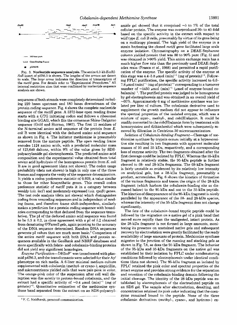
FIG. 3. Nucleotide sequence analysis. The entire 6.3-kb EcoRI- Sal1 insert of p386.3 is shown, The lengths of the arrows are drawn to scale. The large arrow indicates the direction of transcription of the metH gene. For details refer to "Experimental Procedures." All internal restriction sites that were confirmed by nucleotide sequence analysis are shown.
sequences of both strands were completely determined includ- ing 220 bases upstream and 180 bases downstream of the protein coding sequence. Fig. 4 shows the complete nucleotide sequence of the metH gene. A 3372-base open reading frame starts with a GTG initiating codon and follows a ribosomal binding site GGAG, which fits the consensus Shine-Dalgarno sequence (Gold and Stormo, 1987). The first 11 residues of the N-terminal amino acid sequence of the protein from E. coli B were identical with the deduced amino acid sequence as shown in Fig. 4. The initiator methionine is presumably removed from the mature peptide. The open reading frame encodes 1124 amino acids, with a predicted molecular mass of 123,640 daltons, within 9% of the value given by SDS- polyacrylamide gel electrophoresis. The predicted amino acid composition and the experimental value obtained from total amino acid hydrolysis of the homogenous protein from E. coli B are in good agreement (data not shown). The codon usage probability (data not shown) is high in only one of the three frames and supports the verity of the sequence determination. It yields a codon preference statistic of 0.929, a value similar to those for other biosynthetic genes. This overall codon preference statistic of metH puts it in a category between weakly (uiz. lad) and moderately expressed (uiz. rpoB) genes. The test code analysis (data not shown) which distinguishes coding from noncoding sequences and is independent of read- ing frame, and therefore frame shift-independent, similarly makes a prediction for a protein coding sequence with bound- aries corresponding to that deduced from the sequence trans- lation. The PI of the deduced amino acid sequence was found to be 5.3 f 0.2, in close agreement with a PI of 5.2 obtained from denaturing O'Farrell gels, again pointing to the accuracy of the DNA sequence determined. Random DNA sequences generate PI values that are much more basic.' Comparison of the entire metH sequence with both DNA and protein se- quences available in the GenBank and NBRF databases and more specifically with folate- and cobalamin-binding proteins did not yield any significant homologies.
Enzyme Purification-DH5aF' was transformed with plas- mid p4B6.3, and the transformants were selected for their Ap' phenotype on rich media. A 6-liter minimal medium culture supplemented with cobalamin (methyl- or cyano-), ampicillin, and micronutrients yielded cells that were pale pink in color. The orange-pink color of the suspension after cell wall dis- ruption was due mostly to protein-bound cobalamin, and the extract had a specific activity of -0.4 pmol (min)" (mg of protein)". Quantitative estimation of the methionine syn- thase band separated from the extract on an SDS-polyacryl-
F. C . Neidhardt, personal communication.
amide gel showed that it comprised -5 to 7% of the total cellular protein. The enzyme was overproduced 30- to 40-fold based on the specific activity in the extract with respect to wild type E. coli B cells, presumably by virtue of its gene being on a multicopy plasmid. The high yield of the enzyme in a strain harboring the cloned metH gene facilitated large scale enzyme isolation. Chromatography on a DEAE-Sepharose column yielded protein that was 80 to 90% pure (Fig. 5) and was obtained in >90% yield. This anion exchange resin has a much higher flow rate than the previously used DEAE-Seph- adex resin (Frasca et al., 1988) and permitted a rapid purifi- cation of the enzyme. The specific activity of the enzyme at this stage was 4.4-5.8 pmol (rnin)" (mg of protein)". Follow- ing FPLC purification, the specific activity increased to 6.6- 7.8 pmol (min)" (mg of protein)" corresponding to a turnover number of -1450 pmol (rnin)" (pmol of enzyme-bound co- balamin)". The purified protein was judged to be homogenous by gel electrophoresis and was obtained in an overall yield of -60%. Approximately 6 mg of methionine synthase was iso- lated per liter of culture. The cobalamin derivative used to supplement the growth medium did not appear to influence the spectral properties of the isolated enzyme, which was a mixture of aquo-, methyl-, and cob(I1)alamin. It could be readily converted to the cob(I1)alamin form by treatment with homocysteine and dithiothreitol which were subsequently re- moved by filtration in Centricon 30 microconcentrators.
Isolation of Cobalamin-binding Fragment-Cleavage of me- thionine synthase by trypsin occurs initially at a hypersensi- tive site resulting in two fragments with apparent molecular masses of 95 and 35 kDa, respectively, and a corresponding loss of enzyme activity. The two fragments obtained after the first cleavage could be isolated by FPLC. Whereas the 35-kDa fragment is relatively stable, the 95-kDa peptide is further cleaved to 68- and 28-kDa fragments. The 68-kDa fragment is fairly unstable as evidenced by its relatively low intensity on analytical gels, but a 58-kDa fragment, presumably a product, accumulates. Fig. 6 shows the kinetics of formation of the various fragments and traces the origin of the 28-kDa fragment (which harbors the cobalamin-binding site as dis- cussed below) to the 95-kDa and not to the 35-kDa peptide. The kinetics of disappearance of the 95-kDa fragment is nicely paralleled by the appearance of the 58- and 28-kDa species, whereas the intensity of the 35-kDa fragment does not change significantly.
The fate of the cobalamin-bound tryptic peptide could be followed by the migration on a native gel of a pink band that moved more rapidly than the undigested, intact protein. As the 28-kDa fragment is not formed in large amounts, moni- toring its presence on unstained native gels and subsequent recovery by electroelution were greatly facilitated by the ready availability of large amounts of protein. Methionine synthase migrates to the junction of the running and stacking gels as shown in Fig. 7A, as does the 95-kDa fragment. The behavior of the 95-kDa and 35-kDa fragments on the native gel was established by their isolation by FPLC under nondenaturing conditions followed by electrophoresis under identical condi- tions (data not shown). The 95-kDa fragment as isolated by FPLC retained the pink color and spectral properties of the intact enzyme and provides strong evidence for the separation and retention of the cobalamin-binding domain following the initial cleavage. The identity of the 28-kDa peptide was es- tablished by electrophoresis of the electroeluted peptide on an SDS gel. The sample after electroelution, desalting, and concentration retained its pink color indicating that the coen- zyme remained bound to the peptide. None of the three cobalamin derivatives (methyl-, cyano-, and hydroxo-) ex-
Cobalamin-dependent Methionine Synthase CCG GW ATG GCl CCG ACC 1TG ACA GlC TCT 111 1TC TC1 A T C GTG WA AlC All 1TC ATT IT1 AlT GTT AGC TAA 1GC M I LC1 TAC T U ACT LA1 CCG AIG AGT TAA TGT 1 W ACA AAl
CTC AIC 1TC CGl GGT CGT CGC TTl IAC CAC AW TGC GlT T E A G T A T G GTT TGT TW All T T T A E T C T GGG TlG AGC GTG TCG r G C A AGl GIG ACC AGC AAA G I G GAA CAA -35 -10 5 . 0
n s s ~ v f o 1
ClG CGT GCG CAC ITA M I WA CGT AlT ClG GTG CTG WC CGC GCT AlG GGC ACC ATG AlC CAG AGl TAl CW ClG M C WA GCC WT TIT CGl GGT WA CCC 111 CCC GAC TGG CCA TGC
L R A O L Y E R l L V L D G G * G T M I O S Y R L ~ E A D ~ R G E R ~ A D U P C S
LAC CTC AAA GGC M C AAC GIC ClG CTG GlA CTC AGT M A CCG WA GlG A T C GCC GCl AlC CAC M C GCC T 1 C 111 W GCG GGC GCG WT A T C AIC W ACC M C ACC TIC A X TCC ACC
Y V A G V L G P l Y R T A S I S P D V Y D P A ~ R Y l T ~ D G L V A A Y R E S l I54
AM GCG ClG GIG W GGT GGC WG W l CTG AlC ClG All W ACC GlT T1C WC ACE Cll U C GCC MA GCC GCC GlA 111 CCC GIG MA ACC WG 111 W GCG CTG CbC GI1 WG CTG K A L V E G C E D L I L I L l V f D l L ~ A K A A V ~ A V K I E F E A L G V f l
2m
CCC ATT AlG A T C TCC GGC K C AlC ACC WC GCC 1CC GGG CGC ACG ClC 1CC CCG U G ACC ACC GM G U 1Tl TAC U C TU 116 CCC CIC GCC GU GCl ClG ACC 1lT GGC C T G M C TGI P I M I S G T I T D A S C R T L S G O T T E A ~ I Y P L R I A ~ A L ~ ~ G L ~ C
GCG ClG GGG CCC CAT WA CTG CGC CAG TAC GIG CAG GAG ClG TU CGG All GCG GU TGC TAC ClC ACC GCG CAC CCG M C GCC GGG CTA CCC M C GCC 1lT GGT WG TAC W l CTC WC
A ~ G V D E L R O Y V O E L S R I A L C I Y T I R P * L ~ L P * * ~ ~ E I D L D 2m
GCC GAC ACG AIG GCA A M CAG A T A CCT W TGG GCC CAI GCG GG1 TTl ClC M l AlC GlC CGC GGC TCC 1Gl GGC ACC ACG CCA W CAl A T 1 G U GCG AlG AGl CGl GCA GTA WA GCA A D T ~ A K P I R E Y A O A G F L Y I V G G ~ ~ ~ ~ ~ P ~ ~ I ~ A M S R ~ V E G
300
T T A CCG CCG CCC A M CTG CCG GU AT1 CCC G1A GCC TGC CG1 TTG TCC CCC ClG WG CCG ClG M C AlT GGC tu WT A b E CTG 111 GlG U C CTG CGl GU CGC ACC M C GTC ACC CGl L A V R K L P E I P V A C 1 L S G L E P L ~ l G f D S L f V M V G E R l ~ V l G
354
TCC CCT AAG T T C AAG CGC CTG AlC A M GU WG MA T I C AGC WG GCG ClG W l GlC GCG CGl C U U G GIG GU U C GGC GCG U G AT1 AlC W l ATE M C ATC W l GU GGG AlG ClC S A K ~ K ~ L I K E E ~ T S E A L O V A ~ ~ ~ V E ~ ~ A ~ I I D I ~ ~ D ~ G ~ L
400
W l GCC WA GCG GCG AlG GlG CGT llT CTC M l ClG All GCC bGT GU CCG W l ATC GCl CCC GlG CCG All AlG AlC WC TCC T U MA T t b WC G1C AT1 GU MA GGl ClG M G lG7 D A ~ A A ~ V ~ ~ L ~ L I A G ~ P D I A ~ V P I * I O ~ ~ ~ Y O V I E K G L K C
ATC CAG GGC L M GGC AlT GTT M C 1CT ATC TCG AlG A M GAG CGC ClC W l GCC TTl ATC UT CAC GCG AAA llb 11G CGl CGC T I C GGI CCG G U GTG GIG CIA AlG GCC I T 1 WC GAA
l a G C G l V ~ f l S ~ K € G V D A f l W I I X I L I R Y G A A V V V ~ A ~ D E 450
CAG GGA CAG GCC W l ACT CGC GCA CCG A M AlC WG AT1 TGC CGTCGG GCG TAC MA AlC ClC ACC W WC Cll GGC TTC CCG CCA W W l AlC AlC T T C WC CCA AAC AlC 11C GCG a G O A D T R A R K I E I C R R A Y K I L l E E V C f P P E D I I f D P Y I T A
sm GTC GCA ACl GGC IT1 C A I GAG CAC AAC AAC TAC CCG CAG WC 111 hTC GGCGCG 1GT W W C ATC LAR CGC G M ClG CCG U C GCG CTG L T T TCC CCC CCC GTA 1CT M C GTl TC1 1TC
V I T G I E E ~ Y Y I A ~ D ~ ~ G A C E D I K R L L P M A L I S G C V S Y V S ~
TCG TIC CGT GGC AAC GAT CCG GTG CGC WA GCC All U C GU GTG TIC CTC 1AC TIC GCl AlT CCC M I GGC AlC W l ATG GGG ATC GlC LAC GCC G G G C M ClG CCG AlT T I C WC GAC
s r ~ G U D P V R E A I I A V ~ L I Y A I R Y C ~ D ~ C I V Y A G ~ L A l Y D D
554
603
CTA CCC GCT WA ClG CGC W C GCG GIG W GAT GlG AlT CTT M l CGl CGC WC W l GGC ACC G 4 G CGT 111 ClG GAG CTT CCC WG MA 1Al CGC CGC AGC MA ACC WC WC ACC GCC L P A E L R D A V E D V I L Y R R D D G l E R L L E L A f C I R G S K T D D T A
"
A X GCC CAG CAG GCG GAG TGG CGC TCG 1CC W C T G U T A M CGl ClG W 1AC TCG ClG GlC MA CGC A T T ACC WC TIT A T C WG CAG W l ACC W GU GCC CCC U C C A G GCl K G U A ~ ~ A E Y R S Y E Y Y I R L E Y S L V ~ ~ I I E ~ : E O D ~ L ~ A R P ~ A T
CGC CCG A I T GAA Grc AIT WA GGC CCG TTG ATG GAC GGC A x A N GIG GTC GGC GAC CTG ITT GCC w GGC AM ATG ITC CIG CCA CIC GIG GTC MA TCG GCG CGC CTC ATC A M C A G R ~ I E ~ I E G ~ L M D G ~ ~ V V G D L ~ C E ~ K ~ ~ ~ P ~ ~ ~ K ~ ~ ~ ~ M ~ ~
- 650
1M
GCG G T G GCC ikC ClC CIA CCG TTT A T T G A 1 GCC AGC A A A W G CAG GGC ALA K C AAC GGC &AS A T 0 GTG AlC GCC K C GTGAAG CCC GAC CTC CAT GAG ATC 661 kU U T ATC GTT GGl L V A r L E P ~ l E A S K E O G K 1 Y G ~ M V I A T V K G D V Y E I C K Y I V C
G I G GTG C T C C1A TGT M C M C T A C W All GTC D l ClC CCC Cll AlC ClC CCT GCG GU A M AlT CTC CGl ACC GCT MA W G I G AAl GCT WT CTG All GGC CTT 1CC CGG CTT A T C
V V L O C ~ Y Y E I V D L G V * V P A E ~ I L R T A C € V Y A D L I C L S G L I
7%
8m K C CCG TCG CTG D \ C GAG 1 T C ClT AAC GTG GCG A U GAG AlC WG CCl CAG CGC TlC ACT AT1 CCG TlA CTG All CCC CCC CCG ACG ACC T U MA GCC CAC ACG GCG GTG AAA AlC WC
T P S L D E ~ V U V A K E ~ E R O C F ~ I P L L I C C A ~ T S K A W ~ A V I I F
U G Mt T I C AGC CGCCCG ACC CTC T A T G I G CAC All GCC TCG CGT ACC Gl? GGl GIG G I G CCG GCC CTC CTl 1CC GAT K C CAG CCl W l D l TlT GTCGCT CGl ACC CGC AAG GAG TAC
~ Y I S G P T Y Y V ~ Y A S R T V C Y V A A L L S D T ~ t D ~ f V A ~ T R K E ~ 854
GAA ACC GTA CGl A T T CAC CACGGG CGC AAG M A CCG CGC ACA CCA CCG GlC ACG ClC GM GCCGCC CGC CAT M C CAT TTC GCT TlT W C TCG CAC CCT TAC ACGCCC CCG G I G CCC C A C
E T V R I D ~ C ~ K ~ P ~ ~ P P Y T L L A A ~ D ~ D ~ A F D Y ~ A Y T P P V A I w3 -.
CGT CIC GCC GIG U G W GTC GAA GCCACC A1C C A I ACG ClG CGl U T TAC ATC WC TGG ACA CCG T l C T l T ATG ACC TGG TCG CTG CCC GGG AAG TAT CCG CGC A T 1 ClCGAA GAT C I A
R ~ G V O E V E A S I E I L R U I I D ~ l P f ~ ~ I Y S L A G K l P ~ I L € D E 950
G T G GTG GGC 677 GAG GCG CAG CGG CTG I T T AMWC GCC M C GAC ATG ClG GAT M A ITA AGC GCC WG M A ACG ClG AAT CCG CGT GGC GIG GTG GGC CTG TTC CCG GCA M C CGT GTG V Y G V E A ~ ~ L ~ K D A Y D M L D K L S A E K ~ ~ Y P R G V V G L ~ ~ A Y R V
Ima
GCC GAT Wc A T 7 GAL ATC TAC CGl CAC GAL ACG CGT ACC CAT GTG A T C AAC GTC AGC CAC CAI ClC CGT C M CAG ACC W1A.A ACA GCG TlC GCT AAC TAC IC1 CTC GCl WC TTC Gll ~ D D I E I Y R D E T R T U V I Y V S ~ ~ L R O O T E K T A ~ A ~ ~ C L A D ~ V
GCG CCG AAG CTI T C T GCl M A GCA WT TAC ATC CGC CCA T T T CCC GTG ACT GGC GGG ClG G M WG WC GCA C T G GCT GAT GCC TTl W GCG CAG CAC GAl 0 1 1AC M C MA AlC AlG A P K L S C K A D I I C A ~ A V T G C L ~ E D A L A D A ~ E A O ~ D D V ~ ~ I ~
IMD GlG MA GCC tll GCC GAC CGT 1lA GCC GU GCC 1TT CCC WG TA1 ClC CAI GAG CGl GIG CGl AM GlC TAC 1GG CCl AIG CGC CW ACG AW ACC 1CA GCA ACG AAG AGC 1 W TCC GCG
V K A L A D R L A E A f A E l L W E R V R K V Y U A ~ R R l R T S A T K S I IM
MA ACl ACC AGG G U 1CC GTC CGG CAC CGC CC1 AlC CGG CCl GCC CGC AAC AlA CGG A M M G CCA C U IC1 CCG AGC TGC 1GG M G TGG M A U C ACA CTG GCA 10. LAC IC1 " - - r -
Cll K G C U GCC CCC cl(i UI UX "I UX CIT CC! ACT ICA DCC ACC UX K A CU X I ACT ACC Cn TAG U C bAA C H CIC CCC WT CAC CIT W WT TI1
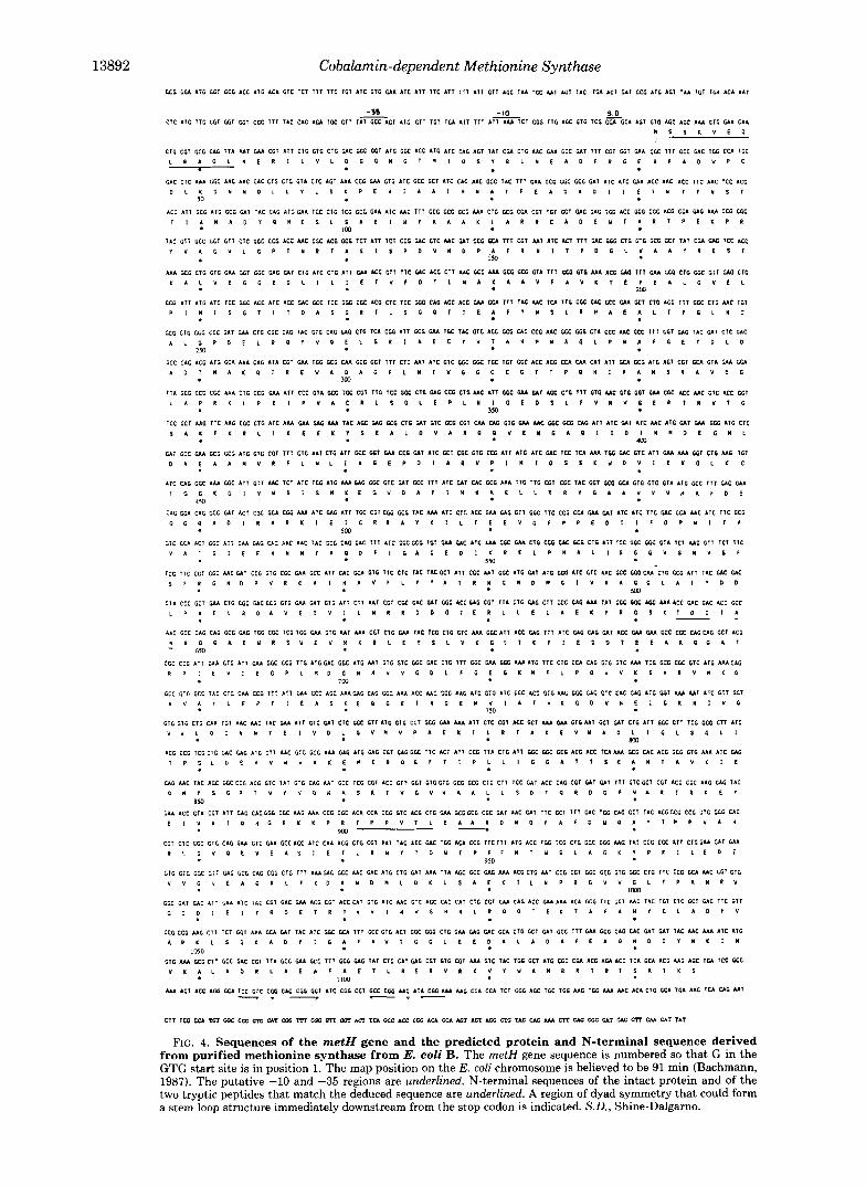
FIG. 4. Sequences of the metH gene and the predicted protein and N-terminal sequence derived from purified methionine synthase from E. coli B. The metH gene sequence is numbered SO that G in the GTG start site is in position 1. The map position on the E. coli chromosome is believed to be 91 min (Bachmann, 1987). The putative -10 and -35 regions are underlined. N-terminal sequences of the intact protein and of the two tryptic peptides that match the deduced sequence are underlined. A region of dyad symmetry that could form a stem loop structure immediately downstream from the stop codon is indicated. S.D. , Shine-Dalgarno.

Cobalamin-dependent Methionine Synthase 13893
A B C
FIG. 5. Polyacrylamide gel electrophoresis of methionine synthase at different stages of purification. Samples were de- natured by boiling for 2 min in a solution containing 5 mM 2- mercaptoethanol, 2% SDS, 10% glycerol, and 62.5 mM Tris-HCI, pH 6.8. Samples were applied to a discontinuous gel (4% polyacrylamide stacking gel, 12% polyacrylamide running gel). After electrophoresis, the gel was stained with Coomassie Blue, destained, and dried on a filter paper. Each lane contained 16 X units of methionine synthase. Lane A, enzyme after chromatography on a DEAE-Sepha- rose column; lune R, enzyme after FPLC purification; lane C, same as B, but with 32 X units of methionine synthase. The numbers on the left indicate the molecular mass of the standards in kilodaltons.
I 2 3 4 5 6 7 """- MS- 95- 68 - 58-
37 - 28-
2 4 8 I6 32 64 DIGESTION TIME (mid
FIG. 6. Limited proteolysis of methionine synthase and ki- netics of formation of the major fragments. Enzyme (223 pg) was incubated with trypsin (2% w/w) a t ambient temperature. Sam- ples (6 pl) were removed at the indicated times and quenched with 1 pl of TLCK (10% w/v). A, lanes 1-7 correspond to 0, 2, 4, 8, 16, 32, and 64 rnin, respectively. The numbers on the left edge correspond to the apparent molecular mass of the peptides in kilodaltons. B, kinetics of appearance and disappearance of the individual peptides expressed as a percentage of their maximum intensity over time.
amined migrated into the native gel. Hence, the identification of both a parent (95-kDa) and a daughter (28-kDa) peptide fragment that retain the pink color of the native enzyme is very strong evidence for the isolation of peptides that retain the cobalamin-binding site.
A 1 2 3 4 5 B
?&- n
FIG. I . Formation and identification of a cobalamin-bind- ing fragment from methionine synthase upon treatment with trypsin. A, enzyme (514 pg) was treated with trypsin (1.18 w/w in lanes 2 and 3, and 11% w/w in lanes 4 and 5) . The reactions were terminated by the addition of TLCK (50 pg). Samples were run out on a 15% acrylamide, non-SDS gel and photographed without stain- ing. A green filter was employed to enhance the contrast of the pale pink bands. Neither methyl-, cyano-, nor hydroxocobalamin migrate into the gel under these conditions. Lane I has undigested methionine synthase. The positions to which the native (124-kDa) and tryptic fragments migrate are marked on the left along with their molecular masses in kilodaltons. i?, the pink protein band that migrated sepa- rately from the undigested protein band was cut out and electroeluted. After desalting and concentration, the protein was separated on a 12.5% acrylamide gel containing SDS. Low molecular weight markers are shown on the /eft.
N-terminal sequence analysis of the 28- and 35-kDa frag- ments established the limits of these peptides and provided evidence for the verity of the deduced amino acid sequence (Fig. 4). The hypersensitive site between residues 900 and 901 resulting in the formation of the 35-kDa fragment lies in one of the most hydrophilic internal regions of the protein as predicted by the Peptide Structure program with the method of Kyte and Doolittle (Kyte and Doolittle, 1982). The 28-kDa fragment with an N-terminal sequence of T-D-D (beginning a t residue 643 of the deduced amino acid sequence) is situated immediately upstream of the 35-kDa fragment. It is charac- terized by the prevalence of hydrophobic residues. Also note- worthy are the high concentration of lysines and threonine (relative to their abundance on the complete chain) and the low cysteine content. A single cysteine is present in this fragment. The position of the cobalamin-binding domain immediately upstream of the 35-kDa fragment and the com- plete loss of enzyme activity following cleavage at the hyper- sensitive site suggest that the downstream region is also required for catalysis. Proteolysis in the presence of CH3H4folate does not affect the initial rate of cleavage but stabilizes the 68-kDa fragment (data not shown). Proteolysis in the presence of both homocysteine and CHaH4folate greatly destabilizes the 95-kDa fragment. These data are consistent with the substrates interacting with the parent 95-kDa pep- tide which also harbors the cobalamin-binding site.
The size of the 28-kDa fragment agrees very well with the apparent size of that fragment (29 kDa) as estimated by gel electrophoresis. On the other hand, the fragment with the apparent mass of 35 kDa has an actual mass of 26 kDa. The discrepancy in the migration of this fragment is not under- stood. Since the "35-kDa" fragment lies at the C terminus of the protein, the possibility of a sequence error leading to a predicted premature termination was considered. The se- quence in the region of the gene containing the stop codon

13894 Cobalamin-dependent Methionine Synthase
and approximately 200 nucleotides upstream of it was deter- mined with two different clones on one strand and two primers on the opposite strand. In each case, the same sequence was read, making it quite unlikely that a sequence determination error has been made.
DISCUSSION
The enzyme cobalamin-dependent methionine synthase has been isolated from an overproducing strain of E. coli bearing p4B6.3. A nutritional auxotroph of E. coli was used to isolate a recombinant clone that complemented the mutation and encoded cobalamin-dependent methionine synthase (Baner- jee et al., 1988). The identity of the cloned gene has been confirmed by nucleotide sequence analysis, maxicell expres- sion, and enzyme activity assays. Cloning of the metH and metE genes has been recently reported independently by another group (Old et al., 1988). They employed TnlOOO insertion-mutagenesis to map and determine the direction of transcription of the metH gene. From the single and slightly
truncated polypeptide identified, they correctly assumed the clockwise direction of transcription from the EcoRI site. Moreover, the approximate map positions of the HindIII sites, in addition to their inability to subclone using the HindIII sites, are in agreement with our data.
Methionine synthase is a protein of 123,640-dalton molec- ular mass with a calculated PI of 5.3 f 0.2. The turnover number of the purified enzyme is 1450 min" and is within the range of fully active enzyme isolated from E. coli B (Frasca et al., 1988). The specific activity of the purified recombinant enzyme is, however, only 60 to 80% of that found with E. coli B methionine synthase (9.3-11.7 pmol (min)" (mg of pro- tein)") and suggests that a portion of the enzyme is present as apoenzyme, which is inactive under standard assay condi- tions. It is possible that the bacteria are unable to transport cobalamin at a rate commensurate with the elevated level of apoenzyme synthesis. The growth medium is pink even after the cells have been harvested by centrifugation, indicating that the cobalamin concentration is not limiting.
ff Twlnr CF Alph8 Hsllces
ff Beta Snsstr
T m E
6UR Alpha Helfces 60 Bstr Shsrtr
Glycosyl. SI tes
FIG. 8. Secondary structure comparisons of segments of three cobalamin-binding proteins. Upper panel, residues 800 to 900 of MetH middle, residues 96 to 196 of BtuR lower, residues 505 to 605 of MutB. CF, Chou-Fasman; GOR, Gamier-Osguthorpe-Robson. The thickness of the black bars is proportional to the strength of the prediction.

Cobalamin-dependent Methionine Synthase 13895
Methionine synthase is one of the largest peptides known in E. coli. Hence, the isolation of a fragment that is less than 25% the size of the intact protein and harbors the cobalamin- binding site will facilitate studies of the interactions of the cobalamin with the apoprotein. This is especially so in light of the fact that no cobalamin-binding proteins have been crystallized to date, and the nature of a “cobalamin pocket” therefore remains unknown. The sequence appears to contain a region of moderate hydrophobicity and is flanked by ex- tended hydrophilic segments. This retains the tertiary struc- ture and ability to bind cobalamin after separation from adjacent peptide segments.
A comparison of both the full length methionine synthase and of the 28-kDa peptide with other cobalamin-binding proteins BtuR (Lundrigan and Kadner, 1989), BtuB (Heller and Kadner, 1985), and MutA and MutB (Marsh et al., 1989) was attempted and failed to pick up any significant homolo- gies. BtuB is the cobalamin receptor protein in the outer membrane of E. coli, while BtuR is a soluble protein from E. coli that has been implicated in metabolism of adenosylco- balamin. MutA and MutB represent the two subunits of methylmalonyl-CoA mutase from Propionibacterium sher- manii. MutB shows a striking primary sequence homology with the human methylmalonyl-CoA mutase.
I t is possible that, as with the heme pocket, similarities may exist instead in the secondary and tertiary structures in which functionally critical regions of the structure are con- served. Comparisons within the globin family reveal that although its members have very different amino acid se- quences, they share remarkably similar secondary and tertiary structures (Lesk and Chothia, 1980). Eight helices assemble in a common pattern, enclosing the heme group in pockets of similar geometry made up from homologous portions of the molecules. Only 5 positions out of the 116 that are involved in conserved interactions have the same residue in all globins, and even they are not contiguous on the primary sequence. A similar comparison of hemoglobin and cytochrome b5 is re- markable not only for the close superposition of the heme groups but also in the conservation of structurally equivalent units (Rossman and Argos, 1975).
Examination of the Chou-Fasman (Chou and Fasman, 1978) and the Garnier-Osguthorpe-Robson (Garnier et al., 1978) predictions of secondary structure for BtuR, MutB, and the 28-kDa fragment reveals a pattern of alternating (Y helices and 0 sheets. This is distinct from the secondary structure elements involved in heme binding, which are primarily a helices. The region of greatest similarity in each of the three proteins is shown in Fig. 8. The spacing and the alternation of the a helices and /3 sheets in the three proteins is quite striking and is suggestive of a Rossman fold type of structure. Dimethylbenzimadazole is an unusual nucleotide that is part of the cobalamin structure. A comparison of the benzimida- zole-ribofuranosyl portion of cobalamin with the adenine ribose of NAD (bound to crystalline malate dehydrogenase) revealed a good alignment of the 2 molecules with a calculated root-mean-square distance between corresponding atoms of
1.05 A (Dieckgraefe et al., 1988). The region of identified secondary structure similarity in the cobalamin binding pro- teins, shown in Fig. 8, may be involved in docking the di- methylbenzimidazoyl group.
The isolation and subcloning of the metH gene allow for the convenient purification of methionine synthase in large amounts. This sets the stage for further characterization of the structural and mechanistic properties of the enzyme.
Acknowledgments-We gratefully acknowledge the generous advice and helpful suggestions from Dr. Martha Ludwig, Dr. Herbert Schweizer, Robert Clark, and Claudius Vincenz at the University of Michigan.
REFERENCES Bachmann, B. (1987) in Escherichia coli and Salmonella typhimurium
(Neidhardt, F. C., ed) Vol. 2, pp. 807-876, American Society of Microbiology, Washington, D. C.
Banerjee, R. V., Frasca, W., Johnston, N. L., Ballou, D. P., Datta, P., and Matthews, R. G. (1988) Biochemistry 27,3101 (abstr.)
Blumenthal, R. M., Reeh, S., and Pederson, S. (1976) Proc. Natl. Acad. Sci. U. S. A. 73,2285-2288
Chou, P. Y., and Fasman, G. D. (1978) Adu. Enzymol. Relat. Areas Mol. Biol. 47, 45-148
Devereaux, J., Haeberli, P., and Smithies, 0. (1984) Nucleic Acids Res. 12,387-395
Dieckgraefe, B. K., Seetharam, B., Banaszak, L., Leykam, J. F., and Alpers, D. H. (1988) Proc. Natl. Acad. Sci. U. S. A. 85,46-50
Fickett, J. W. (1982) Nucleic Acids Res. 10 , 5303-5318 Frasca, V., Banerjee, R. V., Dunham, W. R., Sands, R. H., and
Matthews, R. G. (1988) Biochemistry 2 7 , 8458-8465 Garnier, J., Osguthorpe, D. J., and Robson, B. (1978) J. Mol. Biol.
Gold, L., and Stormo, G. (1987) in Escherichia coti and Salmonella typhimurium (Neidbardt, F. C., ed) Vol. 2, pp. 1302-1307, American Society for Microbiology, Washington, D. C.
Gribskov, M., Devereux, J., and Burgess, R. R. (1984) Nucleic Acids Res. 12,539-549
Heller, K., and Kadner, R. J. (1985) J. Bacteriol. 161, 904-908 Henikoff, S. (1984) Gene (Amst.) 28,351-359 Kyte, J., and Doolittle, R. F. (1982) J. Mol. Biol. 157 , 105-132 Lesk, A. M., and Chothia, C. (1980) J . Mol. Biol. 136, 225-270 Lundrigan, M. D., and Kadner, R. J. (1989) J. Bacteriol. 171, 154-
161 Maniatis, T., Fritsch, E. F., and Sambrook, J. (1982) Molecular
Cloning: A Laboratory Manual ,Cold Spring Harbor Laboratory, Cold Spring Harbor, NY
Marsh, E. N., McKie, N., Davis, N. K., and Leadlay, P. F. (1989) Biochem. J. 260,345-352
Messing, J., and Viera, J. (1982) Gene (Amst.) 19,269-276 Mount, D. W., and Conrad, B. (1986) Nucleic Acids Res. 14,443-454 Neidhardt, F. C., Bloch, P. L., and Smith, D. F. (1974) J. Bacteriol.
O’Farrell, P. H. (1975) J. Biol. Chem. 250, 4007-4021 Old, I. G., Hunter, M. G., Wilson, D. T. R., Knight, S. M., Weather-
ston, C. A., and Glass, R. E. (1988) Mol. & Gen. Genet. 2 11, 78-87 Rossman, M. G., and Argos, P. (1975) J. Bwl. Chem. 250,7525-7532 Sanger, F., Nicklen, S., and Coulson, A. R. (1977) Proc. Natl. Acad.
Sei. U. S. A. 74,5463-5467 Silhavy, T. J., Berman, M. L., and Enquist, L. W. (1984) Experiments
with Gene Fusions, pp. 213-214, Cold Spring Harbor Laboratory, Cold Spring Harbor, NY
Wanner, B., Kodaira, R., and Neidhardt, F. C. (1977) J. Bacteriol.
120,97-120
119 , 736-747
130,212-222