Embed Size (px)
Citation preview

ISSN: 1314-6246 Mohamed et al. J. BioSci. Biotechnol. 2015, 4(3): 291-302.
RESEARCH ARTICLE
http://www.jbb.uni-plovdiv.bg 291
Zeinat K. Mohamed 1
Sherif M. Elnagdy 1
AlaaEddeen Seufi 2
Mohamed Gamal 1
Cloning and molecular analysis of L-
asparaginase II gene (ansB)
Authors’ addresses: 1 Botany and Microbiology Department,
Faculty of Science, Cairo University,
Egypt. 2 Entomology Department, Faculty of
Science, Cairo University, Egypt.
Correspondence:
Sherif M. Elnagdy
Botany and Microbiology Department,
Faculty of Science, Cairo University,
Egypt.
Tel.: +20-1008843357
e-mail: [email protected]
Article info:
Received: 19 April 2015
Accepted: 14 August 2015
ABSTRACT
The deamination of L-asparagine to L-aspartic acid and ammonia is catalyzed by
L-asparaginases (L-asparagine amino hydrolase). The enzyme L-asparaginase is
widely distributed in nature from different living organisms, starting from
bacteria till mammals and plants. It has been recently thought to be a therapeutic
agent in treatment of various lymphoblastic leukemia diseases. There have been
many attempts to isolate microorganisms that produce L-asparaginase. L-ASNase
producing bacteria, Escherichia coli MG27, was previously isolated from the
River Nile and identified. In this study, ansB gene, encoding L-ASNase II from
E. coli MG27, was amplified by PCR, cloned and characterized by DNA
sequencing. The DNA sequence was then analyzed using bioinformatics analysis
and translated into amino acid sequence. Identification of highly conserved amino
acid sequence motifs was conducted by comparison against the InterPro database.
Analysis revealed that the protein sequence had a catalytic domain of L-
asparaginase type II (IPR004550) that belong to asparaginase/glutaminase family
(IPR006034) and has asparaginase/glutaminase conserved site (IPR020827).
According to results predicted using PSIpred tool, ansB consists of eight α-
helices and 13 β-strands.
Key words: L-asparaginase, cloning, Escherichia coli, leukemia, sequencing
Introduction
The enzyme L-asparaginase is widely distributed in
nature from bacteria, yeast, filamentous fungi, mammals and
plants. The amino acid sequence of L-asparaginase II was
determined by protein sequencing in the seventies and the
nucleotides sequence of the ansB gene was reported in 1990.
The L-asparaginase II precursor has a 22-residue N-terminal
secretory signal peptide which is cleaved between alanine
and leucine residues (amino acids 22 and 23, numbering from
the initiating methionine residue) to yield mature protein with
N-terminal leucine residues. The 22-residue amino acid
secretory signal peptide directs the translocation of the
protein through the membrane and has a charged N-terminal
region, amino acids from 1 to 6, followed by hydrophobic
core, amino acids from 7 to 16, and terminated with more
polar region, amino acids from 17 to 22. The mature enzyme
consists of 326 amino acids located in periplasm as a homo
tetramer and has molecular weight of 141 kDa and its
synthesis is 100 to 1000 fold induced in anaerobic cultures.
Each one of the four active sites is located between the N and
C-terminal domains of two adjacent monomers. Thus, the L-
asparaginase II tetramer can be treated as a dimer of dimers.
Despite this fact, the active enzyme is always a tetramer
(Aung et al., 2000; Kozak et al., 2002; Khushoo et al., 2004).
The production of E. coli L-asparaginase II is regulated
by two pleitropic regulatory proteins, the oxygen-sensitive
FNR protein, which activates a number of genes during
anaerobiosis (Partridge et al., 2008; Tolla & Savageau, 2011;
Shan et al., 2012) and the cyclic AMP receptor protein CRP,
which controls the initiation of transcription of genes in
various catabolic pathways (Beatty et al., 2003; Uppal et al.,
2011; Chen et al., 2012; Kraxenberger et al., 2012).
The information from X-ray crystallography and
extensive site-directed mutagenesis studies on E. coli
asparaginase II, revealed a number of amino acid residues
ISSN: 1314-6246 Mohamed et al. J. BioSci. Biotechnol. 2015, 4(3): 291-302.
RESEARCH ARTICLE
http://www.jbb.uni-plovdiv.bg 292
that are important for catalysis and substrate binding. Among
them Thr 12, Tyr 25, Thr 89 and Lys 162 which play a
catalytic role, while the positions of Ser 58, Asp 90, Asn 248
and Glu 283 assist substrate binding (Wehner et al., 1994;
Palm et al., 1996; Aung et al., 2000).
Many attempts have been made to clone L-asparaginase
(ansB) gene from different bacterial and fungal species.
Hüser et al. (1999) cloned a class II glutaminase/asparaginase
coding gene (ansB) from Pseudomonas fluorescens into
pASK-C expression vector that was transformed into E. coli
CU1783. The expressed fusion protein with a C-terminal
His8-tag was purified by affinity chromatography on Talon-
Spin columns.
DNA fragment coding for L-asparaginase from E. coli
AS1.357 was generated using polymerase chain reaction
(PCR) technology. It was cloned into the expression vector
pBV220 and transformed into E. coli strains JM105, JM109,
TG1, DH5α and AS1.357 (Wang et al., 2001). Khushoo et al.
(2004) fused the gene coding L-asparaginase II of E. coli to
an efficient pelB leader sequence and an N-terminal 6x
histidine tag. The fused gene has been cloned downstream of
the T7lac promoter in pET22b expression vector.
Saccharomyces cerevisiae gene coding for the
periplasmic L-asparaginase II was cloned into pPIC9
expression vector in-frame with the S. cerevisiae α-factor
secretion signal under the control of the AOX1 gene promoter
and expressed in the methylotrophic yeast Pichia pastoris
(Ferrara et al., 2006). Furthermore, Kotzia & Labrou (2007)
reported the cloning and expression of L-asparaginase from
Erwinia chrysanthemi 3937 in E. coli BL21 (DE3)pLysS,
using pCR®T7/CT-TOPO® expression vector.
Youssef & Al-Omair (2008) reported the cloning of L-
asparaginase II gene from E. coli W3110 into pGEX-2T
expression vector in-frame with the glutatione S-transferase
fusion protein (GST) in E. coli BL21 (DE3) cells. Also,
Cappelletti et al. (2008) cloned ansB gene from the
pathogenic strain Helicobacter pylori CCUG 17874. The
gene was isolated by PCR using specific primers and the
PCR product was cloned into pCR2.1-TOPO cloning vector
and sequenced.
Vidya et al. (2011) isolated L-asparaginase II-encoding
gene (ansB) with excluding the native signal sequence from
E. coli MTCC 739 by PCR technique. The 981-bp amplicon
was cloned into pET20b expression vector and expressed in
E. coli DE3 cells. Vidya & Pandey (2012) isolated L-
asparaginase II gene from a moderately thermotolerant
bacterium belonging to Enterobacteriaceae by PCR. They
used specific primers that were designed in such a way that
the native signal sequence was excluded and the mature gene
sequence was cloned into pET20b expression vector with a
six histidine sequences at the C-terminal end transformed to
competent BL21 DE3 cells. Also, Pokrovskaya et al. (2012)
cloned ansB gene from Yersinia pseudotuberculosis and
constructed a stable inducible expression system that
overproduce L-asparaginase in E. coli BL21 (DE3) cells.
This study targeted ansB gene, encoding L-ASNase II
from E. coli MG27 amplification by PCR, cloning and
characterization by DNA sequencing. The DNA sequence
was then analyzed using bioinformatics analysis and
translated into amino acid sequence.
Materials and Methods
Amplifying of ansB gene by PCR
Genomic DNA preparation
Genomic DNA was prepared from E. coil MG27 cells
(previously isolated from the River Nile and identified) using
GeneJET™ Genomic DNA Purification Kit (Thermo
Scientific, USA) and according to its protocol. The purified
genomic DNA was store at -20°C.
Primers design for amplification of ansB gene
Primers were designed based on the sequence of E. coli
K-12 from GenBank accession number M34277 that yielded
a single 1047-bp ORF supposed to encode for L-asparaginase
II. Primers were designed for amplification of ansB gene by
excluding the native signal sequence.
A DNA fragment coding for the predicted mature part of
L-asparaginase II (amino acid residues 23-346) was amplified
using primers ansB-F (forward, 5'-GGTGGATCC
TTACCCAATATCACCATTTTAG-3') and ansB-R (reverse,
5'-GGGAAGCTTTTAGTACTGATTGAAGATCTG-3').
BamHI and HindIII restriction sites (underlined) were
incorporated into the primers at their 5' ends, respectively, to
facilitate the directional cloning of the structural asparaginase
II gene.
Polymerase chain reaction (PCR)
The genomic DNA isolated from E. coli strain MG27 was
used as template for the amplification of ansB gene. It was
amplified by polymerase chain reaction without its native
signal sequence using ansB-F and ansB-R primers.
The PCR reaction was carried out in a total volume of
25μl. The PCR conditions were as follows: initial
ISSN: 1314-6246 Mohamed et al. J. BioSci. Biotechnol. 2015, 4(3): 291-302.
RESEARCH ARTICLE
http://www.jbb.uni-plovdiv.bg 293
denaturation at 94°C for 4 min, denaturation at 95°C for 40 s,
annealing at 52°C for 40 s, extension at 72°C for 60 s for 35
cycles, and a final extension at 72°C for 10 min. The PCR
product was then analyzed on 1% agarose gel electrophoresis.
Agarose gel electrophoresis
For analyzing DNA samples, agarose gel electrophoresis
was used. The DNA bands in the gel were visualized using
short wave ultraviolet light provided by a transilluminator
and photographed.
Elution and purification of PCR fragments
To insure high purity of PCR fragments, the amplified
DNA bands were eluted and purified form agarose gels using
QIAquick Gel Extraction Kit according to manufacturer's
protocol.
TA-cloning of ansB gene
Ligation
The gel purified PCR product was ligated into the pGEM-
T Easy vector (Promega crop. Madison, Wi, USA) using T4
DNA ligase provided in the kit according to the
manufacturer's instructions. The concentration of the gel
purified PCR product was measured prior to ligation. One µl
of the resultant recombinant construct was used to transform
E. coli JM109 competent cells
Preparation and Transformation of CaCl2 competent cells
For preparation and transformation of competent cells,
CaCl2-treatment was performed (Sambrook & Russel, 2001).
Screening of positive colonies
E. coli JM109 colonies harboring the recombinant
plasmid were screened for the presence of insert in pGEM-T
easy vector by blue/white color selection on plate containing
ampicillin, 5-bromo-4-chloro-3-indolyl-β-D-galactosidase
(X-Gal) and isopropyl-β-D-thiogalactopyranoside (IPTG).
White colonies were randomly selected. The presence of
insert was verified by PCR and restriction endonuclease
digestion of plasmids isolated from these white colonies.
Plasmid DNA preparation
Plasmid DNA was prepared from white colonies of
transformed E. coli JM109 cells using QIAprep Spin
Miniprep Kit. The purified plasmid DNA was stored at -20°C
until used for confirmation of cloning by PCR amplification
and restriction digestion.
Verification tests for the recombinant ansB clones
Polymerase chain reaction (PCR) screening
Plasmid DNA prepared from white clones was used as
template for confirmation of transformation using PCR. The
presence of ansB gene was checked by PCR using the insert
specific primers ansB-F and ansB-R.
The PCR reaction was carried as previously described.
Plasmids from blue clones served as negative control. The
PCR products were analyzed on 1% agarose gel
electrophoresis for selection of right clones.
Restriction digestion
Cloning of the ansB gene into pGEM-T Easy vector was
confirmed by single and double restriction enzyme digestion
of the recombinant plasmid with specific restriction enzymes.
Single restriction of the recombinant plasmid was done with
BamHI while double restriction was carried out with BamHI
and HindIII.
For linearization of plasmids, one microgram of plasmids
was digested with FastDigest BamHI restriction enzyme in 20
μl volume with FastDigest buffer. Digestion reactions were
incubated at 37°C for 5 minutes. For release the insert,
double digestion was carried out using FastDigest BamHI and
FastDigest HindIII restriction enzymes with FastDigest
buffer. Reactions were incubated at 37°C in heat blocks for 5
minutes, and then electrophoresed into 1% agarose gel.
Agarose gels were visualized by ultraviolet transilluminator.
The released inserts were eluted and purified form agarose
gels using QIAquick Gel Extraction Kit as described before.
Gel purified inserts were used for subcloning into pQE-30
expression vector.
Nucleotide sequencing
The nucleotide sequence of the insert was determined
according to Sanger dideoxy chain-termination method at
GATC Biotech (Konstanz, Germany), using M13 forward
and reverse primers. The DNA sequence was determined by
automated DNA sequencing method using ABI 3730xI
sequence analyzer (Applied Biosystems, Foster City, CA,
USA). The forward and reverse DNA sequence reads were
assembled to obtain the consensus sequence by using DNA
Baser Sequence Assembler software v.3.5.3.
Bioinformatics analysis
The DNA sequence was analyzed and translated into
amino acid sequence using the BioEdit program (Hall, 1999).
Restriction analysis was done using CodonCode Aligner
(version 3.5.6). ProtParam tool (Gasteiger et al., 2005) was
used for computing physicochemical properties that can be
deduced from a protein sequence query, such as molecular
weight, theoretical pI, amino acid composition, instability
index, aliphatic index and grand average of hydropathicity.

ISSN: 1314-6246 Mohamed et al. J. BioSci. Biotechnol. 2015, 4(3): 291-302.
RESEARCH ARTICLE
http://www.jbb.uni-plovdiv.bg 294
Nucleotide and amino acid sequence data were analyzed
against all sequences in the GenBank using the basic local
alignment search tool (BLAST) (Altschul et al., 1997).
Sequence alignments were performed using CLC Main
Workbench (version 6.5).
The Conserved Domain Database tool (CDD) (Marchler-
Bauerwas et al., 2009) was used to search for proteins with
similar domain architecture and superfamilies that show
specific sequence matches complementary to the amino acid
query sequence. PROSITE tool (De Castro et al., 2006) was
used for the detection of known motifs for the classification
of a protein into families sharing functional attributes derived
from a common ancestor. PROSITE is a database of protein
families, domains and associated patterns as well as of
signatures and functional sites. INTERPRO tool (Quevillon
et al., 2005) was used for classification and characterization
of the protein sequence based on consensus sequences
between known families, domains and models and the amino
acid query sequence. Protein secondary structure of the
putative L-asparaginase II was predicted using PSIpred tool
(Jones, 1999).
Results
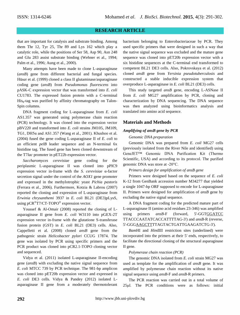
Amplifying of ansB gene by PCR
Amplification of 981-bp fragment was performed using
pair of specific primers ansB-F and ansB-R incorporating the
sequence for the restriction endonucleases BamHI and
HindIII, respectively (Figure 1).
TA-Cloning of ansB into pGEM-T Easy vector
Three white colonies were picked and subjected to
confirmation procedures to detect the recombinant clones
harboring the putative gene encoding L-ASNase II from local
isolate E. coli strain MG27(Figure 2).
Verification tests for the recombinant ansB clones
Three white colonies designated W1, W2 and W3 were
picked from LB/ampicillin/IPTG/X-gal plate. These clones
were plated on LB-ampicillin plates, as well as being cultured
in LB-ampicillin broth for Plasmid DNA minipreps. The
presence of insert was confirmed by PCR and restriction
digestion.
Polymerase chain reaction (PCR) screening
Figure 3 showed the amplified products of clones W1,
W2 and W3 which had the same expected size (981 bp) for
PCR product of ansB gene as in positive control (lane 5).
Positive control was conducted by using genomic DNA of E.
coli MG27 as DNA template. Negative control was
conducted by using plasmid from a blue colony as DNA
template and showed no band indicating no recombination
(lane 4).
Figure 1. Agarose gel electrophoresis of PCR amplification
of putative ansB gene amplified from genomic DNA of E. coli
MG27. Lane 1, DNA marker; lane 2, PCR amplicon of
putative ansB gene. Genomic DNA isolated from E. coli
strain MG27 was used as a template for PCR amplification of
ansB gene without its native signal sequence using ansB-F
and ansB-R specific primers. The PCR product analyzed
using 1% agarose gel electrophoresis showed a fragment of
the expected size (981 bp) of ansB gene.
Figure 2. Blue/White screening of transformants.
LB/ampicillin/IPTG/X-gal plate was plated with 50 µl of
transformation mixture and incubated at 37°C for 18-24 h.
White colonies represent recombinant clones while blue
colonies represent empty clones.

ISSN: 1314-6246 Mohamed et al. J. BioSci. Biotechnol. 2015, 4(3): 291-302.
RESEARCH ARTICLE
http://www.jbb.uni-plovdiv.bg 295
Figure 3. Screening of white clones for recombinant pGEM-
ansB plasmid by PCR. Lane M, DNA marker; lanes 1-3, PCR
products from white clones W1, W2 and W3, respectively.
Lane 4, negative control; lane 5, positive control. Plasmid
DNA was prepared from white clones W1, W2 and W3 and
used as DNA template for PCR reactions using insert-specific
primers ansB-F and ansB-R. Negative control was conducted
by using plasmid from a blue colony as DNA template.
Positive control was conducted by using genomic DNA of E.
coli MG27 as DNA template. PCR amplicons were analyzed
on 1% agarose gel electrophoresis. White clones W1, W2 and
W3 generated fragments of expected size 981 bp confirming
the presence of putative ansB gene.
Confirmation of transformation by restriction digestion
Linearized vector in lane 3 showed one band at the
expected size (~4 Kb) while two bands were observed in case
of double digested vector (lane 4). A DNA fragment about
981 bp was released from the vector backbone (~3.0 kb).
Plasmid extracted from the clone W3 was designated pGEM-
ansB and subjected to the nucleotide sequence analysis
(Figure 4).
Nucleotide sequencing
Nucleotide sequence of the putative ansB gene of E. coli
MG27 was submitted to the NCBI database and an accession
number KC416966 was assigned while the deduced amino
acid sequence was submitted under accession number
AGE81914.
Bioinformatics analysis
Results revealed that, sequence of putative ansB consists
of 981 bp codes for 326 amino acids (Figure 5). Restriction
map of putative ansB gene generated using CodonCode
aligner program was represented in Figure 6.
Using ProtParam tool of ExPASy, The results showed
that the protein contains 20 amino acids with valine in the
highest percentage (10.7%) while tryptophan was the lowest
(0.3%) as detailed in Table 1. Other physiochemical
properties obtained from ProtParam analysis has been
tabulated (Table 2). The instability index of the protein
calculated using ProtParam tool was 19.61. The predicted
theoretical isoelectric point (pI) value was 5.66 and its
molecular weight was estimated to be 34.5788 kDa. The
number of negatively charged residues (Asp + Glu) was
greater than the number of positively charged residues (Arg +
Lys). ProtParam results showed that 33 residues are
negatively charged and 30 residues are positively charged.
Additionally the grand average of hydropathicity (GRAVY)
and aliphatic index were computed to be -0.214 and 84.33,
respectively.
Figure 4. Restriction analysis of constructed pGEM-ansB
vector. Lane 1, 1 kb DNA marker; lane 2, undigested vector;
lane 3, Linearized vector digested with BamHI restriction
enzyme; lane 4, vector digested with BamHI and HindIII
restriction enzymes. Plasmid DNA prepared from clone W3
was single and double digested and analyzed on 1% agarose
gel electrophoresis. Single digestion with BamHI resulted in
a linearized vector at the expected size (~4 kb). Two bands
were observed when vector was double digested with BamHI
and HindIII. Bands in lane 4 represent the expected vector
backbone (~3.0 kb) and the released insert (~981 bp).
The deduced amino acid sequence was utilized for
similarity search through BLAST at NCBI selecting non-
redundant database. BLAST analysis on the deduced amino
acid sequence of putative ansB gene from E. coli MG27
showed 100% identity with L-asparaginases II from Shigella
sonnei Ss046 (accession number YP_312053.1), Escherichia
coli OK1357 (accession number WP_001345951.1) and E.
coli MS 79-10 (accession number WP_001012363.1). In

ISSN: 1314-6246 Mohamed et al. J. BioSci. Biotechnol. 2015, 4(3): 291-302.
RESEARCH ARTICLE
http://www.jbb.uni-plovdiv.bg 296
addition, significant homology with 99-87% similarity was
found with several bacterial L-asparaginases including L-
asparaginase II from Escherichia coli KTE66 (accession
number WP_001559780.1), Shigella boydii 5216-82
(accession number WP_000394146.1), Citrobacter freundii
(accession number ACC85692.1), C. youngae ATCC 29220
(accession number WP_006686837.1), Salmonella enterica
subsp. enterica (accession number WP_000394193.1),
Enterobacter cloacae SCF1 (accession number
YP_003940358.1) and Serratia marcescens VGH107
(accession number WP_004928279.1). Multiple sequence
alignment was conducted on these sequences using CLC
program. Alignment results revealed that several highly
conserved domains were extended along ansB sequences.
Figure 5. Nucleotide sequence of putative L-asparaginase II gene (ansB) and its deduced amino acid sequence. The sequence
extends, 981 nucleotid length and the translation product of the ansB gene was shown below the nucleotide sequence.

ISSN: 1314-6246 Mohamed et al. J. BioSci. Biotechnol. 2015, 4(3): 291-302.
RESEARCH ARTICLE
http://www.jbb.uni-plovdiv.bg 297
Figure 6. Restriction map of the putative ansB gene.
Table 1. Amino acid composition of E. coli MG27 putative
ansB calculated using the ProParam tool of ExPASy.
Amino acid Number of
residues Percentage
Alanine
Arginine
Asparagine
Aspartic acid
Cysteine
Glutamine
Glutamic acid
Glycine
Histidine
Isoleucine
Leucine
Lysine
Methionine
Phenylalanine
Proline
Serine
Threonine
Tryptophan
Tyrosine
Valine
pyrrolysine
Selenocysteine
33
8
24
27
2
13
6
28
3
13
23
22
6
8
13
16
33
1
12
35
0
0
10.1
2.5
7.4
8.3
0.6
4.0
1.8
8.6
0.9
4.0
7.1
6.7
1.8
2.5
4.0
4.9
10.1
0.3
3.7
10.7
0.0
0.0
Table 2. Physicochemical parameters of E. coli MG27
putative ansB computed using ExPASy's ProtParam tool.
Value Parameter
326 Number of amino acids
19.61 The instability index
5.66 Theoretical isoelectric point (pI)
34.5788 kDa Molecular weight
33 Total number of negatively charged
residues (Asp + Glu)
30 Total number of positively charged
residues (Arg + Lys).
-0.214 Grand average of hydropathicity
(GRAVY)
84.33 Aliphatic index
The deduced amino acid sequence of E. coli MG27 ansB
gene was scanned for conserved residues by the Conserved
Domain Database (CDD). The CDD results revealed that the
mature protein contains a conserved domain of L-
asparaginase-like superfamily (CDD accession: cl00216) and
L-asparaginase-like domain (CDD accession: cd00411) as
visualized in Figure 7.

ISSN: 1314-6246 Mohamed et al. J. BioSci. Biotechnol. 2015, 4(3): 291-302.
RESEARCH ARTICLE
http://www.jbb.uni-plovdiv.bg 298
Figure 7. Prosite analysis of the deduced amino acid
sequence of putative ansB showing two active domains.
Active site signatures are ASN_GLN_ASE_1 (PS00144) and
ASN_GLN_ASE_2 (PS00917) at amino acids 6-14 and 82-92,
respectively.
Analysis with PROSITE program revealed that L-
asparaginase II contains two recognizable structural domains
and their locations are marked below (shadowed). The first
structural domain (residues 6-14) is located near the N-
terminal section while the second structural domain located
within residues 82 to 92.
LPNITILATGGTIAGGGDSATKSNYTAGKVGVENLVNAVPQ
LKDIANVKGEQVVNIGSQDMNDNVWLTLAKKINTDCDKTDG
FVITHGTDTMEETAYFLDLTVKCDKPVVMVGAMRPSTSMSA
DGPFNLYNAVVTAADKASANRGVLVVMNDTVLDGRDVTKTN
TTDVATFKSVNYGPLGYIHNGKIDYQRTPARKHTSDTPFDV
SKLNELPKVGIVYNYANASDLPAKALVDAGYDGIVSAGVGN
GNLYKSVFDTLATAAKNGTAVVRSSRVPTGATTQDAEVDDA
KYGFVASGTLNPQKARVLLQLALTQTKDPQQIQQIFNQY
The two asparaginase/glutaminase active site signatures
are ASN_GLN_ASE_1 (PS00144) and ASN_GLN_ASE_2
(PS00917) having active site consensus pattern [LIVM]-x-
{L}-T-G (2)-T-[IV]-[AGS] and [GA]-x-[LIVM]-x (2)-H-G-
T-D-T-[LIVM]. The amino acids 12 and 89 are the active site
residues, respectively. The results of the InterPro database are
summarized below (Table 3; Figure 8). InterProScan analysis
revealed that the protein sequence had a catalytic domain of
L-asparaginase type II (IPR004550) that belong to
asparaginase/glutaminase family (IPR006034) and has
Asparaginase/glutaminase conserved site (IPR020827).
Table 3. Protein signatures and functional domains of ansB protein identified using InterProScan.
Source database accession InterPro accession Signature ID Amino acids E-value
TGRFAMs / TIGR00520 IPR004550 asnASE_II 1-326 3.7e-166
PIRSF / PIRSF001220 IPR006034
L-ASNase_gatD 1-326 1.8e-94
SUPERFAMILY / SSF53774 Asp/Glutamnse 1-326 4.0e-111
PROSITE patterns / PS00144 IPR020827
ASN_GLN_ASE_1 6-14 1.0
PROSITE patterns / PS00917 ASN_GLN_ASE_2 82-92 1.0
Figure 8. Graphical view of InterProScan showing functional domains from the deduced amino acid sequence of L-
asparaginase II. Characteristic signatures of L-ASNase II which were found after analysis of the 326 amino acid sequence in
the integrative protein signature database InterPro.

ISSN: 1314-6246 Mohamed et al. J. BioSci. Biotechnol. 2015, 4(3): 291-302.
RESEARCH ARTICLE
http://www.jbb.uni-plovdiv.bg 299
Figure 9. Predicted secondary structure of L-asparaginase
II. Secondary structure prediction was performed based on
position-specific scoring matrices using the PSIpred method.
The sequences marked as ‘H’, ‘E’ and ‘C’ correspond to
helix, strand and coil, respectively. According to the PSIpred
prediction, the protein has 8 α-helices and 13 β-strands, as
shown.
The PSIpred database predicted the following secondary
structures of the L-asparaginase II from the amino acid
sequence as depicted in Figure 9. PSIpred program revealed
that ansB consists of eight α-helices and 13 β-strands. ansB
was predicted to be formed of approximately 27% of α-
helices (88 residues) and 20% of β-strands (63 residues)
while random coils represents 53% (175 residues).
Discussion
Studying L-Asparaginase (L-ASNase) has recently gained
much attention for its anti-carcinogenic potential. Several
authors documented the use of L-ASNase in cancer therapy
(Avramis & Panosyan 2005; Narta et al., 2007; Pieters et al.,
2011; Tong et al., 2013). Although L-ASNases are present in
many plants, mammalian and bacterial species, only the
enzymes from Escherichia coli and Erwinia chrysanthemii
have been produced on industrial scale as chemotherapeutics
in acute lymphoblastic leukemia. This is due to their high
catalytic activity and specificity towards L-asparagine
(Müller & Boos 1998; Aghaiypour et al., 2001; Duval et al.,
2002). Apart from the therapeutic use, L-ASNase has a potent
application in food industry to reduce acrylamide formation
in heat-processed products (Friedman & Levin, 2008;
Pedreschi et al., 2008; Kukurová et al., 2013).
The mature sequence of ansB gene was amplified from
the genomic DNA of a moderately thermotolerant bacterium
belonging to Enterobacteriaceae by PCR and the amplicon of
~980 bp was cloned into pET20b vector (Vidya & Pandey,
2012). Furthermore, a putative L-asparaginase gene
consisting of 981 bp was amplified by PCR from the
Pyrococcus furiosus genomic DNA and the PCR product was
cloned into a pET14b vector (Bansal et al., 2010). Many
investigators cloned L-asparaginase coding genes from
various bacteria such as Pseudomonas fluorescens (Hüser et
al., 1999), E. coli (Wang et al., 2001), Erwinia carotovora
(Kotzia & Labrou, 2005), E. crysanthemi (Kotzia & Labrou,
2007), Yersinia pseudotuberculosis (Pokrovskaya et al.,
2012) and Bacillus subtilis (Jia et al., 2013).
In the present study, the clone harboring pGEM-ansB
construct was selected for nucleotide sequencing using M13
forward and reverse primers. Nucleotide sequence of the
putative ansB gene of E. coli MG27 was submitted to the
NCBI database and an accession number KC416966 was
assigned while the predicted protein sequence was submitted
under accession number AGE81914. Based on the instability
index of the deduced amino acids predicted by ProtParam

ISSN: 1314-6246 Mohamed et al. J. BioSci. Biotechnol. 2015, 4(3): 291-302.
RESEARCH ARTICLE
http://www.jbb.uni-plovdiv.bg 300
tool, the protein is stable with a value of 19.61.
The grand average of hydropathicity (GRAVY) of the
deduced amino acids was computed to be -0.214. This
negative value of GRAVY suggests the hydrophilicity of the
protein. The aliphatic index of the deduced amino acids
predicted using ProtParam tool was 84.33. This high aliphatic
index indicates that the protein can be stable within a wide
range of temperature.
Based on BLAST analysis, the deduced amino acid
sequence of mature L-ASNase II showed 100% identity with
L-ASNase II from Escherichia coli OK1357
(WP_001345951.1), E. coli MS 79-10 (WP_001012363.1).
BLAST analysis of the deduced amino acid sequence
revealed significant similarity (99- 87%) with L-ASNase II of
Shigella boydii 5216-82 (WP_000394146.1), Citrobacter
freundii (ACC85692.1), C. youngae ATCC 29220
(WP_006686837.1), Salmonella enterica subsp. enterica
(WP_000394193.1), Enterobacter cloacae SCF1
(YP_003940358.1) and Serratia marcescens VGH107
(WP_004928279.1).
The amino acid sequence alignment of putative L-
ASNase II from E. coli MG27 with L-ASNase II sequences
from other 10 strains of bacteria revealed that the sequence of
this enzyme is highly conserved especially with Thr-12, Tyr-
25, Thr-89, Asp-90, and Lys-162. It was suggested that these
residues are essential for reaction the enzymatic activity
(Wehner et al., 1994). Thr-12 and Thr-89 are able to act as
primary nucleophiles (Harms et al., 1991; Palm et al., 1996).
Thr-12 and the adjacent Tyr-25 are components of a mobile
loop that closes over the active site during catalysis while
Thr-89, Asp-90 and Lys-162 are all located in a rigid part of
the structure (Aung et al., 2000; Derst et al., 2000). In this
study, the amino acid sequence differs in two positions from
the sequence published by Jennings & Beacham (1990).
These changes are of substitution type, where alanine is
present instead of valine and asparagine is present instead of
threonine at residues 27 and 263, respectively. These
differences may result in enzymes with different activities
between different strains.
NCBI conserved-domain search of the deduced protein
revealed the presence of a conserved L-asparaginase-like
superfamily domain (CDD accession: cl00216) and L-
asparaginase-like domain (CDD accession: cd00411). As
predicted by the Prosite program, the deduced amino acid
sequence contains two recognizable structural domains.
These asparaginase/glutaminase active site signatures are
ASN_GLN_ASE_1 (PS00144) and ASN_GLN_ASE_2
(PS00917) located within residues 6 to14 and 82 to 92,
respectively. Prosite analysis revealed that two threonine
residues at 12 and 89 are the active site residues. Harms et al.
(1991) provided an evidence for the importance of threonine-
12 for catalytic activity of L-ASNase II that lost its activity
when Thr-12 is mutated to alanine. In addition, Thr-89 was
postulated to play a catalytic role in ASNase II activity
(Swain et al., 1993; Palm et al., 1996).
In the present study, highly conserved amino acid
sequence motifs were identified by comparison against the
InterPro database. InterProScan analysis revealed that the
protein sequence had a catalytic domain of L-asparaginase
type II (IPR004550) that belong to asparaginase/glutaminase
family (IPR006034) and has Asparaginase/glutaminase
conserved site (IPR020827). According to results predicted
using PSIpred tool, ansB consists of eight α-helices and 13 β-
strands. C-terminal domain (residues 213-326) was predicted
to be consisted of four β-strands and four α-helices. These
results agree with Swain et al. (1993).
References
Aghaiypour K, Wlodower A, Lubkowski J. 2001. Structural basis
for the activity and substrate specificity of Erwinia
chrysanthemi L-asparaginase. Biochemistry, 40: 5655-5664.
Altschul SF, Madden TL, Schäffer AA, Zhang J, Zhang Z, Miller
W, Lipman DJ. 1997. Gapped BLAST and PSI-BLAST: a new
generation of protein database search programs. Nucleic Acids
Res., 25: 3389-3402.
Aung HP, Bocola M, Schleper S, Röhm KH. 2000. Dynamics of a
mobile loop at the active site of Escherichia coli asparaginase.
Biochim. Biophys. Acta, 1481: 349-359.
Avramis VI, Panosyan EH. 2005. Pharmacokinetic
/pharmacodynamic relationships of asparaginase formulations:
the past, the present and recommendations for the future. Clin.
Pharmacokinet., 44: 367-393.
Bansal S, Gnaneswari D, Mishra P, Kundu B. 2010. Structural
stability and functional analysis of L-asparaginase from
Pyrococcus furiosus. Biochemistry (Moscow), 75: 375-381.
Beatty CM, Browning DF, Busby SJ, Wolfe AJ. 2003. Cyclic AMP
receptor protein-dependent activation of the Escherichia coli
acsP2 promoter by a synergistic class III mechanism. J.
Bacteriol., 185: 5148-5157.
Cappelletti D, Chiarelli LR, Pasquetto MV, Stivala S, Valentini G,
Scotti C. 2008. Helicobacter pylori L-asparaginase: A promising
chemotherapeutic agent. Biochemical and Biophysical Research
Communications, 377: 1222-1226.
Chen YP, Lin HH, Yang CD, Huang SH, Tseng CP. 2012.
Regulatory role of cAMP receptor protein over Escherichia coli
fumarase genes. J. Microbiol., 50: 426-433.

ISSN: 1314-6246 Mohamed et al. J. BioSci. Biotechnol. 2015, 4(3): 291-302.
RESEARCH ARTICLE
http://www.jbb.uni-plovdiv.bg 301
De Castro E, Sigrist CJA, Gattiker A, Bulliard V, Langendijk-
Genevaux PS, Gasteiger E, Bairoch A, Hulo N. 2006.
ScanProsite: detection of PROSITE signature matches and
ProRule-associated functional and structural residues in
proteins. Nucleic Acids Res., 34 (Web Server issue): W362-365.
Derst C, Henseling J, Röhm KH. 2000. Engineering the substrate
specificity of Escherichia coli asparaginase II. Selective
reduction of glutaminase activity by amino acid replacements at
position 248. Protein Science, 9: 2009-2017.
Duval M, Suciu S, Ferster A, Rialland X, Nelken B, Lutz P, Benoit
Y, Robert A, Manel AM, Vilmer E, Otten J, Philippe N. 2002.
Comparison of Escherichia coli-asparaginase with Erwinia-
asparaginase in the treatment of childhood lymphoid
malignancies: results of a randomized European Organization
for Research and Treatment of Cancer-Children’s Leukemia
Group phase 3 trial. Blood, 99: 2734-2739.
Ferrara MA, Severino NMB, Mansure JJ, Martins AS, Oliveira
EMM, Siani AC, Pereira N, Torres FAG, Bon EBS. 2006.
Asparaginase production by a recombinant Pichia pastoris strain
harbouring Saccharomyces cerevisiae ASP3 gene. Enzyme and
Microbial Technology, 39: 1457-1463.
Friedman M, Levin E. 2008. Review of methods for the reduction of
dietary content and toxicity of acrylamide. Journal of
Agricultural and Food Chemistry, 56: 6113-6140.
Gasteiger E, Hoogland C, Gattiker A, Duvaud S, Wilkins MR,
Appel RD, Bairoch A. 2005. In: John M. Walker (ed): Protein
Identification and Analysis Tools on the ExPASy Server. The
Proteomics Protocols Handbook, Humana Press. pp. 571-607.
Hall TA. 1999. BioEdit: a user-friendly biological sequence
alignment editor and analysis program for Windows 95/98/NT.
Nucl. Acids. Symp., Ser. 41: 95-98.
Harms E, Wehner, A, Aung HP, Röhm KH. 1991. A catalytic role
for threonine-12 of E. coli asparaginase II as established by site-
directed mutagenesis. FEBS Lett., 285: 55-58.
Hüser A, Klöppner U, Röhm KH. 1999. Cloning, sequence analysis,
and expression of ansB from Pseudomonas fluorescens,
encoding periplasmic glutaminase/asparaginase. FEMS
Microbiol. Lett., 178: 327-335.
Jennings MP, Beacham IR. 1990. Analysis of the E. coli gene
encoding L-asparaginase II, ansB, and its regulation by cyclic
AMP receptor and FNR proteins. J. Bacteriol., 172: 1491-1498.
Jia M, Xu M, He B, Rao Z. 2013. Cloning, expression, and
characterization of L-asparaginase from a newly isolated
Bacillus subtilis B11-06. J. Agric. Food Chem., 61: 9428-9434.
Jones DT. 1999. Protein secondary structure prediction based on
position-specific scoring matrices. J. Mol. Biol., 292: 195-202.
Khushoo A, Pal Y, Singh BN, Mukherjee KJ. 2004. Extracellular
expression and single step purification of recombinant
Escherichia coli L-asparaginase II. Protein Expression and
Purification, 38: 29-36.
Kotzia GA, Labrou NE. 2005. Cloning, expression and
characterization of Erwinia carotovora L-asparaginase. J.
Biotechnol., 119: 309-323.
Kotzia GA, Labrou NE. 2007. l-Asparaginase from Erwinia
chrysanthemi 3937: Cloning, expression and characterization. J.
Biotechnol., 127: 657-669.
Kozak M, Borek D, Janowski R, Jaskólski M. 2002. Crystallization
and preliminary crystallographic studies of five crystal forms of
Escherichia coli L-asparaginase II (Asp90Glu mutant). Acta
Crystallogr. D. Biol. Crystallogr., 58: 130-132.
Kraxenberger T, Fried L, Behr S, Jung K. 2012. First insights into
the unexplored two-component system YehU/YehT in
Escherichia coli. J. Bacteriol., 194: 4272-4284.
Kukurová K, Ciesarová Z, Mogol BA, Açar ÖÇ, Gökmen V. 2013.
Raising agents strongly influence acrylamide and HMF
formation in cookies and conditions for asparaginase activity in
dough. Eur. Food Res. Technol., 237: 1-8.
Marchler-Bauer A, Anderson JB, Chitsaz F, Derbyshire MK,
DeWeese-Scott C, Fong JH, Geer LY, Geer RC, Gonzales NR,
Gwadz M, He S, Hurwitz DI, Jackson JD, Ke Z, Lanczycki CJ,
Liebert CA, Liu C, Lu F, Lu S, Marchler GH, Mullokandov M,
Song JS, Tasneem A, Thanki N, Yamashita R A, Zhang D,
Zhang N, Bryant SH. 2009. CDD: specific functional annotation
with the conserved domain database. Nucleic Acids Res., 37:
(D) 205-210.
Müller HJ, Boos J. 1998. Use of L-asparaginase in childhood ALL.
Crit. Rev. Oncol. Hemat., 28: 97-113.
Narta UK, Kanwar SS, Azmi W. 2007. Pharmacological and clinical
evaluation of L-asparaginase in the treatment of leukemia. Crit.
Rev. Oncol/Hematol., 61: 208-221.
Palm GJ, Lubkowski J, Derst C, Schleper S, Röhm KH, Wlodawer
A. 1996. A covalently bound catalytic intermediate in
Escherichia coli asparaginase: crystal structure of a Thr-89-Val
mutant. FEBS Lett., 390: 211-216.
Partridge JD, Browning DF, Xu M, Newnham LJ, Scott C, Roberts
RE, Poole RK, Green J. 2008. Characterization of the
Escherichia coli K-12 ydhYVWXUT operon: regulation by
FNR, NarL and NarP. Microbiology, 154: 608-618.
Pedreschi F; Kaack K, Granby K. 2008. The effect of asparaginase
on acrylamide formation in French fries. Food Chemistry, 109:
386-392.
Pieters R, Hunger SP, Boos J, Rizzari C, Silverman L, Baruchel A,
Goekbuget N, Schrappe M, Pui CH. 2011. L-asparaginase
treatment in acute lymphoblastic leukemia: a focus on Erwinia
asparaginase. Cancer, 117: 238-249.
Pokrovskaya MV, Aleksandrova SS, Pokrovsky VS, Omeljanjuk
NM, Borisova AA, Anisimova NY, Sokolov NN. 2012.
Cloning, expression and characterization of the recombinant
Yersinia pseudotuberculosis L-asparaginase. Protein Expression
and Purification, 82: 150-154.
Quevillon E, Silventoinen V, Pillai S, Harte N, Mulder N, Apweiler
R, Lopez R. 2005. InterProScan: protein domains identifier.
Nucleic Acids Res., 33 (Web Server issue): W116-120.
Sambrook J, Russell DW. 2001. Molecular cloning: A laboratory
manual. 3rd ed. Cold Spring Harbor (NY): Cold Spring Harbor
Laboratory Press.
Shan Y, Pan Q, Liu J, Huang F, Sun H, Nishino K, Yan A. 2012.
Covalently linking the Escherichia coli global anaerobic
regulator FNR in tandem allows it to function as an oxygen
stable dimer. Biochem. Biophys. Res. Commun., 419: 43-48.
Swain AL, Jaskólski M, Housset D, Rao JK, Wlodawer A. 1993.
Crystal structure of Escherichia coli L-asparaginase, an enzyme
used in cancer therapy. Proc. Natl. Acad. Sci. U S A, 90: 1474-
1478.
Tolla DA, Savageau MA. 2011. Phenotypic repertoire of the FNR
regulatory network in Escherichia coli. Mol. Microbiol., 79:
149-165.
Tong WH, van der Sluis IM, Alleman C, van Litsenburg RR,
Kaspers GJ, Pieters R, Uyl-de Groot CA. 2013. Cost-analysis of
treatment of childhood acute lymphoblastic leukemia with

ISSN: 1314-6246 Mohamed et al. J. BioSci. Biotechnol. 2015, 4(3): 291-302.
RESEARCH ARTICLE
http://www.jbb.uni-plovdiv.bg 302
asparaginase preparations: the impact of expensive
chemotherapy. Haematologica, 98: 753-759.
Uppal S, Maurya SR, Hire RS, Jawali N. 2011. Cyclic AMP
receptor protein regulates cspE, an early cold-inducible gene, in
Escherichia coli. J. Bacteriol., 193: 6142-6151.
Vidya J; Vasudevan UM; Soccol CR, Pandey A. 2011. Cloning,
functional expression and characterization of L-asparaginase II
from E. coli MTCC 739. Food Technol. Biotechnol., 49: 286-
290.
Vidya J, Pandey A. 2012. Recombinant expression and
characterization of L-asparaginase II from a moderately
thermotolerant bacterial isolate. Appl. Biochem. Biotechnol.,
167: 973-980.
Wang Y ,Qian S, Meng G, Zhang S. 2001. Cloning and expression
of L-asparaginase gene in Escherichia coli. Appl. Biochem.
Biotechnol., 95: 93-101.
Wehner A, Derst C, Specht V, Aung HP, Röhm KH. 1994. The
catalytic mechanism of Escherichia coli asparaginase II. Hoppe
Seylers Z. Physiol. Chem., 375: 108.
Youssef MM, Al-Omair MA. 2008. Cloning, purification,
characterization and immobilization of L-asparaginase II from
E. coli W3110. Asian Journal of Biochemistry, 3: 337-350.