Embed Size (px)
Citation preview

Redefine clone screening and selection: transform your cell line development workflow
ClonePix™ 2
www.moleculardevices.com2
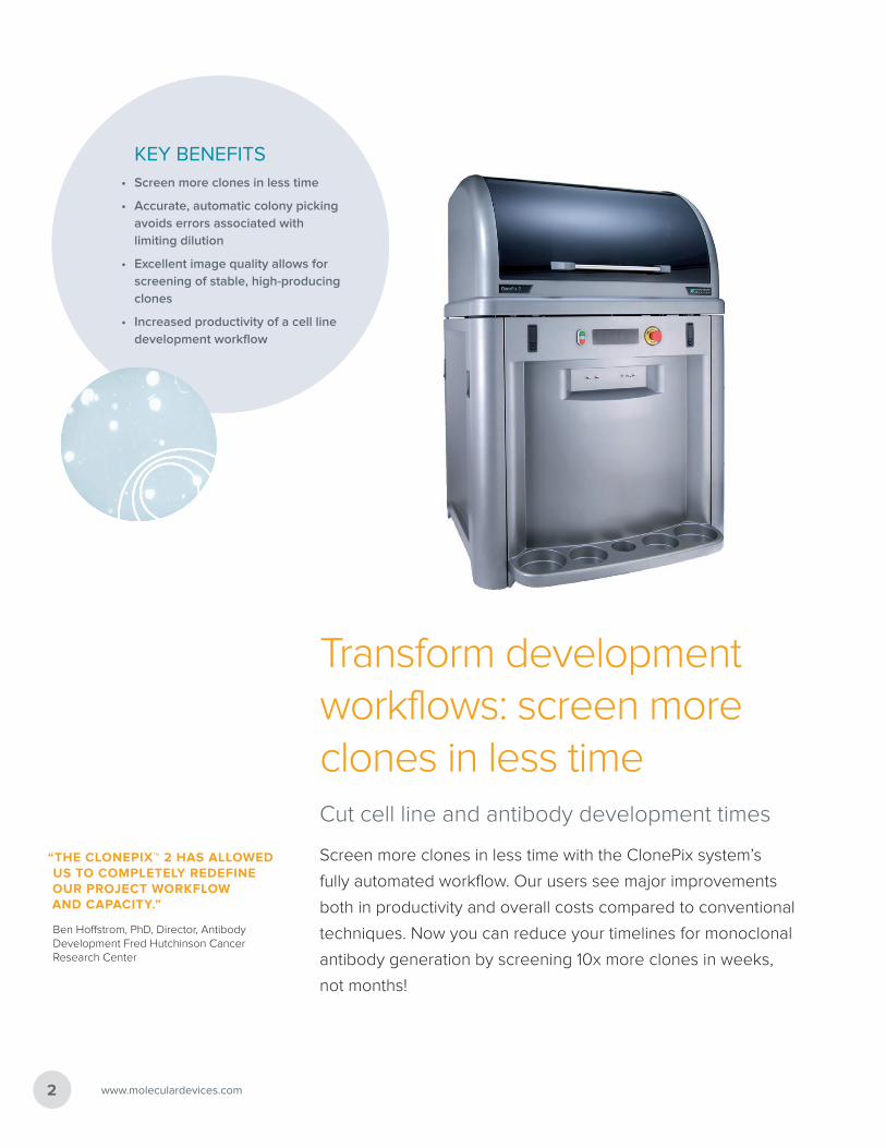
KEY BENEFITS• Screen more clones in less time
• Accurate, automatic colony picking avoids errors associated with limiting dilution
• Excellent image quality allows for screening of stable, high-producing clones
• Increased productivity of a cell line development workflow
Transform development workflows: screen more clones in less timeCut cell line and antibody development times
Screen more clones in less time with the ClonePix system’s
fully automated workflow. Our users see major improvements
both in productivity and overall costs compared to conventional
techniques. Now you can reduce your timelines for monoclonal
antibody generation by screening 10x more clones in weeks,
not months!
“THE CLONEPIX™ 2 HAS ALLOWED US TO COMPLETELY REDEFINE OUR PROJECT WORKFLOW AND CAPACITY.”
Ben Hoffstrom, PhD, Director, Antibody Development Fred Hutchinson Cancer Research Center
ClonePix™ 2 3
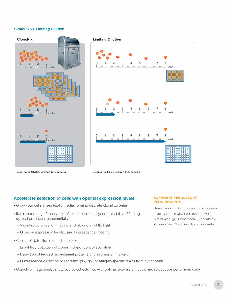
ClonePix vs. Limiting Dilution
Limiting Dilution
...screens 10,000 clones in 3 weeks ...screens 1,000 clones in 8 weeks
ClonePix
0 1 2 3 4 5 6 7 8weeks
0 1 2 3 4 5 6 7 8weeks
0 1 2 3 4 5 6 7 8weeks
0 1 2 3weeks
0 1 2 3weeks
0 1 2 3weeks
Accelerate selection of cells with optimal expression levels
• Grow your cells in semi-solid media, forming discrete clonal colonies
• Rapid screening of thousands of clones increases your probability of finding optimal producers exponentially
– Visualize colonies for imaging and picking in white light
– Observe expression levels using fluorescence imaging
• Choice of detection methods enables:
– Label-free detection of clones independent of secretion
– Detection of tagged recombinant proteins and expression markers
– Fluorescence detection of secreted IgG, IgM, or antigen-specific mAbs from hybridomas
• Objective image analysis lets you select colonies with optimal expression levels and reject poor performers early
SUPPORTS REGULATORY REQUIREMENTS
These products do not contain components
of animal origin when you need to work
with human IgG: CloneMedia, CloneMatrix,
Recombinant CloneDetect, and XP media.

Ensure formation of discrete colonies
www.moleculardevices.com4
Select and pick with more accuracy and confidence
Select your colonies based on the system’s automatic analysis and ranking
Screen and select colonies based on the criteria you define
Cells plated into semi-solid medium
Clonal colonies grow
ClonePix images, analyzes and ranks colonies
ClonePix picks colonies
5 – 14 days
Utilizing semi-solid media as a cloning system is a well-established method. Growing cells in CloneMedia makes it easier to plate out large numbers of cells and ensures formation of discrete colonies—facilitating recovery of many independent clones.
Fast-track recovery of independent clones
• Choose from a range of media optimized for use with ClonePix systems
• Compatible with CHO, HEK, CHOK1SV cells and hybridomas
• Flexibility to prepare your specific media using a CloneMatrix concentrate
Colonies growing in CloneMedia. Images captured using CloneSelect Imager. (Left) CHO colonies, serum-free suspension-adapted, in CloneMedia CHO Growth A imaged on day 8 post-plating. (Right) Hybridoma colonies in CloneMedia Hybridoma/Myeloma imaged on day 7 post-plating.
“EASIER TO PLATE OUT LARGE NUMBERS OF CELLS AND TO RECOVER MANY INDEPENDENT HYBRIDOMA CLONES”
A simple, single-step technique for selecting and cloning hybridomas for the production of monoclonal antibodies (J. Immun. Methods (1982) 50, 161-171)

5
Detect secretion using fluorescence-based methods
Detecting secretion using fluorescently-conjugated CloneDetect
agents enables in situ detection of secreted antibodies.
• Select the detection agent that’s right for you: CloneDetect anti-human, anti-mouse, anti-rat, and anti-sheep detection agent FITC labels
• Add detection agents directly to semi-solid media
• ClonePix systems image, analyze and rank fluorescence levels across thousands of clones in parallel
Choice of detection methods
Simplify target protein detection
Secreting clone
CloneDetect added
Secreted proteins form fluorescent precipitate
Secreted antibody (IgG)
Complex Initiation Factor (CIF)
Antigen (fluorescently conjugated or tagged)
Secreting clone
Fluorescent anti-tag
Secreted tagged protein
ClonePix™ 2
MAb secreting IgG. For in situ detection of human antibodies use CloneDetect fluorescently-conjugated agents.
Antigen-specific MAbs from hybridomas. Generate precipitation using fluorescently-conjugated or tagged antigen plus complex initiation factor (CIF).
Tagged recombinant proteins. Protein construct contains epitope tag(s), e.g. His, FLAG™ or Fc using tag-specific agents.

Higher secretors
Intelligent imaging and analysis
www.moleculardevices.com6
Imaging
Automatically capture images of all your colonies in white light and secreting colonies with fluorescence.
Data analysis
• Automatically generate a 2D map of clones and their secretion levels from a series of images generated in situ
• Screen and select colonies based on:
– Size, roundness, and proximity to neighbors
– Ranking according to fluorescence levels
– Closely placed colonies ignored via user-controlled “proximity” software setting
White light suggest selection of the largest clone Fluorescence reveals the optimal secretor
2D ranking plot You define selection parameters, the system selects clones
Automatic colony picking (can be manually overridden)
7ClonePix™ 2
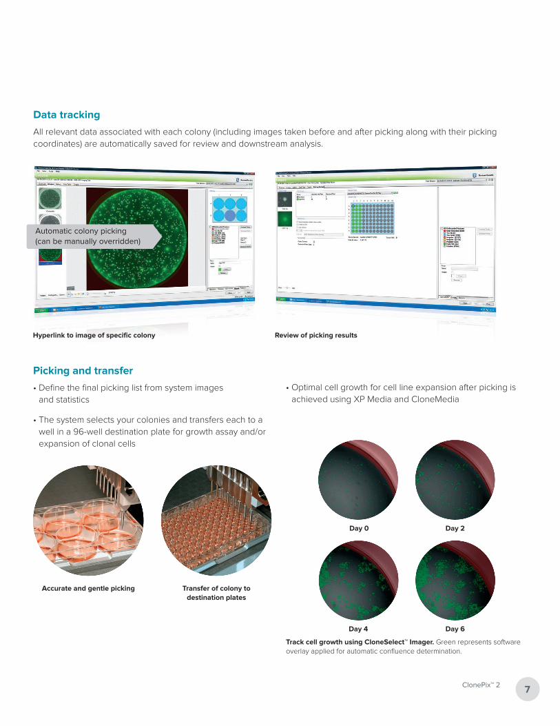
Data tracking
All relevant data associated with each colony (including images taken before and after picking along with their picking coordinates) are automatically saved for review and downstream analysis.
Hyperlink to image of specific colony Review of picking results
Accurate and gentle picking
Picking and transfer
• Define the final picking list from system images and statistics
• The system selects your colonies and transfers each to a well in a 96-well destination plate for growth assay and/or expansion of clonal cells
Transfer of colony to destination plates
Day 0 Day 2
Day 4 Day 6
Track cell growth using CloneSelect™ Imager. Green represents software overlay applied for automatic confluence determination.
• Optimal cell growth for cell line expansion after picking is achieved using XP Media and CloneMedia
www.moleculardevices.com8
Boost generation of research antibodiesAutomate screening and collection of hybridomas from fusions
• Efficient processing lets you screen 10,000 clones and pick 1,000 HAT-selected clones per day
• Minimal loss of positive clones downstream and early elimination of negative clones upstream saves time and resources
• Screen in situ for antigen specificity and /or IgG secretion across a broad range of antigens: 160 kDa multimeric protein to
2.6 kDa phosphopeptide
“WE HAVE INCREASED OUR FUSION PRODUCTIVITY BY APPROXIMATELY 50%, WHILE DECREASING OUR TIME FROM FUSION TO STABLE CLONE BY 50%”
Dr. Robin Barbour, Prothena Corporation
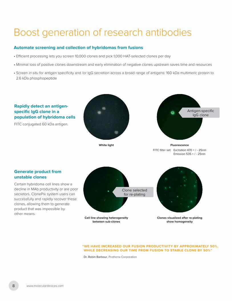
Rapidly detect an antigen-specific IgG clone in a population of hybridoma cells
FITC conjugated 60 kDa antigen.
White light Fluorescence
Antigen-specific IgG clone
FITC filter set: Excitation 470 + / - 25nm Emission 535 + / - 25nm
Generate product from unstable clones
Certain hybridoma cell lines show a decline in MAb productivity or are poor secretors. ClonePix system users can successfully and rapidly recover these clones, allowing them to generate product that was impossible by other means.
Cell line showing heterogeneity between sub-clones
Clones visualized after re-plating show homogeneity
Clone selected for re-plating
9ClonePix™ 2
Elevate productivity for biotherapeuticsAmplify the probability of finding rare high secretors
More clones can be screened in less time on the ClonePix system than with traditional methods, letting you screen larger cell populations. MedImmune LLC compared the effectiveness of the ClonePix system to an already established limiting dilution process for their cell line development workflow. Real-world results validate the ClonePix system increased their overall productivity.
“... A POWERFUL TOOL IN CELL LINE DEVELOPMENT. THIS METHOD MAKES SELECTING THE OPTIMUM PRODUCERS FASTER AND LESS LABOR-INTENSIVE AND SHORTENS CELL LINE DEVELOPMENT TIME.”
Dr. Jianguo Yang, Group Leader in Cell Line Development, MedImmune LLC
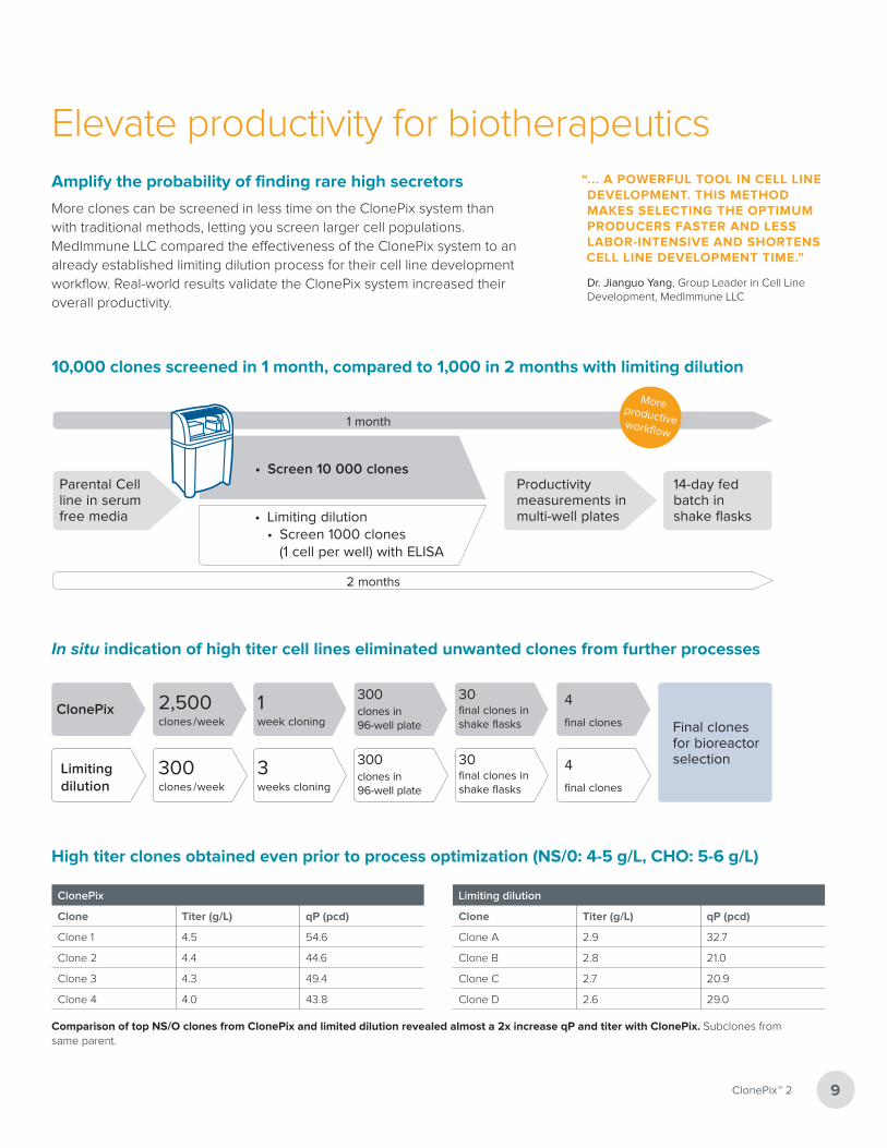
10,000 clones screened in 1 month, compared to 1,000 in 2 months with limiting dilution
Productivity measurements in multi-well plates
14-day fed batch in shake flasks• Limiting dilution
• Screen 1000 clones (1 cell per well) with ELISA
Parental Cell line in serum free media
2 months
1 month
• Screen 10 000 clones
In situ indication of high titer cell lines eliminated unwanted clones from further processes
ClonePix 2,500clones / week
1 week cloning
300 clones in 96-well plate
4
final clones Final clones for bioreactor selection
Limiting dilution
300clones / week
3 weeks cloning
300 clones in 96-well plate
30 final clones in shake flasks
4
final clones
30 final clones in shake flasks
High titer clones obtained even prior to process optimization (NS/0: 4-5 g/L, CHO: 5-6 g/L)
ClonePix
Clone Titer (g/L) qP (pcd)
Clone 1 4.5 54.6
Clone 2 4.4 44.6
Clone 3 4.3 49.4
Clone 4 4.0 43.8
Limiting dilution
Clone Titer (g/L) qP (pcd)
Clone A 2.9 32.7
Clone B 2.8 21.0
Clone C 2.7 20.9
Clone D 2.6 29.0
Comparison of top NS/O clones from ClonePix and limited dilution revealed almost a 2x increase qP and titer with ClonePix. Subclones from same parent.
More productive workflow
Reveal stable clones fasterLack of cell line stability is problematic with early stage transfectants. ClonePix systems can help you isolate stable cell line.
• Select clones and re-plate aliquots into semi-solid media
• Re-image within 4-7 days to verify and compare production rates of the daughter clones. Or, re-screen for sub-clones within
7-14 days.
Selection of stable clones by second round screening
60
50
40
30
20
10
00 50 000 100 000 150 000 200 000
Rel
ativ
e P
rod
uctio
n/C
onf
luen
ce
Fluorescence
80
70
60
50
40
30
20
10
00 20 000 40 000 60 000 80 000
Rel
ativ
e P
rod
uctio
n/C
onf
luen
ce
Fluorescence
Top 2% of transfected population of suspension-adapted CHO cells collected and assayed for productivity. Confluence values determined by CloneSelect Imager.
Productivity versus fluorescence after second round screening. Confluence values determined by CloneSelect Imager.
ClonePix 2 System
www.moleculardevices.com10
Accurate picking and gentle transfer of your colonies
with a robotic arm
Washing and sterilization for picking heads
Barcode reader for precise tracking of your colonies
Automate lid removal and replacement
Stack 6-well source plates and 96-well destination plates

System Specifications
Imaging
Software Dedicated imaging software pre-installed on a high-specification PC, Microsoft Windows 7
White light imaging • Trans-illumination for imaging low contrast colonies such as adherent monolayers or small colonies in suspension• Epi-illumination for imaging colonies as they are collected
Fluorescence imaging Software-controlled switching between up to 5 excitation /emission filter pairs (we recommend no more than 3 filters be multiplexed for optimal performance)
Data tracking Internal barcode reader for source and destination plates enables data tracking for each run
Camera Integrated 16-bit cooled CCD camera
Imaging speed 6-well microplate: 5 min for 2 wavelengths (standard conditions)
Resolution Standard: 28 micron; Maximum: 1.5 micron
Instrumentation
Containment Fully enclosed working environment with Class 100-type, HEPA filtration
Source plate type PetriWell-6 plate, PetriWell-1 plate, Greiner 6 well plate, Nunc 6 well plate, Nunc OmniTray
Destination plate type PetriWell-96 plate, Costar 96 well plate, Greiner 96 well plate, Nunc 96 well plate, Falcon 96 well plate
Source plate capacity 10 plates
Destination plate capacity 10 plates
Picking head 8 x picking pins – each pin independently controlled
Picking pin size Diameter of picking pins is application specific – F1: suspension cells, F2: adherent cells
Picking speed > 200 clones per hour
Wash bath Ethanol wash bath, automatically refilled
Picking system fluids 5 L sterile water supply, 5L waste bottle
Pin drying Proprietary halogen pin drying station
Instrument dimensions 1010 mm (width) x 900 mm (depth) x 1490 mm (height)
Instrument weight 350 kg
Compressed air specifications
Air Clean, oil-free with sub-micron filtration
Minimum operating pressure 6 bar (~90psi)
Minimum operating volume 80 L/min
Optional compressor specifications
Compressor unit Clean, oil-free compressor with sub-micron filtration
Dimensions 250 mm (width) x 600 mm (depth) x 750 mm (height)
Weight 60 kg
Minimum operating pressure 6 bar
Minimum operating volume 80 L/min
Noise level 61 dB(A)
Regulatory approval
Compliance CE
Quality ISO9001:2008 certified
11ClonePix™ 2

Maximize your hybridoma yield with our complete set of culture media
Culture media for every stage of hybridoma cell line generation
Stable hybridoma cell lines are critical for monoclonal antibody production. Our XP Media and CloneMedia portfolio of products is a complete solution that supports all stages of hybridoma cell line development from fusion to scale up. Optimized to support the selection and growth of hybridoma clones using our ClonePix 2 System, the kit is also compatible with other appropriate methods.
• HAT (hypoxanthine-aminopterin-thymidine) selection and cloning of hybridomas are accomplished in one step, which minimizes both time and materials required
• Not only does the semi-solid CloneMedia method eliminate the possible masking of potentially valuable slow-growing clones by fast-growing clones, but it also reduces or eliminates sub-cloning steps
• Reduce hands-on time when the workflow is combined with the ClonePix 2 System
Optimized growth conditions result in stable antibody production
www.moleculardevices.com12
Hybridomas were generated, selected, and screened using our XP Media and CloneMedia suite of hybridoma media. (A) 5 individual hybridoma fusion experiments were conducted on BALB/c mice, immunized to the same antigen, to assess reproducibility of fusions in XP Media suite of products. Fusion efficiency was calculated by dividing the number of hybridoma colonies detected on the ClonePix 2 System by the number of splenocytes grown in XP Media Pre-Fusion Myeloma Growth Medium and Hybridoma Expansion Medium (without HT). (B) Images of hybridomas were captured with the ClonePix 2 System in white light (left panel) and FITC (right panel), after 7 days growth, to determine growth and expression of IgGs, respectively. Colonies grown in the presence of CloneDetect were ranked according to their FITC intensity (indicating total IgG production), with the highest producers picked for further characterization. (C) Software detection of cell confluency, indicated by the green overlay, across a 96-well plate allowing for a quick visualization of plating efficiency. Images were collected on the CloneSelect Imager. 87 out of 96 wells grew to a confluency >5% after 7 days (the initial confluency of all wells was <1%) for a >90% plating efficiency. The real plating efficiency may be even higher because slow growing clones may be classified as non-growing using the >5% confluency criteria. (D) IgG production plotted per well (show in blue) with red lines indicating 2 s.d. away from the mean. Because these are stable hybridomas, we don’t expect a large variation in the total amount of IgG produced per cell, which is confirmed by <5% CV across all clones tested.
A B
C D
E
Single well confluency Single well confluency over time
Confluency across 96-well plate Confluency of 96-well plate over time
>90% plating efficiency Time (days)0 1 2 3 4 5 6 7 8
Coin
fluen
cy(%
)
ELISA Absorbance Values for IgG production
40 hybridoma clones in duplicates
IgG
pro
duct
ion
(ng/
ml)
40 clones (CV < 5%)
Average IgG production1 2 3 4 5 6 7 8 9 10 11 12
A 2.45 2.28 0.30 0.29 0.28 0.27 0.26 0.26 0.26 0.25 0.29 0.29B 2.03 1.94 0.29 0.27 0.26 0.27 0.27 0.27 0.25 0.26 0.25 0.26C 1.44 1.40 0.27 0.26 0.27 0.28 0.27 0.26 0.26 0.27 0.26 0.26D 1.00 0.96 0.28 0.28 0.27 0.28 0.27 0.27 0.27 0.26 0.27 0.27E 0.56 0.52 0.29 0.27 0.26 0.27 0.27 0.26 0.26 0.26 0.25 0.26F 0.30 0.29 0.29 0.28 0.28 0.27 0.27 0.27 0.27 0.26 0.26 0.27G 0.18 0.18 0.29 0.28 0.28 0.29 0.28 0.28 0.27 0.26 0.27 0.27H 0.07 0.07 0.28 0.27 0.28 0.28 0.26 0.26 0.27 0.26 0.27 0.28
F
Stds
1%
Day 0 Day 1 Day 2
Day 3 Day 4 Day 74% 6%
9% 46% 65%C
Confluency across 96-well plate
>90% plating efficiency
A B
C D
E
Single well confluency Single well confluency over time
Confluency across 96-well plate Confluency of 96-well plate over time
>90% plating efficiency Time (days)0 1 2 3 4 5 6 7 8
Coin
fluen
cy(%
)
ELISA Absorbance Values for IgG production
40 hybridoma clones in duplicates
IgG
pro
duct
ion
(ng/
ml)
40 clones (CV < 5%)
Average IgG production1 2 3 4 5 6 7 8 9 10 11 12
A 2.45 2.28 0.30 0.29 0.28 0.27 0.26 0.26 0.26 0.25 0.29 0.29B 2.03 1.94 0.29 0.27 0.26 0.27 0.27 0.27 0.25 0.26 0.25 0.26C 1.44 1.40 0.27 0.26 0.27 0.28 0.27 0.26 0.26 0.27 0.26 0.26D 1.00 0.96 0.28 0.28 0.27 0.28 0.27 0.27 0.27 0.26 0.27 0.27E 0.56 0.52 0.29 0.27 0.26 0.27 0.27 0.26 0.26 0.26 0.25 0.26F 0.30 0.29 0.29 0.28 0.28 0.27 0.27 0.27 0.27 0.26 0.26 0.27G 0.18 0.18 0.29 0.28 0.28 0.29 0.28 0.28 0.27 0.26 0.27 0.27H 0.07 0.07 0.28 0.27 0.28 0.28 0.26 0.26 0.27 0.26 0.27 0.28
F
Stds
1%
Day 0 Day 1 Day 2
Day 3 Day 4 Day 74% 6%
9% 46% 65%
DAverage lgG production
40 clones (CV < 5%)
lgG
pro
du
ctio
n (n
g/m
l)
AHybridoma fusion from 5 separate
immunizations of BALB/c mice
Fu
sio
n e
ffic
ien
cy
B
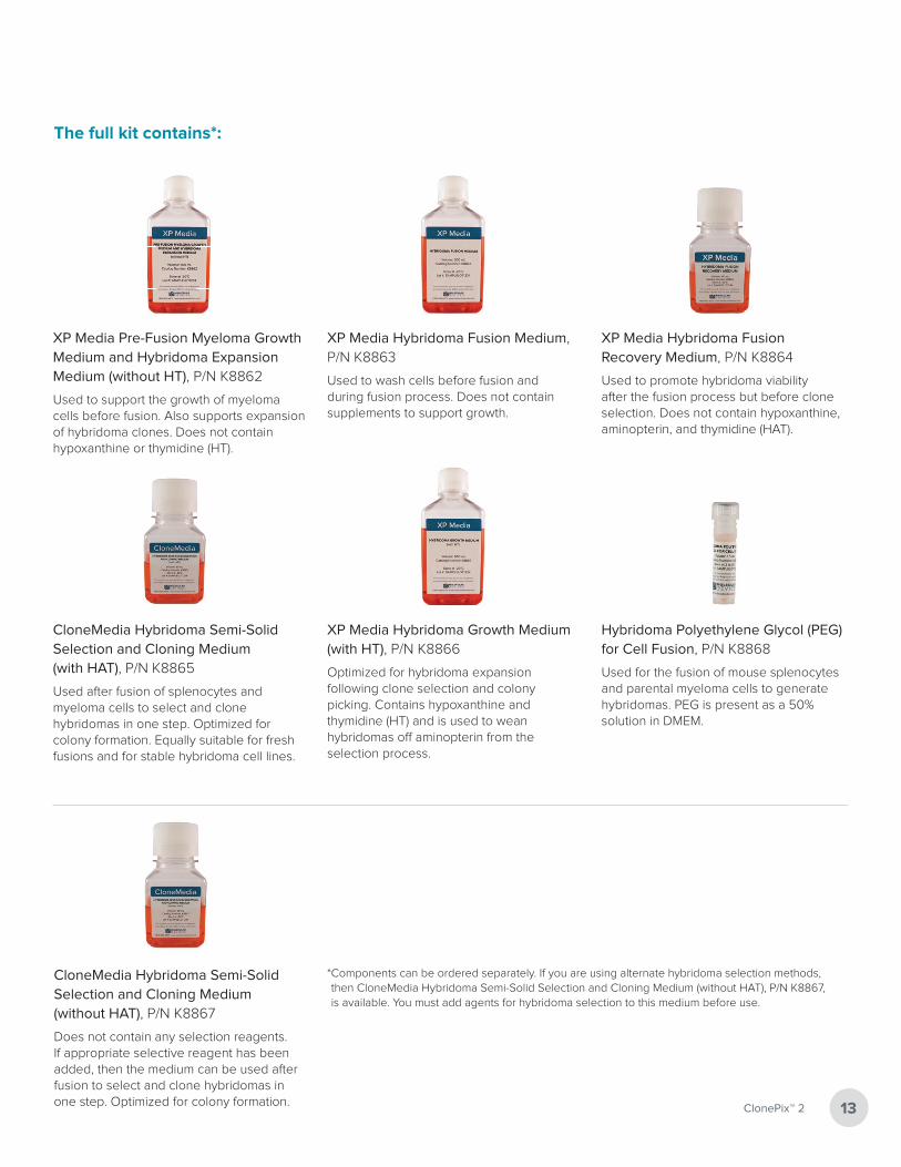
CloneMedia Hybridoma Semi-Solid Selection and Cloning Medium (without HAT), P/N K8867
Does not contain any selection reagents. If appropriate selective reagent has been added, then the medium can be used after fusion to select and clone hybridomas in one step. Optimized for colony formation. 13ClonePix™ 2
XP Media Pre-Fusion Myeloma Growth Medium and Hybridoma Expansion Medium (without HT), P/N K8862
Used to support the growth of myeloma cells before fusion. Also supports expansion of hybridoma clones. Does not contain hypoxanthine or thymidine (HT).
XP Media Hybridoma Fusion Medium, P/N K8863
Used to wash cells before fusion and during fusion process. Does not contain supplements to support growth.
XP Media Hybridoma Fusion Recovery Medium, P/N K8864
Used to promote hybridoma viability after the fusion process but before clone selection. Does not contain hypoxanthine, aminopterin, and thymidine (HAT).
CloneMedia Hybridoma Semi-Solid Selection and Cloning Medium (with HAT), P/N K8865
Used after fusion of splenocytes and myeloma cells to select and clone hybridomas in one step. Optimized for colony formation. Equally suitable for fresh fusions and for stable hybridoma cell lines.
XP Media Hybridoma Growth Medium (with HT), P/N K8866
Optimized for hybridoma expansion following clone selection and colony picking. Contains hypoxanthine and thymidine (HT) and is used to wean hybridomas off aminopterin from the selection process.
Hybridoma Polyethylene Glycol (PEG) for Cell Fusion, P/N K8868
Used for the fusion of mouse splenocytes and parental myeloma cells to generate hybridomas. PEG is present as a 50% solution in DMEM.
* Components can be ordered separately. If you are using alternate hybridoma selection methods, then CloneMedia Hybridoma Semi-Solid Selection and Cloning Medium (without HAT), P/N K8867, is available. You must add agents for hybridoma selection to this medium before use.
The full kit contains*:

Accelerate your hybridoma cell line development with a complete set of platforms and culture media
Hybridoma fusion
Culture myeloma cells in pre-fusion growth medium. Prepare splenocytes for fusion.
Pre-Fusion
Fuse splenocytes and myeloma cells. Fusion
Clone Selection
Accurate and gentle picking
Transfer of colony to destination plates
Pick selected, high-producing clones from semi-solid medium and transfer to liquid medium.
Clone Picking
Monitor growth of picked clones.
Perform secondary screening on clone supernatants (e.g. ELISA).
Functional Characterization
FLFL
FLFL
Cells or beads with antigen
FL
Anti-species IgG-FL
Primary antibody
Scale up clones producing desired antibodies and wean off HT selection.
Clone screening and selection in semi-solid medium.
Clone Stability
Scale-Up Scale-Up Scale-Up Scale-Up and Weanand Weanand Weanand Wean
ExpansionExpand clones producing desired antibodies.
15% Day 3
3% Day 1
5% Day 2
55% Day 7
VIEW EVERY GROWTH CURVE
IN EVERY WELL
15% Day 3
3% Day 1
5% Day 2
55% Day 7
VIEW EVERY GROWTH CURVE
IN EVERY WELL well of interest
Expansion Medium without HT (K8862),
Fusion Medium (K8863)
Fusion Medium (K8863),Polyethylene Glycol (K8868),
Fusion Recovery Medium (K8864)
ClonePix 2 System, CloneDetect,CloneMedia Selection and Cloning
Medium with and without HAT(K8865, K8867)
ClonePix 2 System, Growth Medium with HT (K8866)
CloneSelect Imager
SpectraMax i3x or other MolecularDevices microplate readers
Growth Medium with HT (K8866) and Expansion Medium without
HT (K8862) at 1:1 ratio
Expansion Medium without HT(K8862)
www.moleculardevices.com14
15ClonePix™ 2
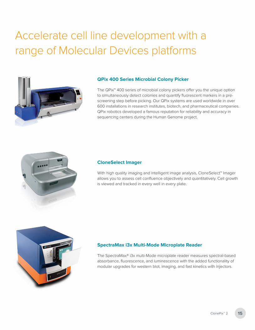
Accelerate cell line development with a range of Molecular Devices platforms
QPix 400 Series Microbial Colony Picker
The QPix™ 400 series of microbial colony pickers offer you the unique option to simultaneously detect colonies and quantify fluorescent markers in a pre-screening step before picking. Our QPix systems are used worldwide in over 600 installations in research institutes, biotech, and pharmaceutical companies. QPix robotics developed a famous reputation for reliability and accuracy in sequencing centers during the Human Genome project.
CloneSelect Imager
With high quality imaging and intelligent image analysis, CloneSelect™ Imager allows you to assess cell confluence objectively and quantitatively. Cell growth is viewed and tracked in every well in every plate.
SpectraMax i3x Multi-Mode Microplate Reader
The SpectraMax® i3x multi-Mode microplate reader measures spectral-based absorbance, fluorescence, and luminescence with the added functionality of modular upgrades for western blot, imaging, and fast kinetics with injectors.

The trademarks used herein are the property of Molecular Devices, LLC or their respective owners. ClonePix, CloneSelect, CellReporter are trademarks of Molecular Devices (New Milton) Ltd. All third party trademarks are the property of their respective owners. For a listing of trademark owners, visit www.moleculardevices.com/genetix. Specifications subject to change without notice. Patents: www.moleculardevices.com/productpatentsFOR RESEARCH USE ONLY. NOT FOR USE IN DIAGNOSTIC PROCEDURES.
©2016 Molecular Devices, LLC 5/16 1943E
Unrivaled solutionsOur products empower you with unrivaled solutions that utilize imaging and intelligent image analysis to support basic
research, pharmaceutical and biotherapeutic development. We are continually establishing industry standards in areas such
as picking microbial colonies for genomic studies or screening and selection of mammalian cell lines. Our systems use
imaging platforms to monitor cell growth, evaluate cellular responses and quantify protein production. We bring you expertise
in robotics, cell and molecular biology, image analysis and interpretation supported by a strong IP portfolio, and are truly
committed to the continual development of innovative solutions for life science applications.
For more information, visit www.moleculardevices.com.
Regional Offices
USA and Canada +1.800.635.5577United Kingdom +44.118.944.8000Europe* 00800.665.32860
China (Beijing) +86.10.6410.8669China (Shanghai) +86.21.3372.1088Hong Kong +852.2248.6000
Japan (Osaka) +81.6.7174.8331Japan (Tokyo) +81.3.6362.5260South Korea +82.2.3471.9531
Brazil +55.11.3616.6607
Contact Us
Phone: 800.635.5577Web: www.moleculardevices.comEmail: [email protected]
Check our website for a current listing of worldwide distributors
*Austria, Belgium, Denmark, Finland, France, Germany, Ireland, Netherlands, Spain, Sweden and Switzerland