Embed Size (px)
Citation preview
ClonePix™ 2Screen and select more clones in less time
www.moleculardevices.com/genetix GenetixNow part of Molecular Devices
02
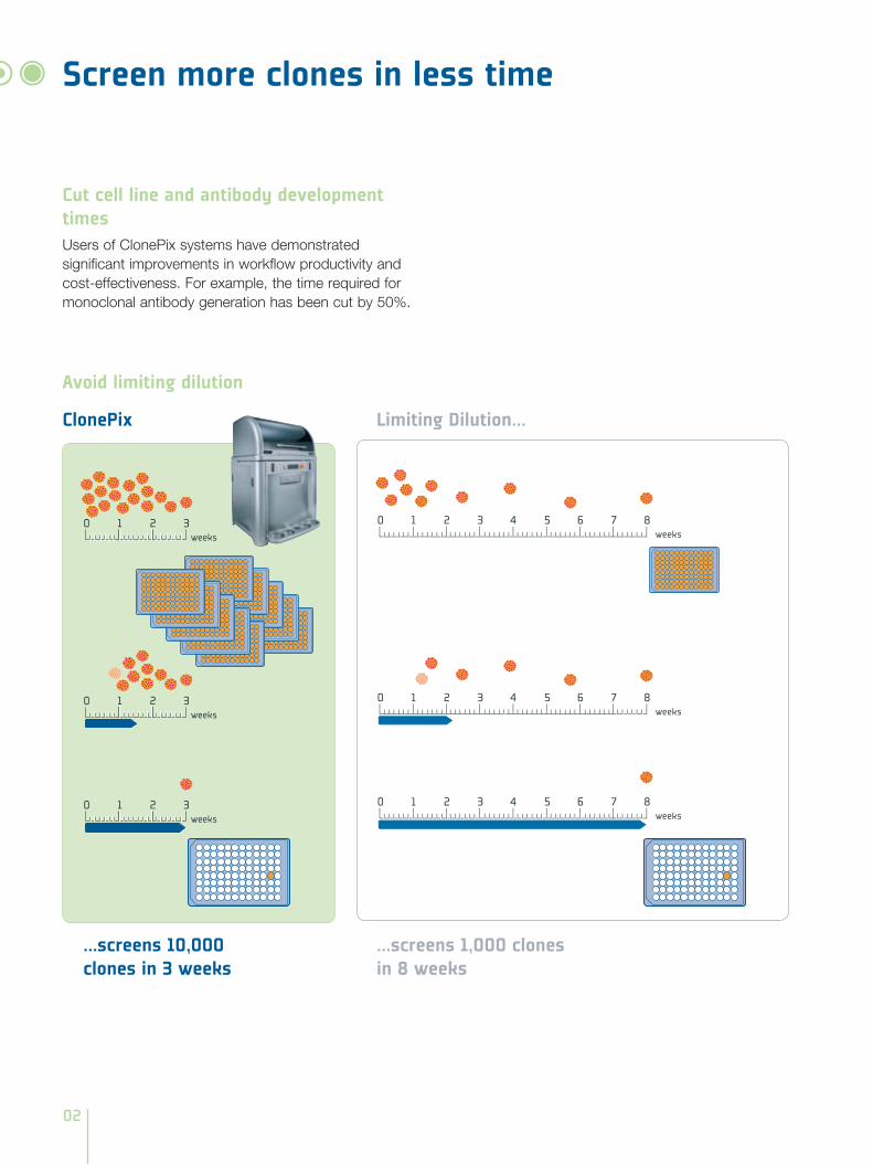
Screen more clones in less time
Limiting Dilution...
...screens 10,000 clones in 3 weeks
...screens 1,000 clones in 8 weeks
ClonePix
Avoid limiting dilution
0 1 2 3 4 5 6 7 8weeks
0 1 2 3 4 5 6 7 8weeks
0 1 2 3 4 5 6 7 8weeks
0 1 2 3weeks
0 1 2 3weeks
0 1 2 3weeks
Cut cell line and antibody development timesUsers of ClonePix systems have demonstrated significant improvements in workflow productivity and cost-effectiveness. For example, the time required for monoclonal antibody generation has been cut by 50%.
03
Select cells with optimal expression levels• Cells grow in semi-solid CloneMedia, forming discrete
clonal colonies
• ClonePix system rapidly screens thousands of clones
- Increasing the probability of finding the best clones
- White light visualizes colonies for imaging and picking
- Fluorescence imaging indicates expression levels
• A choice of detection methods enables:
- Label-free detection of secreted IgG or antigen-specific MAbs from hybridomas
- Detection of tagged recombinant proteins and expression markers
• Objective image analysis enables the user to select colonies with optimal expression levels - eliminating poor performers as early as possible
• Accurate, automatic colony picking avoids errors associated with limiting dilution
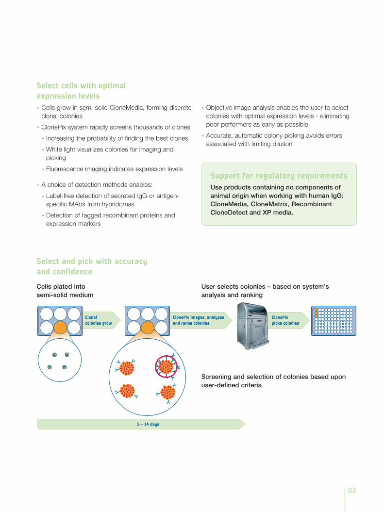
User selects colonies – based on system’s analysis and ranking
Screening and selection of colonies based upon user-defined criteria
Select and pick with accuracy and confidence
Cells plated into semi-solid medium
Support for regulatory requirementsUse products containing no components of animal origin when working with human IgG: CloneMedia, CloneMatrix, Recombinant CloneDetect and XP media.
04
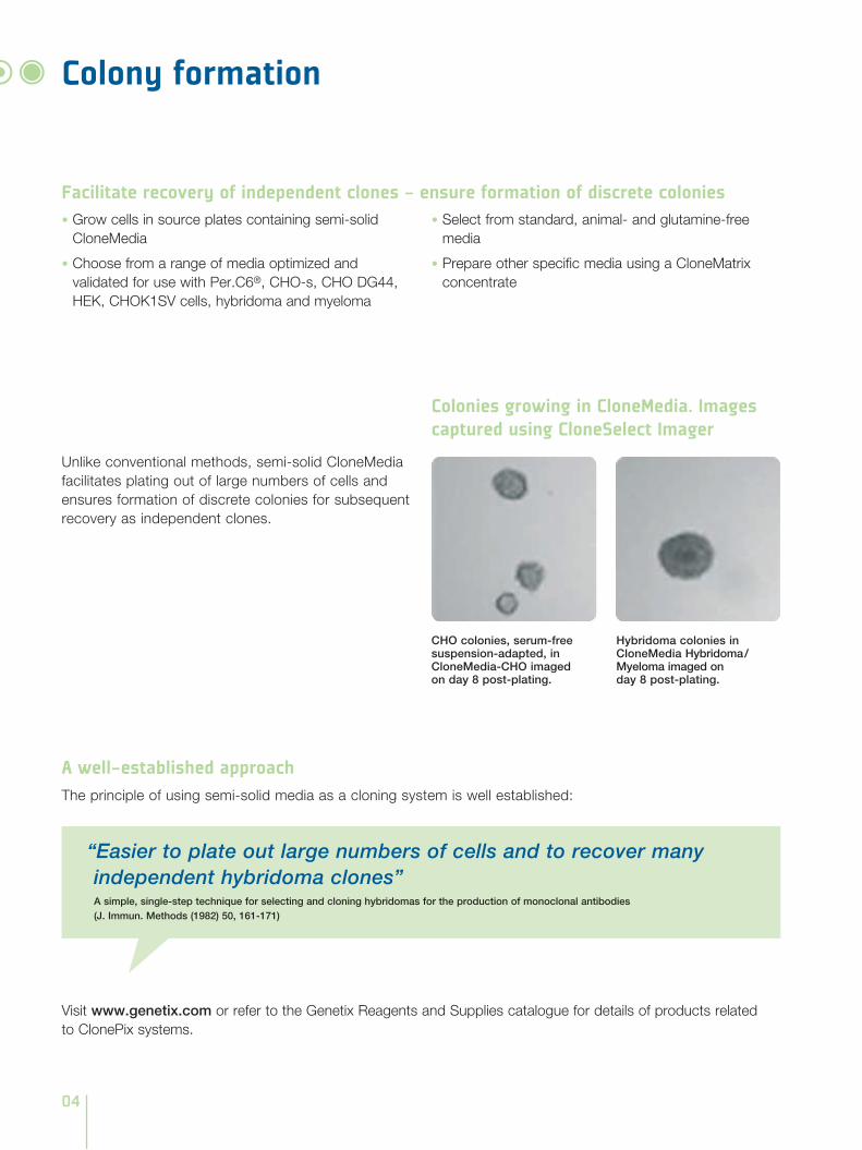
Colony formation
Unlike conventional methods, semi-solid CloneMedia facilitates plating out of large numbers of cells and ensures formation of discrete colonies for subsequent recovery as independent clones.
A well-established approachThe principle of using semi-solid media as a cloning system is well established:
CHO colonies, serum-free suspension-adapted, in CloneMedia-CHO imaged on day 8 post-plating.
Hybridoma colonies in CloneMedia Hybridoma / Myeloma imaged on day 8 post-plating.
Facilitate recovery of independent clones - ensure formation of discrete colonies• Grow cells in source plates containing semi-solid
CloneMedia
• Choose from a range of media optimized and validated for use with Per.C6®, CHO-s, CHO DG44, HEK, CHOK1SV cells, hybridoma and myeloma
• Select from standard, animal- and glutamine-free media
• Prepare other specific media using a CloneMatrix concentrate
Colonies growing in CloneMedia. Images captured using CloneSelect Imager
“Easier to plate out large numbers of cells and to recover many independent hybridoma clones”A simple, single-step technique for selecting and cloning hybridomas for the production of monoclonal antibodies (J. Immun. Methods (1982) 50, 161-171)
Visit www.genetix.com or refer to the Genetix Reagents and Supplies catalogue for details of products related to ClonePix systems.
05
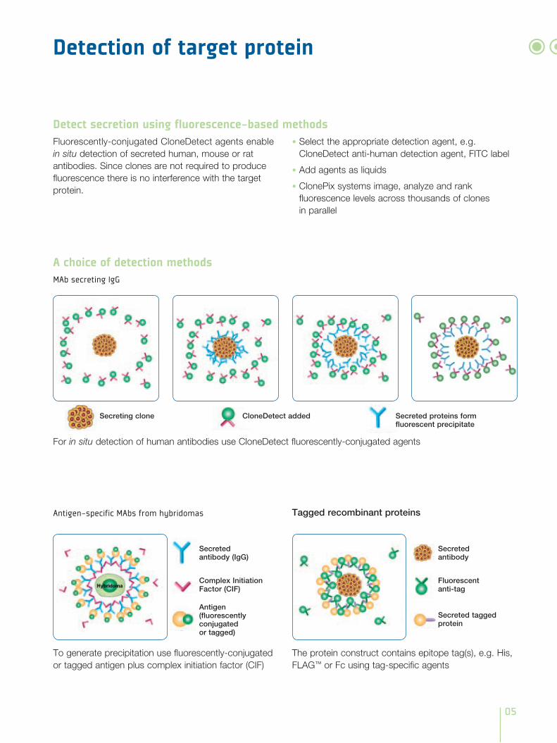
Fluorescently-conjugated CloneDetect agents enable in situ detection of secreted human, mouse or rat antibodies. Since clones are not required to produce fluorescence there is no interference with the target protein.
• Select the appropriate detection agent, e.g. CloneDetect anti-human detection agent, FITC label
• Add agents as liquids
• ClonePix systems image, analyze and rank fluorescence levels across thousands of clones in parallel
Detection of target protein
Detect secretion using fluorescence-based methods
To generate precipitation use fluorescently-conjugated or tagged antigen plus complex initiation factor (CIF)
Antigen-specific MAbs from hybridomas
The protein construct contains epitope tag(s), e.g. His, FLAG™ or Fc using tag-specific agents
For in situ detection of human antibodies use CloneDetect fluorescently-conjugated agents
Tagged recombinant proteins
A choice of detection methodsMAb secreting IgG
Secreting clone CloneDetect added Secreted proteins form fluorescent precipitate
Secreted antibody (IgG)
Complex Initiation Factor (CIF)
Antigen (fluorescently conjugated or tagged)
Secreted antibody
Fluorescent anti-tag
Secreted tagged protein
06
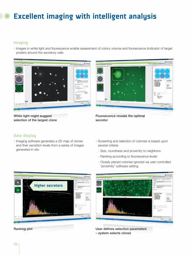
White light might suggest selection of the largest clone
Fluorescence reveals the optimal secretor
Excellent imaging with intelligent analysis
Imaging• Images in white light and fluorescence enable assessment of colony volume and fluorescence (indicator of target
protein) around the secretory cells
Data display• Imaging software generates a 2D map of clones
and their secretion levels from a series of images generated in situ
• Screening and selection of colonies is based upon several criteria:
- Size, roundness and proximity to neighbors
- Ranking according to fluorescence levels
- Closely placed colonies ignored via user-controlled “proximity” software setting
User defines selection parameters – system selects clones
Higher secretors
Ranking plot
07
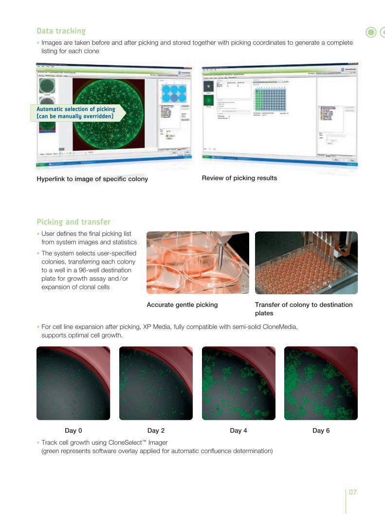
Picking and transfer• User defines the final picking list
from system images and statistics
• The system selects user-specified colonies, transferring each colony to a well in a 96-well destination plate for growth assay and / or expansion of clonal cells
• For cell line expansion after picking, XP Media, fully compatible with semi-solid CloneMedia, supports optimal cell growth.
Day 0 Day 2 Day 4 Day 6
• Track cell growth using CloneSelect™ Imager (green represents software overlay applied for automatic confluence determination)
Data tracking• Images are taken before and after picking and stored together with picking coordinates to generate a complete
listing for each clone
Review of picking resultsHyperlink to image of specific colony
Accurate gentle picking Transfer of colony to destination plates
Automatic selection of picking (can be manually overridden)
08
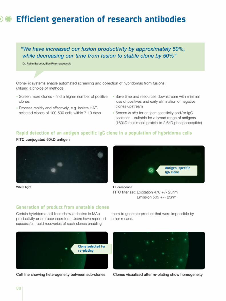
Efficient generation of research antibodies
“We have increased our fusion productivity by approximately 50%, while decreasing our time from fusion to stable clone by 50%”Dr. Robin Barbour, Elan Pharmaceuticals
ClonePix systems enable automated screening and collection of hybridomas from fusions, utilizing a choice of methods.
Cell line showing heterogeneity between sub-clones
Generation of product from unstable clonesCertain hybridoma cell lines show a decline in MAb productivity or are poor secretors. Users have reported successful, rapid recoveries of such clones enabling
them to generate product that were impossible by other means.
• Screen more clones - find a higher number of positive clones
• Process rapidly and effectively, e.g. isolate HAT-selected clones of 100-500 cells within 7-10 days
• Save time and resources downstream with minimal loss of positives and early elimination of negative clones upstream
• Screen in situ for antigen specificity and / or IgG secretion - suitable for a broad range of antigens (160kD multimeric protein to 2.6kD phosphopeptide)
Clones visualized after re-plating show homogeneity
Fluorescence
FITC filter set: Excitation 470 + / - 25nm Emission 535 + / - 25nm
FITC conjugated 60kD antigen
Rapid detection of an antigen specific IgG clone in a population of hybridoma cells
White light
Antigen-specific IgG clone
Clone selected for re-plating
09
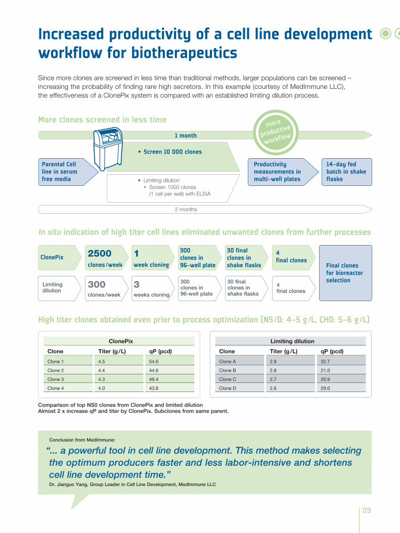
Comparison of top NS0 clones from ClonePix and limited dilution Almost 2 x increase qP and titer by ClonePix. Subclones from same parent.
High titer clones obtained even prior to process optimization (NS / 0: 4-5 g / L, CHO: 5-6 g / L)
Increased productivity of a cell line development workflow for biotherapeuticsSince more clones are screened in less time than traditional methods, larger populations can be screened – increasing the probability of finding rare high secretors. In this example (courtesy of MedImmune LLC), the effectiveness of a ClonePix system is compared with an established limiting dilution process.
Conclusion from MedImmune:
“... a powerful tool in cell line development. This method makes selecting the optimum producers faster and less labor-intensive and shortens cell line development time.”Dr. Jianguo Yang, Group Leader in Cell Line Development, MedImmune LLC
ClonePix 2500clones / week
1 week cloning
300 clones in 96-well plate
30 final clones in shake flasks
4 final clones
Final clones for bioreactor selection
Limiting dilution
300clones / week
3weeks cloning
300clones in 96-well plate
30 final clones in shake flasks
4final clones
More clones screened in less time
In situ indication of high titer cell lines eliminated unwanted clones from further processes
Productivity measurements in multi-well plates
14-day fed batch in shake flasks• Limitingdilution
• Screen 1000 clones (1 cell per well) with ELISA
Parental Cell line in serum free media
2 months
1 month
• Screen 10 000 clones
Clone
Clone 1
Clone 2
Clone 3
Clone 4
ClonePix
Titer (g / L)
4.5
4.4
4.3
4.0
qP (pcd)
54.6
44.6
49.4
43.8
Clone
Clone A
Clone B
Clone C
Clone D
Limiting dilution
Titer (g / L)
2.9
2.8
2.7
2.6
qP (pcd)
32.7
21.0
20.9
29.0
more
productive
workflow
10
60
50
40
30
20
10
00 50 000 100 000 150 000 200 000
Rel
ativ
e P
rod
uctio
n/C
onf
luen
ce
Fluorescence
80
70
60
50
40
30
20
10
00 20 000 40 000 60 000 80 000
Rel
ativ
e P
rod
uctio
n/C
onf
luen
ce
Fluorescence
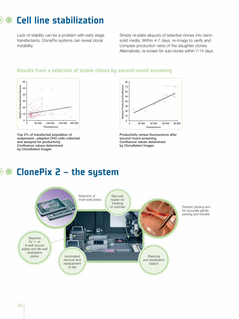
Lack of stability can be a problem with early stage transfectants. ClonePix systems can reveal clonal instability.
Simply re-plate aliquots of selected clones into semi-solid media. Within 4-7 days, re-image to verify and compare production rates of the daughter clones. Alternatively, re-screen for sub-clones within 7-14 days.
Results from a selection of stable clones by second round screening
Cell line stabilization
Top 2% of transfected population of suspension- adapted CHO cells collected and assayed for productivity.Confluence values determined by CloneSelect Imager.
Productivity versus fluorescence after second round screening.Confluence values determined by CloneSelect Imager.
ClonePix 2 – the system
Selection of multi-well plates
Robotic picking arm for accurate gentle picking and transfer
Stackers for 1- or
6-well source plates and 96-well
destination plates Washing
and sterilization station
Automated removal and replacement
of lids
Barcode reader for tracking
of colonies
11
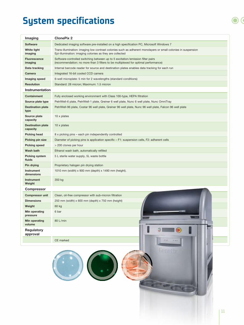
System specificationsImaging
Software
White light imaging
Fluorescence imaging
Data tracking
Camera
Imaging speed
Resolution
Instrumentation
Containment
Source plate type
Destination plate type
Source plate capacity
Destination plate capacity
Picking head
Picking pin size
Picking speed
Wash bath
Picking system fluids
Pin drying
Instrument dimensions
Instrument Weight
Compressor
Compressor unit
Dimensions
Weight
Min operating pressure
Min operating volume
Regulatory approval
ClonePix 2
Dedicated imaging software pre-installed on a high specification PC, Microsoft Windows 7
Trans-illumination: imaging low contrast colonies such as adherent monolayers or small colonies in suspension Epi-illumination: imaging colonies as they are collected
Software-controlled switching between up to 5 excitation / emission filter pairs (recommendation: no more than 3 filters to be multiplexed for optimal performance)
Internal barcode reader for source and destination plates enables data tracking for each run
Integrated 16-bit cooled CCD camera
6-well microplate: 5 min for 2 wavelengths (standard conditions)
Standard: 28 micron; Maximum: 1.5 micron
Fully enclosed working environment with Class 100-type, HEPA filtration
PetriWell-6 plate, PetriWell-1 plate, Greiner 6 well plate, Nunc 6 well plate, Nunc OmniTray
PetriWell-96 plate, Costar 96 well plate, Greiner 96 well plate, Nunc 96 well plate, Falcon 96 well plate
10 x plates
10 x plates
8 x picking pins – each pin independently controlled
Diameter of picking pins is application specific – F1: suspension cells, F2: adherent cells
> 200 clones per hour
Ethanol wash bath, automatically refilled
5 L sterile water supply, 5L waste bottle
Proprietary halogen pin drying station
1010 mm (width) x 900 mm (depth) x 1490 mm (height).
350 kg
Clean, oil-free compressor with sub-micron filtration
250 mm (width) x 600 mm (depth) x 750 mm (height)
60 kg
6 bar
80 L / min
CE marked
ClonePix, CloneSelect, CellReporter are trademarks of Molecular Devices (New Milton) Ltd. All third party trademarks are the property of their respective owners.
For a listing of trademark owners, visit www.moleculardevices.com/genetix
07LB
L102
1.A
3
www.moleculardevices.com/genetix
Unrivalled solutions based on excellent imaging and intelligent image analysis
Products from Molecular Devices offer scientists unrivalled solutions that utilize imaging and intelligent image analysis to support basic research, pharma-ceutical and biotherapeutic development. The company’s systems continue to establish industry standards in areas such as picking microbial colonies for genomic studies or screening and selection of mammalian cell lines. Other systems use imaging platforms to monitor cell growth, evaluate cellular responses and quantify
protein production. Through its expertise in robotics, cell and molecular biology, image analysis and interpretation, supported by a strong IP portfolio, the company is committed to the continual development of innovative solutions for life science applications.
For more information, visit www.moleculardevices.com/genetix